
Elements of Sleep Breathing and Sleep-Deprivation Physiology in the Context of Athletic Performance

 ,
, 
Abstract
:1. Sleep-Disordered Breathing Physiology
1.1. Respiratory Aspect
1.2. Neural Aspect
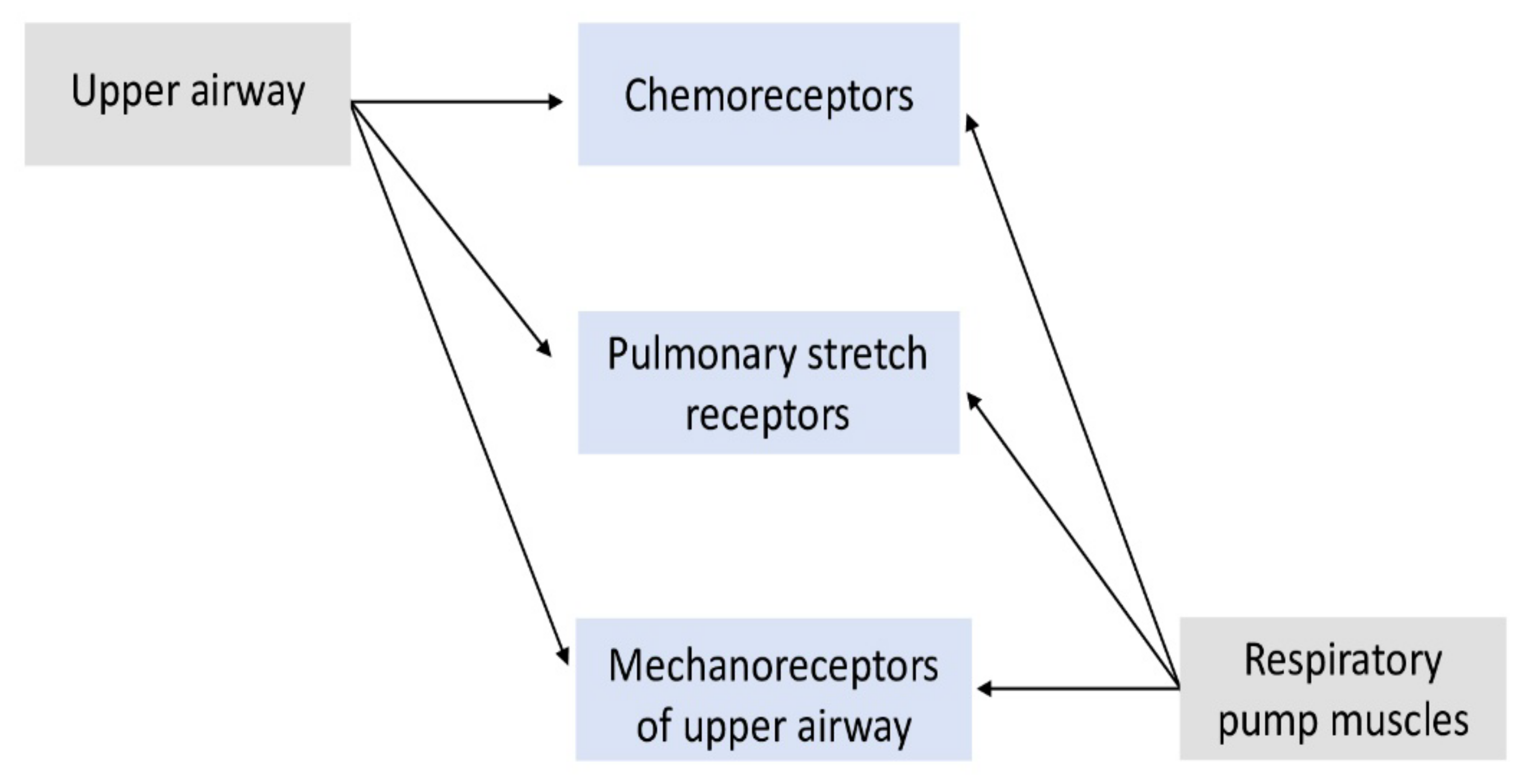
1.3. Input Sensors
1.4. Output Mediators
2. Sleep Deprivation
2.1. Sleep Deprivation and CO2 Retention
2.2. Sleep Deprivation and Exercise: Cognitive Implications
2.3. Sleep Deprivation and Exercise: Cardiovascular Implications—The Example of Sleep Apnea
2.4. Sleep Deprivation and Performance
3. Beneficial Sleep Effects
4. Conclusions
Author Contributions
Funding
Institutional Review Board Statement
Informed Consent Statement
Data Availability Statement
Conflicts of Interest
References
- Patwa, A.; Shah, A. Anatomy and physiology of respiratory system relevant to anaesthesia. Indian J. Anaesth. 2015, 59, 533–541. [Google Scholar] [CrossRef] [PubMed]
- Newton, K.; Malik, V.; Lee-Chiong, T. Sleep and breathing. Clin. Chest Med. 2014, 35, 451–456. [Google Scholar] [CrossRef]
- Sowho, M.; Amatoury, J.; Kirkness, J.P.; Patil, S.P. Sleep and respiratory physiology in adults. Clin. Chest Med. 2014, 35, 469–481. [Google Scholar] [CrossRef] [PubMed] [Green Version]
- Dutschmann, M.; Dick, T.E. Pontine mechanisms of respiratory control. Comprehensive Physiol. 2012, 2, 2443–2469. [Google Scholar]
- Stavrou, V.T.; Astara, K.; Tourlakopoulos, K.N.; Papayianni, E.; Boutlas, S.; Vavougios, G.D.; Daniil, Z.; Gourgoulianis, K.I. Obstructive Sleep Apnea Syndrome: The Effect of Acute and Chronic Responses of Exercise. Front. Med. 2021, 8, 806924. [Google Scholar] [CrossRef] [PubMed]
- Stavrou, V.T.; Astara, K.; Karetsi, E.; Daniil, Z.; Gourgoulianis, K.I. Respiratory Muscle Strength as an Indicator of the Severity of the Apnea-Hypopnea Index: Stepping Towards the Distinction between Sleep Apnea and Breath Holding. Cureus 2021, 13, e14015. [Google Scholar] [CrossRef]
- Stavrou, V.; Bardaka, F.; Karetsi, E.; Daniil, Z.; Gourgoulianis, K.I. Brief Review: Ergospirometry in Patients with Obstructive Sleep Apnea Syndrome. J. Clin. Med. 2018, 31, 191. [Google Scholar] [CrossRef] [Green Version]
- Iturriaga, R. Translating carotid body function into clinical medicine. J. Physiol. 2018, 596, 3067–3077. [Google Scholar] [CrossRef]
- Yumino, D.; Bradley, T.D. Central sleep apnea and Cheyne-Stokes respiration. Proc. Am. Thorac. Soc. 2008, 5, 226–236. [Google Scholar] [CrossRef]
- Halliwill, J.R.; Morgan, B.J.; Charkoudian, N. Peripheral chemoreflex and baroreflex interactions in cardiovascular regulation in humans. J. Physiol. 2003, 552, 295–302. [Google Scholar] [CrossRef]
- Marcus, N.J.; Li, Y.L.; Bird, C.E.; Schultz, H.D.; Morgan, B.J. Chronic intermittent hypoxia augments chemoreflex control of sympathetic activity: Role of the angiotensin II type 1 receptor. Respir. Physiol. Neurobiol. 2010, 171, 36–45. [Google Scholar] [CrossRef] [PubMed] [Green Version]
- Krimsky, W.R.; Leiter, J.C. Physiology of breathing and respiratory control during sleep. Semin. Respir. Crit. Care Med. 2005, 26, 5–12. [Google Scholar] [CrossRef] [PubMed]
- Harris, C.D. Neurophysiology of sleep and wakefulness. Respir. Care Clin. N. Am. 2005, 11, 567–586. [Google Scholar] [PubMed]
- Penzel, T.; Kantelhardt, J.W.; Lo, C.C.; Voigt, K.; Vogelmeier, C. Dynamics of heart rate and sleep stages in normals and patients with sleep apnea. Neuropsychopharmacology 2003, 28 (Suppl. 1), S48–S53. [Google Scholar] [CrossRef]
- Appelberg, J.; Pavlenko, T.; Bergman, H.; Rothen, H.U.; Hedenstierna, G. Lung aeration during sleep. Chest 2007, 131, 122–129. [Google Scholar] [CrossRef]
- Krieger, J. Breathing during sleep in normal subjects. Clin. Chest Med. 1985, 6, 577–594. [Google Scholar] [CrossRef]
- Benarroch, E.E. Control of the cardiovascular and respiratory systems during sleep. Auton. Neurosci. 2019, 218, 54–63. [Google Scholar] [CrossRef]
- Giglio, P.; Lane, J.T.; Barkoukis, T.J.; Dumitru, I. CHAPTER 3-Sleep Physiology. In Review of Sleep Medicine, 2nd ed.; Barkoukis, T.J., Avidan, A.Y., Eds.; Butterworth-Heinemann: Philadelphia, PA, USA, 2007; pp. 29–41. [Google Scholar]
- Han, F.; Chen, E.; Wei, H.; Ding, D.; He, Q. Influence of different sleep stages on respiratory regulation in normal humans. Zhongguo Yi Xue Ke Xue Yuan Xue Bao 2004, 26, 237–240. [Google Scholar]
- Wirth, K.J.; Steinmeyer, K.; Ruetten, H. Sensitization of upper airway mechanoreceptors as a new pharmacologic principle to treat obstructive sleep apnea: Investigations with AVE0118 in anesthetized pigs. Sleep 2013, 36, 699–708. [Google Scholar] [CrossRef]
- Brown, R.E.; Basheer, R.; McKenna, J.T.; Strecker, R.E.; McCarley, R.W. Control of sleep and wakefulness. Physiol. Rev. 2012, 92, 1087–1187. [Google Scholar] [CrossRef] [Green Version]
- Van Eyck, A.; Van Hoorenbeeck, K.; De Winter, B.Y.; Ramet, J.; Van Gaal, L.; De Backer, W.; Verhulst, S.L. Sleep-disordered breathing and C-reactive protein in obese children and adolescents. Sleep Breath. 2014, 18, 335–340. [Google Scholar] [CrossRef]
- Yoshihisa, A.; Takeishi, Y. Sleep Disordered Breathing and Cardiovascular Diseases. J. Atheroscler. Thromb. 2019, 26, 315–327. [Google Scholar] [CrossRef] [Green Version]
- Cummins, E.P.; Keogh, C.E. Respiratory gases and the regulation of transcription. Exp. Physiol. 2016, 101, 986–1002. [Google Scholar] [CrossRef] [Green Version]
- Plataki, M.; Sands, S.A.; Malhotra, A. Clinical consequences of altered chemoreflex control. Respir. Physiol. Neurobiol. 2013, 189, 354–363. [Google Scholar] [CrossRef] [PubMed] [Green Version]
- Brown, M.K.; Naidoo, N. The UPR and the anti-oxidant response: Relevance to sleep and sleep loss. Mol. Neurobiol. 2010, 42, 103–113. [Google Scholar] [CrossRef]
- Yun, A.J.; Lee, P.Y.; Bazar, K.A. Autonomic dysregulation as a basis of cardiovascular, endocrine, and inflammatory disturbances associated with obstructive sleep apnea and other conditions of chronic hypoxia, hypercapnia, and acidosis. Med. Hypotheses 2004, 62, 852–856. [Google Scholar]
- Leproult, R.; Van Cauter, E. Role of sleep and sleep loss in hormonal release and metabolism. Endocr Dev. 2010, 17, 11–21. [Google Scholar] [PubMed] [Green Version]
- Leproult, R.; Deliens, G.; Gilson, M.; Peigneux, P. Beneficial impact of sleep extension on fasting insulin sensitivity in adults with habitual sleep restriction. Sleep 2015, 38, 707–715. [Google Scholar] [CrossRef] [PubMed]
- Wisor, J.P.; Clegern, W.C.; Schmidt, M.A. Toll-like receptor 4 is a regulator of monocyte and electroencephalographic responses to sleep loss. Sleep 2011, 34, 1335–1345. [Google Scholar] [CrossRef] [PubMed] [Green Version]
- Shearer, W.T.; Reuben, J.M.; Mullington, J.M.; Price, N.J.; Lee, B.N.; Smith, E.O.; Szuba, M.P.; Van Dongen, H.P.; Dinges, D.F. Soluble TNF-alpha receptor 1 and IL-6 plasma levels in humans subjected to the sleep deprivation model of spaceflight. J. Allergy Clin. Immunol. 2001, 107, 165–170. [Google Scholar] [CrossRef]
- Chapman, C.L.; Schlader, Z.J.; Reed, E.L.; Worley, M.L.; Johnson, B.D. Renal and segmental artery hemodynamic response to acute, mild hypercapnia. Am. J. Physiol. Regul. Integr. Comp. Physiol. 2020, 318, R822–R827. [Google Scholar] [CrossRef] [PubMed]
- Lei, Y.; Shao, Y.; Wang, L.; Zhai, T.; Zou, F.; Ye, E.; Jin, X.; Li, W.; Qi, J.; Yang, Z. Large-Scale Brain Network Coupling Predicts Total Sleep Deprivation Effects on Cognitive Capacity. PLoS ONE 2015, 10, e0133959. [Google Scholar] [CrossRef] [PubMed]
- Jones, A.; Vennelle, M.; Connell, M.; McKillop, G.; Newby, D.E.; Douglas, N.J.; Riha, R.L. Arterial stiffness and endothelial function in obstructive sleep apnoea/hypopnoea syndrome. Sleep Med. 2013, 14, 428–432. [Google Scholar] [CrossRef] [PubMed]
- Robertson, M.D.; Russell-Jones, D.; Umpleby, A.M.; Dijk, D.-J. Effects of three weeks of mild sleep restriction implemented in the home environment on multiple metabolic and endocrine markers in healthy young men. Metabolism 2013, 62, 204–211. [Google Scholar] [CrossRef] [PubMed] [Green Version]
- Astara, K.; Siachpazidou, D.; Vavougios, G.D.; Ragias, D.; Vatzia, K.; Rapti, G.; Alexopoulos, E.; Gourgoulianis, K.I.; Xiromerisiou, G. Sleep disordered breathing from preschool to early adult age and its neurocognitive complications: A preliminary report. Sleep Sci. 2021, 14, 140–149. [Google Scholar] [CrossRef]
- Alhola, P.; Polo-Kantola, P. Sleep deprivation: Impact on cognitive performance. Neuropsychiatr. Dis. Treat. 2007, 3, 553–567. [Google Scholar]
- Martella, D.; Plaza, V.; Estévez, A.F.; Castillo, A.; Fuentes, L.J. Minimizing sleep deprivation effects in healthy adults by differential outcomes. Acta Psychol. 2012, 139, 391–396. [Google Scholar] [CrossRef]
- Pollicina, I.; Maniaci, A.; Lechien, J.R.; Iannella, G.; Vicini, C.; Cammaroto, G.; Cannavicci, A.; Magliulo, G.; Pace, A.; Cocuzza, S.; et al. Neurocognitive Performance Improvement after Obstructive Sleep Apnea Treatment: State of the Art. Behav. Sci. 2021, 11, 180. [Google Scholar] [CrossRef]
- Roth, T. Effects of excessive daytime sleepiness and fatigue on overall health and cognitive function. J. Clin. Psychiatry 2015, 76, e1145. [Google Scholar] [CrossRef]
- Stavrou, V.T.; Astara, K.; Tourlakopoulos, K.N.; Daniil, Z.; Gourgoulianis, K.I.; Kalabakas, K.; Karagiannis, D.; Basdekis, G. Sleep Quality’s Effect on Vigilance and Perceptual Ability in Adolescent and Adult Athletes. J. Sports Med. 2021, 2021, 5585573. [Google Scholar] [CrossRef]
- Vitale, K.C.; Owens, R.; Hopkins, S.R.; Malhotra, A. Sleep Hygiene for Optimizing Recovery in Athletes: Review and Recommendations. Int. J. Sports Med. 2019, 40, 535–543. [Google Scholar] [CrossRef]
- Axelsson, J.; Ingre, M.; Kecklund, G.; Lekander, M.; Wright, K.P.; Sundelin, T. Sleepiness as motivation: A potential mechanism for how sleep deprivation affects behavior. Sleep 2020, 43, zsz291. [Google Scholar] [CrossRef]
- VanHelder, T.; Radomski, M.W. Sleep deprivation and the effect on exercise performance. Sports Med. 1989, 7, 235–247. [Google Scholar] [CrossRef]
- Fullagar, H.H.K.; Skorski, S.; Duffield, R.; Hammes, D.; Coutts, A.J.; Meyer, T. Sleep and athletic performance: The effects of sleep loss on exercise performance, and physiological and cognitive responses to exercise. Sports Med. 2015, 45, 161–186. [Google Scholar] [CrossRef] [PubMed]
- Dijk, D.-J.; Landolt, H.-P. Sleep Physiology, Circadian Rhythms, Waking Performance and the Development of Sleep-Wake Therapeutics. Handb. Exp. Pharmacol. 2019, 253, 441–481. [Google Scholar] [PubMed] [Green Version]
- Marvel, C.L.; Paradiso, S. Cognitive and neurological impairment in mood disorders. Psychiatr. Clin. N. Am. 2004, 27, 19–36. [Google Scholar] [CrossRef] [Green Version]
- Pires, G.N.; Bezerra, A.G.; Tufik, S.; Andersen, M.L. Effects of acute sleep deprivation on state anxiety levels: A systematic review and meta-analysis. Sleep Med. 2016, 24, 109–118. [Google Scholar] [CrossRef]
- Riemann, D.; Krone, L.B.; Wulff, K.; Nissen, C. Sleep, insomnia, and depression. Neuropsychopharmacology 2020, 45, 74–89. [Google Scholar] [CrossRef]
- Franken, I.H.; van Strien, J.W.; Nijs, I.; Muris, P. Impulsivity is associated with behavioral decision-making deficits. Psychiatry Res. 2008, 158, 155–163. [Google Scholar] [CrossRef] [PubMed]
- Strygin, K.N. Sleep and stress. Ross Fiziol Zh Im I M Sechenova 2011, 97, 422–432. [Google Scholar] [PubMed]
- Kalmbach, D.A.; Anderson, J.R.; Drake, C.L. The impact of stress on sleep: Pathogenic sleep reactivity as a vulnerability to insomnia and circadian disorders. J. Sleep Res. 2018, 27, 12710. [Google Scholar] [CrossRef] [PubMed] [Green Version]
- Kalmbach, D.A.; Cuamatzi-Castelan, A.S.; Tonnu, C.V.; Tran, K.M.; Anderson, J.R.; Roth, T.; Drake, C.L. Hyperarousal and sleep reactivity in insomnia: Current insights. Nat. Sci. Sleep 2018, 10, 193–201. [Google Scholar] [CrossRef] [Green Version]
- MacQueen, G.M.; Memedovich, K.A. Cognitive dysfunction in major depression and bipolar disorder: Assessment and treatment options. Psychiatry Clin. Neurosci. 2017, 71, 18–27. [Google Scholar] [CrossRef] [PubMed] [Green Version]
- Baker, L.D.; Frank, L.L.; Foster-Schubert, K.; Green, P.S.; Wilkinson, C.W.; McTiernan, A.; Plymate, S.R.; Fishel, M.A.; Watson, G.S.; Cholerton, B.A.; et al. Effects of aerobic exercise on mild cognitive impairment: A controlled trial. Arch. Neurol. 2010, 67, 71–79. [Google Scholar] [CrossRef] [PubMed] [Green Version]
- Hernández-Mendo, A.; Reigal, R.E.; López-Walle, J.M.; Serpa, S.; Samdal, O.; Morales-Sánchez, V.; Juárez-Ruiz de Mier, R.; Tristán-Rodríguez, J.L.; Rosado, A.F.; Falco, C. Physical Activity, Sports Practice, and Cognitive Functioning: The Current Research Status. Front. Psychol. 2019, 10, 2658. [Google Scholar] [CrossRef] [PubMed] [Green Version]
- Stavrou, V.; Boutou, A.K.; Vavougios, G.D.; Pastaka, C.; Gourgoulianis, K.I.; Koutedakis, Y.; Daniil, Z.; Karetsi, E. The use of cardiopulmonary exercise testing in identifying the presence of obstructive sleep apnea syndrome in patients with compatible symptomatology. Respir. Physiol. Neurobiol. 2019, 262, 26–31. [Google Scholar] [CrossRef]
- Nagai, M.; Hoshide, S.; Kario, K. Sleep duration as a risk factor for cardiovascular disease- a review of the recent literature. Curr. Cardiol. Rev. 2010, 6, 54–61. [Google Scholar] [CrossRef]
- Parati, G.; Lombardi, C.; Castagna, F.; Mattaliano, P.; Filardi, P.P.; Agostoni, P.; Italian Society of Cardiology (SIC); Working Group on Heart Failure members. Heart failure and sleep disorders. Nat. Rev. Cardiol. 2016, 13, 389–403. [Google Scholar] [CrossRef]
- Ness, K.M.; Strayer, S.M.; Nahmod, N.G.; Schade, M.M.; Chang, A.M.; Shearer, G.C.; Buxton, O.M. Four nights of sleep restriction suppress the postprandial lipemic response and decrease satiety. J. Lipid Res. 2019, 60, 1935–1945. [Google Scholar] [CrossRef]
- Dáttilo, M.; Antunes, H.K.M.; Galbes, N.M.N.; Mônico-Neto, M.; DE Sá Souza, H.; Dos Santos Quaresma, M.V.L.; Lee, K.S.; Ugrinowitsch, C.; Tufik, S.; DE Mello, M.T. Effects of Sleep Deprivation on Acute Skeletal Muscle Recovery after Exercise. Med. Sci. Sports Exerc. 2020, 52, 507–514. [Google Scholar] [CrossRef]
- Alves, E.S.; Lira, F.S.; Santos, R.V.; Tufik, S.; de Mello, M.T. Obesity, diabetes and OSAS induce of sleep disorders: Exercise as therapy. Lipids Health Dis. 2011, 23, 148. [Google Scholar] [CrossRef] [PubMed] [Green Version]
- Vranish, J.R.; Bailey, E.F. Inspiratory Muscle Training Improves Sleep and Mitigates Cardiovascular Dysfunction in Obstructive Sleep Apnea. Sleep 2016, 39, 1179–1185. [Google Scholar] [CrossRef] [PubMed] [Green Version]
- Craighead, D.H.; Heinbockel, T.C.; Freeberg, K.A.; Rossman, M.J.; Jackman, R.A.; Jankowski, L.R.; Hamilton, M.N.; Ziemba, B.P.; Reisz, J.A.; D’Alessandro, A.; et al. Time-Efficient Inspiratory Muscle Strength Training Lowers Blood Pressure and Improves Endothelial Function, NO Bioavailability, and Oxidative Stress in Midlife/Older Adults With Above-Normal Blood Pressure. J. Am. Heart Assoc. 2021, 10, e020980. [Google Scholar] [CrossRef] [PubMed]
- Stavrou, V.T.; Tourlakopoulos, K.N.; Daniil, Z.; Gourgoulianis, K.I. Respiratory Muscle Strength: New Technology for Easy Assessment. Cureus 2021, 13, e14803. [Google Scholar] [CrossRef] [PubMed]
- Simpson, N.S.; Gibbs, E.L.; Matheson, G.O. Optimizing sleep to maximize performance: Implications and recommendations for elite athletes. Scand. J. Med. Sci. Sports 2017, 27, 266–274. [Google Scholar] [CrossRef] [PubMed]
- Mah, C.D.; Mah, K.E.; Kezirian, E.J.; Dement, W.C. The effects of sleep extension on the athletic performance of collegiate basketball players. Sleep 2011, 34, 943–950. [Google Scholar] [CrossRef]
- Nishida, M.; Yamamoto, K.; Murata, Y.; Ichinose, A.; Shioda, K. Exploring the Effect of Long Naps on Handball Performance and Heart Rate Variability. Sports Med. Int. Open. 2021, 5, E73–E80. [Google Scholar] [CrossRef]
- Teece, A.R.; Argus, C.K.; Gill, N.; Beaven, M.; Dunican, I.C.; Driller, M.W. Sleep and Performance during a Preseason in Elite Rugby Union Athletes. Int. J. Environ. Res. Public Health 2021, 18, 4612. [Google Scholar] [CrossRef]
- Lever, J.R.; Murphy, A.P.; Duffield, R.; Fullagar, H.H.K. A Combined Sleep Hygiene and Mindfulness Intervention to Improve Sleep and Well-Being during High-Performance Youth Tennis Tournaments. Int. J. Sports Physiol. Perform. 2020, 16, 250–258. [Google Scholar] [CrossRef]
- Edinger, J.D.; Marsh, G.R.; McCall, W.V.; Erwin, C.W.; Lininger, A.W. Daytime functioning and nighttime sleep before, during, and after a 146-hour tennis match. Sleep 1990, 13, 526–532. [Google Scholar] [CrossRef] [Green Version]
- Lastella, M.; Halson, S.L.; Vitale, J.A.; Memon, A.R.; Vincent, G.E. To Nap or Not to Nap? A Systematic Review Evaluating Napping Behavior in Athletes and the Impact on Various Measures of Athletic Performance. Nat. Sci. Sleep 2021, 13, 841–862. [Google Scholar] [CrossRef]
- Haraldsdottir, K.; Sanfilippo, J.; McKay, L.; Watson, A.M. Decreased Sleep and Subjective Well-Being as Independent Predictors of Injury in Female Collegiate Volleyball Players. Orthop. J. Sports Med. 2021, 9, 23259671211029285. [Google Scholar] [CrossRef] [PubMed]
- Costa, J.; Figueiredo, P.; Nakamura, F.; Rago, V.; Rebelo, A.; Brito, J. Intra-individual variability of sleep and nocturnal cardiac autonomic activity in elite female soccer players during an international tournament. PLoS ONE 2019, 14, e0218635. [Google Scholar] [CrossRef] [PubMed] [Green Version]
- Walsh, N.P.; Halson, S.L.; Sargent, C.; Roach, G.D.; Nédélec, M.; Gupta, L.; Leeder, J.; Fullagar, H.H.; Coutts, A.J.; Edwards, B.J.; et al. Sleep and the athlete: Narrative review and 2021 expert consensus recommendations. Br. J. Sports Med. 2020, 55, 356–368. [Google Scholar] [CrossRef] [PubMed]
- Halson, S.L.; Juliff, L.E. Sleep, sport, and the brain. Prog. Brain Res. 2017, 234, 13–31. [Google Scholar]
- Doherty, R.; Madigan, S.; Warrington, G.; Ellis, J. Sleep and Nutrition Interactions: Implications for Athletes. Nutrients 2019, 11, 822. [Google Scholar] [CrossRef] [Green Version]
- Luyster, F.S.; Strollo, P.J., Jr.; Zee, P.C.; Walsh, J.K. Boards of Directors of the American Academy of Sleep Medicine and the Sleep Research Society. Sleep: A health imperative. Sleep 2012, 35, 727–734. [Google Scholar] [CrossRef]
- Vgontzas, A.N.; Pejovic, S.; Zoumakis, E.; Lin, H.M.; Bixler, E.O.; Basta, M.; Fang, J.; Sarrigiannidis, A.; Chrousos, G.P. Daytime napping after a night of sleep loss decreases sleepiness, improves performance, and causes beneficial changes in cortisol and interleukin-6 secretion. Am. J. Physiol. Endocrinol. Metab. 2007, 292, E253–E261. [Google Scholar] [CrossRef]
- Mograss, M.; Crosetta, M.; Abi-Jaoude, J.; Frolova, E.; Robertson, E.M.; Pepin, V.; Dang-Vu, T.T. Exercising before a nap benefits memory better than napping or exercising alone. Sleep 2020, 43, zsaa062. [Google Scholar] [CrossRef]


| References | Participants | Intervention Protocol | Results |
|---|---|---|---|
| Chapman et al. [32] | Healthy adults (age: 26.0 ± 4.0 yrs, M: n = 7, F: n = 7) | Blood velocity was measured in the renal and segmental arteries with Doppler ultrasound while subjects breathed room air and while they breathed a 3% CO2, 21% O2, 76% N2 gas mixture for 5 min | CO2 decreased blood velocity in the renal and segmental arteries and increased vascular resistance in the renal and segmental arteries (kidneys are hemodynamically responsive to a mild and acute hypercapnic stimulus in healthy humans) |
| Lei et al. [33] | 14 healthy and right-handed adult males (mean age: 25.9 years) with normal or corrected-to-normal vision | fMRI study during RW and after 36 h of TSD | Self-reported scores of sleepiness were higher for TSD than for RW. A subsequent working memory task showed that memory performance was lower after 36 h of TSD. Significant increase of sleep pressure index was observed after 36 h of TSD |
| Van Eyck et al. [22] | 120 children; control (age: 12.0 ± 3.0 y, M: n = 30, F: n = 55), mild OSAS (age: 11.0 ± 3.0 y, M: n = 9, F: n = 11), and moderate-to-severe OSAS (age: 12.0 ± 3.0 y, M: n = 10, F: n = 5) | PSG and a blood sample was taken to determine CRP levels | Relationship between CRP and BMI and between CRP and fat mass |
| Jones et al. [34] | OSAHS patients (age: 44.0 ± 7.0 y, M: n = 13, F: n = 7, AHI: ≥15/h and ESS score ≥ 11) vs. controls (age: 44.0 ± 7.0 y, M: n = 13, F: n = 7) | Evaluation of arterial stiffness (applanation tonometry and cardiovascular MRI) and endothelial function (measuring vascular reactivity after administration of glyceryl trinitrate and salbutamol) | Subjects with OSAHS had increased arterial stiffness and impaired endothelial function and were at increased risk for cardiovascular disease |
| Robertson et al. [35] | Healthy and normal-weight male students aged 20–30 y, BMI: 19–26 kg/m2. They were randomized to either sleep restriction (habitual bedtime minus 1.5 h) or a control condition (habitual bedtime) for three weeks | Weekly assessments of insulin sensitivity by hyperinsulinemic-euglycemic clamp, anthropometry, vascular function, leptin, and adiponectin were made. Sleep was assessed continuously using actigraphy and diaries. | Sleep restriction led to changes in insulin sensitivity, body weight, and plasma concentrations of leptin, which varied during the 3-week period. There was no effect on plasma adiponectin or vascular function. Even minor reductions in sleep duration led to changes in insulin sensitivity, body weight, and other metabolic parameters, which vary during the exposure period. |
Publisher’s Note: MDPI stays neutral with regard to jurisdictional claims in published maps and institutional affiliations. |
© 2022 by the authors. Licensee MDPI, Basel, Switzerland. This article is an open access article distributed under the terms and conditions of the Creative Commons Attribution (CC BY) license (https://creativecommons.org/licenses/by/4.0/).
Share and Cite
Papanikolaou, D.D.; Astara, K.; Vavougios, G.D.; Daniil, Z.; Gourgoulianis, K.I.; Stavrou, V.T. Elements of Sleep Breathing and Sleep-Deprivation Physiology in the Context of Athletic Performance. J. Pers. Med. 2022, 12, 383. https://doi.org/10.3390/jpm12030383
Papanikolaou DD, Astara K, Vavougios GD, Daniil Z, Gourgoulianis KI, Stavrou VT. Elements of Sleep Breathing and Sleep-Deprivation Physiology in the Context of Athletic Performance. Journal of Personalized Medicine. 2022; 12(3):383. https://doi.org/10.3390/jpm12030383
Chicago/Turabian StylePapanikolaou, Dimitra D., Kyriaki Astara, George D. Vavougios, Zoe Daniil, Konstantinos I. Gourgoulianis, and Vasileios T. Stavrou. 2022. "Elements of Sleep Breathing and Sleep-Deprivation Physiology in the Context of Athletic Performance" Journal of Personalized Medicine 12, no. 3: 383. https://doi.org/10.3390/jpm12030383