
The Effects of Chromium and Vanadium on Biomarkers of Carbohydrate and Lipid Metabolism in Workers Exposed to Coal Fly Ash

 ,
,
Abstract
:1. Introduction
2. Materials and Methods
2.1. Study Population
2.2. Blood Sampling and Trace Elements Analysis
2.3. Statistical Methods
3. Results
4. Discussion
5. Conclusions
Author Contributions
Funding
Institutional Review Board Statement
Informed Consent Statement
Data Availability Statement
Conflicts of Interest
References
- Gurusankar, R.; Yenugadhati, N.; Krishnan, K.; Hays, S.; Haines, D.; Zidek, A.; Kuchta, S.; Kinniburgh, D.; Gabos, S.; Mattison, D.; et al. The role of human biological monitoring in health risk assessment. Int. J. Risk Assess. Manag. 2017, 20, 136–197. [Google Scholar] [CrossRef]
- Sexton, K.; Needham, L.; Pirkle, J. Human Biomonitoring of Environmental Chemicals. Am. Sci. 2004, 92, 38–45. [Google Scholar] [CrossRef]
- Zeneli, L.; Daci, N.; Pacarizi, H.; Daci-Ajvazi, M. Impact of Environmental Pollution on Human Health of the Population Which Lives Nearby Kosovo Thermopower Plants. Indoor Built Environ. 2011, 20, 479–482. [Google Scholar] [CrossRef]
- Manno, M.; Viau, C.; Cocker, J.; Colosio, C.; Lowry, L.; Mutti, A.; Nordberg, M.; Wang, S. Biomonitoring for occupational health risk assessment (BOHRA). Toxicol. Lett. 2010, 192, 3–16. [Google Scholar] [CrossRef] [PubMed]
- Zeneli, L.; Sekovanić, A.; Ajvazi, M.; Kurti, L.; Daci, N. Alterations in antioxidant defense system of workers chronically exposed to arsenic, cadmium and mercury from coal flying ash. Environ. Geochem. Health 2016, 38, 65–72. [Google Scholar] [CrossRef] [PubMed]
- Ladeira, C.; Viegas, S. Human Biomonitoring—An overview on biomarkers and their application in Occupational and Environmental Health. Biomonitoring 2016, 3, 15–24. [Google Scholar] [CrossRef]
- Filler, G.; Kobrzynski, M.; Sidhu, H.K.; Belostotsky, V.; Huang, S.-H.S.; McIntyre, C.; Yang, L. A cross-sectional study measuring vanadium and chromium levels in paediatric patients with CKD. BMJ Open 2017, 7, e014821. [Google Scholar] [CrossRef] [PubMed] [Green Version]
- Lagerkvist, B.; Nordberg, G.F.; Vouk, V.V. Handbook on the Toxicology of Metals; Elsevier: Amsterdam, The Netherlands, 1986. [Google Scholar]
- Hosokawa, S.; Yamaguchi, O.; Yoshida, O. Vanadium transfer during haemodialysis. Int. Urol. Nephrol. 1991, 23, 407–409. [Google Scholar] [CrossRef] [PubMed]
- Ochi, A.; Ishimura, E.; Tsujimoto, Y.; Kakiya, R.; Tabata, T.; Mori, K.; Shoji, T.; Yasuda, H.; Nishizawa, Y.; Inaba, M. Trace elements in the hair of hemodialysis patients. Biol. Trace Elem. Res. 2011, 143, 825–834. [Google Scholar] [CrossRef]
- Services USDoHaH. Toxicological Profile for Chromium. Available online: https://www.atsdr.cdc.gov/ToxProfiles/tp7.pdf (accessed on 10 July 2020).
- Services USDoHaH. Toxicological Profile for Vanadium. Available online: http://www.atsdr.cdc.gov/ToxProfiles/tp58.pdf (accessed on 10 July 2020).
- Barceloux, D.G.; Barceloux, D. Vanadium. J. Toxicol. Clin. Toxicol. 1999, 37, 265–278. [Google Scholar] [CrossRef] [PubMed]
- Byczkowski, J.Z.; Kulkarni, A.P. Oxidative stress and pro-oxidantbiological effects of vanadium. In Vanadium in the Environment. Part 1: Chemistry and Biochemistry; Nriagu, J.O., Ed.; John Wiley & Sons: Hoboken, NJ, USA, 1998; pp. 235–263. [Google Scholar]
- Donaldson, J.; LaBella, F. Prooxidant properties of vanadate in vitro on catecholamines and on lipid peroxidation by mouse and rat tissues. J. Toxicol. Environ. Health 1983, 12, 119–126. [Google Scholar] [CrossRef] [PubMed]
- Soares, S.S.; Martins, H.; Duarte, R.O.; Moura, J.J.G.; Coucelo, J.; Gutiérrez-Merino, C.; Aureliano, M. Vanadium distribution, lipid peroxidation and oxidative stress markers upon decavanadate in vivo administration. J. Inorg. Biochem. 2007, 101, 80–88. [Google Scholar] [CrossRef] [PubMed] [Green Version]
- Huang, Y.L.; Chen, C.Y.; Sheu, J.Y.; Chuang, I.C.; Pan, J.H.; Lin, T.H. Lipid peroxidation in workers exposed to hexavalent chromium. J. Toxicol. Environ. Health 1999, 56, 235–247. [Google Scholar] [CrossRef]
- O’Flaherty, E.J. A Physiologically Based Model of Chromium Kinetics in the Rat. Toxicol. Appl. Pharmacol. 1996, 138, 54–64. [Google Scholar] [CrossRef] [PubMed]
- Wu, F.Y.; Wu, W.Y.; Kuo, H.W.; Liu, C.-S.; Wang, R.-Y.; Lai, J.-S. Effect of genotoxic exposure to chromium among electroplating workers in Taiwan. Sci. Total Environ. 2001, 279, 21–28. [Google Scholar] [CrossRef]
- Rehder, D. Biological and medicinal aspects of vanadium. Inorg. Chem. Commun. 2003, 6, 604–617. [Google Scholar] [CrossRef]
- Crebelli, R.; Leopardi, P. Long-term risks of metal contaminants in drinking water: A critical appraisal of guideline values for arsenic and vanadium. Ann. Ist Super Sanita 2012, 48, 354–361. [Google Scholar] [CrossRef] [PubMed]
- Ryan, L.K. Vanadium Pentoxide Effects on Lungs. In Encyclopedia of Metalloproteins; Kretsinger, R.H., Uversky, V.N., Permyakov, E.A., Eds.; Springer: Berlin/Heidelberg, Germany, 2013; pp. 2293–2297. [Google Scholar]
- Ray, R.R. Adverse hematological effects of hexavalent chromium: An overview. Interdiscip. Toxicol. 2016, 9, 55–65. [Google Scholar] [CrossRef] [PubMed] [Green Version]
- Jacobs, L.; Buczynska, A.; Walgraeve, C.; Delcloo, A.; Potgieter-Vermaak, S.; Van Grieken, R.; Demeestere, K.; Dewulf, J.; Van Langenhove, H.; De Backer, H.; et al. Acute changes in pulse pressure in relation to constituents of particulate air pollution in elderly persons. Environ. Res. 2012, 117, 60–67. [Google Scholar] [CrossRef]
- Facchini, D.M.; Yuen, V.G.; Battell, M.L.; McNeill, J.; Grynpas, M. The effects of vanadium treatment on bone in diabetic and non-diabetic rats. Bone 2006, 38, 368–377. [Google Scholar] [CrossRef] [PubMed]
- Beam, H.A.; Parsons, J.R.; Lin, S.S. The effects of blood glucose control upon fracture healing in the BB Wistar rat with diabetes mellitus. J. Orthop. Res. 2002, 20, 1210–1216. [Google Scholar] [CrossRef]
- Rehder, D. Perspectives for vanadium in health issues. Future Med. Chem. 2016, 8, 325–338. [Google Scholar] [CrossRef] [PubMed]
- Lai, M.H. Antioxidant effects and insulin resistance improvement of chromium combined with vitamin C and e supplementation for type 2 diabetes mellitus. J. Clin. Biochem. Nutr. 2008, 43, 191–198. [Google Scholar] [CrossRef] [Green Version]
- Pieper, G.M. Peroxidative stress in diabetic blood vessels. Diabetes 1995, 44, 884–889. [Google Scholar] [CrossRef] [PubMed]
- Duman, B.S. Thiols, malonaldehyde and total antioxidant status in the Turkish patients with type 2 diabetes mellitus. Tohoku J. Exp. Med. 2003, 201, 147–155. [Google Scholar] [CrossRef] [PubMed] [Green Version]
- Oberley, L.W. Free radicals and diabetes. Free Radic. Biol. Med. 1988, 5, 113–124. [Google Scholar] [CrossRef]
- Zeneli, L.; Sekovanić, A.; Daci, N. Chronic exposure to aluminum, nickel, thallium and uranium and their relationship with essential elements in human whole blood and blood serum. J. Environ. Sci. Health A Tox. Hazard. Subst. Environ. Eng. 2015, 50, 540–546. [Google Scholar]
- Živković, T.; Tariba, B.; Pizent, A. Multielement analysis of human seminal plasma by octopole reaction cell ICP-MS. J. Anal. At. Spectrom. 2014, 9, 2114–2126. [Google Scholar] [CrossRef]
- Di Bona, K.R.; Love, S.; Rhodes, N.R.; McAdory, D.A.; Sinha, S.H.; Kern, N.; Kent, J.; Strickland, J.; Wilson, A.; Beaird, J.; et al. Chromium is not an essential trace element for mammals: Effects of a low-chromium diet. J. Biol. Inorg. Chem. 2011, 16, 381–390. [Google Scholar] [CrossRef] [PubMed]
- Vincent, J.B. Chromium: Celebrating 50 years as an essential element? Dalton Trans. 2010, 39, 3787–3794. [Google Scholar] [CrossRef] [PubMed]
- Anderson, R.A. Chromium as an essential nutrient for humans. Regul. Toxicol. Pharmacol. 1997, 26, 35–41. [Google Scholar] [CrossRef] [PubMed]
- Sørensen, M.; Schins, R.P.F.; Hertel, O.; Loft, S. Transition metals in personal samples of PM2.5 and oxidative stress in human volunteers. Cancer Epidemiol. Biomark. Prev. 2005, 14, 1340–1343. [Google Scholar] [CrossRef] [PubMed] [Green Version]
- IARC. Cobalt in Hard Metals and Cobalt Sulfate, Gallium Arsenide, Indium Phosphide and Vanadium Pentoxide; International Agency for Research on Cancer (IARC): Lyon, France, 2006. [Google Scholar]
- Mukherjee, A.B. Chromium in the environment of Finland. Sci. Total Environ. 1998, 217, 9–19. [Google Scholar] [CrossRef]
- Ellis, A.S.; Johnson, T.M.; Bullen, T.D. Chromium isotopes and the fate of hexavalent chromium in the environment. Science 2002, 295, 2060–2062. [Google Scholar] [CrossRef] [PubMed] [Green Version]
- Rowbotham, A.L.; Levy, L.S.; Shuker, L.K. Chromium in the environment: An evaluation of exposure of the UK general population and possible adverse health effects. J. Toxicol. Environ. Health B Crit. Rev. 2000, 3, 145–178. [Google Scholar] [PubMed]
- Strimbu, K.; Tavel, J.A. What are biomarkers? Curr. Opin. HIV AIDS 2010, 5, 463–466. [Google Scholar] [CrossRef]
- Davis, C.M.; Vincent, J.B. Chromium in carbohydrate and lipid metabolism. JBIC J. Biol. Inorga. Chem. 1997, 2, 675–679. [Google Scholar] [CrossRef]
- Sidorova, Y.S.; Skalnaya, M.G.; Tinkov, A.A.; Mazo, V.K. The effect of vanadium compounds on carbohydrate and lipid metabolism disorders. Probl. Endokrinol. 2019, 65, 184–190. [Google Scholar] [CrossRef] [PubMed]
- Kerger, B.D.; Paustenbach, D.J.; Corbett, G.E.; Finley, B.L. Absorption and elimination of trivalent and hexavalent chromium in humans following ingestion of a bolus dose in drinking water. Toxicol. Appl. Pharmacol. 1996, 141, 145–158. [Google Scholar] [CrossRef]
- Wetterhahn, K.E.; Hamilton, J.W.; Aiyar, J.; Borges, K.M.; Floyd, R. Mechanisms of Chromium(VI) carcinogenesis—Reactive intermediates and effect on gene-expression. Biol. Trace Elem. Res. 1989, 21, 405–411. [Google Scholar] [CrossRef] [PubMed]
- Reynolds, M.; Zhitkovich, A. Cellular vitamin C increases chromate toxicity via a death program requiring mismatch repair but not p53. Carcinogenesis 2007, 28, 1613–1620. [Google Scholar] [CrossRef] [PubMed]
- Petrilli, F.L.; Rossi, G.A.; Camoirano, A.; Romano, M.; Serra, D.; Bennicelli, C.; De Flora, A.; De Flora, S. Metabolic reduction of chromium by alveolar macrophages and its relationships to cigarette smoke. J. Clin. Investig. 1986, 77, 1917–1924. [Google Scholar] [CrossRef] [PubMed] [Green Version]
- Suzuki, Y.; Fukuda, K. Reduction of hexavalent chromium by ascorbic acid and glutathione with special reference to the rat lung. Arch. Toxicol. 1990, 64, 169–176. [Google Scholar] [CrossRef] [PubMed]
- Sanna, D.; Serra, M.; Micera, G.; Garribba, E. Interaction of antidiabetic vanadium compounds with hemoglobin and red blood cells and their distribution between plasma and erythrocytes. Inorg. Chem. 2014, 53, 1449–1464. [Google Scholar] [CrossRef] [PubMed]
- De Cremer, K.; Van Hulle, M.; Chéry, C.; Cornelis, R.; Strijckmans, K.; Dams, R.; Lameire, N.; Vanholder, R. Fractionation of vanadium complexes in serum, packed cells and tissues of Wistar rats by means of gel filtration and anion-exchange chromatography. J. Biol. Inorg. Chem. 2002, 7, 884–890. [Google Scholar] [CrossRef]
- Delgado, T.C.; Tomaz, A.I.; Correia, I.; Pessoa, J.C.; Jones, J.G.; Geraldes, C.F.; Castro, M.M.C. Uptake and metabolic effects of insulin mimetic oxovanadium compounds in human erythrocytes. J. Inorg. Biochem. 2005, 99, 2328–2339. [Google Scholar] [CrossRef] [Green Version]
- Cakir, Y.; Yildiz, D. Efflux of glutathione and glutathione complexes from human erythrocytes in response to vanadate. Blood Cells Mol. Dis. 2013, 50, 1–7. [Google Scholar] [CrossRef]
- Sanna, D.; Micera, G.; Garribba, E. On the transport of vanadium in blood serum. Inorg. Chem. 2009, 48, 5747–5757. [Google Scholar] [CrossRef]
- Zhang, Y.; Zhang, Q.; Feng, C.; Ren, X.; Li, H.; He, K.; Wang, F.; Zhou, D.; Lan, Y. Influence of vanadium on serum lipid and lipoprotein profiles: A population-based study among vanadium exposed workers. Lipids Health Dis. 2014, 13, 39. [Google Scholar] [CrossRef] [Green Version]
- Yang, X.G.; Yuan, L.; Wang, K.; Crans, D. The permeability and cytotoxicity of insulin-mimetic vanadium compounds. Pharm. Res. 2004, 21, 1026–1033. [Google Scholar] [CrossRef]
- Cohen, M.D.; Sisco, M.; Prophete, C.; Chen, L.C.; Zelikoff, J.T.; Ghio, A.J.; Stonehuerner, J.D.; Smee, J.; Holder, A.; Crans, D. Pulmonary immunotoxic potentials of metals are governed by select physicochemical properties: Vanadium agents. J. Immunotoxicol. 2007, 4, 49–60. [Google Scholar] [CrossRef] [PubMed]
- Treviño, S.; Díaz, A.; Sánchez-Lara, E.; Sanchez-Gaytan, B.L.; Perez-Aguilar, J.M.; González-Vergara, E. Vanadium in Biological Action: Chemical, Pharmacological Aspects, and Metabolic Implications in Diabetes Mellitus. Biol. Trace Elem. Res. 2019, 188, 68–98. [Google Scholar] [CrossRef] [PubMed] [Green Version]
- Wang, Z.Q.; Cefalu, W.T. Current concepts about chromium supplementation in type 2 diabetes and insulin resistance. Curr. Diab. Rep. 2010, 10, 145–151. [Google Scholar] [CrossRef] [PubMed]
- Balk, E.M.; Tatsioni, A.; Lichtenstein, A.H.; Lau, J.; Pittas, A.G. Effect of chromium supplementation on glucose metabolism and lipids: A systematic review of randomized controlled trials. Diabetes Care 2007, 30, 2154–2163. [Google Scholar] [CrossRef] [PubMed] [Green Version]
- Ngala, R.A.; Awe, M.A.; Nsiah, P. Chromium is well known to play substantial roles in lipid and metabolism if carbohydrates and it affects etiology of diabetes, obesity and cardiovascular diseases. PLoS ONE 2018, 13, e0197977. [Google Scholar] [CrossRef] [Green Version]
- Anderson, R.A.; Cheng, N.; Bryden, N.A.; Polansky, M.M.; Cheng, N.; Chi, J.; Feng, J. Elevated intakes of supplemental chromium improve glucose and insulin variables in individuals with type 2 diabetes. Diabetes 1997, 46, 1786–1791. [Google Scholar] [CrossRef]
- Lai, M.H.; Chen, Y.Y.; Cheng, H.H. Chromium yeast supplementation improves fasting plasma glucose and LDL-cholesterol in streptozotocin-induced diabetic rats. Int. J. Vitam. Nutr. Res. 2006, 76, 391–397. [Google Scholar] [CrossRef]
- Yang, J.; Xu, Y.; Qian, K.; Zhang, W.; Wu, D.; Wang, C. Effects of chromium-enriched Bacillus subtilis KT260179 supplementation on growth performance, caecal microbiology, tissue chromium level, insulin receptor expression and plasma biochemical profile of mice under heat stress. Br. J. Nutr. 2016, 115, 774–781. [Google Scholar] [CrossRef] [Green Version]
- Cong, X.-Q.; Piao, M.-H.; Li, Y.; Xie, L.; Liu, Y. Bis(maltolato)oxovanadium(IV) (BMOV) Attenuates Apoptosis in High Glucose-Treated Cardiac Cells and Diabetic Rat Hearts by Regulating the Unfolded Protein Responses (UPRs). Biol. Trace Element Res. 2016, 173, 390–398. [Google Scholar] [CrossRef]
- Bâlici, Ş.; Wankeu-Nya, M.; Rusu, D.; Nicula, G.Z.; Rusu, M.; Florea, A.; Matei, H. Ultrastructural analysis of in vivo hypoglycemiant effect of two polyoxometalates in rats with streptozotocin-induced diabetes. Microsc. Microanal. 2015, 21, 1236–1248. [Google Scholar] [CrossRef]
- Blondel, O.; Simon, J.; Chevalier, B.; Portha, B. Impaired insulin action but normal insulin receptor activity in diabetic rat liver: Effect of vanadate. Am. J. Phys. 1990, 258, 459–467. [Google Scholar] [CrossRef] [PubMed]
- Korbecki, J.; Baranowska-Bosiacka, I.; Gutowska, I.; Chlubek, D. Biochemical and medical importance of vanadium compounds. Acta Biochim. Pol. 2012, 59, 195–200. [Google Scholar] [CrossRef] [PubMed] [Green Version]
- Xie, M.; Chen, D.; Zhang, F.; Willsky, G.R.; Crans, D.C.; Ding, W. Effects of vanadium (III, IV, V)-chlorodipicolinate on glycolysis and antioxidant status in the liver of STZ-induced diabetic rats. J. Inorg. Biochem. 2014, 136, 47–56. [Google Scholar] [CrossRef]
- Francik, R.; Kryczyk-Kozioł, J.; Francik, S.; Gryboś, R.; Krośniak, M. Bis(4,4′-dimethyl-2,2′-bipyridine)oxidovanadium(IV) sulfate dehydrate: Potential candidate for controlling lipid metabolism? BioMed Res. Int. 2017, 2017, 6950516. [Google Scholar] [CrossRef] [PubMed]


| Characteristic | ThPP Workers | Control Subjects | T | p |
|---|---|---|---|---|
| Total Number of Individuals | 70 | 30 | - | - |
| Occupational Exposure Duration (years) | 21 (4–43) | 0 | - | - |
| Age (years) | 51 (31–64) | 42 (30–65) | 0.67 | <0.5 |
| BMI | 27.4 ± 0.17 | 26.9 ± 3.8 | 4.5 | <0.0001 |
| BCr (µg/L) | 1.0 ± 0.3 | 0.73 ± 0.12 | 7.3 | <0.0001 |
| BV (µg/L) | 0.7 ± 0.16 | 0.47 ± 3.6 | 6.6 | <0.0001 |
| SCr (µg/L) | 0.87 ± 0.1 | 1.07 ± 0.07 | −9.3 | <0.0001 |
| SV (µg/L) | 1.38 ± 0.1 | 1.70 ± 0.07 | −14 | <0.0001 |
| Glu (mmol/L) | 5.62 ± 1.5 | 5.88 ± 1.4 | −1.0 | 0.283 |
| TG (µg/L) | 1.65 ± 0.4 | 1.84 ± 0.5 | −3.2 | <0.002 |
| Chol (µg/L) | 5.63 ± 1.1 | 6.4 ± 1.0 | −1.9 | <0.05 |
| ThPP Workers | Control Subjects | T | p | |
|---|---|---|---|---|
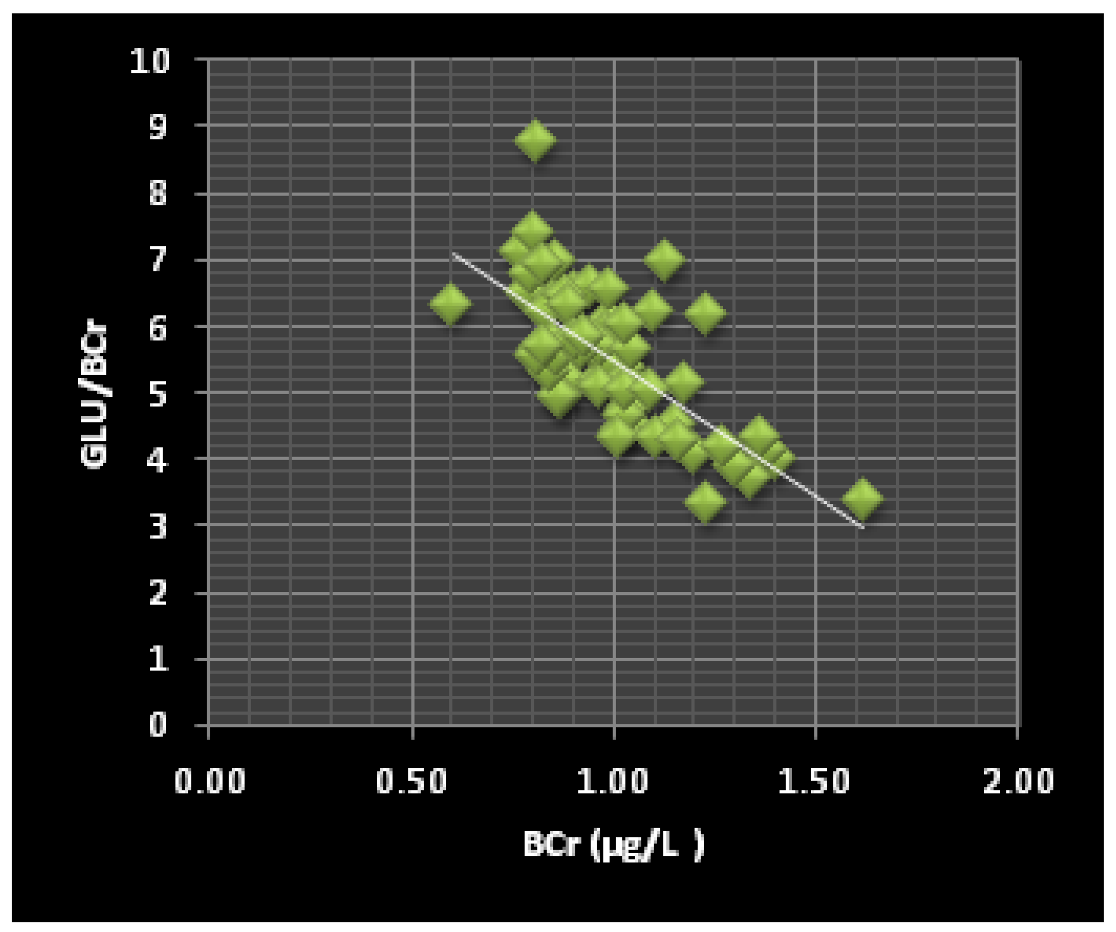
| Glu/BCr | 5.62 | 8.05 | −6.8 | <0.0001 |
| Chol/BCr | 5.63 | 8.76 | −7.9 | <0.0001 |
| TG/BCr | 1.56 | 2.52 | −5.7 | <0.0001 |
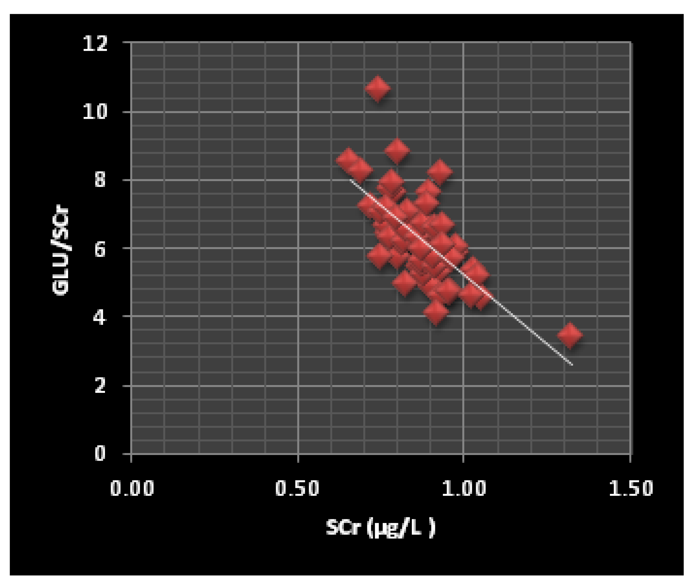
| Glu/SCr | 6.45 | 5.49 | 3.4 | <0.005 |
| Chol/SCr | 6.47 | 5.98 | 1.64 | 0.115 |
| TG/SCr | 1.79 | 1.71 | 1.13 | 0.275 |
| Glu/BV | 8.08 | 13.36 | −9.1 | <0.0001 |
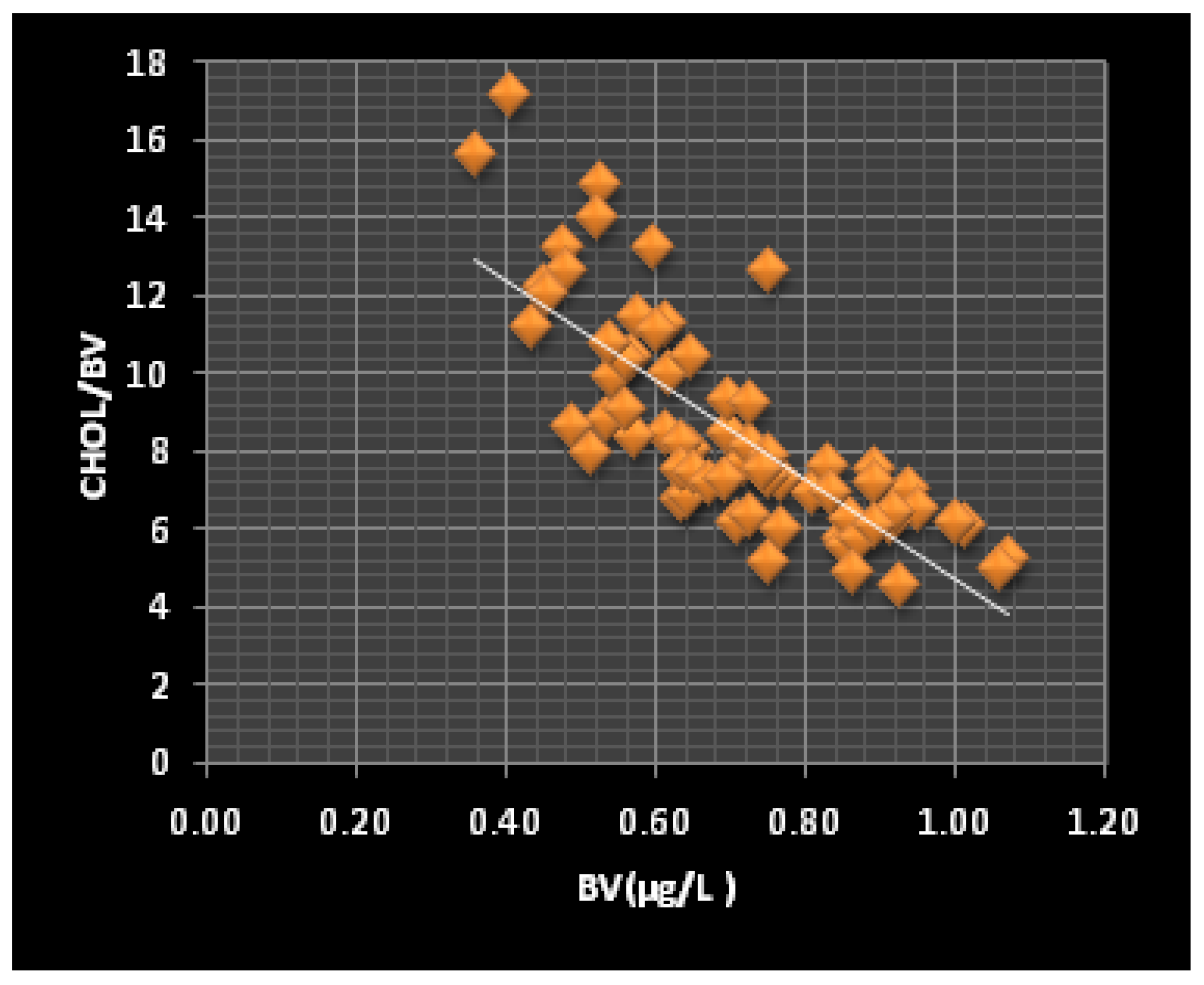
| Chol/BV | 8.04 | 13.61 | −9.3 | <0.0001 |
| TG/BV | 2.22 | 3.91 | −5.6 | <0.0001 |
| Glu/SV | 4.19 | 3.45 | −3.8 | 0.0002 |
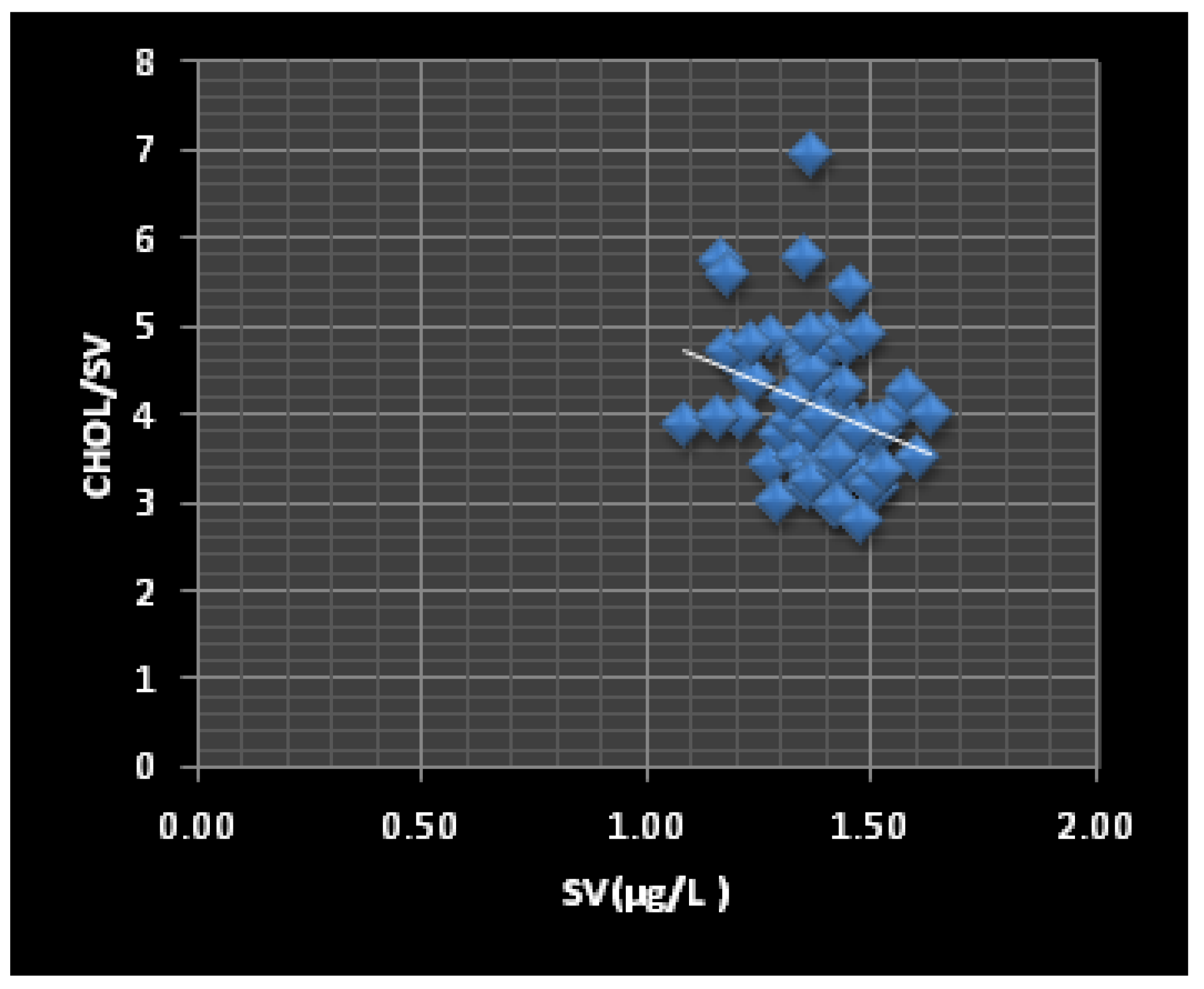
| Chol/SV | 4.08 | 3.76 | −1.65 | 0.105 |
| TG/SV | 1.13 | 1.02 | −1.06 | 0.226 |
| B 1 Cr (µg/L) | S 2 Cr (µg/L) | BV (µg/L) | SV (µg/L) | |
|---|---|---|---|---|
| Glu/BCr | −0.713 a | |||
| Chol/BCr | ||||
| TG/BCr | ||||
| Glu/SCr | −0.678 a | |||
| Chol/SCr | −0.955 a | |||
| TG/SCr | −0.358 c | |||
| Glu/BV | −0.895 a | |||
| Chol/BV | −0.825 a | |||
| TG/BV | −0.672 b | |||
| Glu/SV | −0.288 d | |||
| Chol/SV | −0.203 d | |||
| TG/SV |
Publisher’s Note: MDPI stays neutral with regard to jurisdictional claims in published maps and institutional affiliations. |
© 2022 by the authors. Licensee MDPI, Basel, Switzerland. This article is an open access article distributed under the terms and conditions of the Creative Commons Attribution (CC BY) license (https://creativecommons.org/licenses/by/4.0/).
Share and Cite
Zeneli, L.; Daci-Ajvazi, M.; Sekovanić, A.; Jurasović, J.; Bajraktari, D. The Effects of Chromium and Vanadium on Biomarkers of Carbohydrate and Lipid Metabolism in Workers Exposed to Coal Fly Ash. J. Xenobiot. 2022, 12, 307-316. https://doi.org/10.3390/jox12040021
Zeneli L, Daci-Ajvazi M, Sekovanić A, Jurasović J, Bajraktari D. The Effects of Chromium and Vanadium on Biomarkers of Carbohydrate and Lipid Metabolism in Workers Exposed to Coal Fly Ash. Journal of Xenobiotics. 2022; 12(4):307-316. https://doi.org/10.3390/jox12040021
Chicago/Turabian StyleZeneli, Lulzim, Majlinda Daci-Ajvazi, Ankica Sekovanić, Jasna Jurasović, and Demush Bajraktari. 2022. "The Effects of Chromium and Vanadium on Biomarkers of Carbohydrate and Lipid Metabolism in Workers Exposed to Coal Fly Ash" Journal of Xenobiotics 12, no. 4: 307-316. https://doi.org/10.3390/jox12040021