Article

Phylogenetic and Morphological Evidence Reveal Five New Species of Boletes from Southern China

1
Bioengineering and Technological Research Centre for Edible and Medicinal Fungi, Jiangxi Agricultural University, Nanchang 330045, China
2
Jiangxi Key Laboratory for Conservation and Utilization of Fungal Resources, Jiangxi Agricultural University, Nanchang 330045, China
3
College of Bioscience and Bioengineering, Jiangxi Agricultural University, Nanchang 330045, China
4
Key Laboratory of Crop Physiology, Ecology and Genetic Breeding, Jiangxi Agricultural University, Ministry of Education, Nanchang 330045, China
5
Jiangxi Wuyishan National Nature Reserve Administration Bureau, Wuyishan National Nature Reserve, Shangrao 334500, China
*
Authors to whom correspondence should be addressed.
J. Fungi 2023, 9(8), 814; https://doi.org/10.3390/jof9080814
Submission received: 9 June 2023 / Revised: 26 July 2023 / Accepted: 27 July 2023 / Published: 31 July 2023

Abstract

:
Fungi of the order Boletales are extremely important in both ecology and economy, since most of them are ectomycorrhizal fungi, which play vital roles in maintaining forest ecosystems, water and soil protection, vegetation restoration and so on. Although previous studies have shown that this order has a very high species diversity in China, there are few reports on the species diversity of boletes in Jiangxi Province, China. Based on morphological (macroscopic and microscopic morphological characteristics) and phylogenetic analyses (ITS, LSU, and TEF1-α sequences), in this study, the wild boletes in Jiangxi Province were investigated, and five new species are described: Austroboletus albus, Xanthoconium violaceipes, Xanthoconium violaceofuscum, Xerocomus rutilans and Xerocomus subsplendidus. Descriptions and hand drawings of the new species are presented.

1. Introduction

The order Boletales is one of the largest groups of Basidiomycota and most of them are not only ectomycorrhizal fungi but also edible fungi. They are economically and ecologically important [1]. They could form mycorrhizal relationships with more than ten families of plants, including Betulaceae, Dipterocarpaceae, Fagaceae, Pinaceae and Salicaceae, which play vital roles in maintaining forest ecosystems, water and soil protection, vegetation restoration, and contributing to the diversification of both fungi and their host plants [2].
In the past few decades, over 500 species of Boletales, including nearly 100 new species, have been reported from China [3,4,5,6,7,8,9,10,11,12,13,14,15,16,17,18,19,20,21,22,23,24,25,26,27,28]. Before the 21st century, taxonomy relied primarily on morphological and chemotaxonomic characters, which led to the problem of homonyms and synonyms [29,30,31]. With the rapid development of molecular technology, molecular methods have been continuously applied to the taxonomy, systematics, phylogeny and biogeography of macrofungi, and the classification of boletes has made unprecedented progress. Since 2016, more than 100 new species from China have been described [1,2,32,33,34,35,36,37,38,39,40,41,42,43,44,45], but few of them have been reported in Jiangxi Province [2,36,38,44,46,47].
Jiangxi has the second highest forest coverage in China, which is dominated by evergreen broad-leaved forests with a subtropical warm and humid monsoon climate and abundant rainfall. The climatic conditions and vegetation types are very suitable for the growth and reproduction of macrofungi. It is predicted that there are more than 12,000 species of fungi in Jiangxi Province, including more than 1000 species of macrofungi [48].
Until 2016, 659 species had been reported in Jiangxi Province [49], and there are many macrofungi waiting to be discovered. In this study, five new species of boletes were collected and described during the investigation of the macrofungi in Wuyishan National Nature Reserve in Jiangxi Province.

2. Materials and Methods

2.1. Sample Collection

In this study, 140 boletes specimens were collected in Jiangxi Province. All specimens were completely dug out with a shovel after taking photos of the habitat and related characteristics of the basidiomata, then a part of the context was cut from the connection between pileus and stipe and dried with silica gel to extract DNA, and the remaining specimens were dried at 41–56 °C by a food-grade fruit dryer.

2.2. Morphological Studies

Twelve specimens of five new boletes species were stored in the Herbarium of Fungi of Jiangxi Agricultural University (HFJAU). The macroscopic morphological characteristics mainly come from field records and photographs of basidiomata. Color codes were obtained from Kornerup & Wanscher [50]. Micromorphological descriptions were based on dried materials rehydrated in 5% KOH and stained with ammoniacal Congo red. Freehand sections were performed by using a Nikon SMZ1270 (NIKON Corporation, Japan) stereomicroscope, following the standard method described in previous studies [19,22,51,52]. Microstructures were observed with a Nikon Y–TV55 (NIKON Corporation, Japan) compound microscope. Basidiospores with special structure were examined with a ZEISS EVO18 (GER) scanning electron microscope (SEM).
The number of measured basidiospores is given as n/m/p, which means that the measurements were created on n basidiospores from m basidiomata of p collections. Dimensions of basidiospores are given as (a)b–c(d), where the range b–c represents a minimum of 90% of the measured values (5th to 95th percentile), and extreme values (a and d), whenever present (a < 5th percentile, d > 95th percentile), are in parentheses. Q represents the ratio of length/width of the spores. Qm refers to the average Q of basidiospores ± sample standard deviation [53].

2.3. DNA Extraction, Amplification, and Sequencing

Whole-genome DNA was extracted from approximately 0.5 g of dried specimens by optimized CTAB method [54,55]. The primer pairs ITS1/ITS4 were used to amplify the ITS region [56], LR0R/LR5 were used to amplify the large subunit ribosomal region (nrLSU) [57,58] and TEF1-983F/TEF1-1567R were used to amplify the translation elongation factor 1-α region (TEF1-α) [24,59].
The PCR reaction was under the following conditions: 94 °C for 4 min, then 35 cycles of 94 °C for 60 s (50 °C for ITS, 53 °C for nrLSU and TEF1-α) for 40 s, and 72 °C for 80 s, followed by a final extension step of 72 °C for 8 min [24].
PCR products were detected by NanoDrop One (Thermo Fisher Scientific, Waltham, MA, USA) and 1% agarose gels, and then sequenced by TSINGKE Biological Technology (Hunan, China) using the same primers used for PCR amplification.

2.4. Phylogenetic Analyses

Sequences obtained by sequencing were visualized and edited with BioEdit v7.0.9 [60], and then submitted to NCBI online website for Nucleotide BLAST search (https://blast.ncbi.nlm.nih.gov/Blast.cgi, (accessed on 3 October 2022)) to determine which genus the specimens belonged to. Based on BLAST results, all available nrLSU and TEF1-α sequences were downloaded from NCBI, and were used to identify the relationships among all of our samples and known related species in the GenBank, and to evaluate the variability of the TEF1-α intron region by single-gene analysis and a later polygene phylogenetic analysis [31]. The data ultimately used for phylogenetic analysis are shown in Table 1.
Sequence datasets were aligned on the online website MAFFT version 7 (http://mafft.cbrc.jp/alignment/server/, (accessed on 6 October 2022)) [61], the concatenation of the sequences of the two or three genes was completed in PhyloSuite [62]. The gene fragments of some taxa that could be found or sequenced were regarded as missing data. The intron regions of protein-coding genes were retained in the final analyses [27]. These datasets were then analyzed using RAxML version 8 [63] and MrBayes v3.2 [64] for maximum likelihood (ML) and Bayesian inference (BI), respectively. For ML analyses, under GTRGAMMAI model [65], nonparametric bootstrap analysis with 1000 repetitions [66] was used to determine the statistical support of phylogeney. For BI analyses, substitution models of partition in the datasets were determined using the Bayesian information criterion (BIC) implemented in PartitionFinder 2 [67]. Two or four MCMC runs and trees were sampled every 1000 generations. At the end of the runs, the average deviation of split frequencies was below 0.01. Other parameters were kept at their default settings. Trees were summarized, and posterior probabilities (PPs) were calculated after discarding the first 25% of generations as burn-in. Figtree v1.4.4 was used for visualization of phylogenetic analysis results. Branches that received bootstrap support for maximum likelihood (ML) and Bayesian posterior probabilities (BPP) greater than or equal to 50% (BS) and 0.95 (PP) are shown above.

3. Results

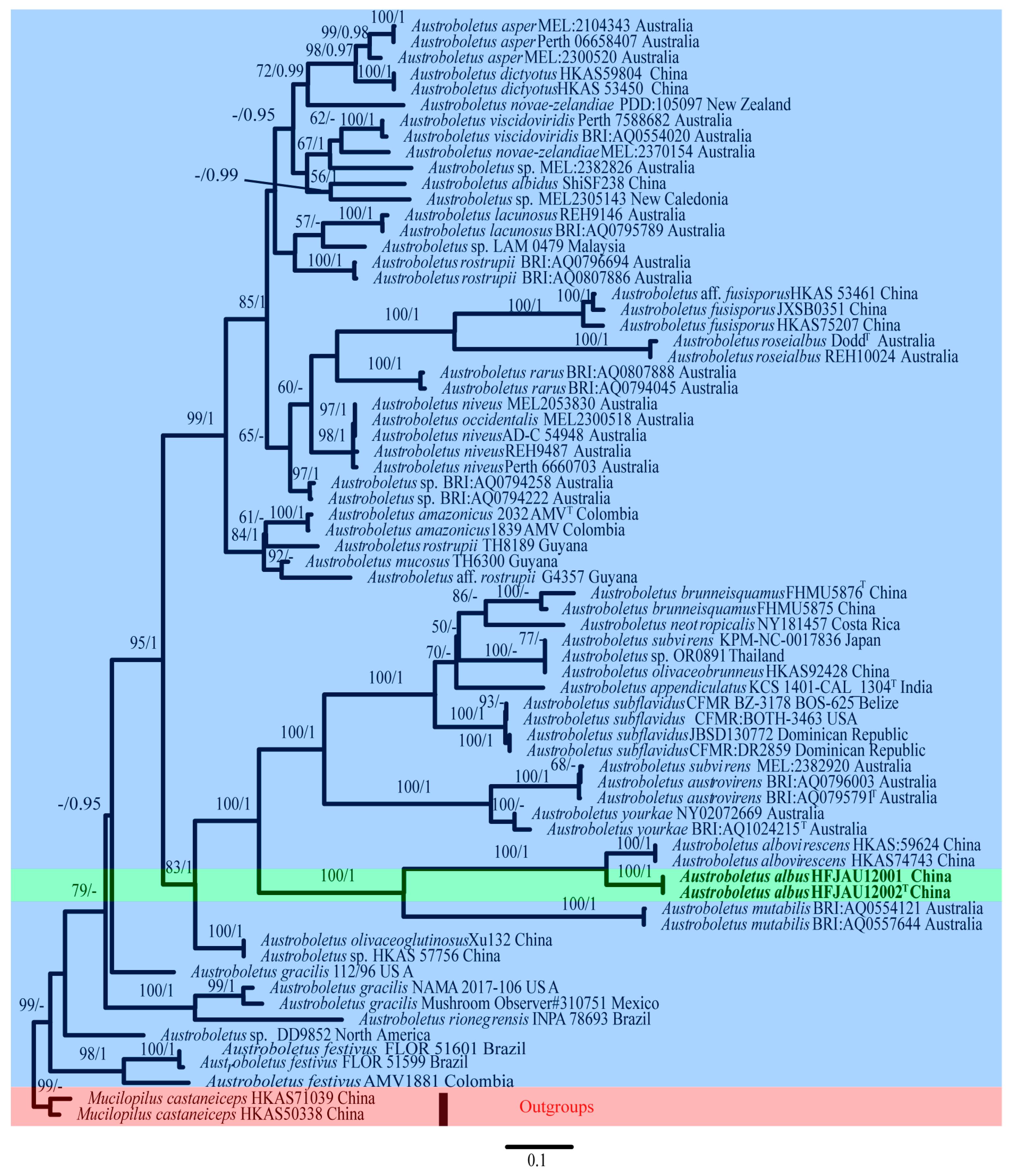
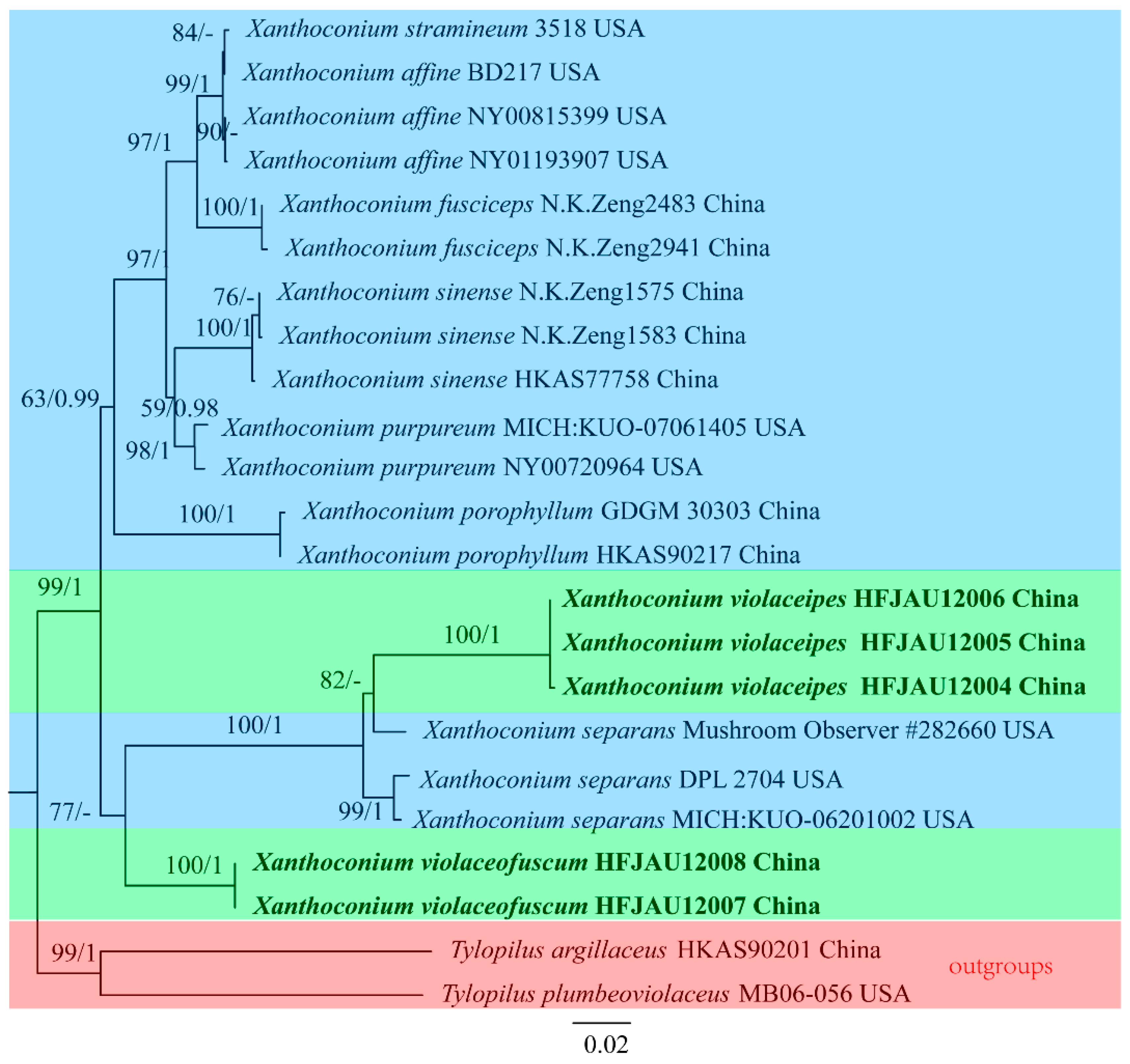
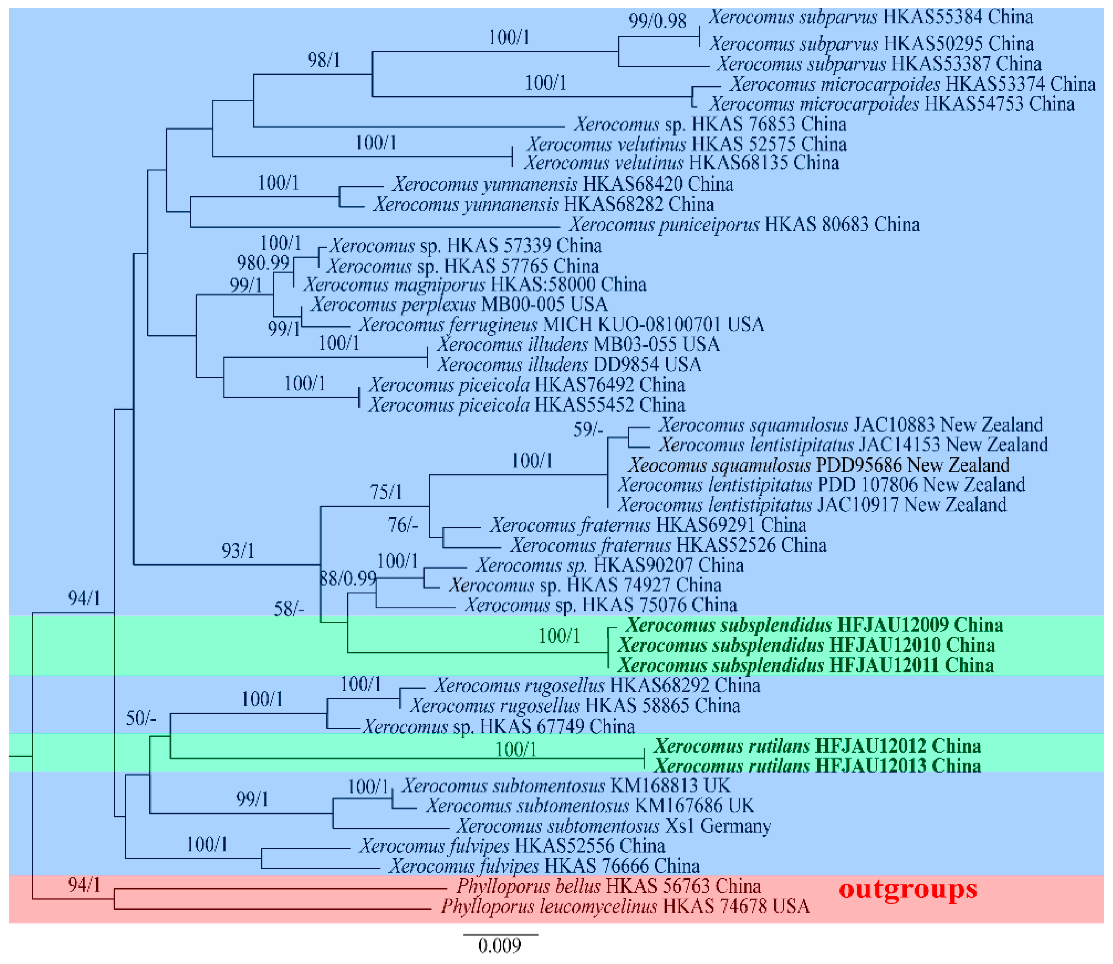
3.1. Molecular Phylogenetic Results
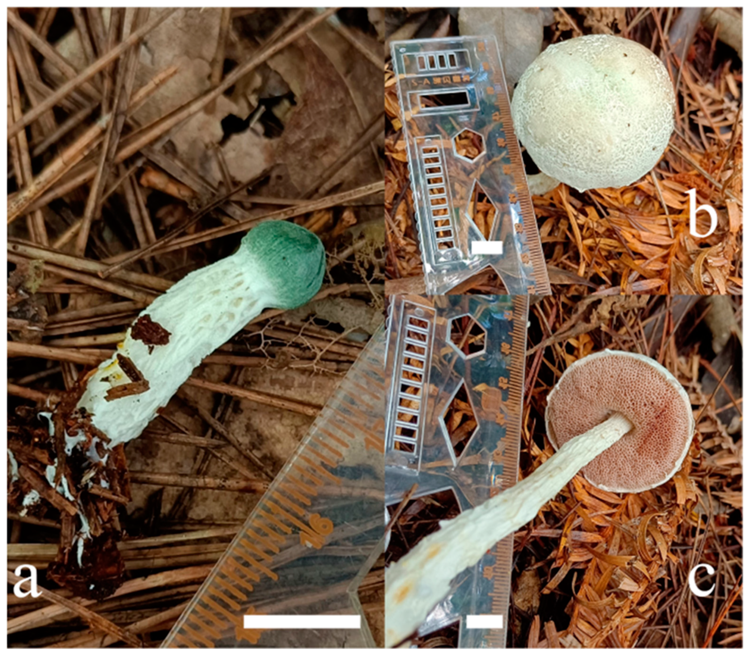
For Austroboletus, six new sequences (two of ITS, two of nrLSU, two of TEF1-α) from two collections were generated. For the combined dataset, HKY + I + G, TIM + I + G and TRNEF + G were evaluated as best-fit substitution models for the ITS, nrLSU and TEF1-α partitions, respectively. The ITS dataset consisted of 43 taxa and 1097 characters. The nrLSU dataset included 60 taxa and 926 characters. The TEF1-α dataset comprised 23 accessions and 623 characters. The combined nuclear dataset (ITS + nrLSU + TEF1-α) contains 126 sequences with 2646 nucleotide sites, and the alignment was deposited in TreeBASE (S30554). The maximum likelihood and Bayesian phylograms have no conflict in topology, the ML trees with both BS and PP values are shown in Figure 1, the green background represents the new species identified in this study, the blue represents other known species of the genus, and the red represents the outgroup.
For Xanthoconium, ten new sequences (five of nrLSU, five of TEF1-α) were generated. For the combined datasets, TRNEF + I + G and TRNEF + G were the best-fit substitution models for the nrLSU and TEF1-α partitions, respectively. The nrLSU dataset included 23 taxa and 883 characters. The TEF1-α dataset comprised 20 accessions and 633 characters, and the alignment was deposited in TreeBASE (S30556). The Maximum likelihood and Bayesian phylograms have no conflict in topology, the ML trees with both BS and PP values are shown in Figure 2, the green background represents the new species identified in this study, the blue represents other known species of the genus, and the red represents the outgroup.
For Xerocomus, ten new sequences (five of nrLSU, five of TEF1-α) were generated. For the combined dataset, TRN + I + G and TRNEF + I + G were the best-fit substitution models for the nrLSU and TEF1-α partitions, respectively. The nrLSU dataset included 45 taxa and 897 characters. The TEF1-α dataset comprised 36 accessions and 619 characters, and the alignment was deposited in TreeBASE (S30557). The Maximum likelihood and Bayesian phylograms have no conflict in topology, the ML trees with both BS and PP values are shown in Figure 3, the green background represents the new species identified in this study, the blue represents other known species of the genus, and the red represents the outgroup.

3.2. Taxonomy

Austroboletus albus F. Zhou, H.Y. Song and D.M. Hu, sp. nov. (Figure 4, Figure 5 and Figure 6).
Mycobank: MB844812.
Etymology—Latin “albus” refers to the color of pileus, which is white.
Diagnosis: Austroboletus albus is characterized by a gray-green reticulate texture pileus, emerald green when young, white to pale brown when mature and an uneven stipe, similar to a gully, covered with obvious white to yellowish white reticulation, subfusiform to amygdaliform basidiospores measuring 13–17 × 6.5–7.5 μm, surface with intricate reticulum or irregular pits, becoming shorter to nearly smooth toward both poles, yellow to golden yellow in KOH.
Holotype: China. Jiangxi Province: Shangrao City, Yanshan County, Wuyishan Nature Reserve, 117°42′56″ E, 27°48′57″ N, elev. 900 m, 23 August 2021, F. Zhou, HFJAU12002 (WYS215).
Description: Basidiomata are small to medium-sized. Pileus 2–5 cm diam, hemispherical when young, becoming convex to subhemispherical with maturity, surface dry, emerald green when young, and white (5A1) to pale brown (4A2) when mature, with gray-green (26B3) reticulate texture, margin decurved, marginal veil white (5A1), serrated to irregular. Context is 0.5–1.6 cm in thickness in the center of the pileus, white (5A1), no change when injured. Hymenophore adnate to slightly depressed around the stipe; pores circular to angular, 1–2 per mm, white (7A1) when young and pinkish white (11A2) to pink (12B3) when old; tubes up to 1.3 cm long, pinkish (9A2), no change in color when injured. Stipe 4–12 × 0.5–1.1 cm, central, cylindrical to clavate, solid; surface dry, uneven, similar to a gully, covered with obvious white (3A1) to yellowish white (1A2) reticulation. Context is white (2A1), no change when injured; basal mycelium white (2A1). Odor indistinct.
Basidia 30–40 × 10.5–12.5 μm, thin-walled, clavate, four-spored; sterigmata 2–5 μm long. Basidiospores [40/2/2] (12–)13–17(–17.5) × (6–)6.5–7.5(–8.5) μm, Q = (1.63–)1.75–2.2(–2.5), Qm = 2.10 ± 0.21, including ornamentation, subfusiform to amygdaliform and slightly angular; surface with intricate reticulum or irregular pits becomes shorter to nearly smooth toward the both poles and yellow to golden yellow in KOH. Hymenophoral trama boletoid is composed of thin- to slightly thick-walled hyphae, 3–8 μm wide and hyaline in KOH. Cheilocystidia are 36–67 × 7.4–10.3 μm, subfusiform to subfusoid-mucronate, sometimes narrowly mucronate, rostrate, thin-walled and hyaline to grayish brown in KOH. Pleurocystidia are absent. Pileipellis is a trichoderm 65–160 μm thick, composed of hyaline to grayish yellow in KOH, thin-walled, and 3–7.5 μm diam; terminal cells are 22–60 × 3–10 μm, clavate or subterete, with an obtuse apex. Pileus trama is composed of thin-walled hyphae 4–11 μm in diameter. Clamp connections are absent in all tissues.
Habitat: Solitary or group on the wet ground under mixed forests of Fagaceae (Fagus longipetiolata) and Pinaceae (Tsuga chinensis and Pinus massoniana).
Distribution: Jiangxi Province, China.
Additional specimens examined: China, Jiangxi Province: Shangrao City, Yanshan County, Wuyishan Nature Reserve, 117°42′54″ E, 27°48′59″ N, elev. 1050 m, 23 August 2021, F. Zhou, HFJAU12001 (WYS222).
Xanthoconium violaceipes F. Zhou, H.Y. Song and D.M. Hu, sp. nov. (Figure 7 and Figure 8).
Mycobank: MB847090.
Etymology—Latin “violaceipes” refers to the color of stipe, which is purple.
Diagnosis: Xanthoconium violaceipes have a hemispherical to convex to applanate pileus, purple-black to purple when young, brownish green when mature, a obvious dark purple stripes, slightly interwoven near the pileus. Basidiospores measure approximately 12.5–16.5 × 4–5.5 μm, ellipsoid to subfusiform to cylindrical, pileipellis is 60–156 μm thick and stipitipellis is 55–110 μm thick.
Holotype: China, Jiangxi Province: Shangrao City, Yanshan County, Wuyishan Nature Reserve, 117°45′44″ E, 27°50′1″ N, elev. 1880 m, 23 August 2022, F. Zhou, HFJAU12006 (WYS642).
Description: Basidiomata are medium-sized to large. Pileus 1–10 cm in diameter, initially hemispherical, becomes convex to applanate with maturity, surface is dry, purple-black (11F1) to purple (11B3) when young, brownish green (30B6–30E6) when mature, margin is lighter and deeper in the center. Context is 0.3–0.7 cm in thickness in the center of the pileus, white (1A1) and does not change when injured. Hymenophore adnate around the stipe are quite crowded; pores are circular to angular, 2–3 per mm, white (1A1) when young and yellow (1A5) to yellow-brown (1B5) when old; tubes up to 1 cm long are white (2A1) to pale brown (2B2) and do not change in color when injured. Stipe 2–10.5 × 0.6–1.5 cm, central, cylindrical, solid; surface dry, with obvious dark purple (16D3) stripes, slightly interwoven near the pileus; context is white (16A1) to light pink (16A2), no reaction when bruised; basal mycelium white. Odor indistinct.
Basidia are 20–36 × 11.5–16 μm, thin-walled, clavate and four-spored; sterigmata are 3–7 μm long, colorless to hyaline in KOH. Basidiospores [40/2/2] (11.5–)12.5–16.5(–17) × (3.5–)4–5.5(–6) μm, Q = (2.18–)2.27–3.33(–3.71), Qm = 3.00 ± 0.35, ellipsoid to subfusiform to cylindrical, slightly thick-walled (up to 0.5 μm), golden yellow in KOH, smooth. Hymenophoral trama are composed of subparallel hyphae 5–12 μm broad, colorless to hyaline in 5% KOH. Cheilocystidia are 35–55 × 8–12 μm, clavate to subfusiform, rarely mucronate, rostrate, thin-walled and colorless to hyaline in KOH. Pleurocystidia are 45–80 × 21–24 μm, clavate and cystiform. Pileipellis is a trichoderm 60–156 μm thick, composed of hyaline hyphae in KOH, thin-walled, and 4–13 μm in diameter; terminal cells are 14–46.5 × 8–18.5 μm, clavate to pyriform or cystidioid, with obtuse apex. Pileus trama is composed of thin-walled hyphae 5–12 μm in diameter. Stipitipellis is 55–110 μm thick, clavate with an obtuse apex, terminal cells (18–25 × 5–11 μm), and colorless in KOH. Stipe trama is composed of parallel hyphae 5–10 μm wide. Clamp connections are absent in all tissues.
Habitat: Scattered on soil in subtropical forests of Fagaceae, including Lithocarpus spp., Castanopsis spp. and Quercus spp.
Distribution: Jiangxi Province, China.
Additional specimens examined: China, Jiangxi Province: Shangrao City, Yanshan County, Wuyishan Nature Reserve, 117°45′42″ E, 27°50′3″ N, elev. 1750 m, 1 August 2021, F. Zhou, HFJAU12004 (WYS121); Shangrao City, Yanshan County, Wuyishan Nature Reserve, 117°45′44″ E, 27°50′3″ N, elev. 1840 m, 23 August 2022, F. Zhou, HFJAU12005 (WYS619).
Xanthoconium violaceofuscum F. Zhou, H.Y. Song and D.M. Hu, sp. nov. (Figure 9 and Figure 10).
Mycobank: MB847089.
Etymology—Latin “violaceofuscum” refers to the color of pileus, which is purple-brown.
Diagnosis: Xanthoconium violaceofuscum is characterized by purplish brown pileus, surface normally densely covered with dark reddish brown tomentose scales, and the context of pileus is pale purple, changing to blue first, then black when injured, the tubes are purplish brown to yellow to yellowish brown, change from blue to black, and has a cylindrical or clavate stipe, purple-brown to black when young and black when mature. Basidiospores are 7–9 × 3.7–5 μm, ellipsoid to subfusiform and golden yellow in KOH.
Holotype: China, Jiangxi Province: Shangrao City, Yanshan County, Wuyishan Nature Reserve, 117°45′43″ E, 27°50′29″ N, elev. 1700 m, 27 August 2022, F. Zhou, HFJAU12007 (WYS717).
Description: Basidiomata are medium-sized to large. Pileus is 3–13 cm diameter, hemispherical to subhemispherical to applanate, surface is dry, purplish brown (7C2), surface is normally densely covered with dark reddish brown (7E3) tomentose scales. Context are up to 1.5 cm in thickness in the center, pale purple (7B2), change to blue (19B5) first, then black (7E1) when injured. Hymenophore adnate around the stipe, scattered; pores circular to angular, 1–2 per mm, purplish brown (7D4) when young and yellow (5B5) to yellowish brown (5D5) when old; tubes are up to 1.5 cm long, purplish brown (7D4) to yellow (5B5) to yellowish brown (5D5), change to blue (19B5) to black (7E1) when injured. Stipe is 3.5–7 × 1–3 cm, central, cylindrical or clavate, solid; surface is dry, purple-brown (7E3) to black (7E1) when young and black (6F1) when mature. Context is pale purple-brown (6D2), changes to black (6F1) when bruised; basal mycelium is purple-gray (6D1). Odor indistinct.
Basidia are 22–34 × 7.5–13 μm, thin-walled, clavate and four-spored; sterigmata are 2–6 μm long. Basidiospores are [40/2/2] 7–9(–11.5) × (3.5–)3.7–5(–6) μm, Q = (1.67–)1.75–2.29(–2.36), Qm = 1.97 ± 0.19, ellipsoid to subfusiform, thin-walled and golden yellow in KOH, smooth. Hymenophoral trama is composed of subparallel hyphae 6–10 μm broad, yellowish white to hyaline in 5% KOH. Cheilocystidia are 37.5–64 × 7–10 μm, subfusiform to subfusoid-mucronate, sometimes narrowly mucronate, rostrate, thin-walled, hyaline to grayish brown in KOH. Pleurocystidia are 33–118 × 12–23 μm, clavate or cystiform. Pileipellis is a trichoderm 30–65 μm thick, composed of hyaline to grayish yellow in KOH, thin-walled, 7–12 μm in diameter; terminal cells are 30–65 × 10–13.5 μm, clavate or subterete, with an obtuse apex. Pileus trama is composed of thin-walled hyphae 7–12 μm in diameter. Stipitipellis ca. is 150 μm thick, with clavate and cystiform terminal cells (37–74 × 8–15.5 μm), colorless and sometimes yellowish brown in KOH. Stipe trama is composed of parallel hyphae 6–12 μm wide. Clamp connections are absent in all tissues.
Habitat: Scattered on soil in subtropical forests of Fagaceae, including Lithocarpus spp., Castanopsis spp. and Quercus spp.
Distribution: Currently only known from Jiangxi Province, China.
Additional specimens examined: China, Jiangxi Province: Shangrao City, Yanshan County, Wuyishan Nature Reserve, 117°45′13″ E, 27°50′14″ N, elev. 1550 m, 27 August 2022, F. Zhou, HFJAU12008 (WYS724).
Xerocomus rutilans F. Zhou, H.Y. Song and D.M. Hu, sp. nov. (Figure 11 and Figure 12).
Mycobank: MB847091.
Etymology—Latin “rutilans” refers to the color of pileus, which is ochre.
Diagnosis: The pileus of Xerocomus rutilans is purple to ochre when young, brownish yellow when mature, the pores are yellow when young and yellowish brown when old and the stipes are covered with white tomentose scales, ellipsoidal to elongated to fusiform basidiospores measuring 8.5–11.5 × 4–5.5 μm, pale yellow to yellow brown in KOH.
Holotype: China, Jiangxi Province: Shangrao City, Yanshan County, Wuyishan Nature Reserve, 117°46′52″ E, 27°59′31″ N, elev. 430 m, 27 August 2022, F. Zhou, HFJAU12013 (WYS693).
Description: Basidiomata are medium-sized to large. Pileus is 2.9–9 cm in diameter, initially hemispherical, becomes convex to applanate with maturity and the surface is dry, purple (11A2) to ochre (9B4) when young, brownish yellow (5B6) when mature. Context is up to 1 cm in thickness in the center of the pileus, white (5A1) and does not change when injured. Hymenophore adnate to slightly depressed around the stipe, quite crowded; pores iiragular, 2–3 per mm, yellow (2A6) when young and yellowish brown (5C6) when old, slightly higher around the stipe; tubes are up to 1 cm long, golden (2A7) to yellowish brown (5C6) and do not change in color when injured. Stipe is 4–9 × 1–3 cm, central, cylindrical, solid, dry and covered with white (2A1) tomentose scales; context is white (2A1) and has no reaction when bruised; basal mycelium is white (1A1). Odor indistinct.
Basidia are 26–45 × 9–11.5 μm, thin-walled, clavate and four-spored; sterigmata are 3.5–6 μm long. Basidiospores are [40/2/2] (8–)8.5–11.5(–12) × (3.5–)4–5.5(–6) μm, Q = (1.83–)2–2.56(–2.75), Qm = 2.27 ± 0.23, ellipsoidal to elongated to fusiform, thin-walled, pale yellow to yellow-brown in KOH, smooth. Hymenophoral trama is composed of subparallel hyphae 5–13 μm broad, colorless to hyaline in KOH. Cheilocystidia are 22–36 × 9–12 μm, common, clavate to subclavate, thin-walled and hyaline to grayish brown in KOH. Pleurocystidia are 28–48 × 8–11 μm, common, clavate to subfusiform or cystidioid. Pileipellis is a trichoderm 60–140 μm thick composed of hyaline to yellow hyphae in KOH, thin-walled, and 6–11 μm in diameter; terminal cells 26–81 × 6–10 μm, clavate to cylindrical. Pileus trama is composed of thin-walled hyphae 5–12 μm in diameter. Stipitipellis is up to 100 μm thick, with clavate to ventricose terminal cells (25–47 × 7–16 μm), hyaline to grayish brown in KOH. Stipe trama is composed of parallel hyphae 4–12.5 μm wide. Clamp connections are absent in all tissues.
Habitat: Solitary or group on the wet ground under mixed forests of Fagaceae (Fagus longipetiolata) and Pinaceae (Tsuga chinensis and Pinus massoniana).
Distribution: Jiangxi Province, China.
Additional specimens examined: China, Jiangxi Province: Shangrao City, Yanshan County, Wuyishan Nature Reserve, 117°46′50″ E, 27°59′25″ N, elev. 400 m, 6 July 2022, F. Zhou, HFJAU12012 (WYS531).
Xerocomus subsplendidus F. Zhou, H.Y. Song and D.M. Hu, sp. nov. (Figure 13 and Figure 14).
Mycobank: MB847092.
Etymology—Latin “subsplendidus” refers to the color of pileus, which is yellowish brown.
Diagnosis: Xerocomus subsplendidus has a subhemispherical to applanate to infundibulate pileus, and the surface is always covered with yellowish brown tomentose when young, cracking into brown to dark brown scales with age, always has a crooked stipe, covered with pale brown to reddish brown tomentose in the upper part, and pores and tubes change to blue when injured, has ellipsoid to subfusiform to cylindrical; basidiospores are 9–15.5 × 4–5.5 μm, pale yellow to yellow-brown in KOH.
Holotype: China, Jiangxi Province: Shangrao City, Yanshan County, Wuyishan Nature Reserve, 117°46′7″ E, 27°50′36″ N, elev. 1750 m, 27 August 2022, F. Zhou, HFJAU12010 (WYS704).
Description: Basidiomata are small to medium-sized. Pileus is 2.2–5.6 cm in diameter, subhemispherical to applanate to infundibulate, initially earth-yellow (6B5) to yellowish brown (6B7), and brown (6C7) to dark brown (6E5) later and have lighter margins; surface dry, always covered with yellowish brown (6C5) tomentose when young, cracking into brown (6A4) to dark brown (6E5) scales with age. Context is white (6A1), up to 0.8 cm in thickness in the center of the pileus, and does not change when injured. Hymenophore is slightly depressed and decurrent around the apex of the stipe, quite scattered; pores are irregular, 1 per mm or less, yellow (3A6), changes to blue (22D5) when bruised; tubes are up to 1 cm long, golden (2A6) to yellowish brown (2D7), changes to blue (22D5) when injured. Stipe is 2.5–5 × 0.4–0.7 cm, central, cylindrical or crooked, solid; covered with pale brown (2B3) to reddish brown (9B4) tomentose in the upper part; context is white (3A1) to pale brown (3A2), no reaction when bruised; basal mycelium white. Odor indistinct.
Basidia are 27–38.5 × 9–11 μm, thin-walled, clavate and four-spored; sterigmata are 3–7 μm long. Basidiospores are [60/3/3] 9–15.5(–16) × 4–5.5(–6) μm, Q = (1.82–)2–3.11(–3.56), Qm = 2.40 ± 0.32, ellipsoid to subfusiform to cylindrical, pale yellow to yellow-brown in KOH and smooth. Hymenophoral trama is composed of subparallel hyphae 5–13 μm broad, colorless to hyaline in KOH. Cheilocystidia are 39–65 × 8.5–12 μm, lanceolate, clavate to ventricose, thin-walled and hyaline to grayish brown in KOH. Pleurocystidia are 31–82 × 10–13 μm, common, similar to cheilocystidia. Pileipellis is a trichoderm 70–230 μm thick composed of hyaline hyphae in KOH, thin-walled, 5–13 μm in diameter; terminal cells are 27.5–54.5 × 10⁻¹⁶ μm, clavate or cystidioid. Pileus trama is composed of thin-walled hyphae 5–12 μm diam. Stipitipellis is 45–140 μm thick, with clavate to pyriform terminal cells (18–28.5 × 8.5–15 μm) and hyaline to grayish brown in KOH. Stipe trama is composed of parallel hyphae 6–18 μm wide. Clamp connections are absent in all tissues.
Habitat: Solitary or group on the wet ground under mixed forests of Fagaceae (Fagus longipetiolata) and Pinaceae (Tsuga chinensis and Pinus massoniana).
Distribution: Jiangxi Province, China.
Additional specimens examined: China, Jiangxi Province: Shangrao City, Yanshan County, Wuyishan Nature Reserve, 117°45′56″ E, 27°50′22″ N, elev. 1700 m, 23 August 2021, F. Zhou, HFJAU12009 (WYS232); Shangrao City, Yanshan County, Wuyishan Nature Reserve, 117°45′43″ E, 27°50′29″ N, elev. 1740 m, 27 August 2022, F. Zhou, HFJAU12011 (WYS718).

4. Discussion

The macro and micromorphology of Austroboletus albus conforms to the characteristics of Austroboletus. It shared similar morphological characteristics with A. subflavidus, A. albidus and A. roseialbus. However, A. subflavidus has larger basidia [27–49(51) × 12–19 μm] and basidiospores [(13.1)15.9 ± 1.15(19.5) × (5.5)7.0 ± 0.58(8.7) μm], slightly shorter and coarser stipe [(2.9)4.5–7.5(10.2) × (0.4)0.6–1.8(2.0) cm], pileus surface often cracks with matures [68]; A. albidus can be easily differentiated by wider basidia (28–39 × 14–20 μm), shorter stipe (4–7 × 0.4–0.6 cm), and the color of pileus is white to cream when young, cream to grayish yellow when mature, covered with light orange to brownish nubby squamules [2]; A. roseialbus has smaller basidiospores (11.2–14 × 6.3–7 μm), wider basidia (28–35 × 10–14 μm), shorter stipe (7–8 cm), and the color of the pileus is whitish with obvious pale pink tinges [69].
Phylogenetically, the specimens we collected formed an independent clade with strong support (BS = 100%, PP = 1), and were closed to A. albovirescens (HKAS:59624 and HKAS74743) [1,2,31] with high statistical support (BS = 100%, PP = 1). However, A. albovirescens was described by Li and Yang [2], which is characterized by the matte green to gray-green pileus, the ornamentation of basidiospores with unequal pits, and the ixocutis pileipellis. Meanwhile, they formed a large clade with A. mutabilis (BS = 100%, PP = 1), a species recorded in Australia [70]. However, A. mutabilis can be easily distinguished by shorter basidiospores (11.9–14.7 × 4.9–7 µm) and basidia (25–35 × 10–13 µm), and the color of the pileus is dark red to brownish red when young, soon fading to brownish orange, becoming orangish yellow, then eventually yellowish.
Morphologically, Xa. violaceipes is similar to Boletus violaceofuscus and Xa. separans. Howener, B. violaceofuscus has longer and narrower basidia [(29)32–40(43) × 10.5–13.5 μm], smaller pleurocystidia [(47)52–60 × 7–10 μm] and longer cheilocystidia [(26)37–67 × 7–10 μm], slightly larger basidiospores [(14.0)15.5 ± 0.8(17.8) × (5.0)5.6 ± 0.3(6.2) μm], larger stipitipellis terminal cells [20–40 × (4)8–15(18) μm] with rare tw- spored and four-spored basidia [71]. Xa. separans can be easily distinguished by larger pileus [60–200(220) mm] and stipe [60–150 × 10–30 mm], longer basidia [(27)32–41(43) × 10–12 μm], smaller pleurocystidia [(52)55–70 × 11–14 μm] and larger cheilocystidia [(24)30–65(80) × 8–15(18) μm] [71].
Based on the phylogenetic analyses, the collections of Xa. violaceipes are clustered on one branch within Xanthoconium with 100% bootstrap support, and sister to Xa. separans with 82% statistical support, but they have a long genetic distance.
Morphologically, Xa. violaceofuscum has similar pileus and stipe colors to Boletus violaceofuscus [3,71]. However, B. violaceofuscus has larger basidiospores [(14.0)15.5 ± 0.8(17.8) × (5.0)5.6 ± 0.3(6.2) μm] and basidia [(29)32–40(43) × 10.5–13.5 μm], has smaller pleurocystidia [(47)52–60 × 7–10 μm], and the tubes are white or whitish at first, stipe has net–like ornamentation and at the base has white tomentum, has a few two-spored and four-spored basidia in stipitipellis, and the flesh is white.
In phylogenetic analyses, the collections of Xa. violaceofuscum are clustered on an independent branch within Xanthoconium with 100% bootstrap support.
Morphologically, Xerocomus rutilans is similar to Aureoboletus zangii [1,72] by its bright yellow hymenophore and reddish gray to grayish red pileus color. However, Au. zangii can be distinguished by its fox-red to English-red stipe, viscid pileus and stipe. Au. zangii has shorter basidia [20–30 μm], larger cheilocystidia (24–51 × 9–16 μm) and pleurocystidia [40–68(77) × 11.5–18 μm].
Phylogenetically, the samples of Xerocomus rutilans are clustered into an unique branch within Xerocomus with high statistical support (BS = 100%), and related to X. rugosellus [1,3] with 50% bootstrap support. However, X. rugosellus is characterized by the slowly bluing hymenophore and context when injured, wider basidia (12–15 μm), and t larger basidiospores [(12)14–15.5(18) × (4.5)5–5.5(7) μm] and pleuro- and cheilocystidia (45–80 × 10–14 μm).
Morphologically, Xerocomus subsplendidus is similar to X. fraternus, X. subparvus and X. yunnanensis [1]. However, X. fraternus can be distinguished by the changes in pileus and stipe context when injured, pileus context changing from cream to yellowish and staining bluish slowly when injured, stipe context cream on upper part, staining pale blue slowly when injured, and lower part pale red-brown near stipe base; X. fraternus has shorter basidiospores [(8.5)9.5–12(13) μm], and the shape of pileipellis terminal cells is subcylindrical. X. subparvus has cream to yellowish pileus context and a red tinge near pileipellis, staining bluish slowly or indistinctly when injured, context of the stipe is pale yellow at the upper part, staining bluish slowly when injured and pale brown to pale-red brown at the lower part; X. subparvus has smaller basidiospores [(8.5)9–10.5(11.5) × (3)3.5–4(4.5) μm], and the shape of pileipellis terminal cells is subcylindrical. X. yunnanensis is characterized by the tubes staining red-brown slowly when young and becoming bluish when mature on injury, the fresh yellow context of pileus near the pileipellis, and the white to yellowish context of stipe, bluing indistinctly when cut [1]; X. yunnanensis has smaller basidiospores [(9)10–11.5(13) × 4–4.5(5) μm], and the terminal cells of pileipellis are tapered or bullet-shaped.
According to our phylogeny, the sequences of Xerocomus subsplendidus form an independent branch within Xerocomus with 100% bootstrap support, and are sistered to Xerocomus sp. (voucher: HKAS90207, HKAS 74927 and HKAS 75076) with 58% statistical support, but they have a long genetic distance.

Author Contributions

Conceptualization, F.Z. and D.-M.H.; Methodology, F.Z., H.-Y.S., W.-J.Y. and W.Z.; Performing the experiment, F.Z.; Formal analysis, F.Z.; Resources, F.Z., Y.G., L.-Y.L., Y.F., L.C. and H.-J.H.; Writing—original draft preparation, F.Z.; Writing—review and editing, D.-M.H. and Y.G.; Supervision, D.-M.H.; Project administration, D.-M.H.; Funding acquisition, D.-M.H. All authors have read and agreed to the published version of the manuscript.

Funding

Funds for research were provided by the National Natural Science Foundation of China (NSFC 32060014, NSFC 32070023, NSFC 32160010, and NSFC 32160673), the earmarked fund for Jiangxi Agriculture Research System (2023), and the Forestry Science and Technology Innovation Project of Jiangxi Forestry Bureau of China (Innovation Special [2022] No. 9).

Institutional Review Board Statement

Not applicable.

Informed Consent Statement

Not applicable.

Data Availability Statement

All newly generated sequences were deposited in GenBank (https://www.ncbi.nlm.nih.gov/genbank/, Table 1 (Submitted on 30 December 2022)). All new taxa were deposited in MycoBank (https://www.mycobank.org/).

Acknowledgments

The author thanks Liu Kun, Jia (Kunming Institute of Botany, CAS) and Li Rong, Liu for their help, and the rangers at the Wuyishan Nature Reserve in China, for their help in fieldwork.

Conflicts of Interest

The authors declare no conflict of interest.

References

  1. Wu, G.; Li, Y.C.; Zhu, X.T.; Zhao, K.; Han, L.H.; Cui, Y.Y.; Li, F.; Xu, J.P.; Yang, Z.L. One hundred noteworthy boletes from China. Fungal Divers. 2016, 81, 25–188. [Google Scholar] [CrossRef]
  2. Li, Y.C.; Yang, Z.L. The Boletes of China—Tylopilus s.l.; Springer Nature: Berlin/Heidelberg, Germany; Beijing Science Press: Beijing, China, 2021; p. 418. [Google Scholar]
  3. Chiu, W.F. The Boletes of Yunnan. Mycologia 1948, 40, 199–231. [Google Scholar] [CrossRef]
  4. Teng, S.C. Fungi of China; Science Press: Beijing, China, 1963. [Google Scholar]
  5. Yeh, K.W.; Chen, Z.C. The Boletes of Taiwan (I). Taiwania 1980, 25, 166–184. [Google Scholar]
  6. Bi, Z.; Lu, D.; Zheng, G. Basidiomycetes from Dinghu Mountain of China. II. Some species of Boletaceae (1). Acta Bot. Yunnanica 1982, 4, 55–64. [Google Scholar]
  7. Bi, Z.; Li, T.; Zheng, G.; Li, C. Basidiomycetes from Dinghu Mountain of China. III. Some species of Boletaceae (2). Acta Mycol. Sin. 1984, 3, 199–206. [Google Scholar]
  8. Ying, J.Z.; Ma, Q.M. New taxa and records of the genus Strobilomyces in China. Acta Mycol. Sin. 1985, 4, 95–102. [Google Scholar]
  9. Zang, M. Notes on the Boletales from eastern Himalayas and adjacent of China. Acta Bot Yunnanica 1985, 7, 383–401. [Google Scholar]
  10. Ying, J.Z. New species of the genus Xerocomus from China. Acta Microbiol. Sin. Suppl. 1986, 1, 309–315. [Google Scholar]
  11. Zang, M. Notes on the Boletales from eastern Himalayas and adjacent of China (2). Acta Bot Yunnanica 1986, 8, 1–22. [Google Scholar]
  12. Bi, Z.; Li, T. New taxon and new records of the genus Suillus from Guangdong. Acta Microbiol. Sin. 1990, 9, 20–24. [Google Scholar]
  13. Bi, Z.S.; Zheng, G.Y.; Li, T.H.; Wang, Y.Z. Macrofungus Flora of the Mountainous District of North Guangdong; Guangdong Science and Technology Press: Guangdong, China, 1990. [Google Scholar]
  14. Zang, M. Sinoboletus, a new genus of Boletaceae from China. Mycotaxon 1992, 45, 223–227. [Google Scholar]
  15. Bi, Z.S.; Zheng, G.Y.; Li, T.H. The Macrofungus Flora of China’s Guangdong Province; Chinese University Press: Hong Kong, China, 1993. [Google Scholar]
  16. Zang, M.; Li, B.; Xi, J.; Zhang, D. Fungi of Hengduan Mountains; Science Press: Beijing, China, 1996. [Google Scholar]
  17. Zang, M.; Li, T.; Petersen, R. Five new species of Boletaceae from China. Mycotaxon 2001, 80, 481–488. [Google Scholar]
  18. Zang, M. Flora Fungorum Sinicorum. Boletaceae (I); Science Press: Beijing, China, 2006; Volume 22, p. 215. [Google Scholar]
  19. Li, Y.C.; Feng, B.; Yang, Z.L. Zangia, a new genus of Boletaceae supported by molecular and morphological evidence. Fungal Divers. 2011, 49, 125–143. [Google Scholar] [CrossRef]
  20. Zhang, M.; Li, T.H.; Bau, T.; Song, B. A new species of Xerocomus from Southern China. Mycotaxon 2012, 121, 23–27. [Google Scholar] [CrossRef]
  21. Zang, M. Flora Fungorum Sinicorum. Boletaceae (II); Science Press: Beijing, China, 2013; Volume 44, p. 152. [Google Scholar]
  22. Zeng, N.K.; Liang, Z.Q.; Yang, Z.L. Boletus orientialbus, a new species with white basidioma from subtropical China. Mycoscience 2013, 55, 159–163. [Google Scholar] [CrossRef]
  23. Li, Y.C.; Li, F.; Zeng, N.K.; Cui, Y.Y.; Yang, Z.L. A new genus Pseudoaustroboletus (Boletaceae, Boletales) from Asia as inferred from molecular and morphological data. Mycol. Prog. 2014, 13, 1207–1216. [Google Scholar] [CrossRef]
  24. Li, Y.C.; Santana, B.O.; Zeng, N.K.; Feng, B.; Yang, Z.L. Molecular phylogeny and taxonomy of the genus Veloporphyrellus. Mycologia 2014, 106, 291–306. [Google Scholar] [CrossRef]
  25. Zhang, M.; Li, T.H.; Song, B. A new slender species of Aureoboletus from southern China. Mycotaxon 2014, 128, 195–202. [Google Scholar] [CrossRef]
  26. Wu, G.; Zhao, K.; Li, Y.C.; Zeng, N.K.; Feng, B.; Halling, R.E.; Yang, Z.L. Four new genera of the fungal family Boletaceae. Fungal Divers. 2015, 81, 1–24. [Google Scholar] [CrossRef]
  27. Cui, Y.Y.; Feng, B.; Wu, G.; Xu, J.P.; Yang, Z.L. Porcini mushrooms (Boletus sect. Boletus) from China. Fungal Divers. 2015, 81, 189–212. [Google Scholar] [CrossRef]
  28. Zeng, N.K.; Tang, L.P.; Li, Y.C.; Tolgor, B.; Zhu, X.T.; Zhao, Q.; Yang, Z.L. The genus Phylloporus (Boletaceae, Boletales) from China: Morphological and multilocus DNA sequence analyses. Fungal Divers. 2012, 58, 73–101. [Google Scholar] [CrossRef]
  29. Snell, W. The genera of the Boletaceae. Mycologia 1941, 33, 415–423. [Google Scholar] [CrossRef]
  30. Horak, E. Revision of Malaysian Species of Boletales s.l. (Basidiomycota) Described by EJH Corner (1972, 1974); Forest Research Institute Malaysia: Kuala Lumpur, Malaysia, 2011; Volume 51, p. 283. [Google Scholar]
  31. Wu, G.; Feng, B.; Xu, J.P.; Zhu, X.T.; Li, Y.C.; Zeng, N.K.; Hosen, M.I.; Yang, Z.L. Molecular phylogenetic analyses redefine seven major clades and reveal 22 new generic clades in the fungal family Boletaceae. Fungal Divers. 2014, 69, 93–115. [Google Scholar] [CrossRef]
  32. Qi, L.L.; Fu, Y.P.; Wang, F.J.; Song, B.; Li, Y. Suillus foetidus (Boletales, Basidiomycota), a new species from northeast China. Phytotaxa 2016, 260, 167–175. [Google Scholar] [CrossRef]
  33. Liang, Z.Q.; Chai, H.; Jiang, S.; Ye, Z.K.; Zeng, N.K. The genus Xanthoconium (Boletaceae, Boletales) in tropical China. Phytotaxa 2017, 295, 246–254. [Google Scholar] [CrossRef]
  34. Qi, L.L.; Fu, Y.P.; Lang, N.; Bai, X.; Li, Y. A new species of Gomphidius from Northeast China. Phytotaxa 2017, 316, 181–188. [Google Scholar] [CrossRef]
  35. Chai, H.; Liang, Z.Q.; Xue, R.; Jiang, S.; Luo, S.H.; Wang, Y.; Wu, L.L.; Tang, L.P.; Chen, Y.; Hong, D.; et al. New and noteworthy boletes from subtropical and tropical China. MycoKeys 2019, 46, 55–96. [Google Scholar] [CrossRef] [PubMed]
  36. Fang, J.Y.; Wu, G.; Zhao, K. Aureoboletus rubellus, a new species of bolete from Jiangxi Province, China. Phytotaxa 2019, 420, 72–78. [Google Scholar] [CrossRef]
  37. Xue, R.O.U.; Liang, Z.Q.; Chai, H.U.I.; Jiang, S.; Tang, L.P.; Fu, Y.Q.; Fan, Y.G.; Wu, L.L.; Zeng, N.K. The Suillus spraguei complex (Suillaceae, Boletales): New taxon, new hosts and amended descriptions. Phytotaxa 2019, 401, 239–256. [Google Scholar] [CrossRef]
  38. Zhang, M.; Li, T.H.; Wang, C.Q.; Zeng, N.K.; Deng, W.Q. Phylogenetic overview of Aureoboletus (Boletaceae, Boletales), with descriptions of six new species from China. MycoKeys 2019, 61, 111–145. [Google Scholar] [CrossRef]
  39. Han, L.H.; Wu, G.; Horak, E.; Halling, R.E.; Xu, J.; Ndolo, E.S.T.; Sato, H.; Fechner, N.; Sharma, Y.P.; Yang, Z.L. Phylogeny and species delimitation of Strobilomyces (Boletaceae), with an emphasis on the Asian species. Persoonia 2020, 44, 113–139. [Google Scholar] [CrossRef] [PubMed]
  40. Wang, Y.I.; Su, M.S.; Jiang, S.; Xue, R.O.U.; Wu, L.L.; Xie, H.J.; Zhang, Y.Z.; Liang, Z.Q.; Zeng, N.K. The genus Hourangia in China and a description of Aureoboletus erythraeus sp. nov. Phytotaxa 2020, 472, 87–106. [Google Scholar] [CrossRef]
  41. Huang, C.; Zhang, M.; Wu, X.L.; Wu, G.; Xu, J.P.; Yang, Z.L.; Li, Y.C. Cyanescent Gyroporus (Gyroporaceae, Boletales) from China. MycoKeys 2021, 81, 165–183. [Google Scholar] [CrossRef] [PubMed]
  42. Jiang, S.; Mi, H.X.; Xie, H.J.; Zhang, X.; Chen, Y.; Liang, Z.Q.; Zeng, N.K. Neoboletus infuscatus, a new tropical bolete from Hainan, southern China. Mycoscience 2021, 62, 205–211. [Google Scholar] [CrossRef] [PubMed]
  43. Zhang, M.; Xie, D.C.; Wang, C.Q.; Deng, W.Q.; Li, T.H. New insights into the genus Gyroporus (Gyroporaceae, Boletales), with establishment of four new sections and description of five new species from China. Mycology 2022, 13, 223–242. [Google Scholar] [CrossRef]
  44. Zhou, F.; Gao, Y.; Song, H.Y.; Hu, H.J.; Li, S.X.; Liu, J.H.; Hu, D.M. Retiboletus atrofuscus (Boletaceae, Boletales), a new species from China. Arch. Microbiol. 2022, 204, 381. [Google Scholar] [CrossRef]
  45. Chen, X.N.; Zhang, M.; Li, T.H.; Zeng, N.K. A new species of Heimioporus (Boletaceae) from southern China. Phytotaxa 2019, 415, 179–188. [Google Scholar] [CrossRef]
  46. Zeng, N.K.; Liang, Z.Q.; Wu, G.; Li, Y.C.; Yang, Z.L. The genus Retiboletus in China. Mycologia 2016, 108, 363–380. [Google Scholar] [CrossRef]
  47. Zhao, K.; Zhang, F.M.; Zeng, Q.Q.; Han, L.H.; Li, Y.C. Tylopilus jiangxiensis, a new species of Tylopilus s. str. from China. Phytotaxa 2020, 434, 281–291. [Google Scholar] [CrossRef]
  48. Huo, G.H.; Yan, J.Q.; Zhang, L.P.; Hu, D.M. Illustrated Handbook of Macrofungi in Jiangxi; Jiangxi Science And Technology Press: Nanchang, China, 2020; p. 296. [Google Scholar]
  49. Zhang, J.; Song, H.; Hu, D. A Checklist of Macrofungi in Jiangxi Province. Biol. Disaster Sci. 2016, 39, 1–13. [Google Scholar]
  50. Kornerup, A.; Wanscher, J.H. Taschenlexikon der Farben 3; Aufl; Muster-Schmid: Northeim, Germany, 1981; p. 242. [Google Scholar]
  51. Zeng, N.K.; Cai, Q.; Yang, Z.L. Corneroboletus, a new genus to accommodate the southeastern Asian Boletus indecorus. Mycologia 2012, 104, 1420–1432. [Google Scholar] [CrossRef] [PubMed]
  52. Hosen, I.; Feng, B.; Wu, G.; Zhu, X.-T.; Li, Y.-C.; Yang, Z.L. Borofutus, a new genus of Boletaceae from tropical Asia: Phylogeny, morphology and taxonomy. Fungal Divers. 2013, 58, 215–226. [Google Scholar] [CrossRef]
  53. Zeng, N.K.; Chai, H.; Jiang, S.; Xue, R.; Wang, Y.; Hong, D.; Liang, Z.Q. Retiboletus nigrogriseus and Tengioboletus fujianensis, two new boletes from the south of China. Phytotaxa 2018, 367, 45–54. [Google Scholar] [CrossRef]
  54. Doyle, J. A rapid DNA isolation procedure for small quantities of fresh leaf tissue. Phytochem. Bull. 1987, 19, 11–15. [Google Scholar]
  55. Huang, J.C.; Ge, X.J.; Sun, M. Modified CTAB Protocol Using a Silica Matrix for Isolation of Plant Genomic DNA. BioTechniques 2000, 28, 432–434. [Google Scholar] [CrossRef]
  56. White, T.J.; Bruns, T.D.; Lee, S.B.; Taylor, J.W.; Innis, M.A.; Gelfand, D.H.; Sninsky, J. Amplification and Direct Sequencing of Fungal Ribosomal RNA Genes for Phylogenetics; Academic Press: Cambridge, MA, USA, 1990; Volume 31, pp. 315–322. [Google Scholar]
  57. Vilgalys, R.; Gonzalez, D. Organization of ribosomal DNA in the basidiomycete Thanatephorus praticola. Curr. Genet. 1990, 18, 277–280. [Google Scholar] [CrossRef]
  58. James, T.Y.; Kauff, F.; Schoch, C.L.; Matheny, P.B.; Hofstetter, V.; Cox, C.J.; Celio, G.; Gueidan, C.; Fraker, E.; Miadlikowska, J.; et al. Reconstructing the early evolution of Fungi using a six-gene phylogeny. Nature 2006, 443, 818–822. [Google Scholar] [CrossRef]
  59. Rehner, S.A.; Buckley, E. A Beauveria phylogeny inferred from nuclear ITS and EF1-α sequences: Evidence for cryptic diversification and links to Cordyceps teleomorphs. Mycologia 2005, 97, 84–98. [Google Scholar] [CrossRef]
  60. Hall, T.A. BioEdit: A user-friendly biological sequence alignment editor and analysis program for Windows 95/98/NT. Nucleic Acids Symp. Ser. 1999, 41, 95–98. [Google Scholar]
  61. Katoh, K.; Standley, D.M. MAFFT multiple sequence alignment software version 7: Improvements in performance and usability. Mol. Biol. Evol. 2013, 30, 772–780. [Google Scholar] [CrossRef]
  62. Zhang, D.; Gao, F.; Jakovlić, I.; Zou, H.; Zhang, J.; Li, W.X.; Wang, G.T. PhyloSuite: An integrated and scalable desktop platform for streamlined molecular sequence data management and evolutionary phylogenetics studies. Mol. Ecol. Resour. 2020, 20, 348–355. [Google Scholar] [CrossRef] [PubMed]
  63. Stamatakis, A. RAxML Version 8: A Tool for Phylogenetic Analysis and Post-Analysis of Large Phylogenies. Bioinformatics 2014, 30, 1312–1313. [Google Scholar] [CrossRef] [PubMed]
  64. Ronquist, F.; Teslenko, M.; Mark, P.V.D.; Ayres, D.L.; Darling, A.; Höhna, S.; Larget, B.; Liu, L.; Suchard, M.A.; Huelsenbeck, J. MrBayes 3.2: Efficient Bayesian phylogenetic inference and model choice across a large model space. Syst. Biol. 2012, 61, 539–542. [Google Scholar] [CrossRef] [PubMed]
  65. Stamatakis, A. RAxML-VI-HPC: Maximum likelihood-based phylogenetic analyses with thousands of taxa and mixed models. Bioinformatics 2006, 22, 2688–2690. [Google Scholar] [CrossRef]
  66. Joseph, F. Confidence Limits on Phylogenies: An Approach Using the Bootstrap. Evolution 1985, 39, 783–791. [Google Scholar]
  67. Lanfear, R.; Frandsen, P.B.; Wright, A.M.; Senfeld, T.; Calcott, B. PartitionFinder 2: New Methods for Selecting Partitioned Models of Evolution for Molecular and Morphological Phylogenetic Analyses. Mol. Biol. Evol. 2016, 34, 772–773. [Google Scholar] [CrossRef]
  68. Gelardi, M.; Angelini, C.; Costanzo, F.; Ercole, E.; Ortiz-Santana, B.; Vizzini, A. Outstanding Pinkish Brown-Spored Neotropical Boletes: Austroboletus subflavidus and Fistulinella gloeocarpa (Boletaceae, Boletales) from the Dominican Republic. Mycobiology 2020, 49, 24–45. [Google Scholar] [CrossRef]
  69. Fechner, N.; Bonito, G.; Bougher, N.L.; Lebel, T.; Halling, R.E. New species of Austroboletus (Boletaceae) in Australia. Mycol. Prog. 2017, 16, 769–775. [Google Scholar] [CrossRef]
  70. Halling, R.E.; Osmundson, T.W.; Neves, M.A. Austroboletus mutabilis sp. nov. from northern Queensland. Muelleria 2006, 24, 31–36. [Google Scholar] [CrossRef]
  71. Simonini, G.; Floriani, M.; Binder, M.; Besl, H. Two close extraeuropean Boletes: Boletus violaceofuscus and Boletus separans. Micol. E Veg. Mediterr. 2001, 16, 148–170. [Google Scholar]
  72. Shi, X.F.; Liu, P.G. Aureoboletus zangii (Boletaceae), a new species from China. Mycotaxon 2013, 123, 451–456. [Google Scholar] [CrossRef]
Figure 1. Maximum likelihood phylogenetic tree of Austroboletus inferred from the combined nuclear dataset (ITS + nrLSU + TEF1-α). Bootstrap frequencies ≥ 50% and posterior probabilities ≥ 0.95 are shown above supported branches. New sequences are shown in bold. The different colors are for decoration. The green background represents the sequence of new species discovered in this study, the red background represents the outer group, and the blue background represents other known species of this genus used in this study.
Jof 09 00814 g001
Figure 2. Maximum likelihood phylogenetic tree of Xanthoconium inferred from the combined nuclear dataset (nrLSU + TEF1-α). Bootstrap frequencies ≥ 50% and posterior probabilities ≥ 0.95 are shown above supported branches. New sequences are shown in bold. Different colors are used to highlight different contents. The green background represents the new species identified in this study, the blue represents other known species of the genus, and the red represents the outgroup.
Jof 09 00814 g002
Figure 3. Maximum likelihood phylogenetic tree of Xerocomus inferred from the combined nuclear dataset (nrLSU + TEF1-α). Bootstrap frequencies ≥ 50% and posterior probabilities ≥ 0.95 are shown above supported branches. New sequences are shown in bold. Different colors are used to highlight different contents. The green background represents the new species identified in this study, the blue represents other known species of the genus, and the red represents the outgroup.
Jof 09 00814 g003
Figure 4. Habitat of Austroboletus albus. (a): HFJAU12001. (b,c): HFJAU12002 (Holotype). Bars = 1 cm. Photos by F. Zhou.
Jof 09 00814 g004
Figure 5. SEM of basidiospores from dried specimen of Austroboletus albus (HFJAU12002, Holotype). (a): Mag = 1.00k×, scale bars = 10 μm. (b): Mag = 3.00k×, scale bars = 2 μm. (c): Mag = 4.00k×, scale bars = 1 μm.
Jof 09 00814 g005
Figure 6. Austroboletus albus (HFJAU12002, Holotype). (a) Basidiospores. (b) Basidia. (c) Cheilocystidia. (d) Pileipellis. Scale bars = 10 μm. Drawings by F. Zhou.
Jof 09 00814 g006
Figure 7. Habitat of Xanthoconium violaceipes. (a,b) HFJAU12004. (c) HFJAU12005. (d,e) HFJAU12006 (Holotype). Photos by F. Zhou.
Jof 09 00814 g007
Figure 8. Xanthoconium violaceipes (HFJAU12006, Holotype). (a) Basidia. (b) Basidiospores. (c) Pleurocystidia. (d) Cheilocystidia. (e) Pileipellis. (f) Stipitipellis scale bars = 10 μm. Drawings by F. Zhou.
Jof 09 00814 g008
Figure 9. Habitat of Xanthoconium violaceofuscum. (a) HFJAU12008. (b,c) HFJAU12007 (Holotype). Photos by F. Zhou.
Jof 09 00814 g009
Figure 10. Xanthoconium violaceofuscum (HFJAU12007, Holotype). (a) Basidiospores. (b) Basidia. (c) Pileipellis. (d) Pleurocystidia. (e) Cheilocystidia. (f) Stipitipellis scale bars = 10 μm. Drawings by F. Zhou.
Jof 09 00814 g010
Figure 11. Habitat of Xerocomus rutilans. (a,b) HFJAU12012. (c,d) HFJAU12013(Holotype). Photos by F. Zhou.
Jof 09 00814 g011
Figure 12. Xerocomus rutilans. (HFJAU12013, Holotype). (a) Basidiospores. (b) Cheilocystidia. (c) Pileipellis. (d) Basidia. (e) Pleurocystidia. (f) Stipitipellis Scale bars = 10 μm. Drawings by F. Zhou.
Jof 09 00814 g012
Figure 13. Habitat of Xerocomus subsplendidus. (a,b) HFJAU12009. (c,d) HFJAU12010 (Holotype). (e,f) HFJAU12011. Photos by F. Zhou.
Jof 09 00814 g013
Figure 14. Xerocomus subsplendidus. (HFJAU12010, Holotype). (a) Basidiospores. (b) Basidia. (c) Pileipellis. (d) Cheilocystidia. (e) Pleurocystidia. (f) Stipitipellis Scale bars = 10 μm. Drawings by F. Zhou.
Jof 09 00814 g014
Table 1. Names, voucher numbers, localities and corresponding GenBank accession numbers of the sequences used in phylogenetic analyses in this study. Sequences in bold were generated in this study.
SpeciesVoucherLocalityGenBank Accession No.
ITSLSUTEF1-α
Austroboletus
Austroboletus aff. fusisporusHKAS53461ChinaKF112486 KF112214
Austroboletus aff. rostrupii G4357 Guyana KJ786636
Austroboletus albusHFJAU12001ChinaON207028ON207254ON221310
Austroboletus albusHFJAU12002ChinaON207029ON207255ON221311
Austroboletus albidusShiSF238ChinaMT154756
Austroboletus albovirescensHKAS:59624ChinaKF112485KF112217
Austroboletus albovirescensHKAS74743China KT990527 KT990730
Austroboletus amazonicus2032 AMVColombiaKF937309 KF714510
Austroboletus amazonicus1839 AMVColombiaKF937307 KF714508
Austroboletus appendiculatusKCS 1401-CAL_1304IndiaKX530028
Austroboletus asperPerth 06658407AustraliaKP242216 KP242277
Austroboletus asperMEL:2104343Australia KP242174 KP242260
Austroboletus asperMEL:2300520Australia KP242186 KP242253
Austroboletus austrovirensBRI:AQ0796003Australia KP242212 KP242228
Austroboletus austrovirensBRI:AQ0795791Australia KP242211 KP242225
Austroboletus brunneisquamusFHMU5875ChinaMZ855494MW506828
Austroboletus brunneisquamusFHMU5876ChinaMZ855495MW506829MW512637
Austroboletus dictyotusHKAS59804 ChinaJX901138
Austroboletus dictyotusHKAS53450ChinaKF112487 KF112215
Austroboletus festivusFLOR 51599 Brazil KY886202 KY888001
Austroboletus festivusFLOR 51601 Brazil KY886203 KY888000
Austroboletus festivusAMV1881 Colombia KT724086 KT724095
Austroboletus fusisporusJXSB0351ChinaMK765810
Austroboletus fusisporusHKAS75207ChinaJX889719JX889720 JX889718
Austroboletus gracilis112/96 USA DQ534624 KF030425
Austroboletus gracilisNAMA 2017-106 USA MH979242
Austroboletus gracilisMushroom Observer#310751Mexico MH167935
Austroboletus lacunosusBRI:AQ0795789 Australia KP242162 KP242271
Austroboletus lacunosusREH9146 Australia JX889669 JX889709
Austroboletus mucosusTH6300 Guyana AY612798
Austroboletus mutabilisBRI:AQ0554121 Australia KP242192 KP242241
Austroboletus mutabilisBRI:AQ0557644 Australia KP242196 KP242237
Austroboletus neotropicalisNY181457 Costa Rica JQ924301 JQ924334
Austroboletus niveusAD-C 54948 Australia KP242220 KP242280
Austroboletus niveusPerth 6660703 Australia KP242217 KP242279
Austroboletus niveusMEL2053830 Australia KC552016 KC552058 KC552099
Austroboletus niveusREH9487 Australia JX889668 JX889708
Austroboletus novae-zelandiaePDD:105097 New Zealand MH594051
Austroboletus novae-zelandiaeMEL:2370154 Australia KP242175 KP242256
Austroboletus occidentalisMEL2300518AustraliaKC552017KC552059KC552100
Austroboletus olivaceobrunneusHKAS92428ChinaMT110363
Austroboletus olivaceoglutinosusXu132ChinaMT154753MW165263
Austroboletus rarusBRI:AQ0794045 Australia KP242197 KP242236
Austroboletus rarusBRI:AQ0807888 Australia KP242200
Austroboletus rionegrensisINPA 78693 Brazil KY886201
Austroboletus roseialbusDodd Australia KY872653 KY872650
Austroboletus roseialbusREH10024 Australia KY872652 KY872651
Austroboletus rostrupiiBRI:AQ0807886 Australia KP242163 KP242270
Austroboletus rostrupiiBRI:AQ0796694 Australia KP242179 KP242258
Austroboletus rostrupiiTH8189 Guyana JN168683
Austroboletus sp. MEL:2382826 Australia KP242213 KP242283
Austroboletus sp. BRI:AQ0794258 Australia KP242182 KP242255
Austroboletus sp. BRI:AQ0794222 Australia KP242215 KP242234
Austroboletus sp. MEL2305143 New Caledonia KC552018 KC552060 KC552101
Austroboletus sp. HKAS57756China KF112383 KF112212
Austroboletus sp. OR0891 Thailand MH614706
Austroboletus sp. LAM 0479 Malaysia KY091070
Austroboletus sp. DD9852 North America AY612797
Austroboletus subflavidusCFMR BZ-3178 BOS-625 Belize MK601716 MK721070
Austroboletus subflavidusJBSD130772Dominican RepublicMT581526 MT580903
Austroboletus subflavidusCFMR:DR2859Dominican RepublicMT581523 MT580901
Austroboletus subflavidusCFMR:BOTH-3463 USAMT581521 MT580900
Austroboletus subvirensKPM-NC-0017836 Japan JN378518 JN378458
Austroboletus subvirensMEL:2382920 Australia KP012789
Austroboletus viscidoviridisPerth 7588682 Australia KP242219 KP242282
Austroboletus viscidoviridisBRI:AQ0554020 Australia KP242189 KP242243
Austroboletus yourkaeBRI:AQ1024215AustraliaMZ358814
Austroboletus yourkaeNY02072669AustraliaMZ358815
Mucilopilus castaneicepsHKAS50338ChinaKT990555 KT990755
Mucilopilus castaneicepsHKAS71039ChinaKT990547 KT990748
Xanthoconium
Xanthoconium affineNY00815399USA/KT990661KT990850
Xanthoconium affineNY01193907USA/KT990660KT990849
Xanthoconium affineBD217USA/HQ161854
Xanthoconium fuscicepsN.K.Zeng2941China/KY271035
Xanthoconium fuscicepsN.K.Zeng2483China/KY271034KY271046
Xanthoconium porophyllumHKAS90217China/KT990662KT990851
Xanthoconium porophyllumGDGM 30303China/KC561775
Xanthoconium purpureumNY00720964USA/KT990663KT990852
Xanthoconium purpureumMICH:KUO-07061405USA/MK601816MK721170
Xanthoconium separansMushroom Observer #282660USA/MH244206MH347319
Xanthoconium separansDPL 2704USA/KF030329KF030431
Xanthoconium separansMICH KUO-06201002USA/MK601723MK721077
Xanthoconium sinenseN.K.Zeng1583China/KY271032KY271044
Xanthoconium sinenseHKAS77758China/KT990665KT990854
Xanthoconium sinenseN.K.Zeng1575China/KY271031KY271043
Xanthoconium stramineum3518USA/KF030353KF030428
Xanthoconium violaceipesHFJAU12004China/OQ146964OQ162207
Xanthoconium violaceipesHFJAU12005China/OQ146965OQ162208
Xanthoconium violaceipesHFJAU12006China/OQ146966OQ162209
Xanthoconium violaceofuscumHFJAU12007China/OQ146967OQ162210
Xanthoconium violaceofuscumHFJAU12008China/OQ146968OQ162211
Tylopilus argillaceusHKAS90201China/KT990588KT990783
Tylopilus plumbeoviolaceusMB06-056USA/KF030350KF030439
Xerocomus
Xerocomus ferrugineusMICH KUO-08100701USA/MK601820MK721174
Xerocomus fraternusHKAS69291China/KT990683KT990871
Xerocomus fraternusHKAS52526China/KT990682KT990870
Xerocomus fulvipesHKAS52556China/KT990672KT990860
Xerocomus fulvipesHKAS76666China/KF112390KF112292
Xerocomus illudensMB03-055USA/JQ003705
Xerocomus illudensDD9854USA/AY612840
Xerocomus lentistipitatusPDD 107806New Zealand/OP141603
Xerocomus lentistipitatusJAC14153New Zealand/OP141550
Xerocomus lentistipitatusJAC10917New Zealand/OP141509
Xerocomus magniporusHKAS:58000China/KF112392KF112293
Xerocomus microcarpoidesHKAS54753China/KT990680KT990868
Xerocomus microcarpoidesHKAS53374China/KT990679KT990867
Xerocomus perplexusMB00-005USA/JQ003702KF030438
Xerocomus piceicolaHKAS76492China/KT990684KT990872
Xerocomus piceicolaHKAS55452China/KT990685
Xerocomus puniceiporusHKAS80683China/KU974141KU974138
Xerocomus rugosellusHKAS58865China/KF112389KF112294
Xerocomus rugosellusHKAS68292China/KT990686KT990873
Xerocomus rutilansHFJAU12012China/OQ146972OQ162215
Xerocomus rutilansHFJAU12013China/OQ146973OQ162216
Xerocomus squamulosusJAC10883New Zealand/OP141507
Xerocomus squamulosusPDD95686New Zealand/JQ924327
Xerocomus subparvusHKAS53387China/KF112397KF112297
Xerocomus subparvusHKAS50295 China/KT990667
Xerocomus subparvusHKAS55384China/KT990687KT990874
Xerocomus subtomentosusKM168813UK/KC215223KC215249
Xerocomus subsplendidusHFJAU12009China/OQ146969OQ162212
Xerocomus subsplendidusHFJAU12010China/OQ146970OQ162213
Xerocomus subsplendidusHFJAU12011China/OQ146971OQ162214
Xerocomus subtomentosusKM167686UK/KC215222KC215248
Xerocomus subtomentosusXs1Germany/AF139716JQ327035
Xerocomus velutinusHKAS68135China/KT990673KT990861
Xerocomus velutinusHKAS52575China/KF112393KF112295
Xerocomus sp.HKAS67749China/KT990676KT990864
Xerocomus sp.HKAS76853 China/KF112394KF112296
Xerocomus sp.HKAS75076 China/KF112387KF112290
Xerocomus sp.HKAS57339China/KT990674KT990862
Xerocomus sp.HKAS57765China/KT990675KT990863
Xerocomus sp.HKAS74927China/KF112395KF112291
Xerocomus sp.HKAS90207 China/KT990677KT990865
Xerocomus yunnanensisHKAS68420China/KT990690KT990877
Xerocomus yunnanensisHKAS68282China/KT990691KT990878
Phylloporus bellusHKAS56763China/JQ967196JQ967153
Phylloporus leucomycelinusHKAS74678 USA/JQ967206 JQ967163
/” means the data is not applied in this study, “—“ means the data is missing.
Disclaimer/Publisher’s Note: The statements, opinions and data contained in all publications are solely those of the individual author(s) and contributor(s) and not of MDPI and/or the editor(s). MDPI and/or the editor(s) disclaim responsibility for any injury to people or property resulting from any ideas, methods, instructions or products referred to in the content.

