Article

Lycosin-II Exhibits Antifungal Activity and Inhibits Dual-Species Biofilm by Candida albicans and Staphylococcus aureus

1
Department of Bioinformatics, Kongju National University, Kongju 32588, Korea
2
Department of Biomedical Science, Chosun University, Gwangju 61452, Korea
3
Research Center for Proteineous Materials, Chosun University, Gwangju 61452, Korea
*
Author to whom correspondence should be addressed.
J. Fungi 2022, 8(9), 901; https://doi.org/10.3390/jof8090901
Submission received: 8 July 2022 / Revised: 21 August 2022 / Accepted: 22 August 2022 / Published: 24 August 2022

Abstract

:
The increase and dissemination of antimicrobial resistance is a global public health issue. To address this, new antimicrobial agents have been developed. Antimicrobial peptides (AMPs) exhibit a wide range of antimicrobial activities against pathogens, including bacteria and fungi. Lycosin-II, isolated from the venom of the spider Lycosa singoriensis, has shown antibacterial activity by disrupting membranes. However, the mode of action of Lycosin-II and its antifungal activity have not been clearly described. Therefore, we confirmed that Lycosin-II showed antifungal activity against Candida albicans (C. albicans). To investigate the mode of action, membrane-related assays were performed, including an evaluation of C. albicans membrane depolarization and membrane integrity after exposure to Lycosin-II. Our results indicated that Lycosin-II damaged the C. albicans membrane. Additionally, Lycosin-II induced oxidative stress through the generation of reactive oxygen species (ROS) in C. albicans. Moreover, Lycosin-II exhibited an inhibitory effect on dual-species biofilm formation by C. albicans and Staphylococcus aureus (S. aureus), which are the most co-isolated fungi and bacteria. These results revealed that Lycosin-II can be utilized against C. albicans and dual-species strain infections.

1. Introduction

Increasing antimicrobial resistance is a severe threat to public health, warranting the development of effective drugs to combat infections that are caused by multidrug-resistant pathogens, such as bacteria and fungi [1]. The abuse of antibiotics to combat infections has increased, resulting in the development of various multidrug-resistant pathogens [2]. Fungal infections, such as candidiasis, are also becoming increasingly problematic. Three classes of antifungal agents—azoles, polyenes, and echinocandins—are only used in clinical settings. Therefore, an increase in resistant fungal pathogens raises concerns regarding fungal diseases and public health [3].
C. albicans is an opportunistic and prevalent fungal pathogen [4]. C. albicans can cause nosocomial bloodstream infections with a mortality rate of 40%, despite the use of antifungal agents [5,6,7]. In particular, C. albicans can cause superficial infections in immunocompromised individuals receiving organ transplants and chemotherapy [8]. Moreover, C. albicans can form biofilms, which are related to C. albicans infections. C. albicans biofilms are a community of yeast-form, pseudohyphal, and hyphal cells encased in extracellular polymeric substances (EPS) that are resistant to the host immune system and existing antifungal agents [9]. Resistance to antifungal agents by C. albicans biofilms and the ability to colonize surfaces, such as implanted medical devices, negatively impacts patient health [10]. C. albicans is the most common fungal pathogen that is regularly found in bacterial-fungal co-infections.
S. aureus is a major pathogen that causes clinical infections, such as bacteremia and osteoarticular, skin, pleuropulmonary, and device-associated infections. Moreover, S. aureus is frequently isolated with C. albicans in bloodstream infections [11]. Importantly, C. albicans and S. aureus co-infections have increased mortality compared to monomicrobial infections [12]. Therefore, a wide range of novel antimicrobials should be developed to treat these polymicrobial infections.
AMPs play a key role in the innate immune system, functioning as a defense against harmful microbes in various organisms. Additionally, AMPs are a promising alternative to conventional antibiotics. In particular, AMPs yielding broad-spectrum antimicrobial activity and decreased rates of resistance have become potential therapies for the control of infections [13,14].
Lycosin-II, a 21-amino-acid peptide isolated from the venom of the spider Lycosa singoriensis, is an α-helix peptide with antibacterial effects against multi-drug resistant nosocomial bacterial pathogens [15]. In a previous study, we confirmed that Lycosin-II, which disrupted the bacterial membrane, was active against oxacillin-resistant S. aureus and meropenem-resistant Pseudomonas aeruginosa [16]. However, it is not clear how Lycosin-II has antifungal activity and can inhibit the formation of C. albicans and S. aureus mono- and multispecies biofilms.
In this study, we investigate the antifungal activity and the mechanism of action of Lycosin-II against C. albicans by examining membrane depolarization and integrity after exposure to Lycosin-II. Moreover, we confirm that Lycosin-II can inhibit biofilm formation by C. albicans and multispecies biofilm formation by C. albicans and S. aureus.

2. Materials and Methods

2.1. Materials

Yeast extract dextrose peptone (YPD) broth and Luria–Bertani (LB) broth were purchased from LPS Solution (Daejeon, Korea). Agar and crystal violet were purchased from Duksan (Ansan, Korea). Additionally, Rosewell Park Memorial Institute (RPMI) 1640 medium was purchased from Welgene (Daegu, Korea). Then, calcofluor white, bis(1,3-dibutylbarbituric acid) trimethine oxonol (DiBAC4(3)), 2, 7-dichlorofluorescin diacetate (DCFH-DA), SYTOX green, N-acetyl cysteine (NAC), propidium iodide (PI) fluconazole, and amphotericin B were obtained from Sigma-Aldrich (St. Louis, MO, USA).
C. albicans (Korea Collection for Type Culture [KCTC] 7270) was used as a reference strain, and it was isolated from a human skin lesion of erosion interdigitalis in Uruguay and purchased from the KCTC. C. albicans (Culture Collection of Antibiotics Resistant Microbes [CCARM] 14001, CCARM 14004, and CCARM 14007) isolated in 1999, as well as C. albicans (CCARM 14020) isolated in 2002, were used as representative drug-resistant strains and were purchased from the CCARM [17]. S. aureus (American Type Culture Collection [ATCC] 25923) was purchased from the ATCC.

2.2. Peptide Synthesis, Purification, and Mass Analysis

Lycosin-II and melittin were synthesized, purified, and analyzed as previously described [16]. Briefly, the peptides were synthesized using solid-phase methods with Fmoc (N-(9-fluorenyl)methoxycarbonyl)-protected amino acids in a Liberty microwave peptide synthesizer (CEM Co., Matthews, NC, USA). Coupling was initiated with 0.1 M N-hydroxy benzotriazole (in piperidine), dimethylformamide, 0.45 M 2-(1H-benzotriazole-1-yil)-1,1,3,3-tetramethyluronium hexafluorophosphate (in dimethylformamide), and 2 M N,N-diisopropylethylamine (in N-methyl-pyrrolidone). The peptides were purified by reversed-phase high-performance liquid chromatography with a Jupiter C18 column (250 × 21.2 Zmm, 15 μM, 300 Å; Phenomenex, Torrance, CA, USA). The molecular weight of the peptides was analyzed using a matrix-assisted laser desorption ionization mass spectrometer (Kratos Analytical Inc., Chestnut Ridge, NY, USA).

2.3. Antifungal Activity

To determine the antifungal activity of Lycosin-II, melittin, and the antifungal agents, the minimum inhibitory concentrations (MICs) were determined using a broth microdilution method with some modification [18,19]. Lycosin-II, melittin, fluconazole, and amphotericin B were serially diluted two-fold in 10 mM sodium phosphate buffer in 96-well plates. C. albicans was cultured overnight at 28 °C in YPD media and then seeded into 96-well plates (2 × 10⁴ colony-forming units (CFUs)/mL; 50 μL/well). After incubation for 16–24 h at 28 °C, the growth of the C. albicans strains was determined by measuring the absorbance at 600 nm using a Spectra Max M3 microplate reader. The MICs were defined as the lowest peptide or antifungal agent concentration that could inhibit 90% of the growth of C. albicans compared to the negative control.

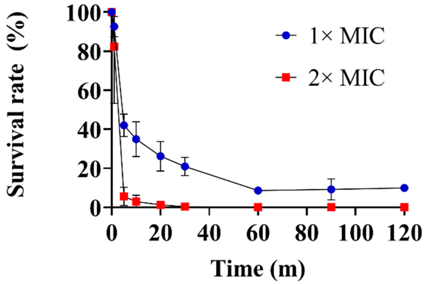
2.4. Time-Kill Kinetics Assay

The time-kill kinetics of Lycosin-II against C. albicans (KCTC 7270) were determined. C. albicans was grown to mid-log phase in YPD medium at 28 °C. Then, C. albicans was diluted in the YPD medium to a final concentration of 2 × 10⁴ CFU/mL. The suspensions were incubated with Lycosin-II at 1× and 2× MICs and with 10 mM sodium phosphate buffer as a control. Next, aliquots were plated on the YPD agar plates and incubated for 16–24 h at 28 °C. After incubation, the CFUs were counted. The survival rate was calculated using the following equation [20]:
Percentage survival rate = (CFUs of C. albicans in the Lycosin-II solution–CFUs of C. albicans in the control solution) × 100%

2.5. Membrane Depolarization Assay

Membrane depolarization in C. albicans (KCTC 7270) by Lycosin-II was measured using DiBAC4(3) [21]. DiBAC4(3) is a membrane-potential-sensitive dye that can enter depolarized cells and increase the fluorescence intensity. Briefly, C. albicans was cultured in YPD medium at 28 °C and then washed with phosphate-buffered saline (PBS) three times. The C. albicans (5 × 10⁶ CFU/mL) in the PBS was incubated with Lycosin-II at 0.5×, 1×, 2×, and 4× MICs for 3 h at 28 °C. The cells were centrifuged and resuspended in 1 mL PBS supplemented with 2 μg/mL DiBAC4(3) for 10 min. The fluorescence intensity of DiBAC4(3) was measured using flow cytometry (Beckman, Brea, CA, USA).

2.6. PI Uptake Assay

The membrane integrity of C. albicans (KCTC 7270) after exposure to Lycosin-II was determined using a PI uptake assay. Propidium iodide is an impermeable dye that is used to determine the permeability of cells by agents [22,23]. C. albicans (2 × 10⁷ CFU/mL) in PBS was incubated with Lycosin-II at 0.5×, 1×, 2×, and 4× MICs at 28 °C for 10 min. Then, C. albicans was harvested by centrifugation and resuspended in PBS. Next, PI (10 μg/mL) was added to the cells, and these were incubated for 10 min. To remove unbound PI, the cells were washed with PBS. The fluorescence intensity of the PI was measured using flow cytometry (Beckman). Additionally, the PI uptake was visualized using fluorescence microscopy and an EVOS FL Auto 2 imaging system (Invitrogen, Waltham, MA, USA).

2.7. SYTOX Green Uptake Assay

To further investigate C. albicans membrane damage by Lycosin-II, we used SYTOX green, which is an impermeable dye that interacts with nucleic acids, resulting in enhanced fluorescent intensity. C. albicans (KCTC 7270) was washed and resuspended in PBS. Then, C. albicans (2 × 10⁷ CFU/mL) in PBS loaded with SYTOX green (1 μM) was treated with Lycosin-II, and the fluorescence was measured using excitation and emission wavelengths of 485 nm and 520 nm, respectively [24]. Additionally, the SYTOX green uptake was visualized using fluorescence microscopy and an EVOS FL Auto 2 imaging system (Invitrogen).

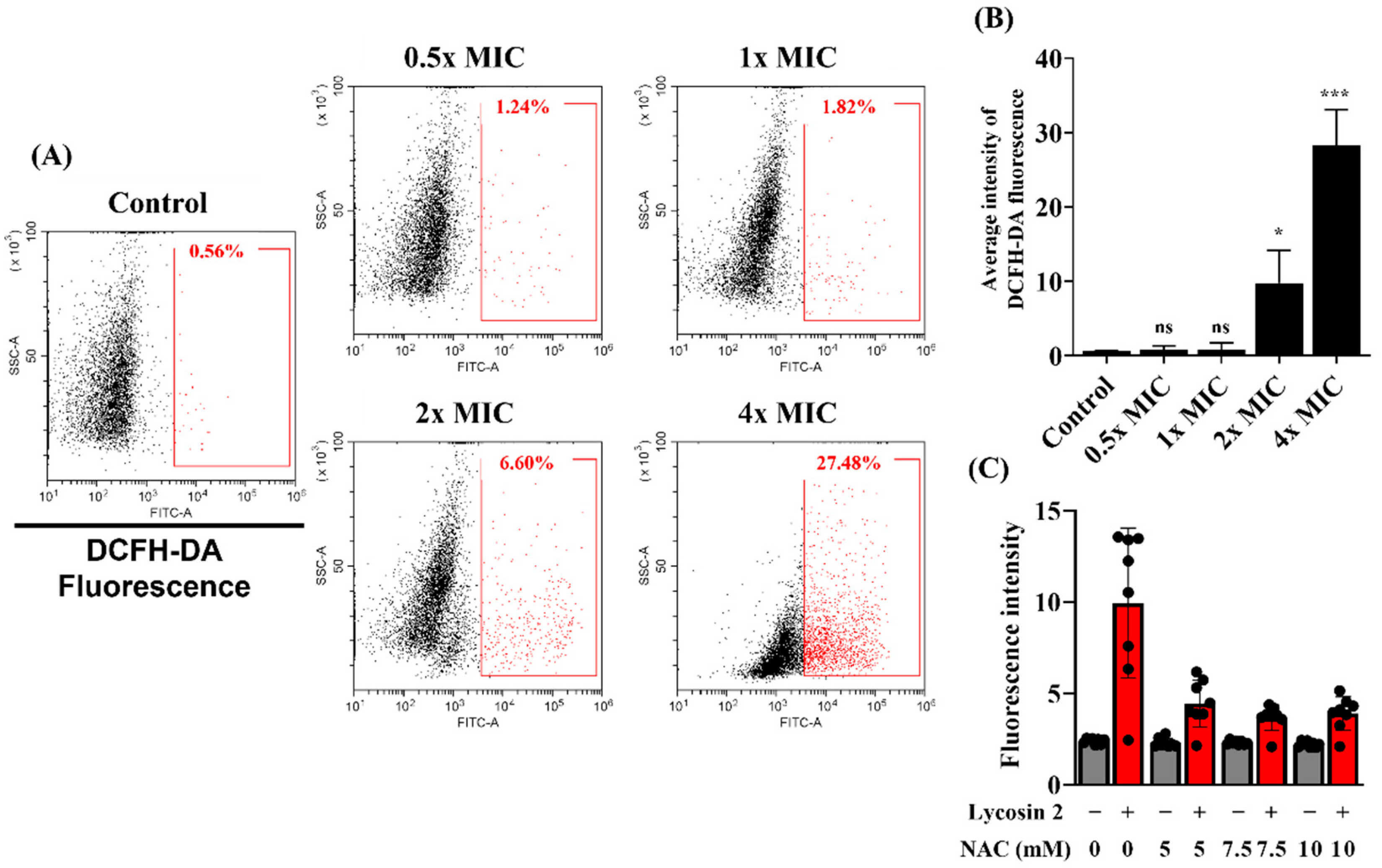
2.8. Measurement of ROS

The accumulation of reactive oxygen species (ROS) in C. albicans (KCTC 7270) due to Lycosin-II was detected using DCFH-DA [25]. DCFH-DA is oxidized by ROS to dichlorofluorescein (DCF), which emits green fluorescence [26]. C. albicans was washed with YPD medium and resuspended at a final concentration of 1 × 10⁷ CFU/mL in YPD medium. Next, suspensions of C. albicans were exposed to Lycosin-II at 0.5×, 1×, 2×, and 4× MICs for 1 h at 28 °C. After centrifugation, C. albicans was incubated with 10 μM DCFH-DA for 1 h and then washed with PBS. The fluorescence intensity of DCFH-DA was measured using flow cytometry (Beckman).

2.9. Analysis of the Effect of NAC on the Antifungal Activity of Lycosin-II

To determine the effect of ROS production on the mode of action of Lycosin-II, we used the ROS scavenger NAC [27]. C. albicans (KCTC 7270) was washed and resuspended with PBS at 1 × 10⁷ CFU/mL, and SYTOX green at 1 μM was added to the suspension. Then, NAC was added to the suspension at 5, 7, 7.5, and 10 mM, which was then incubated with Lycosin-II at 1× MIC for 1 h. The fluorescence intensity of the SYTOX green was measured at excitation and emission wavelengths of 485 nm and 520 nm using a Spectramax M3 spectrophotometer (Molecular Devices, Sunnyvale, CA, USA).

2.10. Biofilm Inhibition Assays

To analyze the effect of Lycosin-II on C. albicans single-species biofilms and polymicrobial biofilms, we used C. albicans (CCARM 14020), S. aureus (ATCC 25923), and S. aureus (ATCC 25923) expressing green fluorescent protein (GFP). We demonstrated in a previous study that the strain CCARM 14020 of C. albicans could form a large biofilm [23,28]. C. albicans (CCARM 14020) and S. aureus (ATCC 25923) were incubated in YPD medium and LB medium. The two strains were washed with RPMI medium supplemented with 2% glucose and adjusted to 1 × 10⁶ CFU/mL for the single-species biofilm evaluation. A total of 180 μL aliquots of C. albicans or S. aureus and the two mixed strains was added to 96-well plates and incubated with 20 μL Lycosin-II at 0.5–8 μM for 24 h at 37 °C. The supernatant was discarded, and 100% methanol was added to fix the biofilm for 20 min. Then, the methanol was discarded, and the sample was dried completely. The biofilm formation was stained with 0.1% crystal violet for 20 min and then washed three times with distilled water. Next, ethanol (95%) was added to the stained biofilms for dissolution. Biofilm quantification was determined by measuring absorbance at 595 nm using a Versa-Max microplate enzyme-linked immunosorbent assay reader (Molecular Devices) [29,30]. The percentage of biofilm formation was determined using the following equation:
Biofilm formation (%) = (A595 of treated biofilm/A595 of untreated biofilm) × 100

2.11. Determination of C. albicans and S. aureus Viability in a Dual-Species Biofilm

The biofilms that were formed by C. albicans and S. aureus were suspended by pipetting. The biofilm suspension was transferred to a 1.5 mL tube, diluted in 1 mL PBS, and vortexed for 5 min [31,32]. Then, YPD and LB agar plates containing vancomycin and amphotericin B were used to distinguish between C. albicans and S. aureus, respectively. The aliquots were spread on agar plates and incubated at 37 °C for 16 h. The numbers of C. albicans and S. aureus were determined by counting the CFUs.

2.12. Visualization of the Biofilm

To visualize the biofilm after treatment with Lycosin-II, we used calcofluor white (10 μg/mL) to stain C. albicans in the biofilm. Moreover, the S. aureus (ATCC 25923) strain expressing GFP allowed for the visualization of this bacterium. The images were visualized using EVOS FL Auto 2 fluorescence (Invitrogen).

2.13. Statistics

All the data were expressed as three independent experiments using the mean ± standard deviation. Two-tailed Student’s t-tests were used to determine the significance of the differences (*, p < 0.05; **, p < 0.01; ***, p < 0.001; ****, p < 0.0001; ns, not significant compared to the control).

3. Results

3.1. Antifungal Activity against C. albicans

The antifungal activities of Lycosin-II, melittin, fluconazole, and amphotericin B against the C. albicans strains are summarized in Table 1. Melittin is a lytic peptide with antimicrobial activity that was isolated from Iranian honeybee (Apis mellifera meda) venom and was used as a control in this study. Lycosin-II showed antifungal activity against C. albicans with a MIC of 2 μM. Melittin showed antifungal activity against C. albicans at concentrations ranging from 2 to 4 μM. We also found that Lycosin-II showed antifungal activity against fluconazole-resistant C. albicans strains. A time-kill kinetics assay was performed to determine the time that was required to kill C. albicans cells, and Lycosin-II showed antifungal activity in dose- and time-dependent manners. Within 60 min, Lycosin-II at 1× MIC reduced C. albicans to >10% viability. In addition, Lycosin-II at 2× MIC was able to completely kill C. albicans in 60 min (Figure 1).

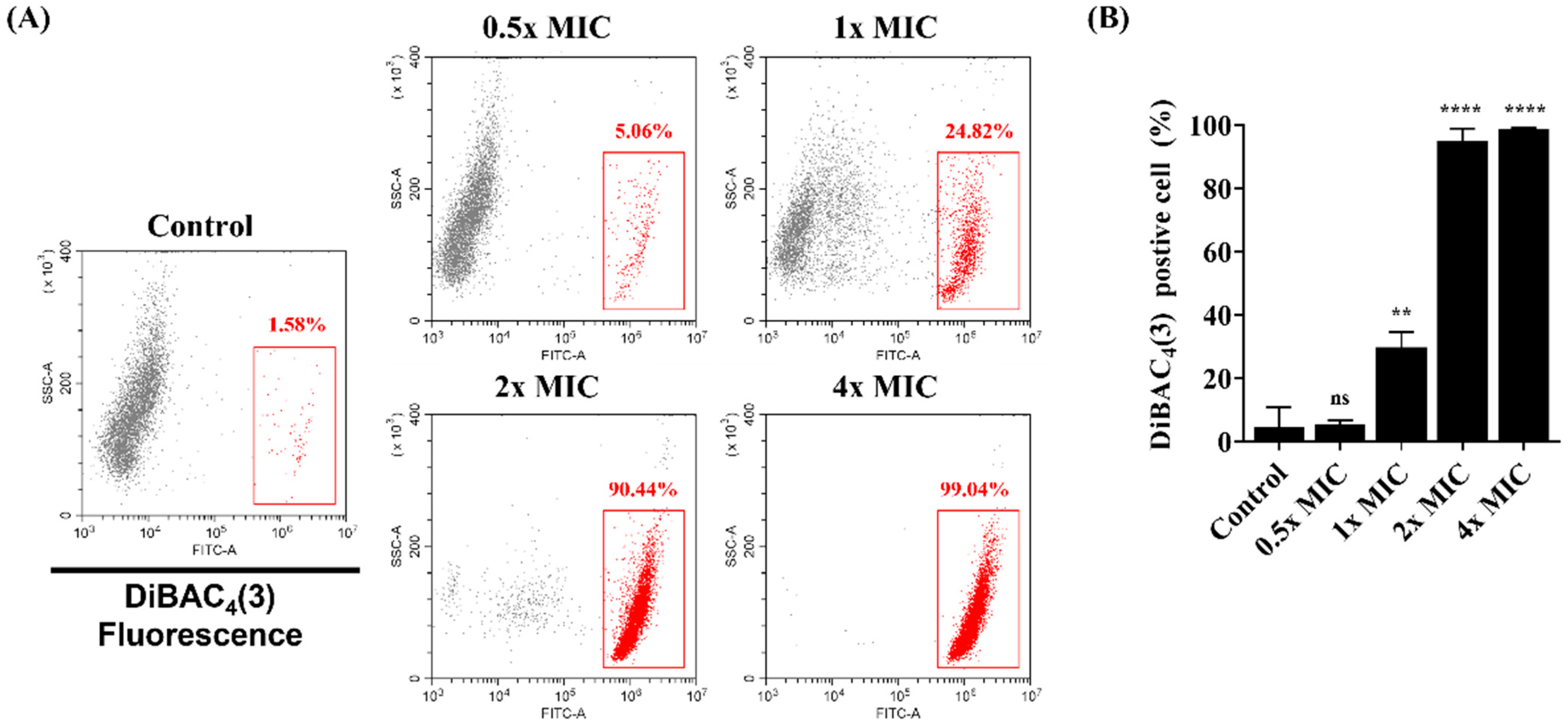
3.2. Membrane Potential Levels of C. albicans

DiBAC4(3) was used to analyze the effect of the membrane potential in C. albicans after exposure to Lycosin-II. C. albicans was treated with Lycosin-II at 0.5×, 1×, 2×, and 4× MICs. As shown in Figure 2A,B, 1.58% of the C. albicans control group stained positive for DiBAC4(3). However, in C. albicans exposed to Lycosin-II at 0.5×, 1×, 2×, and 4× MICs, the fluorescence intensity increased by 5.06%, 24.82%, 90.44%, and 99.04%, respectively, compared to those of the control groups (Figure 2A,B). These results suggested that Lycosin-II has membrane-depolarizing activity against C. albicans.

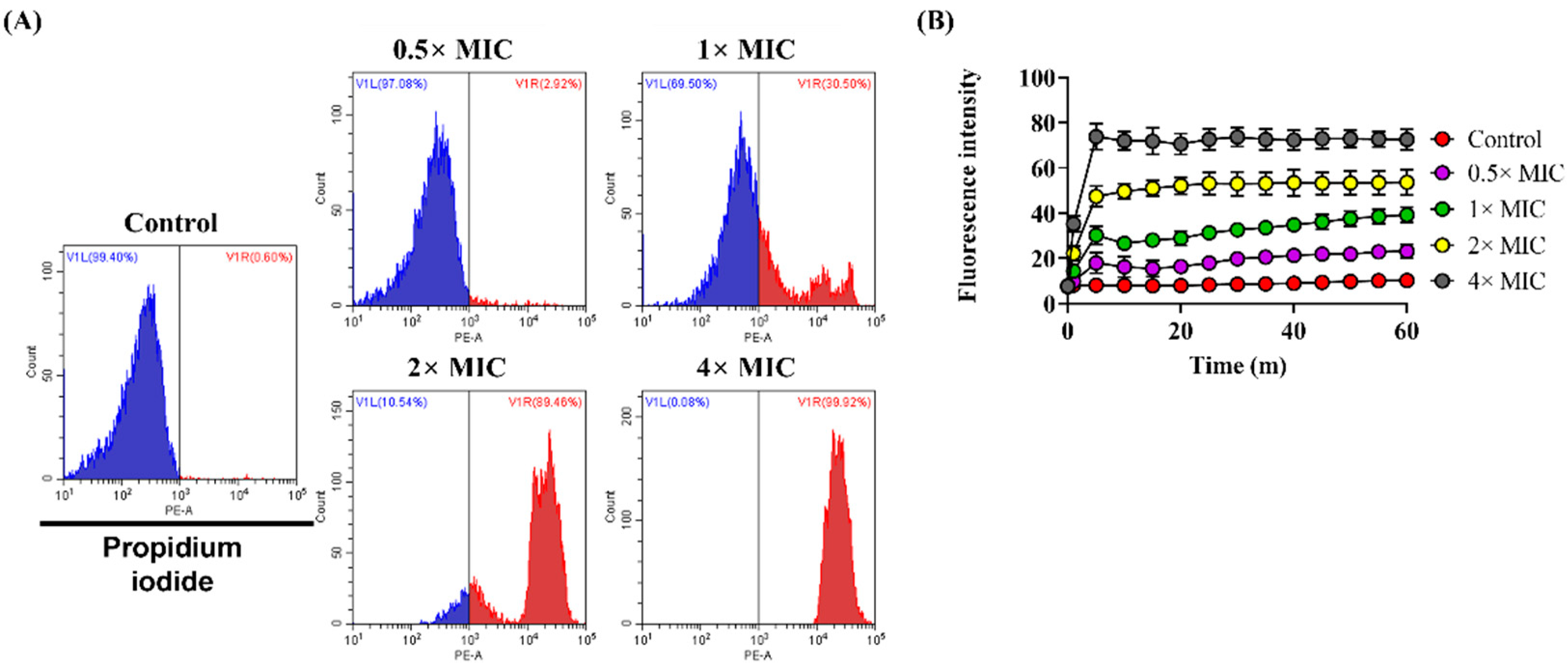
3.3. Effect of Lycosin-II on Membrane Integrity

We analyzed whether the integrity of the fungal membrane was damaged by Lycosin-II using PI and SYTOX green. These dyes cannot penetrate intact membranes. However, they interact with nucleic acids after membranes are compromised with antimicrobial agents, thereby increasing the fluorescence intensity. We demonstrated the disruption of C. albicans membrane integrity by flow cytometric analysis using PI. Among the control groups, the proportion of C. albicans stained with PI was 0.6%, indicating an intact cell membrane. After treatment with Lycosin-II at 0.5×, 1×, 2×, and 4× MICs, the proportions of PI-positive C. albicans increased to 2.92%, 30.50%, 89.48%, and 99.92%, respectively (Figure 3A). We further confirmed the integrity of the C. albicans membrane using SYTOX green. We also demonstrated that treatment with Lycosin-II induced an increase in the fluorescence intensity of SYTOX in a dose-dependent manner (Figure 3B). Moreover, PI and SYTOX green stained the nucleic acid DNA in C. albicans after treatment with Lycosin-II in a dose-dependent manner. Additionally, we confirmed that Lycosin-II damaged the C. albicans membranes, causing the uptake of PI and SYTOX green, as demonstrated by the increase in the red and green fluorescence intensities, respectively (Figure S1).

3.4. Antifungal Activity against C. albicans

The induction of ROS production by antimicrobial agents is acknowledged as an antifungal mechanism that damages membrane integrity [33]. To analyze intracellular ROS generation by C. albicans after Lycosin-II exposure, we used a fluorescent dye, DCFH-DA, which is widely used as an indicator of ROS. We confirmed that Lycosin-II induced ROS production by using flow cytometry to quantify DCF fluorescence. A total of 0.56% of C. albicans exhibited ROS-positive staining within the control group. However, dose-dependent ROS generation was observed, with 1.24%, 1.82%, 6.60%, and 27.48% exhibiting ROS-positive staining after treatment with 0.5×, 1×, 2×, and 4× MICs of Lycosin-II, respectively (Figure 4A,B). We used NAC as an ROS scavenger to demonstrate the association between ROS generation due to Lycosin-II and antifungal activity. We confirmed that pretreatment with NAC reduced the fluorescence intensity of SYTOX green (Figure 4C). These results suggested that Lycosin-II induces ROS generation, leading to oxidative and fungal membrane damage.

3.5. Inhibitory Effects of Lycosin-II on Biofilm

Biofilms produced by pathogens such as bacteria and fungi worsen disease progression and increase drug resistance [34]. C. albicans and S. aureus were tested to assess the biofilm inhibitory effect of Lycosin-II. The results show that Lycosin-II at 8 and 2 μM inhibited more than 90% of the biofilm formation by a single species (C. albicans or S. aureus; Figure 5A,B). Lycosin-II also inhibited the formation of dual-species biofilms containing C. albicans and S. aureus (Figure 5C). Furthermore, to investigate the viability of C. albicans and S. aureus in dual-species biofilms, a colony count assay was performed. Lycosin-II resulted in the inhibition of C. albicans and S. aureus within the biofilm (Figure 5D,E). To visualize biofilm inhibition by Lycosin-II, we used calcofluor white to distinguish the fungi and GFP-expressing S. aureus. Treatment with 4 μM Lycosin-II decreased the number of C. albicans in the biofilm. Moreover, 2 μM Lycosin-II markedly reduced S. aureus in the biofilms (Figure 5F). These results indicate that Lycosin-II effectively inhibited biofilms that contained C. albicans and S. aureus.

4. Discussion

Antibiotics are highly useful for the treatment of infections. However, antimicrobial resistance is increasing globally and is considered a serious threat to public health. The development of antimicrobial agents is a key factor in the treatment of infectious diseases. The increasing incidence of infections by pathogens, such as bacteria and fungi, that are resistant to conventional antimicrobial agents warrants the development of novel antimicrobial agents [35].
Azoles are commonly used to treat Candida spp. infections. Azoles have antifungal activity due to inhibiting the ergosterol biosynthesis pathway and increasing membrane permeability. Recently, many studies have reported that Candida species can be resistant to antifungal agents [36]. Amphotericin B is commonly used to treat fungal infections. However, amphotericin B has side effects, including chills, headaches, kidney damage, and inflammation [37]. Therefore, new antifungal agents should be developed to prevent resistance and toxicity.
Therefore, AMPs offer an effective therapeutic strategy against a wide range of pathogens. The mechanism of action of AMPs is associated with membrane disruption and the targeting of intracellular components, including nucleic acids [38]. Moreover, resistance to AMPs develops slower than it does to conventional antibiotics [39]. Thus, AMPs may be used to overcome antimicrobial resistance.
We previously reported that Lycosin-II, which is derived from the venom of the spider L. singoriensis, has antibacterial activity and can act on Gram-negative and Gram-positive bacteria by disrupting the membrane within a concentration range of 1–2 μM. Lycosin-II also exhibited antimicrobial activity against multidrug-resistant (MDR)-S. aureus and MDR-P. aeruginosa. Moreover, Lycosin-II showed lower cytotoxicity than melittin, which is a known lytic peptide [16]. Lycosin-II also has anti-inflammatory activity by reducing pro-inflammatory cytokines, such as IL-6, IL-8, TNF-α, and IL-1β, in mammalian cells during S. aureus and P. aeruginosa infections. However, the antifungal activity and mechanism of action of Lycosin-II are not known. Thus, this study aimed to investigate the Lycosin-II antifungal activity, mechanism, and prevention of biofilm formation by C. albicans and S. aureus.
The MICs were determined to confirm the antifungal activity of Lycosin-II against C. albicans. We found that Lycosin-II showed antifungal activity against both standard C. albicans (KCTC 7270) and fluconazole-resistant C. albicans (CCARM 14001, 14004, 14007, and 14020; Table 1).
The fungal cell wall is an important target in the development of antifungal agents [40]. Therefore, we investigated whether Lycosin-II affects the membrane of C. albicans. The mechanisms of action of AMPs include depolarization of the cytoplasmic membrane, which induces membrane permeation [41]. In a previous study, Bac8c, the peptide analog of Ba2A, induced a change in the membrane potential in C. albicans [42]. To investigate the mechanism of action of Lycosin-II, we verified if Lycosin-II could depolarize the membrane using DiBAC4(3). This dye is a potential sensing probe that can enter a depolarized membrane and bind to intracellular components, increasing the fluorescence intensity. C. albicans treated with Lycosin-II showed a greater increase in DiBAC4(3) fluorescence intensity than untreated C. albicans. These results showed that Lycosin-II induced the depolarization of the plasma membrane of C. albicans (Figure 2). Previous studies reported that the membrane of C. albicans was damaged by antifungal peptides, such as protonectin isolated from Agelaia pallipes pallipes [33]. In contrast, peptides consisting of lysine and tryptophan 4 repeats showed antifungal activity against C. albicans but did not affect the membrane integrity [23]. To explore the antifungal mechanism of Lycosin-II in the C. albicans membrane, the integrity of the fungal membranes was analyzed using PI and SYTOX green. Both dyes are nucleic acid affinity red and green dyes that enter the fungal cell after membrane integrity is compromised by antimicrobial agents [43]. Therefore, increases in PI and SYTOX green fluorescence intensities indicate a loss in fungal membrane integrity. We confirmed the increased uptake of PI and SYTOX green after Lycosin-II treatment compared to untreated C. albicans using flow cytometry and fluorescence spectrophotometry (Figure 3). Moreover, the fluorescence microscopy data showed that Lycosin-II treatment led to increased PI and SYTOX green fluorescence in dose- and time-dependent manners. These data were consistent with the results of the flow cytometry and spectrophotometry (Supplement S1). The natural production of ROS plays a role in homeostasis and cellular metabolism. Previous studies have reported the mechanisms of conventional antifungal agents associated with ROS production. The excessive production of ROS induced by antifungal agents affects lipid retention and induces membrane permeability [22,44]. For example, the antifungal agent fluconazole can increase oxidative stress in fungal strains by inducing ROS production [45]. Therefore, we used the ROS probes DCFH-DA and NAC as general scavengers of ROS. Our results showed that Lycosin-II induced ROS generation in C. albicans in a dose-dependent manner. In addition, NAC indicated reduced membrane permeability of C. albicans, which was caused by Lycosin-II. These results indicated that enhanced ROS production after exposure to Lycosin-II affected membrane permeability in C. albicans (Figure 4).
Biofilms are communities of microorganisms that are trapped by EPS and are protected from the host immune system and antibiotics. Microbes in multispecies infections can have altered virulence, proliferation, and antioxidant tolerance. Because microorganisms within mixed fungal–bacterial biofilms have high levels of resistance to antimicrobial agents, fungal–bacterial infections are complex and challenging to treat. C. albicans and S. aureus are major opportunistic fungi and bacterial pathogens, respectively [46]. Approximately 27% of C. albicans infections are polymicrobial with S. aureus [47]. S. aureus and C. albicans exhibit synergistic pathogenicity. For example, C. albicans can enhance the resistance of S. aureus to vancomycin [48]. Therefore, approaches to combat both bacteria and fungi are urgently needed. In this study, we explored whether Lycosin-II inhibited single-species biofilm formation using a crystal violet assay for biofilm quantification. Additionally, we used calcofluor white and S. aureus expressing GFP for biofilm visualization. Lycosin-II prevented biofilm formation by C. albicans and S. aureus in a dose-dependent manner. Moreover, we confirmed that Lycosin-II inhibited the formation of dual-species biofilms. Additionally, the numbers of live C. albicans and S. aureus in the biofilms were significantly reduced by Lycosin-II (Figure 5). These results indicated that Lycosin-II had inhibitory activity against dual-species biofilms.

5. Conclusions

In conclusion, Lycosin-II had antifungal activity against C. albicans. Lycosin-II increased membrane depolarization and permeabilization in C. albicans, and Lycosin-II-induced ROS generation was associated with antifungal activity against C. albicans. Lycosin-II effectively inhibited the formation of single- and dual-species biofilms. Our results suggested that Lycosin-II may be a useful therapeutic alternative against polymicrobial infections.

Supplementary Materials

The following supporting information can be downloaded at: https://www.mdpi.com/article/10.3390/jof8090901/s1, Figure S1: Fluorescence microscopy of C. albicans stained with propidium iodide (PI) and SYTOX green after treatment with Lycosin-II.

Author Contributions

Supervision, Y.P.; writing—original draft preparation, J.P.; data curation, H.K. and H.-K.K.; formal analysis, M.-C.C. All authors have read and agreed to the published version of the manuscript.

Funding

This work was supported by a National Research Foundation of Korea (NRF) grant funded by the Korean Government (NRF-2017M3A9E4077206 and NRF-2021R1C1C2009855) and an Institute for Information and Communications Technology Promotion (IITP) grant funded by the Korean Government (MSIT; No. 2017-0-01714, Development of Antimicrobial Peptide using Deep Learning).

Institutional Review Board Statement

Not applicable.

Informed Consent Statement

Not applicable.

Data Availability Statement

The data showed in this study are available on request from the corresponding author.

Conflicts of Interest

The authors declare no conflict of interest.

References

  1. Khezerlou, A.; Alizadeh-Sani, M.; Azizi-Lalabadi, M.; Ehsani, A. Nanoparticles and their antimicrobial properties against pathogens including bacteria, fungi, parasites and viruses. Microb. Pathog. 2018, 123, 505–526. [Google Scholar] [CrossRef] [PubMed]
  2. Bugli, F.; Massaro, F.; Buonocore, F.; Saraceni, P.R.; Borocci, S.; Ceccacci, F.; Bombelli, C.; Di Vito, M.; Marchitiello, R.; Mariotti, M. Design and characterization of myristoylated and non-myristoylated peptides effective against Candida spp. Clinical isolates. Int. J. Mol. Sci. 2022, 23, 2164. [Google Scholar] [CrossRef] [PubMed]
  3. Kim, J.H.; Cheng, L.W.; Chan, K.L.; Tam, C.C.; Mahoney, N.; Friedman, M.; Shilman, M.M.; Land, K.M. Antifungal drug repurposing. Antibiotics 2020, 9, 812. [Google Scholar] [CrossRef] [PubMed]
  4. Burgain, A.; Tebbji, F.; Khemiri, I.; Sellam, A. Metabolic reprogramming in the opportunistic yeast candida albicans in response to hypoxia. Msphere 2020, 5, e00913–e00919. [Google Scholar] [CrossRef] [PubMed]
  5. Lee, Y.; Puumala, E.; Robbins, N.; Cowen, L.E. Antifungal drug resistance: Molecular mechanisms in candida albicans and beyond. Chem. Rev. 2020, 121, 3390–3411. [Google Scholar] [CrossRef]
  6. Chen, H.; Zhou, X.; Ren, B.; Cheng, L. The regulation of hyphae growth in candida albicans. Virulence 2020, 11, 337–348. [Google Scholar] [CrossRef]
  7. Nile, C.; Falleni, M.; Cirasola, D.; Alghamdi, A.; Anderson, O.F.; Delaney, C.; Ramage, G.; Ottaviano, E.; Tosi, D.; Bulfamante, G. Repurposing pilocarpine hydrochloride for treatment of candida albicans infections. Msphere 2019, 4, e00689-18. [Google Scholar] [CrossRef]
  8. Rafat, Z.; Hashemi, S.J.; Ashrafi, K.; Nikokar, I.; Jafari, A.; Foroushani, A.R.; Roohi, B.; Boroujeini, Z.B.; Rashidi, N.; Najar-Shahri, N. Fungal isolates of the respiratory tract in symptomatic patients hospitalized in pulmonary units: A mycological and molecular epidemiologic study. J. Multidiscip. Healthc. 2020, 13, 661. [Google Scholar] [CrossRef]
  9. Cavalheiro, M.; Teixeira, M.C. Candida biofilms: Threats, challenges, and promising strategies. Front. Med. 2018, 5, 28. [Google Scholar] [CrossRef]
  10. Lohse, M.B.; Gulati, M.; Johnson, A.D.; Nobile, C.J. Development and regulation of single-and multi-species candida albicans biofilms. Nat. Rev. Microbiol. 2018, 16, 19–31. [Google Scholar] [CrossRef]
  11. Carolus, H.; Van Dyck, K.; Van Dijck, P. Candida albicans and staphylococcus species: A threatening twosome. Front. Microbiol. 2019, 10, 2162. [Google Scholar] [CrossRef] [PubMed]
  12. Sovari, S.N.; Radakovic, N.; Roch, P.; Crochet, A.; Pavic, A.; Zobi, F. Combatting amr: A molecular approach to the discovery of potent and non-toxic rhenium complexes active against C. albicans-mrsa co-infection. Eur. J. Med. Chem. 2021, 226, 113858. [Google Scholar] [CrossRef] [PubMed]
  13. Mwangi, J.; Yin, Y.; Wang, G.; Yang, M.; Li, Y.; Zhang, Z.; Lai, R. The antimicrobial peptide zy4 combats multidrug-resistant pseudomonas aeruginosa and acinetobacter baumannii infection. Proc. Natl. Acad. Sci. USA 2019, 116, 26516–26522. [Google Scholar] [CrossRef] [PubMed]
  14. Elnagdy, S.; AlKhazindar, M. The potential of antimicrobial peptides as an antiviral therapy against COVID-19. ACS Pharmacol. Transl. Sci. 2020, 3, 780–782. [Google Scholar] [CrossRef]
  15. Wang, Y.; Wang, L.; Yang, H.; Xiao, H.; Farooq, A.; Liu, Z.; Hu, M.; Shi, X. The spider venom peptide lycosin-ii has potent antimicrobial activity against clinically isolated bacteria. Toxins 2016, 8, 119. [Google Scholar] [CrossRef]
  16. Oh, J.H.; Park, J.; Park, Y. Anti-biofilm and anti-inflammatory effects of lycosin-ii isolated from spiders against multi-drug resistant bacteria. Biochim. Biophys. Acta-Biomembr. 2022, 1864, 183769. [Google Scholar] [CrossRef]
  17. Park, S.-C.; Kim, J.-Y.; Kim, E.-J.; Cheong, G.-W.; Lee, Y.; Choi, W.; Lee, J.R.; Jang, M.-K. Hydrophilic linear peptide with histidine and lysine residues as a key factor affecting antifungal activity. Int. J. Mol. Sci. 2018, 19, 3781. [Google Scholar] [CrossRef]
  18. Radhakrishnan, V.S.; Mudiam, M.K.R.; Kumar, M.; Dwivedi, S.P.; Singh, S.P.; Prasad, T. Silver nanoparticles induced alterations in multiple cellular targets, which are critical for drug susceptibilities and pathogenicity in fungal pathogen (candida albicans). Int. J. Nanomed. 2018, 13, 2647. [Google Scholar] [CrossRef]
  19. Hong, M.J.; Kim, M.K.; Park, Y. Comparative antimicrobial activity of hp404 peptide and its analogs against acinetobacter baumannii. Int. J. Mol. Sci. 2021, 22, 5540. [Google Scholar] [CrossRef]
  20. Dong, N.; Chou, S.; Li, J.; Xue, C.; Li, X.; Cheng, B.; Shan, A.; Xu, L. Short symmetric-end antimicrobial peptides centered on β-turn amino acids unit improve selectivity and stability. Front. Microbiol. 2018, 9, 2832. [Google Scholar] [CrossRef]
  21. Zorić, N.; Kosalec, I.; Tomić, S.; Bobnjarić, I.; Jug, M.; Vlainić, T.; Vlainić, J. Membrane of candida albicans as a target of berberine. BMC Complement. Altern. Med. 2017, 17, 268. [Google Scholar] [CrossRef] [PubMed]
  22. Seyedjavadi, S.S.; Khani, S.; Eslamifar, A.; Ajdary, S.; Goudarzi, M.; Halabian, R.; Akbari, R.; Zare-Zardini, H.; Imani Fooladi, A.A.; Amani, J. The antifungal peptide mch-amp1 derived from matricaria chamomilla inhibits candida albicans growth via inducing ros generation and altering fungal cell membrane permeability. Front. Microbiol. 2020, 10, 3150. [Google Scholar] [CrossRef] [PubMed]
  23. Ramamourthy, G.; Park, J.; Seo, C.; Vogel, H.J.; Park, Y. Antifungal and antibiofilm activities and the mechanism of action of repeating lysine-tryptophan peptides against candida albicans. Microorganisms 2020, 8, 758. [Google Scholar] [CrossRef]
  24. Wong, J.H.; Ng, T.B.; Legowska, A.; Rolka, K.; Hui, M.; Cho, C.H. Antifungal action of human cathelicidin fragment (ll13–37) on candida albicans. Peptides 2011, 32, 1996–2002. [Google Scholar] [CrossRef] [PubMed]
  25. Hwang, B.; Hwang, J.-S.; Lee, J.; Lee, D.G. The antimicrobial peptide, psacotheasin induces reactive oxygen species and triggers apoptosis in candida albicans. Biochem. Biophys. Res. Commun. 2011, 405, 267–271. [Google Scholar] [CrossRef] [PubMed]
  26. Li, Y.; Chang, W.; Zhang, M.; Li, X.; Jiao, Y.; Lou, H. Diorcinol d exerts fungicidal action against candida albicans through cytoplasm membrane destruction and ros accumulation. PLoS ONE 2015, 10, e0128693. [Google Scholar]
  27. Chang, C.-K.; Kao, M.-C.; Lan, C.-Y. Antimicrobial activity of the peptide lfcinb15 against candida albicans. J. Fungi 2021, 7, 519. [Google Scholar] [CrossRef]
  28. Luo, Y.; McAuley, D.F.; Fulton, C.R.; Sá Pessoa, J.; McMullan, R.; Lundy, F.T. Targeting candida albicans in dual-species biofilms with antifungal treatment reduces staphylococcus aureus and mrsa in vitro. PLoS ONE 2021, 16, e0249547. [Google Scholar] [CrossRef]
  29. Kim, M.K.; Kang, N.H.; Ko, S.J.; Park, J.; Park, E.; Shin, D.W.; Kim, S.H.; Lee, S.A.; Lee, J.I.; Lee, S.H. Antibacterial and antibiofilm activity and mode of action of magainin 2 against drug-resistant acinetobacter baumannii. Int. J. Mol. Sci. 2018, 19, 3041. [Google Scholar] [CrossRef]
  30. Wang, Y.; Pei, Z.; Lou, Z.; Wang, H. Evaluation of anti-biofilm capability of cordycepin against candida albicans. Infect. Drug Resist. 2021, 14, 435. [Google Scholar] [CrossRef]
  31. Cruz, C.D.; Shah, S.; Tammela, P. Defining conditions for biofilm inhibition and eradication assays for gram-positive clinical reference strains. BMC Microbiol. 2018, 18, 173. [Google Scholar] [CrossRef] [PubMed]
  32. Galdiero, E.; Di Onofrio, V.; Maione, A.; Gambino, E.; Gesuele, R.; Menale, B.; Ciaravolo, M.; Carraturo, F.; Guida, M. Allium ursinum and allium oschaninii against klebsiella pneumoniae and candida albicans mono-and polymicrobic biofilms in in vitro static and dynamic models. Microorganisms 2020, 8, 336. [Google Scholar] [CrossRef] [PubMed]
  33. Wang, K.; Dang, W.; Xie, J.; Zhu, R.; Sun, M.; Jia, F.; Zhao, Y.; An, X.; Qiu, S.; Li, X. Antimicrobial peptide protonectin disturbs the membrane integrity and induces ros production in yeast cells. Biochim. Biophys. Acta-Biomembr. 2015, 1848, 2365–2373. [Google Scholar] [CrossRef] [PubMed]
  34. Rasamiravaka, T.; Labtani, Q.; Duez, P.; El Jaziri, M. The formation of biofilms by pseudomonas aeruginosa: A review of the natural and synthetic compounds interfering with control mechanisms. BioMed Res. Int. 2015, 2015, 759348. [Google Scholar] [CrossRef]
  35. Chen, C.H.; Lu, T.K. Development and challenges of antimicrobial peptides for therapeutic applications. Antibiotics 2020, 9, 24. [Google Scholar] [CrossRef]
  36. Gao, J.; Wang, H.; Li, Z.; Wong, A.H.-H.; Wang, Y.-Z.; Guo, Y.; Lin, X.; Zeng, G.; Liu, H.; Wang, Y. Candida albicans gains azole resistance by altering sphingolipid composition. Nat. Commun. 2018, 9, 4495. [Google Scholar] [CrossRef]
  37. Aigner, M.; Lass-Flörl, C. Encochleated amphotericin b: Is the oral availability of amphotericin b finally reached? J. Fungi 2020, 6, 66. [Google Scholar] [CrossRef]
  38. Falanga, A.; Lombardi, L.; Franci, G.; Vitiello, M.; Iovene, M.R.; Morelli, G.; Galdiero, M.; Galdiero, S. Marine antimicrobial peptides: Nature provides templates for the design of novel compounds against pathogenic bacteria. Int. J. Mol. Sci. 2016, 17, 785. [Google Scholar] [CrossRef]
  39. Kumar, S.; Varela, M.F. Molecular mechanisms of bacterial resistance to antimicrobial agents. Chemotherapy 2013, 14, 522–534. [Google Scholar]
  40. Ma, H.; Zhao, X.; Yang, L.; Su, P.; Fu, P.; Peng, J.; Yang, N.; Guo, G. Antimicrobial peptide amp-17 affects candida albicans by disrupting its cell wall and cell membrane integrity. Infect. Drug Resist. 2020, 13, 2509. [Google Scholar] [CrossRef]
  41. Lee, H.; Hwang, J.-S.; Lee, J.; Kim, J.I.; Lee, D.G. Scolopendin 2, a cationic antimicrobial peptide from centipede, and its membrane-active mechanism. Biochim. Biophys. Acta-Biomembr. 2015, 1848, 634–642. [Google Scholar] [CrossRef] [PubMed]
  42. Lee, W.; Lee, D.G. Fungicidal mechanisms of the antimicrobial peptide bac8c. Biochim. Biophys. Acta-Biomembr. 2015, 1848, 673–679. [Google Scholar] [CrossRef] [PubMed]
  43. Sun, Y.; Dong, W.; Sun, L.; Ma, L.; Shang, D. Insights into the membrane interaction mechanism and antibacterial properties of chensinin-1b. Biomaterials 2015, 37, 299–311. [Google Scholar] [CrossRef] [PubMed]
  44. Hwang, B.; Hwang, J.-S.; Lee, J.; Kim, J.-K.; Kim, S.R.; Kim, Y.; Lee, D.G. Induction of yeast apoptosis by an antimicrobial peptide, papiliocin. Biochem. Biophys. Res. Commun. 2011, 408, 89–93. [Google Scholar] [CrossRef]
  45. Dbouk, N.H.; Covington, M.B.; Nguyen, K.; Chandrasekaran, S. Increase of reactive oxygen species contributes to growth inhibition by fluconazole in cryptococcus neoformans. BMC Microbiol. 2019, 19, 243. [Google Scholar] [CrossRef]
  46. Kong, E.F.; Tsui, C.; Kucharíková, S.; Andes, D.; Van Dijck, P.; Jabra-Rizk, M.A. Commensal protection of staphylococcus aureus against antimicrobials by candida albicans biofilm matrix. MBio 2016, 7, e01365-16. [Google Scholar] [CrossRef]
  47. Kean, R.; Rajendran, R.; Haggarty, J.; Townsend, E.M.; Short, B.; Burgess, K.E.; Lang, S.; Millington, O.; Mackay, W.G.; Williams, C. Candida albicans mycofilms support staphylococcus aureus colonization and enhances miconazole resistance in dual-species interactions. Front. Microbiol. 2017, 8, 258. [Google Scholar] [CrossRef]
  48. Hu, Y.; Niu, Y.; Ye, X.; Zhu, C.; Tong, T.; Zhou, Y.; Zhou, X.; Cheng, L.; Ren, B. Staphylococcus aureus synergized with candida albicans to increase the pathogenesis and drug resistance in cutaneous abscess and peritonitis murine models. Pathogens 2021, 10, 1036. [Google Scholar] [CrossRef]
Figure 1. The time-kill kinetics curves of Lycosin-II against Candida albicans. C. albicans (Korea Collection for Type Culture 7270) was exposed to Lycosin-II for 0, 1, 5, 10, 20, 30, 60, 90, and 120 min. The survival rates were determined by counting the number of colony-forming units at each time point. Abbreviation: MIC, minimum inhibitory concentration.
Jof 08 00901 g001
Figure 2. Analysis of Candida albicans membrane depolarization after treatment with Lycosin-II. (A) Changes in membrane depolarization in C. albicans after treatment with Lycosin-II with the peptide at 0.5×, 1×, 2×, and 4× MICs were investigated using flow cytometry with DiBAC4(3). (B) The statistical analyses are displayed as averages (n = 3). The graph is expressed as the mean ± standard deviation. Significance: **, p < 0.01; ****, p < 0.0001; ns, not significant compared to the control. Abbreviations: MIC, minimum inhibitory concentration; DiBAC4(3), bis(1,3-dibutylbarbituric acid) trimethine oxonol.
Jof 08 00901 g002
Figure 3. Analysis of Candida albicans membrane integrity after incubation with Lycosin-II. Lycosin-II at 0.5×, 1×, 2×, and 4× MICs were incubated with C. albicans. (A) A flow cytometry analysis of membrane permeabilization using PI (10 μg/mL) in C. albicans. (B) The kinetics of the membrane disruption of C. albicans were confirmed using SYTOX green (1 μM), as well as by monitoring the fluorescence intensity. Abbreviations: PI, propidium iodide; MIC, minimum inhibitory concentration.
Jof 08 00901 g003
Figure 4. Analysis of reactive oxygen species (ROS) induced by Lycosin-II in Candida albicans. (A) ROS production in C. albicans added to Lycosin-II was measured using DCFH-DA detection and flow cytometry. (B) The statistical analyses are displayed as averages (n = 3). (C) The effect of N-acetyl cysteine (NAC) at different concentrations on membrane integrity using SYTOX green. Black dot indicated fluorescence intensity of SYTOX green. The graph is expressed as the mean ± standard deviation. Significance: *, p < 0.05; ***, p < 0.001; ns, not significant compared to the control. Abbreviations: DCFH-DA, 2, 7-dichloroflurorescin diacetate; MIC, minimum inhibitory concentration.
Jof 08 00901 g004
Figure 5. The inhibitory effect of Lycosin-II on biofilms formed by one or two microbial species. Biofilms formed by a single strain, (A) Candida albicans (CCARM 14020) or (B) Staphylococcus aureus (ATCC 25922), and (C) dual-species biofilms containing C. albicans and S. aureus were incubated with Lycosin-II. The biofilm mass was quantified by using crystal violet and measuring the optical density at 595 nm. The colony-forming unit (CFU) numbers of (D) C. albicans and (E) S. aureus in the dual-species biofilms were analyzed using a plating method on yeast extract peptone dextrose and Luria–Bertani medium supplemented with vancomycin and amphotericin B, respectively. Black dot indicated number of C. albicans and S. aureus. (F) The viability of the dual-species biofilm was analyzed using fluorescence microscopy. C. albicans was stained using calcofluor white (blue), and S. aureus expressing green fluorescent protein (GFP; green) was used to detect the bacterial cells. The graph is expressed as the mean ± standard deviation. Significance: *, p < 0.05; ***, p < 0.001; ns, not significant compared with control.
Jof 08 00901 g005
Table 1. Minimum inhibitory concentrations (MICs) of Lycosin-II and antifungal agents against Candida albicans.
StrainsMIC (μM)
Lycosin-IIMelittinFluconazoleAmphotericin B
C. albicans
(KCTC * 7270)
22161
C. albicans
(CCARM * 14001)
22>320.5
C. albicans
(CCARM 14004)
24>320.5
C. albicans
(CCARM 14020)
22>321
* Abbreviations: KCTC, Korea Collection for Type Culture; CCARM, Culture Collection of Antibiotics Resistant Microbes.
Publisher’s Note: MDPI stays neutral with regard to jurisdictional claims in published maps and institutional affiliations.

