Article

The Relationship between Endophytic Fungi of Chimonanthus praecox and Volatile Metabolites under Different Circadian Rhythms and Blooming Stages

1
Key Laboratory of Underforest Resource Protection and Utilization in Yunnan Province, College of Landscape and Horticulture of Southwest Forestry University, Kunming 650224, China
2
Key Laboratory of Ministry of Education on Forest Resources Conservation and Utilization in Southwest Mountainous Area, Kunming International Research and Development Center of Ecological Forestry Industry, Kunming 650233, China
*
Authors to whom correspondence should be addressed.
These authors contributed equally to this work.
J. Fungi 2024, 10(2), 145; https://doi.org/10.3390/jof10020145
Submission received: 8 January 2024 / Revised: 7 February 2024 / Accepted: 9 February 2024 / Published: 11 February 2024
(This article belongs to the Special Issue Advances in Fungal Endophyte Research)

Abstract

:
Chimonanthus praecox is an aromatic plant that flowers in winter. The composition of the floral volatiles of C. praecox is influenced by different blooming stages, circadian rhythms and species. However, the relationship between floral volatiles and plant endophytic fungi has not received much research attention. Here, we used high-throughput sequencing technology to compare and analyze the changes in the structure and diversity of the endophytic fungal communities in C. praecox under different circadian rhythms (7:00 a.m., 1:00 p.m., and 7:00 p.m.) and in different blooming stages (unopened flowers and opened flowers). The endophytic fungi of C. praecox consisted of nine phyla, 34 classes, 79 orders, 181 families, 293 genera, and 397 species, and Ascomycota was the dominant phylum. Under a diurnal rhythm, the diversity (Chao1 and Shannon indices) of endophytic fungi gradually decreased in the unopened flowers, while an increasing and then decreasing trend was found for the opened flowers. In the different blooming stages, the endophytic fungal diversity was significantly higher at 7:00 a.m. in the unopened flowers compared to the opened flowers. Humidity was the key factors that significantly affected the endophytic fungal diversity and community. Moreover, 11 endophytic fungi were significantly positively or negatively correlated with seven floral volatiles. In conclusion, the community structure and diversity of endophytic fungi in C. praecox were affected by the different blooming stages and circadian rhythms, and a correlation effect related to floral volatiles was found, but there are other possible reasons that were not tested. This study provides a theoretical basis for elucidating the interrelationships between endophytic fungi, floral volatiles, and environmental factors in C. praecox.

1. Introduction

Plant endophytes, mainly including fungi, bacteria, and actinomycetes, commonly inhabit healthy plant tissues without causing apparent disease [1]. Endophytic fungi can promote plant growth [2], enhance tissue colonization [3], improve stress resistance [4], and produce bioactive secondary metabolites [5,6]. They can synthesize the same or similar compounds as their hosts, and increase key metabolite production in horticultural plants, such as paclitaxel (Taxus chinensis) [7], methyl eugenol (Rosa spp.) [5], and 4-penten-2-ol (Lilium brownii) [8]. Similar phenomena occur in medicinal plants. For instance, the endophytes in Azadirachta indica [9] and Rhodiola sachalinensis [10] can produce the AL4 and rhodioside metabolites. These results suggest that endophytic fungi and their hosts share secondary metabolic pathways or production sources. Therefore, revealing the close relationship between endophytic fungi and metabolites can clarify the sources and metabolic pathways of plant metabolites.
The community composition, abundance, and diversity of plant endophytic fungi are primarily influenced by the host species [11,12], growth period [13], and geographic habitat [14], among other factors. Taxa of endophytic fungi significantly differ in different ornamental plant species: for example, Rhododendron spp. [11], Paeonia ludlowii [15], and Lonicera japonica [16] were found to contain 43, 14, and 19 genera, respectively. Similar results were observed in different floral organs for instance, 11 and 4 genera were found in Eucalyptus globulus [17] and Moinga oleifera flowers [18], respectively. Moreover, endophytic fungal diversity and community composition vary between different plant tissues (roots, stems, and leaves) across growth stages. For instance, endophytic fungal diversity was higher in Bletilla mycorrhizae in the flowering and emergence periods than in other periods [19]. The changes in the major genera (Penicillium, Fusarium, Xylaria) in Capsicum annuum stems across the seedling, flowering, and fruiting stages were inconsistent [20]. Similar variability was found in the leaves of Stellera chamaejasme [21]. In addition, environmental factors such as temperature and humidity have significant impacts on the diversity of endophytes. Previous results showed that endophytic fungal diversity in angiosperms is richer in tropical versus temperate/cold forests [22]. Humid climates were found to be more suitable for the survival of endophytes than arid environments [23].
Endophytic fungi can not only produce secondary metabolites in plant tissue, but are also one of the critical pathways for producing volatile compounds with a floral fragrance in plants [24]. Five aromatic metabolites (cyclo-(Trp-Ala), indole-3-carboxylic acid (ICA), in-dole-3-carbaldehyde, mellein, and 2-phenylethanol) were identified in endophytic fungi of Viscum coloratum flowers [25], confirming that endophytic fungi in flowers may be an essential resource for natural plant aromas. Moreover, the emission and composition of floral volatile compounds in different plants are affected by the blooming stage [26,27,28,29], circadian rhythms [28,30], environmental factors [31,32,33,34], and species [35]. However, it is unclear whether floral substance changes caused by biological and abiotic factors can affect some endophytic fungal populations. At the same time, environmental factors can directly affect not only floral volatiles, but also the diversity of endophytic fungi. Therefore, exploring the relationship between floral volatiles and endophytic fungi under different conditions is a necessary way to clarify the source and production of floral volatiles.
Chimonanthus praecox is a deciduous shrub that flowering in winter, belonging to the genus Chimonanthus in the family Calycanthaceae, and it is a fragrant and unique traditional plant in China. Most previous studies showed that the components of floral volatiles in C. praecox are altered by the blooming stage [35,36], circadian rhythms [37], geographic locations [38], and species [39]. Although a close relationship between endophytic fungi and floral volatiles has been demonstrated, the relationship between endophytic fungi diversity, floral volatiles, and environmental factors in C. praecox is still unclear. Based on our previous study of the variation in the floral volatiles of C. praecox in different blooming stages (unopened flowers and opened flowers) and under different circadian rhythms (7:00 a.m., 1:00 p.m., and 7:00 p.m.) in Kunming, Yunnan Province [37], we further analyzed the community structure and diversity of the endophytic fungi in C. praecox using high-throughput sequencing technology. This study aimed to (1) investigate the influence of blooming stages and circadian rhythms on the community structure and diversity of the endophytic fungi in C. praecox, and (2) elucidate the relationship between endophytic fungal diversity, floral volatiles, and environmental factors in C. praecox.

2. Materials and Methods

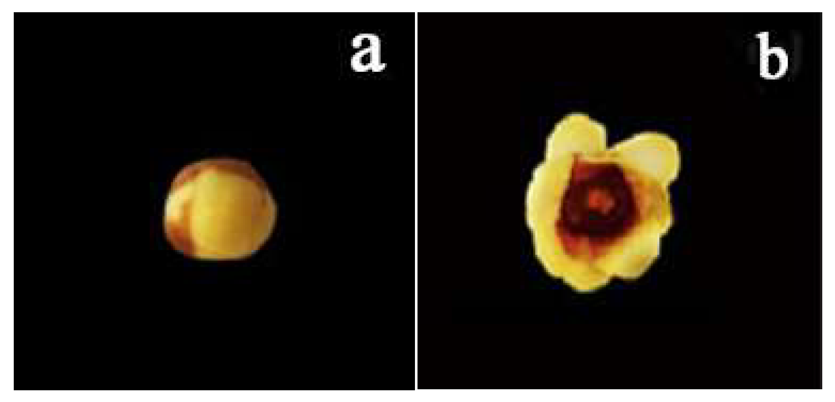
2.1. Plant Materials

In this experiment, the flowers of var. grandiflorus in different blooming stages were used as research materials. The unopened flowers were yellow with upright stamens and buds beginning to increase, while the opened flowers were yellow with completely opened petals and a purple flower center (Figure 1). All the flowers were collected from the campus of Southwest Forestry University in Kunming, Yunnan Province.

2.2. Flower Samples Collection

On 25 December 2019, 24 unopened and 18 opened flowers of C. praecox were collected at 7:00 a.m., 1:00 p.m., and 7:00 p.m.. Two opened flowers and four unopened flowers (about 0.5 g) were placed in a 20 mL head space bottle, which was quickly sealed with an aluminum cover, to determine the floral volatiles. Additionally, twelve unopened and opened flowers of C. praecox were selected and placed in sterile self-sealing bags, and then immediately brought back to the laboratory for surface disinfection and biocidal treatment. The disinfection process involved the following steps: 75% alcohol for 60 s, 0.5% sodium hypochlorite for 90 s, and 75% alcohol for 30 s. After each step finished, sterile excess water was applied 3–4 times to rinse the samples. Subsequently, all the flower samples were dried with sterile filter paper, placed in a 50 mL sterile centrifugal tube, and stored at −80 °C for DNA extraction and sequencing.

2.3. Floral Volatile Collection and Identification

Headspace solid-phase microextraction (HS-SPME) and gas chromatography–mass spectrometry (GC/MS) were carried out to extract the floral scent compounds in C. praecox. An empty capped vial was used as the negative control. The SPME device was equipped with an SPME fiber (50/30 μm) coated with polydimethylsiloxane (PDMS) (TriPlus 300, Thermo Fisher Scientific, Waltham, MA, USA). In detail, the protocols for HS-SPME followed the methods of Chen et al. [40]. Subsequently, a GC/MS system (Trace GC Ultra/ITQ900, Thermo Fisher Scientific, Waltham, MA, USA) with an HP-5MS capillary column (30 m × 0.25 mm × 0.25 μm, Agilent J & W Scientific, Santa Clara, CA, USA) was performed to identify the floral volatiles. The variation in the GC oven temperature and the data recorded from the mass spectrometer in electron impact mode (MS/EI) were based on the methods of Li et al. [37].

2.4. DNA Extraction, PCR Amplification, and Sequencing

DNA was extracted from 0.5 g of frozen flowers using the FastDNA® Spin KitDNA (MP Biomedicals, Southern California, USA) kit according to the manufacturer’s protocol. The obtained DNA was quantified by using a NanoDrop® spectrophotometer (ND-2000c, Thermo Scientific, Waltham, MA, USA) for nucleic acid concentration and purity. The entire fungal ITS1 region was amplified using the primers ITS1F (5′-TCC GTA GGT GAA CCT GCG G-3′) and ITS2R (5′-TCC TCC GCT TAT TGA TAT GC-3′) [41]. The amplification system consisted of 5 × Fast Pfu Buffer 4 µL, 2.5 mM dNTPs 2 µL, Forward Primer (5 µM) 0.8 µL, Reverse Primer (5 µM) 0.8 µL, Fast Pfu Polymerase 0.4 µL, BSA 0.2 µL, Template DNA 10 ng, and ddH2O added to 20 µL. The PCR protocols were as follows: pre-denaturation at 95 °C for 3 min; denaturation at 95 °C for 20 s, annealing at 55 °C for 30 s, and extension at 72 °C for 30 s, over 30 cycles; and extension at 72 °C for 5 min. Three PCR products as three replicates were selected to detect the DNA quality using 2% agarose gel electrophoresis. The qualified PCR products were purified with an AxyPrep DNA Gel Extraction Kit and quantified using a Quantus™ Fluorometer. Amplicons from different samples were mixed for library construction using a NEXTFLEX Rapid DNA-Seq Kit. Finally, high-throughput sequencing was performed using the MiSeq PE300 platform (Shanghai MajorBio Technology Co, China). The sequences have been deposited in the National Center for Biotechnology Information Sequence Read Archive under Accession No. PRJNA875038 (ITS data).

2.5. Data Processing and Statistical Analysis

In this experiment, microbiome bioinformatics was implemented in QIIME 1.9.1, with slight modifications according to the official tutorials (www.majorbio.com, accessed on 19 November 2022). Briefly, raw sequence data were quality-controlled using the Trimmomatic plugin, followed by base splicing using the FLASH plugin. Finally, the sequences were quality-frequency-analyzed, denoised, and merged, and chimeras were removed using the UCHIME plugin. The RDP classifier was used to annotate the species classification for each sequence, and representative sequences were assigned by comparing to Silva database (ssu123), with the comparison threshold being 70%. Sequence data analysis was performed using QIIME 1.9.1, during which non-fungal sequences were removed to avoid contamination of the chloroplasts and mitochondria of the host plant.
The mothur plugin was employed to calculate the alpha diversity (Chao1 and Shannon indices). PCoA plots based on the Bray–Curtis index were analyzed using the vegan and ggplot2 packages in R (version 3.3.1). Venn diagrams based on an OTU table with a 97% similarity level and a heat map of the correlation clustering for the top 30 genera and 10 main floral volatiles (our pre-study) were generated using R (version 3.3.1). RDA analysis was performed using the vegan package in R to analyze the relationship between endophytic fungal communities and environmental factors.

3. Results

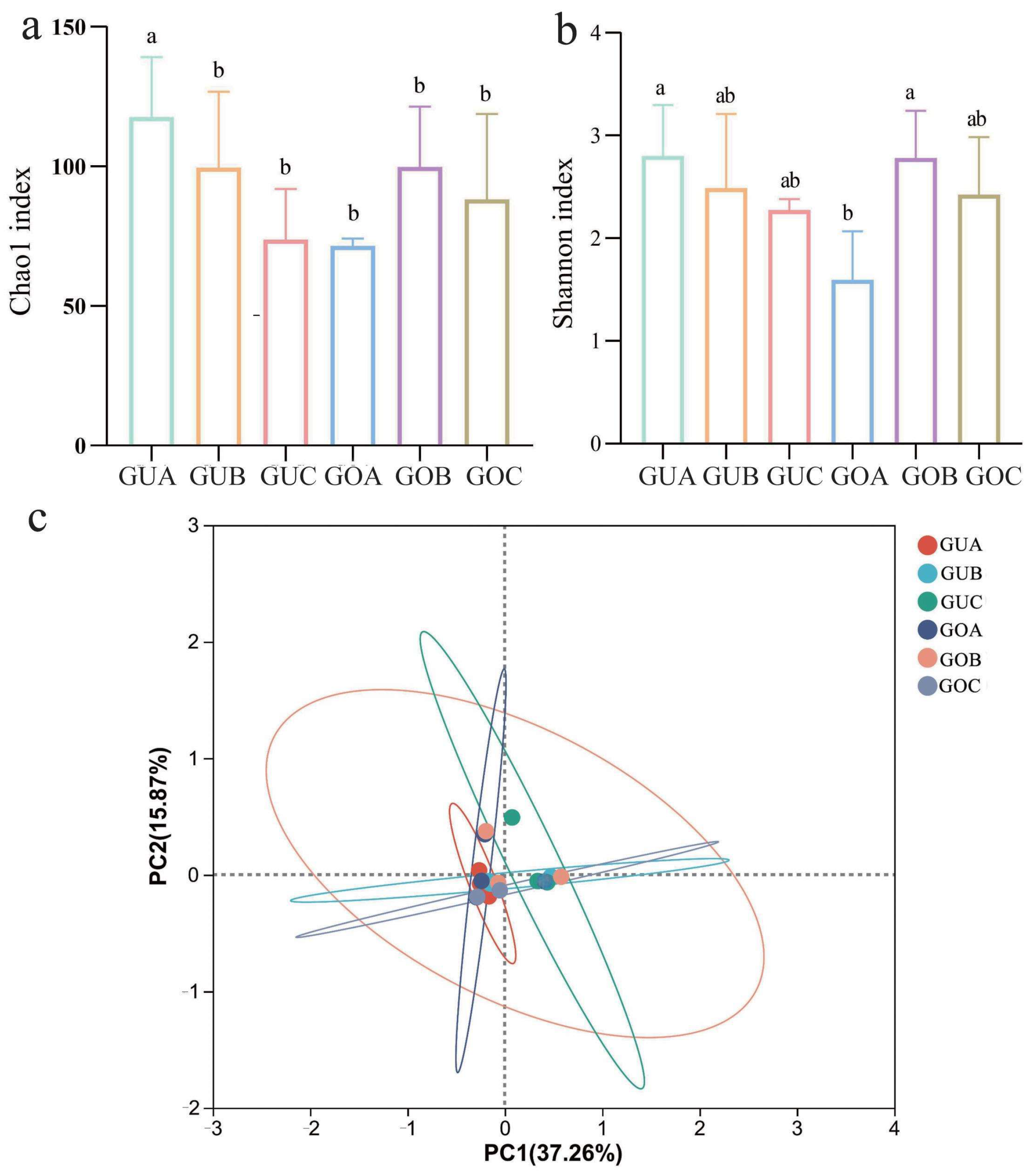
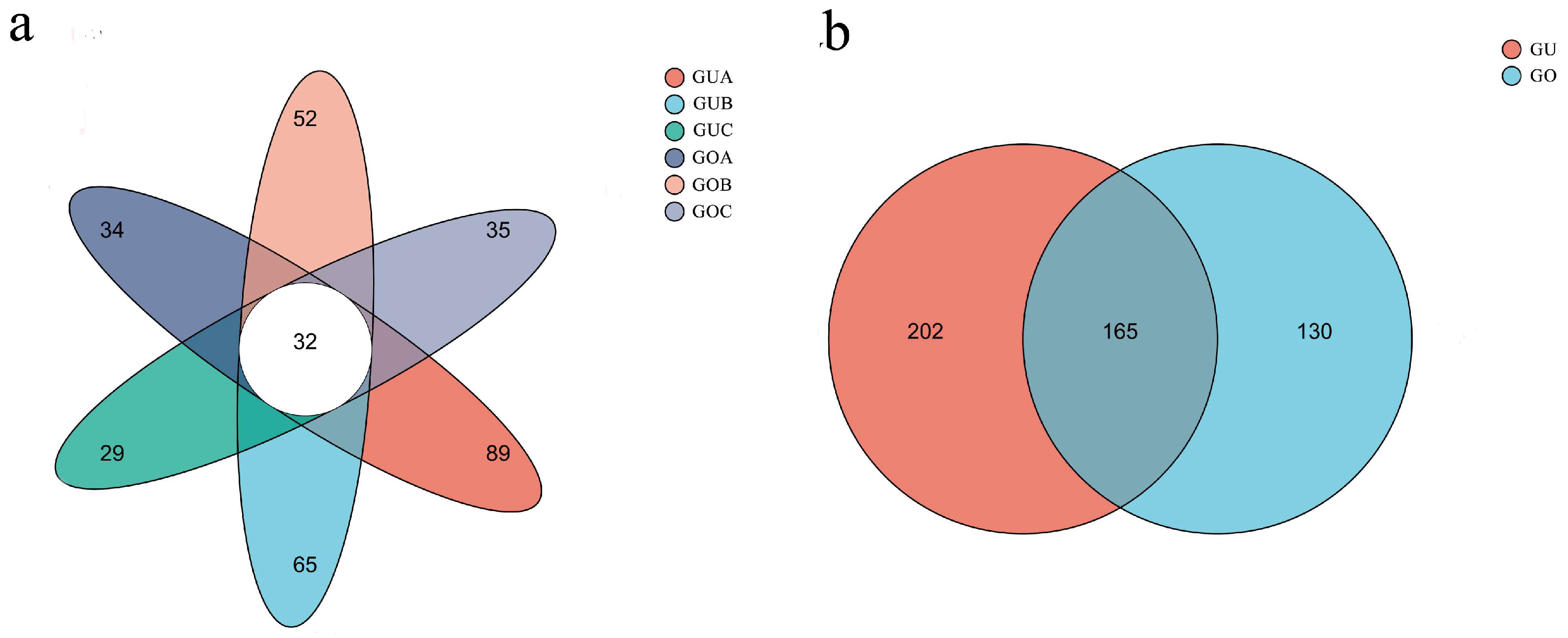
3.1. Alpha and Beta Diversity of Endophytic Fungi

The detailed sequencing information of the endophytic fungi in C. praecox in different blooming stages and under different circadian rhythms can be found in the attached table (Table S1). The rarefaction analysis results (Figure S1) indicated an adequate sequencing depth for all samples. The α diversity (Chao1 and Shannon indices) of the endophytic fungal community in C. praecox was analyzed using Qiime software (1.9.1) (Figure 2a,b). Our findings indicated a circadian rhythm-dependent variation in these indices. Specifically, endophytic fungal α diversity showed a gradual decrease in the unopened flowers with the circadian rhythm, whereas an initial increase followed by a decrease was found in the opened flowers. In the different blooming stages, the Chao1 and Shannon indices of the endophytic fungi were significantly higher at 7:00 a.m. in the unopened flowers compared to the opened flowers (p < 0.05). Additionally, the community abundance and diversity of the endophytic fungi at the other two time points (1:00 p.m. and 7:00 p.m.) showed a no differences between the unopened and opened flowers. The beta diversity of endophytic fungi was further analyzed using the Bray–Curtis distance measure. The PCoA plot (Figure 2c) showed that the contributions of PC1 and PC2 were 32.3% and 13.29%, respectively, with no significant differences between the groups (R2 = 0.22, p > 0.05).

3.2. Taxonomic Analysis of Endophytic Fungal Communities

Following clustering at the 97% similarity level, the Venn diagram (Figure 3a) revealed that the number of Operational Taxonomic Units (OTUs) of the endophytic fungi in the unopened flowers of C. praecox decreased gradually with the circadian rhythm, while in the opened flowers, an initial increase followed by a decrease was observed. The GUA, GUB, GUC, GOA, GOB, and GOC samples contained 89, 65, 29, 34, 52, and 35 OTUs, respectively, with a total of 32 shared OTUs. Among these, GUA had the highest number of OTUs, with 252. The top three most abundant endophytic fungi were unclassified Ascomycota (35.37%), Didymella (13.63%), and Apiotrichum (6.54%). The Venn diagrams for the different blooming stages (Figure 3b) showed that the number of OTUs in the unopened flowers (202) was higher than in the opened flowers (130), with a total of 165 OTUs for both stages. The top three endophytic fungi were unclassified Ascomycota (30.40%), Didymella (11.49%), and Apiotrichum (5.85%).
OTU representative sequences with 97% or higher similarity levels were taxonomically analyzed using the RDP classifier Bayesian algorithm. The C. praecox sample contained various endophytic fungal species types, including nine phyla, 34 classes, 79 orders, 181 families, 293 genera, and 397 species. The top five endophytic fungal communities were identified at the phylum, class, order, family, genus, and species levels (Table 1). Ascomycota, Dothideomycetes, and Pleosporales were the most dominant phylum, class, and order, respectively, while fungi belonging to unclassified Ascomycota were the most prevalent at the family, genus, and species levels. The percentages of Ascomycota and Dothideomycetes in the unopened and opened flower stages decreased with the circadian rhythm and were the highest at 7:00 a.m. Similar results were observed in the most dominant order (Pleosporales) and family (unclassified Ascomycota). At the genus and species levels, the percentage of unclassified Ascomycota decreased first and then increased with the circadian rhythm, and was the highest at 7:00 a.m. in the unopened and opened flower stages.

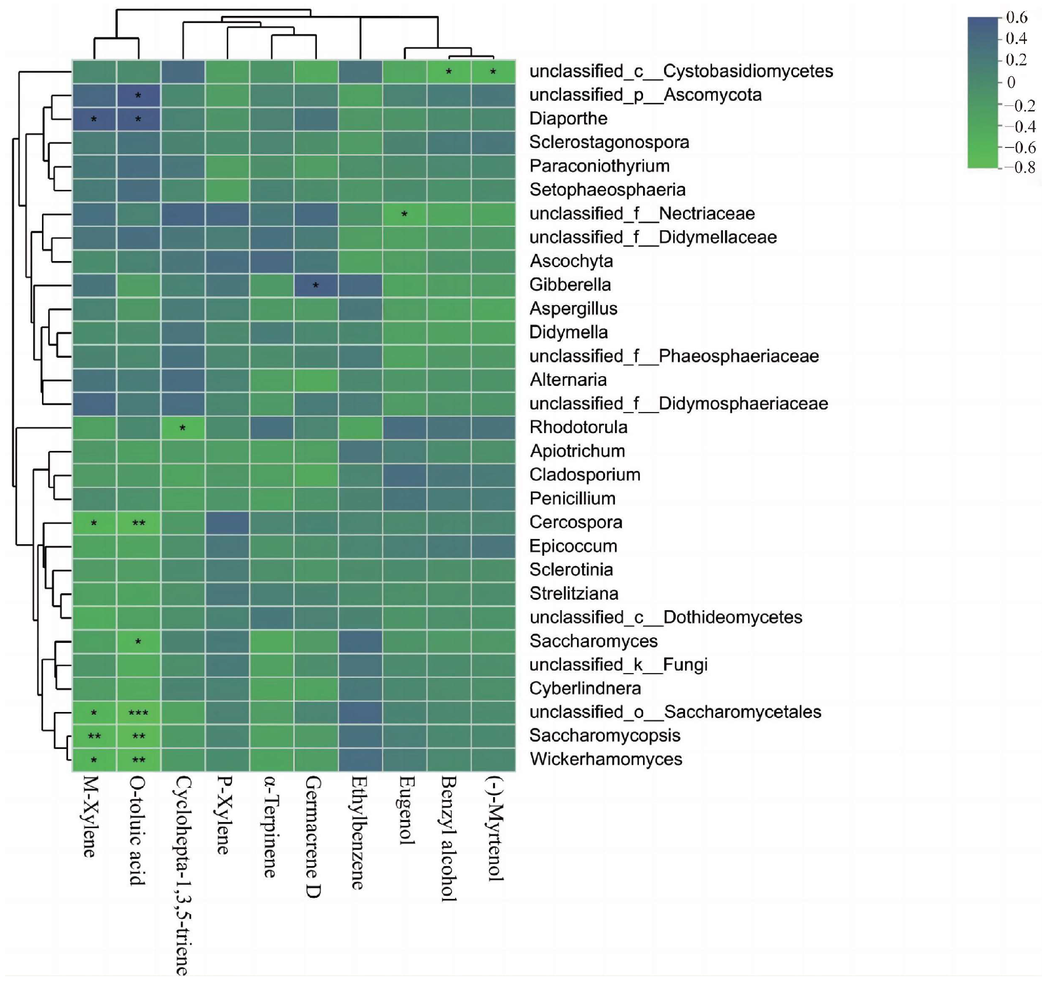
3.3. Correlation Analysis between Endophytic Fungi and Floral Volatiles in C. praecox

A classification analysis was conducted at the 97% similarity level using the Unite database for 293 genera, and then the top 30 genera in terms of relative abundance were selected for the correlation analysis (Figure S2). Didymella was the dominant genus in unopened and opened flowers. The abundance of Didymella was increased in the unopened flowers and decreased in the opened flowers with the circadian rhythm, respectively. Additionally, the correlation analysis between the top 30 main genera of endophytic fungi and 10 main floral compounds showed that only 11 fungi were significantly associated with seven floral volatiles (Table S2, Figure 4). Unclassified Saccharomycetales (R = −0.56; R = −0.75), Saccharomycosis (R = −0.59; R = −0.67), Wickerhamomyces (R = −0.56; R = −0.64), and Cercospora (R = −0.56; R = −0.65) were negatively correlated with M-Xylene and o-toluic acid, and positively correlated with Diaporthe (R = 0.55; R = 0.54). Meanwhile, Saccharomyces (R = −0.56) and unclassified Ascomycota (R = 0.58) were associated with o-toluic acid, respectively. In addition, Rhodotorula was negatively correlated with Cyclohepta-1,3,5-triene (R = −0.58), unclassified Necritiaceae and Eugenol (R = −0.49), unclassified Cystobasidiomycetes and Benzyl alcohol (R = −0.57) and (-)-Myrtenol (R = −0.54), and Gibberella was significantly positively correlated with Germacrene D (R = 0.50). Furthermore, the community composition and diversity of endophytic fungi in C. praecox was positively correlated with humidity (Figure S3; Table S3).

4. Discussion

4.1. Endophytic Fungal Diversity in C. praecox Is Altered by Circadian Rhythm and Blooming Stage

The diversity and community structure of endophytic fungi are predominantly determined by the host species, growth stages, and environmental conditions [12,13,14]. Our study indicated a circadian rhythm-dependent variation in the Chao1 and Shannon indices of the endophytic fungi in C. praecox. Specifically, these indices exhibited a gradual decrease in the unopened flowers, while in the opened flowers, an initial increase followed by a decrease was observed. Despite the fact that limited research is available about the impact of circadian rhythms on plant endophytic fungi, it has been established that the microbial flora exhibits heightened sensitivity to temperature fluctuations, particularly over short durations such as daily variations [42]. However, the correlation analysis revealed a positive correlation between the diversity and community structure of C. praecox’s endophytic fungi and humidity. This aligns with findings from studies on Taxus Chinensis [43], and can be attributed to the organism’s preference for dark and humid environments. Given these observations, we propose that the diversity of endophytic fungi in C. praecox is modulated by humidity, which is, in turn, influenced by circadian rhythms. Additionally, we observed inconsistent variations in endophytic fungal diversity in different blooming stages (unopened and opened flowers), a phenomenon also reported in other studies [19,20,21,44]. It is known that endophytic fungi in different growth stages have distinct nutrient requirements, which could significantly influence their behavior during the flowering process [45]. Our study also suggested that the β diversity of the endophytic fungi remained relatively consistent across the different blooming stages and circadian rhythms. This could imply that endophytic fungi are not easily selected, being subjected to environmental filtering over short periods or throughout growth [46], especially within the same plant. In addition, the endophytic fungi in C. praecox are influenced by both circadian rhythms and blooming stages, similar to the dynamic changes observed in floral volatiles [47,48]. As insect pollinators act rhythmically, the variations in floral scent with rhythm are associated with the activity of the corresponding pollinators [37]. This leads us to speculate about the potential close relationship between plant endophytic fungi and their secondary metabolites. In other words, changes in floral volatiles may be an important reason for changes in endophytic fungi.

4.2. Dominant Fungal Communities under Different Circadian Rhythms and in Different Blooming Stages

We analyzed the species composition and relative abundance of endophytic fungi in C. praecox. Our analysis revealed that the relative abundance of endophytic fungi in C. praecox at various taxonomic levels fluctuated with the circadian rhythm. Specifically, the number of endophytic fungal OTUs decreased gradually in the unopened flowers, whereas an initial increase followed by a decrease was observed with the circadian rhythm in the opened flowers. Moreover, the species composition of endophytic fungi in C. praecox spans nine phyla, 34 classes, 79 orders, 181 families, 293 genera, and 397 species. Notably, Ascomycota was dominant in C. praecox and prevalent in different tissue parts of other plants, including the seeds, leaves, roots, and bark [49,50,51,52,53]. Aromatic compounds produced by Ascomycota through biotransformation pathways [54] can be used as food by some endophytic fungi [55], and these endophytic fungi can also resist the toxicity of the compounds [56], which could potentially account for their high presence in C. praecox. However, this finding contrasts with the primary fungal communities of local flowers in Yunnan, which predominantly belong to the Deuteromycotina, suggesting a potential influence of the host species [57]. In addition to Ascomycota, Didymella holds a dominant position at the genus level among the endophytic fungi of C. praecox. As the most phytopathologically important genus in the Didymellaceae, Didymella may serve as a significant reservoir of species of the Septosporaceae in plants [58]. Furthermore, the endophytic fungi of C. praecox consisted of Phaeosphaeriaceae, Aspergillus, and Gibberella. Among these, some species within the Phaeosphaeriaceae have been identified as a source of polyketide biosynthesis in desert plants [59]; Aspergillus synthesizes a variety of secondary metabolites [60]; and Gibberella has been reported to promote plant growth [61]. To summarize, our findings are expected to enrich the understanding of the types and resources of endophytic fungi in C. praecox.

4.3. Relationship between Endophytic Fungi and Floral Volatiles in C. praecox

Eleven endophytic fungi were positively or negatively correlated with seven floral volatiles in C. praecox. This may be attributed to the ability of endophytic fungi to be directly selected from within the plant, thereby influencing its metabolic pathways [62]. Furthermore, some aromatic hydrocarbons (e.g., α-Terpinene, M-xylene, O-toluic acid, and Germacrene D) present in the floral volatiles of C. praecox exhibited a positive correlation with specific endophytic fungi (Ascomycota, Gibberella, and Diaporthe). This contrasts with previous findings that phenolic substances in medicinal plants are more likely to coexist with certain endophytic fungi, possibly due to variations in the host species or metabolic responses [63]. The dominance of aromatic hydrocarbons in C. praecox, an ornamental flower, suggests a different metabolic preference compared to medicinal plants, where phenolic substances enhance the antioxidant capacity. Diaporthe fungi, a significant source of functional natural products, have been found to produce polyketones, alkaloids, terpenoids, anthraquinones, and other novel structural metabolites [64]. Similarly, most Gibberella fungi are known to produce bioactive metabolites [65]. Interestingly, M-xylene and O-toluic acid showed significant negative correlations (p < 0.01) with unclassified Saccharomycetales, Saccharomycopsis, and Wickerhamomyces. This parallels findings of negative correlations between endophytic fungi and some precursors for the synthesis of paclitaxel in Taxus Chinensis [66], implying a close relationship between endophytic fungi and plant metabolism. In conclusion, the endophytic fungi of C. praecox can directly affect the production of secondary metabolites, which may be influenced by multiple strains of fungi. This study further deepens our understanding of the relationship between plant endophytic fungi and their floral volatiles.

5. Conclusions

This study represents the first investigation into the community structure and diversity of endophytic fungi in relation to the circadian rhythms and blooming stages of C. praecox using high-throughput sequencing technology. Under a diurnal rhythm, the diversity (Chao1 and Shannon indices) of endophytic fungi gradually decreased in the unopened flowers, while it showed an increasing and then decreasing trend in the opened flowers. In the different blooming stages, the endophytic fungal diversity was significantly higher at 7:00 a.m. in the unopened flowers compared to the opened flowers. Ascomycota was found to be dominant in C. praecox flowers. Humidity was the key factors that significantly affected the endophytic fungal diversity and community. Notably, 11 endophytic fungi were significantly positively or negatively correlated with seven floral volatiles. Our findings are expected to enrich the knowledge of the types and resources of endophytic fungi in C. praecox and further elucidate the relationship between endophytic fungi and floral volatiles.

Supplementary Materials

The following supporting information can be downloaded at: https://www.mdpi.com/article/10.3390/jof10020145/s1. Table S1. Summary of sample sequencing information; Table S2. Floral volatile emitted from C. praecox with different blooming stages and circadian rhythms; Table S3. Correlations between endophytic fungal diversity in C. praecox with environmental factors; Figure S1. Sequencing data results in endophytic fungi of C. praecox OTU dilution curve; Figure S2. Community structure of endophytic fungi of C. praecox at genus level. Figure S3. Correlations between the endophytic fungal community in C. praecox and environmental factors (T: temperature; H: humidity).

Author Contributions

Y.L.: Methodology, Data curation, Writing—original draft, Writing—review and editing, Visualization, Methodology, Investigation, Data curation. J.H.: Investigation, Data curation, Writing—review and editing. X.H.: Conceptualization, Resources, Project administration. R.R.: Methodology, Formal analysis, Writing—original draft. S.W.: Writing—review and editing, Supervision, Funding acquisition. All authors have read and agreed to the published version of the manuscript.

Funding

This work was supported by the National Natural Science Foundation of China (31860229), Yunnan Ten Thousand People Plan Youth Top Talent Project (YNWR-QNBJ-2019–028), China Agriculture Research System of MOF & MARA (CARS-21-05B), Major Science and Technology Project of Yunnan (202102AE090042), Major Science and Technology Project of Kunming (2021JH002).

Institutional Review Board Statement

Not applicable.

Informed Consent Statement

Not applicable.

Data Availability Statement

The sequences have been deposited in the National Center for Biotechnology Information Sequence Read Archive under Accession no. PRJNA875038 (ITS data).

Conflicts of Interest

The authors declare no conflicts of interest.

References

  1. Arnold, A.E. Understanding the diversity of foliar endophytic fungi: Progress, challenges, and frontiers. Fungal Biol. Rev. 2007, 21, 51–66. [Google Scholar] [CrossRef]
  2. Li, P.Q.; Mou, Y.; Shan, T.J.; Xu, J.M.; Li, Y.; Lu, S.Q.; Zhou, L.G. Effects of polysaccharide elicitors from endophytic Fusarium oxysporium Dzf17 on growth and diosgenin production in cell suspension culture of Dioscorea zingiberensis. Molecules 2011, 16, 9003–9016. [Google Scholar] [CrossRef]
  3. Sun, Y.; Wang, Q.; Lu, X.D.; Okane, I.; Kakishima, M. Endophytic fungal community in stems and leaves of plants from desert areas in China. Mycol. Prog. 2012, 11, 781–790. [Google Scholar] [CrossRef]
  4. Muhammad, W.; Abdul, L.K.; Muhammad, H.; Raheem, S.; Sang, M.K.; Jong, G.K.; In, J.L. Endophytic fungi promote plant growth and mitigate the adverse effects of stem rot: An example of Penicillium citrinum and Aspergillus terreus. J. Plant Interact. 2015, 10, 280–287. [Google Scholar]
  5. Kornsakulkarn, J.; Choowong, W.; Rachtawee, P.; Boonyuen, N.; Kongthong, S.; Isaka, M.; Thongpanchang, C. Bioactive hydroanthraquinones from endophytic fungus Nigrospora sp. Phytochem. Lett. 2018, 24, 46–50. [Google Scholar] [CrossRef]
  6. Li, S.J.; Zhang, X.; Wang, X.H.; Zhao, C.Q. Novel natural compounds from endophytic fungi with anticancer activity. Eur. J. Med. Chem. 2018, 157, 960–961. [Google Scholar] [CrossRef] [PubMed]
  7. Stierle, A.; Strobel, G.; Stierle, D. Taxol and taxane production by Taxomyces andreanae, an endophytic fungus of Pacific yew. Science 1993, 260, 214–216. [Google Scholar] [CrossRef] [PubMed]
  8. Wang, L.J.; Liu, S.M.; Chu, Z.M. Isolation and Identification Endophytic Fungi from Lilium Siberia and Analysis of Volatile Components. Food Ind. 2013, 34, 203–205. [Google Scholar]
  9. Chukalo, C.E.; Krishna, C.R. Endophytic mycoflora and their bioactive compounds from azadirachta indica: A comprehensive review. J. Fungi 2018, 4, 42. [Google Scholar]
  10. Wu, X.M.; Ren, W.M.; Yang, X.D.; Wang, Y.H. Isolation of endophytic fungi and screening of salidrosid-producing strains from Rhodiola sachalinensis. Lishizhen Med. Mater. Medica 2014, 25, 2769–2772. Available online: https://kns.cnki.net/kcms/detail/detail.aspx?FileName=SZGY201411087&DbName=CJFQ2014 (accessed on 6 June 2022).
  11. Li, H.; Yan, S.; Mi, Z.; Zuo, P.; Li, T.; Wei, Y.L.; Lin, L.B. Diversity and cold adaptation of endophytic fungi from five dominant plant species collected from the Baima Snow Mountain Southwest China. Fungal Divers. 2012, 54, 79–86. [Google Scholar] [CrossRef]
  12. Martin, U.; Almut, R.; Knut, F.; Peter, O.; Wilfried, M. Species richness and distribution patterns of leaf-inhabiting endophytic fungi in a temperate forest canopy. Mycol. Prog. 2007, 6, 201–212. [Google Scholar]
  13. Shi, Y.W.; Li, C.; Yang, H.M.; Zhang, T.; Gao, Y.; Zeng, J.; Lin, Q.; Mahemuti, O.; Li, Y.G.; Huo, X.D.; et al. Endophytic fungal diversity and space-time dynamics in sugar beet. Eur. J. Soil Biol. 2016, 77, 77–85. [Google Scholar] [CrossRef]
  14. Hoffman, M.T.; Arnold, A.E. Geographic locality and host identity shape fungal endophyte communities in cupressaceous trees. Mycol. Res. 2007, 112, 331–344. [Google Scholar] [CrossRef] [PubMed]
  15. Lu, Y.Z.; Zhang, E.H.; Yin, X.; Cai, H.; Yuan, L.; Li, L.Q.; Zhao, K.T.; Lan, X.Z. Diversity of endophytic fungi and the rhizosphere soil fungi communities of the endangered plant Paeonia ludlowii in Tibe. J. Biol. 2020, 37, 5. [Google Scholar]
  16. Li, H.L.; Yu, L. Diversity of endophytic fungi in Zhanjiang honeysuckle. Guangdong Agric. Sci. 2014, 41, 5. [Google Scholar]
  17. Lupo, S.; Tiscornia, S.; Bettucci, L. Endophytic fungi from flowers, capsules and seeds of eucalyptus globulus. Rev. Iberoam. Micol. 2001, 18, 38–41. [Google Scholar]
  18. Rajeswari, S.; Umamaheswari, S.; Prasanth, D.A.; Rajamanikandan, K. Study of endophytic fungal community of Moringa oleifera from Omalur Region-Salem. Int. J. Pharm. Sci. Res. 2014, 5, 4887. [Google Scholar]
  19. Zeng, X.H.; Diao, H.X.; Ni, Z.Y.; Shao, L.; Jiang, K.; Hu, C.; Huang, Q.J.; Huang, W.C. Temporal Variation in Community Composition of Root Associated Endophytic Fungi and Carbon and Nitrogen Stable Isotope Abundance in Two Bletilla Species (Orchidaceae). Plants 2020, 10, 18. [Google Scholar] [CrossRef]
  20. Paul, N.C.; Jian, X.D.; Sang, H.K.; Choi, Y.P.; Yu, S.H. Distribution and Antifungal Activity of Endophytic Fungi in Different Growth Stages of Chili Pepper (Capsicum annuum L.) in Korea. Plant Pathol. J. 2012, 28, 10–19. [Google Scholar] [CrossRef]
  21. Jin, H.; Yan, Z.Q.; Liu, Q.; Yang, X.Y.; Chen, J.X.; Qin, B. Diversity and dynamics of fungal endophytes in leaves, stems and roots of Stellera chamaejasme L. in northwestern China. Antonie Van Leeuwenhoek 2013, 104, 949–963. [Google Scholar] [CrossRef]
  22. Arnold, A.E.; Lutzoni, F. Diversity and host range of foliar ungal endophytes: Are tropical leaves biodiversity hotspots. Ecology 2007, 88, 541–549. [Google Scholar] [CrossRef]
  23. Mishra, A.; Gond, S.K.; Kumar, A.; Sharma, V.K.; Verma, S.K.; Kharwar, R.N.; Sieber, T.N. Season and tissue type affect fungal endophyte communities of the Indian medicinal plant Tinospora cordifolia more strongly than geographic location. Microb. Ecol. 2012, 64, 388–398. [Google Scholar] [CrossRef]
  24. Lemfack, M.C.; Nickel, J.; Dunkel, M.; Preissner, R.; Piechulla, B. mVOC: A database of microbial volatiles. Nucleic Acids Res. 2014, 42, D744–D748. [Google Scholar] [CrossRef]
  25. Qian, C.D.; Fu, Y.H.; Jiang, F.S.; Xu, Z.H.; Cheng, D.Q.; Ding, B.; Gao, C.X.; Ding, Z.S. Lasiodiplodia sp. ME4-2, an endophytic fungus from the floral parts of Viscum coloratum, produces indole-3-carboxylic acid and other aromatic metabolites. BMC Microbiol. 2014, 14, 297. [Google Scholar] [CrossRef] [PubMed]
  26. Yakir, E.; Hilman, D.; Harir, Y.; Green, R.M. Regulation of output from the plant circadian clock. FEBS J. 2007, 274, 335–345. [Google Scholar] [CrossRef] [PubMed]
  27. Mc-Tavish, H.S.; Davies, N.W.; Menary, R.C. Emission of Volatiles from brown boronia flowers: Some comparative observations. Ann. Bot. 2000, 86, 347–354. [Google Scholar] [CrossRef]
  28. Fan, J.J.; Zhang, W.X.; Zhang, D.L.; Wang, G.B.; Cao, F.L. Flowering Stage and Daytime Affect Scent Emission of Malus ioensis “Prairie Rose”. Molecules 2019, 24, 2356. [Google Scholar] [CrossRef] [PubMed]
  29. Kanani, M.; Chamani, E.; Shokouhian, A.A.; Torabi, G.M. Plant secondary metabolism and flower color changes in damask rose at different flowering development stages. Acta Physiol. Plant. 2021, 43, 55. [Google Scholar] [CrossRef]
  30. Chuang, Y.C.; Lee, M.C.; Chang, Y.L.; Chen, W.H.; Chen, H.H. Diurnal regulation of the floral scent emission by light and circadian rhythm in the Phalaenopsis orchids. Bot. Stud. 2017, 58, 50. [Google Scholar] [CrossRef] [PubMed]
  31. Cna’ani, A.; Muhlemann, J.K.; Ravid, J.; Masci, T.; Klempien, A.; Nguyen, T.T.H.; Dudareva, N.; Pichersky, E.; Vainstein, A. Petunia × hybrida floral scent production is negatively affected by high-temperature growth conditions. Plant Cell Environ. 2015, 38, 1333–1346. [Google Scholar] [CrossRef]
  32. Hu, Z.; Zhang, H.; Leng, P.; Zhao, J.; Wang, W.H.; Wang, S.D. The emission of floral scent from lilium ‘siberia’ in response to light intensity and temperature. Acta Physiol. Plant. 2013, 35, 1691–1700. [Google Scholar] [CrossRef]
  33. Rasulov, B.; Hüve, K.; Välbe, M.; Laisk, A.; Niinemets, Ü. Evidence that light, carbon dioxide, and oxygen dependencies of leaf isoprene emission are driven by energy status in hybrid aspen. Plant Physiol. 2009, 151, 448–460. [Google Scholar] [CrossRef] [PubMed]
  34. Kawoosa, T.; Singh, H.; Kumar, A.; Sharma, S.K.; Devi, K.; Dutt, S.; Vats, S.K.; Sharma, M.; Ahuja, P.S.; Kumar, S. Light and temperature regulated terpene biosynthesis: Hepatoprotective monoterpene picroside accumulation in picrorhiza kurrooa. Funct. Integr. Genomic. 2010, 10, 393–404. [Google Scholar] [CrossRef] [PubMed]
  35. Zhou, J.R.; Ni, D.J. Changes in Flower Aroma Compounds of Cultivars of Chimonanthus Praecox (L.) Link and at Different Stages Relative to Chimonanthus Tea Quality. Acta Hortic. Sin. 2010, 37, 1621–1628. [Google Scholar] [CrossRef]
  36. Li, Z.G.; Cao, H.; Lee, M.R.; De, L.S. Analysis of volatile compounds emitted from Chimonanthus praecox (L.) in different florescence and QSRR study of GC retention indices. Chromatographia 2009, 70, 1153–1162. [Google Scholar] [CrossRef]
  37. Li, Y.; Jia, W.J.; Wang, Q.; Wang, B.; Wang, S. Comparative analysis of floral scent profiles between two Chimonanthus praecox plants under different rhythms and blooming stages. Sci. Hortic. 2022, 301, 111129. [Google Scholar] [CrossRef]
  38. Yu, L. The Study of Violate Compounds and Flower Pigments in Chimonanthus praecox L. Master’s Thesis, Huazhong Agricultural University, Wuhan, China, 2013. Available online: https://kns.cnki.net/kcms/detail/detail.aspx?FileName=1016190451.nh&DbName=CMFD2017 (accessed on 27 July 2022).
  39. Meng, L.B.; Shi, R.; Wang, Q.; Wang, S. Analysis of Floral Fragrance Compounds of Chimonanthus praecox with Different Floral Colors in Yunnan, China. Separations 2021, 8, 122. [Google Scholar] [CrossRef]
  40. Chen, S.Y.; Rui, R.; Wang, S.; He, X.H. Comparative Analysis of the Floral Fragrance Compounds of Panax notoginseng Flowers under the Panax notoginseng-pinus Agroforestry System Using SPME-GC-MS. Molecules 2022, 27, 3565. [Google Scholar] [CrossRef] [PubMed]
  41. Jia, W.J.; Wang, S.; He, X.H.; Zhao, X.Y. Different factors drive the assembly of pine and Panax notoginseng-associated microbiomes in Panax notoginseng-pine agroforestry systems. Front. Microbiol. 2022, 13, 1018989. [Google Scholar] [CrossRef]
  42. Hu, A.; Nie, Y.; Yu, G.; Han, C.; He, J.; He, N.; Liu, S.; Deng, J.; Shen, W.; Zhang, G. Diurnal Temperature Variation and Plants Drive Latitudinal Patterns in Seasonal Dynamics of Soil Microbial Community. Front. Microbiol. 2019, 10, 674. [Google Scholar] [CrossRef]
  43. Wu, L.S. Diversity and Secondary Metabolites of Endophytic fungi from Taxus Chinensis var. mairei and Their Correlations with the Host. Ph.D. Thesis, Second Military Medical University, Shanghai, China, 2012. Available online: https://kns.cnki.net/KCMS/detail/detail.aspx?dbname=CDFD1214&filename=1012404569.n (accessed on 29 November 2022).
  44. Zhang, P.L.; Wang, G.K.; Yu, Y.; Liu, J.S.; Wang, J.T.; Xu, F.Q.; Ma, Z.H.; Zhang, N.; Wang, G. Isolation and Identification of Endophytic Fungi from Paeonia ostii and Their Diversity in Different Growth Periods. J. Anhui Univ. Chin. Med. 2016, 35, 78–82. [Google Scholar]
  45. Ercole, E.; Adamo, M.; Rodda, M.; Gebauer, G.; Girlanda, M.; Perotto, S. Temporal variation in mycorrhizal diversity and carbon and nitrogen stable isotope abundance in the wintergreen meadow orchid Anacamptis morio. New Phytol. 2015, 205, 1308–1319. [Google Scholar] [CrossRef] [PubMed]
  46. Stegen, J.C.; Lin, X.; Fredrickson, J.K.; Chen, X.; Kennedy, D.W.; Murray, C.J.; Rockhold, M.L.; Konopka, A. Quantifying community assembly processes and identifying features that impose them. ISME J. 2013, 7, 2069–2079. [Google Scholar] [CrossRef]
  47. Donna, J.L.; Juliette, T.J.; Colleen, J.D. Only a matter of time: The impact of daily and seasonal rhythms on phytochemicals. Phytochem. Rev. Fundam. Perspect. Nat. Prod. Res. 2019, 18, 1409–1433. [Google Scholar]
  48. Dudareva, N.; Gershenzon, P.J. Biochemistry of Plant Volatiles. Plant Physiol. 2004, 135, 1893–1902. [Google Scholar] [CrossRef] [PubMed]
  49. Ma, W.; Liu, Z.P.; Sun, L.Y.; Zhang, K.X.; Xu, J.; Wen, D.; Zhao, R.; Liu, X.B. Endophytic Fungi Diversity of Ginseng Seeds by Using 454 GS FLX. Inf. Tradit. Chin. Med. 2017, 34, 28–32. [Google Scholar]
  50. Yang, L.C.; Wu, Q.S.; Gao, Y.; Chen, Q.Q.; Wang, Y.Q.; Niu, X.J.; Weng, Q.B. Analysis of Composition of Endophytic Community of Dendrobium nobile Lindl. Seeds Based on High-throughput Sequencing. Seed 2020, 39, 5. [Google Scholar]
  51. Yang, J.; Dong, C.B.; Chen, W.H.; Liang, J.D.; Han, Y.F.; Liang, Z.Q. Community composition and ecological functional structural analysis of endophytic fungi in bark of Eucommia ulmoides in different areas. China J. Chin. Mater. Medica 2019, 6, 1126–1134. [Google Scholar]
  52. Zhang, S.P.; Xu, T.; Yang, L.Q.; Song, Y.; Li, X.Y.; Zhang, H.W. Endophytic fungal communities of Stipa sp. roots in different types of steppes in northern China. Chin. J. Appl. Ecol. 2014, 12, 3475–3482. [Google Scholar]
  53. Yang, Q.; Zhou, K.; Chen, A.J.; Guo, X.; Liu, X.R.; Liang, Y.L. Endophytic fungal communities structure and variation in Ginkgo biloba leaf tissues with different ages. J. Hunan Agric. Univ. (Nat. Sci.) 2019, 45, 42–49. [Google Scholar]
  54. Serra, S.; De Simeis, D. Fungi-mediated biotransformation of the isomeric forms of the apocarotenoidsionone, damascone and theaspirane. Molecules 2019, 24, 19. [Google Scholar] [CrossRef] [PubMed]
  55. Huang, L.H.; Yuan, M.Q.; Ao, X.; Ren, A.Y.; Zhang, H.B.; Yang, M.Z. Endophytic fungi specifically introduce novel metabolites into grape flesh cells in vitro. PLoS ONE 2018, 13, e0196996. [Google Scholar] [CrossRef]
  56. Schulz, B.; Boyle, C. The endophytic continuum. Mycol. Res. 2005, 109, 661–686. [Google Scholar] [CrossRef] [PubMed]
  57. Zhang, C.F. Diversity of Endophytic Fung in Five Flower Plants in Yunnan and Their Effects on Plant Growth. Master’s Thesis, Kunming University of Science and Technology, Kunming, China, 2013. Available online: https://kns.cnki.net/kcms/detail/detail.aspx?FileName=1016037043.nh&DbName=CMFD2016 (accessed on 24 July 2022).
  58. Huang, Y. The Study of the Didymellaceae and Plectosphaerella in Endophytic fungi of Aquatic Plants. Master’s Thesis, Yunnan University, Kunming, China, 2018. [Google Scholar] [CrossRef]
  59. Xu, Z.L.; Yan, D.J.; Tan, X.M.; Niu, S.B.; Yu, M.; Sun, B.D.; Ding, C.F.; Zhang, Y.G.; Ding, G. Phaeosphspirone (1/1′), a pair of unique polyketide enantiomers with an unusual 6/5/5/6 tetracyclic ring from the desert plant endophytic fungus Phaeosphaeriaceae sp. Phytochemistry 2022, 194, 112969. [Google Scholar] [CrossRef] [PubMed]
  60. Liang, H.Q.; Chen, W.F.; Fan, Y.K.; Zhu, Z.D.; Ma, G.X.; Chen, D.L.; Tian, R.R. Research progress on secondary metabolites and activities of endophytic fungi of genuses Aspergillus and Trichoderma from mangroves. J. Trop. Oceanogr. 2023, 42, 12–24. Available online: http://kns.cnki.net/kcms/detail/44.1500.P.20221208.1416.002.html (accessed on 13 June 2023).
  61. Zhang, J. Research on Diversity of Endosymbiotic Fungi in Roots of Cymbidium. Master’s Thesis, Chinese Academy of Forestry, Beijing, China, 2012. Available online: https://kns.cnki.net/kcms/detail/detail.aspx?FileName=1012423572.nh&DbName=CMFD2012 (accessed on 29 June 2022).
  62. Rai, M.; Agarkar, G. Plant-fungal interactions: What triggers the fungi to switch among lifestyles. Crit. Rev. Microbiol. 2016, 42, 428–438. [Google Scholar] [CrossRef]
  63. Huang, W.Y.; Cai, Y.Z.; Hyde, K.D.; Corke, H.; Sun, M. Endophytic fungi from Nerium oleander L (Apocynaceae): Main constituents and antioxidant activity. World J. Microbiol. Biotechnol. 2007, 23, 1253–1263. [Google Scholar] [CrossRef]
  64. Cai, J.; Zhou, W.Y.; Xu, G.M.; Zhou, X.J. Research progress on natural products of fungi of Diaporthe sp. China J. Chin. Mater. Medica 2021, 46, 1717–1726. [Google Scholar] [CrossRef]
  65. Cui, Y.F.; Huang, Y.; Jiang, L.H. Research Progress on Gibberella Species of Agricultural Importance. Chin. Agric. Sci. Bull. 2007, 7, 441–446. [Google Scholar]
  66. Li, H.K. Correlation between the Diversity of Endophytic Fungi and Taxoids Accumulation in Taxus cuspidata and the Application of the High-Yield Strain. Master’s Thesis, Northeast Forestry University, Heilongjiang, China, 2021. [Google Scholar] [CrossRef]
Figure 1. Flower morphology of var. grandiflorus ((a): unopened flowers; (b): opened flowers).
Jof 10 00145 g001
Figure 2. Analysis of fungal diversity and community in the flower of C. praecox in different blooming stages and under different circadian rhythms. (a) Chao1 index; (b) Shannon index; (c) fungal community. GUA: unopened flowers at 7:00 a.m.; GUB: unopened flowers at 1:00 p.m.; GUC: unopened flowers at 7:00 p.m.; GOA: opened flowers at 7:00 a.m.; GOB: opened flowers at 1:00 p.m.; GOC: opened flowers at 7:00 p.m.. Different letters in each column indicate significant differences (p < 0.05).
Jof 10 00145 g002
Figure 3. Venn analysis for the OTU distribution of endophytic fungi. (a,b) indicate the different blooming stages and circadian rhythms, respectively. GUA: unopened flowers at 7:00 a.m.; GUB: unopened flowers at 1:00 p.m.; GUC: unopened flowers at 7:00 p.m.; GOA: opened flowers at 7:00 a.m.; GOB: opened flowers at 1:00 p.m.; GOC: opened flowers at 7:00 p.m..
Jof 10 00145 g003
Figure 4. Correlation analysis for the relationship between the main floral compounds and endophytic fungi in C. praecox. ***, ** and * indicates a significant correlation at the 0.001, 0.01 and 0.05 levels, respectively.
Jof 10 00145 g004
Table 1. Relative abundance of the top five fungi at different levels of taxonomy in the six samples.
Taxonomy
Levels
Top FiveGUAGUBGUCGOAGOBGOC
PhylumAscomycota96.57%90.84%87.70%98.40%85.16%82.32%
Basidiomycota2.88%7.03%8.85%1.55%13.60%13.60%
unclassified_k__Fungi0.29%1.95%3.07%0.04%0.86%3.31%
ClassDothideomycetes46.20%37.16%44.57%44.03%36.64%19.90%
unclassified_p__Ascomycota35.23%20.61%7.43%50.52%26.31%37.55%
Eurotiomycetes8.30%7.34%8.39%2.41%8.81%4.32%
Sordariomycetes5.73%16.97%4.06%0.89%1.30%14.92%
Tremellomycetes1.94%5.66%6.93%1.00%10.83%11.45%
OrderPleosporales33.37%31.01%18.37%40.96%50.53%30.30%
unclassified_p__Ascomycota46.20%20.61%41.66%7.43%41.98%26.13%
Eurotiales7.81%4.45%16.93%5.94%1.30%7.42%
Hypocreales4.51%16.82%1.57%1.99%0.49%0.29%
Trichosporonales1.73%5.49%1.63%5.06%0.73%9.67%
Familyunclassified_p__Ascomycota46.20%20.61%7.43%50.52%26.13%37.55%
Didymellaceae18.86%23.55%25.44%28.41%19.57%9.13%
Phaeosphaeriaceae10.68%6.00%14.21%4.20%7.45%4.29%
Aspergillaceae7.80%4.45%5.94%1.29%7.20%2.45%
Nectriaceae4.49%16.46%1.62%0.47%1.63%13.67%
Genusunclassified_p__Ascomycota46.20%20.61%24.58%50.52%26.13%37.55%
Didymella17.39%19.57%7.43%26.91%14.77%8.70%
unclassified_f__Phaeosphaeriaceae9.88%4.61%12.15%2.84%4.05%3.57%
Aspergillus7.29%5.49%5.06%1.19%6.32%1.63%
Gibberalla3.86%13.87%1.28%0.73%1.63%13.28%
Speciesunclassified_p__Ascomycota46.20%20.61%24.58%50.52%26.13%37.56%
unclassified_g__Didymella17.39%19.56%7.43%26.91%14.77%8.70%
unclassified_f__Phaeosphaeriaceae9.88%4.61%12.15%2.84%4.05%3.57%
Aspergillus amstelodami6.49%2.08%2.12%0.84%1.41%10.71%
unclassified_g__Gibberella3.61%13.13%1.05%8.99%9.67%13.28%
GUA: unopened flowers at 7:00 a.m.; GUB: unopened flowers at 1:00 p.m.; GUC: unopened flowers at 7:00 p.m.; GOA: opened flowers at 7:00 a.m.; GOB: opened flowers at 1:00 p.m.; GOC: opened flowers at 7:00 p.m..
Disclaimer/Publisher’s Note: The statements, opinions and data contained in all publications are solely those of the individual author(s) and contributor(s) and not of MDPI and/or the editor(s). MDPI and/or the editor(s) disclaim responsibility for any injury to people or property resulting from any ideas, methods, instructions or products referred to in the content.

