Article

Al2O3-Phosphate Bioceramic Fabrication via Spark Plasma Sintering-Reactive Synthesis: In Vivo and Microbiological Investigation

1
Department of Nuclear Technology, Far Eastern Federal University, 10 Ajax Bay, Russky Island, 690922 Vladivostok, Russia
2
Pacific State Medical University, 2, Ostryakov Aven., Vladivostok 690990, Russia
3
Shanghai Institute of Ceramics, Chinese Academy of Sciences, Shanghai 201899, China
4
Center of Materials Science and Optoelectronics Engineering, University of Chinese Academy of Sciences, Beijing 100049, China
*
Authors to whom correspondence should be addressed.
J. Compos. Sci. 2023, 7(10), 409; https://doi.org/10.3390/jcs7100409
Submission received: 13 September 2023 / Revised: 21 September 2023 / Accepted: 28 September 2023 / Published: 3 October 2023

Abstract

:
This research introduces a method to enhance the biocompatibility of bioinert Al2O3-based ceramics by incorporating calcium phosphates (hydroxyapatite (HAp) and tricalcium phosphate (TCP)) into alumina via spark plasma sintering-reactive sintering (SPS-RS). TGA/DTG/DTA and XRD revealed phase formation of HAp and TCP and determined the main temperature points of solid-phase reactions occurring in situ during the sintering of the CaO-CaHPO4 mixture within the volume of Al2O3 under SPS-RS conditions in the range of 900–1200 °C. SEM, EDX, low temperature, and nitrogen physisorption were used to monitor changes in the morphology, structure, and elemental composition of bioceramics. Structural meso- and macroporosity, with a mean mesopore size of 10 nm, were revealed in the ceramic volume, while sintering temperature was shown to play a destructive role towards the porous inorganic framework. The physico-chemical characterization demonstrated increased relative density (up to 95.1%), compressive strength (640 MPa and above), and Vickers microhardness (up to 700 HV) depending on the HAp and TCP content and sintering temperature. Four bioceramic samples with different contents of HAP (20 and 50 wt.%) were bio-tested in in vivo models. The samples were implanted into the soft tissues under the superficial fascia of the thorax of a laboratory animal (a New Zealand White rabbit, female) in the area of the trapezius muscle and the broadest muscle of the back. Based on the results of the assessment of the surrounding tissue reaction, the absence of specific inflammation, necrosis, and tumor formation in the tissues during the implantation period of 90 days was proven. Microbial tests and dynamics of Pseudomonas aeruginosa bacterial film formation on bioceramic surfaces were studied with respect to HAp content (20 and 50 wt.%) and holding time (18, 24, and 48 h) in the feed medium.

1. Introduction

From the perspective of bone prosthetics and orthopedic applications, the most promising materials are synthetic ceramics and their composites with bioactive properties, specifically bioceramics [1,2]. Bioinert ceramics, such as titanium and its alloys, as well as oxide ceramics like zirconia, titania, and alumina, are commonly used for bone reconstruction and defect healing while maintaining their chemical, physical, and mechanical properties under physiological conditions [3,4,5]. However, the main drawback of bioinert ceramics is their relatively short lifespan due to uneven stress distribution, which leads to the resorption of bone tissue and the subsequent failure of the implant [6,7]. This is caused by significant differences in the physicochemical and mechanical properties between the implant and the bone tissue, hindering osteointegration—the formation of a structural connection between the bone tissue and the implant during the regeneration phase [8].
One way to address these issues is by enhancing the bioactive properties of ceramics through the incorporation of resorbable phases into their composition. Such resorbable composite ceramics become able to participate in the process of bone regeneration owing to the activation of osteosynthesis, as has been shown previously in a number of reports for ZrO2 [9,10,11,12,13], Al2O3 [14,15,16], CaSiO3 [17,18,19,20,21,22], etc. Commonly used bioactive resorbable phases are calcium phosphates like tricalcium phosphate (TCP), calcium pyrophosphate, and also hydroxyapatite (HAp) [23,24,25,26]. Inorganic composites containing these calcium phosphates demonstrate efficient osteointegration and withstand long-term mechanical skeletal load in the case of body and limb implants [27,28]. If the bone skull is considered, bioactive implants containing calcium phosphates show growth of alveolar appendices in the lower and upper jaws as a result of dental implanting [29]. In particular, the powder ceramic composite Al2O3-HAp, produced by hydrothermal synthesis [30], demonstrated excellent compatibility (80%) with cell cultures similar to human osteoblasts MG63. Similarly, Al2O3-HAp biocomposites match perfectly with xenogeneic grafts, showing outstanding biocompatibility on the model utilizing L929 cell lines [31]. Ceramic coatings based on Al2O3-HAp biocomposites, e.g., produced by plasma sputtering on metal implants, are also highly biocompatible, with the osteointegrative properties of the resulting grafts being significantly improved [32]. Cu-doping of Al2O3-HAp ceramics, synthesized by wet chemistry methods, leads to clear antibacterial effects against highly resistant infections [33,34].
A new synthetic strategy was previously developed and implemented for the fabrication of bioinert ZrO2-based ceramics modified with HAp and TCP using spark plasma sintering-reactive sintering (SPS-RS) [35,36]. A number of advantages of SPS-RS were shown, including “soft” temperature regimes and short sintering cycles, which allow the fabrication of high-quality ZrO2-phosphate bioceramics owing to suppressed grain growth, absence of microstructural defects, high density, and mechanical stability of the samples, while leaving room for tailoring ceramic porous structures. On top of that, exceptional bioactive properties were demonstrated by the aforementioned zirconia composite with HAp [35]. Highly efficient osteointegration was observed for the ZrO2-(15 wt.%) HAp sample, taking place in the bone skull defect of a lab animal via ossification of newly formed cartilaginous tissue and rearrangement of the bone matrix in the volume of the bone defect. This has proven great prospects for potential applications of such zirconia composites, prepared via a modern technological approach, in the field of regenerative and reconstructive surgery.
Alumina-based bioceramics (Al2O3) exhibit several significant advantages compared to zirconia-based bioceramics (ZrO2) in the academic literature. Firstly, Al2O3 demonstrates higher mechanical strength and hardness, which is crucial for implants subjected to high loads. Secondly, Al2O3 possesses superior corrosion resistance compared to ZrO2, ensuring material stability in aggressive environments and prolonged functioning within the body. Thirdly, Al2O3 exhibits better biocompatibility and is less prone to biodegradation and ion leaching, thereby reducing the risks of toxicity. Additionally, Al2O3 has higher thermal stability and can be operated at elevated temperatures. Equally important, Al2O3 facilitates the formation of porous hydroxyapatite coatings, which enhance compatibility with the bone tissue. Finally, the processing and fabrication of Al2O3 are simpler and more cost-effective compared to ZrO2 [30,37,38,39,40,41].
Related research on the fabrication of Al2O3-phosphate bioceramics by SPS-RS remains unexplored. Also, there is a lack of biocompatibility studies for these kinds of ceramics, including antibacterial properties, which would allow a preliminary justification for preclinical trials, leading to the potential practical application of synthetic bone implants. In this respect, this work aims to develop Al2O3-based ceramics containing calcium phosphates by means of reactive spark plasma sintering. Particularly, we studied the formation of HAp and TCP crystalline phases in the volume of Al2O3 bioceramics as a result of in situ reactions between the components of the reactive mixture under SPS conditions. In addition, we conducted biotests for the prepared bioceramics on an in vivo model under the conditions of contact with the soft tissues of a lab animal (a rabbit) and evaluated the antibacterial properties of the studied materials.
The results of the investigation imply that a non-conventional SPS-RS approach will allow for the fabrication of bioceramics, which is promising for implants in regenerative and reconstructive bone surgery.

2. Experimental Section

2.1. Reagents

Alumina powder (Al2O3) was used in the sintering blend together with the reactive mixture (RM) containing calcium oxide (CaO) and calcium hydrophosphate (CaHPO4) to provide the formation of bioactive HAp phase constituting 20 and 50 wt.% of the ceramics. For the synthesis of HAp, the ratio of Ca to P was considered equal to 1.67. The sintering mixture was prepared on a planetary mill at a rotation rate of 600 rpm for 30 min. All reagents used had high purity, with a key component content of 99.98% (Sigma Aldrich, St. Louis, MO, USA).

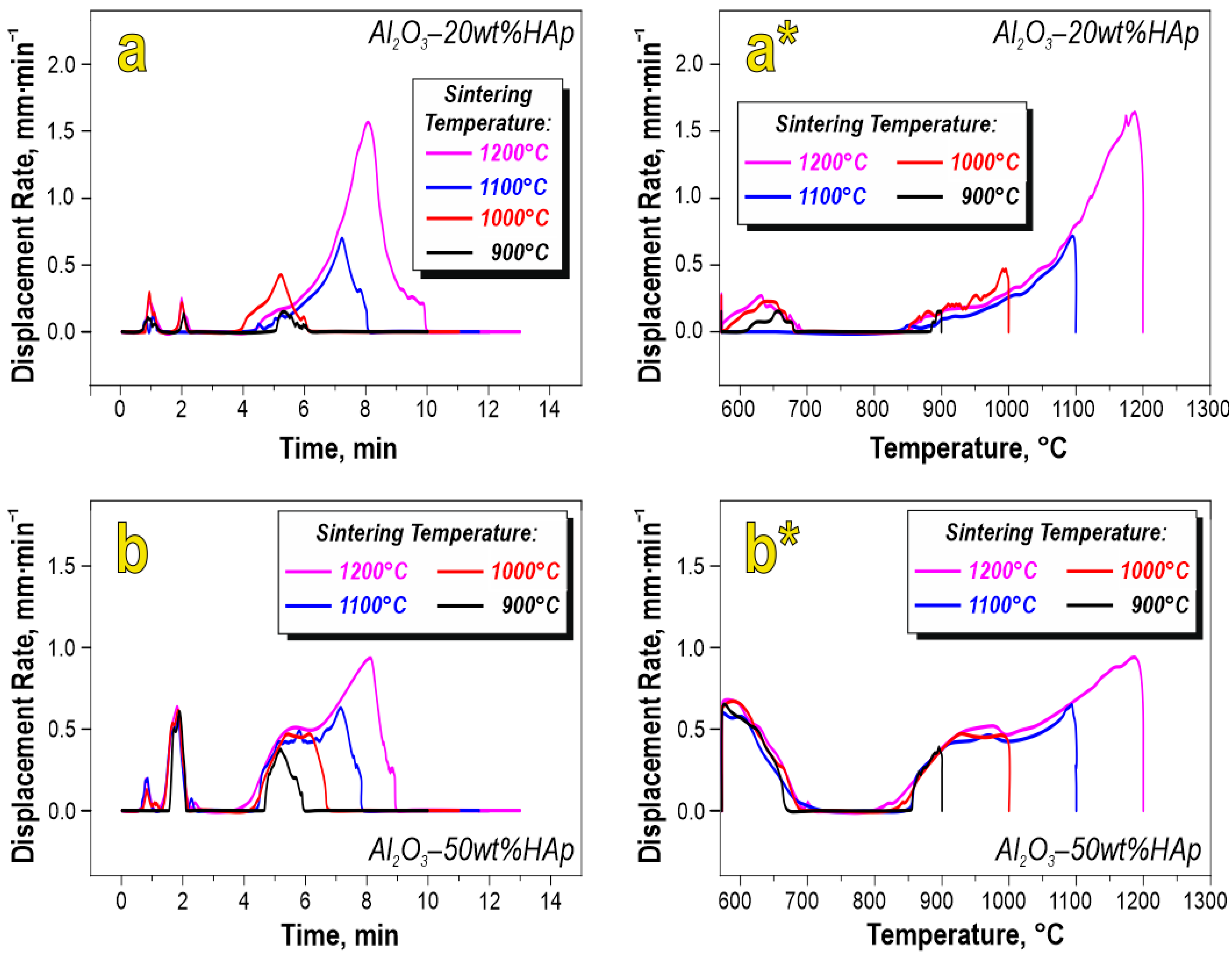
2.2. Bioceramics Fabrication

Bioceramic samples were fabricated by means of SPS-RS technology via powder consolidation on a SPS-515S setup by “Dr.Sinter·LABTM” (Japan), according to the following scheme: 1 g of starting powder mix was placed into a graphite die (internal diameter 10.5 mm), prepressed (pressure 20.7 MPa), and then the green body was transferred into a vacuum chamber (10−5 atm), followed by sintering. Sample’s heating was provided by unipolar low-voltage pulse current in On/Off regime (12 “On” pulses, 2 “Off” phases); thus, the duration of the pulse packet was 39.6/6.6 ms. SPS temperature was controlled with an optical pyrometer (low detector threshold 650 °C), focused on a 5.5 mm deep gap in the middle of the outer die wall. To prevent sintered powder from baking onto the die and plungers and to ease the extraction of the consolidated sample, we used a 200 µm thick graphite foil. The die was wrapped in a thermally insulating fabric to reduce heat loss. The geometric dimensions of the obtained ceramic samples of cylindrical shape were: diameter 10.3 mm and height 4–6 mm (depending on sintering regime).
Samples were sintered at temperatures of 900, 1000, 1100, and 1200 °C. The heating rate was changed stepwise to 300 °C/min in the range 0–650 °C and 90 °C/min above 650 °C. Samples were held at the final temperature for 5 min and then cooled down to room temperature in 30 min. Compacting pressure was 50 MPa.

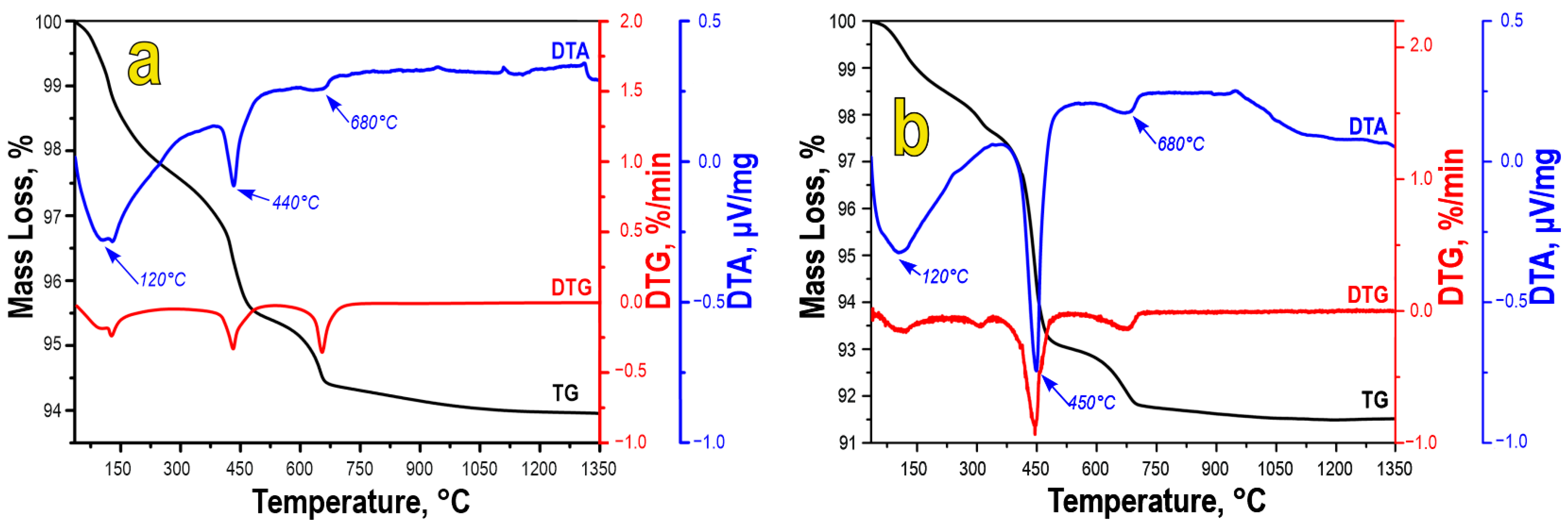
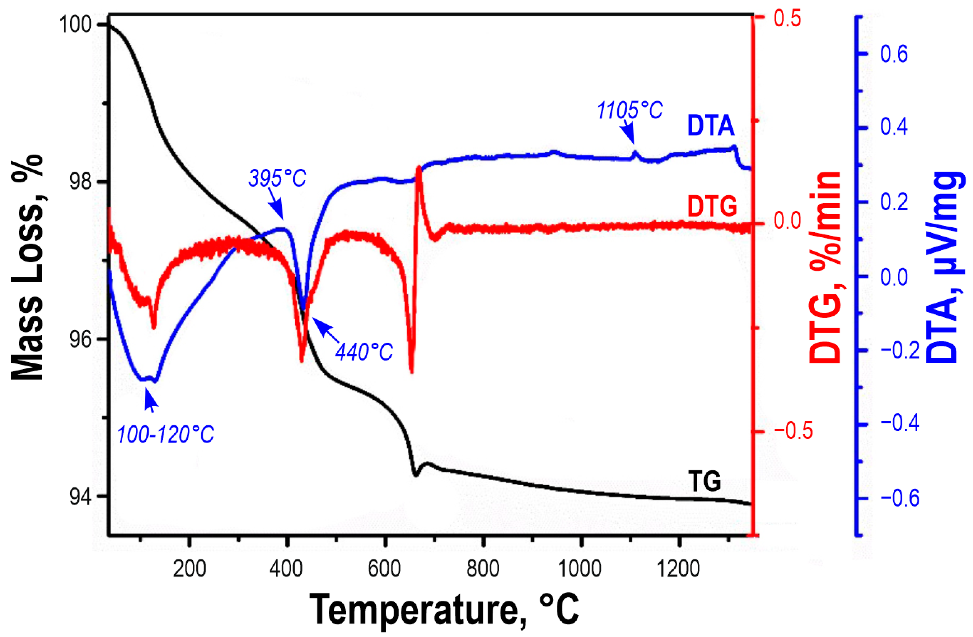
2.3. Characterization Methods

Powder particle size was evaluated on a laser analyzer, the Analysette-22 NanoTec/MicroTec/XT “Fritsch” (Idar-Oberstein, Germany), with averaging over 3 scans for each sample. Phase identification was conducted by means of XRD on a diffractometer D8 Advance “Bruker AXS” (Bruker, Bremen, Germany), using CuKα-source, Ni-filter, angle range 10–80°, scanning step 0.02°, and scanning rate 5°/min. The thermogravimetric curves were recorded on the DTG-60 H “Shimadzu” thermal analyzer (Japan) in platinum crucibles with a pierced lid in a dry argon stream (20 mL/min) in the temperature range of 35–1300 °C and a heating rate of 10 °C/min. Specific surface area was measured by low-temperature nitrogen physisorption at 77 K on an automated gas sorption analyzer, the Autosorb IQ “Quantachrome” (Boynton Beach, FL, USA). The results were analyzed at the level of the BET and BJH models. Compressive strength (σcs.) of cylindrically shaped samples (diameter 10.3 mm and height 4–6 mm) was determined by compressing at a rate of 0.5 mm/min on the tensile machine Autograph AG-X plus 100 kN “Shimadzu” (Kyoto, Japan). Surface imaging of the fabricated samples was performed by means of SEM on a CrossBeam 1540 XB “Carl Zeiss” microscope (ZEISS, Jena, Germany) with an EDX add-on by Bruker (Germany). Specific density (ρspec) was assessed via hydrostatic weighing on an AdventurerTM “OHAUS Corporation” balance (Parsippany, NJ, USA). Vickers microhardness (HV) was estimated at a force of 0.2 N on the HMV-G-FA-D “Shimadzu” microhardness tester (Kyoto, Japan).

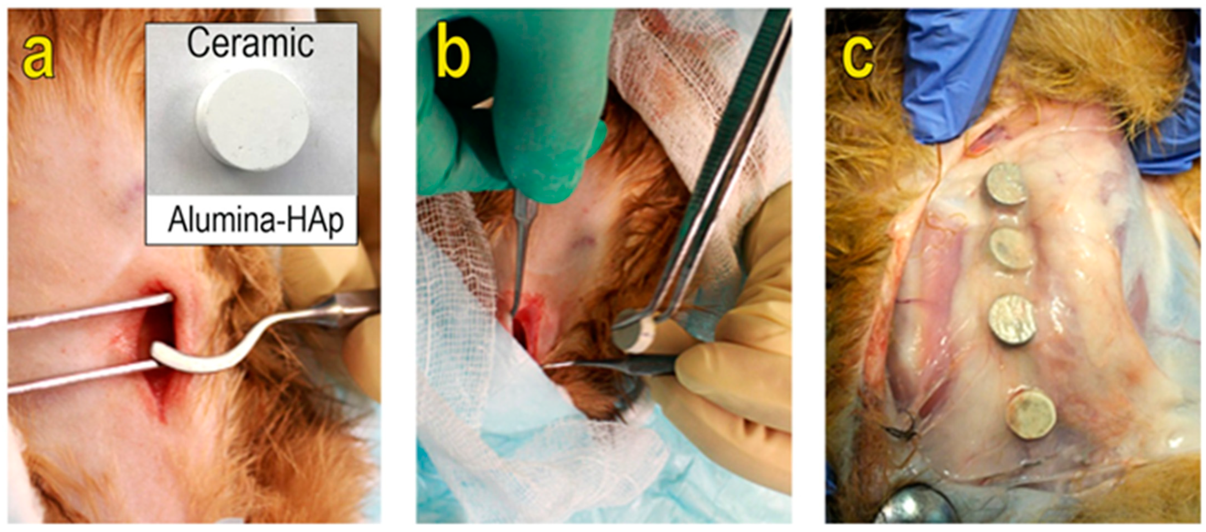
2.4. In Vivo Biocompatibility Tests

Bioceramic (samples obtained at 1000 °C) implanting into a lab animal was carried out under anesthesia (2% xylazine (5 mg/kg) and 5% ketamine hydrochloride (30 mg/kg) were injected intramuscularly). Prior to implanting, the skin in the area of the withers was shaved, and after treatment with sanitizer (Betadine, Bridgewater, NJ, USA), a skin incision was made (Figure 1a). Sterilized at 120 °C, bioceramic samples were placed into the premade cavity (Figure 1b). The skin incision was sutured with non-absorbable monofilament suture Prolen “Ethicon” (Bridgewater, NJ, USA). After surgery, an analgetic injection (tramadol, 10 mg/kg) was given to the animal intramuscularly once. Daily wound dressing of the post-operative suture was performed once a day during the first 10 days. Sutures were removed 12 days after surgery.
Sample extraction was conducted after lab animal euthanasia (2% xylazine, 5 mg/kg, and 5% ketamine hydrochloride, 40 mg/kg, were injected intramuscularly) 90 days after surgery (3 months). After 15 min, 1% propofol (5 mg/kg) and 19.1% potassium chloride (1 mL/kg) were injected intravenously. Every sample was extracted separately with surrounding tissues (Figure 1c). Further soft tissues (capsules), formed around each bioceramic sample, were separated and retained in 10% neutral-buffered formalin. The extracted biomaterial was made into paraffin blocks, cut into 16–20 µm thick shears, and stained with Ehrlich’s hematoxylin and eosin. The histological studies were carried out on a microscope CX41 equipped with a digital camera, the U-TV0.35XC-2 “Olympus” (Shinjuku, Japan).
The state and condition of the capsule tissues formed around the ceramics were subjected to histological investigation in order to reveal plausible inflammatory processes, metabolic injuries, and adhesive and cicatrical formations in the contact zone.

2.5. Antibacterial Tests

Microbiological studies were based on the assessment of Pseudomonas aeruginosa biofilm formation on the fabricated ceramic samples. Samples were placed into liquid feed medium (MPB, Obolensk, Russia) with P. Aeruginosa (0.5 standard McFarland) for 10 min, followed by transfer into Petri dishes on 5% blood agar gel, seeded with the same microorganisms. Cultivation was performed at 37 °C for 18, 24, and 48 h. Biofilm retention was conducted via washing with 4% formalin and 1% phosphate buffer solutions, followed by treatment with 1% osmium tetroxide for 1 h. Dehydration was conducted by sequential treatment in ethanol solutions of different concentrations, with the exposure time varying correspondingly (30%—10 min; 50%—10 min; 70%—10 min; 96%—10 min; 100%—20 min), and then finally treated with acetone. Biofilm morphology was studied by SEM.

3. Results and Discussion

The choice of the composition of the reactive mixture (RM) is based on the feasibility of initiating the in situ interaction of the components in the blend, which yields crystalline hydroxyapatite within the Al2O3 volume under SPS-RS conditions according to the reaction:
4CaO + 6CaHPO4 = Ca10(PO4)6(OH)2 + 2H2O
Similar approaches were first applied earlier in our studies for other bioceramics [42].
The starting powder blend both for 20 (Figure 2a) and 50 wt.% (Figure 2b) HAp in the Al2O3 composites consists of two main particle size fractions of 0.2–5.0 µm (50 vol.%) and 5–15 µm (50 vol.%), which is also confirmed by SEM data (Figure 2(a*,b*)). EDX mapping revealed that the large size fraction is mainly contributed by Al2O3 particles (Figure 2(a**,b**)). The fraction of particles with smaller sizes is contributed mainly by the components of the RM powder blend, i.e., CaO and CaHPO4. The RM components were found to be uniformly distributed within the powder blend, as confirmed by the mapping of the Ca and P elements.
According to TGA data, annealing of the (20 wt.% HAp)-Al2O3 mix starts with 1.5–2.0 wt.% loss at 100–120 °C, which is associated with loss of residual moisture (Figure 3a). A further weight loss of 2.0–2.5 wt.% at higher temperatures is caused by solid-phase interaction within the RM leading to HAp and water formation according to Reaction (1), with a maximum on DTA at 440 °C. Further annealing leads to a weight loss of 1 wt.% with a maximum at 680 °C, which can be attributed to partial HAp decomposition into TCP according to Reaction (2), as was previously shown in [43]. The aforementioned paper reported that HAp decomposition is sensitive to gaseous mediums.
Ca10(PO4)6(OH)2 = 3Ca3(PO4)2+ CaO +H2O
The overall weight loss is around 5.5 wt.%.
The thermal annealing of (50 wt.% HAp)-Al2O3 proceeds through a similar pathway (Figure 3b). The only difference is the amount of weight loss, which is 4.5–5.0 wt.% at 440 °C on DTA due to the higher content of RM in the powder blend. The overall weight loss is around 8 wt.%.
The described results also agree well with previously observed phenomena related to interactions within the RM in the absence of alumina [35]. Thus, the obtained data prove the formation of HAp and its decomposition into TCP under the thermal treatment of alumina. Additionally, we conducted TGA of the starting powder blend in an inert gaseous medium to imitate the conditions of spark plasma sintering. According to the obtained data (Figure 4), the (20 wt.% HAp)-Al2O3 powder blend undergoes similar transformations. Three main stages of weight loss are indicated with maxima at 100–120, 440, and 680 °C, referring to residual water removal, HAp formation (Reaction 1), and decompositions (Reaction 2), respectively. The total weight loss is around 5.5 wt.%, as was found previously for the same sample in air. Thus, changes in the gaseous medium have no effect on the HAp formation process, proving that this phosphate phase should be expected to form during spark plasma sintering.
The displacement rate curves vs. time and temperature (Figure 5), recorded during RM-Al2O3 powder blend consolidation, reveal that sintering proceeds through two main stages regardless of RM content. Stage I starts after 1–2 min of hearting and compaction at 680° (Figure 5a,b). Stage I is relatively short and displays a low rate of powder compaction, arising mainly from particle rearrangement and packing under applied compressive force and RM interaction producing HAp. Stage II is based on densification under thermal treatment on sintered powder alongside the applied compressive force, occurring in the range 850–1200 °C and lasting 4–10 min after the start of the sintering (Figure 5(a*,b*)). It is obvious that the displacement rate peaks at 1200 °C but does not evolve further with time. Stage II results from the overall sintering of Al2O3 powder together with the formed HAp and the products of its decomposition, particularly TCP. A very pronounced temperature difference in displacement rate is observed in the 50 wt.% and 20 wt.% HAP samples (Figure 5(a*,b*)). The sample with a higher HAp content possesses a higher starting displacement rate, which is caused by a larger amount of products related to HAp formation and decomposition. The sintering kinetics of those products are faster as compared to more refractory Al2O3.
The XRD data (Figure 6) show that the consolidation of the powder blend consisting of Al2O3, CaO, and CaHPO4 proceeds with initiating chemical interaction not only between calcium-containing components but also involving alumina. The phase composition of bioceramics includes a mixture of TCP and HAp. At 900 °C, alongside the HAp and Al2O3 phases, there are some traces of CaO observed, which indicates both a non-complete Reaction (1) and the partial decomposition of HAp according to Reaction (2). At 1100 °C, there is no HAP phase observed on the XRD of the (20 wt.% HAP)-Al2O3 sample (Figure 6a) due to the complete decomposition of hydroxyapatite. However, even after sintering at 1200 °C, the (50 wt.% HAp)-Al2O3 sample still contains HAp (Figure 6b). Apparently, large amounts of HAp cannot undergo complete decomposition owing to rapid heating rates under SPS; a considerable part of hydroxiapatite remains in the system, as indicated in previous reports, where HAp retained stability up to 1300 °C [44]. At the same time, decomposition of HAp yields CaO, which in turn can interact with Al2O3, forming crystalline CaAl2O4 at 1100 °C according to Reaction (3) [45,46]:
Ca10(PO4)6(OH)2 + Al2O3 → 3Ca3(PO4)2 + CaAl2O4 + H2O
The low-temperature polymorph of pristine Ca3(PO4)2 [β-Ca3(PO4)2] is stable up to 1180 °C, above which it turns into α-Ca3(PO4)2 [47], giving rise to the reflections we observe on XRD bioceramics prepared at 1200 °C. Crystalline HAp and TCP are both important for biomedical applications. However, if the thermal decomposition of HAp cannot be avoided above 1000 °C, the TCP phase is stable in α-Ca3(PO4)2 modification up to 1400 °C [48,49]. This can be useful for sintering Al2O3 at higher temperatures (>1200 °C) to achieve the maximal density of the ceramics. Stability improvement is managed by the introduction of mono-, bi-, or trivalent cations into the crystal structure of TCP [47,50,51]. In our case, Al3+ can act as such a substituent coming from Al2O3, as shown in [14], which further confirms the beneficial role of the composition chosen for the bioceramics studied here. All three phases, HAp, TCP, and calcium aluminate, are represented by monolithic agglomerates on SEM images, thus making their separation impossible. However, these agglomerates are visible. This study did not aim to separately identify each of these phases, and therefore, samples with the aluminate phase were deemed suboptimal and were not considered for further biological investigation. Samples containing both the HAp and TCP phases do not necessitate a phase separation in regard to their bioceramic properties and utilization since both phases possess comparable biocompatibility.
Low-temperature nitrogen physisorption and mercury porosimetry were used to study the porous structure of the sintered bioceramics. The applicability of the aforementioned method for SPS ceramic investigations was previously demonstrated in [52]. Low-temperature nitrogen absorption–desorption isotherms, regardless of the HAp content, correspond to type IV isotherms of the IUPAC classification, turning into type II isotherms with increased sintering temperature (Figure 7 and Figure 8). Type IV isotherms are characteristic of mesoporous structures with narrow pore size distributions. Particularly, samples fabricated at low temperatures (900 °C) observe increased sorption hysteresis in the medium relative pressure range, indicating the presence of mesopores (Figure 7a and Figure 8a). This is further confirmed by BJH pore size distributions, with mesopore size spanning the 5–40 nm range and the mean value being 10 nm (Figure 7(a*) and Figure 8(a*)). At higher SPS temperatures, sorption hysteresis is reduced as well, as the isotherm type turns from type IV to type II of the IUPAC classification (Figure 7b and Figure 8b,c). Type II isotherms are characteristic of non-porous or macroporous materials with undefined pore size and shape. The number of mesopores decreases in these samples with pore size broadening and emerging macropores, which is further confirmed by BJH distributions (Figure 7(b*,c*) and Figure 8(b*,c*)). Porous structure changes under heat treatment are also confirmed by a significant reduction in the specific surface area from 39.7 to 7.5 m2/g and from 25 to 3.6 m2/g for 20 and 50 wt.% of HAp, respectively. This is caused by the activation of deformation phenomena in the solid phase during the material’s sintering, followed by grain growth and porous volume collapse.
As was shown above, sorption isotherms do not depend much on the HAp content in the sample. The only difference is the specific surface area reduction at higher HAp concentrations (Figure 7 and Figure 8), which is due to the porous volume being occupied by hydroxyapatite formed in situ.
The aforementioned structural changes in the bioceramics were further confirmed by SEM (Figure 9 and Figure 10). The samples sintered at low temperatures (900 °C) are characterized by a loose porous structure formed by interparticle reorientation and densification of the consolidated particles (Figure 9(a,a*,b,b*)). An increase in sintering temperature up to 1100 °C and above leads to intense particle sintering and the exclusion of volume porosity, yielding a monolithic surface (Figure 9(c,c*,d,d*)). This is similar for the sample containing 50 wt.% HAp (Figure 10(c,c*,d,d*)).
It was shown that HAp formation leads to the emergence of monolithic agglomerates in the bioceramic samples (Figure 9(a*–d*)). The agglomerate size increases substantially at higher (50 wt.%) HAp content (Figure 10(a*–d*)). HAp occupies free porous volume in the samples, therefore reducing the number of open pores, which was previously shown by nitrogen physisorption (Figure 7 and Figure 8).
According to EDX (Figure 11), all elements are distributed uniformly throughout the volume of the ceramic samples. In samples with a higher HAp content (50 wt.%), calcium and phosphorus concentrations are higher, which is reflected on the element distribution maps.
An analysis of mechanical properties (Figure 12) shows that SPS-RS allows for the preparation of samples with high relative density (up to 95.1%) and high compressive strength (>640 MPa) depending on temperature and HAp content. Samples with 20 wt.% HAp have higher relative density and compressive strength compared to 50 wt.%, which is most likely due to the formation of a large amount of calcium aluminate, which is less dense than aluminum oxide.
The box-and-whiskers plots in Figure 13 show that microhardness is uniform across the volume of the studied bioceramics, with minimal variance for each sample. The microhardness of the 20 wt.% HAp sample sintered at 900 °C is below the detection threshold; therefore, its data are not on the diagram (Figure 13a). This indicates that sintering is not complete and has stopped at the initial stage. Microhardness increases with sintering temperature due to enhanced packing density. At the same time, HAp has a positive effect on the sintering efficiency because microhardness is found to be higher for samples containing 50 wt.% HAp and sintered at 1100 °C and below (Figure 13b). The most intense sintering occurs above 1100 °C in both cases. A sharp increase in microhardness at 1200 °C is caused, particularly by the sintering of Al2O3. Samples with less phosphate content contain more alumina, which in turn determines their higher microhardness. Also, at 1200 °C, a monolithic structure forms in the samples, as was shown previously on SEM (Figure 9(d*) and Figure 10(d*)).
To study bioactive properties in vivo, we used four bioceramic samples sintered at 1000 °C with 20 and 50 wt.% HAp content, two samples for each composition. Samples were inserted into soft tissues beneath the superficial fascia of a lab animal’s chest (a female rabbit) and into the chest section of the trapezius and lats muscles. The response of the contacting tissues on the implanted ceramic samples was also assessed.
During the extraction of the implanted bioceramics, we noted that the position of the samples after insertion did not change. The samples did not migrate and were fixed in the volume of the soft tissues and formed capsules of connective tissue (Figure 1c). This agrees well with the reported data that Al2O3 implants tend to form rather thick connective capsule tissue around themselves as compared to titanium- and yttrium-containing implants [53]. A visual assessment of the implanted regions did not reveal any inflammatory reactions or changes in the surrounding tissues. The capsules of connective tissue of all the ceramic samples were of light-gray color and possessed a rather small thickness. There were identified separate structural parts and implant contours.
A histological study of the capsules formed around the implants with 20 and 50 wt.% of HAp revealed similar trends of capsule stratification into three layers. The internal capsule shell was composed of fibers with low cellularity; the presence of pseudo-hairs and the thickening of the capsules in places of dense implant contact are indicated in Figure 14a,b, respectively. The middle part of the capsule consisted of homogeneous connective tissue with capillary-type vessels. The external capsule part was formed by fascial fibers with moderate cellularity passing into fat tissue (Figure 14c). Giant multinuclear cells were observed on the internal surface of the capsule obtained from the (20 wt.% HAp)-Al2O3 implant (Figure 14d). Thus, the histological studies of the capsules around the implants revealed an aseptic inflammatory positive response with non-specific giant-cell formation regardless of HAp content. Such a response does not have a specific character and can be met in capsules formed by the organism around extraneous bodies [54]. The giant-cell response of the tissues of the recipient is characteristic of various ceramic types containing hydroxyapatite, titania, zirconia, and calcium oxide.
Microbiological research was conducted to assess the intensity of biofilm formation by P. aeruginosa on the surface of the ceramic samples. This bacteria type inhabits the environment (water, soil) and is one of the leading infectious pathogens, causing the necessity of medical help and infection after surgery [55]. The investigation results reveal an obvious difference in the rate and intensity of biofilm formation on the samples depending on HAp content. After 18 h of incubation, all samples exhibit disconnected single bacterial cells (Figure 15a–d).
After 24 h, the biofilm has become more mature but still shows single bacteria on the surface of the Al2O3 samples containing 20 and 50 wt.% HAp and on pure synthetic HAp (Figure 16). After 48 h, the densest biofilm is formed with a large amount of extracellular matrix on pristine Al2O3 (without calcium phosphate addition) and on alumina containing 20 and 50 wt.% of HAp (Figure 16). Bacteria are dipped into a thick layer of matrix, which indicates a more stable biofilm and better bacteria protection in its volume from the negative factors of the environment. The extracellular matrix is particularly pronounced on pristine alumina (Figure 16). Synthetic HAp forms a rather loose and thin biofilm (Figure 16). After 24 h, there are single bacterial cells (Figure 16), and the growth is slightly higher than after 18 h. Only after 48 h does the biofilm form (Figure 16), but with a rather small amount of extracellular matrix than on pristine alumina without calcium phosphate additives (Figure 16). Bacterial biofilm forms the most intensively and with a large amount of extracellular matrix on the alumina samples without phosphates and (20 wt.% HAp)-Al2O3. Synthetic HAp without alumina displays rather sharp antibacterial properties as compared to pristine Al2O3. Thus, Hap’s presence in the composite ceramic samples is very promising for risk reduction in the inflammatory process during implanting.

4. Conclusions

XRD and TGA data showed that the formation of Al2O3-based bioceramic composites with hydroxyapatite and tricalcium phosphate (20 and 50 wt.% with respect to HAp content) can be initiated by the solid-phase interaction of a reactive mixture (CaO and CaHPO4) under SPS-RS conditions in situ during the sintering of Al2O3. The thermal stability of HAp was found to span up to 1000 °C, above which it decomposes into the tricalcium phosphate phase. The products of HAp decomposition initiate the formation of calcium aluminate at 1100 °C and above. Low-temperature nitrogen physisorption porosimetry revealed bimodal meso- and macroporous structures in the volume of the ceramics, with an average mesopore size of 10 nm. Pore size and volume depend on the amount of calcium phosphate added and on sintering temperature, while an increase in these parameters leads to pore isolation and collapse with the formation of a monolithic surface without porous volume. A significant increase in relative density (up to 95.1%), compressive strength (up to 640 MPa), and Vickers microhardness (up to 700 HV) is demonstrated depending on the amount of calcium phosphates in the composition of the samples and the SPS-RS sintering temperature.
In vivo biotests of bioceramic composites under conditions of implanting into the soft tissues of a lab animal (a female rabbit) allowed us to find that Al2O3 bioceramic implants together with HAp form dense capsules around the implant as a result of an aseptic positive inflammatory reaction with a non-specific giant-cellular reaction regardless of HAp content. There was no specific inflammation, necrosis, or tumor granulation revealed in the tissues during the implantation period (90 days).
Microbiological studies showed that the efficiency of P. aeruginosa’s biofilm formation depends on the HAp content in Al2O3. The biofilm forms faster and is more active on the Al2O3 samples without phosphate additives. The presence of synthetic HAp in the Al2O3 composition reduces the risk of infection during implantation.

Author Contributions

E.P.: Project administration, Funding acquisition, Editing. O.S.: Writing—review and editing, Results discussion. N.P.: Conceptualization, Methodology, Writing—original draft. A.B. (Anton Belov): Synthesis of ceramics. I.B.: Validation. A.P.: Formal analysis, Results discussion, Editing. V.M.: N2 adsorption–desorption measurements. Y.S.: Validation. A.F.: Validation. A.B. (Anastasiya Buravleva): Validation. E.G.: Validation. Y.S.: Conceptualization, Data curation, Formal analysis. A.V.: Conceptualization, Data curation, Formal analysis. All authors have read and agreed to the published version of the manuscript.

Funding

This investigation was financially supported by the Russian Science Foundation (project No. 18-73-10107). Link to information about the project: https://rscf.ru/project/21-73-03028/ (accesed on 13 Septmeber 2023). This investigation was also financially supported by the Far Eastern Federal University (Program “PRIORITY-2030”: Physics and Materials Science). The synthesis and characterization of ceramic samples were financially supported by Papynov E.K. and the State Assignment of the Ministry of Science and Higher Education of the Russian Federation under topic number FZNS-2023-0003.

Institutional Review Board Statement

This study was conducted without human participants; all experiments were conducted in vivo Bioceramic biocompatibility was assessed by means of implanting samples into the soft tissues (withers area) of a lab animal—a female rabbit of the “New Zealand White” breed, weight 3 kg, 1.5 years old. All manipulations were conducted in accordance with the requirements of Directive 2010/63/EU of the European Parliament and of the Council of 22 September 2010 on the protection of animals used for scientific purposes. Animal handling was performed in accordance with the law “On animal protection from violent handling”; the World Medical Association Declaration of Helsinki, 1975, and its revised version of 2000 [56]; and the “Rules on handling and care over lab-gnawing animals and rabbits” GOST 33216-2014 [57]. The research protocol was authorized by the interdisciplinary ethics committee of Pacific State Medical University, No. 3, on 16 November 2020.

Informed Consent Statement

No experiments involving the use of human tissue have been performed (Not Applicable).

Data Availability Statement

Not Applicable.

Acknowledgments

The equipment of the interdisciplinary CUC in the field of nanotechnologies and new functional materials (FEFU, Vladivostok, Russia) used in this research is gratefully acknowledged.

Conflicts of Interest

The authors declare that they have no known competing financial interests or personal relationships that could have appeared to influence the work reported in this paper. The authors declare no financial or commercial conflicts of interest.

References

  1. Shekhawat, D.; Singh, A.; Banerjee, M.K.; Singh, T.; Patnaik, A. Bioceramic Composites for Orthopaedic Applications: A Comprehensive Review of Mechanical, Biological, and Microstructural Properties. Ceram. Int. 2021, 47, 3013–3030. [Google Scholar] [CrossRef]
  2. Sprio, S.; Tampieri, A.; Dapporto, M.; Iafisco, M.; Montesi, M. Bioceramics in Regenerative Medicine. Encycl. Mater. Tech. Ceram. Glas. 2021, 601–613. [Google Scholar] [CrossRef]
  3. Song, C.; Liu, L.; Deng, Z.; Lei, H.; Yuan, F.; Yang, Y.; Li, Y.; Yu, J. Research Progress on the Design and Performance of Porous Titanium Alloy Bone Implants. J. Mater. Res. Technol. 2023, 23, 2626–2641. [Google Scholar] [CrossRef]
  4. Davaie, S.; Hooshmand, T.; Ansarifard, S. Different Types of Bioceramics as Dental Pulp Capping Materials: A Systematic Review. Ceram. Int. 2021, 47, 20781–20792. [Google Scholar] [CrossRef]
  5. Vargas-Moreno, V.F.; de Ribeiro, M.C.O.; Gomes, R.S.; Faot, F.; Del Bel Cury, A.A.; Marcello-Machado, R.M. Clinical Performance of Short and Extrashort Dental Implants with Wide Diameter: A Systematic Review with Meta-Analysis. J. Prosthet. Dent. 2023. [Google Scholar] [CrossRef]
  6. Juhasz, J.A.; Best, S.M. Bioactive Ceramics: Processing, Structures and Properties. J. Mater. Sci. 2012, 47, 610–624. [Google Scholar] [CrossRef]
  7. Zhang, C.; Zeng, C.; Wang, Z.; Zeng, T.; Wang, Y. Optimization of Stress Distribution of Bone-Implant Interface (BII). Biomater. Adv. 2023, 147, 213342. [Google Scholar] [CrossRef]
  8. Fini, M.; Giavaresi, G.; Torricelli, P.; Borsari, V.; Giardino, R.; Nicolini, A.; Carpi, A. Osteoporosis and Biomaterial Osteointegration. Biomed. Pharmacother. 2004, 58, 487–493. [Google Scholar] [CrossRef]
  9. Quan, R.; Tang, Y.; Huang, Z.; Xu, J.; Wei, X.; Yang, D. Effects of HA/ZrO2 Composite Powder on Mesenchymal Stem Cells Proliferation and Osteogenic Differentiation. Ceram. Int. 2012, 38, 6621–6628. [Google Scholar] [CrossRef]
  10. Rapacz-Kmita, A.; Ślósarczyk, A.; Paszkiewicz, Z. HAp-ZrO2 Composite Coatings Prepared by Plasma Spraying for Biomedical Applications. Ceram. Int. 2005, 31, 567–571. [Google Scholar] [CrossRef]
  11. Wang, L.N.; Luo, J.L. Enhancing the Bioactivity of Zirconium with the Coating of Anodized ZrO2 Nanotubular Arrays Prepared in Phosphate Containing Electrolyte. Electrochem. Commun. 2010, 12, 1559–1562. [Google Scholar] [CrossRef]
  12. Zhao, Y.T.; Zhang, Z.; Dai, Q.X.; Lin, D.Y.; Li, S.M. Microstructure and Bond Strength of HA(+ZrO2+Y2O3)/Ti6Al4V Composite Coatings Fabricated by RF Magnetron Sputtering. Surf. Coatings Technol. 2006, 200, 5354–5363. [Google Scholar] [CrossRef]
  13. Zhang, L.; Liu, H.; Yao, H.; Zeng, Y.; Chen, J. Preparation, Microstructure, and Properties of ZrO2(3Y)/Al2O3 Bioceramics for 3D Printing of All-Ceramic Dental Implants by Vat Photopolymerization. Chinese J. Mech. Eng. Addit. Manuf. Front. 2022, 1, 100023. [Google Scholar] [CrossRef]
  14. Nandha Kumar, P.; Ferreira, J.M.F.; Kannan, S. Phase Transition Mechanisms Involved in the Formation of Structurally Stable β-Ca3(PO4)2-α-Al2O3 Composites. J. Eur. Ceram. Soc. 2017, 37, 2953–2963. [Google Scholar]
  15. Bezzina, S.; Khoshaim, A.B. Effects of the Sintering Process on the Different Properties of Alumina/Hydroxyapatite Nanobiocomposites. Appl. Phys. A 2021, 127, 428. [Google Scholar] [CrossRef]
  16. Djouallah, S.; Belhouchet, H.; Kenzour, A.; Kherifi, D. Sintering Behavior of Fluorapatite-Based Composites Produced from Natural Phosphate and Alumina. Ceram. Int. 2021, 47, 3553–3564. [Google Scholar] [CrossRef]
  17. Papynov, E.K.; Shichalin, O.O.; Apanasevich, V.I.; Afonin, I.S.; Evdokimov, I.O.; Mayorov, V.Y.; Portnyagin, A.S.; Agafonova, I.G.; Skurikhina, Y.E.; Medkov, M.A. Synthetic CaSiO3 Sol-Gel Powder and SPS Ceramic Derivatives: “In Vivo” Toxicity Assessment. Prog. Nat. Sci. Mater. Int. 2019, 29, 569–575. [Google Scholar] [CrossRef]
  18. Papynov, E.K.; Shichalin, O.O.; Buravlev, I.Y.; Portnyagin, A.S.; Belov, A.A.; Maiorov, V.Y.; Skurikhina, Y.E.; Merkulov, E.B.; Glavinskaya, V.O.; Nomerovskii, A.D.; et al. Reactive Spark Plasma Synthesis of Porous Bioceramic Wollastonite. Russ. J. Inorg. Chem. 2020, 65, 263–270. [Google Scholar] [CrossRef]
  19. Papynov, E.K.; Shichalin, O.O.; Apanasevich, V.I.; Portnyagin, A.S.; Yu, M.V.; Yu, B.I.; Merkulov, E.B.; Kaidalova, T.A.; Modin, E.B.; Afonin, I.S.; et al. Sol-Gel (Template) Synthesis of Osteoplastic CaSiO3/HAp Powder Biocomposite: “In Vitro” and “in Vivo” Biocompatibility Assessment. Powder Technol. 2020, 367, 762–773. [Google Scholar] [CrossRef]
  20. Mei, L.; Yin, J.; Xia, Y.; Yao, D.; Liang, H.; Zuo, K.; Zeng, Y.-P. Preparation of High-Strength β-CaSiO3 Bioceramic with B2O3 and SiO2 Sintering Additives. Ceram. Int. 2020, 46, 25970–25978. [Google Scholar] [CrossRef]
  21. Papynov, E.K.; Mayorov, V.Y.; Portnyagin, A.S.; Shichalin, O.O.; Kobylyakovt, S.P.; Kaidalova, T.A.; Nepomnyashiy, A.V.; Sokol’nitskaya, T.A.; Zub, Y.L.; Avramenko, V.A. Application of Carbonaceous Template for Porous Structure Control of Ceramic Composites Based on Synthetic Wollastonite Obtained via Spark Plasma Sintering. Ceram. Int. 2015, 41, 1171–1176. [Google Scholar] [CrossRef]
  22. Zhang, N.; Molenda, J.A.; Fournelle, J.H.; Murphy, W.L.; Sahai, N. Effects of Pseudowollastonite (CaSiO3) Bioceramic on in Vitro Activity of Human Mesenchymal Stem Cells. Biomaterials 2010, 31, 7653–7665. [Google Scholar] [CrossRef]
  23. Stevens, M.M. Biomaterials for Bone Tissue Engineering. Mater. Today 2008, 11, 18–25. [Google Scholar] [CrossRef]
  24. Zhou, G.S.; Su, Z.Y.; Cai, Y.R.; Liu, Y.K.; Dai, L.C.; Tang, R.K.; Zhang, M. Different Effects of Nanophase and Conventional Hydroxyapatite Thin Films on Attachment, Proliferation and Osteogenic Differentiation of Bone Marrow Derived Mesenchymal Stem Cells. Biomed. Mater. Eng. 2007, 17, 387–395. [Google Scholar] [PubMed]
  25. Cai, Y.; Liu, Y.; Yan, W.; Hu, Q.; Tao, J.; Zhang, M.; Shi, Z.; Tang, R. Role of Hydroxyapatite Nanoparticle Size in Bone Cell Proliferation. J. Mater. Chem. 2007, 17, 3780. [Google Scholar] [CrossRef]
  26. Kumar, R.; Mohanty, S. Hydroxyapatite: A Versatile Bioceramic for Tissue Engineering Application. J. Inorg. Organomet. Polym. Mater. 2022, 32, 4461–4477. [Google Scholar] [CrossRef]
  27. Rapacz-Kmita, A.; Ślósarczyk, A.; Paszkiewicz, Z. Mechanical Properties of HAp-ZrO2 Composites. J. Eur. Ceram. Soc. 2006, 26, 1481–1488. [Google Scholar] [CrossRef]
  28. Silva, V.V.; Lameiras, F.S.; Domingues, R.Z. Microstructural and Mechanical Study of Zirconia-Hydroxyapatite (ZH) Composite Ceramics for Biomedical Applications. Compos. Sci. Technol. 2001, 61, 301–310. [Google Scholar] [CrossRef]
  29. Engstrand, T.; Kihlström, L.; Neovius, E.; Skogh, A.C.D.; Lundgren, T.K.; Jacobsson, H.; Bohlin, J.; Åberg, J.; Engqvist, H. Development of a Bioactive Implant for Repair and Potential Healing of Cranial Defects: Technical Note. J. Neurosurg. 2014, 120, 273–277. [Google Scholar] [CrossRef]
  30. Vignesh Raj, S.; Rajkumar, M.; Meenakshi Sundaram, N.; Kandaswamy, A. Synthesis and Characterization of Hydroxyapatite/Alumina Ceramic Nanocomposites for Biomedical Applications. Bull. Mater. Sci. 2018, 41, 93. [Google Scholar] [CrossRef]
  31. Yelten, A.; Karal-Yilmaz, O.; Akguner, Z.P.; Bal-Ozturk, A.; Yilmaz, S. In-Vitro Bioactivity Investigation of Sol-Gel Derived Alumina-Bovine Hydroxyapatite (Bha) Composite Powders. Gazi Univ. J. Sci. 2020, 33, 690–700. [Google Scholar] [CrossRef]
  32. Melnikova, I.P.; Nikolaev, A.L.; Lyasnikova, A.V. Improving the Osseointegration Properties of Biocompatible Plasma-Sprayed Coatings Based on Hydroxyapatite and Al2O3. In Physics and Mechanics of New Materials and Their Applications; Springer International Publishing: Cham, Switzerland, 2021; pp. 79–89. [Google Scholar]
  33. Nair Silva-Holguín, P.; de Jesús Ruíz-Baltazar, Á.; Yobanny Reyes-López, S. Antimicrobial Study of the Al2O3-Cu and Al2O3-Hydroxiapatite-Cu Spheres. Inorg. Chem. Commun. 2022, 138. [Google Scholar] [CrossRef]
  34. Silva-Holguín, P.N.; Reyes-López, S.Y. Synthesis of Hydroxyapatite-Ag Composite as Antimicrobial Agent. Dose-Response 2020, 18, 155932582095134. [Google Scholar] [CrossRef] [PubMed]
  35. Papynov, E.K.; Shichalin, O.O.; Skurikhina, Y.E.; Turkutyukov, V.B.; Medkov, M.A.; Grishchenko, D.N.; Portnyagin, A.S.; Merkulov, E.B.; Apanasevich, V.I.; Geltser, B.I.; et al. ZrO2-Phosphates Porous Ceramic Obtained via SPS-RS “in Situ” Technique: Bacteria Test Assessment. Ceram. Int. 2019, 45, 13838–13846. [Google Scholar] [CrossRef]
  36. Papynov, E.K.; Shichalin, O.O.; Medkov, M.A.; Grishchenko, D.N.; Tkachenko, I.A.; Fedorets, A.N.; Pechnikov, V.S.; Golub, A.V.; Buravlev, I.Y.; Tananaev, I.G.; et al. Spark Plasma Sintering of Special-Purpose Functional Ceramics Based on UO2, ZrO2, Fe3O4/α-Fe2O3. Glas. Phys. Chem. 2018, 44, 632–640. [Google Scholar] [CrossRef]
  37. Nazirah, R.; Zuhailawati, H.; Siti Nur Hazwani, M.R.; Abdullah, T.K.; Azzura, I.; Dhindaw, B.K. The Influence of Hydroxyapatite and Alumina Particles on the Mechanical Properties and Corrosion Behavior of Mg-Zn Hybrid Composites for Implants. Materials 2021, 14, 6246. [Google Scholar] [CrossRef]
  38. Li, J.; Fartash, B.; Hermansson, L. Hydroxyapatite—Alumina Composites and Bone-Bonding. Biomaterials 1995, 16, 417–422. [Google Scholar] [CrossRef]
  39. Başar, B.; Tezcaner, A.; Keskin, D.; Evis, Z. Synthesis, Phase Transitions and Cellular Biocompatibility of Nanophase Alumina–Hydroxyapatite Composites. Adv. Appl. Ceram. 2011, 110, 238–243. [Google Scholar] [CrossRef]
  40. Gunawan, G.; Arifin, A.; Yani, I.; Indrajaya, M. Characterization of Porous Hydroxyapatite-Alumina Composite Scaffold Produced via Powder Compaction Method. IOP Conf. Ser. Mater. Sci. Eng. 2019, 620, 012107. [Google Scholar] [CrossRef]
  41. Bartonickova, E.; Vojtisek, J.; Tkacz, J.; Porizka, J.; Masilko, J.; Moncekova, M.; Parizek, L. Porous HA/Alumina Composites Intended for Bone-Tissue Engineering. Mater. Tehnol. 2017, 51, 631–636. [Google Scholar] [CrossRef]
  42. Papynov, E.K.; Shichalin, O.O.; Buravlev, I.Y.; Belov, A.B.; Portnyagin, A.S.; Mayorov, V.Y.; Merkulov, E.B.; Kaydalova, T.I.; Skurikhina, Y.E.; Turkutyukov, V.B.; et al. CaSiO 3-HAp Structural Bioceramic by Sol-Gel and SPS-RS Techniques: Bacteria Test Assessment. J. Funct. Bioceram. 2020, 11, 41. [Google Scholar] [CrossRef]
  43. Ergun, C. Enhanced Phase Stability in Hydroxylapatite/Zirconia Composites with Hot Isostatic Pressing. Ceram. Int. 2011, 37, 935–942. [Google Scholar] [CrossRef]
  44. Epure, L.M.; Dimitrievska, S.; Merhi, Y.; Yahia, L.H. The Effect of Varying Al2O3 Percentage in Hydroxyapatite/Al2O3 Composite Materials: Morphological, Chemical and Cytotoxic Evaluation. J. Biomed. Mater. Res. Part A 2007, 1009–1023. [Google Scholar] [CrossRef] [PubMed]
  45. Viswanath, B.; Ravishankar, N. Interfacial Reactions in Hydroxyapatite/Alumina Nanocomposites. Scr. Mater. 2006, 55, 863–866. [Google Scholar] [CrossRef]
  46. Horng Yih Juang; Min Hsiung Hon Fabrication and Mechanical Properties of Hydroxyapatite-Alumina Composites. Mater. Sci. Eng. C 1994, 2, 77–81. [CrossRef]
  47. Quillard, S.; Paris, M.; Deniard, P.; Gildenhaar, R.; Berger, G.; Obadia, L.; Bouler, J.M. Structural and Spectroscopic Characterization of a Series of Potassium- and/or Sodium-Substituted β-Tricalcium Phosphate. Acta Biomater. 2011, 7, 1844–1852. [Google Scholar] [CrossRef]
  48. Nandha Kumar, P.; Mishra, S.K.; Udhay Kiran, R.; Kannan, S. Preferential Occupancy of Strontium in the Hydroxyapatite Lattice in Biphasic Mixtures Formed from Non-Stoichiometric Calcium Apatites. Dalt. Trans. 2015, 44, 8284–8292. [Google Scholar] [CrossRef]
  49. Kumar, P.N.; Mishra, S.K.; Kannan, S. Probing the Limit of Magnesium Uptake by β-Tricalcium Phosphate in Biphasic Mixtures Formed from Calcium Deficient Apatites. J. Solid State Chem. 2015, 231, 13–19. [Google Scholar] [CrossRef]
  50. Matsumoto, N.; Sato, K.; Yoshida, K.; Hashimoto, K.; Toda, Y. Preparation and Characterization of β-Tricalcium Phosphate Co-Doped with Monovalent and Divalent Antibacterial Metal Ions. Acta Biomater. 2009, 5, 3157–3164. [Google Scholar] [CrossRef]
  51. Bessière, A.; Benhamou, R.A.; Wallez, G.; Lecointre, A.; Viana, B. Site Occupancy and Mechanisms of Thermally Stimulated Luminescence in Ca 9Ln(PO 4) 7 (Ln = Lanthanide). Acta Mater. 2012, 60, 6641–6649. [Google Scholar] [CrossRef]
  52. Papynov, E.K.; Portnyagin, A.S.; Modin, E.B.; Mayorov, V.Y.; Shichalin, O.O.; Golikov, A.P.; Pechnikov, V.S.; Gridasova, E.A.; Tananaev, I.G.; Avramenko, V.A. A Complex Approach to Assessing Porous Structure of Structured Ceramics Obtained by SPS Technique. Mater. Charact. 2018, 145, 294–302. [Google Scholar] [CrossRef]
  53. IGARASHI, K.; NAKAHARA, K.; HAGA-TSUJIMURA, M.; KOBAYASHI, E.; WATANABE, F. Hard and Soft Tissue Responses to Three Different Implant Materials in a Dog Model. Dent. Mater. J. 2015, 34, 692–701. [Google Scholar] [CrossRef] [PubMed]
  54. Anderson, J.M.; Rodriguez, A.; Chang, D.T. Foreign Body Reaction to Biomaterials. Semin. Immunol. 2008, 20, 86–100. [Google Scholar] [CrossRef] [PubMed]
  55. Callejas-Díaz, A.; Fernández-Pérez, C.; Ramos-Martínez, A.; Múñez-Rubio, E.; Sánchez-Romero, I.; Vargas Núñez, J.A. Impact of Pseudomonas Aeruginosa Bacteraemia in a Tertiary Hospital: Mortality and Prognostic Factors. Med. Clin. (Barc). 2018, 152, 83–89. [Google Scholar] [CrossRef]
  56. World Medical Association World Medical Association Declaration of Helsinki. Bull. world Heal. Organ. 2001, 79, 373–374.
  57. Ratuski, A.S.; Weary, D.M. Environmental Enrichment for Rats and Mice Housed in Laboratories: A Metareview. Animals 2022, 12, 414. [Google Scholar] [CrossRef]
Figure 1. Implanting Al2O3 bioceramics containing 20 and 50 wt.% of Hap (samples obtained at 1000 ºC) into soft tissues of the lab animal (female rabbit): (a)—implanting zone (inset shows a general view of a ceramic sample); (b)—process of implantation; (c)—extraction of the implanted samples after 3 months.
Jcs 07 00409 g001
Figure 2. Particle size distributions (a,b), SEM images (a*,b*), and EDX mapping (a**,b**) of the starting powder blend consisting of Al2O3 and reactive mixture (CaO and CaHPO4), taken in a ratio to provide the corresponding HAp content: (a,a*,a**)—20 wt.%; (b,b*,b**)—50 wt.%.
Jcs 07 00409 g002
Figure 3. Thermogravimetric analysis (in air) of Al2O3 and reactive mixture (CaO and CaHPO4) powder blend, taken in a ratio to provide the corresponding HAp content: (a)—20 wt.%; (b)—50 wt.%.
Jcs 07 00409 g003
Figure 4. Thermogravimetric analysis (in argon) of Al2O3 and reactive mixture (CaO and CaHPO4) powder blend, taken in a ratio to provide the corresponding 20 wt.% HAp content.
Jcs 07 00409 g004
Figure 5. Displacement rate vs. temperature and time during consolidation of Al2O3 and reactive mixture (CaO and CaHPO4) powder blend, taken in a ratio to provide the corresponding HAp content: (a,a*)—20 wt.%; (b,b*)—50 wt.%.
Jcs 07 00409 g005
Figure 6. XRD of SPS-RS Al2O3 bioceramics sintered at various temperatures and containing (a)—20 wt.% HAp and (b)—50 wt.% HAp.
Jcs 07 00409 g006
Figure 7. Low-temperature (77 K) nitrogen adsorption–desorption, measured for Al2O3 bioceramics containing 20 wt.% HAp and sintered at various SPS-RS temperatures: (a,a*)—900 °C; (b,b*)—1000 °C; (c,c*)—1100 °C.
Jcs 07 00409 g007
Figure 8. Low-temperature (77 K) nitrogen adsorption–desorption, measured for Al2O3 bioceramics containing 50 wt.% HAp and sintered at various SPS-RS temperatures: (a,a*)—900 °C; (b,b*)—1000 °C; (c,c*)—1100 °C.
Jcs 07 00409 g008
Figure 9. SEM images of SPS-RS Al2O3 bioceramics containing 20 wt.% HAp, sintered at various temperatures: (a,a*)—900 °C; (b,b*)—1000 °C; (c,c*)—1100 °C; (d,d*)—1200 °C.
Jcs 07 00409 g009
Figure 10. SEM images of SPS-RS Al2O3 bioceramics containing 50 wt.% HAp, sintered at various temperatures: (a,a*)—900 °C; (b,b*)—1000 °C; (c,c*)—1100 °C; (d,d*)—1200 °C.
Jcs 07 00409 g010
Figure 11. Element map distribution (EDX analysis) on the surface of Al2O3 bioceramics sintered at 900 °C and containing (a)—20 wt.% HAp and (b)—50 wt.% HAp.
Jcs 07 00409 g011
Figure 12. Compressive strength and relative density dependence on sintering temperature for Al2O3 bioceramic samples fabricated by SPS-RS at 900, 1000, 1100, and 1200 °C, containing (a)—20 wt.% HAp and (b)—50 wt.% HAp.
Jcs 07 00409 g012
Figure 13. Microhardness vs. sintering temperature, measured at HV0,1 (980,7 mN) for Al2O3 bioceramics containing 20 wt.% (a) and 50 wt.% (b) of HAp.
Jcs 07 00409 g013
Figure 14. Histological study of connective capsules after contact with Al2O3 bioceramic samples containing 20 wt.% of HAp: (a)—capsule region with moderate fibrosis, thickening of internal capsule shell due to pseudo-hairs (indicated by arrow); (b)—thickening of the internal capsule shell in place of intimate attachment of the implant (indicated by arrow); (c)—implant surrounded by a fibrous capsule with a transition into fat tissue (indicated by arrow); (d)—pseudo-hairs of the internal capsule shell with the formation of giant-cellular reaction. Hematoxylin and eosin staining and ×100 magnification.
Jcs 07 00409 g014
Figure 15. Formation of P. aeruginosa’s biofilm (18 h of incubation) on the surface of SPS-RS bioceramics. (a)—Al2O3 without additives; (b)—Al2O3 with addition of 20 wt.% HAp; (c)—Al2O3 with addition of 50 wt.% HAp; (d)—synthetic HAp.
Jcs 07 00409 g015
Figure 16. Evolution of Pseudomonas aeruginosa’s biofilm on the surface of SPS-RS ceramics in time: pristine Al2O3; 20 wt.%; HAp-Al2O3; 50 wt.% HAp-Al2O3; synthetic HAp.
Jcs 07 00409 g016
Disclaimer/Publisher’s Note: The statements, opinions and data contained in all publications are solely those of the individual author(s) and contributor(s) and not of MDPI and/or the editor(s). MDPI and/or the editor(s) disclaim responsibility for any injury to people or property resulting from any ideas, methods, instructions or products referred to in the content.

