
Tumor Embolic Stroke: The Importance of Pathological Assessment of Clots after Thrombectomy
 ,
, 
Abstract
:1. Introduction
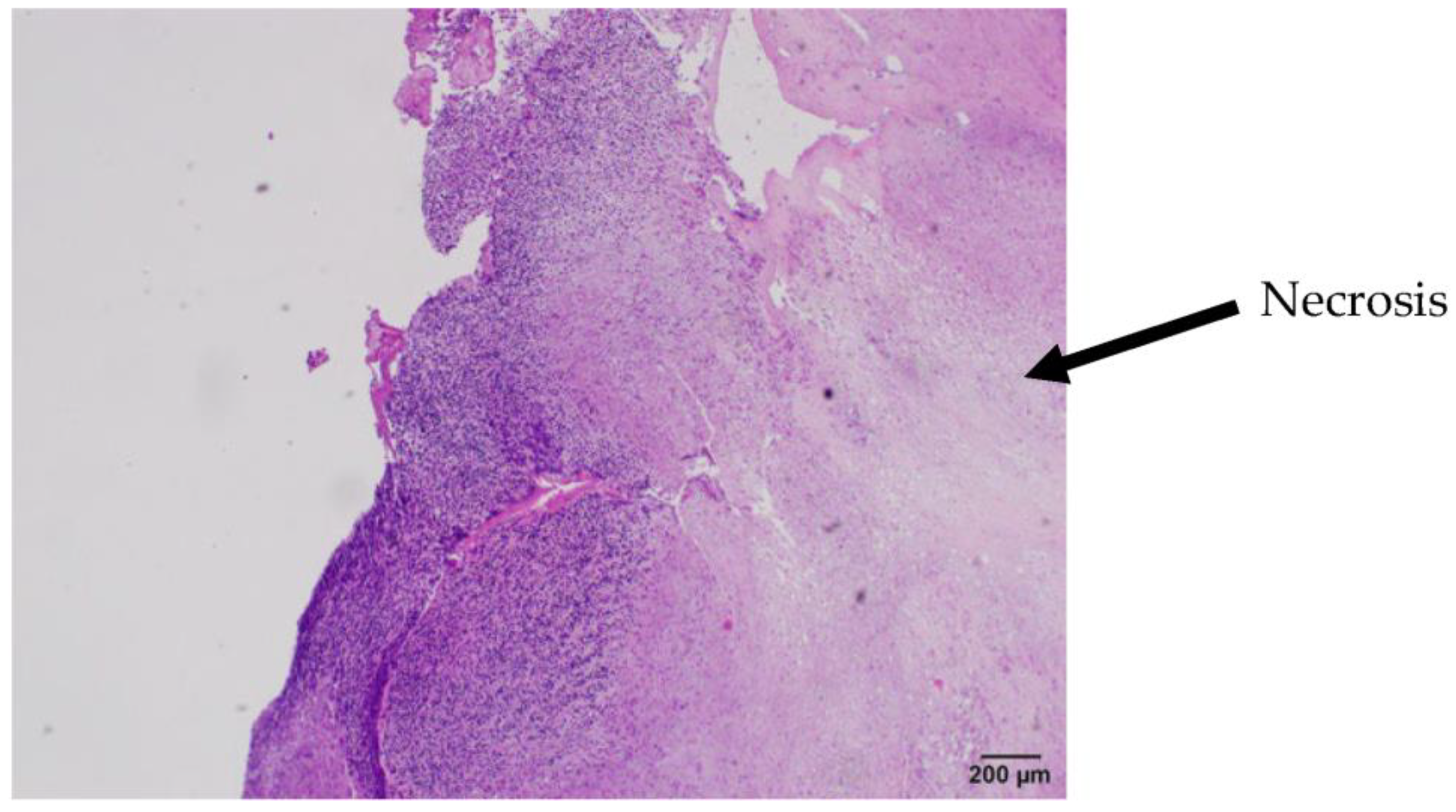
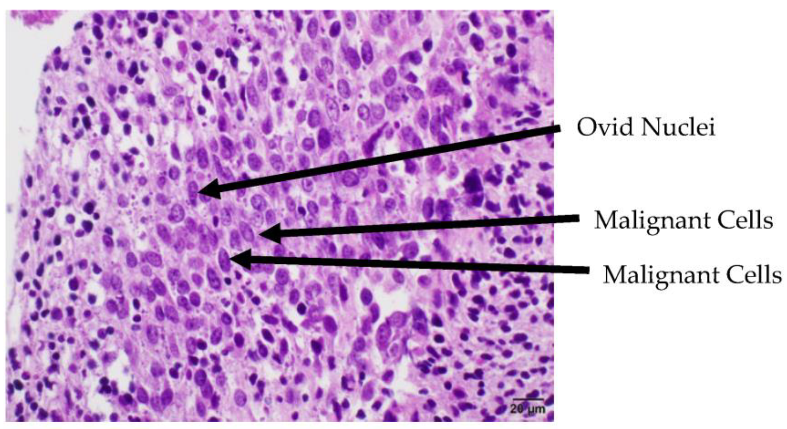
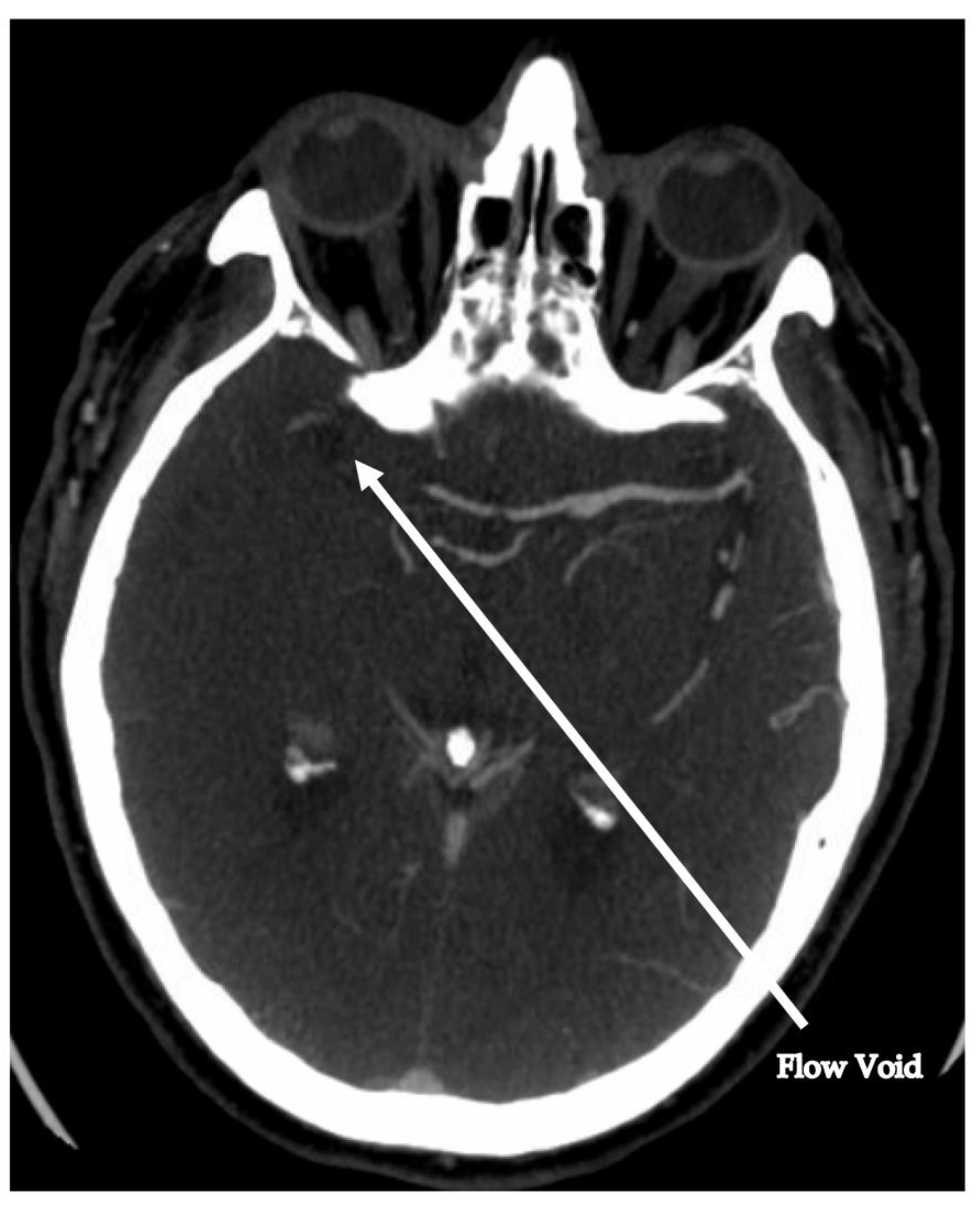
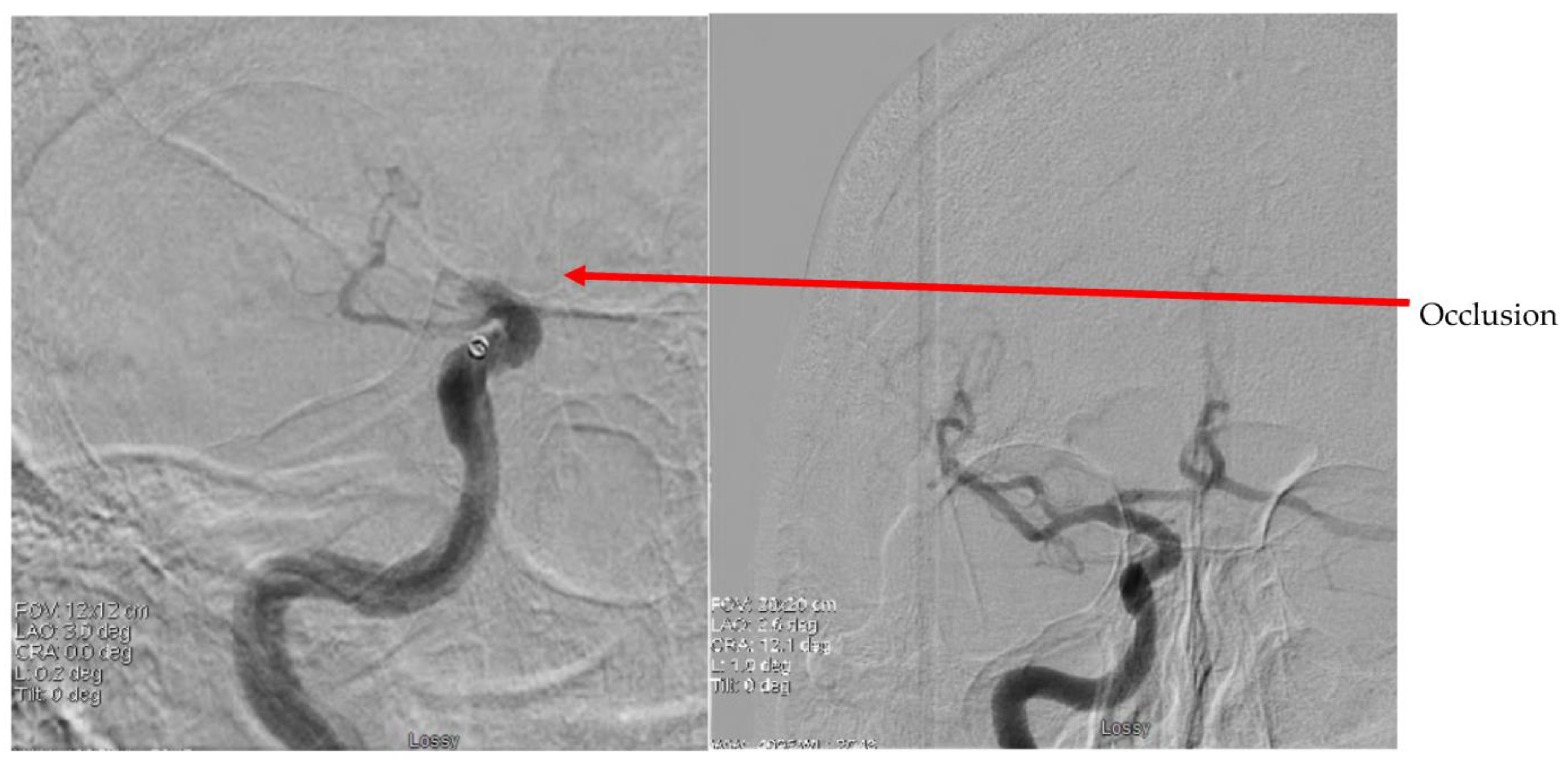
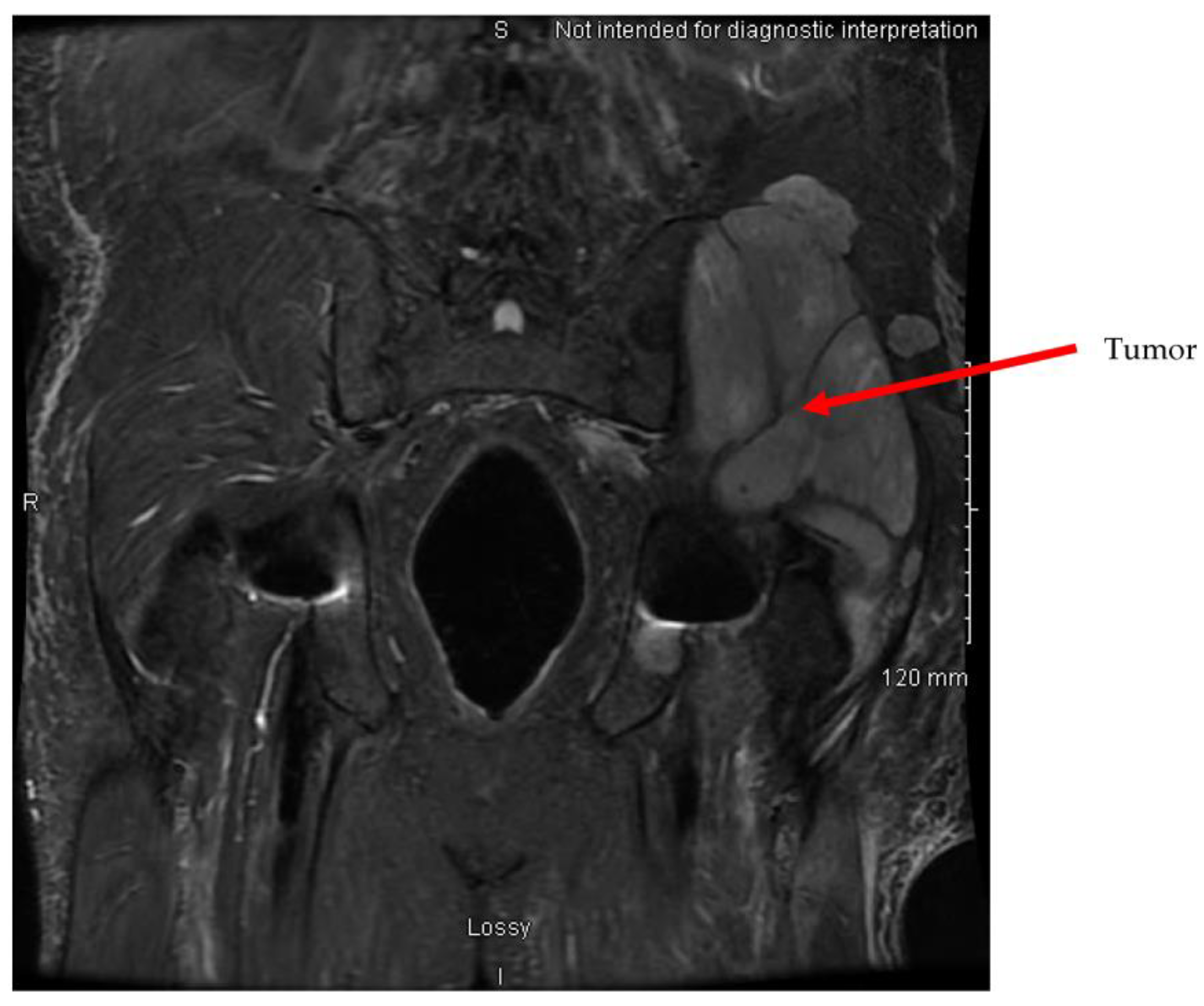
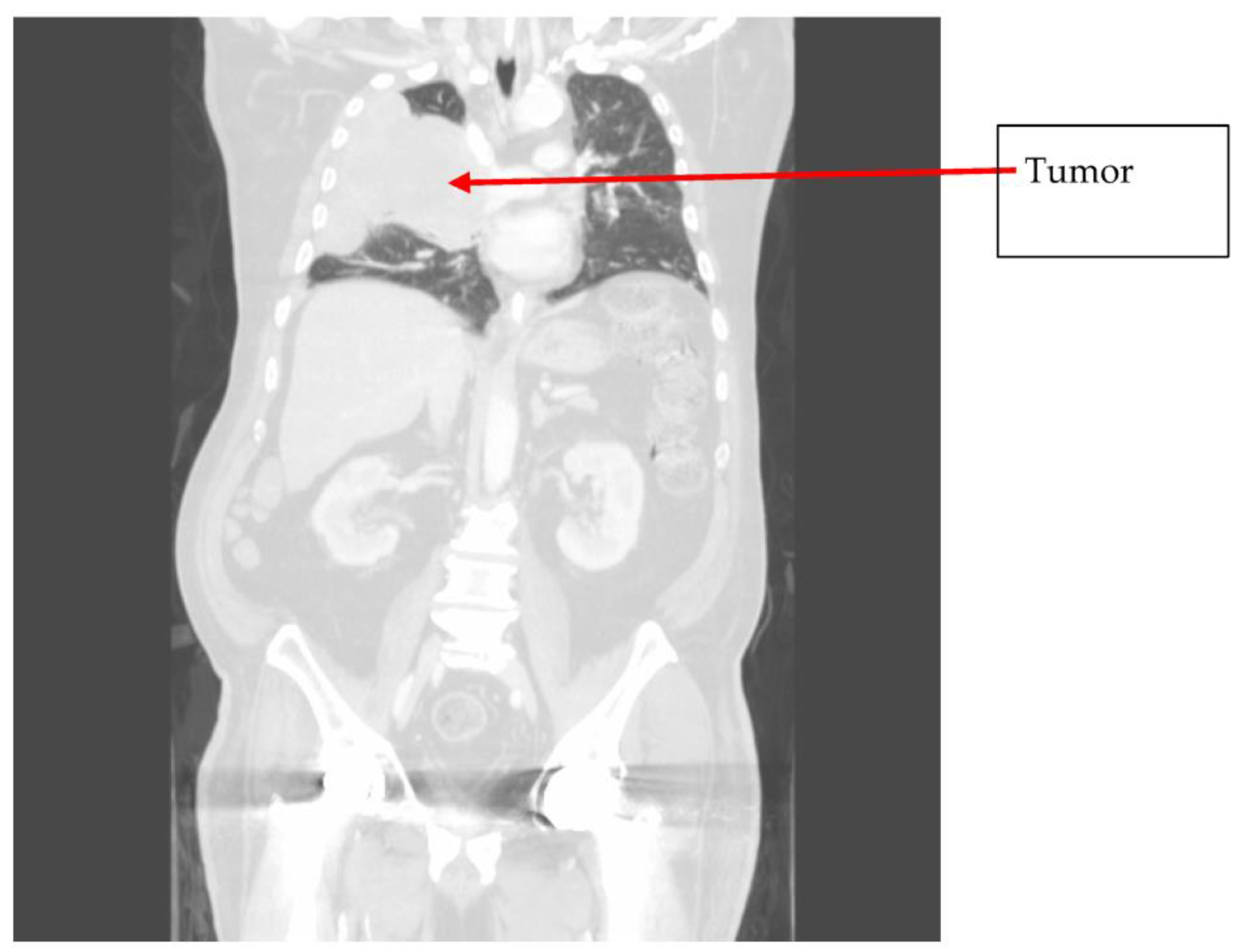
2. Case
3. Discussion
4. Conclusions
Author Contributions
Funding
Institutional Review Board Statement
Informed Consent Statement
Data Availability Statement
Conflicts of Interest
References
- Imaizumi, K.; Murate, T.; Ohno, J.; Shimokata, K. Cerebral infarction due to a spontaneous tumor embolus from lung cancer. Respiration 1995, 62, 155–156. [Google Scholar] [CrossRef] [PubMed]
- Navi, B.B.; Kawaguchi, K.; Hriljac, I.; Lavi, E.; DeAngelis, L.M.; Jamieson, D.G. Multifocal stroke from tumor emboli. Arch. Neurol. 2009, 66, 1174–1175. [Google Scholar] [CrossRef] [PubMed]
- Stergiopoulos, K.; Vasu, S.; Bilfinger, T.; Poon, M. Embolic stroke in a patient with metastatic renal cell cancer. Hellenic J. Cardiol. 2011, 52, 256–258. [Google Scholar] [PubMed]
- Grazziotin, M.U.; Turnipseed, W.D. Arterial tumor embolism caused by metastatic melanoma: Case report and literature review. J. Vasc. Surg. 2002, 36, 191–193. [Google Scholar] [CrossRef] [PubMed]
- Xiromeritis, N.; Klonaris, C.; Papas, S.; Valsamis, M.; Bastounis, E. Recurrent peripheral arterial embolism from pulmonary cancer. Case report and review of the literature. Int. Angiol. 2000, 19, 79–83. [Google Scholar]
- Singh, A.; Jenkins, D.P.; Dahdal, M.; Dhar, S.; Ratnatunga, C.P. Recurrent arterial embolization from a metastatic germ cell tumor invading the left atrium. Ann. Thorac. Surg. 2000, 70, 2155–2156. [Google Scholar] [CrossRef]
- Ramchandani, P.; Morris, M.C.; Zeit, R.M. Acute limb ischemia due to arterial embolism of tumor. Cardiovasc. Interv. Radiol. 1990, 13, 372–374. [Google Scholar] [CrossRef]
- Bloch, R.S.; Jacobs, L.A.; Lewis, L.S.; Bernys, C.F. Malignant tumor embolism: A rare presentation of malignant disease. J. Cardiovasc. Surg. 1986, 27, 630–631. [Google Scholar]
- Gómez, J.-R.; Vañó, J.; Luengo, L.; Escuder, J.; Castellote, M.; Ros, S.; Diaz, J.; Martin-Paredero, V. Tumor embolism after pneumonectomy for primary pulmonary neoplasia. Ann. Vasc. Surg. 1995, 9, 199–203. [Google Scholar] [CrossRef]
- Stein, M.E.; Drumea, K.; Ben-Itshak, O.; Hoffman, A.; Eyal, A.; Moshkovitz, B.; Haim, N. Acute limb ischemia due to malignant arterial embolism from a metastatic germ cell tumor. Med. Pediatr. Oncol. 1995, 25, 126–129. [Google Scholar] [CrossRef]
- Fernandez, B.B.; Grove, M.; Carman, T.L. An unusual presentation of simultaneous bilateral popliteal artery embolism—A case report. Angiology 1998, 49, 573–576. [Google Scholar] [CrossRef]
- Harris, R.W.; Andros, G.; Dulawa, L.B.; Oblath, R.W. Malignant melanoma embolus as a cause of acute aortic occlusion: Report of a case. J. Vasc. Surg. 1986, 3, 550–553. [Google Scholar] [CrossRef]
- ElBardissi, A.W.; Dearani, J.A.; Daly, R.C.; Mullany, C.J.; Orszulak, T.A.; Puga, F.J.; Schaff, H.V. Embolic potential of cardiac tumors and outcome after resection: A case-control study. Stroke 2009, 40, 156–162. [Google Scholar] [CrossRef]
- Reed, M.; Kerndt, C.C.; Nicolas, D. Alteplase. In StatPearls; StatPearls Publishing: Treasure Island, FL, USA, 2023. Available online: https://www.ncbi.nlm.nih.gov/books/NBK499977/ (accessed on 9 November 2023).
- Wold, L.E.; Lie, J.T. Cardiac myxomas: A clinicopathologic profile. Am. J. Pathol. 1980, 101, 219–240. [Google Scholar]
- Hart, R.G.; Diener, H.C.; Coutts, S.B.; Easton, J.D.; Granger, C.B.; O’Donnell, M.J.; Sacco, R.L.; Connolly, S.J. Embolic strokes of undetermined source: The case for a new clinical construct. Lancet Neurol. 2014, 13, 429–438. [Google Scholar] [CrossRef] [PubMed]
- Navi, B.B.; Kasner, S.E.; Elkind, M.S.V.; Cushman, M.; Bang, O.Y.; DeAngelis, L.M. Cancer and Embolic Stroke of Undetermined Source. Stroke 2021, 52, 1121–1130. [Google Scholar] [CrossRef]
- Dardiotis, E.; Aloizou, A.-M.; Markoula, S.; Siokas, V.; Tsarouhas, K.; Tzanakakis, G.; Libra, M.; Kyritsis, A.P.; Brotis, A.G.; Aschner, M.; et al. Cancer-associated stroke: Pathophysiology, detection and management (Review). Int. J. Oncol. 2019, 54, 779–796. [Google Scholar] [CrossRef]
- Gore, M.; Bansal, K.; Khan Suheb, M.Z.; Asuncion, R.M.D. Lacunar Stroke. In StatPearls; StatPearls Publishing: Treasure Island, FL, USA, 2023. Available online: https://www.ncbi.nlm.nih.gov/books/NBK563216/ (accessed on 9 November 2023).
- Graus, F.; Rogers, L.R.; Posner, J.B. Cerebrovascular complications in patients with cancer. Medicine 1985, 64, 16–35. [Google Scholar] [CrossRef] [PubMed]
- Navi, B.B.; Reiner, A.S.; Kamel, H.; Iadecola, C.; Elkind, M.S.; Panageas, K.S.; DeAngelis, L.M. Association between incident cancer and subsequent stroke. Ann. Neurol. 2015, 77, 291–300. [Google Scholar] [CrossRef]
- Navi, B.B.; Howard, G.; Howard, V.J.; Zhao, H.; Judd, S.E.; Elkind, M.S.; Iadecola, C.; DeAngelis, L.M.; Kamel, H.; Okin, P.M.; et al. New diagnosis of cancer and the risk of subsequent cerebrovascular events. Neurology 2018, 90, e2025–e2033. [Google Scholar] [CrossRef]
- Navi, B.B.; Iadecola, C. Ischemic stroke in cancer patients: A review of an underappreciated pathology. Ann. Neurol. 2018, 83, 873–883. [Google Scholar] [CrossRef] [PubMed]
- Bang, O.Y.; Chung, J.W.; Lee, M.J.; Seo, W.K.; Kim, G.M.; Ahn, M.J.; OASIS-Cancer Study Investigators. Cancer-Related Stroke: An Emerging Subtype of Ischemic Stroke with Unique Pathomechanisms. J. Stroke 2020, 22, 1–10. [Google Scholar] [CrossRef]
- Bick, R.L. Cancer-associated thrombosis. N. Engl. J. Med. 2003, 349, 109–111. [Google Scholar] [CrossRef]
- Bang, O.Y.; Chung, J.-W.; Lee, M.J.; Kim, S.J.; Cho, Y.H.; Kim, G.-M.; Chung, C.-S.; Lee, K.H.; Ahn, M.J.; Moon, G.J. Cancer Cell-Derived Extracellular Vesicles Are Associated with Coagulopathy Causing Ischemic Stroke via Tissue Factor-Independent Way: The OASIS-CANCER Study. PLoS ONE 2016, 11, e0159170. [Google Scholar] [CrossRef] [PubMed]
- Diener, H.-C.; Sacco, R.L.; Easton, J.D.; Granger, C.B.; Bernstein, R.A.; Uchiyama, S.; Kreuzer, J.; Cronin, L.; Cotton, D.; Grauer, C.; et al. Dabigatran for Prevention of Stroke after Embolic Stroke of Undetermined Source. N. Engl. J. Med. 2019, 380, 1906–1917. [Google Scholar] [CrossRef] [PubMed]
- Ntaios, G. Embolic Stroke of Undetermined Source: JACC Review Topic of the Week. J. Am. Coll. Cardiol. 2020, 75, 333–340. [Google Scholar] [CrossRef] [PubMed]
- Hart, R.G.; Connolly, S.J.; Mundl, H. Rivaroxaban for Stroke Prevention after Embolic Stroke of Undetermined Source. N. Engl. J. Med. 2018, 379, 987. [Google Scholar] [CrossRef] [PubMed]
- Gon, Y.; Okazaki, S.; Terasaki, Y.; Sasaki, T.; Yoshimine, T.; Sakaguchi, M.; Mochizuki, H. Characteristics of cryptogenic stroke in cancer patients. Ann. Clin. Transl. Neurol. 2016, 3, 280–287. [Google Scholar] [CrossRef]


Disclaimer/Publisher’s Note: The statements, opinions and data contained in all publications are solely those of the individual author(s) and contributor(s) and not of MDPI and/or the editor(s). MDPI and/or the editor(s) disclaim responsibility for any injury to people or property resulting from any ideas, methods, instructions or products referred to in the content. |
© 2024 by the authors. Licensee MDPI, Basel, Switzerland. This article is an open access article distributed under the terms and conditions of the Creative Commons Attribution (CC BY) license (https://creativecommons.org/licenses/by/4.0/).
Share and Cite
Baker, R.; Bakali, Z.; Crocker, J.S.; Mowla, A.; Smith, M.; Grossman, A.; Hagen, M.C.; Prestigiacomo, C.J.; Shirani, P. Tumor Embolic Stroke: The Importance of Pathological Assessment of Clots after Thrombectomy. J. Clin. Med. 2024, 13, 1834. https://doi.org/10.3390/jcm13071834
Baker R, Bakali Z, Crocker JS, Mowla A, Smith M, Grossman A, Hagen MC, Prestigiacomo CJ, Shirani P. Tumor Embolic Stroke: The Importance of Pathological Assessment of Clots after Thrombectomy. Journal of Clinical Medicine. 2024; 13(7):1834. https://doi.org/10.3390/jcm13071834
Chicago/Turabian StyleBaker, Richard, Zohabe Bakali, Jeffrey S. Crocker, Ashkan Mowla, Matthew Smith, Aaron Grossman, Matthew C. Hagen, Charles J. Prestigiacomo, and Peyman Shirani. 2024. "Tumor Embolic Stroke: The Importance of Pathological Assessment of Clots after Thrombectomy" Journal of Clinical Medicine 13, no. 7: 1834. https://doi.org/10.3390/jcm13071834