Article

Augmented Reality-Assisted Transcanal Endoscopic Ear Surgery for Middle Ear Cholesteatoma

Department of Otorhinolaryngology, Jikei University School of Medicine, Minato-ku, Tokyo 105-8461, Japan
*
Author to whom correspondence should be addressed.
J. Clin. Med. 2024, 13(6), 1780; https://doi.org/10.3390/jcm13061780
Submission received: 19 February 2024 / Revised: 14 March 2024 / Accepted: 15 March 2024 / Published: 20 March 2024
(This article belongs to the Special Issue Recent Advances in Otitis Media and Cholesteatoma)

Abstract

:
Background: The indications for transcanal endoscopic ear surgery (TEES) for middle ear cholesteatoma have expanded for cases involving mastoid extension. However, TEES is not indicated for all cases with mastoid extension. In addition, predicting the extent of external auditory canal (EAC) removal needed for cholesteatoma resection is not always easy. The purpose of this study was to use augmented reality (AR) to project the lesion onto an intraoperative endoscopic image to predict EAC removal requirements and select an appropriate surgical approach. Methods: In this study, patients showing mastoid extension were operated on using a navigation system with an AR function (Stryker). Results: The results showed that some cases with lesions slightly extending into the antrum required extensive resection of the EAC, while cases with lesions extending throughout the antrum required smaller resection of the EAC, indicating TEES. Conclusions: By predicting the extent of the needed EAC removal, it is possible to determine whether TEES (a retrograde approach) or canal wall-up mastoidectomy, which preserves as much of the EAC as possible, should be performed. We believe that our findings will contribute to the success of middle ear surgeries and the implementation of robotic surgery in the future.

1. Introduction

Cholesteatomas are benign collections of keratinized squamous epithelium, mostly found within the middle ear; however, they are an intractable chronic proliferative disease that can cause fatal complications, such as bone destruction and brain abscesses. The only treatment option is surgery, and the long-term recurrence rate is reported to be as high as approximately 20–60% [1,2,3,4,5,6,7]. Recurrence rates vary widely depending on the method of statistical analysis and period of observation, making it difficult to determine the optimal strategy for selecting a procedure. Nevertheless, the goal should be to ensure all lesions are removed and reconstructed while preserving as much of the normal structure as possible. The extent of the lesion, particularly its extension into the mastoid, affects the choice of the surgical approach, such as transcanal endoscopic ear surgery (TEES) or microscopic ear surgery (MES) with a postauricular incision [8]. TEES, which does not require a postauricular incision, is less invasive [9] but is not indicated for all cases. The external auditory canal (EAC) is often missing when TEES is applied in cases with mastoid extension because the EAC is drilled to approach the lesion in TEES. Excessive drilling can make ear canal reconstruction difficult and lead to an incomplete postoperative state. The ideal approach is to resect the lesion while preserving the ear canal using TEES and to ensure reconstruction of the defect. We have actively used a 70°endoscope in TEES to reduce excessive EAC drilling [10,11]. In cases that require extensive EAC drilling, even with an angled endoscope, MES, a transcortical approach in which the EAC can be preserved (canal wall-up mastoidectomy), should be applied. Imaging examinations, such as computed tomography (CT) and magnetic resonance imaging (MRI), are used to predict the extent of progress and select a surgical approach. However, this decision is not easy to make, and sometimes unintentional excessive drilling or an intraoperative change of approach is necessary, leading to prolonged operative time [11]. Therefore, we focused on using augmented reality (AR) to make optimal intraoperative decisions. AR is a generic term for advanced technologies that superimpose computer-generated images onto scenery that can be viewed with the naked eye. The term AR was first defined in 1992 [12], and AR technology applications date back to the early 1900s, with the first optical devices used to assist military personnel in shooting [13]. The AR technology currently used in the medical field is associated with navigation systems, and its basic principle is to overlay preoperative images with a conventional endoscopic view [14,15,16,17]. In addition to projecting onto endoscopic images, AR glasses can be used to project information into the clinician’s field of vision. This has already been applied in spine, hip, and skull base surgeries [18,19,20]. In otologic surgery, there have been reports of cochlear implantation using AR with robotic assistance for cadavers [21] and preoperative AR projection in lateral temporal bone surgery to confirm the location of critical structures [22]. These are all types of MES, and to date, there are no reports on the use of AR in TEES. In TEES, AR can be used more simply and practically than in MES because AR can be projected directly onto the endoscopic image. Therefore, we aimed to investigate the possibility of using AR to determine the appropriate range for EAC drilling by projecting a lesion onto an intraoperative endoscopic image. The first aim of this study was to determine whether AR lesion projection is practical for TEES. The second aim was to determine whether AR allows the surgeon to select an appropriate technique. For example, the surgeon may continue with TEES or switch to a transcortical approach with MES.

2. Materials and Methods

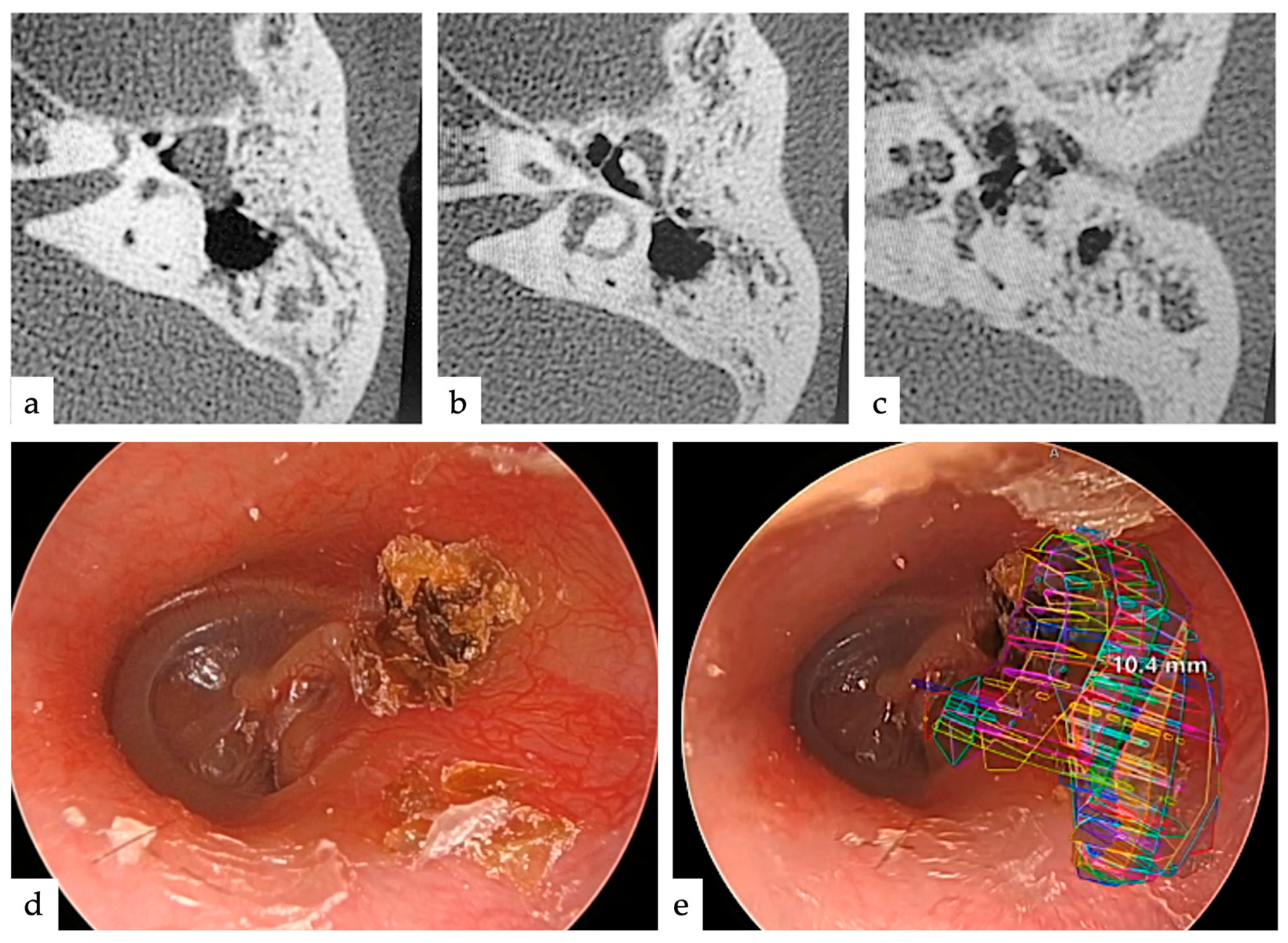
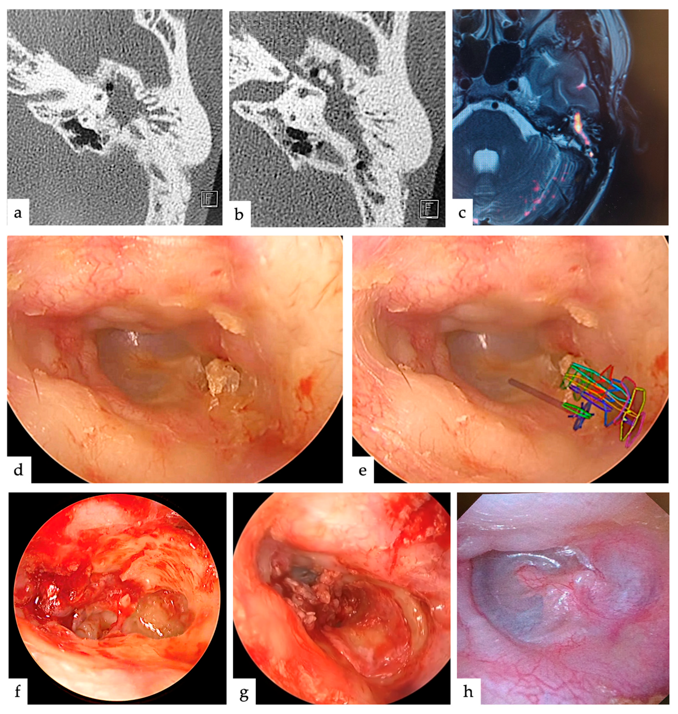
2.1. Cases and Treatment Strategies

Cases in which mastoid extension was anticipated and could be considered an indication for TEES were included, while cases with very extensive extension and no obvious indication for TEES were excluded. We performed tympanoplasty using AR in five cases of cholesteatoma from January 2023 to September 2023. Our surgical treatment strategy for cholesteatomas with mastoid extension was as follows: TEES was indicated if the cholesteatoma could be removed by drilling the lateral wall of the attic using an angled endoscope. If the lesion was extensive and wide drilling of the posterior wall of the EAC was necessary, MES with a posterior ear incision was performed to preserve the posterior wall of the EAC. In both techniques, the ear canal defect was reconstructed using cartilage.

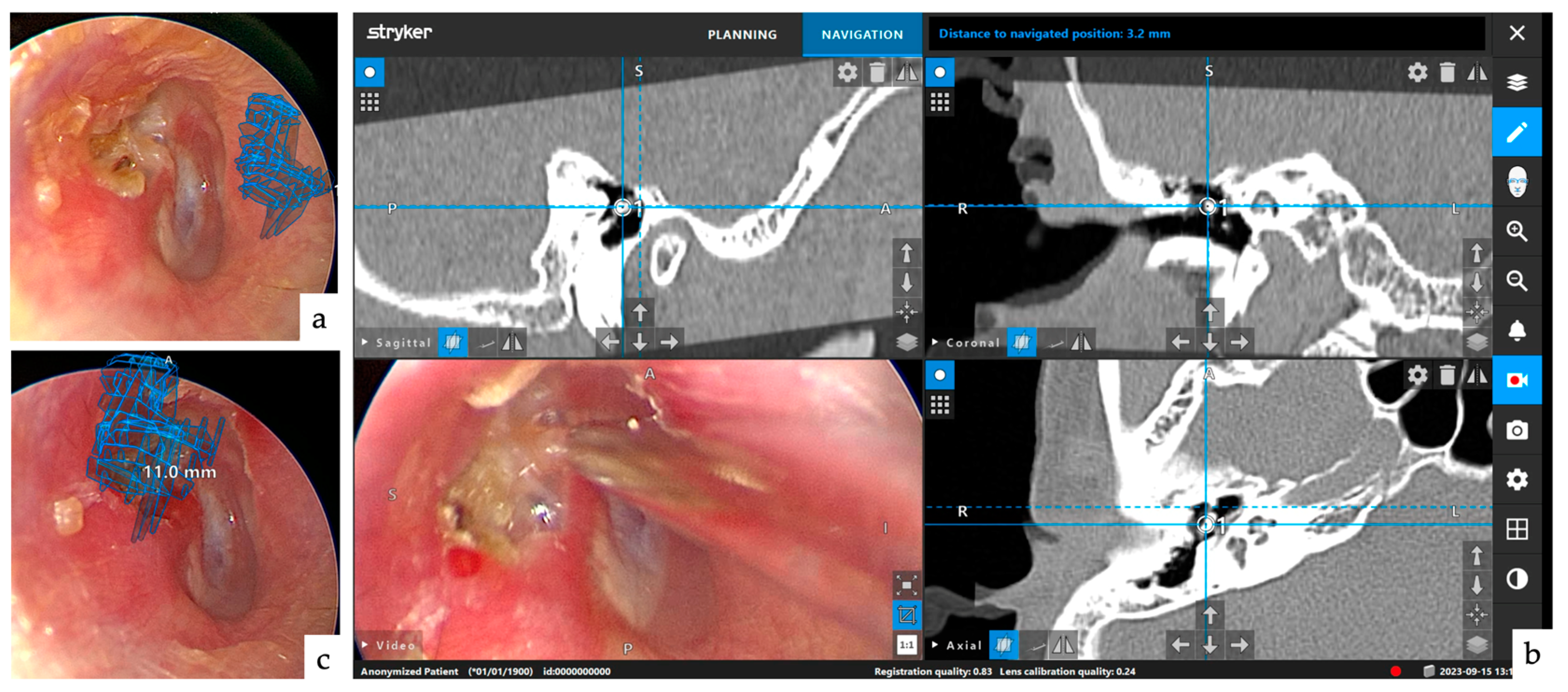
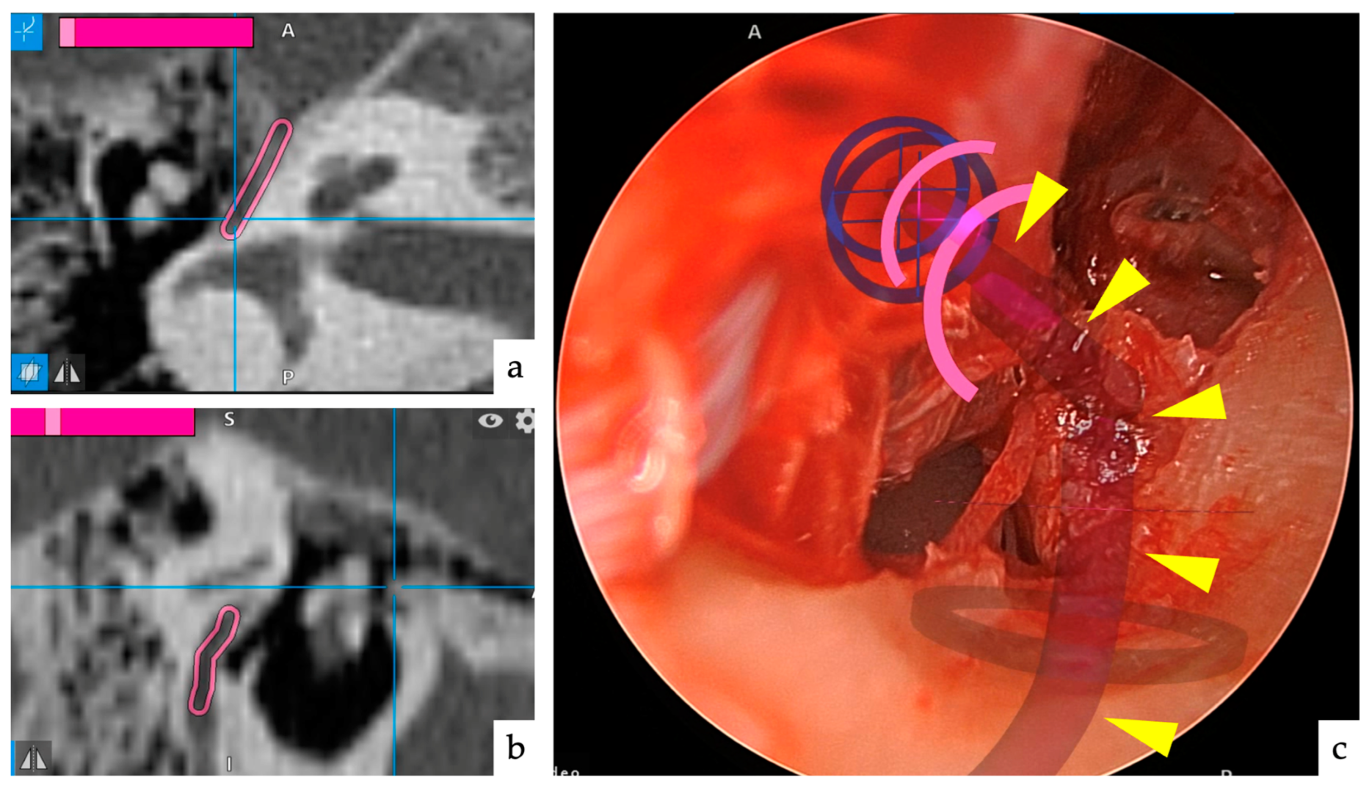
2.2. Navigation Systems
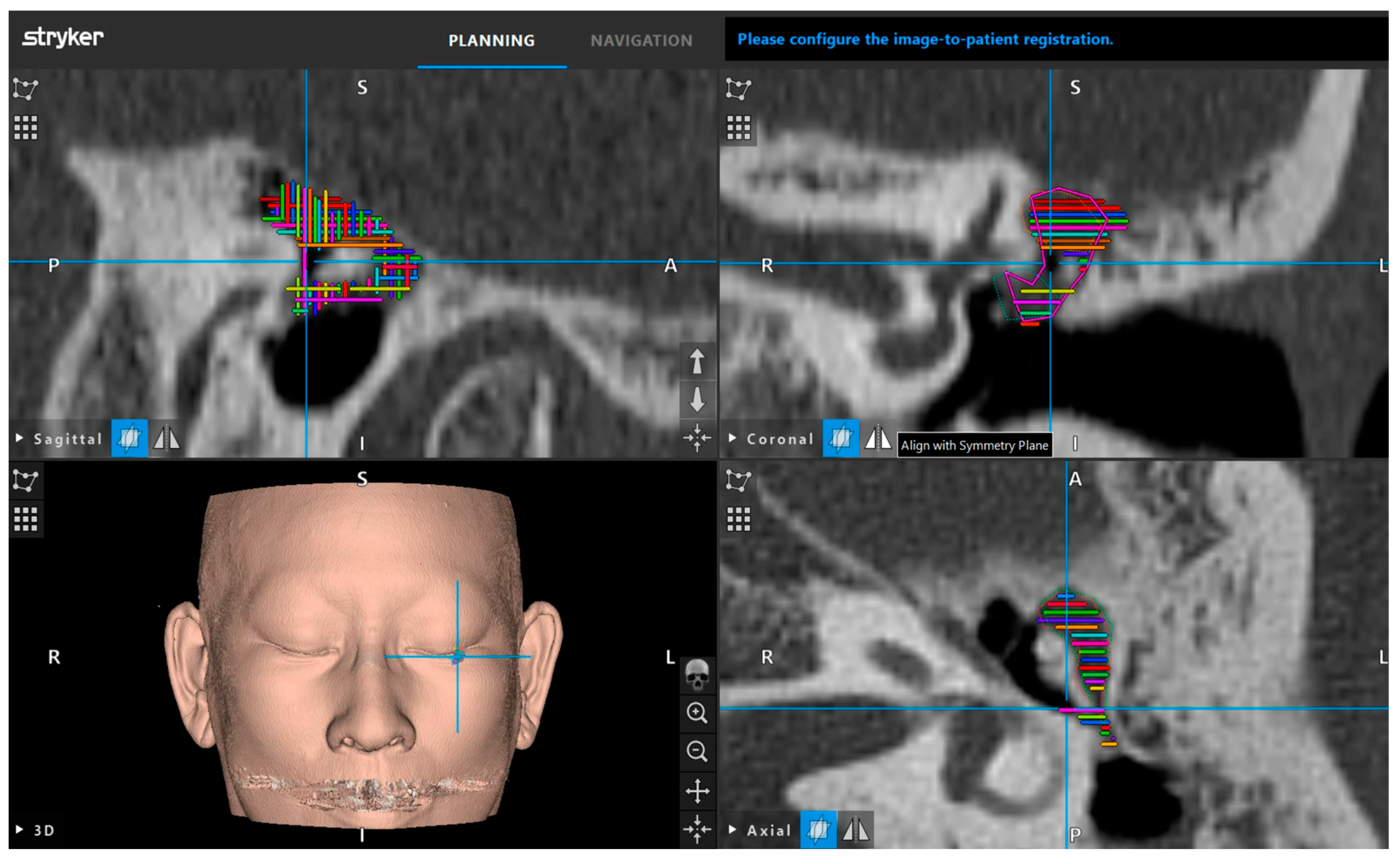
The navigation system used in this study is specialized for use in the field of otorhinolaryngology and is an electromagnetic tracking system (ENT navigationTM, Striker, Kalamazoo, MI, USA). AR images can be used by the software built into this machine. Rigid endoscopes with angles of 0°, 30°, 45°, or 70° (length: 14 cm, outer diameter: 3.1 mm; Stryker, Kalamazoo, MI, USA) for the transcanal endoscopic approach and a microscope (Zeiss OPMI Pentero 800, Carl Zeiss, Jena, Germany) for the transcortical approach were used. The endoscopes were connected to a camera head (Karl Storz Spies Image1 System TC200/TC300/TH100, Karl Storz, Tuttlingen, Germany), and a high-definition monitor was positioned in front of the surgeon.

2.3. Setup

All patients underwent CT of the temporal bone, while MRI was performed if the extent of the lesion could not be determined by CT. Lesions were marked every 1.2 mm using CT in three directions for AR imaging (Figure 1). CT without contrast was performed using a 64-MDCT scanner (SOMATOM Perspective; Siemens AG, Munich, Germany). The scanning parameters were as follows: collimation, 64 × 0.6 mm; rotation time, 1.0 s; detector-row width, 0.6 mm; pitch, 1.0; and scanning field of view (FOV), 25 cm. The peak tube voltage was maintained at 130 kV. The reconstruction parameters were as follows: section thickness, 0.6 mm, and 0.6 mm reconstruction in the axial plane. The CT threshold was adjusted using the bone algorithm (window center and width were fixed at 700 and 4000, respectively).

2.4. Registration

The field generator was placed on the cephalic side of the parietal lobe, and a reference marker was attached to the midline of the forehead to use relative coordinates. Registration was performed by tracing the facial skin using the pointer instruments provided with the navigation system (surface matching registration). Because the error increases with depth in surface matching registration, the malleus was used as a guide for correction (paired point registration). After completion of registration, the endoscope served as a navigation pointer by attaching instrument clamps to the endoscope. This made it possible to project AR images onto the endoscope screen.

2.5. Questionnaires

Ten otolaryngologists who had 5 to 33 years of experience (average 15.7 years) specializing in otologic surgery at our hospital and who utilized this strategy were questioned regarding their decision to use or not to use TEES in these cases on the basis of imaging findings, and they were also asked to show how they made the decision by presenting AR images. Four options were considered, including TEES without hesitation, probably TEES, probably MES, and MES without hesitation.

2.6. Statistical Analysis
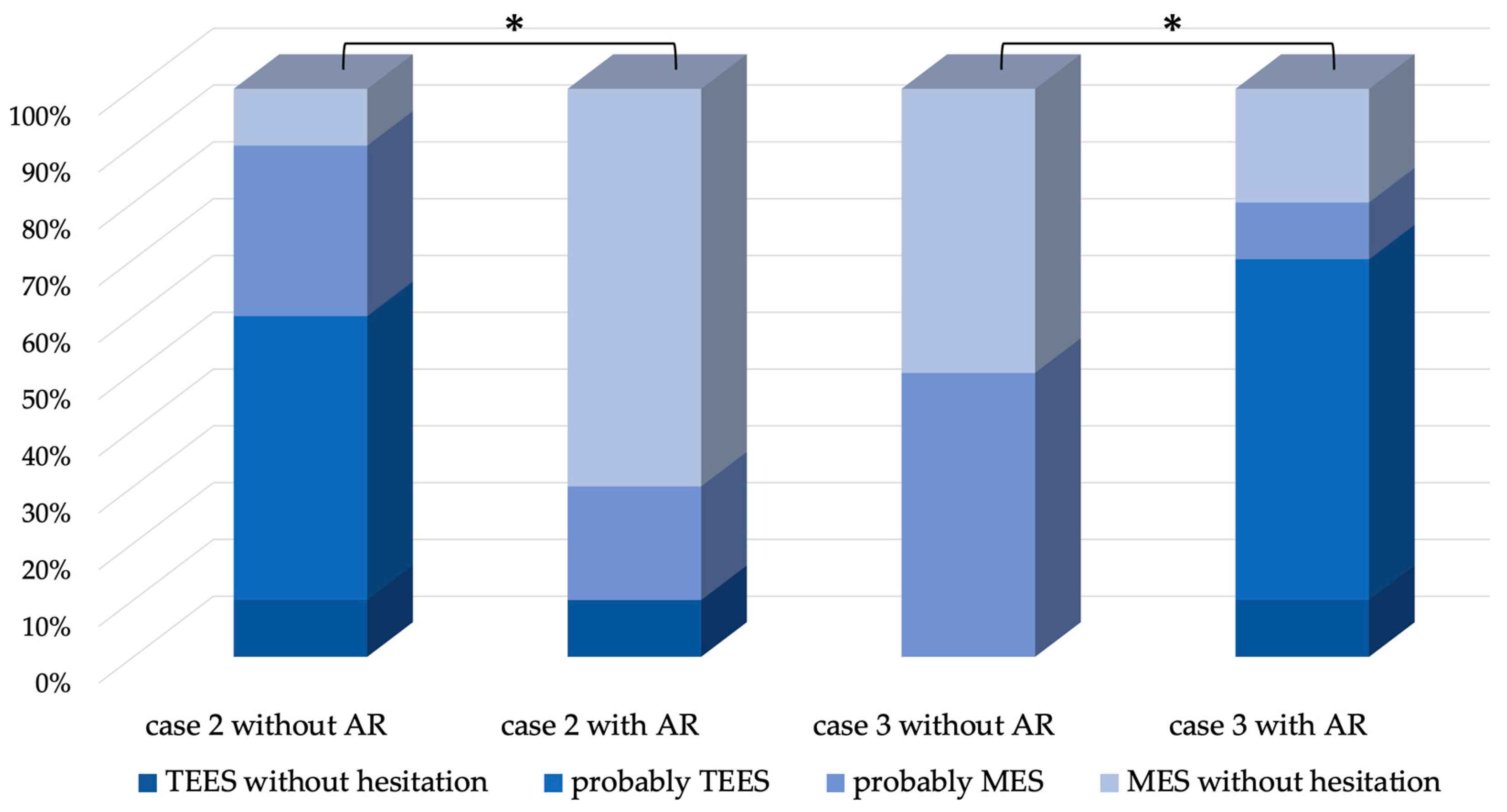
Statistical analysis was performed using a statistical software package (JMP version 13; JMP Pro 14.0.0; SAS Institute Japan, Tokyo, Japan), and the Wilcoxon signed-rank test with continuity correction was used. A p-value of less than 0.05 denoted the presence of a statistically significant difference.

3. Results

The five patients who underwent surgery were aged between 5 and 51 years, including two males and three females, and the type of cholesteatoma was pars flaccida in four and congenital in one. All cases were operated on without complications, including four treated with TEES and one treated with MES. In all cases, AR images were projected onto the endoscopic image, and the planned approach was not changed during surgery. The mean operative time was 176.2 min, which did not change significantly with the use of AR. The extent of lesion extension identified during surgery was from the attic to the mastoid in three patients, and further to the tympanic cavity in two patients (Table 1). In three of the five cases, there was no significant difference between the choice of surgical approach based on imaging evaluation and on AR; however, in two cases (Cases 2 and 3), the choice changed significantly (Figure 2, p < 0.05). One patient (Case 2) had a slight mastoid extension (Figure 3a–c); therefore, 60% of the physicians thought they would adopt TEES. However, after seeing the AR images, 70% of the physicians thought they would adopt canal wall-up mastoidectomy (MES) without hesitation because it was considered to be more extensive than EAC drilling, judging from the AR images (Figure 3d,e). In the other case (Case 3), the antrum was filled with cholesteatoma (Figure 4a–c); therefore, 100% of the physicians thought that they would adopt MES. However, the extent of EAC drilling was not so large, judging from the AR images (Figure 4d,e), that 70% of the physicians thought they would adopt TEES. The cholesteatoma could be removed by TEES, and the ear canal defect was reconstructed using a single piece of tragal cartilage (Figure 4f,g). No re-retraction pockets were observed 1 year postoperatively (Figure 4h).
The five patients, including two males and three females, were aged between 5 and 51 years. The type of cholesteatoma was pars flaccida in four patients and congenital in one patient. All cases were operated on without complications, including four treated with TEES and one treated with MES. The extent of lesion extension identified during surgery was AM in three patients and TAM in two patients.
T: tympanic cavity, A: attic, M: mastoid referred to by the European Academy of Otology and Neurotology/Japan Otological Society (EAONO/JOS) staging system [23]; TEES: trans-canal endoscopic ear surgery; MES: canal wall-up mastoidectomy using a microscope.

4. Discussion

Techniques for middle ear cholesteatomas can be broadly divided into transcanal retrograde and transcortical approaches. The former mostly uses an endoscope currently, which avoids a retro-auricular incision, although it is also applied in MES. The latter, on the other hand, often uses a microscope and can preserve the EAC in canal wall-up mastoidectomy. The indications for endoscopic ear surgery for cholesteatoma have expanded to cases with mastoid extension in addition to the traditional indications for the tympanic cavity and attic. However, not all mastoid extension cases can be treated with TEES. Moreover, it is not always easy to predict the extent of EAC drilling required to remove the lesion and decide which approach should be applied. This is also indicated by the results of the questionnaire survey conducted in this study. That is, the survey showed that different doctors made different decisions, even though they shared the same strategy.
We attempted to predict the appropriate removal range of the EAC to resect the lesion by projecting a lesion on an intraoperative endoscopic image using AR preoperatively. As a result, the AR display was used as a reference to proceed with the surgery, and in two of five cases, the use of AR changed the approach considered based on the imaging findings. If AR had not been used, the approach would have to be changed during surgery, unnecessarily destroying normal structures, such as the EAC, and requiring more surgical time. For example, in Case 2, the cholesteatoma was localized in the mastoid cavity on preoperative images and was considered an indication for transcanal attico-antorotomy using a powered device [8,24]. However, AR projection at the time of surgery revealed that the lesion was located deep in the antrum, and extensive posterior wall drilling of the EAC was required to remove the cholesteatoma. Therefore, a transcortical approach was selected at the beginning of the surgery, and the posterior wall of the EAC was preserved. In contrast, in Case 3, although the cholesteatoma was extensive on the images, it was confirmed that a wide osteotomy was unnecessary based on the AR image. Because the area of reconstruction was small (Figure 4f), the EAC defect was completely and easily reconstructed with a single piece of tragal cartilage (Figure 4g), with favorable postoperative findings (Figure 4h). The main reason for this difference is that the size of the attic and antrum vary from person to person. The extent of EAC removal may depend not only on its size but also on its positional relationship with other structures, such as the width and angle of the EAC.
Tympanoplasty, a reconstructive procedure of the eardrum and ossicles, necessitates precise and minimal removal of the affected areas to maintain hearing. Minimizing the extent of osteotomy is required to ensure reconstruction of the posterior wall of the EAC and the lateral wall of the attic for cholesteatoma treatment [10], but this is not easy with conventional tympanoplasty because the location of the target lesion is unclear during surgery. The integration of AR technology enables surgeons to objectively visualize lesions and select the most appropriate surgical technique during the surgery.
While imaging of the position of instruments matched with image data is a basic function of navigation systems in various fields, newer systems allow for additional features such as integrated elements of AR, image fusion, acoustic warning signals, and the use of intraoperative CT [25,26,27,28,29]. The application of AR in otorhinolaryngology, particularly in endoscopic sinus surgery (ESS), has been previously reported [30], and its role in MES for cochlear implantation has also been explored [31]. However, the use of AR in TEES and cholesteatoma has not been reported, making our study pioneering in this field. One concern regarding the use of navigation systems, including AR, in temporal bone surgery is their accuracy. Many structures, such as nerves and blood vessels, are located within the temporal bone, and their anatomy is such that they cannot be touched or observed until bone drilling, such as mastoidectomy, is performed. In addition, important organs, such as the cochlea, as well as vestibular and facial nerves, are densely located within a small area, with large individual differences in their movements; therefore, a highly accurate navigation system is required to ensure safety. The surface-matching registration method commonly used in sinus surgery is simple and noninvasive. However, its accuracy varies widely, with an error margin of approximately 2 mm [32]. In temporal bone surgery, changes in skin surface coordinates due to head torsion cause further variations in accuracy, and unacceptable levels of error are likely to occur in the deep temporal bone area. A paired-point method, in which a titanium screw is implanted in the bone surface in advance and CT scans are taken for registration, has been developed to improve accuracy. Although this method improves accuracy, it is psychologically and physically burdensome for patients because it requires an invasive procedure with patients in the awake state before surgery [33,34]. In this study, ENT navigationTM (Striker, Kalamazoo, MI, USA), which allows for intraoperative modifications, was used when the surface-matching method did not provide sufficient accuracy. In cases where high accuracy was achieved at the surface but errors were noticeable at depths, such as in the tympanic cavity, high accuracy was achieved by re-registering the lateral process of the malleus as a reference (Figure 5a–c). The lateral process of the malleus was chosen as the reference because of its proximity to the site to be considered and because its position is unlikely to change with diseases or surgical manipulations. This modification was important because it allowed for reproducibility.
We have shown that a navigation system using AR can minimize osteotomies to preserve the normal structure in tympanoplasty, and objectively select the appropriate technique intraoperatively. To effectively use AR, proper preoperative diagnosis by CT/MRI is important, especially for the accurate evaluation of the range of cholesteatoma extension from the attic to the mastoid cavity to select the appropriate surgical approach. In order to accurately evaluate images, it is necessary to predict the extent of mastoid cavity extension based on the magnification of the mastoid cavity opening [35], the prediction by the temporal subtraction method [36], and the prediction of mastoid cavity extension by artificial intelligence (AI) [37]. These are based on CT images. However, despite the issues of the costs of MRI facilities and imaging, MRI is very useful [38,39].
Despite the potential of AR to be applied to any temporal bone surgery, a challenge exists in AR-assisted surgery, which is the lack of three-dimensional features. Although AR is effective in identifying the site of the lesion, it does not reveal the depth of normal structures that require attention, such as facial nerves and blood vessels, and thus, the full potential of AR cannot be exploited (Figure 6a–c). However, because endoscopic images are originally 2D and their orientation can be determined, they may be of great value. Therefore, we believe that when robotic surgery is introduced in otology in the future, it will be performed with AR assistance. This is because robotic surgery, which has difficulty utilizing the surgeon’s hand sensations, requires more information to be obtained from the monitor.
Other challenges are the time required and the cost. The navigation unit is expensive, and peripheral equipment must also be purchased. CT must be used more extensively than usual for navigation, although special imaging conditions are not required to use AR images. Extra time is required to preoperatively plot the lesion, although this can be done in 10–15 min once the doctor is accustomed to the process. In addition to the usual registration of the probe, a registration of the endoscope must also be performed. Therefore, at this time, AR is used for limited cases; however, once the process is further simplified, it will be used for all cases.
This study has some limitations. First, the sample size was small (five patients) and included both pediatric and adult patients with different types of cholesteatoma. To make a universal statement, we must increase the number of cases studied. The pathogenesis of cholesteatoma varies widely, and there are naturally significant differences between congenital and acquired lesions. However, for the following reasons, we decided to present this report in its developing state: We had intended to report our findings after having examined many more cases; however, early on, we encountered a case in which the preoperative prediction and the actual surgical technique differed. The use of AR is promising, and there are indeed cases that differ from preoperative predictions. We believe that the difference will not be significant, regardless of the number of cases we include to demonstrate these points. Even if the pathology and etiology differ, the goal of complete removal of the lesion while preserving as much of the normal structure as possible remains (although the method of reconstruction may differ). In this study, we focused on cases that could benefit from TEES; we believe that it is problematic when extensive bone resection or posterior auricular incision is required despite TEES being scheduled. Second, ideally, the surgical approach should be determined using AR during the outpatient consultation phase in the process of surgical scheduling, which is, however, impossible because this is a navigation-based technique and registration is required. Third, this study was based on the assumption that our surgical strategy was the best option. Despite the variety of surgical techniques available, this report was limited to canal wall-up (TEES + transcortical mastoidectomy) or TEES with minimal EAC drilling. Although these should prove to be the best, they are difficult to achieve. As previous reports of surgical results show, the pathophysiology of cholesteatoma is so diverse that it is difficult to single out one strategy as the “best” one. The main goal in the treatment of cholesteatoma is to reduce the recurrence rate. Although residual disease has been shown to decrease with the use of endoscopes [40], recurrent disease remains a problem [2,3,4,5]. Thus, the idea of more reliable reconstruction is certainly an important strategy. It would be natural to aim for reliable resection of the lesion while preserving as much normal structure as possible for this purpose.

5. Conclusions

We report for the first time the use of AR imaging in TEES and show that AR-assisted surgery, which can minimize EAC drilling, is useful in TEES and facilitates the intraoperative selection of the most suitable technique. Although the number of cases was small in this study, we demonstrated that AR may change the approach considered preoperatively based on imaging findings. Further studies should be conducted to find a more reliable and simple registration method for better imaging of structures that require attention. Although there are still many issues to be addressed before AR can be used as a standard, this is an area where further growth is expected.

Author Contributions

Conceptualization, M.T. and Y.Y.; formal analysis, M.T., K.T. and Y.Y.; investigation, M.T., K.T., T.N., S.K., K.Y., Y.Y. and H.K.; writing—original draft preparation, M.T. and K.T.; writing—review and editing, M.T., K.T., T.N., S.K., K.Y., Y.Y. and H.K. All authors have read and agreed to the published version of the manuscript.

Funding

This study was supported by the Japan Society for the Promotion of Science (JSPS) KAKENHI (grant number 22K16936).

Institutional Review Board Statement

The study was conducted in accordance with the Declaration of Helsinki, and approved by the Human Ethics Review Committee of Jikei University School of Medicine (approval number: 32-036 [10111], approval date: 18 May 2020).

Informed Consent Statement

Informed consent was obtained from all subjects involved in the study.

Data Availability Statement

Data are available upon reasonable request to the corresponding authors.

Acknowledgments

I would like to thank the people who helped me conduct this research. I thank my supervisors, N. Otori and Y. Sakurai, for their support and encouragement, respectively.

Conflicts of Interest

The authors declare no conflicts of interest directly relevant to the content of this article.

References

  1. Hashimoto-Ikehara, M.; Mishiro, Y.; Kitahara, T.; Sakagami, M. The 10-year disease-free rate of attic cholesteatoma based on a new staging system. J. Int. Adv. Otol. 2011, 7, 289–292. [Google Scholar]
  2. Nakazawa, T.; Yamamoto, Y.; Kanno, K.; Kurihara, S.; Takahashi, M.; Morino, T.; Motegi, M.; Yamamoto, K.; Sakurai, Y.; Kojima, H. Time analysis of recidivism in acquired cholesteatoma using the Kaplan-Meier method. Otol. Neurotol. 2023, 44, e393–e397. [Google Scholar] [CrossRef] [PubMed]
  3. Stangerup, S.E.; Drozdziewicz, D.; Tos, M.; Hougaard-Jensen, A. Recurrence of attic cholesteatoma: Different methods of estimating recurrence rates. Otolaryngol. Head Neck Surg. 2000, 123, 283–287. [Google Scholar]
  4. Mishiro, Y.; Sakagami, M.; Kitahara, T.; Kondoh, K.; Okumura, S. The investigation of the recurrence rate of cholesteatoma using Kaplan-Meier survival analysis. Otol. Neurotol. 2008, 29, 803–806. [Google Scholar] [CrossRef]
  5. Møller, P.R.; Pedersen, C.N.; Grosfjeld, L.R.; Faber, C.E.; Djurhuus, B.D. Recurrence of cholesteatoma—A retrospective study including 1,006 patients for more than 33 years. Int. Arch. Otorhinolaryngol. 2020, 24, e18–e23. [Google Scholar] [CrossRef] [PubMed]
  6. Eggink, M.C.; de Wolf, M.J.F.; Ebbens, F.A.; Dikkers, F.G.; van Spronsen, E. Assessing the prognostic value of the ChOLE classification in predicting the severity of acquired cholesteatoma. Otol. Neurotol. 2022, 43, 472–480. [Google Scholar] [CrossRef]
  7. Presutti, L.; Anschuetz, L.; Rubini, A.; Ruberto, M.; Alicandri-Ciufelli, M.; Dematte, M.; Caversaccio, M.; Marchioni, D. The impact of the transcanal endoscopic approach and mastoid preservation on recurrence of primary acquired attic cholesteatoma. Otol. Neurotol. 2018, 39, 445–450. [Google Scholar] [CrossRef]
  8. Kakehata, S.; Watanabe, T.; Ito, T.; Kubota, T.; Furukawa, T. Extension of indications for transcanal endoscopic ear surgery using an ultrasonic bone curette for cholesteatomas. Otol. Neurotol. 2014, 35, 101–107. [Google Scholar] [CrossRef]
  9. Kakehata, S.; Furukawa, T.; Ito, T.; Kubota, T.; Futai, K.; Watanabe, T. Comparison of postoperative pain in patients following transcanal endoscopic versus microscopic ear surgery. Otol. Neurotol. 2018, 39, 847–853. [Google Scholar] [CrossRef]
  10. Takahashi, M.; Motegi, M.; Yamamoto, K.; Yamamoto, Y.; Kojima, H. Quantitative study of bone removal region in transcanal endoscopic approach to the attic and antrum using a 70° endoscope. J. Int. Adv. Otol. 2022, 18, 232–235. [Google Scholar] [CrossRef]
  11. Takahashi, M.; Yamamoto, Y.; Kojima, H. Transcanal endoscopic approach for pars flaccida cholesteatoma using a 70-degree angled endoscope. Eur. Arch. Otorhinolaryngol. 2021, 278, 1283–1288. [Google Scholar] [CrossRef] [PubMed]
  12. Caudell, T.; Mizell, D. Augmented reality: An application of heads-up display technology to manual manufacturing processes. In Proceedings of the Twenty-Fifth Hawaii International Conference on System Sciences, Kauai, HI, USA, 7–10 January 1992. [Google Scholar] [CrossRef]
  13. Howard, G. A new collimating-telescope gunsight for large and small ordnance. Sci. Trans. R. Dublin Soc. 1901, 7, 321–330. [Google Scholar]
  14. Citardi, M.J.; Batra, P.S. Intraoperative surgical navigation for endoscopic sinus surgery: Rationale and indications. Curr. Opin. Otolaryngol. Head Neck Surg. 2007, 15, 23–27. [Google Scholar] [CrossRef] [PubMed]
  15. Besharati Tabrizi, L.; Mahvash, M. Augmented reality-guided neurosurgery: Accuracy and intraoperative application of an image projection technique. J. Neurosurg. 2015, 123, 206–211. [Google Scholar] [CrossRef] [PubMed]
  16. Suzuki, N.; Hattori, A.; Iimura, J.; Otori, N.; Onda, S.; Okamoto, T.; Yanaga, K. Development of AR surgical navigation systems for multiple surgical regions. Stud. Health Technol. Inform. 2014, 196, 404–408. [Google Scholar] [PubMed]
  17. Winne, C.; Khan, M.; Stopp, F.; Jank, E.; Keeve, E. Overlay visualization in endoscopic ENT surgery. Int. J. Comput. Assist. Radiol. Surg. 2011, 6, 401–406. [Google Scholar] [CrossRef] [PubMed]
  18. Ghaednia, H.; Fourman, M.S.; Lans, A.; Detels, K.; Dijkstra, H.; Lloyd, S.; Sweeney, A.; Oosterhoff, J.H.F.; Schwab, J.H. Augmented and virtual reality in spine surgery, current applications and future potentials. Spine J. 2021, 21, 1617–1625. [Google Scholar] [CrossRef]
  19. Sun, P.; Zhao, Y.; Men, J.; Ma, Z.-R.; Jiang, H.-Z.; Liu, C.-Y.; Feng, W. Application of virtual and augmented reality technology in hip surgery: Systematic review. J. Med. Internet Res. 2023, 25, e37599. [Google Scholar] [CrossRef]
  20. Begagic, E.; Bečulić, H.; Pugonja, R.; Memić, Z.; Balogun, S.; Džidić-Krivić, A.; Milanović, E.; Salković, N.; Nuhović, A.; Skomorac, R.; et al. Augmented reality integration in skull base neurosurgery: A systematic review. Medicina 2024, 60, 335. [Google Scholar] [CrossRef]
  21. Liu, W.P.; Azizian, M.; Sorger, J.; Taylor, R.H.; Reilly, B.K.; Cleary, K.; Preciado, D. Cadaveric feasibility study of da Vinci Si–assisted cochlear implant with augmented visual navigation for otologic surgery. JAMA Otolaryngol. Head Neck Surg. 2014, 140, 208–214. [Google Scholar] [CrossRef]
  22. Schwam, Z.G.; Kaul, V.F.; Bu, D.D.; Iloreta, A.-M.C.; Bederson, J.B.; Perez, E.; Cosetti, M.K.; Wanna, G.B. The utility of augmented reality in lateral skull base surgery: A preliminary report. Am. J. Otolaryngol. 2021, 42, 102942. [Google Scholar] [CrossRef] [PubMed]
  23. Yung, M.; Tono, T.; Olszewska, E.; Yamamoto, Y.; Sudhoff, H.; Sakagami, M.; Mulder, J.; Kojima, H.; İncesulu, A.; Trabalzini, F.; et al. EAONO/JOS Joint Consensus statements on the definitions, classification and staging of middle ear cholesteatoma. J. Int. Adv. Otol. 2017, 13, 1–8. [Google Scholar] [CrossRef] [PubMed]
  24. Ito, T.; Mochizuki, H.; Watanabe, T.; Kubota, T.; Furukawa, T.; Koike, T.; Kakehata, S. Safety of ultrasonic bone curette in ear surgery by measuring skull bone vibrations. Otol. Neurotol. 2014, 35, e135–e139. [Google Scholar] [CrossRef] [PubMed]
  25. Batra, P.S.; Kanowitz, S.J.; Citardi, M.J. Clinical utility of intraoperative volume computed tomography scanner for endoscopic sinonasal and skull base procedures. Am. J. Rhinol. 2008, 22, 511–515. [Google Scholar] [CrossRef] [PubMed]
  26. Dixon, B.J.; Daly, M.J.; Chan, H.; Vescan, A.; Witterick, I.J.; Irish, J.C. Augmented real-time navigation with critical structure proximity alerts for endoscopic skull base surgery. Laryngoscope 2014, 124, 853–859. [Google Scholar] [CrossRef] [PubMed]
  27. Jackman, A.H.; Palmer, J.N.; Chiu, A.G.; Kennedy, D.W. Use of intraoperative CT scanning in endoscopic sinus surgery: A preliminary report. Am. J. Rhinol. 2008, 22, 170–174. [Google Scholar] [CrossRef] [PubMed]
  28. Leong, J.L.; Batra, P.S. Citardi MJ. CT-MRT image fusion for the management of skull base lesions. Otolaryngol. Head Neck Surg. 2006, 134, 868–876. [Google Scholar] [CrossRef] [PubMed]
  29. Li, L.; Yang, J.; Chu, Y.; Wu, W.; Xue, J.; Liang, P.; Chen, L. A novel augmented reality navigation system for endoscopic sinus and skull base surgery: A feasibility study. PLoS ONE 2016, 11, e0146996. [Google Scholar] [CrossRef]
  30. Maximilian, L. Augmented Reality-Enhanced Navigation in Endoscopic Sinus Surgery: A Prospective, Randomized, Controlled Clinical Trial. Laryngoscope Investig. Otol. 2020, 5, 621–629. [Google Scholar]
  31. Guigou, C.; Hussain, R.; Lalande, A.; Bozorg Grayeli, A. Augmented reality based transmodiolar cochlear implantation. Otol. Neurotol. 2022, 43, 190–198. [Google Scholar] [CrossRef]
  32. Miller, R.S.; Hashisaki, G.T.; Kesser, B.W. Image-guided localization of the internal auditory canal via the middle cranial fossa approach. Otolaryngol. Head Neck Surg. 2006, 134, 778–782. [Google Scholar] [CrossRef]
  33. Metzger, M.C.; Rafii, A.; Holhweg-Majert, B.; Pham, A.M.; Strong, B. Comparison of 4 registration strategies for computer-aided maxillofacial surgery. Otolaryngol. Head Neck Surg. 2007, 137, 93–99. [Google Scholar] [CrossRef]
  34. Kohan, D.; Jethanamest, D. Imageguided surgical naevigation in otology. Laryngoscope 2012, 122, 2291–2299. [Google Scholar] [CrossRef]
  35. Baba, A.; Kurihara, S.; Ogihara, A.; Matsushima, S.; Yamauchi, H.; Ikeda, K.; Kurokawa, R.; Ota, Y.; Takahashi, M.; Sakurai, Y.; et al. Preoperative predictive criteria for mastoid extension in pars flaccida cholesteatoma in assessments using temporal bone high-resolution computed tomography. Auris Nasus Larynx 2021, 48, 609–614. [Google Scholar] [CrossRef]
  36. Baba, A.; Kurokawa, R.; Kurokawa, M.; Ota, Y.; Matsushima, S.; Fukuda, T.; Yamauchi, H.; Kano, R.; Shoji, T.; Kurihara, S.; et al. Preoperative prediction for mastoid extension of middle ear cholesteatoma using temporal subtraction serial HRCT studies. Eur. Radiol. 2022, 32, 3631–3638. [Google Scholar] [CrossRef] [PubMed]
  37. Takahashi, M.; Noda, K.; Yoshida, K.; Tsuchida, K.; Yui, R.; Nakazawa, T.; Kurihara, S.; Baba, A.; Motegi, M.; Yamamoto, K.; et al. Preoperative prediction by artificial intelligence for mastoid extension in pars flaccida cholesteatoma using temporal bone high-resolution computed tomography: A retrospective study. PLoS ONE 2022, 17, e0273915. [Google Scholar] [CrossRef] [PubMed]
  38. Baba, A.; Kurihara, S.; Fukuda, T.; Yamauchi, H.; Matsushima, S.; Ikeda, K.; Kurokawa, R.; Ota, Y.; Takahashi, M.; Sakurai, Y.; et al. Non-echoplanar diffusion weighed imaging and T1-weighted imaging for cholesteatoma mastoid extension. Auris Nasus Larynx 2021, 48, 846–851. [Google Scholar] [CrossRef] [PubMed]
  39. Migirov, L.; Wolf, M.; Greenberg, G.; Eyal, A. Non-EPI DW MRI in planning the surgical approach to primary and recurrent cholesteatoma. Otol. Neurotol. 2014, 35, 121–125. [Google Scholar] [CrossRef]
  40. Noschang Lopes da Silva, M.; Selaimen da Costa, S.; André Selaimen, F.; da Costa Huve, F.; Lang Silva, A.; Dias Toshiaki Koga, F.; Martins-Costa, L.L.; Bernard Rosa Nery, M.; Zanardini, M.; Sperling, N. Residual cholesteatoma after endoscopic-assisted canal wall-up tympanomastoidectomy: A randomized controlled trial. Otol. Neurotol. 2022, 43, 803–807. [Google Scholar] [CrossRef] [PubMed]
Figure 1. Navigation monitor after completion of tracing of lesion. The lesion was traced every 1.2 mm in three computed tomography (CT) directions and was ready for the augmented reality (AR) display after pre-planning was performed. The upper-left, upper-right, and lower-right images are sagittal, coronal, and axial slices, respectively. The tracing colors are freely selectable, and a variety of colors were used in this case.
Jcm 13 01780 g001
Figure 2. Questionnaire results of the two cases (Cases 2 and 3) in which the planned approach changed based on the AR display. The surgical approach in Case 2 and Case 3 changed significantly due to the AR display (*: p < 0.05). In Case 2, the proportion of performing MES increased significantly, while in Case 3, the proportion of performing TEES increased significantly. AR: augmented reality; MES: microscopic ear surgery; TEES: transcanal endoscopic ear surgery.
Jcm 13 01780 g002
Figure 3. Imaging findings and AR display in Case 2. The AR display shows that the area of EAC drilling to be performed is large, whereas the area of progress is not very large. (a) Axial CT slice of the temporal bone showing a slight extension into the antrum. (b) Axial CT slice of the temporal bone showing extension into the attic. (c) Axial CT slice of the temporal bone showing extension into the tympanic cavity. The lesion extended close to the stapes; however, the superstructure did not exhibit any erosive changes. (d) Typical endoscopic findings of cholesteatoma with debris in the pars flaccida. (e) Endoscopic image with AR display overlaid on image (d). Extensive EAC drilling was required to remove the lesions. AR: augmented reality; EAC: external auditory canal; CT: computed tomography.
Jcm 13 01780 g003
Figure 4. Imaging findings and AR display in Case 3. The AR display shows that the area of the EAC drilling to be performed is not very large, whereas the area of progress was relatively large. (a) Axial CT slice of the temporal bone showing extension into the entire antrum. (b) Axial CT slice of the temporal bone showing extension from the attic to the entire antrum. (c) MRI (non-echoplanar imaging (EPI)-based diffusion-weighted imaging (DWI) fused with T2) showing extension from the attic to the entire antrum, as well as (b). (d) Endoscopic findings of cholesteatoma with debris in the pars flaccida. Multiple mild osteomas were also observed in the EAC. (e) Endoscopic image with AR display overlaid on image (d). Extensive EAC drilling was not required to remove the lesion. (f) Endoscopic image after lesion removal. The range of the EAC drilling was consistent with that on the AR image. (g) The defect is covered with a single piece of tragus cartilage. (h) Otological findings 1 year postoperatively do not show retraction pockets.
Jcm 13 01780 g004
Figure 5. Correction of faulty registrations. (a) The lesion and augmented reality (AR) images (shapes in blue) do not match significantly. (b) Re-registration based on the lateral process of the malleolus. (c) After correction, the lesion and AR images match.
Jcm 13 01780 g005
Figure 6. Augmented reality (AR) image of the facial nerve. (a) Marking the facial nerve with a pink line on the axial computed tomography (CT) slice of the temporal bone. (b) Marking the facial nerve with a pink line on the sagittal CT slice of the temporal bone. (c) An AR image of the facial nerve overlaid on the endoscopic image (yellow arrowhead tip). It is not a 3D display; therefore, its depth is not known, but its direction is indicated.
Jcm 13 01780 g006
Table 1. Characteristics of the five cases using augmented reality (AR).
CaseAgeSexTypeRangeTEES/MES
144Malepars flaccidaAMTEES
251Femalepars flaccidaTAMMES
341Malepars flaccidaAMTEES
45FemalecongenitalTAMTEES
541Malepars flaccidaAMTEES
Disclaimer/Publisher’s Note: The statements, opinions and data contained in all publications are solely those of the individual author(s) and contributor(s) and not of MDPI and/or the editor(s). MDPI and/or the editor(s) disclaim responsibility for any injury to people or property resulting from any ideas, methods, instructions or products referred to in the content.

