Article

A Change in Global Sagittal Alignment after Transforaminal Epidural Steroid Injections in Lumbar Spinal Stenosis

by
Park Manakul
1,
Koopong Siribumrungwong
2,* and
Naphakkhanith Dhanachanvisith
1
1
Department of Orthopedics, Faculty of Medicine, Thammasat University, Pathum Thani 12120, Thailand
2
Chulabhorn International College of Medicine, Thammasat University, Pathum Thani 12120, Thailand
*
Author to whom correspondence should be addressed.
J. Clin. Med. 2023, 12(14), 4727; https://doi.org/10.3390/jcm12144727
Submission received: 2 June 2023 / Revised: 12 July 2023 / Accepted: 12 July 2023 / Published: 17 July 2023
(This article belongs to the Special Issue Advances in Scoliosis, Spinal Deformity and Other Spinal Disorders)

Abstract

:
Patients’ functional adaptation to pain can affect global sagittal alignment. This study evaluated the short-term spinal sagittal alignment change after transforaminal epidural steroid injection (TFESI) in lumbar spinal stenosis patients. Patients with lumbar spinal stenosis who underwent TFESI were retrospectively examined. Clinical outcomes were assessed using the Visual Analog Scale (VAS) and Oswestry Disability Index (ODI). Before and two weeks after the intervention, whole-spine lateral standing view radiographs were taken. Radiographic parameters including the Sagittal Vertical Axis (SVA), C2C7 Cobb, Thoracic Kyphosis (TK), Lumbar Lordosis (LL), Pelvic Incidence (PI), Pelvic Tilt (PT), Sacral Slope (SS), and Lumbopelvic Mismatch (PI-LL) were measured. Ninety-nine patients (mean age 64.3 ± 9.2 years) were included in this study. Both VAS and ODI outcomes were statistically improved after two weeks of intervention. Radiographic parameters showed that SVA, PT, and PI-LL mismatch were significantly decreased, while C2C7 Cobb, TK, SS, and LL were significantly increased after the intervention. SVA was improved by 29.81% (52.76 ± 52.22 mm to 37.03 ± 41.07 mm, p < 0.001). PT also decreased significantly from 28.71° ± 10.22° to 23.84° ± 9.96° (p < 0.001). Transforaminal epidural steroid injection (TFESI) significantly improves VAS, ODI, and global sagittal parameters in lumbar spinal stenosis patients.

1. Introduction

Global sagittal balance is important for patient well-being. Previous studies have reported that spinal imbalance causes more muscle energy expenditure [1] to maintain body balance and movement, resulting in pain, fatigue, and disability [2,3]. For this reason, accurate planning for global sagittal alignment correction is essential for spinal surgeons to achieve the best outcomes and improve patients’ quality of life [4].
Other than structural spinal deformity, which causes global spinal imbalance, a patient’s functional adaptation can also affect global sagittal alignment [5]. Lumbar canal stenosis may compensate for the pain they feel by leaning forward or adopting a flexion posture. This has been proven to alleviate pain due to an increase in the spinal canal diameter [6,7]. From previous studies, spinal surgical procedures for treating spinal canal stenosis, such as decompression alone [8], discectomy [9,10], or short-segment fusion [11], result in an improvement in sagittal spinal imbalance. However, to our knowledge, no study is yet to report the effect of transforaminal epidural steroid injection (TFESI) in sagittal spinal alignment.
TFESI is recognized as a conservative treatment in lumbar spinal stenosis to alleviate symptoms [12,13], and as a diagnostic procedure [14] to identify pathologic levels in complex cases. TFESI is commonly used as an initial procedure before considering surgical management, and this study aims to evaluate the short-term spinal sagittal alignment change after TFESI.

2. Materials and Methods

All patients provided written informed consent. We retrospectively reviewed cases from electronic medical records and radiographs of lumbar spinal stenosis patients aged between 18 and 85 years old at Thammasat University Hospital, who underwent transforaminal epidural steroids injections from January 2017 to January 2020. The inclusion criteria were patients who have a history of clinical radiculopathy from spinal stenosis and lumbar disc herniation with complete pre-intervention and post-intervention data records. The exclusion criteria were spinal infection, clinical progressive neurologic deficit, symptoms of cauda equina or conus medullaris syndrome, a history of spinal surgery, ankylosing spondylitis, active hip disease, and a history of other conditions that can mimic spine pathology (such as urologic, gynecologic, or great vessel disease).
Demographic data were collected from the electronic medical records, including age, sex, and the vertebra level injected. Clinical outcomes were recorded pre-intervention and two weeks after intervention. The Visual Analog Scale (VAS) and Oswestry Disability Index (ODI) (Thai version) [15] were collected.

2.1. Radiological Measurement
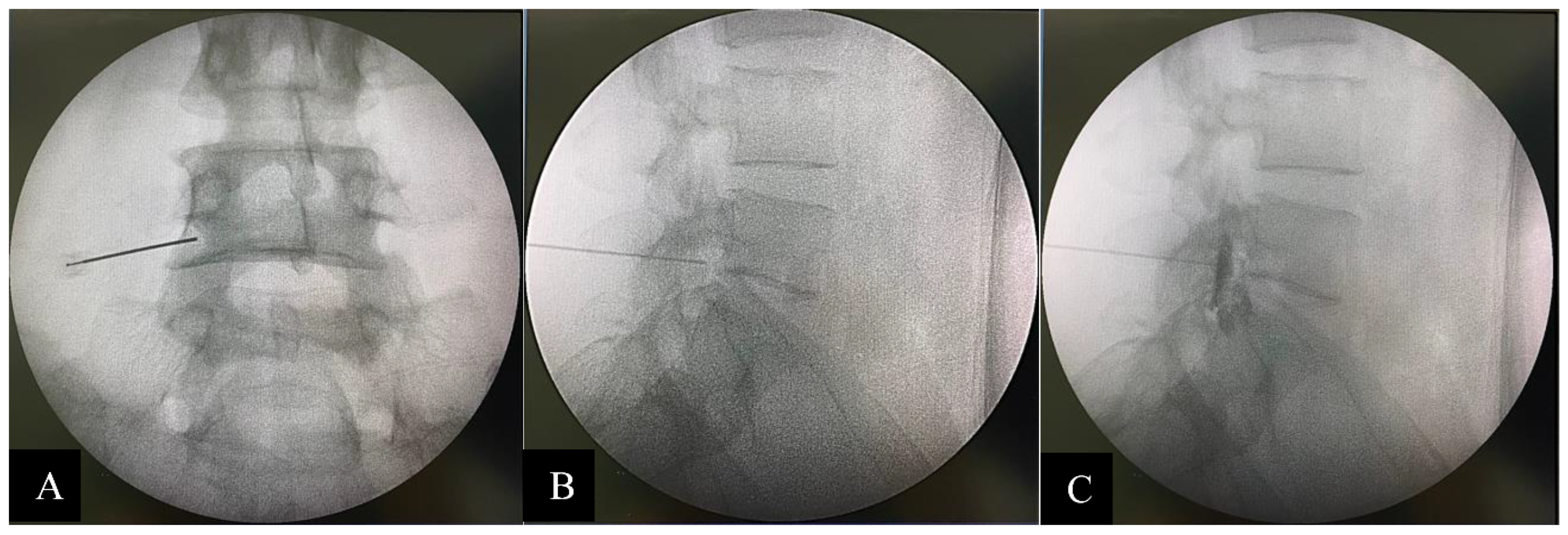
Whole-spine anteroposterior (AP) and lateral standing radiographs were taken before intervention (pre-intervention) and two weeks after intervention (post-intervention) by using 36-inch full-length films. To standardize the imaging process, all patients were instructed to stand in a comfortable position with full hip and knee extension, and with the elbow flexed at 45° [16].
Two spine surgeons independently performed digital radiograph interpretations. The patients’ data and identifications were blinded to the evaluators. The sagittal parameters were measured by PACS (SYNAPSE, Fujifilm’s) measurement tools on the 27-inch monitor. The Sagittal Vertical Axis (SVA), Thoracic Kyphosis (TK), Lumbar Lordosis (LL), Pelvic Incidence (PI), Pelvic Tilt (PT), Sacral Slope (SS), and PI-LL (PI minus LL) were measured according to the Scoliosis Research Society-Schwab classification [1]. C2C7 Cobb was measured from the angle between C2 and the C7 lower endplate [17] (Figure 1).

2.2. Intervention

TFESI was performed with triamcinolone acetonide 40 mg/1 cc (40 mg for one-level injection and 80 mg for two- to four-level injection) and normal saline mixed up to 2 cc for each injection point. In addition, 0.5–1 cc of Iohexol (Omipaque 300 Contrast) was administered as a contrast media to confirm the position prior to steroid injection under biplanar (AP and lateral) fluoroscopic guidance (C-arm Fluoroscope, Philips BV Pulsera, Amsterdam, The Netherlands). Quincke spinal needles, size 23, gauge 9 cm, were used, and they were angled towards the safe triangle [18,19] in the AP view and towards the middle of the neural foramen in the lateral view (Figure 2).

2.3. Statistical Analysis

Percentages were used for categorical data. The mean and standard deviation were used for continuous data after the normality assumption was validated, and a paired t-test was used in the analysis. Characteristics of the data between groups were analyzed by analysis of variance (ANOVA). The correlation of the data was measured using Pearson correlation coefficients. An r value of more than 0.3 or less than −0.3 confirmed a statistical correlation [20], and a p value of less than 0.05 indicated statistical significance. Interobserver reliability testing was performed using the Intraclass Correlation Coefficient (ICC). All statistical calculations were performed on IBM SPSS Statistic version 25.0 (IBM Corporation, Armonk, NY, USA).

3. Results

We retrospectively reviewed 120 individual cases with 99 patients. A total of 21 males (21.2%) and 78 females (78.8%) underwent TFESI and were included in the study. The average age was 64.3 ± 9.2 years. The average vertebrae levels injected were 1.8 ± 0.8 levels.

3.1. Pre-Intervention and Post-Intervention Outcomes

Both VAS and ODI outcomes were statistically improved after the intervention: VAS decreased from 8.31 ± 1.11 to 3.38 ± 1.47 and ODI decreased from 31.06 ± 3.48 to 18.03 ± 4.17. All sagittal parameters were statistically significantly improved. SVA, PT, and lumbopelvic mismatch were significantly decreased, while C2C7 Cobb, TK, SS, and LL were significantly increased after the intervention. Post-intervention SVA showed an improvement of 30.12% (53.26 ± 51.67 mm to 37.22 ± 40.71 mm). Pelvic Incidence (PI) was not statistically affected by TFESI. All data are shown in Table 1.

3.2. Difference between the Number of Injection Levels and Sagittal Parameters

The collected data were divided according to the total number of vertebrae levels injected: 1 level, 2 levels, 3 levels, and 4 levels. The differences in outcomes and sagittal parameters in each group were assessed. The variations of VAS score and ODI among the different numbers of injection levels were not statistically significant, both before and after the intervention. The C2C7 Cobb and TK were found to not be statistically significant between injection levels. The sagittal parameters, including SVA, PT, and PI-LL mismatch, were statistically significantly higher in multiple-level injection groups compared to single-level injection groups, both before and after the intervention. In contrast, SS and LL were statistically significantly lower in multiple-level injection groups compared to single-level injection groups, both before and after the intervention. All data are shown in Table 2.

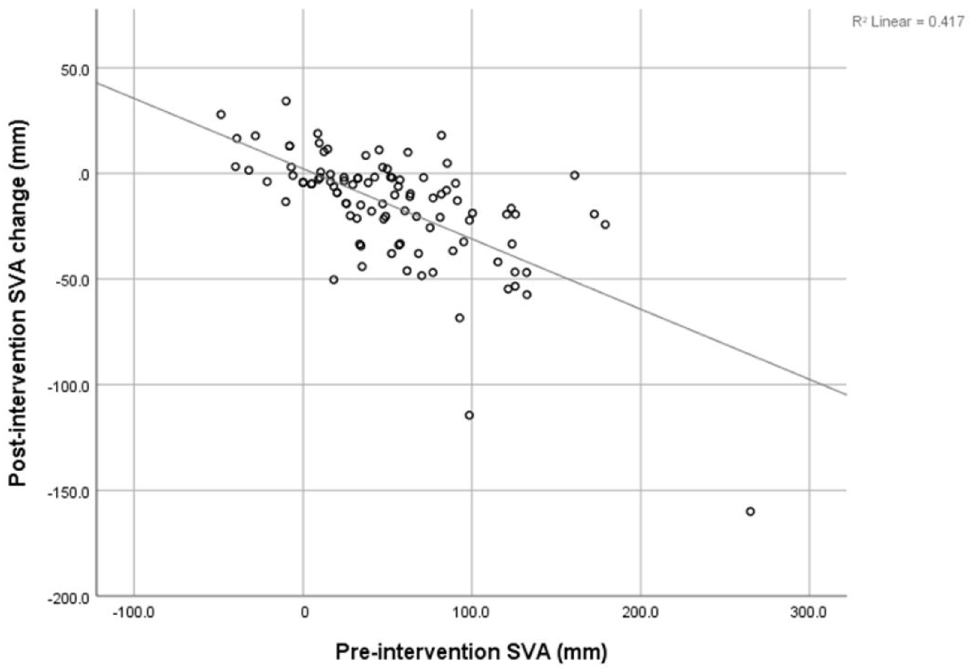
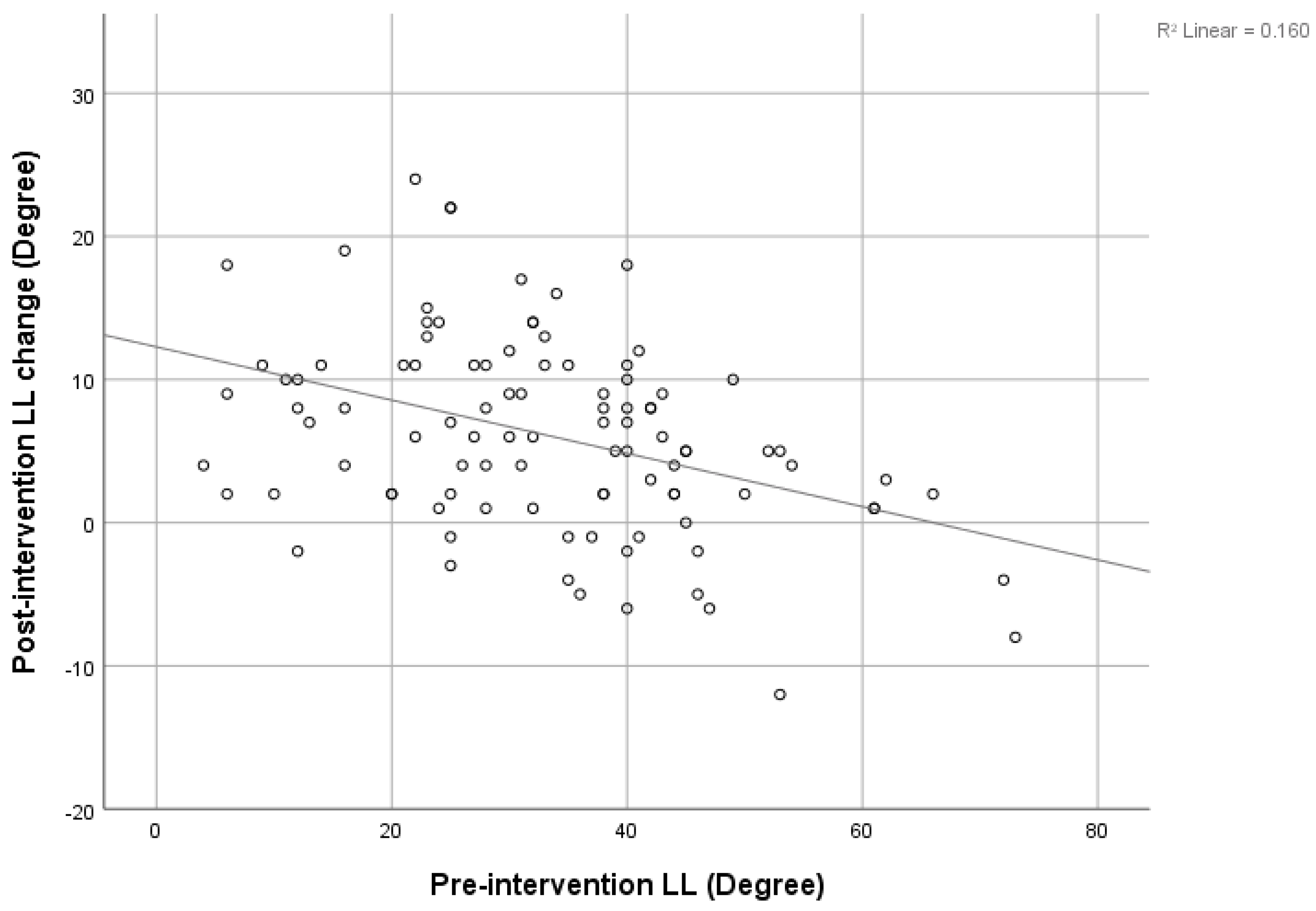
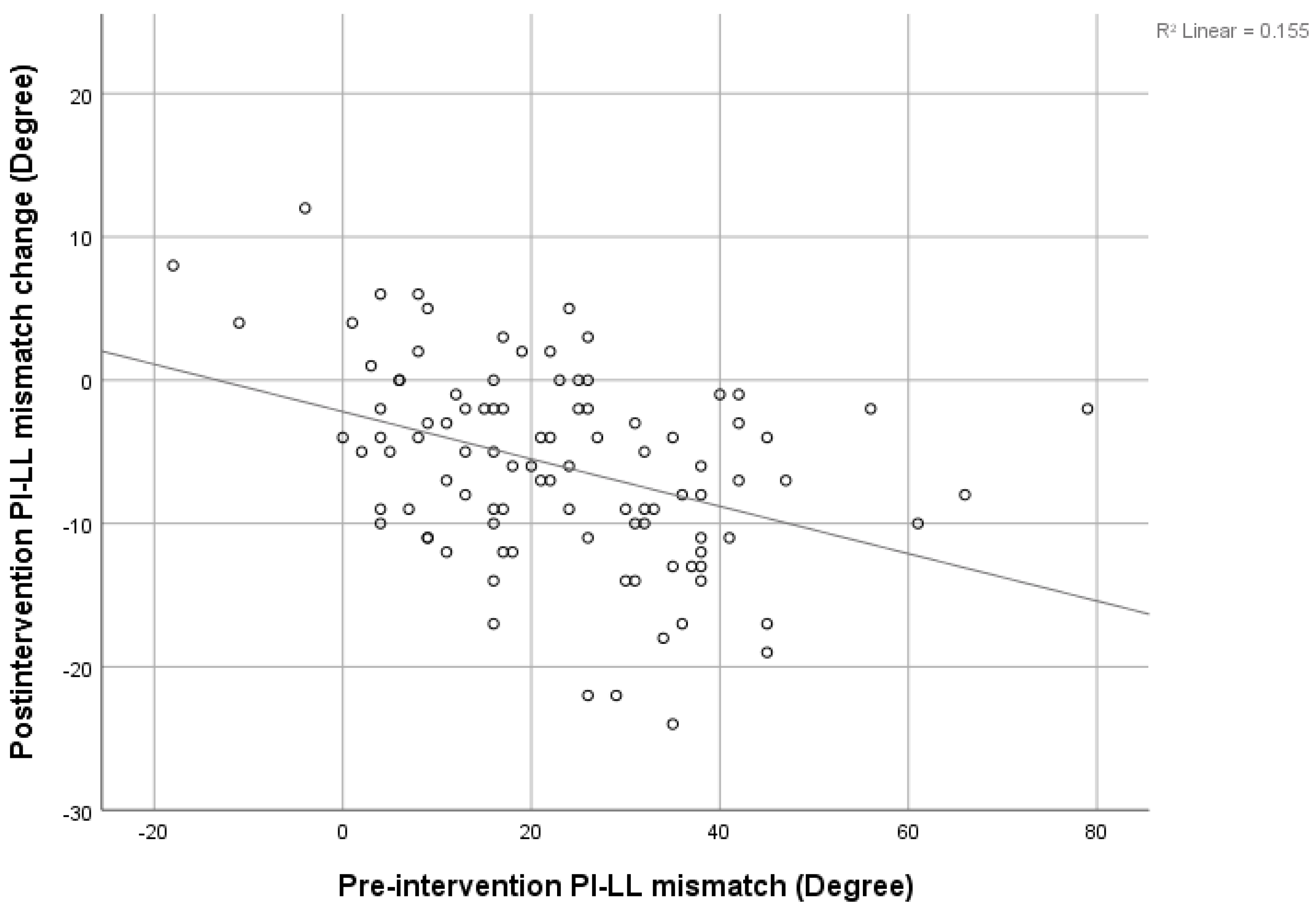
3.3. Correlation between Sagittal Parameters

There was a strong negative correlation between the pre-intervention SVA and post-intervention SVA decrement (r = −0.646), shown in Figure 3. The pre-intervention LL showed a negative correlation with the post-intervention LL increment, shown in Figure 4 (r = −0.4). The pre-intervention PI-LL mismatch exhibited a negative correlation with the post-intervention PI-LL decrement (r = −0.394), shown in Figure 5. The significant correlation between pre-intervention parameters and post-intervention changes is shown in Table 3.

3.4. Interobserver Reliability
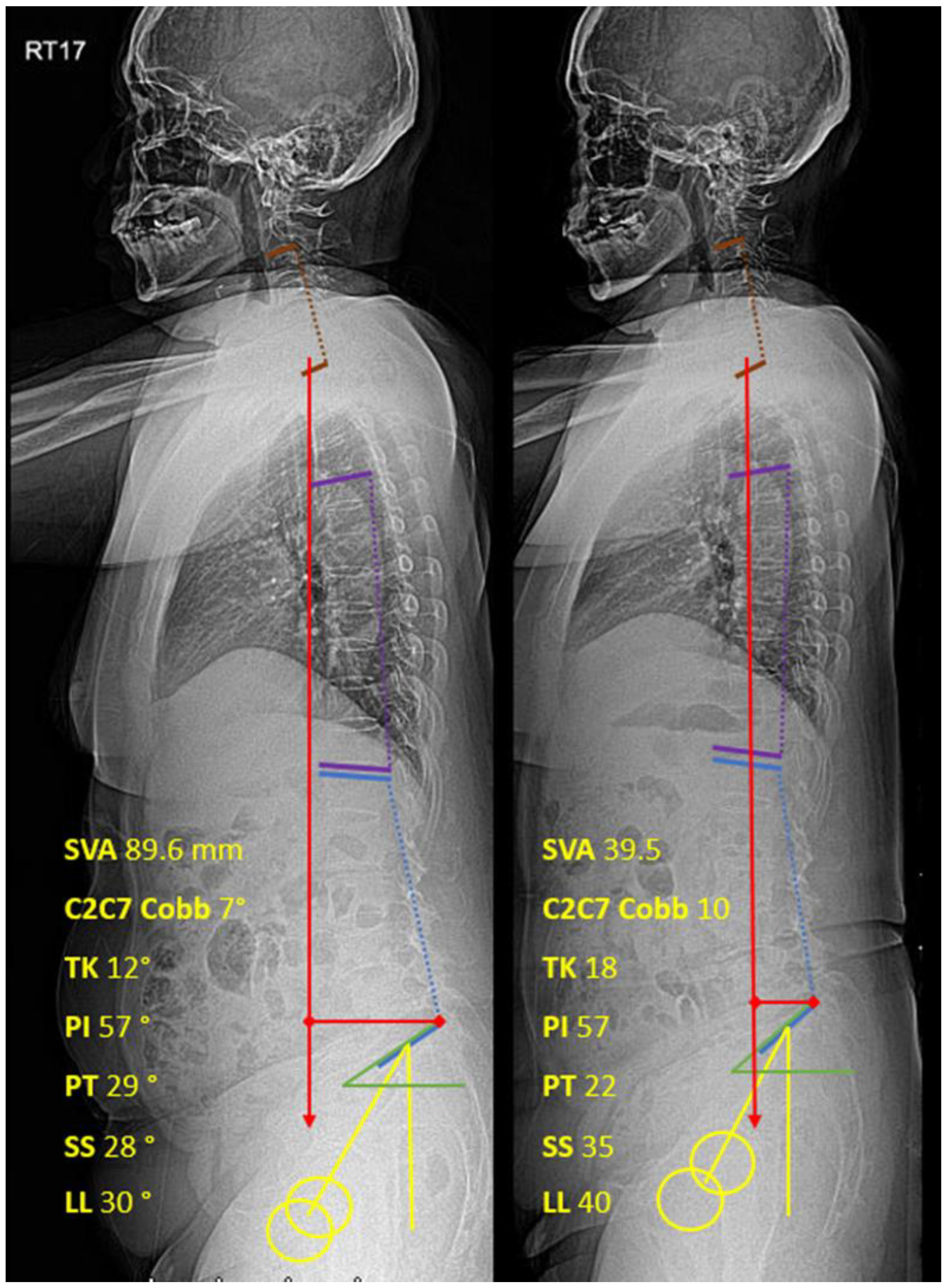
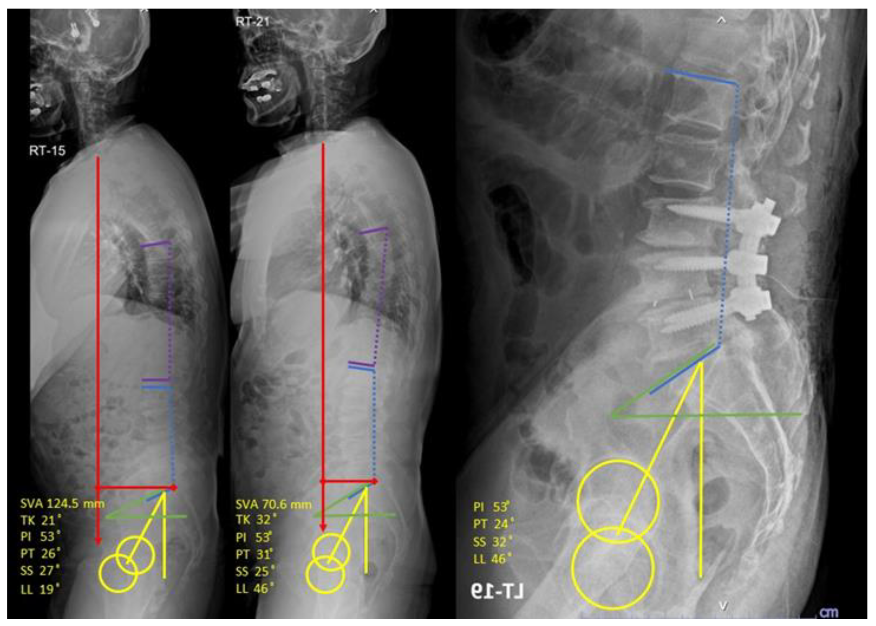
Interobserver reliability was calculated using the Intraclass Correlation Coefficient. The results were greater than 0.9 in all sagittal parameters. Thus, it was considered that the measurements were valid and achieved excellent reliability.

3.5. Case Example

A 63-year-old Thai man with low back pain and positive balance had pre-intervention parameters: SVA 124.5 mm, TK 21°, PI 53°, PT 26°, SS 27°, and LL 19°; and post-intervention parameters: SVA 70.6 mm, TK 32°, PI 53°, PT 31°, SS 25°, and LL 46°. After post-intervention parameters were evaluated, the operative planning was changed from long-segment fusion for correct SVA and multiple osteotomies for correct LL to short-segment fusion and single-level osteotomies instead (Figure 6).

4. Discussion

In our study, we collected data from patients with lumbar spinal stenosis who received TFESI. The results showed that both the VAS and the ODI improved significantly after the intervention. Ghahreman A. [21], Kabatas S. [22], and McCormick Z. [23] studied the short-term effects of TFESI, which significantly improved VAS and ODI in a similar study. The study of Karppinen et al. [24] showed that at 2 weeks of follow-up, a significant improvement from baseline was observed in every outcome parameter (leg pain, back pain, ODI, degree of straight-leg-raising test). This could support the treatment of pain using TFESI for short-term efficacy in lumbar spinal stenosis.
Liang C. [10] conducted a study on lumbar disc herniation and found significant immediate improvement in all sagittal imbalance parameters on day one post-operation, and this improvement continued for three months before the parameters became close to their normal ranges. SVA improved from 11.6 ± 6.6 cm to 2.9 ± 6.1 cm, and three months post-operation, LL improved from 25.3° ± 14.0° to 42.4° ± 10.2°. Moreover, Fujii K. [8] retrospectively reviewed lumbar decompression without fusion in lumbar spinal stenosis and concluded that the SVA, TK, PT, LL, and PI-LL mismatch improved post-operatively. SVA was decreased from 49.1° ± 38.6° to 28.6° ± 30.7°, and LL was changed from 38° ± 13° to 44° ± 11°. A strong correlation was found between pre-op SVA/PI-LL and post-op SVA/PI-LL decrement post-operatively. Likewise, Salimi H et al. [25] also reported that minimally invasive lumbar decompression surgery could convert sagittal malalignment to normal alignment in 2 years and 5 years follow-up. The previous studies mentioned above indicated that spinal decompressive procedures without instrumentation have the ability to improve sagittal spinal parameters. Therefore, we believe that spinal interventions that reduce radicular pain, such as TFESI, can partially improve sagittal spinal parameters because when radicular pain was improved, compensating forward bending subsided.
This study may be the first study to collect data on the non-operative management of lumbar spinal stenosis. The present procedure did not interfere with the anatomical structure, but decreased the inflammation process to the neural structure and improved radicular pain. We found a significant change in SVA, C2C7 Cobb, TK, PT, SS, and LL after patients underwent TFESI. The SVA was improved by about 30% (from 52.76 ± 52.22 mm to 37.03 ± 41.07 mm), and PT and LL were improved by about 17% and 18%, respectively. This is compared to 42%, 15%, and 16% improvements reported by Fujii K. [8]. Patients with multiple levels of stenosis tend to have significantly more severe positive SVA, higher PT, and lower LL and SS. Furthermore, we found a strong negative correlation between pre-intervention SVA and post-intervention SVA decrement. This indicates that the more positive imbalanced patients were, the greater the resulting improvement in the SVA. This correlation has also been found in recent studies [8,10,26]. Similarly, a strong negative correlation between pre-intervention PI-LL and post-intervention PI-LL was found. The greater the PI-LL mismatch, the larger the PI-LL improvement could be predicted to be. In contrast, the pre-intervention LL was negatively correlated with the post-intervention LL increment, and this means that in a small cohort of pre-intervention LL patients, there may be more improvement in the post-intervention LL.
From a review of previous literature, it can be observed that many spinal pathologies are caused by sagittal imbalance, reduced muscle strength [10,27,28], adjacent disc degeneration [29], disc herniation [9], and spinal stenosis. Several authors have proposed that spinal stenosis patients have limited lumbar lordosis (LL) [5] due to the decreased pressure of the epidural venous plexus when bending forward. Furthermore, compensatory lumbar flexion posture lowers epidural pressure, thus reducing pain and neurogenic claudication [9,27,30,31]. The anatomical study showed that flexion for the lumbar spine increased spinal canal diameters [6,7]. We hypothesized that in global sagittal imbalance patients, there might be two factors that are involved in the imbalance. The first one is a structural imbalance, and the second one is the “functional compensation” of patients to radicular pain. We believe that after undergoing TFESI and the pain becoming less severe, compensation of lumbar flexion may be diminished. In this study, after TSESI, we found that SVA, PT, and LL were significantly improved. Recently, there has been little focus on the functional compensation of sagittal alignment before spinal surgical correction. We believe that it is better to evaluate the spinal surgical balance when the clinical pain of patients is subsiding, rather than when the pain remains severe.
Our most recent concern with this main issue was that we were uncertain whether we had to correct the deformity if a global sagittal imbalance existed in the surgical treatment of degenerative lumbar spinal stenosis. This study found that a considerable number of patients’ global sagittal alignment significantly improved following TFESI. For this reason, in patients with degenerative lumbar spinal stenosis and global sagittal imbalance, reassessing global sagittal alignment after TFESI might show more accurate structural global sagittal imbalance. We advise obtaining whole-spine AP and lateral standing radiographs again after patients begin improving in terms of pain following TFESI.
This study has some limitations. First, this is a retrospective review of the database, so recall bias and selection bias may be present. Second, this radiographic study focuses on sagittal alignment, but the dynamic compensation of lower limbs, such as hip and knee flexion, is not investigated. It should be noted that we instructed all patients who received the whole-spine film to extend their hip and knee before imaging [32]. Third, due to the short-term effect of TFESI, the outcome and sagittal parameter data were collected only at a short-term follow-up. We suggest a long-term follow-up in future studies.

5. Conclusions

Transforaminal epidural steroid injection (TFESI) can improve SVA, C2C7 Cobb, TK, PT, SS, and LL parameters, as well as VAS and ODI, in a short-term follow-up study and also has benefits in that it is effective in correcting functional compensation to evaluate sagittal alignment correction before surgery to avoid postoperative overcorrection alignment. However, this could be the choice of treatment to improve quality of life factors in terms of pain and disability in sagittal malalignment patients who have contraindications or deny surgery.

Author Contributions

Methodology, K.S.; Formal analysis, P.M.; Writing—review & editing, K.S. and N.D. All authors have read and agreed to the published version of the manuscript.

Funding

This research received funding from Chulabhorn International College of Medicine.

Institutional Review Board Statement

Human Ethics Committee of Thammasat University (MTU-EC-OT-6-105/63).

Informed Consent Statement

Informed consent was obtained from all subjects involved in the study.

Conflicts of Interest

The authors declare no conflict of interest.

References

  1. Iyer, S.; Sheha, E.; Fu, M.C.; Varghese, J.; Cunningham, M.E.; Albert, T.J.; Schwab, F.J.; Lafage, V.C.; Kim, H.J. Sagittal Spinal Alignment in Adult Spinal Deformity: An Overview of Current Concepts and a Critical Analysis Review. JBJS Rev. 2018, 6, e2. [Google Scholar] [CrossRef] [PubMed]
  2. Harroud, A.; Labelle, H.; Joncas, J.; Mac-Thiong, J.M. Global sagittal alignment and health-related quality of life in lumbosacral spondylolisthesis. Eur. Spine J. 2013, 22, 849–856. [Google Scholar] [CrossRef] [PubMed] [Green Version]
  3. Schwab, F.; Lafage, V.; Patel, A.; Farcy, J.P. Sagittal plane considerations and the pelvis in the adult patient. Spine 2009, 34, 1828–1833. [Google Scholar] [CrossRef]
  4. Yeh, K.T.; Lee, R.P.; Chen, H.; Yu, T.C.; Liu, K.L.; Peng, C.H.; Wang, J.H.; Wu, W.T. Correlation of Functional Outcomes and Sagittal Alignment After Long Instrumented Fusion for Degenerative Thoracolumbar Spinal Disease. Spine 2018, 43, 1355–1362. [Google Scholar] [CrossRef] [PubMed]
  5. Pourtaheri, S.; Sharma, A.; Savage, J.; Kalfas, I.; Mroz, T.E.; Benzel, E.; Steinmetz, M.P. Pelvic retroversion: A compensatory mechanism for lumbar stenosis. J. Neurosurg. Spine 2017, 27, 137–144. [Google Scholar] [CrossRef] [Green Version]
  6. Schonstrom, N.; Lindahl, S.; Willen, J.; Hansson, T. Dynamic changes in the dimensions of the lumbar spinal canal: An experimental study in vitro. J. Orthop. Res. 1989, 7, 115–121. [Google Scholar] [CrossRef] [PubMed]
  7. Miao, J.; Wang, S.; Park, W.M.; Xia, Q.; Fang, X.; Torriani, M.P.; Wood, K.B.; Li, G. Segmental spinal canal volume in patients with degenerative spondylolisthesis. Spine J. 2013, 13, 706–712. [Google Scholar] [CrossRef] [PubMed] [Green Version]
  8. Fujii, K.; Kawamura, N.; Ikegami, M.; Niitsuma, G.; Kunogi, J. Radiological improvements in global sagittal alignment after lumbar decompression without fusion. Spine 2015, 40, 703–709. [Google Scholar] [CrossRef] [PubMed]
  9. Endo, K.; Suzuki, H.; Tanaka, H.; Kang, Y.; Yamamoto, K. Sagittal spinal alignment in patients with lumbar disc herniation. Eur. Spine J. 2010, 19, 435–438. [Google Scholar] [CrossRef] [PubMed] [Green Version]
  10. Liang, C.; Sun, J.; Cui, X.; Jiang, Z.; Zhang, W.; Li, T. Spinal sagittal imbalance in patients with lumbar disc herniation: Its spinopelvic characteristics, strength changes of the spinal musculature and natural history after lumbar discectomy. BMC Musculoskelet. Disord. 2016, 17, 305. [Google Scholar] [CrossRef] [Green Version]
  11. Kim, C.H.; Chung, C.K.; Park, S.B.; Yang, S.H.; Kim, J.H. A Change in Lumbar Sagittal Alignment After Single-level Anterior Lumbar Interbody Fusion for Lumbar Degenerative Spondylolisthesis with Normal Sagittal Balance. Clin. Spine Surg. 2017, 30, 291–296. [Google Scholar] [CrossRef]
  12. Manchikanti, L.; Kaye, A.D.; Manchikanti, K.; Boswell, M.; Pampati, V.; Hirsch, J. Efficacy of epidural injections in the treatment of lumbar central spinal stenosis: A systematic review. Anesth. Pain Med. 2015, 5, e23139. [Google Scholar] [CrossRef] [PubMed] [Green Version]
  13. Davis, N.; Hourigan, P.; Clarke, A. Transforaminal epidural steroid injection in lumbar spinal stenosis: An observational study with two-year follow-up. Br. J. Neurosurg. 2017, 31, 205–208. [Google Scholar] [CrossRef]
  14. Bartleson, J.D.; Maus, T.P. Diagnostic and therapeutic spinal interventions: Epidural injections. Neurol. Clin. Pract. 2014, 4, 347–352. [Google Scholar] [CrossRef] [Green Version]
  15. Sanjaroensuttikul, N. The Oswestry low back pain disability questionnaire (version 1.0) Thai version. J. Med. Assoc. Thai. 2007, 90, 1417–1422. [Google Scholar] [PubMed]
  16. Lafage, V.; Schwab, F.; Vira, S.; Patel, A.; Ungar, B.; Farcy, J.P. Spino-pelvic parameters after surgery can be predicted: A preliminary formula and validation of standing alignment. Spine 2011, 36, 1037–1045. [Google Scholar] [CrossRef] [PubMed]
  17. Alijani, B.; Rasoulian, J. The Sagittal Balance of the Cervical Spine: Radiographic Analysis of Interdependence between the Occipitocervical and Spinopelvic Alignment. Asian Spine J. 2020, 14, 287. [Google Scholar] [CrossRef] [Green Version]
  18. Lee, I.S.; Kim, S.H.; Lee, J.W.; Hong, S.H.; Choi, J.Y.; Kang, H.S.; Song, J.W.; Kwon, A.K. Comparison of the temporary diagnostic relief of transforaminal epidural steroid injection approaches: Conventional versus posterolateral technique. AJNR Am. J. Neuroradiol. 2007, 28, 204–208. [Google Scholar]
  19. Lutz, G.E.; Vad, V.B.; Wisneski, R.J. Fluoroscopic transforaminal lumbar epidural steroids: An outcome study. Arch. Phys. Med. Rehabil. 1998, 79, 1362–1366. [Google Scholar] [CrossRef]
  20. Mukaka, M.M. Statistics corner: A guide to appropriate use of correlation coefficient in medical research. Malawi Med. J. 2012, 24, 69–71. [Google Scholar]
  21. Ghahreman, A.; Ferch, R.; Bogduk, N. The efficacy of transforaminal injection of steroids for the treatment of lumbar radicular pain. Pain Med. 2010, 11, 1149–1168. [Google Scholar] [CrossRef] [PubMed] [Green Version]
  22. Kabatas, S.; Cansever, T.; Yilmaz, C.; Kocyigit, O.I.; Coskun, E.; Demircay, E.; Akar, A.; Caner, H. Transforaminal epidural steroid injection via a preganglionic approach for lumbar spinal stenosis and lumbar discogenic pain with radiculopathy. Neurol. India 2010, 58, 248–252. [Google Scholar] [CrossRef]
  23. McCormick, Z.; Kennedy, D.J.; Garvan, C.; Rivers, E.; Temme, K.; Margolis, S.; Zander, E.; Rohr, A.; Smith, M.C.; Plastaras, C. Comparison of Pain Score Reduction Using Triamcinolone vs. Betamethasone in Transforaminal Epidural Steroid Injections for Lumbosacral Radicular Pain. Am. J. Phys. Med. Rehabil. 2015, 94, 1058–1064. [Google Scholar] [CrossRef]
  24. Karppinen, J.; Malmivaara, A.; Kurunlahti, M.; Kyllönen, E.; Pienimäki, T.; Nieminen, P.; Ohinmaa, A.; Tervonen, O.; Vanharanta, H. Periradicular infiltration for sciatica: A randomized controlled trial. Spine 2001, 26, 1059–1067. [Google Scholar] [CrossRef]
  25. Salimi, H.; Toyoda, H.; Yamada, K.; Terai, H.; Hoshino, M.; Suzuki, A.; Takahashi, S.; Tamai, K.; Hori, Y.; Yabu, A.; et al. The effect of minimally invasive lumbar decompression surgery on sagittal spinopelvic alignment in patients with lumbar spinal stenosis: A 5-year follow-up study. J. Neurosurg. Spine 2021, 35, 177–184. [Google Scholar] [CrossRef] [PubMed]
  26. Cho, J.H.; Joo, Y.S.; Lim, C.; Hwang, C.J.; Lee, D.H.; Lee, C.S. Effect of one- or two-level posterior lumbar interbody fusion on global sagittal balance. Spine J. 2017, 17, 1794–1802. [Google Scholar] [CrossRef] [PubMed]
  27. Enomoto, M.; Ukegawa, D.; Sakaki, K.; Tomizawa, S.; Arai, Y.; Kawabata, S.; Kato, T.; Yoshii, T.; Shinomiya, K.; Okawa, A. Increase in paravertebral muscle activity in lumbar kyphosis patients by surface electromyography compared with lumbar spinal canal stenosis patients and healthy volunteers. J. Spinal Disord. Tech. 2012, 25, E167–E173. [Google Scholar] [CrossRef]
  28. Masaki, M.; Ikezoe, T.; Yanase, K.; Ji, X.; Umehara, J.; Aoyama, J.; Minami, S.; Fukumoto, Y.; Watanabe, Y.; Kimura, M.; et al. Association of Pain History and Current Pain with Sagittal Spinal Alignment and Muscle Stiffness and Muscle Mass of the Back Muscles in Middle-aged and Elderly Women. Clin. Spine Surg. 2019, 32, E346–E352. [Google Scholar] [CrossRef]
  29. Makhni, M.C.; Shillingford, J.N.; Laratta, J.L.; Hyun, S.J.; Kim, Y.J. Restoration of Sagittal Balance in Spinal Deformity Surgery. J. Korean Neurosurg. Soc. 2018, 61, 167–179. [Google Scholar] [CrossRef] [Green Version]
  30. Lee, J.H.; Kim, K.T.; Suk, K.S.; Lee, S.H.; Jeong, B.O.; Kim, J.S.; Eoh, J.H.; Kim, Y.J. Analysis of spinopelvic parameters in lumbar degenerative kyphosis: Correlation with spinal stenosis and spondylolisthesis. Spine 2010, 35, E1386–E1391. [Google Scholar] [CrossRef]
  31. Suzuki, H.; Endo, K.; Kobayashi, H.; Tanaka, H.; Yamamoto, K. Total sagittal spinal alignment in patients with lumbar canal stenosis accompanied by intermittent claudication. Spine 2010, 35, E344–E346. [Google Scholar] [CrossRef] [PubMed]
  32. Lafage, V.; Smith, J.S.; Bess, S.; Schwab, F.J.; Ames, C.P.; Klineberg, E.; Arlet, V.; Hostin, R.; Burton, D.C.; Shaffrey, C.I.; et al. Sagittal spino-pelvic alignment failures following three column thoracic osteotomy for adult spinal deformity. Eur. Spine J. 2012, 21, 698–704. [Google Scholar] [CrossRef] [PubMed] [Green Version]
Figure 1. Whole-spine lateral standing films with measured sagittal parameters ((left): pre-intervention; (right): post-intervention). Red: Sagittal vertical axis, Purple: Thoracic kyphosis, Blue: Lumbar lordosis, Yellow: Pelvic tilt, Green: Sacral slope.
Jcm 12 04727 g001
Figure 2. (A) AP fluoroscopic view. The end of the needle is located in a safe triangle. (B) Lateral fluoroscopic view. The end of the needle is located in the middle of the neural foramen. (C) Lateral fluoroscopic view. After the L4 nerve root, contrast injection was outlined.
Jcm 12 04727 g002
Figure 3. Correlation between pre-intervention SVA and post-intervention SVA change.
Jcm 12 04727 g003
Figure 4. Correlation between pre-intervention LL and post-intervention LL change.
Jcm 12 04727 g004
Figure 5. Correlation between pre-intervention PI-LL mismatch and post-intervention PI-LL mismatch change.
Jcm 12 04727 g005
Figure 6. Whole-spine lateral standing films and post-operative films with measured sagittal parameters ((left): pre-intervention; (middle): post-intervention; (right): post-operative). Red: Sagittal vertical axis, Purple: Thoracic kyphosis, Blue: Lumbar lordosis, Yellow: Pelvic tilt, Green: Sacral slope.
Jcm 12 04727 g006
Table 1. Pre-Intervention and Post-Intervention Outcomes and Sagittal Parameters.
ParameterPre-InterventionPost-Interventionp-Value α
VAS8.31 ± 1.113.38 ± 1.47<0.001
ODI31.06 ± 3.4818.03 ± 4.17<0.001
SVA, mm53.26 ± 51.6737.22 ± 40.71<0.001
C2C7 Cobb12.15 ± 10.8613.63 ± 10.260.004
TK 19.22 ± 10.8020.9 ± 10.460.001
PI 56.25 ± 10.1856.31 ± 10.130.296
PT 28.65 ± 10.1723.66 ± 9.96<0.001
SS 27.61 ± 9.0032.65 ± 8.36<0.001
LL 33.35 ± 14.8039.56 ± 13.34<0.001
PI—LL 22.89 ± 16.3516.75 ± 14.82<0.001
α Calculated with paired t-test.
Table 2. Outcomes and Sagittal Parameters between Number of Injection Levels.
ParameterNumber of TFESI Injection Levels
1 (n = 40)2 (n = 42)3 (n = 14)4 (n = 3)p Value α
Pre-interventionVAS8.30 ± 1.048.17 ± 1.158.71 ± 1.148.67 ± 1.530.42
ODI30.93 ± 2.7630.74 ± 3.7832.29 ± 3.7931.67 ± 7.230.53
SVA25.59 ± 33.2964.08 ± 45.2972.44 ± 57.25164.76 ± 98.35<0.001
C2C7 cobb12.05 ± 8.4511.12 ± 10.9313.57 ± 6.7619.00 ± 39.150.619
TK19.40 ± 8.9016.60 ± 11.4518.21 ± 12.2510.00 ± 8.710.487
PI56.55 ± 8.5953.81 ± 9.3160.57 ± 12.1567.33 ± 13.610.027
PT2.45 ± 9.6528.19 ± 8.7836.14 ± 9.2144.61 ± 13.78<0.001
SS31.13 ± 8.2625.69 ± 9.1824.43 ± 7.9822.67 ± 0.570.011
LL39.5 ± 13.5730.90 ± 12.7627.0 ± 15.8012.33 ± 1.52<0.001
PI-LL17.05 ± 15.3822.90 ± 12.4633.57 ± 16.7955.0 ± 14.93<0.001
Post-interventionVAS3.17 ± 1.503.37 ± 1.483.69 ± 1.105.00 ± 1.730.17
ODI17.47 ± 3.7017.69 ± 4.4219.84 ± 3.9322.33 ± 4.930.08
SVA19.34 ± 28.9644.8 ± 39.3053.22 ± 47.4497.76 ± 64.280.0002
C2C7 cobb13.70 ± 8.3013.27 ± 10.6713.5 ± 8.4617.83 ± 31.050.907
TK21.83 ± 8.6021.83 ± 10.9318.19 ± 136.83 ± 4.250.071
PI56.7 ± 8.7854.13 ± 9.9859.84 ± 12.567.16 ± 11.620.067
PT20.8 ± 8.7323.40 ± 9.2429.00 ± 10.5741.00 ± 10.440.0007
SS35.88 ± 6.7530.60 ± 8.8430.84 ± 9.2226.16 ± 1.600.011
LL43.78 ± 11.8538.61 ± 12.833.61 ± 15.6722.5 ± 3.50.006
PI-LL12.9 ± 13.2115.52 ± 12.0126.23 ± 18.344.67 ± 14.90.0001
α Calculated with ANOVA test.
Table 3. Significant Correlation between Pre-Intervention Parameter and Post-Intervention Change α.
Post-Intervention ChangeVASODISVAPTLLPI-LL
Pre-Intervention
VAS−0.444 *−0.104−0.04−0.1510.034−0.038
ODI−0.18−0.43 *−0.2550.0480.104−0.104
SVA0.2680.133−0.646 *0.0970.34 *−0.298
PI0.151−0.004−0.2310.0370.060−0.041
PT0.2380.197−0.253−0.317 *0.226−0.226
LL−0.236−0.2640.2600.162−0.4 *0.411 *
PI-LL0.304 *0.235−0.374 *−0.1230.369 *−0.394 *
* r value more than 0.3 or less than −0.3; α Calculated with Pearson correlation coefficient.
Disclaimer/Publisher’s Note: The statements, opinions and data contained in all publications are solely those of the individual author(s) and contributor(s) and not of MDPI and/or the editor(s). MDPI and/or the editor(s) disclaim responsibility for any injury to people or property resulting from any ideas, methods, instructions or products referred to in the content.

