Article

Clinical Factors Associated with Severity of Colonic Diverticular Bleeding and Impact of Bleeding Site

1
Department of Medicine, Teikyo University School of Medicine, Tokyo 173-8605, Japan
2
Department of Gastroenterology, Nagoya Central Hospital, Nagoya 453-0801, Japan
*
Author to whom correspondence should be addressed.
J. Clin. Med. 2023, 12(5), 1826; https://doi.org/10.3390/jcm12051826
Submission received: 7 February 2023 / Revised: 20 February 2023 / Accepted: 22 February 2023 / Published: 24 February 2023

Abstract

:
Factors associated with serious colonic diverticular bleeding (CDB) are unclear, although the incidence of CDB has increased. We carried out this study to clarify factors associated with serious CDB and rebleeding. Subjects included 329 consecutive patients hospitalized for confirmed or suspected CDB between 2004 and 2021. Patients were surveyed regarding backgrounds, treatment, and clinical course. Of 152 with confirmed CDB, 112 showed bleeding from the right colon, and 40 did from the left colon. Patients received red blood cell transfusions in 157 (47.7%), interventional radiology in 13 (4.0%), and surgery in 6 (1.8%) cases. Early rebleeding within one month occurred in 75 (22.8%) patients, and late rebleeding within one year occurred in 62 (18.8%). Factors associated with red blood cell transfusion included confirmed CDB, anticoagulants, and high shock index. The only factor related to interventional radiology or surgery was confirmed CDB, which was also associated with early rebleeding. Late rebleeding was associated with hypertension, chronic kidney disease and past CDB. Right CDB showed higher rates of transfusion and invasive treatment than left CDB. Confirmed CDB had high frequencies of transfusion, invasive treatment, and early rebleeding. Right CDB seemed to be a risk for serious disease. Factors related to late rebleeding were different from those related to early rebleeding of CDB.

1. Introduction

The prevalence of colonic diverticula is increasing as the population ages in many countries, including Japan [1]. The number of patients with diverticular bleeding is rising in combination with the increasing use of antithrombotic and anti-inflammatory drugs. Colonic diverticular bleeding (CDB) is therefore becoming the main cause of lower gastrointestinal bleeding [2,3,4]. CDB is consequently a troublesome condition, because it occurs repeatedly and can sometimes be severe [5,6,7,8,9,10]. Recently, relevant academic societies published guidelines for treating CDB in Japan [11].
Reports to date have suggested that the use of nonsteroidal anti-inflammatory drugs (NSAIDs) and antithrombotic drugs, comorbidities, past diverticular bleeding, and physical findings such as shock are factors associated with exacerbation and rebleeding in CDB. However, the evidence remains weak, and predicting exacerbation and rebleeding in initial consultations is presently difficult [3,11,12,13,14]. Examining the predictability of associated factors using actual clinical data may therefore be of great significance.
Additionally, in this study, we focused on differences among clinical factors depending on the site of diverticular bleeding. The locations of colonic diverticula vary greatly by race and age. For example, colonic diverticula are mainly in the cecum, ascending colon, and right transverse colon in Asians, including the Japanese. Conversely in Caucasians, left colonic diverticula, mainly in the sigmoid colon, account for the majority of cases. In Japan, left colonic diverticula are more common in the elderly than in the young [1,15,16,17,18]. Diverticulitis, as another major complication of colonic diverticula, is known to have a higher rate of exacerbation in the left colon than in the right colon [19]. However, the site-specific clinical features of diverticular bleeding have not been sufficiently studied, and the amount of information on the condition is consequently small [20,21,22]. If the dependence of severity on the site of bleeding and differences in progression could be clarified, this could contribute to the selection of treatment methods and the prediction of prognoses during initial consultations.
This study therefore examined cases in which red blood cell (RBC) transfusion and invasive treatment were necessary, as well as predictors in cases of recurrence among patients with CDB. We also examined differences in the clinical features of diverticular bleeding between the right colon from the cecum to the transverse colon and the left colon from the descending colon to the rectum in confirmed cases of CDB where the site of bleeding was clear.

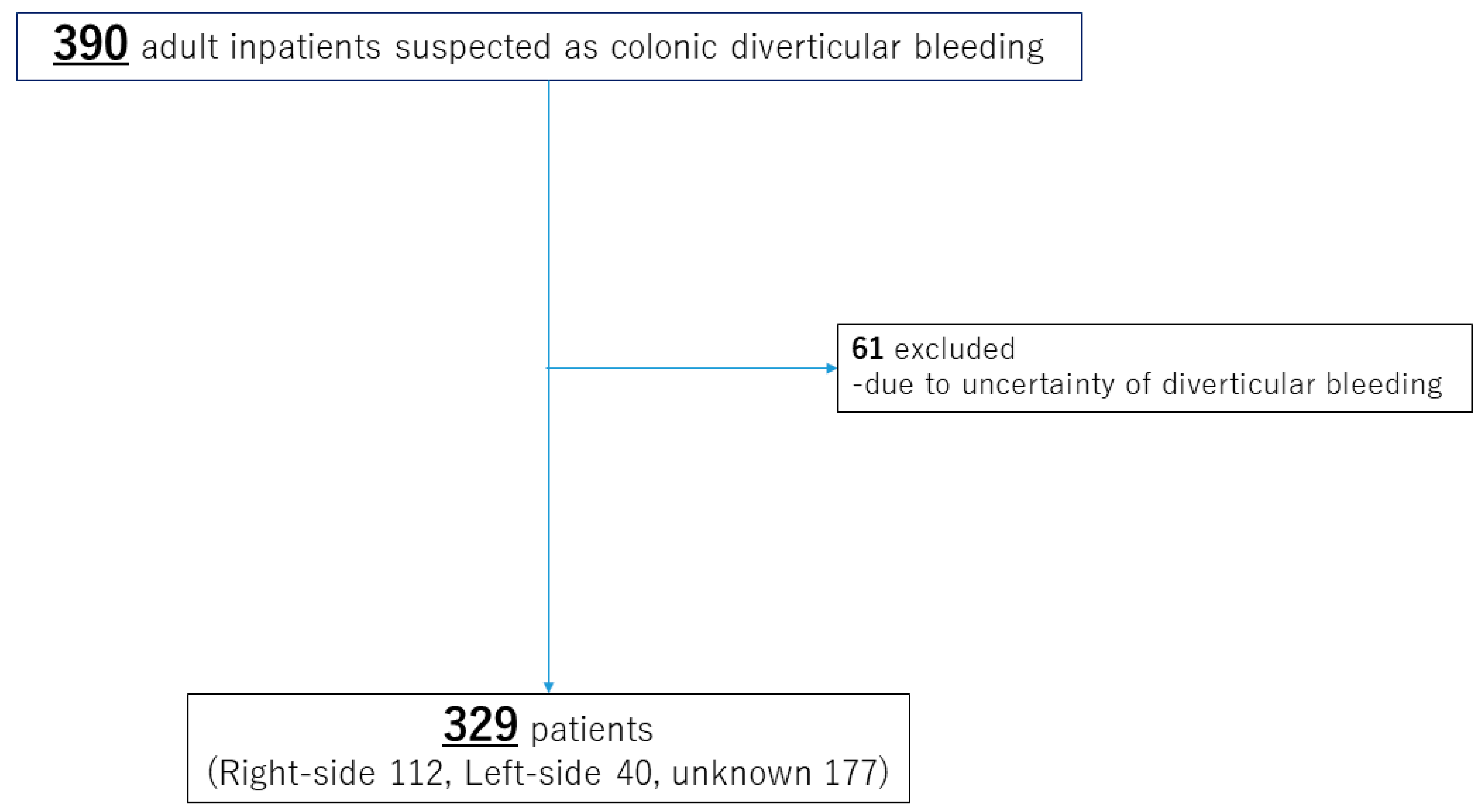
2. Materials and Methods

This study was a retrospective investigation of a single facility. The primary outcome of this study was to identify the factors related to the aggravation and recurrence of CDB in the patients. As the secondary outcome, we investigated the differences in clinical course according to the laterality of the bleeding site. Subjects were consecutive patients over 20 years old who had undergone inpatient treatment following a confirmed or suspected diagnosis of CDB (Figure 1).
Patients were selected from among all the patients who underwent colonoscopy in the Department of Medicine at Teikyo University Hospital (Tokyo, Japan) between January 2004 and May 2021. In the case of patients with a history of multiple hospitalizations during the investigated period, only the first hospitalization was evaluated. Diverticular bleeding was diagnosed based on colonoscopy and/or CT findings. Confirmed cases were those in which: (1) bleeding from diverticula or exposed vessels was directly confirmed on endoscopy (Figure 2A); or (2) leakage of contrast medium in the colon was confirmed with contrast-enhanced CT (Figure 2B), and colonoscopy confirmed that gastrointestinal bleeding and diverticula existed and there were no other lesions that cause bleeding.
Suspected cases were those in which gastrointestinal bleeding was evident, and the presence of colonic diverticula was confirmed during endoscopic observation, no lesions other than the diverticula were apparent as potential causes of bleeding, and no flow of blood from the ileocecal valve to the colon was observed. In confirmed cases, the site of bleeding was judged to be the right colon in cases involving the cecum, ascending colon, or transverse colon, and to be the left colon in cases involving the descending colon, sigmoid colon, or rectum. Cases that did not fit the above definitions or in which CDB could not be diagnosed were excluded from being subjects. These judgments were independently made for each case by two endoscopists. When the two endoscopists were not in agreement, another endoscopist made the final judgment.
Each of the patients was investigated for background factors comprising physical data (age, height, and weight), smoking history, drinking history, comorbidities (hypertension, diabetes, dyslipidemia, ischemic heart disease, constipation, cerebral infarction, and history of diverticular bleeding), and concomitant medications (NSAIDs, antiplatelet drugs, and anticoagulants), as well as for information on diverticular bleeding, comprising information during bleeding (confirmed/suspected diagnosis, site of bleeding, blood pressure, heart rate, and hemoglobin level), treatment (endoscopic hemostasis, vascular embolization (IVR), surgical resection, and RBC transfusion), rebleeding, and survival. The occurrence or absence of rebleeding was evaluated and confirmed as either early rebleeding (occurring within one month of initial bleeding) or late rebleeding (occurring after one month but within one year of initial bleeding) based on medical records.
Statistical analyses were performed using SPSS version 28 software (IBM Japan, Tokyo, Japan). The Mann–Whitney U test and chi-squared test were used for comparison of the two groups. The effect of clinical factors was evaluated using a generalized linear model, after adjusting for background factors using the inverse probability of treatment, weighting with propensity scores calculated for each case. The level of statistical significance was 0.05 in all cases. The study protocol was approved by the ethics committee of Teikyo University before the study was started (approval no. TU 21-066). All procedures were performed in accordance with the relevant national and international guidelines, including the Declaration of Helsinki and its amendments. The need to obtain informed consent was waived by the ethics committee that approved the study, given the retrospective design of the study.

3. Results

3.1. Background Characteristics

A total of 329 patients was analyzed (Figure 1). Background data are presented in Table 1. Median age was 73 years, and approximately two-thirds (222, 67.5%) were men. Obese patients with a body mass index ≥ 30 kg/m2 only accounted for 18 patients (5.5%). A smoking history was seen in 95 (28.9%), and a drinking history was seen in 134 patients (40.7%). In addition, a high rate of patients (148, 45.0%) had a history of diverticular bleeding. In terms of comorbidities, 224 (68.1%) had hypertension, and 123 (37.4%) had dyslipidemia. Concomitant medications were anticoagulants in 53 (16.1%), antiplatelet drugs in 90 (27.4%), and NSAIDs in 66 (20.1%).

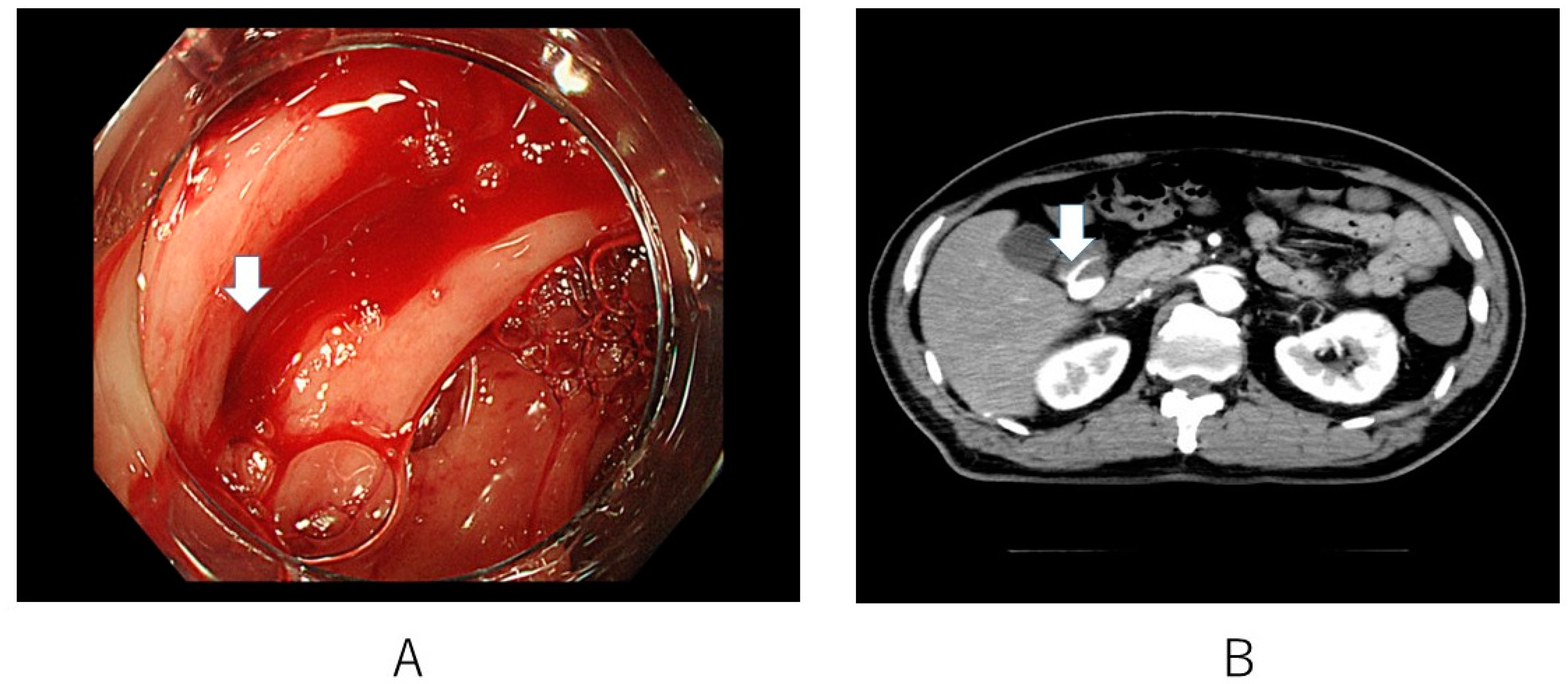
3.2. Data on Colonic Diverticular Bleeding

CDB was confirmed in 152 cases, including 112 cases with right colonic bleeding and 40 with left colonic bleeding. The remaining 177 cases had suspected CDB. Hemoglobin levels during bleeding were ≤10 g/dL in 146 cases (44.4%), and the shock index (heart rate/systolic blood pressure) was ≥1 in 19 cases (5.8%). As for treatment, 157 patients (47.7%) required RBC transfusion, 93 patients (28.3%) underwent endoscopic hemostasis, and a hemostatic clip was used in all patients. Interventional radiology (IVR) was performed in 13 cases (4.0%), and surgery was performed in 6 cases (1.8%). No cases of death during bleeding were seen. Early rebleeding within one month of initial bleeding occurred in 75 cases (22.8%), and late rebleeding within one year occurred in 62 cases (18.8%; Table 1).
Association of clinical factors with outcome
Table 2 shows the associations between RBC transfusion, invasive treatment, and rebleeding and clinical factors. Factors significantly associated with RBC transfusion were confirmed CDB, anticoagulants, high shock index during bleeding, and low hemoglobin level. The only factor associated with invasive treatment was confirmed CDB, and all patients who underwent surgery or IVR were confirmed cases. Factors significantly associated with early rebleeding were confirmed CDB and hypertension, while factors significantly associated with late rebleeding were hypertension, chronic kidney disease, and past CDB (Table 2). No significant associations were seen between NSAIDs and RBC transfusion, invasive treatment, or early or late rebleeding in this study.
The hemostatic effect of endoscopic treatment was not observed. Instead, early rebleeding tended to be more common in patients who underwent endoscopic hemostasis (adjusted odds ratio 7.590, 95% confidence interval [CI] 4.307–13.374, p < 0.001). Furthermore, no significant association was seen between late rebleeding and the performance or absence of endoscopic hemostasis (adjusted odds ratio 1.152, 95%CI 0.631–2.106, p = 0.645).

3.3. Difference between Right and Left CDB

Table 3 shows the differences in RBC transfusion, invasive treatment, and rate of rebleeding in right and left CDB. Median patient age was significantly lower for right CDB (71 years) than for left CDB (81 years) (p = 0.002, Mann–Whitney test). Patients with right CDB also tended to be male (p = 0.02, chi-squared test). Rates of patients who received RBC transfusion and underwent surgery or IVR were significantly higher for right CDB, but no significant differences were seen for early or late rebleeding between right and left CDB (Table 3).

4. Discussion

The main findings in this study were (1) confirmed CDB, concomitant use of antithrombotic drugs, shock upon hospital admission, and decreased hemoglobin were identified as factors associated with blood transfusion and invasive treatment; (2) confirmed cases of CDB showed a high rate of early rebleeding in addition to blood transfusion and invasive treatment; (3) patients with right CDB required RBC transfusion and invasive treatment more often than patients with left CDB; and (4) hypertension, chronic kidney disease, and past diverticular bleeding were risk factors for late rebleeding.
Contrast-enhanced computed tomography (CT) and colonoscopy are the main measures used to diagnose lower gastrointestinal bleeding, because contrast-enhanced CT is useful for identifying sites of bleeding and diagnosing underlying diseases [23,24], and colonoscopy allows hemostatic treatment as needed, in addition to high diagnostic performance [25,26,27]. All patients in this study had undergone endoscopy, and many had also undergone contrast-enhanced CT as well. Patients with confirmed CDB in whom bleeding was confirmed during the initial examination using these tests were assumed to have a considerable amount of bleeding at the time of or just before examination. This was likely associated with subsequent continuous or massive bleeding and may have resulted in a higher frequency of invasive treatment, RBC transfusion, and early rebleeding. Especially for invasive treatment, it is expected that treatment was not considered in unconfirmed cases, which may lead to selection bias. In other reports, continuous bleeding and early rebleeding in CDB are accompanied by a high risk of requiring RBC transfusion and invasive treatments such as surgery and arterial embolization [13,22]. Previously reported predictors included symptoms of shock, bright red blood in stool without diarrhea or abdominal pain, use of NSAIDs and/or aspirin, past bleeding, two or more comorbidities, and low hemoglobin level. However, these predictors alone were reportedly insufficient to predict present severity, so other predictors are required [11,12,13,14]. This study found the same associations with shock, antithrombotic drugs, and low hemoglobin level as previous reports, but invasive treatment, RBC transfusion, and early rebleeding also had significantly higher incidence in confirmed cases of CDB, suggesting their usefulness as new prognostic factors.
This study produced interesting results regarding the association between the site of bleeding and the severity. A significantly higher rate of patients with right CDB required RBC transfusion and invasive treatment compared with patients with left CDB. Kinjo et al. reported bleeding from diverticula in the right colon as a risk factor for severe diverticular bleeding, based on multivariate analysis, and suggested potential reasons, such as the following: right colonic diverticula are more susceptible to injury than left colonic diverticula because they are larger; the intestinal wall is thinner in the right colon than in the left colon and has relatively high blood flow; and bleeding is detected more slowly in the right colon because the distance from the anus is greater [20]. The causes of such differences were not evident in this study, but clinicians must keep in mind the high risk of requiring surgery or IVR in cases of bleeding from right colonic diverticula. CDB is a disease with a high rate of spontaneous hemostasis (73–88%) [5,6], and expecting spontaneous hemostasis with conservative treatment is valid in suspected cases where the site of bleeding has not been identified. However, active treatment is preferable from an early stage in confirmed cases of right CDB.
While endoscopic treatment is the predominant method of hemostasis, no inhibitory effect on early rebleeding was seen with endoscopic hemostasis in this study. Instead, rebleeding tended to be more common in the group that underwent endoscopic hemostasis. The cause of this unexpected result is not clear, since the details of endoscopic treatment have not been examined in the present study. Endoscopic hemostasis is generally performed in cases of severe disease or confirmed CDB with extravasation of contrast medium, which is a known situation in which rebleeding is common [28,29]. It is speculated that this result may be due to selection bias, as confirmed cases of CDB in the high-risk group for rebleeding underwent endoscopic hemostasis more often. Furthermore, these results may suggest that a hemostatic clip is not sufficiently effective, because a hemostatic clip was applied for hemostasis in all the cases. One of the countermeasures mentioned is selecting an endoscopic hemostatic method such as one using endoscopic band ligation (EBL) or a snare, as such methods have recently been reported to be effective [30,31,32,33,34,35,36,37,38,39,40]. According to a recent meta-analysis, rebleeding occurred significantly more with hemoclip than with EBL [41]. If the stigmata of recent hemorrhage (SRH) is at the bottom of the diverticulum or at the small orifice of the diverticulum, it is too difficult to clip the SRH directly, and inadequate clipping may lead to rebleeding. Niikura et al. reported that performing endoscopic hemostasis within 24 h, hemostasis performed by a specialist endoscopist, and use of a tip hood or water jet device, among other approaches, are useful in identifying the site of bleeding and could benefit future hemostatic procedures [42]. However, irrespective of the method of endoscopic treatment applied, late hemostatic effects cannot be expected, and the limitations of endoscopic treatment itself must be acknowledged [43].
In this study, late rebleeding within one year was seen in 18.8% of cases. Previous reports have described similar or slightly higher rates of rebleeding within one year, ranging from 20% to 35% [6,9,10]. The difference may be due to the retrospective study design of this study. Among the cases of rebleeding within one year, cases of visits to other facilities are not counted, and the possibility that the incidence rate has decreased cannot be ruled out. Hypertension, chronic kidney disease, past CDB, and other comorbidities associated with arteriosclerosis have been suggested to be significant risk factors. Furthermore, because risk factors for late rebleeding differed significantly from those in the acute phase and exacerbation, particular caution must be taken with patients with the above-mentioned risks during follow-up for bleeding. However, we believe that the details of risk factors for rebleeding should be reassessed in a prospective multicenter review to be precise.
The limitations of this study included its implementation in a single facility, the small number of subjects, and the retrospective study design. In particular, the influence of sample size seemed to be large. Although there were statistically significant differences in the relationship between shock and RBC transfusion, and between bleeding site and invasive treatment, the 95% CI was very wide. In order to improve the reliability of the results, it is essential to consider increasing the number of subjects. The entry period was also long, and medical advances and changes to treatment made during this period may have impacted the results. Furthermore, it is desirable to consider a prospective design in order to confirm the details of accurate factors affecting the prognosis of CDB, because retrospective cohort assessment of incidence is limited. It is actually hard to separately inquire into the different bleeding situations in the different kinds of CDB, especially in a retrospective study. A prospective study involving multiple facilities is required in the future, if feasible.

5. Conclusions

The present study showed that confirmed CDB had high frequencies of transfusion, invasive treatment, and early rebleeding. Right CDB seemed to be a risk factor for serious disease. Factors related to late rebleeding included hypertension, chronic kidney disease, and past CBD, indicating difference from factors related to early rebleeding of CDB.

Author Contributions

Conceptualization, T.Y. and H.A. (Hirohito Amano); Data curation, H.A. (Hirohito Amano), K.I., N.A., M.S., T.H., A.I. and S.K.; Formal analysis, H.A. (Hirohito Amano), K.I. and N.A.; Investigation, M.S., T.H., K.M., H.A. (Hitoshi Aoyagi), A.I., K.A. and Y.A.; Methodology, K.A. and S.K.; Project administration, A.T.; Resources, K.M. and H.A. (Hitoshi Aoyagi); Validation, T.Y.; Writing—original draft, H.A. (Hirohito Amano); Writing—review & editing, T.Y., Y.A. and A.T. All authors have read and agreed to the published version of the manuscript.

Funding

This research received no external funding.

Institutional Review Board Statement

The study protocol was approved by the ethics committee of Teikyo University before the study was started (approval no. TU 21-066). All procedures were performed in accordance with the relevant national and international guidelines, including the Declaration of Helsinki and its amendments.

Informed Consent Statement

The need to obtain informed consent was waived by the ethics committee that approved the study, given the retrospective design of the study.

Data Availability Statement

The data supporting the findings of this study are available on request from the corresponding author, Takatsugu Yamamoto ([email protected]) at Teikyo University School of Medicine. The data are not publicly available due to the inclusion of information that could compromise the privacy of research participants.

Conflicts of Interest

The authors declare that there are no conflict of interest regarding the publication of this article.

References

  1. Yamamichi, N.; Shimamoto, T.; Takahashi, Y.; Sakaguchi, Y.; Kakimoto, H.; Matsuda, R.; Kataoka, Y.; Saito, I.; Tsuji, Y.; Yakabi, S.; et al. Trend and risk factors of diverticulosis in Japan: Age, gender, and lifestyle/metabolic-related factors may cooperatively affect on the colorectal diverticula formation. PLoS ONE 2015, 10, e0123688. [Google Scholar] [CrossRef] [Green Version]
  2. Nagata, N.; Niikura, R.; Aoki, T.; Shimbo, T.; Itoh, T.; Goda, Y.; Suda, R.; Yano, H.; Akiyama, J.; Yanase, M.; et al. Increase in colonic diverticulosis and diverticular hemorrhage in an aging society: Lessons from a 9-year colonoscopic study of 28,192 patients in Japan. Int. J. Color. Dis. 2014, 29, 379–385. [Google Scholar] [CrossRef]
  3. Kinjo, K.; Matsui, T.; Hisabe, T.; Ishihara, H.; Maki, S.; Chuman, K.; Koga, A.; Ohtsu, K.; Takatsu, N.; Hirai, F.; et al. Increase in colonic diverticular hemorrhage and confounding factors. World J. Gastrointest. Pharmacol. Ther. 2016, 7, 440–446. [Google Scholar] [CrossRef] [Green Version]
  4. Niikura, R.; Nagata, N.; Shimbo, T.; Aoki, T.; Yamada, A.; Hirata, Y.; Sekine, K.; Okubo, H.; Watanabe, K.; Sakurai, T.; et al. Natural history of bleeding risk in colonic diverticulosis patients: A long-term colonoscopy-based cohort study. Aliment. Pharmacol. Ther. 2015, 41, 888–894. [Google Scholar] [CrossRef] [PubMed]
  5. Tanaka, Y.; Motomura, Y.; Akahoshi, K.; Iwao, R.; Komori, K.; Nakama, N.; Osoegawa, T.; Itaba, S.; Kubokawa, M.; Hisano, T.; et al. Predictive factors for colonic diverticular rebleeding: A retrospective analysis of the clinical and colonoscopic features of 111 patients. Gut Liver 2012, 6, 334–338. [Google Scholar] [CrossRef] [PubMed]
  6. Niikura, R.; Nagata, N.; Yamada, A.; Akiyama, J.; Shimbo, T.; Uemura, N. Recurrence of colonic diverticular bleeding and associated risk factors. Color. Dis. 2012, 14, 302–305. [Google Scholar] [CrossRef] [PubMed]
  7. Aytac, E.; Stocchi, L.; Gorgun, E.; Ozuner, G. Risk of recurrence and long-term outcomes after colonic diverticular bleeding. Int. J. Color. Dis. 2014, 29, 373–378. [Google Scholar] [CrossRef]
  8. Poncet, G.; Heluwaert, F.; Voirin, D.; Bonaz, B.; Faucheron, J.L. Natural history of acute colonic diverticular bleeding: A prospective study in 133 consecutive patients. Aliment. Pharmacol. Ther. 2010, 32, 466–471. [Google Scholar] [CrossRef] [Green Version]
  9. Okamoto, T.; Watabe, H.; Yamada, A.; Hirata, Y.; Yoshida, H.; Koike, K. The association between arteriosclerosis related diseases and diverticular bleeding. Int. J. Color. Dis. 2012, 27, 1161–1166. [Google Scholar] [CrossRef]
  10. Nishikawa, H.; Maruo, T.; Tsumura, T.; Sekikawa, A.; Kanesaka, T.; Osaki, Y. Risk factors associated with recurrent hemorrhage after the initial improvement of colonic diverticular bleeding. Acta Gastroenterol. Belg. 2013, 76, 20–24. [Google Scholar]
  11. Nagata, N.; Ishii, N.; Manabe, N.; Tomizawa, K.; Urita, Y.; Funabiki, T.; Fujimori, S.; Kaise, M. Guidelines for colonic diverticular bleeding and colonic diverticulitis: Japan Gastroenterological Association. Digestion 2019, 99 (Suppl. 1), 1–26. [Google Scholar] [CrossRef] [PubMed]
  12. Strate, L.L.; Orav, E.J.; Syngal, S. Early predictors of severity in acute lower intestinal tract bleeding. Arch. Intern. Med. 2003, 163, 838–843. [Google Scholar] [CrossRef] [PubMed] [Green Version]
  13. Aoki, T.; Nagata, N.; Shimbo, T.; Niikura, R.; Sakurai, T.; Moriyasu, S.; Okubo, H.; Sekine, K.; Watanabe, K.; Yokoi, C.; et al. Development and validation of a risk scoring system for severe acute lower gastrointestinal bleeding. Clin. Gastroenterol. Hepatol. 2016, 14, 1562–1570.e2. [Google Scholar] [CrossRef] [PubMed] [Green Version]
  14. Das, A.; Ben-Menachem, T.; Cooper, G.S.; Chak, A.; Sivak, M.V.; Gonet, J.A.; Wong, R.C. Prediction of outcome in acute lower-gastrointestinal haemorrhage based on an artificial neural network: Internal and external validation of a predictive model. Lancet 2003, 362, 1261–1266. [Google Scholar] [CrossRef]
  15. Peery, A.F.; Keku, T.O.; Martin, C.F.; Eluri, S.; Runge, T.; Galanko, J.A.; Sandler, R.S. Distribution and characteristics of colonic diverticular in a United States screening population. Clin. Gastroenterol. Hepatol. 2016, 14, 980–985. [Google Scholar] [CrossRef] [PubMed] [Green Version]
  16. Nagata, N.; Niikura, R.; Shimbo, T.; Kishida, Y.; Sekine, K.; Tanaka, S.; Aoki, T.; Watanabe, K.; Akiyama, J.; Yanase, M.; et al. Alcohol and smoking affect risk of uncomplicated colonic diverticulosis in Japan. PLoS ONE 2013, 8, e81137. [Google Scholar] [CrossRef] [PubMed]
  17. Soh, Y.S.A.; Ooi, S.Q.D.; Chan, Y.H.; Siah, T.H.K.; Lee, S.E.; Lee, W.J.J.; Zhu, F.; Yeoh, K.G.; Gwee, K.A. Rising prevalence of colonic diverticulosis in a westernized multi-ethnic Asian community. J. Gastroenterol. Hepatol. 2021, 36, 413–420. [Google Scholar] [CrossRef] [PubMed]
  18. Nakaji, S.; Danjo, K.; Munakata, A.; Sugawara, K.; MacAuley, D.; Kernohan, G.; Baxter, D. Comparison of etiology of right-sided diverticula in Japan with that of left-sided diverticula in the West. Int. J. Color. Dis. 2002, 17, 365–373. [Google Scholar] [CrossRef]
  19. Manabe, N.; Haruma, K.; Nakajima, A.; Yamada, M.; Maruyama, Y.; Gushimiyagi, M.; Yamamoto, T. Characteristics of colonic diverticulitis and factors associated with complications: A Japanese multicenter, retrospective, cross-sectional study. Dis. Colon Rectum 2015, 58, 1174–1181. [Google Scholar] [CrossRef]
  20. Kinjo, K.; Matsui, T.; Hisabe, T.; Ishihara, H.; Kojima, T.; Chuman, K.; Yasukawa, S.; Beppu, T.; Koga, A.; Ishikawa, S.; et al. Risk factors for severity of colonic diverticular hemorrhage. Intest. Res. 2018, 16, 458–466. [Google Scholar] [CrossRef]
  21. Gilshtein, H.; Kluger, Y.; Khoury, A.; Issa, N.; Khoury, W. Massive and recurrent diverticular hemorrhage, risk factors and treatment. Int. J. Surg. 2016, 33 Pt A, 136–139. [Google Scholar] [CrossRef]
  22. Strate, L.L.; Saltzman, J.R.; Ookubo, R.; Mutinga, M.L.; Syngal, S. Validation of a clinical prediction rule for severe acute lower intestinal bleeding. Am. J. Gastroenterol. 2005, 100, 1821–1827. [Google Scholar] [CrossRef] [PubMed]
  23. García-Blázquez, V.; Vicente-Bártulos, A.; Olavarria-Delgado, A.; Plana, M.N.; Van Der Winden, D.; Zamora, J.; for the EBM-Connect Collaboration. Accuracy of CT angiography in the diagnosis of acute gastrointestinal bleeding: Systematic review and meta-analysis. Eur. Radiol. 2013, 23, 1181–1190. [Google Scholar] [CrossRef] [PubMed]
  24. Obana, T.; Fujita, N.; Sugita, R.; Hirasawa, D.; Sugawara, T.; Harada, Y.; Oohira, T.; Maeda, Y.; Koike, Y.; Suzuki, K.; et al. Prospective evaluation of contrast-enhanced computed tomography for the detection of colonic diverticular bleeding. Dig. Dis. Sci. 2013, 58, 1985–1990. [Google Scholar] [CrossRef] [PubMed]
  25. Strate, L.L.; Syngal, S. Predictors of utilization of early colonoscopy vs. radiography for severe lower intestinal bleeding. Gastrointest. Endosc. 2005, 61, 46–52. [Google Scholar] [CrossRef] [PubMed]
  26. Sengupta, N.; Tapper, E.B.; Feuerstein, J.D. Early versus delayed colonoscopy in hospitalized patients with lower gastrointestinal bleeding: A meta-analysis. J. Clin. Gastroenterol. 2017, 51, 352–359. [Google Scholar] [CrossRef] [PubMed]
  27. Kouanda, A.M.; Somsouk, M.; Sewell, J.L.; Day, L.W. Urgent colonoscopy in patients with lower gastrointestinal bleeding: A systematic review and meta-analysis. Gastrointest. Endosc. 2017, 86, 107–117. [Google Scholar] [CrossRef] [PubMed] [Green Version]
  28. Yamagishi, T.; Kashiura, M.; Shindo, Y.; Yamanaka, K.; Tsuboi, K.; Shinhata, H. Effectiveness of endoscopic hemostasis in preventing diverticular bleeding with extravasation detected by contrast-enhanced computed tomography: A single-center retrospective cohort study. Medicine 2021, 100, e24736. [Google Scholar] [CrossRef]
  29. Ochi, M.; Kamoshida, T.; Hamano, Y.; Ohkawara, A.; Ohkawara, H.; Kakinoki, N.; Yamaguchi, Y.; Hirai, S.; Yanaka, A. Early colonoscopy and urgent contrast enhanced computed tomography for colonic diverticular bleeding reduces risk of rebleeding. World J. Clin. Cases 2021, 9, 2446–2457. [Google Scholar] [CrossRef]
  30. Witte, J.T. Band ligation for colonic bleeding: Modification of multiband ligating devices for use with a colonoscope. Gastrointest. Endosc. 2000, 52, 762–765. [Google Scholar] [CrossRef] [PubMed]
  31. Farrell, J.J.; Graeme-Cook, F.; Kelsey, P.B. Treatment of bleeding colonic diverticula by endoscopic band ligation: An in-vivo and ex-vivo pilot study. Endoscopy 2003, 35, 823–829. [Google Scholar] [CrossRef]
  32. Setoyama, T.; Ishii, N.; Fujita, Y. Enodoscopic band ligation (EBL) is superior to endoscopic clipping for the treatment of colonic diverticular hemorrhage. Surg. Endosc. 2011, 25, 3574–3578. [Google Scholar] [CrossRef] [PubMed]
  33. Ishii, N.; Setoyama, T.; Deshpande, G.A.; Omata, F.; Matsuda, M.; Suzuki, S.; Uemura, M.; Iizuka, Y.; Fukuda, K.; Suzuki, K.; et al. Endoscopic band ligation for colonic diverticular hemorrhage. Gastrointest. Endosc. 2012, 75, 382–387. [Google Scholar] [CrossRef] [PubMed]
  34. Ikeya, T.; Ishii, N.; Nakano, K.; Omata, F.; Shimamura, Y.; Ego, M.; Takagi, K.; Nakamura, K.; Fukuda, K.; Fujita, Y. Risk factors for early rebleeding after endoscopic band ligation for colonic diverticular hemorrhage. Endosc. Int. Open 2015, 03, E523–E528. [Google Scholar] [CrossRef] [Green Version]
  35. Shibata, S.; Shigeno, T.; Fujimori, K.; Kanai, K.; Yoshizawa, K. Colonic diverticular hemorrhage: The hood method for detecting responsible diverticula and endoscopic band ligation for hemostasis. Endoscopy 2014, 46, 66–69. [Google Scholar] [CrossRef] [PubMed]
  36. Akutsu, D.; Narasaka, T.; Wakayama, M.; Terasaki, M.; Kaneko, T.; Matsui, H.; Suzuki, H.; Hyodo, I.; Mizokami, Y. Endoscopic detachable snare ligation: A new treatment method for colonic diverticular hemorrhage. Endoscopy 2015, 47, 1039–1042. [Google Scholar] [CrossRef]
  37. Takahashi, S.; Inaba, T.; Tanaka, N. Delayed perforation after endoscopic band ligation for treatment of colonic diverticular bleeding. Dig. Endosc. 2016, 28, 484. [Google Scholar] [CrossRef] [PubMed]
  38. Ishii, N.; Fujita, Y. Colonic diverticulitis after endoscopic band ligation performed for colonic diverticular hemorrhage. ACG Case Rep. J. 2015, 2, 218–220. [Google Scholar] [CrossRef]
  39. Honda, H.; Ishii, N.; Takasu, A.; Shiratori, Y.; Omata, F. Rsik factors of early rebleeding in the endoscopic management of colonic diverticular bleeding. J. Gastroenterol. Hepatol. 2019, 34, 1784–1792. [Google Scholar] [CrossRef]
  40. Kawanishi, K.; Kato, J.; Kakimoto, T.; Hara, T.; Yoshida, T.; Ida, Y.; Maekita, T.; Iguchi, M.; Kitano, M. Risk of colonic diverticular rebleeding according to endoscopic appearance. Endosc. Int. Open 2018, 6, E36–E42. [Google Scholar] [CrossRef] [Green Version]
  41. Nagata, N.; Niikura, R.; Ishii, N.; Kaise, M.; Omata, F.; Tominaga, N.; Kitagawa, T.; Ikeya, T.; Kobayashi, K.; Furumoto, Y.; et al. Cumulative evidence for reducing recurrence of colonic diverticular bleeding using endoscopic clipping versus band ligation: Systematic review and meta-analysis. J. Gastroenterol. Hepatol. 2021, 36, 1738–1743. [Google Scholar] [CrossRef] [PubMed]
  42. Niikura, R.; Nagata, N.; Aoki, T.; Shimbo, T.; Tanaka, S.; Sekine, K.; Kishida, Y.; Watanabe, K.; Sakurai, T.; Yokoi, C.; et al. Predictors for identification of stigmata of recent hemorrhage on colonic diverticula in lower gastrointestinal bleeding. J. Clin. Gastroenterol. 2015, 49, e24–e30. [Google Scholar] [CrossRef] [PubMed]
  43. Nakano, K.; Ishii, N.; Ikeya, T.; Ego, M.; Shimamura, Y.; Takagi, K.; Nakamura, K.; Fukuda, K.; Fujita, Y. Comparison of long-term outcomes between endoscopic band ligation and endoscopic clipping for colonic diverticular hemorrhage. Endosc. Int. Open 2015, 3, E529–E533. [Google Scholar] [CrossRef] [PubMed] [Green Version]
Figure 1. Flowchart of selecting subjects.
Jcm 12 01826 g001
Figure 2. Definite colonic diverticular bleeding. (A) Bleeding from diverticula is observed with endoscopy (arrow). (B) Extraversation of the contrast medium into the colon lumen is demonstrated with computed tomography (arrow).
Jcm 12 01826 g002
Table 1. Background characteristics of the subjects.
Characteristics
 Number of patients329
 Age (year), median (min-max)73 (35–98)
 Age >= 75 years old, n (%)145
 Sex (male), n (%)222 (67.5)
 Height (m), median (IQR)1.62 (1.53–1.68)
 Weight (kg), median (IQR)59.9 (50.8–67.8)
 Body mass index (kg/m2) > 30, n (%)18 (5.5)
 Smoking, n (%)95 (28.9)
 Alcohol, n (%)134 (40.7)
Comorbidities, n (%)
 Hypertension224 (68.1)
 Dyslipidemia123 (37.4)
 Diabetes mellitus63 (19.1)
 Constipation52 (15.8)
 Ischemic heart disease69 (21.0)
 Cerebrovascular disease43 (13.1)
 Chronic kidney disease166 (50.5)
 Past diverticular bleeding148 (45.0)
Concomitant medicines, n (%)
 Antiplatelets143 (43.5)
 Anticoagulants53 (16.1)
 Nonsteroidal anti-inflammatory drug66 (20.1)
Diverticular bleeding
 Location of bleeding diverticula (right-left-unknown)112-40-177
 Hemoglobin < 10 g/dL, n (%)146 (44.4)
 Heart rate/systolic blood pressure > 1, n (%)19 (5.8)
Treatment
 Endoscopic hemostasis93 (28.3)
 Surgery6 (1.8)
 Interventional radiology13 (4.0)
 Blood transfusion157 (47.7)
Rebleeding
 Early (within 1 month)75 (22.8)
 Late (within 1 year)62 (18.8)
Abbreviations: IQR—interquantile range; n—number.
Table 2. Impact of clinical factors on blood transfusion, invasive treatment, and rebleeding in patients with diverticular bleeding.
FactorsRed Blood Cell
Transfusion, n; 157
Surgery and/or IVR, n; 19Rebleeding within 1 Month, n; 75Rebleeding within 1 Year, n; 62
Older age (≥75)
n; 145
78(54)
1.63(0.71–3.76)
p = 0.25
6(4)
0.34(0.11–1.04)
p = 0.06
27(19)
1.21(0.34–4.33)
p = 0.77
33(23)
0.79(0.34–1.84)
p = 0.59
Hypertension, n; 224115(51)
1.39(0.73–2.68)
p = 0.32
13(6)
0.82(0.25–2.72)
p = 0.75
57(25)
3.16(1.62–6.18)
p < 0.001
49(22)
2.63(1.22–5.67)
p = 0.01
Chronic kidney diseasen; 16683(50)
0.81(0.48–1.36)
p = 0.42
9(5)
0.90(0.34–2.37)
p = 0.83
36(22)
0.85(0.46–1.54)
p = 0.58
43(26)
2.79(1.44–5.40)
p < 0.01
Past DBn; 14859(40)0.66(0.42–1.06)
p = 0.08
10(7)
1.44(0.55–3.77)
p = 0.46
33(22)
1.070(0.62–1.84)
p = 0.81
39(26)
2.10(1.13–3.85)
p = 0.02
Antiplatelets, n; 9052(58)
1.05(0.42–2.64)
p = 0.92
8(9)
2.97(0.60–14.83)
p = 0.18
23(26)
2.00(0.74–5.38)
p = 0.17
15(17)
0.43(0.15–1.21)
p = 0.11
Anticoagulants n; 5334(64)
2.23(1.21–4.10)
p = 0.01
5(9)
1.95(0.67–5.66)
p = 0.22
16(11)
1.590(0.83–3.06)
p = 0.16
11(21)
1.155(0.56–2.40)
p = 0.70
NSAIDs
n; 66
24(36)
0.32(0.39–1.37)
p = 0.73
1(2)
0.26(0.03–1.99)
p = 0.19
12(18)
0.74(0.36–1.53)
p = 0.42
16(24)
1.26(0.62–2.53)
p = 0.52
Confirmed DB
n; 152
96(63)
2.88(1.77–4.69)
p < 0.001
19(13)
N/A *
57(38)
6.19(3.34–11.48)
p < 0.001
28(18)
1.27(0.71–2.28)
p = 0.42
Hb < 10 g/dL
n; 146
102(70)
5.40(3.36–8.67)
p < 0.001
7(5)
0.72(0.28–1.87)
p = 0.50
35(24)
1.13(0.67–1.89)
p = 0.65
33(23)
1.55(0.89–2.70)
p = 0.12
HR/sBP > 1
n; 19
18(95)
22.1(2.9–168.0)
p < 0.01
19(100)
N/A *
7(37)
2.08(0.79–5.48)
p = 0.14
1(5)
0.23(0.03–1.73)
p = 0.15
*: Odds ratios are not available because all the cases that received surgery and/or IVR showed definite DB and HR/sBP above 1. Values express adjusted number (%) and odds ratios (95% confidence interval), which were evaluated with a generalized linear estimating equation model using a propensity score (older elderly (75 years or more), sex, body mass index more than 30, alcohol, smoking, hypertension, diabetes mellitus, dyslipidemia, chronic heart failure, ischemic heart disease, chronic kidney disease, cerebral vascular disease, past history of colonic diverticular bleeding, constipation, nonsteroidal anti-inflammatory drugs, antiplatelets, anticoagulants, detection of bleeding site, hemoglobin below 10 g/dL, heart rate/systolic blood pressure < 1), weighting by the inverse probability weighting method. Abbreviations: IVR—interventional radiology, DB—diverticular bleeding, NSAIDs—non-steroidal anti-inflammatory drugs, Hb—hemoglobin level, sBP—systolic blood pressure, HR—heart rate.
Table 3. Clinical differences between right-sided and left-sided diverticular bleeding.
CharacteristicsRight-Sided 112 Patients
n (%)
Left-Sided
40 Patients
n (%)
Adjusted Odds Ratio
(95% CI)
p-Value
Red blood cell transfusion76
(67.9)
20
(50.0)
3.38
(1.36–8.38)
0.009
Invasive treatment (IVR or surgery)18
(16.1)
1
(2.5)
27.49
(3.46–218.51)
0.002
Early rebleeding
(within 1 month)
44
(39.3)
13
(32.5)
0.85
(0.31–2.31)
0.747
Late rebleeding
(within 1 year)
18
(16.1)
10
(25.0)
0.90
(0.32–2.51)
0.843
Adjusted odds ratios were evaluated with a generalized linear estimating equation model using a propensity score (older elderly (75 years or more), sex, body mass index more than 30, alcohol, smoking, hypertension, diabetes mellitus, dyslipidemia, chronic heart failure, ischemic heart disease, chronic kidney disease, cerebral vascular disease, past history of colonic diverticular bleeding, constipation, nonsteroidal anti-inflammatory drugs, antiplatelets, anticoagulants, detection of bleeding site, hemoglobin below 10 g/dL, heart rate/systolic blood pressure > 1), weighting by the inverse probability weighting method. Abbreviations: CI—confidence interval, IVR—interventional radiology.
Disclaimer/Publisher’s Note: The statements, opinions and data contained in all publications are solely those of the individual author(s) and contributor(s) and not of MDPI and/or the editor(s). MDPI and/or the editor(s) disclaim responsibility for any injury to people or property resulting from any ideas, methods, instructions or products referred to in the content.

