
Predictors of Clinically Significant Prostate Cancer in Patients with PIRADS Categories 3–5 Undergoing Magnetic Resonance Imaging-Ultrasound Fusion Biopsy of the Prostate
 , and
, and
Abstract
:1. Introduction
2. Materials and Methods
2.1. MRI-Ultrasound Fusion Biopsy
2.2. Definitions
2.3. Outcome Measurements and Statistical Analysis
3. Results
4. Discussion
5. Conclusions
Author Contributions
Funding
Institutional Review Board Statement
Informed Consent Statement
Data Availability Statement
Conflicts of Interest
References
- Rawla, P. Epidemiology of prostate cancer. World J. Oncol. 2019, 10, 63–89. [Google Scholar] [CrossRef] [PubMed] [Green Version]
- Wang, L.; Lu, B.; He, M.; Wang, Y.; Wang, Z.; Du, L. Prostate Cancer Incidence and Mortality: Global Status and Temporal Trends in 89 Countries From 2000 to 2019. Front. Public Health 2022, 10, 811044. [Google Scholar] [CrossRef] [PubMed]
- Butler, S.S.; Muralidhar, V.; Zhao, S.G.; Sanford, N.N.; Franco, I.; Fullerton, Z.H.; Chavez, J.; D’Amico, A.V.; Feng, F.Y.; Rebbeck, T.R.; et al. Prostate cancer incidence across stage, NCCN risk groups, and age before and after USPSTF Grade D recommendations against prostate-specific antigen screening in 2012. Cancer 2020, 126, 717–724. [Google Scholar] [CrossRef] [PubMed]
- Padhani, A.R.; Weinreb, J.; Rosenkrantz, A.B.; Villeirs, G.; Turkbey, B.; Barentsz, J. Prostate Imaging-Reporting and Data System Steering Committee: PI-RADS v2 Status Update and Future Directions. Eur. Urol. 2019, 75, 385–396. [Google Scholar] [CrossRef] [PubMed] [Green Version]
- Borghesi, M.; Bianchi, L.; Barbaresi, U.; Vagnoni, V.; Corcioni, B.; Gaudiano, C.; Fiorentino, M.; Giunchi, F.; Chessa, F.; Garofalo, M.; et al. Diagnostic performance of MRI/TRUS fusion-guided biopsies vs. systematic prostate biopsies in biopsy-naïve, previous negative biopsy patients and men undergoing active surveillance. Minerva Urol. Nephrol. 2021, 73, 357–366. [Google Scholar] [CrossRef]
- European Association of Urology. EAU Guidelines. In Proceedings of the 2022 EAU Annual Congress, Amsterdam, The Netherlands, 1–4 July 2022; Available online: https://uroweb.org/guidelines/prostate-cancer/chapter/citation-information (accessed on 3 October 2022).
- Mazzone, E.; Stabile, A.; Pellegrino, F.; Basile, G.; Cignoli, D.; Cirulli, G.O.; Sorce, G.; Barletta, F.; Scuderi, S.; Bravi, C.A.; et al. Positive Predictive Value of Prostate Imaging Reporting and Data System Version 2 for the Detection of Clinically Significant Prostate Cancer: A Systematic Review and Meta-analysis. Eur. Urol. Oncol. 2021, 4, 697–713. [Google Scholar] [CrossRef]
- Watts, K.L.; Frechette, L.; Muller, B.; Ilinksy, D.; Kovac, E.; Sankin, A.; Aboumohamed, A. Systematic review and meta-analysis comparing cognitive vs. image-guided fusion prostate biopsy for the detection of prostate cancer. Urol. Oncol. Semin. Orig. Investig. 2020, 38, 734.e19–734.e25. [Google Scholar] [CrossRef]
- Oerther, B.; Engel, H.; Bamberg, F.; Sigle, A.; Gratzke, C.; Benndorf, M. Cancer detection rates of the PI-RADSv2.1 assessment categories: Systematic review and meta-analysis on lesion level and patient level. Prostate Cancer Prostatic Dis. 2022, 25, 256–263. [Google Scholar] [CrossRef]
- Patel, H.D.; Koehne, E.L.; Shea, S.M.; Bhanji, Y.; Gerena, M.; Gorbonos, A.; Quek, M.L.; Flanigan, R.C.; Goldberg, A.; Gupta, G.N. Risk of prostate cancer for men with prior negative biopsies undergoing magnetic resonance imaging compared with biopsy-naive men: A prospective evaluation of the PLUM cohort. Cancer 2022, 128, 75–84. [Google Scholar] [CrossRef]
- Washino, S.; Okochi, T.; Saito, K.; Konishi, T.; Hirai, M.; Kobayashi, Y.; Miyagawa, T. Combination of prostate imaging reporting and data system (PI-RADS) score and prostate-specific antigen (PSA) density predicts biopsy outcome in prostate biopsy naïve patients. BJU Int. 2017, 119, 225–233. [Google Scholar] [CrossRef]
- Radtke, J.P.; Wiesenfarth, M.; Kesch, C.; Freitag, M.T.; Alt, C.D.; Celik, K.; Distler, F.; Roth, W.; Wieczorek, K.; Stock, C.; et al. Combined Clinical Parameters and Multiparametric Magnetic Resonance Imaging for Advanced Risk Modeling of Prostate Cancer—Patient-tailored Risk Stratification Can Reduce Unnecessary Biopsies. Eur. Urol. 2017, 72, 888–896. [Google Scholar] [CrossRef] [PubMed]
- Huynh-Le, M.-P.; Myklebust, T.Å.; Feng, C.H.; Karunamuni, R.; Johannesen, T.B.; Dale, A.M.; Andreassen, O.A.; Seibert, T.M. Age dependence of modern clinical risk groups for localized prostate cancer—A population-based study. Cancer 2020, 126, 1691–1699. [Google Scholar] [CrossRef] [PubMed]
- Zhang, J.; Tian, W.-Z.; Hu, C.-H.; Niu, T.-L.; Wang, X.-L.; Chen, X.-Y. Age-related changes of normal prostate: Evaluation by MR diffusion tensor imaging. Int. J. Clin. Exp. Med. 2015, 8, 11220–11224. [Google Scholar] [PubMed]
- Palsdottir, T.; Nordström, T.; Aly, M.; Lindberg, J.; Clements, M.; Egevad, L.; Grönberg, H.; Eklund, M. Are Prostate Specific-Antigen (PSA) and age associated with the risk of ISUP Grade 1 prostate cancer? Results from 72 996 individual biopsy cores in 6 083 men from the Stockholm3 study. PLoS ONE 2019, 14, e0218280. [Google Scholar] [CrossRef] [Green Version]
- Stevens, E.; Truong, M.; Bullen, J.A.; Ward, R.D.; Purysko, A.S.; Klein, E.A. Clinical utility of PSAD combined with PI-RADS category for the detection of clinically significant prostate cancer. Urol. Oncol. Semin. Orig. Investig. 2020, 38, 846.e9–846.e16. [Google Scholar] [CrossRef]
- Hong, S.K.; Poon, B.Y.; Sjoberg, D.D.; Scardino, P.T.; Eastham, J.A. Prostate Size and Adverse Pathologic Features in Men Undergoing Radical Prostatectomy. Urology 2014, 84, 153–157. [Google Scholar] [CrossRef] [Green Version]
- Al-Khalil, S.; Ibilibor, C.; Cammack, J.T.; de Riese, W. Association of prostate volume with incidence and aggressiveness of prostate cancer. Res. Rep. Urol. 2016, 8, 201–205. [Google Scholar] [CrossRef] [Green Version]
- Tan, N.; Lin, W.-C.; Khoshnoodi, P.; Asvadi, N.H.; Yoshida, J.; Margolis, D.J.A.; Lu, D.S.K.; Wu, H.; Sung, K.H.; Huang, J.; et al. In-Bore 3-T MR-guided Transrectal Targeted Prostate Biopsy: Prostate Imaging Reporting and Data System Version 2–based Diagnostic Performance for Detection of Prostate Cancer. Radiology 2017, 283, 130–139. [Google Scholar] [CrossRef] [Green Version]
- Syed, J.S.; Nguyen, K.A.; Nawaf, C.B.; Bhagat, A.M.; Huber, S.; Levi, A.; Humphrey, P.; Weinreb, J.C.; Schulam, P.G.; Sprenkle, P.C. Prostate zonal anatomy correlates with the detection of prostate cancer on multiparametric magnetic resonance imaging/ultrasound fusion-targeted biopsy in patients with a solitary PI-RADS v2-scored lesion. Urol. Oncol. 2017, 35, 542.e19–542.e24. [Google Scholar] [CrossRef]
- Kim, T.J.; Lee, M.S.; Hwang, S.I.; Lee, H.J.; Hong, S.K. Outcomes of magnetic resonance imaging fusion-targeted biopsy of prostate imaging reporting and data system 3 lesions. World J. Urol. 2018, 37, 1581–1586. [Google Scholar] [CrossRef]
- Felker, E.R.; Raman, S.S.; Margolis, D.J.; Lu, D.S.K.; Shaheen, N.; Natarajan, S.; Sharma, D.; Huang, J.; Dorey, F.; Marks, L.S. Risk stratification among men with Prostate Imaging Reporting and Data System version 2 category 3 transition zone lesions: Is biopsy always necessary? Am. J. Roentgenol. 2017, 209, 1272–1277. [Google Scholar] [CrossRef] [PubMed] [Green Version]
- Chang, C.M.; McIntosh, A.G.; Shapiro, D.D.; Davis, J.W.; Ward, J.F.; Gregg, J.R. Does a screening digital rectal exam provide actionable clinical utility in patients with an elevated PSA and positive MRI? BJUI Compass 2021, 2, 188–193. [Google Scholar] [CrossRef] [PubMed]
- Omri, N.; Alex, S.; Jacob, B.; Ofer, N. The additive value of mpMRI on prostate cancer detection: Comparison between patients with and without a suspicious digital rectal examination (DRE). Urol. Oncol. Semin. Orig. Investig. 2021, 39, 728.e7–728.e11. [Google Scholar] [CrossRef] [PubMed]
- Sheridan, A.D.; Nath, S.K.; Syed, J.S.; Aneja, S.; Sprenkle, P.C.; Weinreb, J.C.; Spektor, M. Risk of Clinically Significant Prostate Cancer Associated With Prostate Imaging Reporting and Data System Category 3 (Equivocal) Lesions Identified on Multiparametric Prostate MRI. Am. J. Roentgenol. 2018, 210, 347–357. [Google Scholar] [CrossRef] [Green Version]
- Schoots, I.G.; Padhani, A.R. Risk-adapted biopsy decision based on prostate magnetic resonance imaging and prostate-specific antigen density for enhanced biopsy avoidance in first prostate cancer diagnostic evaluation. Br. J. Urol. 2021, 127, 175–178. [Google Scholar] [CrossRef]
- Dell’Oglio, P.; Stabile, A.; Soligo, M.; Brembilla, G.; Esposito, A.; Gandaglia, G.; Fossati, N.; Bravi, C.A.; Dehò, F.; De Cobelli, F.; et al. There Is No Way to Avoid Systematic Prostate Biopsies in Addition to Multiparametric Magnetic Resonance Imaging Targeted Biopsies. Eur. Urol. Oncol. 2020, 3, 112–118. [Google Scholar] [CrossRef]
- Xiang, J.; Yan, H.; Li, J.; Wang, X.; Chen, H.; Zheng, X. Transperineal versus transrectal prostate biopsy in the diagnosis of prostate cancer: A systematic review and meta-analysis. World J. Surg. Oncol. 2019, 17, 31. [Google Scholar] [CrossRef] [Green Version]
- Luzzago, S.; Petralia, G.; Musi, G.; Catellani, M.; Alessi, S.; Di Trapani, E.; Mistretta, F.; Serino, A.; Conti, A.; Pricolo, P.; et al. Multiparametric Magnetic Resonance Imaging Second Opinion May Reduce the Number of Unnecessary Prostate Biopsies: Time to Improve Radiologists’ Training Program? Clin. Genitourin. Cancer 2019, 17, 88–96. [Google Scholar] [CrossRef]


| Characteristic | All Patients (n = 740) | Center 1 (n = 298) | Center 2 (n = 122) | Center 3 (n = 320) |
|---|---|---|---|---|
| Median age, years (IQR) | 65 (60, 69) | 64 (58, 69) | 66 (62, 69) | 65 (61, 69) |
| Biopsy-naïve (%) | 416 (56.2) | 165 (55.4) | 56 (45.9) | 195 (60.9) |
| Median PSA, ng/mL (IQR) | 6.9 (4.9, 9.7) | 7.0 (5.1, 10.0) | 7.5 (5.4, 10.0) | 6.2 (4.7–9.1) |
| Median PV, mL (IQR) | 42.7 (33.1, 59.7) | 41.0 (32.8, 55.9) | 48.9 (37.3, 63.0) | 42.4 (33.0, 60.0) |
| Median max. lesion size, mm (IQR) | 13 (10, 17) | 13 (10, 16) | 14 (10, 18) | 13 (9, 16) |
| PIRADS category (%) | ||||
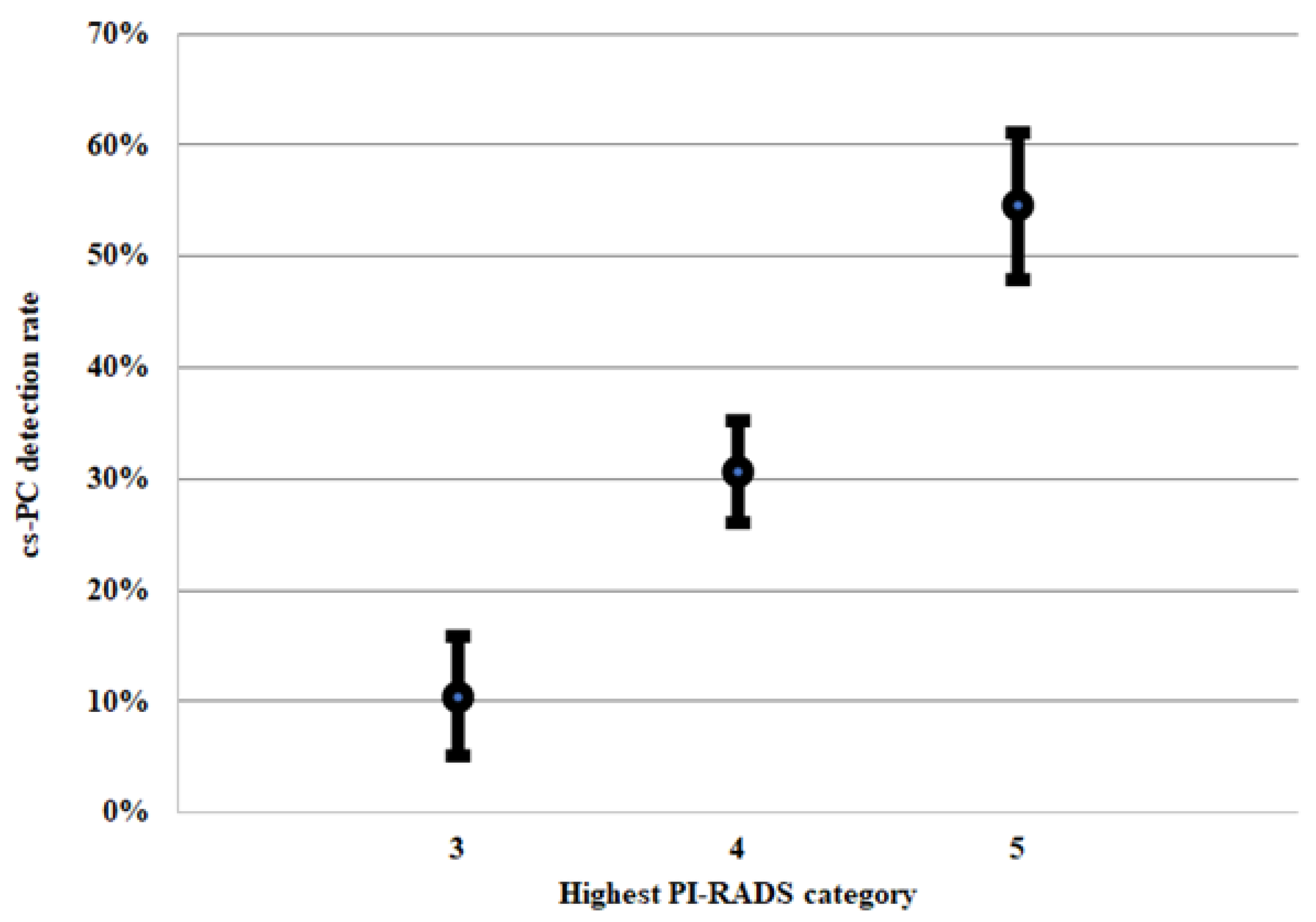
| 3 | 124 (16.8) | 63 (21.1) | 11 (9.0) | 50 (15.6) |
| 4 | 398 (53.8) | 144 (48.3) | 66 (54.1) | 188 (58.8) |
| 5 | 218 (29.5) | 91 (30.5) | 45 (36.9) | 82 (25.6) |
| Lesion in the peripheral zone (%) | 485 (69.6) a | 158 (62.0) a | 101 (82.8) | 226 (70.1) |
| Positive DRE b | 131 (18.5) b | 56 (21.0) b | 13 (10.7) | 62 (19.4) |
| Biopsy approach, n | ||||
| Transperineal | 615 (83.1) | 182 (61.1) | 113 (92.4) | 320 (100.0) |
| Transrectal | 125 (16.9) | 116 (38.9) | 9 (7.6) | 0 (0.0) |
| Median number of cores (IQR) | 11 (9, 15) | 17 (16, 19) | 19 (18, 23) | 11 (9, 14) |
| Diagnosis of csPC (%) | 254 (34.3) | 114 (38.3) | 35 (28.7) | 105 (32.8) |
| UVA vs. csPC OR (95% CI), p-Value | MVA vs. csPC OR (95% CI), p-Value | |
|---|---|---|
| Age, years | 1.05 (1.03–1.07), <0.001 | 1.05 (1.03–1.08), <0.001 |
| Biopsy-naïve | 1.42 (1.04–1.93), 0.027 | 1.57 (1.08–2.29), 0.017 |
| PSA > 10 ng/mL | 2.57 (1.81–3.65), <0.001 | 2.36 (1.53–3.64), <0.001 |
| PV, mL | 0.98 (0.98–0.99), <0.001 | 0.98 (0.97–0.98), <0.001 |
| Max. lesion size, mm | 1.07 (1.05–1.10), <0.001 | 1.05 (1.02–1.08), 0.001 |
| PIRADS > 3 | 5.49 (3.02–9.10), <0.001 | 3.14 (1.63–6.05), 0.001 |
| Lesion in PZ | 2.05 (1.43–2.95), <0.001 | 1.86 (1.24–2.79), 0.003 |
| Positive DRE | 3.14 (2.12–4.63), <0.001 | 1.74 (1.12–2.70), 0.014 |
| MVA vs. csPC OR (95% CI), p-Value | |
|---|---|
| Age, years | 1.05 (1.03–1.08), <0.001 |
| Biopsy-naïve | 1.69 (1.17–2.44), 0.005 |
| PSA > 10 ng/mL | 2.43 (1.58–3.75), <0.001 |
| PV, mL | 0.98 (0.97–0.98), <0.001 |
| Max. lesion size, mm | 1.06 (1.03–1.09), <0.001 |
| PIRADS > 3 | 3.27 (1.69–6.31), <0.001 |
| Lesion in PZ | 1.95 (1.30–2.92), 0.001 |
| MVA vs. csPC, DRE Included OR (95% CI), p-Value | MVA vs. csPC, DRE Excluded OR (95% CI), p-Value | |
|---|---|---|
| Age, years | 1.08 (0.98–1.19), NS | 1.08 (0.98–1.19), NS |
| Biopsy-naïve | 0.81 (0.20–3.39), NS | 0.86 (0.21–3.43), NS |
| PSA > 10 ng/mL | 1.69 (0.32–8.86), NS | 1.46 (0.28–7.52), NS |
| PV, mL | 0.94 (0.90–0.99), 0.019 | 0.94 (0.89–0.99), 0.017 |
| Max. lesion size, mm | 1.02 (0.88–1.19), NS | 1.03 (0.88–1.19), NS |
| Lesion in PZ | 3.17 (0.58–17.37), NS | 3.09 (0.58–16.34), NS |
| Positive DRE | 2.93 (0.42–20.31), NS | N.A. |
Disclaimer/Publisher’s Note: The statements, opinions and data contained in all publications are solely those of the individual author(s) and contributor(s) and not of MDPI and/or the editor(s). MDPI and/or the editor(s) disclaim responsibility for any injury to people or property resulting from any ideas, methods, instructions or products referred to in the content. |
© 2022 by the authors. Licensee MDPI, Basel, Switzerland. This article is an open access article distributed under the terms and conditions of the Creative Commons Attribution (CC BY) license (https://creativecommons.org/licenses/by/4.0/).
Share and Cite
Szempliński, S.; Kamecki, H.; Dębowska, M.; Zagożdżon, B.; Mokrzyś, M.; Zawadzki, M.; Sosnowski, R.; Tokarczyk, A.; Poletajew, S.; Kryst, P.; et al. Predictors of Clinically Significant Prostate Cancer in Patients with PIRADS Categories 3–5 Undergoing Magnetic Resonance Imaging-Ultrasound Fusion Biopsy of the Prostate. J. Clin. Med. 2023, 12, 156. https://doi.org/10.3390/jcm12010156
Szempliński S, Kamecki H, Dębowska M, Zagożdżon B, Mokrzyś M, Zawadzki M, Sosnowski R, Tokarczyk A, Poletajew S, Kryst P, et al. Predictors of Clinically Significant Prostate Cancer in Patients with PIRADS Categories 3–5 Undergoing Magnetic Resonance Imaging-Ultrasound Fusion Biopsy of the Prostate. Journal of Clinical Medicine. 2023; 12(1):156. https://doi.org/10.3390/jcm12010156
Chicago/Turabian StyleSzempliński, Stanisław, Hubert Kamecki, Małgorzata Dębowska, Bartłomiej Zagożdżon, Mateusz Mokrzyś, Marek Zawadzki, Roman Sosnowski, Andrzej Tokarczyk, Sławomir Poletajew, Piotr Kryst, and et al. 2023. "Predictors of Clinically Significant Prostate Cancer in Patients with PIRADS Categories 3–5 Undergoing Magnetic Resonance Imaging-Ultrasound Fusion Biopsy of the Prostate" Journal of Clinical Medicine 12, no. 1: 156. https://doi.org/10.3390/jcm12010156