Article

Impact of Intracranial Volume and Brain Volume on the Prognostic Value of Computed Tomography Perfusion Core Volume in Acute Ischemic Stroke

by
Jan W. Hoving
1,*,
Praneeta R. Konduri
1,2,*,
Manon L. Tolhuisen
1,2,
Miou S. Koopman
1,
Henk van Voorst
1,2,
Laura M. Van Poppel
1,2,
Jasper D. Daems
3,4,
Adriaan C. G. M. van Es
5,
Marianne A. A. van Walderveen
5,
Hester F. Lingsma
4,
Diederik W. J. Dippel
4,
Wim H. Van Zwam
6,
Henk A. Marquering
1,2,
Charles B. L. M. Majoie
1 and
Bart J. Emmer
1 on behalf of the MR CLEAN Registry Investigators
1
Department of Radiology and Nuclear Medicine, Amsterdam University Medical Center, University of Amsterdam, 1105 AZ Amsterdam, The Netherlands
2
Department of Biomedical Engineering and Physics, Amsterdam University Medical Center, University of Amsterdam, 1105 AZ Amsterdam, The Netherlands
3
Department of Public Health, Erasmus University Medical Center, P.O. Box 2040 Rotterdam, The Netherlands
4
Department of Neurology, Erasmus University Medical Center, P.O. Box 2040 Rotterdam, The Netherlands
5
Department of Radiology, Leiden University Medical Center, 2333 ZA Leiden, The Netherlands
6
Department of Radiology and Nuclear Medicine, Cardiovascular Research Institute Maastricht (CARIM), Maastricht University Medical Center+, 6202 AZ Maastricht, The Netherlands
*
Authors to whom correspondence should be addressed.
J. Cardiovasc. Dev. Dis. 2024, 11(3), 80; https://doi.org/10.3390/jcdd11030080
Submission received: 18 December 2023 / Revised: 5 February 2024 / Accepted: 26 February 2024 / Published: 28 February 2024

Abstract

:
Background: Computed tomography perfusion (CTP)-estimated core volume is associated with functional outcomes in acute ischemic stroke. This relationship might differ among patients, depending on brain volume. Materials and Methods: We retrospectively included patients from the MR CLEAN Registry. Cerebrospinal fluid (CSF) and intracranial volume (ICV) were automatically segmented on NCCT. We defined the proportion of the ICV and total brain volume (TBV) affected by the ischemic core as ICVcore and TBVcore. Associations between the core volume, ICVcore, TBVcore, and functional outcome are reported per interquartile range (IQR). We calculated the area under the curve (AUC) to assess diagnostic accuracy. Results: In 200 patients, the median core volume was 13 (5–41) mL. Median ICV and TBV were 1377 (1283–1456) mL and 1108 (1020–1197) mL. Median ICVcore and TBVcore were 0.9 (0.4–2.8)% and 1.7 (0.5–3.6)%. Core volume (acOR per IQR 0.48 [95%CI 0.33–0.69]), ICVcore (acOR per IQR 0.50 [95%CI 0.35–0.69]), and TBVcore (acOR per IQR 0.41 95%CI 0.33–0.67]) showed a lower likelihood of achieving improved functional outcomes after 90 days. The AUC was 0.80 for the prediction of functional independence at 90 days for the CTP-estimated core volume, the ICVcore, and the TBVcore. Conclusion: Correcting the CTP-estimated core volume for the intracranial or total brain volume did not improve the association with functional outcomes in patients who underwent EVT.

1. Introduction

Computed tomography perfusion (CTP) allows for the quantification of the perfusion status of brain tissue in patients with acute ischemic stroke [1]. The CTP-estimated ischemic core volume is associated with functional outcomes [2,3]. However, accurately predicting functional outcomes for individual patients with acute ischemic stroke remains challenging. Patient-specific brain imaging characteristics, such as intracranial volume (ICV) and total brain volume (TBV), are associated with outcome and may influence the association between the ischemic core volume and functional outcome [4,5]. Brain atrophy—which is characterized by a decrease in TBV due to the loss of brain cells and intercellular connections—is commonly considered when assessing outcomes in qualitative and quantitative neuroimaging research and is associated with functional outcomes after endovascular treatment (EVT) in patients with acute ischemic stroke [6,7,8,9]. Previous studies have shown that the degree of cerebral atrophy—which affects the total brain volume (TBV)—is significantly and independently associated with functional outcomes after EVT [6,7,8]. In addition, it has been shown that a ratio of the CTP-estimated core volume to CSF volume more accurately predicts malignant middle cerebral artery infarction [10]. Differences in ICV exist between different ethnic populations, gender, and age groups [11]. Furthermore, TBV may be affected by restricted CSF absorption (i.e., hydrocephalus), medication use, previous stroke, neurodegenerative diseases, and age itself [12]. In this study, we aim to investigate whether the association between the CTP-estimated ischemic core and functional outcome at 90 days can be improved by correcting the CTP-estimated ischemic core for the ICV or TBV.

2. Materials and Methods

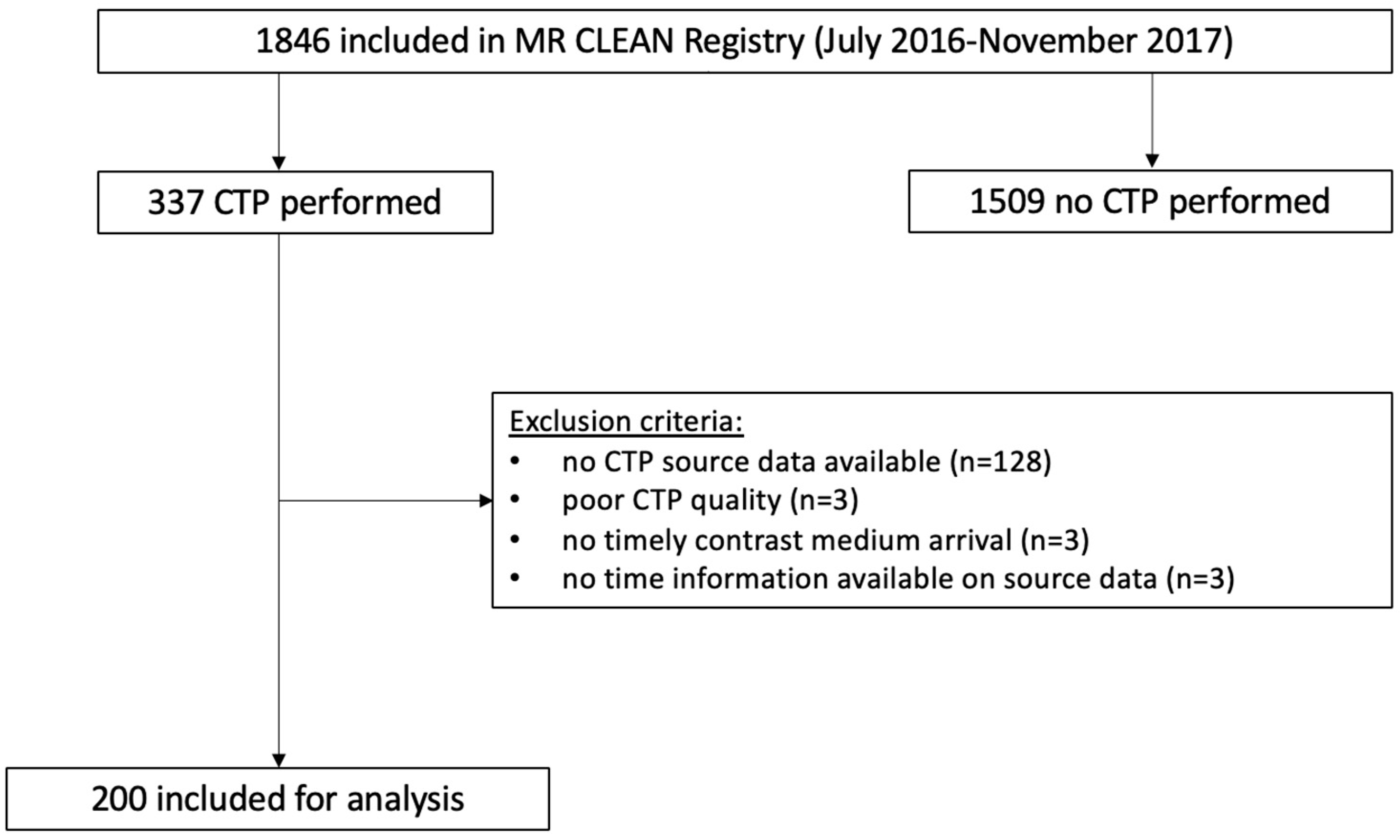
2.1. Patient Selection

We retrospectively included patients with proximal large vessel occlusion of the anterior cerebral circulation and available baseline CTP source data were included in the MR CLEAN Registry between July 2016 and November 2017. The MR CLEAN Registry is an observational, prospective registry of all consecutive patients undergoing EVT for acute ischemic stroke in the Netherlands [13]. Patients were excluded if CTP source data could not be processed by the CTP analysis software (syngo.via, version VB40) due to motion artifacts or the inadequate caption of contrast medium arrival (Figure 1).

2.2. Baseline Image Acquisition, Post-Processing, and Quality Assessment

CTP acquisition was performed according to site-specific protocols. CTP data were centrally processed using syngo.via CT Neuro Perfusion software (version VB40, Siemens Healthineers, Forchheim, Germany). Ischemic core and penumbra were defined as CBV < 1.2 mL/100 mL/s and CBF < 27 mL/100 mL/min, respectively. A default smoothing filter was applied [14]. The CTP results were visually checked by two experienced readers (>5 years of experience). Recanalization success was scored based on the extended thrombolysis in cerebral infarction (eTICI) score on post-treatment digital subtraction angiography (DSA) and ranged from 0 (no antegrade recanalization) to 3 (complete antegrade recanalization) [15].

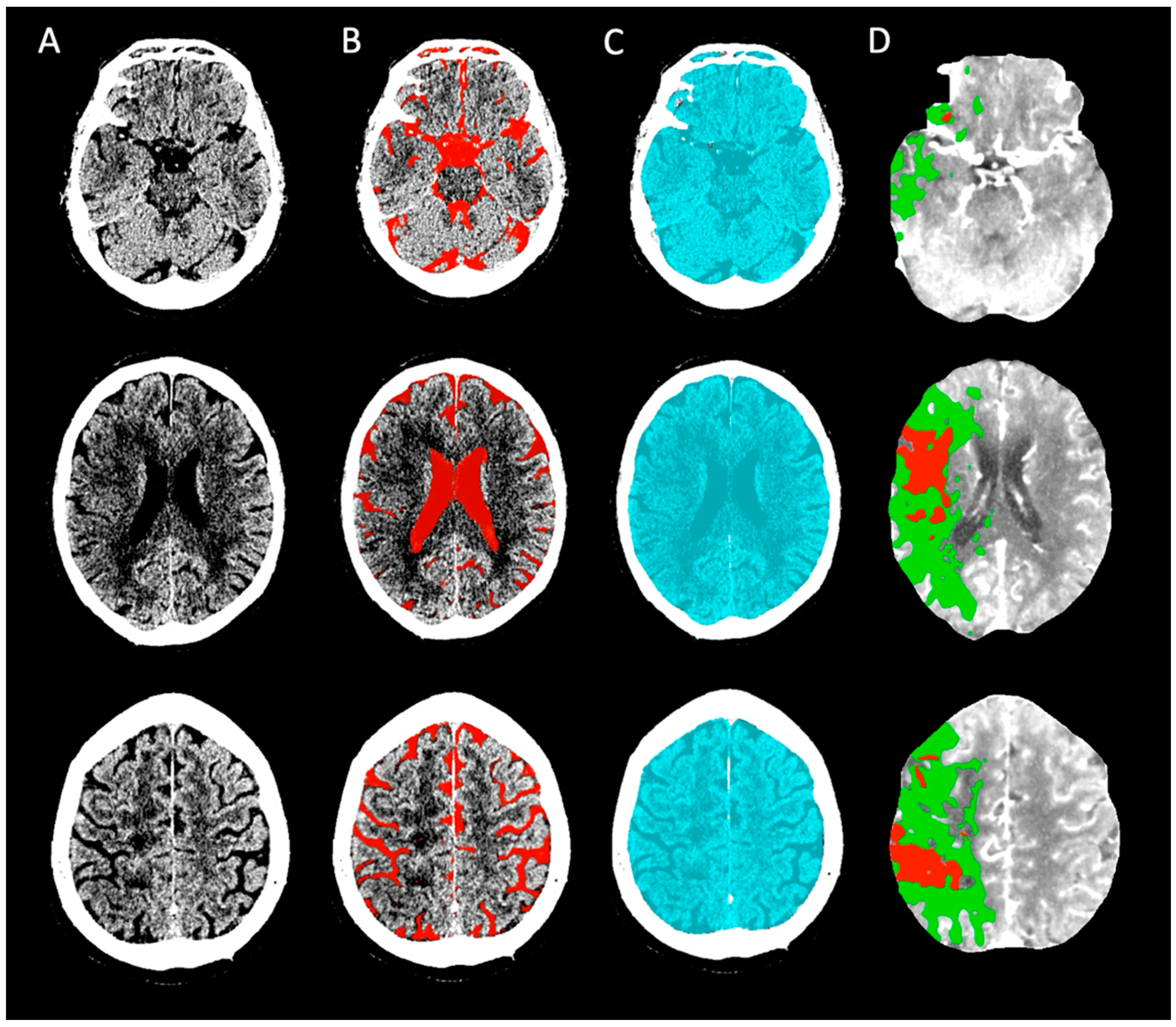
2.3. ICV and Cerebrospinal Fluid (CSF) Assessment

Baseline NCCT images were post-processed using an automated segmentation algorithm (https://github.com/WCHN/CTseg, accessed on 4 July 2021). This algorithm performed the spatial normalization of the CT images and automatically segmented the CSF volume after skull stripping of the image using a Bayesian approach [16]. ICV was segmented as the complete volume within the skull on baseline NCCT. CSF segmentations were visually checked by an expert neuroradiologist (>15 years of experience) (Figure 2). We determined the ICV and CSF volumes by multiplying the total number of voxels in the segmented intracranial area and CSF with the size of the image voxels, respectively. We calculated TBV by subtracting the CSF volume from ICV. The adjusted CTP-estimated core volumes as a proportion of ICV and TBV were defined as ICVcore and TBVcore and reported as percentages.

2.4. Statistical Analyses

The primary outcome was the 90-day functional outcome scored on the ordinal modified Rankin Scale (mRS) [17]. The secondary outcome was 90-day functional independence (mRS 0–2). We report the crude (cOR) and adjusted common odds ratio (acOR) with 95% confidence intervals (95% CI) for a shift towards improved functional outcomes on the 90-day mRS. We used uni- and multivariable binary and ordinal logistic regression to assess the associations of CTP-estimated core volume, ICVcore, and TBVcore with functional outcome. We identified age, gender, pre-stroke mRS, onset-to-groin time, the administration of intravenous thrombolysis, and baseline NIHSS as potential confounders. Since we measured and calculated continuous variables on different units and scales (i.e., milliliters and percentages), we standardized the odds ratios for ischemic core volume, ICVcore, and TBVcore by calculating the odds ratio (OR) per interquartile range. The ORs for crude ICV and TBV are presented per 10 mL. We calculated (Tjur’s and Nagelkerke’s) pseudo R2 and calculated log-likelihood to determine which model best fits the data. We performed receiver operating characteristic (ROC) analyses to determine the diagnostic accuracy of the unadjusted and adjusted CTP core variables, and the area under the curve (AUC) results were reported. Patients with missing CTP or outcome variables were excluded from our analyses. A p-value < 0.05 was considered statistically significant. Statistical analyses were performed in R (R, V3.6.0, R: A language and environment for statistical computing, R Foundation for Statistical Computing, Vienna, Austria).

2.5. Protocol Approval and Patient Consent

The Central Medical Ethics Committee of the Erasmus MC, Rotterdam, Netherlands, granted permission to carry out the MR CLEAN Registry (MEC-2014-235). The Ethics Committee waived the requirement of written informed consent for participation.

2.6. Data Availability

The datasets presented in this article are not readily available since individual patient data cannot be made available under Dutch law if no consent is obtained. All syntax files are available from the corresponding author upon reasonable request.

3. Results

We included 200 patients. A schematic representation of the patient selection is shown in Figure 1. The median age was 71 (IQR 56–80) years, and most patients were men (59%). The median ischemic core volume was 13 (IQR 5–41) mL. The median ASPECTS was 8 (IQR 9–10), the median ICV was 1377 (IQR 1283–1456) mL, the median TBV was 1108 (IQR 1020–1197) mL, the median ICVcore was 0.9% (IQR 0.4–2.8%) and the median TBVcore was 1.2 (IQR 0.5–3.6) %. Successful recanalization was achieved in 136 (71%) patients. Ninety (48%) patients were functionally independent at 90 days. A detailed overview of the baseline characteristics and outcome is given in Table 1. Pre-stroke mRS and NIHSS at the baseline were not available for 6 (3%) and 3 (2%) patients, respectively. Onset-to-groin time was not available for four (2%) patients. Fourteen (7%) patients were lost to follow-up and had missing outcome variables.

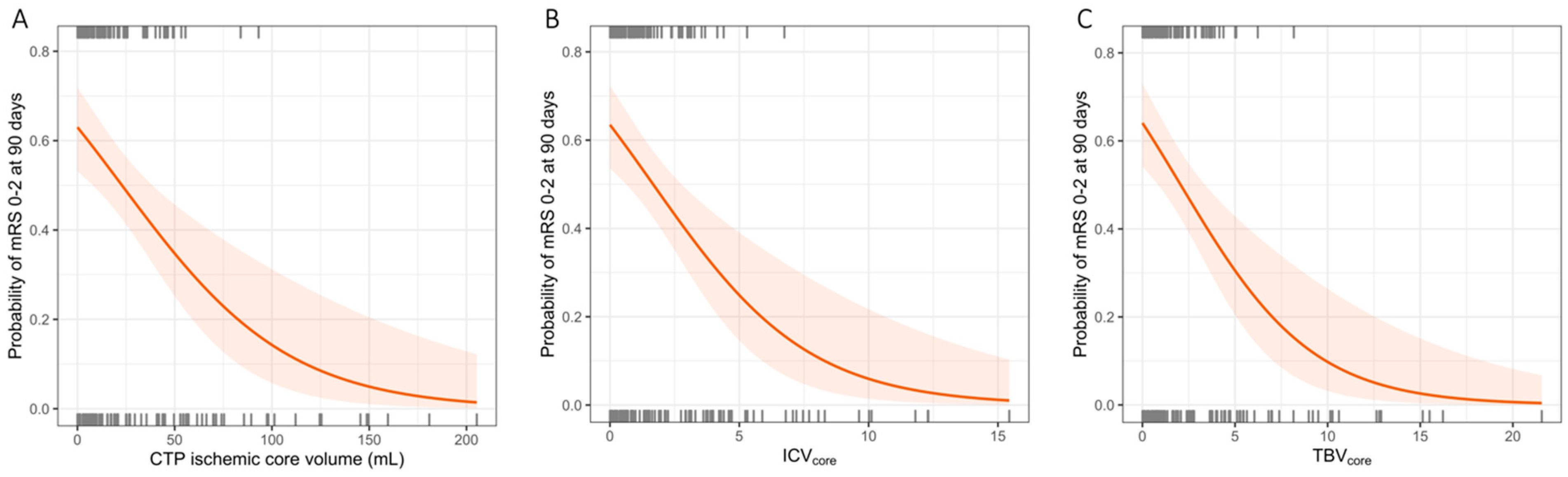
3.1. Associations between CTP-Estimated Core Volume, ICV, TBV, and Functional Outcome

The CTP-estimated core volume (cOR per IQR [mL] 0.48 [95%CI 0.33–0.69]), ICVcore (cOR per IQR [%] 0.51 [95%CI 0.39–0.69]), and TBVcore (cOR per IQR [%] 0.50 [95%CI 0.38–0.67]) were associated with a lower likelihood of improved functional outcomes at 90 days in univariable analyses. TBV was associated with improved functional outcomes (cOR per 10 mL 1.03 [95%CI 1.01–1.05]), whereas ICV was not (cOR per 10 mL 1.01 [95%CI 0.99–1.03]). After adjusting for confounders, the CTP-estimated core volume (acOR per IQR [mL] 0.48 [95%CI 0.33–0.69]), ICVcore (acOR per IQR [%] 0.50 [95%CI 0.35–0.69]), and TBVcore (acOR per IQR [%] 0.41 [95%CI 0.33–0.67]) were associated with improved functional outcomes. After adjusting for confounders, we did not observe a significant association between either TBV or ICV and improved functional outcomes. ICV and TBV were not statistically significantly associated with the CTP-estimated core volume. Detailed results, including log-likelihood and R2 values from the multivariable analyses for CTP-estimated core volume, ICVcore, and TBVcore, are provided in Supplementary Table S1 (Supplementary Materials).

3.2. Associations between CTP-Estimated Core Volume, ICV, TBV, and Functional Independence

CTP-estimated core volume (cOR per IQR 0.49 [95%CI 0.23–0.70], p < 0.001), TBV (OR per IQR 1.10 [95%CI 1.03–1.20]), ICVcore (cOR per IQR 0.45 [95%CI 0.28–0.65]) and TBVcore (cOR per IQR 0.43 [95%CI 0.26–0.63], p < 0.001) were associated with functional independence at 90 days in univariable analysis. The associations between the CTP-estimated ischemic core volume, ICVcore, and TBVcore and functional independence at 90 days are shown in Figure 3. After adjusting for potential confounders, these associations persisted for the CTP-estimated core volume (acOR per IQR 0.34 [95%CI 0.16–0.70]), ICVcore (acOR per IQR 0.36 [95%CI 0.19–0.65], and TBVcore (acOR per IQR 0.35 [95%CI 0.17–0.63], p = 0.002). Details of the multivariable regression analyses for the functional independence of the CTP-estimated core volume, ICVcore, and TBVcore are provided in Supplementary Table S2 of the Supplemental Materials. ROC analysis showed an AUC of 0.80 for the prediction of functional independence at 90 days for the CTP-estimated core volume, ICVcore, and TBVcore. The results of the ROC analysis are shown in Figure 4.

4. Discussion

This post hoc analysis of the MR CLEAN Registry showed that correcting the CTP-estimated ischemic core volume for the ICV or TBV did not result in improved functional outcome predictions compared to the CTP-estimated core volume alone. The ROC analyses showed similar diagnostic performance for all prognostic models in terms of the AUC. This could be explained by the fact that the AUC is relatively insensitive to the additional contribution of a biometric when this is estimated on a continuous scale, especially when the investigated models contain the same biometric in adjusted and unadjusted forms [18]. Therefore, it has been suggested that ORs obtained from regression analyses are more useful for explaining associations of (imaging) metrics with clinical events, such as functional independence at 90 days in patients with acute ischemic stroke [18].
Previous studies showed that both the CTP-estimated core volume and—surrogates of—brain atrophy are associated with functional outcomes after EVT [3,6,7,19,20,21,22]. For example, a post hoc analysis from the MR CLEAN trial found that cerebral atrophy modifies the effect of EVT and that the benefit of EVT was larger in patients with more severe atrophy [20]. Another retrospective cohort study showed that an increased CSF volume, as an imaging marker for biological brain age, was associated with a reduced likelihood of achieving functional independence at 90 days in patients who underwent EVT [6]. Two recent MRI-based studies confirmed this by demonstrating that TBV is an important prognostic marker of functional outcomes after stroke [4,22]. In line with these studies, we found that TBV was associated with functional outcomes. However, since none of the previous studies on the effect of brain atrophy considered CTP results, the question of whether brain imaging metrics provide additional information assessing the CTP-estimated ischemic core volume cannot be answered yet. Our observation that the association between CTP-estimated ischemic core and functional outcome was not improved by determining the proportion of affected ICV or TBV confirms—at least in part—that the relationships between baseline (imaging) characteristics and functional outcomes in acute ischemic stroke are complex and likely to be multifactorial. For example, although it is generally considered that patients with increased CSF volumes have a larger buffer in the case of edema formation [19], patients with smaller brain volumes (e.g., due to a higher frequency of other cerebrovascular comorbidities) are generally older and have worse functional outcomes, despite increased CSF volumes [23].
Several limitations to this study should be noted. First, since patients in our study cohort had relatively small ischemic core volumes (i.e., median 1% of the ICV) and the variation in ICV was limited, our results are probably not generalizable to populations with larger core volumes or more diverse intracranial volumes. Future studies focusing on the effect of the ischemic core volume in relation to brain volume should restrict their brain volume measurements to the parenchymal volume of a single hemisphere or to the specific affected vascular territory from both the affected and the contralateral hemisphere.
Second, it is important to consider that only data from EVT-treated patients were included in the MR CLEAN Registry, as well as the actual treatment effect; therefore, any potential treatment effect modification by any of the studied imaging metrics could not be measured. Third, we were not able to validate our models on an external cohort. Finally, selection bias might have occurred as patients with poor clinical and imaging profiles might have been excluded from EVT. Similarly, patients with a high clinical suspicion of LVO may not have received CTP imaging and directly underwent EVT. Our findings should be validated in a setting where CTP imaging is routinely performed, preferably including data from patients who did not undergo EVT.

5. Conclusions

Correcting the CTP-estimated ischemic core volume for the ICV or TBV does not improve the association with functional outcomes in patients who underwent EVT compared to using the CTP-estimated core volume alone.

Supplementary Materials

The following supporting information can be downloaded at: https://www.mdpi.com/article/10.3390/jcdd11030080/s1, Table S1: Multivariable analysis of improved functional outcomes (mRS) at 90 days; Table S2: Multivariable analysis of functional independence (mRS 0–2).

Author Contributions

J.W.H. and P.R.K. designed the study and collected and processed the CTP imaging data. P.R.K. performed the automated CSF and ICV segmentations. J.W.H. and P.R.K. visually checked all segmentations. J.W.H. and P.R.K. both planned and performed the statistical analysis together. J.W.H. and P.R.K. wrote the manuscript together. writing—review: M.L.T., M.S.K., H.v.V., L.M.V.P., J.D.D., A.C.G.M.v.E., M.A.A.v.W., H.F.L., D.W.J.D., W.H.V.Z., H.A.M., C.B.L.M.M. and B.J.E. All authors have read and agreed to the published version of the manuscript.

Funding

The MR CLEAN Registry was funded and carried out by the Erasmus University Medical Center, the Academic Medical Center Amsterdam, and the Maastricht University Medical Center. The Registry was additionally funded by the Applied Scientific Institute for Neuromodulation (Toegepast Wetenschappelijk Instituut voor Neuromodulatie). J.W.H. was funded by Leading the Change (LtC) (grant number 80-85009-98-2015). LtC is financed by Zorgverzekeraars Nederland and supports various healthcare evaluations in The Netherlands as part of the Healthcare Evaluation Netherlands. PK was funded by INSIST (www.insist-h2020.eu, accessed 31 January 2024), a European Union’s Horizon 2020 research and innovation programme (grant agreement number: 777072), and the RadPath AI project.

Institutional Review Board Statement

Institutional Review Board approval was obtained. The Central Medical Ethics Committee of the Erasmus MC, Rotterdam, Netherlands, granted permission to carry out the MR CLEAN Registry (MEC-2014-235). The Ethics Committee waived the requirement of written informed consent for participation.

Informed Consent Statement

The Central Medical Ethics Committee of the Erasmus MC, Rotterdam, Netherlands, granted permission to carry out the MR CLEAN Registry (MEC-2014-235). The Ethics Committee waived the requirement of written informed consent for participation.

Data Availability Statement

The datasets presented in this article are not readily available since individual patient data cannot be made available under Dutch law if no consent is obtained. All syntax files are available from the corresponding author upon reasonable request.

Acknowledgments

We would like to thank all MR CLEAN Registry and trial centers and investigators, interventionists, core lab members, research nurses, the Executive Committee, and PhD student coordinators of the MR CLEAN trial and Registry for their endless efforts in data collection, completion, and methodological and logistical support. For the names of all MR CLEAN Registry group authors, please see Appendix A.

Conflicts of Interest

B.J.E. reports grants from LtC (ZonMW and TKI-PPP of Health Holland) outside the submitted work. W.H.v.Z. reports speaker fees from Cerenovus, NicoLab, and Stryker and consultancy fees from Philips, all paid to the institution and outside the submitted work. D.W.J.D. reports funding from the Dutch Heart Foundation, Brain Foundation Netherlands, The Netherlands Organisation for Health Research and Development, Health Holland Top Sector Life Sciences & Health, and unrestricted grants from Penumbra Inc., Stryker, Medtronic, Thrombolytic Science, LLC and Cerenovus for research, all paid to the institution outside the submitted work. C.B.L.M.M. reports grants from TWIN during the conduct of the study and grants from CVON/Dutch Heart Foundation, European Commission, Healthcare Evaluation Netherlands, and from Stryker outside the submitted work (paid to institution), and they are also a shareholder of Nicolab. The MR CLEAN Registry was additionally funded by the Applied Scientific Institute for Neuromodulation (TWIN). All other authors report no conflicts of interest.

Appendix A

MR CLEAN Registry investigators:
Executive committee
Diederik W.J. Dippel1; Aad van der Lugt2; Charles B.L.M. Majoie3; Yvo B.W.E.M. Roos4; Robert J. van Oostenbrugge5,44; Wim H. van Zwam6,44; Jelis Boiten14; Jan Albert Vos8
Study coordinators
Ivo G.H. Jansen3; Maxim J.H.L. Mulder1,2; Robert- Jan B. Goldhoorn5,6,44; Kars C.J. Compagne2; Manon Kappelhof3; Josje Brouwer4; Sanne J. den Hartog1,2,40; Wouter H. Hinsenveld5,6; Lotte van den Heuvel1,40.
Local principal investigators
Diederik W.J. Dippel1; Bob Roozenbeek1; Aad van der Lugt2; Pieter Jan van Doormaal2, Charles B.L.M. Majoie3; Yvo B.W.E.M. Roos4; Bart J. Emmer3; Jonathan M. Coutinho4; Wouter J. Schonewille7; Jan Albert Vos8; Marieke J.H. Wermer9; Marianne A.A. van Walderveen10; Adriaan C.G.M. van Es10; Julie Staals5,44; Robert J. van Oostenbrugge5,44; Wim H. van Zwam6,44; Jeannette Hofmeijer11; Jasper M. Martens12; Geert J. Lycklama à Nijeholt13; Jelis Boiten14; Sebastiaan F. de Bruijn15; Lukas C. van Dijk16; H. Bart van der Worp17; Rob H. Lo18; Ewoud J. van Dijk19; Hieronymus D. Boogaarts20; J. de Vries22; Paul L.M. de Kort21; Julia van Tuijl21; Issam Boukrab26; Jo P. Peluso26; Puck Fransen22; Jan S.P. van den Berg22; Heleen M. den Hertog22; Boudewijn A.A.M. van Hasselt23; Leo A.M. Aerden24; René J. Dallinga25; Maarten Uyttenboogaart28; Omid Eschgi29; Reinoud P.H. Bokkers29; Tobien H.C.M.L. Schreuder30; Roel J.J. Heijboer31; Koos Keizer32; Rob A.R. Gons32; Lonneke S.F. Yo33; Emiel J.C. Sturm35,47, Tomas Bulut35; Paul J.A.M. Brouwers34; Anouk D. Rozeman42; Otto Elgersma41, Michel J.M. Remmers43; Thijs E.A.M. de Jong46.
Imaging assessment committee
Charles B.L.M. Majoie3(chair); Wim H. van Zwam6,44; Aad van der Lugt2; Geert J. Lycklama à Nijeholt13; Marianne A.A. van Walderveen10; Marieke E.S. Sprengers3; Sjoerd F.M. Jenniskens27; René van den Berg3; Albert J. Yoo38; Ludo F.M. Beenen3; Alida A. Postma6.45; Stefan D. Roosendaal3; Bas F.W. van der Kallen13; Ido R. van den Wijngaard13; Adriaan C.G.M. van Es10; Bart J. Emmer,3; Jasper M. Martens12; Lonneke S.F. Yo33; Jan Albert Vos8; Joost Bot36; Pieter-Jan van Doormaal2; Anton Meijer27; Elyas Ghariq13; Reinoud P.H. Bokkers29; Marc P. van Proosdij37; G. Menno Krietemeijer33; Jo P. Peluso26; Hieronymus D. Boogaarts20; Rob Lo18;Wouter Dinkelaar41; Auke P.A. Appelman29; Bas Hammer16; Sjoert Pegge27; Anouk van der Hoorn29; Saman Vinke20; Sandra Cornelissen2; Christiaan van der Leij6; Rutger Brans6; Jeanette Bakker41; Maarten Uyttenboogaart28; Miou Koopman3; Lucas Smagge2; Olvert A. Berkhemer1,3,6; Jeroen Markenstein3; Eef Hendriks3; Patrick Brouwer10; Dick Gerrits35.
Writing committee
Diederik W.J. Dippel1(chair); Aad van der Lugt2; Charles B.L.M. Majoie3; Yvo B.W.E.M. Roos4; Robert J. van Oostenbrugge5,44; Wim H. van Zwam6,44; Geert J. Lycklama à Nijeholt13; Jelis Boiten14; Jan Albert Vos8; Wouter J. Schonewille7; Jeannette Hofmeijer11; Jasper M. Martens12; H. Bart van der Worp17; Rob H. Lo18
Adverse event committee
Robert J. van Oostenbrugge5,44(chair); Jeannette Hofmeijer11; H. Zwenneke Flach23
Trial methodologist
Hester F. Lingsma40
Research nurses / local trial coordinators
Naziha el Ghannouti1; Martin Sterrenberg1; Wilma Pellikaan7; Rita Sprengers4; Marjan Elfrink11; Michelle Simons11; Marjolein Vossers12; Joke de Meris14; Tamara Vermeulen14; Annet Geerlings19; Gina van Vemde22; Tiny Simons30; Gert Messchendorp28; Nynke Nicolaij28; Hester Bongenaar32; Karin Bodde24; Sandra Kleijn34; Jasmijn Lodico34; Hanneke Droste34; Maureen Wollaert5; Sabrina Verheesen5; D. Jeurrissen5; Erna Bos9; Yvonne Drabbe15; Michelle Sandiman15; Nicoline Aaldering11; Berber Zweedijk17; Jocova Vervoort21; Eva Ponjee22; Sharon Romviel19; Karin Kanselaar19; Denn Barning10 ; Laurine van der Steen3.
Clinical/imaging data aquisition
Esmee Venema40; Vicky Chalos1,40; Ralph R. Geuskens3; Tim van Straaten19; Saliha Ergezen1; Roger R.M. Harmsma1; Daan Muijres1; Anouk de Jong1; Olvert A. Berkhemer1,3,6; Anna M.M. Boers3,39; J. Huguet3; P.F.C. Groot3; Marieke A. Mens3; Katinka R. van Kranendonk3; Kilian M. Treurniet3; Manon L. Tolhuisen3,39; Heitor Alves3; Annick J. Weterings3; Eleonora L.F. Kirkels3; Eva J.H.F. Voogd11; Lieve M. Schupp3; Sabine L. Collette28,29; Adrien E.D. Groot4; Natalie E. LeCouffe4; Praneeta R. Konduri39; Haryadi Prasetya39; Nerea Arrarte-Terreros39; Lucas A. Ramos39 ; Nikki Boodt1,2,40; Anne F.A.V Pirson5; Agnetha A.E. Bruggeman3; Nadinda A.M. van der Ende 1,2, Rabia Deniz3, Susanne G.H. Olthuis5,44, Floor Pinckaers6,44
List of affiliations
Department of Neurology1, Radiology2, Public Health40, Erasmus MC University Medical Center;
Department of Radiology and Nuclear Medicine3, Neurology4, Biomedical Engineering & Physics39, Amsterdam UMC, location University of Amsterdam;
Department of Neurology5, Radiology & Nuclear Medicine6, Maastricht University Medical Center+; School for Cardiovascular Diseases Maastricht (CARIM)44; and MHeNs School for Mental Health and Neuroscience, Maastricht, the Netherlands45;
Department of Neurology7, Radiology8, Sint Antonius Hospital, Nieuwegein;
Department of Neurology9, Radiology10, Leiden University Medical Center;
Department of Neurology11, Radiology12, Rijnstate Hospital, Arnhem;
Department of Radiology13, Neurology14, Haaglanden MC, the Hague;
Department of Neurology15, Radiology16, HAGA Hospital, the Hague;
Department of Neurology17, Radiology18, University Medical Center Utrecht;
Department of Neurology19, Neurosurgery20, Radiology27, Radboud University Medical Center, Nijmegen;
Department of Neurology21, Radiology26, Elisabeth-TweeSteden ziekenhuis, Tilburg;
Department of Neurology22, Radiology23, Isala Klinieken, Zwolle;
Department of Neurology24, Radiology25, Reinier de Graaf Gasthuis, Delft;
Department of Neurology28, Radiology29, University Medical Center Groningen;
Department of Neurology30, Radiology31, Atrium Medical Center, Heerlen;
Department of Neurology32, Radiology33, Catharina Hospital, Eindhoven;
Department of Neurology34, Radiology35, Medisch Spectrum Twente, Enschede, (currently Deventer Hospital47);
Department of Radiology36, Amsterdam UMC, Vrije Universiteit van Amsterdam, Amsterdam;
Department of Radiology37, Noordwest Ziekenhuisgroep, Alkmaar;
Department of Radiology38, Texas Stroke Institute, Texas, United States of America;
Department of Neurology42, Radiology41, Albert Schweitzer Hospital, Dordrecht;
Department of Neurology43, Radiology46, Amphia Hospital, Breda.

References

  1. Demeestere, J.; Wouters, A.; Christensen, S.; Lemmens, R.; Lansberg, M.G. Review of perfusion imaging in acute ischemic stroke: From time to tissue. Stroke 2020, 51, 1017–1024. [Google Scholar] [CrossRef]
  2. Koopman, M.S.; Hoving, J.W.; Kappelhof, M.; Berkhemer, O.A.; Beenen, L.F.M.; van Zwam, W.H.; de Jong, H.W.A.M.; Dankbaar, J.W.; Dippel, D.W.J.; Coutinho, J.M.; et al. Association of Ischemic Core Imaging Biomarkers With Post-Thrombectomy Clinical Outcomes in the MR CLEAN Registry. Front. Neurol. 2022, 12, 771367. [Google Scholar] [CrossRef] [PubMed]
  3. Campbell, B.C.V.; Majoie, C.B.L.M.; Albers, G.W.; Menon, B.K.; Yassi, N.; Sharma, G.; van Zwam, W.H.; van Oostenbrugge, R.J.; Demchuk, A.M.; Guillemin, F.; et al. Penumbral imaging and functional outcome in patients with anterior circulation ischaemic stroke treated with endovascular thrombectomy versus medical therapy: A meta-analysis of individual patient-level data. Lancet Neurol. 2019, 18, 46–55. [Google Scholar] [CrossRef]
  4. Schirmer, M.D.; Donahue, K.L.; Nardin, M.J.; Dalca, A.V.; Giese, A.-K.; Etherton, M.R.; Mocking, S.J.; McIntosh, E.C.; Cole, J.W.; Holmegaard, L.; et al. Brain Volume: An Important Determinant of Functional Outcome After Acute Ischemic Stroke. Mayo Clin. Proc. 2020, 95, 955–965. [Google Scholar] [CrossRef]
  5. Ganesan, V.; Ng, V.; Chong, W.K.; Kirkham, F.J.; Connelly, A. Lesion volume, lesion location, and outcome after middle cerebral artery territory stroke. Arch. Dis. Child. 1999, 81, 295–300. [Google Scholar] [CrossRef]
  6. Diprose, W.K.; Diprose, J.P.; Wang, M.T.; Tarr, G.P.; McFetridge, A.; Barber, P.A. Automated Measurement of Cerebral Atrophy and Outcome in Endovascular Thrombectomy. Stroke 2019, 50, 3636–3638. [Google Scholar] [CrossRef] [PubMed]
  7. Pedraza, M.I.; de Lera, M.; Bos, D.; Calleja, A.I.; Cortijo, E.; Gómez-Vicente, B.; Reyes, J.; Coco-Martín, M.B.; Calonge, T.; Agulla, J.; et al. Brain Atrophy and the Risk of Futile Endovascular Reperfusion in Acute Ischemic Stroke. Stroke 2020, 51, 1514–1521. [Google Scholar] [CrossRef] [PubMed]
  8. Lauksio, I.; Lindström, I.; Khan, N.; Sillanpää, N.; Hernesniemi, J.; Oksala, N.; Protto, S. Brain atrophy predicts mortality after mechanical thrombectomy of proximal anterior circulation occlusion. J. NeuroInterventional Surg. 2020, 13, 415–420. [Google Scholar] [CrossRef]
  9. Johnson, S.C.; Saykin, A.J.; Baxter, L.C.; Flashman, L.A.; Santulli, R.B.; McAllister, T.W.; Mamourian, A.C. The Relationship between fMRI Activation and Cerebral Atrophy: Comparison of Normal Aging and Alzheimer Disease. NeuroImage 2000, 11, 179–187. [Google Scholar] [CrossRef]
  10. Minnerup, J.; Wersching, H.; Ringelstein, E.B.; Heindel, W.; Niederstadt, T.; Schilling, M.; Schäbitz, W.-R.; Kemmling, A. Prediction of malignant middle cerebral artery infarction using computed tomography-based intracranial volume reserve measurements. Stroke 2011, 42, 3403–3409. [Google Scholar] [CrossRef]
  11. Kijonka, M.; Borys, D.; Psiuk-Maksymowicz, K.; Gorczewski, K.; Wojcieszek, P.; Kossowski, B.; Marchewka, A.; Swierniak, A.; Sokol, M.; Bobek-Billewicz, B. Whole Brain and Cranial Size Adjustments in Volumetric Brain Analyses of Sex- and Age-Related Trends. Front. Neurosci. 2020, 14, 278. [Google Scholar] [CrossRef]
  12. Dieleman, N.; Koek, H.L.; Hendrikse, J. Short-term mechanisms influencing volumetric brain dynamics. NeuroImage Clin. 2017, 16, 507–513. [Google Scholar] [CrossRef]
  13. Jansen, I.G.H.; Mulder, M.J.H.L.; Goldhoorn, R.-J.B. Endovascular treatment for acute ischaemic stroke in routine clinical practice: Prospective, observational cohort study (MR CLEAN Registry). BMJ 2018, 360, k949. [Google Scholar] [CrossRef]
  14. Koopman, M.S.; A Berkhemer, O.; Geuskens, R.R.E.G.; Emmer, B.J.; A A van Walderveen, M.; Jenniskens, S.F.M.; van Zwam, W.H.; van Oostenbrugge, R.J.; van der Lugt, A.; Dippel, D.W.J.; et al. Comparison of three commonly used CT perfusion software packages in patients with acute ischemic stroke. J. NeuroInterventional Surg. 2019, 11, 1249–1256. [Google Scholar] [CrossRef]
  15. Liebeskind, D.S.; Bracard, S.; Guillemin, F.; Jahan, R.; Jovin, T.G.; Majoie, C.B.; Mitchell, P.J.; van der Lugt, A.; Menon, B.K.; Román, L.S.; et al. eTICI reperfusion: Defining success in endovascular stroke therapy. J. NeuroInterventional Surg. 2019, 11, 433–438. [Google Scholar] [CrossRef]
  16. Brudfors, M.; Balbastre, Y.; Flandin, G.; Nachev, P.; Ashburner, J. Flexible Bayesian Modelling for Nonlinear Image Registration. In Lecture Notes in Computer Science, Proceedings of the Medical Image Computing and Computer Assisted Intervention–MICCAI 2020, Lima, Peru, 4–8 October 2020; Martel, A.L., Abolmaesumi, P., Stoyanov, D., Mateus, D., Zuluaga, M.A., Zhou, S.K., Racoceanu, D., Joskowicz, L., Eds.; Springer: Cham, Switzerland, 2020; Volume 12263. [Google Scholar] [CrossRef]
  17. van Swieten, J.C.; Koudstaal, P.J.; Visser, M.C.; Schouten, H.J.; van Gijn, J. Interobserver agreement for the assessment of handicap in stroke patients. Stroke 1988, 19, 604–607. [Google Scholar] [CrossRef]
  18. Grund, B.; Sabin, C. Analysis of biomarker data: Logs, odds ratios, and receiver operating characteristic curves. Curr. Opin. HIV AIDS 2010, 5, 473–479. [Google Scholar] [CrossRef]
  19. Kauw, F.; E Bernsen, M.L.; Dankbaar, J.W.; de Jong, H.W.; Kappelle, L.J.; Velthuis, B.K.; van der Worp, H.B.; van der Lugt, A.; Roos, Y.B.; Yo, L.S.; et al. Cerebrospinal fluid volume improves prediction of malignant edema after endovascular treatment of stroke. Int. J. Stroke 2022, 18, 187–192. [Google Scholar] [CrossRef] [PubMed]
  20. Luijten, S.P.; Compagne, K.C.; van Es, A.C.; Roos, Y.B.; Majoie, C.B.; van Oostenbrugge, R.J.; van Zwam, W.H.; Dippel, D.W.; Wolters, F.J.; van der Lugt, A.; et al. Brain atrophy and endovascular treatment effect in acute ischemic stroke: A secondary analysis of the MR CLEAN trial. Int. J. Stroke 2021, 17, 881–888. [Google Scholar] [CrossRef] [PubMed]
  21. Kaginele, P.; Beer-Furlan, A.; Joshi, K.C.; Kadam, G.; Achanaril, A.; Levy, E.; Waqas, M.; Siddiqui, A.; Rai, H.; Snyder, K.; et al. Brain Atrophy and Leukoaraiosis Correlate with Futile Stroke Thrombectomy. J. Stroke Cerebrovasc. Dis. 2021, 30, 105871. [Google Scholar] [CrossRef] [PubMed]
  22. Bu, N.; Khlif, M.S.; Lemmens, R.; Wouters, A.; Fiebach, J.B.; Chamorro, A.; Ringelstein, E.B.; Norrving, B.; Laage, R.; Grond, M.; et al. Imaging Markers of Brain Frailty and Outcome in Patients With Acute Ischemic Stroke. Stroke 2021, 52, 1004–1011. [Google Scholar] [CrossRef] [PubMed]
  23. McDonough, R.V.; Ospel, J.M.; Campbell, B.C.; Hill, M.D.; Saver, J.L.; Dippel, D.W.; Demchuk, A.M.; Majoie, C.B.; Brown, S.B.; Mitchell, P.J.; et al. Functional Outcomes of Patients ≥85 Years With Acute Ischemic Stroke Following EVT: A HERMES Substudy. Stroke 2022, 53, 2220–2226. [Google Scholar] [CrossRef]
Figure 1. Flowchart of patient selection.
Jcdd 11 00080 g001
Figure 2. Example of baseline CT imaging with ICV and CSF volume assessments; three levels in the brain are shown. (A) Non-contrast CT of a 71-year-old patient with a right-sided M1 occlusion who received IV alteplase before EVT (eTICI 2b). (B) CSF segmentation (red) shows a CSF volume of 250 mL. (C) ICV segmentation (blue) on baseline NCCT shows an ICV of 1374 mL. (D) CTP-estimated core volume (red) was 50 mL. Penumbral volume (green) was 210 mL. CSF = cerebrospinal fluid; CTP = CT perfusion; eTICI = expanded treatment in cerebral infarction; EVT = endovascular treatment; and ICV = intracranial volume.
Jcdd 11 00080 g002
Figure 3. Plot showing the association of (A) CTP ischemic core volume, (B) the proportion of ICV affected by the CTP ischemic core (ICVcore), and (C) the proportion of TBV affected by the CTP ischemic core (TBVcore) with the probability of achieving functional independence (mRS 0–2) at 90 days.
Jcdd 11 00080 g003
Figure 4. Receiver operating characteristic (ROC) curve for prediction of functional independence (i.e., mRS 0–2) at 90 days based on CTP-estimated core volume (blue line), TBVcore (green line), and ICVcore (red line). CTP = computed tomography perfusion; ICV = intracranial volume; and TBV = total brain volume.
Jcdd 11 00080 g004
Table 1. Baseline characteristics and outcome data of the MR CLEAN Registry subpopulation included in this analysis compared to the overall MR CLEAN Registry cohort. ASPECTS = Alberta Stroke Program Early CT Score; CSF = cerebrospinal fluid; CTP = CT perfusion; ICA = intracranial carotid artery; ICA-T = intracranial carotid artery terminus; IVT = IV alteplase; IQR = interquartile range; M1 = M1 segment of the middle cerebral artery; M2 = M1 segment of the middle cerebral artery; mRS = modified Rankin Score; and NIHSS = National Institute of Health Stroke Scale. If the [known in] number is not shown, the variable was known in all patients. * = Time between symptom onset and imaging at a comprehensive stroke center.
MR CLEAN Registry CTP Subgroup (n = 200)Overall MR CLEAN Registry (n = 1755)
Clinical
Age (yr)—median (IQR)71 (56–80)72 (62–81)
Female—n (%)83 (42)889 (51)
NIHSS at baseline—median (IQR) [known in]16 (12–20) [n = 197]16 (11–19)
Transfer from primary stroke center—n (%)19 (10)962 (55)
IVT administered—n (%)144 (72)1282 (73)
Onset-to-imaging time (min) *—median (IQR) [known in]79 (56–137) [N = 194]76(53–128) [N = 1279]
Onset-to-groin time (min)—median (IQR) [known in]153 (120–223) [N = 196]185(144–243) [N = 1740]
Imaging
Occlusion location on baseline CTA—n (%) [known in]
Intracranial ICA
ICA-T
M1
M2
Other
[N = 198]
6 (3)
35 (18)
121 (61)
35 (18)
1 (1)
[N = 1657]
76 (4)
342 (20)
974 (56)
295 (17)
6 (0.3)
ASPECTS—median (IQR) [known in]9 (8–10) [N = 199]9 (8–10) [N = 1713]
Collateral status—n (%) [known in]
0
1
2
3
[N = 197]
8 (4)
79 (40)
82 (42)
28 (14)
[N = 1693]
89 (5)
635 (36)
643 (37)
290 (17)
Baseline ischemic core volume on CTP (mL)—median (IQR)13 (5–41)NA
Baseline penumbra volume on CTP (mL)median (IQR)96 (58–123)NA
Intracranial volume (ICV) (mL)—median (IQR)
Males
Females
1377 (1283–1456)
1435 (1378–1502)
1280 (1224–1330)
NA
Total brain volume (TBV) (mL)median (IQR)
Males
Females
1109 (1020–1196)
1170 (1106–1233)
1020 (976–1082)
NA
CSF volume (mL)—median (IQR)
Males
Females
258 (229–296)
266 (237–303)
246 (216–286)
NA
Disclaimer/Publisher’s Note: The statements, opinions and data contained in all publications are solely those of the individual author(s) and contributor(s) and not of MDPI and/or the editor(s). MDPI and/or the editor(s) disclaim responsibility for any injury to people or property resulting from any ideas, methods, instructions or products referred to in the content.

