Review

Invertebrate Immunity, Natural Transplantation Immunity, Somatic and Germ Cell Parasitism, and Transposon Defense

by
Malgorzata Kloc
1,2,3,*,
Marta Halasa
1,2,
Jacek Z. Kubiak
4,5 and
Rafik M. Ghobrial
1,2
1
Houston Methodist Research Institute, Transplant Immunology, Houston, TX 77030, USA
2
Department of Surgery, Houston Methodist Hospital, Houston, TX 77030, USA
3
Department of Genetics, MD Anderson Cancer Center, University of Texas, Houston, TX 77030, USA
4
Laboratory of Molecular Oncology and Innovative Therapies, Military Institute of Medicine-National Research Institute (WIM-PIB), Szaserow 128, 04-141 Warsaw, Poland
5
Dynamics and Mechanics of Epithelia Group, Faculty of Medicine, Institute of Genetics and Development of Rennes, University of Rennes, CNRS, UMR 6290, 35043 Rennes, France
*
Author to whom correspondence should be addressed.
Int. J. Mol. Sci. 2024, 25(2), 1072; https://doi.org/10.3390/ijms25021072
Submission received: 6 December 2023 / Revised: 11 January 2024 / Accepted: 12 January 2024 / Published: 16 January 2024

Abstract

:
While the vertebrate immune system consists of innate and adaptive branches, invertebrates only have innate immunity. This feature makes them an ideal model system for studying the cellular and molecular mechanisms of innate immunity sensu stricto without reciprocal interferences from adaptive immunity. Although invertebrate immunity is evolutionarily older and a precursor of vertebrate immunity, it is far from simple. Despite lacking lymphocytes and functional immunoglobulin, the invertebrate immune system has many sophisticated mechanisms and features, such as long-term immune memory, which, for decades, have been exclusively attributed to adaptive immunity. In this review, we describe the cellular and molecular aspects of invertebrate immunity, including the epigenetic foundation of innate memory, the transgenerational inheritance of immunity, genetic immunity against invading transposons, the mechanisms of self-recognition, natural transplantation, and germ/somatic cell parasitism.

1. Introduction

All organisms, even unicellular ones, must have the mechanisms to distinguish the self from the non-self and defend against pathogens. Progressive improvements of such mechanisms over millions of years of evolution culminated in a marvel—the jawed vertebrates’ immune system with two mechanistically distinct but interacting branches: innate and adaptive immunity. While innate immunity is the fast and generic first line of defense, adaptive immunity is specific, anticipatory, long-lasting, and able to memorize previous encounters with antigens. This traditional view of the strict division of immune defense tasks does not hold ground in the view of recent advancements in molecular and cellular studies [1]. In the traditional view, innate immunity relies on motile, ameba-like cells (amebocytes, hemocytes, coelomocytes, monocytes, and macrophages), which, for many decades, were assumed to perform ancestral, straightforward functions of moving toward the target (pathogen or any foreign object), engulfing it, and digesting (destroying) it. However, many studies, especially those on invertebrates (worms, insects, crustaceans, chelicerates, mollusks, echinoderms, and tunicates) whose immune system consists only of the innate branch, showed that such a simplistic view of innate immune cells is inaccurate and that these cells can also memorize previous encounters with pathogens and develop lasting immune memory—the features of adaptive immunity [2,3,4]. Such innate immune memory, also called “trained immunity”, supplements adaptive immune memory and regulates adaptive responses (in organisms that have both immune branches} and involves cell metabolism modifications and epigenetic reprogramming. Another aspect of innate immunity is the humoral response, traditionally attributed to antibody-producing B cells, that acts through the functional ancestors of antibodies, such as pentraxins, collectins, and ficolins, which activate the complement, opsonize, agglutinate, and kill microorganisms [5,6,7,8]. On the other hand, adaptive immune cells (B and lymphocytes) can recognize and phagocytose targets using pattern recognition, thus performing functions originally attributed to innate immune cells [1,9,10].
Despite immunologic commonalities between different invertebrate groups, genomic studies showed that even closely related invertebrate species employ very different approaches in dealing with these universal challenges, which, in turn, suggests that invertebrate immune systems have evolved many times, independently from a common ancient precursor [1,4,11,12,13].
The following paragraphs describe some of the complexities of the invertebrate immune system and the role of natural transplantation immunity, somatic and germ cell parasitism, and transposon defense mechanisms.

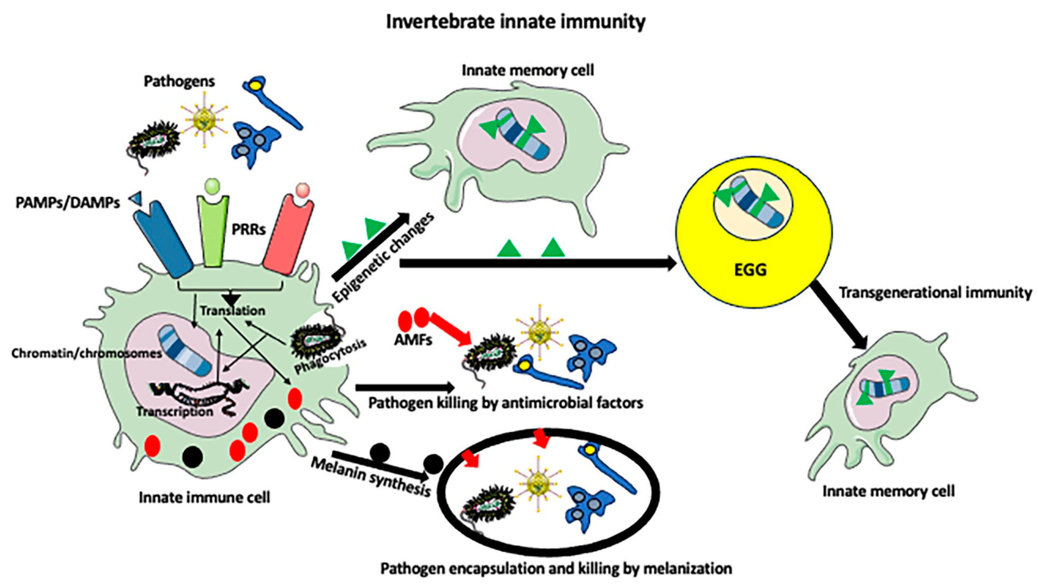
2. Invertebrate Immunity

Although invertebrates are tremendously diversified in morphology, body plan, lifestyles, lifespan, and evolutionary history, all must cope with similar infectious agents (viruses, bacteria, protists, and fungi) and recognize genetically identical and allogeneic individuals. In invertebrates and vertebrates, the recognition of the invading pathogens is based on the same principle of all pathogenic organisms having certain molecular components that are different from those in the host. All these pathogen-specific components, which are usually located on the pathogen’s surface, are collectively called pathogen-related molecular patterns (PAMPs). There are many categories of PAMPs, such as various polysaccharides, lipids, peptides, proteins, and nucleic acids. PAMPs are recognized by the pattern recognition receptors (PRRs) expressed by innate immune cells (Figure 1). Different invertebrate species have different PRRs. For example, the PRRs typical for Lepidoptera are C-type lectin (CTL), peptidoglycan recognition protein (PGRP), β-1,3-glucan recognition protein (βGRP), and Gram-negative binding protein (GNBP) [11,14]. Other compounds also recognized by PRRs are damage-associated molecular patterns (DAMPs)—the molecules released from stressed, damaged, or dying cells, i.e., they represent an endogenous danger signal. Protein DAMPs include heat-shock proteins and molecules derived from the extracellular matrix of injured tissues, and non-protein DAMPs include nuclear and mitochondrial DNA, RNAs, ATP, components of ER, and cell membranes. Although DAMPs and PAMPs activate similar inflammatory responses, those caused by DAMPs are called the aseptic (infection-unrelated) type of inflammation. One of the insect-specific DAMPs is the dorsal switch protein 1 (DSP1)—an ortholog of mammalian high-mobility group box 1 protein (HMGB, amphoterin) released during autophagy [14]. Other groups of receptors recognizing both PAMPs and DAMPs and that are common between vertebrates and invertebrates are Toll-like receptors (TLRs) and scavenger receptors (SRs). Some TLRs are known to have a very high affinity to specific PAMPs. For example, TLR1 and TLR2 have a high affinity for bacterial lipoproteins and peptidoglycans, TLR3 has a high affinity for viral double-stranded RNA, TLR5 has a high affinity for bacterial flagellin, TLR9 has a high affinity for the unmethylated CpG of viral and bacterial DNA, and TLr11 and 12 have high affinity for bacterial profilin [15]. TLRs occur in all invertebrate groups, and because they are also present in the simplest invertebrates, such as cnidarians and sponges, they are evolutionarily the most ancient receptors [11]. For example, in shrimps, the PmToll receptor in Panaeus monodon, lToll in Litopenaeus vannamei, and MjToll in Marsupenaeus japonicus have been cloned and described functionally [16,17,18,19]. The SRs are diversified structurally, which allows them to bind a wide range of ligands and cooperate with other PRRs, such as TLRs, in activating phagocytosis and inflammation pathways [11,20,21,22,23]. Recently, various PRRs, including TRLs, have been identified in the transcriptome of the dung beetle Copris tripartitus [24] and giant African snail Achatina immaculata [25]. In Drosophila embryo, macrophages and hemocytes express scavenger receptor SR-CI (dSR-CI) [26], and shrimp M. japonicus has the B class type III scavenger receptor, SRB2, which induces an immune deficiency (IMD) pathway (see below) [27]. IMD and antimicrobial (AMP) pathway components were also described recently in the transcriptome of tick Amblyomma americanum [28].
The ability to make antimicrobial compounds (AMPs) is one of the hallmark functions of invertebrate innate immune cells. AMPs are made by all invertebrate and vertebrate species. AMPs are usually hydrophobic and cationic and effective against a wide range of microorganisms. A detailed description of various invertebrate and vertebrate AMPs and their promise for therapeutic use is given in [29,30]. Pathways regulating AMP production are especially well described in insects such as Drosophila, Tenebrio, and Triatoma. Two main pathways mediated by the nuclear transcription factor-kappa B (NF-κB) are as follows: Toll pathway against Gram-positive bacteria and fungi and the immune deficiency (IMD, named after the mutation in Drosophila causing severe immune deficiency) pathway against Gram-negative bacteria [31,32,33,34,35,36]. In the Toll pathway, the TLRs, upon recognizing PAMPs, recruit specific adaptor molecules and activate transcription factors NF-κB and, ultimately, the expression of proinflammatory genes and AMPs. In the IMD pathway, the recognition of PAMPs by PRRs, such as PGRP-LC and PGRP-LE, leads to the formation of PGRP homo- and heterodimers and the recruitment of the IMD protein, which in insects shares similarities with the receptor-interacting protein (RIP) of the mammalian tumor necrosis factor receptor (TNFR), followed by the activation of intracellular signaling and the phosphorylation and cleaving of NF-κB transcription factor Relish into N-terminal (Rel-68) and C terminal (Rel-49) fragments. Subsequently, Rel-68 translocates into the nucleus and activates the expression of AMPs [35,37].
Another form of antimicrobial defense is the programmed cell death of immune cells (ETosis), in which, in response to a pathogen, nuclear chromatin is decondensed and dispelled from immune cells. The strands of released DNA create web-like structures (extracellular traps, ETs) decorated with antimicrobial compounds, which capture and kill pathogens. ETosis occurs in the neutrophiles (NETosis) and macrophages (METosis) of vertebrates and the hemocytes of invertebrates [38,39,40].
Additionally, antimicrobial defense is supported by the proteasomal and autophagy pathways. Proteasomes are giant cytoplasmic and nuclear multi-subunit proteases that digest proteins tagged for degradation via polyubiquitination. The proteasome ubiquitination pathway occurs in all animals and plants and, by targeting foreign proteins for proteolysis, defends against microorganisms and bacterial protein toxins [41,42]. Similarly, in the defense against invading pathogens, macroautophagy and autophagy eliminate intracellular components and pathogens through a lysosome-dependent degradation pathway and activate a series of immune responses through pattern recognition receptors (PRRs) [43,44].

3. Invertebrate Immune Cells

Except for the simplest invertebrates, such as cnidarians, most invertebrates contain immune cells. However, despite lacking specialized, mobile immune cells, cnidarians, such as Hydra, have a robust immune defense that is sometimes called “the mucosal immunity” because of its similarity to the immune responses of epithelia in the gastrointestinal and upper respiratory tracts’ mucosa in vertebrates [45,46,47]. Hydra has tissues but does not have organs. The Hydra body is a simple tube built from two epithelial (ectoderm and endoderm) layers with interspersed multipotent stem cells, which can differentiate into nematocytes (stinging cells), gametes, and secretory and nerve cells [48,49]. The Hydra epithelium is covered by a multilayered glycocalyx that forms a physicochemical barrier against pathogens [47]. Although Hydra lacks mobile phagocytes, stationary epithelial cells can phagocytose and destroy pathogens. Additionally, hydra’s epithelium has pattern recognition molecules, based on lectin–carbohydrate interactions, that recognize compounds present in different microorganisms, and it produces potent antimicrobial peptides (AMPs), which fight pathogens. The Hydra genome (which has only ~20,000 genes) lacks conventional Toll-like receptors (TLRs), and the production of AMPs is induced by the interaction between proteins containing leucine-rich repeats (LRRs) with proteins lacking LRRS but containing the Toll/Interleukin-1 Receptor (TIR) domain [45]. The expression of AMPs is also controlled by the forkhead box O (FOXO) transcription factor [47,50].
The more advanced invertebrates have specialized immune cells that are similar in morphology and functions to vertebrate macrophages, but different taxonomic groups and species have different types of immune cells. Arthropods and mollusks have open (lacking the vasculature) circulatory systems, and the body cavity is filled with hemolymph, which is analogous to vertebrate blood. Hemolymph usually does not contain red blood cells or hemoglobin (although the intercellular hemoglobin can be present in various insect tissues such as tracheal system and fat body in Drosophila) but contains hormones, nutrients, ions, carbohydrates, lipids, pigments, and various types of immune cells (hemocytes). Single-cell RNA sequencing showed that the hematopoietic organ (lymph gland) of Drosophila larvae contains several types of myeloid cells: stem-like prohemocytes, intermediate prohemocytes, prohemocytes, plasmatocytes, lamellocytes, adipohemocytes, and crystal cells [51]. Plasmatocytes are very similar to vertebrate macrophages; they phagocytose, remodel tissues, mount an immune response, and express genes involved in metabolism, the cell cycle, and antimicrobial responses [52]. Lamellocytes, which in healthy insects are rare, form in response to parasitic wasps and some environmental factors. They encapsulate pathogens that are too large to be phagocytosed. Adipohemocytes play a role in lipid metabolism, and crystal cells contain a prophenoloxidase (ProPO) enzyme that participates in the melanization immune response (see below) [51,53,54]. Interestingly, as shown in Tenebrio, Manduca sexta, and Drosophila, the activation of the ProPO cascade and melanization pathway shares serine proteases with the Toll pathway [55,56,57]. Studies using single-cell RNA sequencing (scRNA-seq) identified subsets within lamellocytes that express fibroblast growth factor (FGF) receptor breathless and crystal cells that express FGF ligand branchless, which play a role in the response against parasitic wasps [52]. A comprehensive list of genes expressed by Drosophila hemocyte subtypes was recently published in [58]. A recent cross-species comparison of single-cell RNA-sequencing data showed that Drosophila plasmocytes are homologous to vertebrate monocytes and macrophages and prohemocyte 1 (PH1) is homologous to vertebrate hematopoietic stem cells. Additionally, a subpopulation of Drosophila prohemocytes with hematopoietic features was identified as a homolog of vertebrate hematopoietic progenitors [59]. Single-cell RNA sequencing studies of immune cells in shrimp identified three major types of hemocytes: prohemocytes, monocytic hemocytes, and granulocytes. Monocytic hemocytes are like vertebrate phagocytotic macrophages and express specific markers, such as apoptotic and inflammatory-response-related genes: the nucleotide-binding domain and leucine-rich repeat protein 3 (NLRP3) Nlrp3 and Casp1 [59]. Another study on shrimp hemocytes revealed that they also share some features with mammalian immune cells. Li et al. [60] analyzed the transcription factor FOXO in shrimp Marsupenaeus japonicus challenged with a Gram-negative bacteria Vibrio anguillarum. FOXO plays a role in mucosal immune responses in mammals, and it plays a role in gut humoral immune response and low-oxygen immune-like response in invertebrates [61,62,63]. In shrimp, FOXO maintains hemolymph and intestinal microbiota homeostasis by inducing the expression of transcription factor Relish, which belongs to the immune deficiency (IMD) pathway involved in the expression of antimicrobial peptides. Bacteria-challenged shrimps activated FOXO and induced its translocation to the nucleus, where it regulated antibacterial Amp and Relish genes. Additionally, FOXO induced phagocytosis via the upregulation of the phagocytotic receptor—the scavenger receptor C (Src)—and Rab5 and Rab7, two small GTPases regulating phagosome trafficking to the lysosomes [61].
There are also many studies analyzing the immune cells in colonial tunicates, a group of marine invertebrate chordates that are the closest living ancestors of vertebrates. Their circulatory system consists of the heart and extracorporeal vasculature, which share the blood supply within the colony [64]. Studies of colonial tunicate Botryllus schlosseri showed that their hematopoietic system has a mixture of invertebrate and vertebrate features. Rosental et al. [65] identified three subpopulations of phagocytic cells: major phagocytic amoebocytes; large phagocytes, which mainly contributed to allogeneic phagocytosis; and myeloid cells, which were the main contributors to phagocytosis. They also identified large granular lymphocyte-like (LGL) cells (also called the morula cells) with cytotoxic properties, and they were similar in morphology and function to mammalian natural killer (NK) cells. Despite similarities to mammalian lymphocytes, these cells mainly express tunicate-specific genes [65].
Another type of invertebrate innate immune cells is multinucleated giant hemocytes (MGHs), which fight parasitoid infections (such as wasp eggs and larvae) in insects [66]. Invertebrate MGHs are probably an evolutionary precursor of macrophage-derived multinucleated giant cells—a hallmark of chronic inflammation, aging, and cancer in vertebrates [67]. MGHs circulating in insect hemolymph are highly mobile, and when they encounter the parasite, they encapsulate and kill it. The multinuclearity of MGHs serves to amplify the expression of anti-parasitic genes. The ultrastructural and live cell imaging of Drosophila ananassae infected with parasitoid wasps showed that MGHs form an intricate system of intracytoplasmic membrane structures and exosomes, which are used for parasite encapsulation. MGHs also express high levels of hemolysin-like proteins and pore-forming toxins of prokaryotic origin, which are used for parasite elimination [66]. Studies on Anopheles gambiae showed that in response to Plasmodium infection, its hemocytes differentiate via a homolog of the Drosophila Dorsal Rel 1 signaling pathway, into giant cells (megacytes), which are recruited to the midgut surface where they eliminate Plasmodium ookinetes [68].

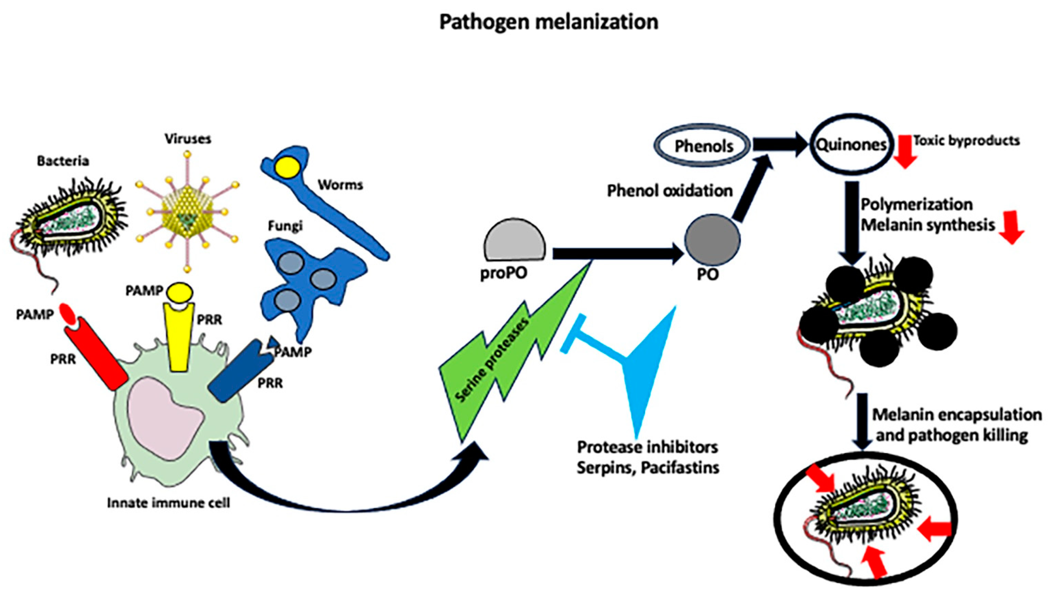
4. Melanization Immune Response

In many arthropods, including insects such as Drosophila and mosquitoes, the immune response against invading pathogens involves the melanization process, which is when the host produces melanin that encapsulates, isolates, prevents dissemination, and kills the pathogen (Figure 2). Because melanization occurs within a few minutes after infection, it is a much faster response than taking several hours to fully mobilize and activate immune cells/effectors [69,70]. Melanin is a generic name for a group of natural pigments found in most organisms that are produced in the process of melanogenesis, which involves the oxidation of phenols to quinones followed by their polymerization [53,70,71,72,73]. In healthy organisms, melanin is produced and stored in specialized cells—melanocytes, which besides making melanin secrete signaling molecules, such as cytokines, POMC peptides (the source of adrenocorticotropic hormone (ACTH), β-lipotropin, β-endorphin, and neuropeptides), catecholamines, and nitric oxide [74]. Upon infection, the pattern recognition receptors on host cells induce a cascade of serine proteases, which activate phenol oxidase (PO), an enzyme directly involved in melanin biosynthesis. PO catalyzes the oxidation of phenols to quinones, which cross-link to form melanin over the pathogen’s surface. Melanin is either produced directly from circulating precursors, or melanin-producing cells adhere tightly to each other and surround the pathogen, forming a tight capsule around it, and, in the next step, they deposit melanin [57]. Because the quinones and reactive oxygen species produced during melanization are toxic, melanization, besides sequestering the pathogen, also kills it. The toxicity of melanization intermediates requires the tight regulation of the melanization process to prevent damage to the host. Thus, the organisms contain PO pathway inhibitors, such as serpins and pacifastins. Genetic studies in Drosophila showed that the mutations of melanization inhibitors (serpins) cause spontaneous melanization and lethality [72,75]. The enzymes and pathways involved in the melanization process are described in detail in [70].

5. Innate Immune Memory

In the most basic definition, immune memory is the ability of a quiescent immune system to recall and respond to challenges encountered in the past, resulting either in the development of tolerance or the potentiation of an immune response. Since its discovery in 1997 in B cells, immune memory has been, for many years, attributed only to adaptive immunity [76]. However, recent studies showed that mouse macrophages have three types of immunological memory: innate nonspecific immunological memory; mediated by the pattern recognition receptor (PRR); innate antigen-specific immunological memory, which is dependent on the macrophage MHC-I receptor (the paired immunoglobulin-like receptor-A); and adaptive immunological memory, created with assistance from various subpopulations of adaptive immune cells, such as T and B cells [77,78,79,80,81]. In vitro studies and mice lacking B and T cells showed that vaccination with Bacillus Calmette-Guérin (BCG) resulted in immune priming, the development of trained immunity, and cross-protection against non-mycobacterial infections, which involved the epigenetic reprogramming of monocytes and macrophages (see below). For three months after vaccination, these innate immune cells upregulated inflammatory factors and reactive oxygen species and increased microbicidal and phagocytic responses after a second exposure to the same or similar pathogen [82,83,84,85,86,87,88]. Because innate immunity evolved before the emergence of adaptive immunity, the innate immune memory of invertebrates must thus be a prototype for adaptive immune memory. In Drosophila melanogaster, after the uptake of viral RNA, the hemocytes produce anti-viral short interfering RNAs (siRNAs), which, after being secreted in exosomes, act like vertebrate antibodies by conferring protection against the virus in naive flies. In Aedes aegypti, viral DNA integrated into the host genome is transcribed into PIWI (P-element induced wimpy testis in Drosophila)-interacting RNAs (piRNAs), which confer heritable protection on next generations against subsequent encounters with the virus [79,85,89,90,91]. Even plants that do not have motile immune cells have immune memory, the so-called systemic acquired resistance (SAR), which can last for a lifetime and depends on salicylic acid (SA) signaling and heritable epigenetic changes in the plant genome [92,93,94,95,96].

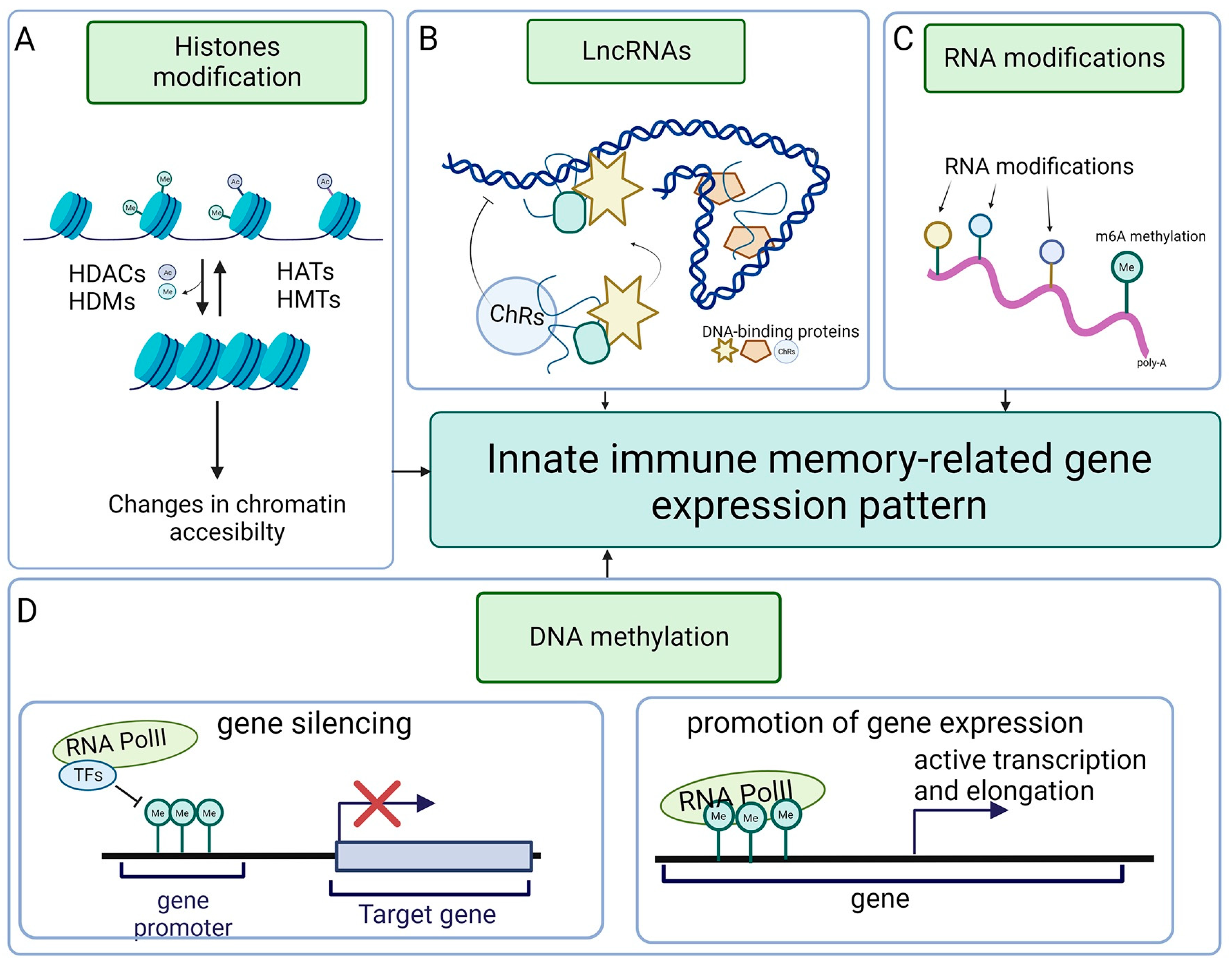
6. Epigenetics of Invertebrate Immunity

In the broadest sense, epigenetics refers to heritable traits that do not depend on changes in the DNA sequence (Figure 3). The main categories of epigenetic modifications are those changing the structure and transcriptional accessibility of chromatin, DNA methylation, histone modifications, noncoding RNAs, and RNA epigenetics that consist of post-transcriptional modifications of tRNA, mRNA, and rRNA, which modulate gene expression [97,98,99]. Another recently discovered category is the so-called RNA recoding, which is an epigenetic process of RNA editing that alters the amino acid sequence of proteins [100,101,102,103]. Epigenetic changes can be induced not only by environmental factors but also by pathogens, which train and boost innate immune responses but also hijack epigenetic mechanisms for their benefit to subdue immunity [98]. Although some of the epigenetic modifications are rapidly reversible, others can be long-lived, persisting not only through many generations of mitotically dividing cells but also by being passed on through germ cells from parents to the offspring, contributing to the development of heritable innate immune memory [103,104].
In invertebrates and vertebrates, chromatin architecture and changes from open (accessible for the transcription factors) to condensed (transcriptionally silent) configurations are regulated by reversible histone acetylation or methylation [104]. The reversible acetylation of histones depends on histone acetyltransferases (HATs), which transfer the acetyl group from acetyl-CoA to the lysine amino acids of histone tails and histone deacetylases (HDACs), which remove the acetyl groups [105]. The dysregulation of HAT/HDAC affects invertebrate antimicrobial immune response. In honeybees, the downregulation of HDACs upregulates histone acetylation, the Janus kinase/signal transducer and activator of the transcription (JAK/STAT) pathway, and the expression of antimicrobial peptides (AMPs) [106]. In mosquitoes, viral infection induces the expression of histone acetyltransferase CBP, opens chromatin structure, and switches on immune genes, while its downregulation increases viral titer and mosquito mortality [107]. Also, insect parasitoids modulate the expression of HAT/HDAC genes and suppress host immunity [108]. Epigenetic modifications are heritable: for example, heat stress changes histone acetylation patterns and induces the epigenetic transgenerational inheritance of heat resistance in the parthenogenetic brine shrimp model [101,109].
Another epigenetic modification that regulates gene expression is DNA methylation catalyzed by DNA methyltransferases (DMMTs) [110]. Although average global DNA methylation in invertebrates is lower (20–40%) than in vertebrates (80%), in some invertebrate species, such as prawns, only 2% of the genome is methylated, while in other species, such as clam worms, 80% of the genome is methylated [111]. Although it is well documented that changes in methylation patterns can lead to immune disorders in humans [112], much less is known about its effect on invertebrate immunity. Recent studies showed that oyster hemocytes express a high level of DNMT3 transferase, and the bacterial challenge upregulates the expression of DNMT3 mRNA [113], while the treatment with epigenetic modifying drug decitabine (DNA-specific methyltransferases inhibitor) upregulates immune-related genes [114]. It seems that, at least in marine invertebrates, there is a tendency to increase DNA methylation in response to heat stress and other environmental challenges [115,116]. In insects, such as silkworms, the suppression of DNMTs increases the apoptosis of virus-infected cells and suppresses virus proliferation [117]. The DNMT inhibitor (5-AZA) upregulates the expression of antimicrobial peptides (AMPs) and inhibits bacterial replication in silkworms [118]. A similar correlation between DNA methylation and the expression of AMPs was also described in other insects [119,120,121]. Recent studies indicate that epigenetic changes responsible for the development of intragenerational or intergenerational innate immune memory in invertebrates may depend not only on DNA but also on RNA modifications and/or recoding [100,122]. In invertebrates and vertebrates, the adenosine deaminases acting on RNA (ADARs) edit RNAs by changing the adenosine to inosine (A-to-I). mRNA editing within the coding region results in recoding (codon changes), which, in turn, produces protein variants with a changed composition of amino acids and creates phenotypic plasticity [123,124]. Thus, it is possible that RNA recoding may produce protein variants of different functionality not only in response to environmental factors but also to pathogens. Although RNA recoding is a rare process in humans, in some invertebrates, such as cephalopods (squid, cuttlefish, and octopus), around 60% of all mRNAs are recoded [124,125]. Besides adenosine-to-inosine (A-to-I) RNA recoding, other modifications of mRNA, rRNA, tRNA, snRNA, and miRNA, such as N6-methyladenosine (m6A), 5-methylcytosine (m5C), N1-methyladenosine (m1A), N7-methylguanosine (m7G), N4-acetylcytosine (ac4C), pseudouridine (Ψ), and uridylation, modulate immune responses by regulating immune cell polarization, activation, migration, and differentiation [120]. There are many examples of RNA modification/recoding affecting macrophage and monocyte functions in mammals. For example, m6A affects the antiviral and anticancer response of monocytes and macrophages; directs, through the Foxo-1-IL-10 axis, macrophage polarization toward M2 phenotype; and promotes macrophage pyroptosis [122,126,127]. Studies of the DNA and RNA methylation status in the beetle Tenebrio molitor showed the presence of methylated cytosine residues (5mC) in RNA during immune priming after a second challenge with bacteria and fungus [128], which suggests that RNA modification/recoding may also affect innate immune memory in invertebrates.

7. Intergenerational and Transgenerational Immunity

In many animal and plant species, the memory of an encounter with pathogens can be transmitted to the next generation (intergenerational immunity) or several subsequent generations (transgenerational immunity, transgenerational immune priming, and TGIP) [129,130,131,132]. The transmission of short-lived immunity, based on non-genetic factors, between the mother and offspring, is well known in mammals, where the antibodies developed by the mother pass through the placenta and are transferred via breast milk to the newborn, and this also occurs in other vertebrates and invertebrates whose eggs may contain fragments of pathogens, maternal antibodies, antimicrobial agents, or silencing RNAs [130,131,133,134]. Interestingly, in animals with male pregnancy, such as seahorses, immunity can be passed to the egg from the father [135]. However, as intergenerational and transgenerational immunity must be long-lived, it requires a more permanent solution, i.e., an immune inheritance. TGIP has been described in many species of Crustacea, Coleoptera, Hymenoptera, Orthoptera, and mollusks [130]. Studies in the worm C. elegans showed that the immune memory of pathogen encounters transferred between generations regulates immune gene expression levels and imprints protective behavior with respect to pathogen avoidance in the progeny [132]. For example, parental exposure to Pseudomonas vranovensis induces the upregulation of cysteine synthase genes cysl-1 and cysl-2 and rhy-1 [136]. Cysteine and other amino acid metabolism and rhy-1(which encodes a multi-pass transmembrane protein and inhibits a hypoxia inducible factor in C. elegans) are known to promote immune cell functions [137,138]. Some studies suggested the involvement of RNAi in the establishment of transgenerational immunity in C. elegans after virus exposure, but other studies did not confirm the involvement of antiviral interfering RNAs in this process [139,140,141].
A global proteome analysis of mealworm beetle Tenebrio molitor eggs from mothers primed with Gram-positive and -negative bacteria showed the abundance of immunologically active proteins and peptides, such as heat shock proteins; annexin; prophenoloxidase; transferins; perilipin; tenecins 1, 2, and 4; defensin; coleoptericin; and attacin, but no RNAs were found [131]. Although this study clearly showed that these immune factors protected Tenebrio eggs from infection, it is still unknown what mechanisms (most probably epigenetic, see below) are involved in the establishment of transgenerational immunity in this species. Studies in another model insect, house fly Musca domestica, showed that adults primed with heat-killed C. albicans can transfer immunity to the next generations. A transcriptome analysis of primed adults identified 24 upregulated and 6 downregulated genes, including metabolic genes and Toll signaling and phagosome pathways, and 154 differentially expressed (80 upregulated and 74 downregulated) genes were identified in their larvae when exposed to a lethal dose of C. albicans [142]. These results indicate that the immune response of the parent induces differential gene expression in the offspring, but again, there is no information on how this happens and what the exact mechanisms underlying the emergence of transgenerational immunity are.
The analysis of transgenerational immunity is complicated not only by differences in pathogen virulence but also by the longevity and habituation mode (philopatry versus dispersal) of different host species. By analyzing published data on 21 invertebrate species, Pigeault et al. [129] developed a theoretical host–parasite model to understand how the above parameters affect the evolution of transgenerational immunity. One of the conclusions is that transgenerational immunity will only develop when the mother is exposed to a moderately virulent pathogen. If virulence is too high, the chances of survival and reproduction of the individual are so low that there is no reason to invest in the maternal transfer of immunity. Similarly, short-lived species would not benefit from transgenerational immunity because they naturally die very fast, and the chances of encountering the same pathogen they are immune to are minuscule. There is also a strong correlation with the dispersal behavior. When the species changes location, the chance of encountering the same pathogen the mother was exposed to is so low that there is no benefit in having mother-derived immunity. In summary, only long-lived and philopatric species have an incentive to develop transferable immune memory [129].

8. Mechanisms of Self and Non-Self-Recognition, Natural Transplantation, and Germ and Somatic Cell Parasitism

The ability to recognize itself and genetically similar individuals is not only indispensable for self-preservation but also for sexual reproduction and the resulting exchange of genetic material between mating partners. In jawed vertebrates, including mammals, the main molecules responsible for allorecognition are the major histocompatibility complex (MHC) molecules, which present self- or non-self-antigens to the T cell receptors. The question is if there are any orthologs of MHC in the invertebrates and, even more generally, if the mechanisms of allorecognition in invertebrates are related to those in vertebrates [143]. Excellent models for studying the mechanisms of allorecognition in invertebrates are the compound organisms (colonies) of asexually propagating polyps of cnidarians, such as Hydractinia, and the zooids of colonial ascidians, such as Botryllus. When an expanding colony touches a different but genetically identical colony, they permanently fuse. Such a process of natural transplantation may have various outcomes. If the encountered colony is only partially compatible, they fuse partially (fuse for a short time and separate or cyclically fuse and reject), while meeting with a genetically non-compatible (allogeneic) colony leads to autophagy, necrosis, and rejection [143,144,145,146].
In Botryllus, genetic compatibility depends on a polymorphic locus fuhc [147], which consists of two independent genes with hundreds of alleles, separated by a 227 bp intergenic region. One gene encodes a secreted form of protein, and another encodes a membrane-bound form. These proteins contain immunoglobulin (Ig) domains that are not related to vertebrate MHC [147]. Another polymorphic locus fester, separated by ~300 kb from fuhc, codes for activating and inhibiting receptors for histocompatibility reactions and plays a role in both initiating the reaction and discriminating between fuhc alleles [148,149]. Additionally, a related gene, called the uncle fester, which is co-expressed with fester, is required for incompatible but not compatible reactions [148].
Although cnidarians do not have immune cells, some can discriminate between self- and non-self-antigens. Colonial cnidarians have two histocompatibility loci (alr1 and alr2), which encode membrane-bound proteins. Despite this, they also contain immunoglobulin-like domains that are not homologous to those in Botryllus. Detailed analyses of the Hydractinia genome showed that the alr2 locus contains the coding sequence for the CDS7 protein, which is a transmembrane receptor with three extracellular immunoglobulin Ig-like domains [147,150,151]. Hydractinia polyps will fuse when they are genetically identical or share one allele at both loci (rr/rr × rr/rf), while the different alleles (rr/rr × ff/ff) result in rejection and tissue necrosis. It seems that mechanisms of allorecognition are highly variable between different species and groups of invertebrates. For example, in demosponges such as Amphimedon, allorecognition is controlled by a cluster of aggregation-related genes. They encode proteoglycan aggregation factors (containing an extracellular Calx-β domain), which besides allorecognition also control cell adhesion [152]. In some invertebrates, such as solitary freshwater cnidarian Hydra, allorecognition does not lead to rejection [153], suggesting that solitary cnidarians do not have self-/non-self-recognition systems present in colonial cnidarian species.
Another fascinating issue is the fate of genetically different cell lineages within a fused organism. The fusion of conspecific organisms may lead to chimerism and genetic heterogeneity, where the somatic and germ cells of two different genotypes coexist, or to a competition resulting in somatic or germ cell parasitism, or a combination of these processes [154,155]. In addition, there is also a phenomenon of allogeneic/chimeric resorption when, after successful fusion, one partner undergoes massive phagocytosis and is resorbed by the other [145]. Such a resorption process is genetically controlled by the histocompatibility alleles. Studies showed that in Botryllus schloserri, allogeneic resorption depends on the fuhc locus and resorption/histocompatibility loci rehc1 and rehc2, which represent three levels of resorption screening, with the general rule that the more heterozygotic partner will resorb the more homozygotic one. The first compatibility screening occurs at the fuhc level, resulting in the resorption of a more homozygous partner. If the resorption cannot be determined at the fuhc level, it will be dictated by heterozygosity/homozygosity at rehc1, and subsequently, if this fails, it will be dictated by rehc2. Interestingly, quite often, although the eliminated partner is not genetically represented in the soma, its germline cells survive and partially or completely take over the gonads of the winning partner. Such germline parasitism will result in the offspring bearing the genotype of the parasitic germ cells [146]. Studies of the genotype signature using amplified fragment length polymorphisms (AFLPs) as the genetic markers of experimentally created fusion chimeras of juvenile Botryllus schlosseri showed that there were several outcomes of fusion in terms of somatic/germline participation [154]. Germline and somatic tissues within a chimera had identical genetic markers, which resulted from complete stem cell parasitism in germline and somatic tissues or the coexistence of both genotypes. Germline matched one of the somatic signatures, indicating germline parasitism and the coexistence of somatic genotypes. All germline cells had the same genotype that was different from the somatic genotype, and finally, all somatic cells had the same genotype, but the germline was chimeric, indicating somatic parasitism or resorption dominance but genotype coexistence in the germline [154]. Because these studies were performed on juveniles, which have abundant stem cell populations, further studies are needed to establish the participation of stem cells in juvenile versus adult fusion, what the mechanisms of these different post-fusion outcomes are, and their advantages/disadvantages for environmental fitness and survival.

9. Defense against Transposons

The immune defense operates not only at the organismal level but also at the level of the genome. Genomic immunity is necessary to protect against selfish transposable parasitic DNAs (transposable elements, TEs), which, in contrast to regular pathogens, do not bear typical antigens that are readily recognizable by the immune system. Additionally, these selfish DNAs must be somehow distinguished from the self-DNA. Because TEs can replicate and insert themselves into various locations within the genome, they play a positive role in the evolution of genomes but also pose a significant threat by promoting DNA breaks and spurious recombination. As preserving genomic integrity is especially important for germ-line cells, they developed a sophisticated defense mechanism against TEs and other potentially detrimental foreign nucleic acids [156]. The germ cells have a special RNA–protein complex containing guide-dependent Argonaute nucleases belonging to the PIWI (P-element induced wimpy testis in Drosophila) subfamily and small (24–31 nucleotides in length), non-coding piRNA (PIWI protein binding RNA). The piRNA pathway silences TEs via two complementary mechanisms: suppressive alterations of chromatin around the targeted genes and cleaving TE transcripts in the cytoplasm [157,158,159,160,161]. The piRNA pathway operates in all animals but is best described in insect and mouse germ cells [161,162,163,164]. piRNAs are either transcribed from the TE copies or the clusters (200 kilobases long in flies) located in the pericentromeric and sub-telomeric regions of chromosomes. The piRNA transcriptional clusters are highly enriched in the dysfunctional remnants of TEs and, by serving as a molecular memory of TE invasion, are the basis of immune defense against TEs [161,165,166]. Transcription from piRNA clusters generates antisense sequences relative to TE mRNAs. Their ultimate role is to find complementary TE mRNAs and guide them toward PIWI proteins, which cleave and destroy them. After transcription, piRNA cluster transcripts are transported to the cytoplasm where they are processed into piRNA intermediates. Subsequently, piRNA intermediates bind PIWI proteins, and after further modification (usually methylation and trimming), mature functional PIWI-piRNA complexes are ready to destroy the invading TEs [161,167].
In C. elegans, the piRNA pathway collaborates with other small RNA pathways to silence the invading TEs and create a trans-generational memory that protects self-mRNAs but allows piRNAs to recognize, without prior exposure, foreign sequences [168,169]. The transcriptome of C. elegans gonads generates a population of diverse small RNAs called 22G- RNAs, which are associated with Argonaute CSR-1 (chromosome segregation and RNAi deficient) proteins. The 22G-RNA/CSR1 complexes are transmitted to the progeny where they recognize the self (parental) transcripts and protect them from destruction by the piRNA pathway, which in C. elegans consist of PIWI family proteins PRG1/WAGOs and the 21U-RNA in piRNA [161,170,171,172]. Recent studies identified a novel protein, PID-5, which plays a role in RNA-induced epigenetic silencing in the C. elegans embryo. It regulates proteolytic cleavage in the silencing pathway and facilitates the balanced production of 22G-RNA signals for transgenerational silencing [171]. The analogous mechanisms of genomic self-recognition and protection against transposable sequences, based on siRNAs and RNA binding proteins, operate in most unicellular and multicellular organisms and progenitor cells [161,173,174,175,176].

10. Conclusions

The invertebrate models give an insight into the functioning and evolution of the innate immune system and help define conserved features of innate immunity. They also provide invaluable information on short and long immune memory and the possible mechanisms of transgenerational immunity. The absence of cellular and humoral components of adaptive immunity makes invertebrates a perfect model for studying how sensu stricto innate immunity functions in isolation without being influenced by an alliance with adaptive immunity. Additionally, invertebrate models allow the discovery of novel pathogens and antimicrobial agents. Analyses of immune responses in invertebrate models may be a powerful tool for the fast and low-cost screening of the immunomodulatory effects of various environmental factors and for exploring the interactions between the host and microbiome. Further studies in all the above-described areas are necessary to fully understand the scope of invertebrate immunity, make valid conclusions and comparisons between invertebrate and vertebrate immune cells and processes, and shed light on the evolution and development of different branches of immunity.
Finally, we want to draw the readers’ attention to the emerging field of immunometabolism. Studies of the last decade have indicated that extracellular signals, including those derived from pathogens, affect the uptake and catabolism of nutrients in immune cells and have shown how metabolic pathways shape immune cells’ fate, phenotypes, and effector functions [177,178,179].

Author Contributions

M.K. and M.H., conceptualization; M.K. and M.H., writing; J.Z.K. and R.M.G., discussion and editing; R.M.G., funding acquisition. All authors have read and agreed to the published version of the manuscript.

Funding

MK, MH and RMG research was supported by the J.C. Walter Jr. Transplant Center fund at the Houston Methodist Hospital Foundation. The funders had no role in study design, data collection and analysis, decision to publish, or preparation of the manuscript. We are also grateful for the support from William Stamps Farish Fund. JZK research was funded by subvention of the Polish Ministry of Education and Science, project no 612/2023 to JZK at WIM-PIB.

Acknowledgments

Figure drawings were made using biorender (https://www.biorender.com/) and Servier Medical ART: SMART (smart.servier.com) programs.

Conflicts of Interest

All authors declare that they do not have any conflicts of interest.

Abbreviations

Ac—acetyl group; ChRs—chromatin remodelers; HATs—Histone acetyltransferases; HDACs—Histone acetyltransferases; HDMs—histone demethylases; HMTs—Histone methyltransferases; lncRNA—long non-coding RNA; Me—methyl group; m6A—M6-methyladenosine; RNA PolII—RNA polymerase II; TFs—transcription factors.

References

  1. Černý, J.; Stříž, I. Adaptive innate immunity or innate adaptive immunity? Clin. Sci. 2019, 133, 1549–1565. [Google Scholar] [CrossRef] [PubMed]
  2. Kurtz, J. Specific memory within innate immune systems. Trends Immunol. 2005, 26, 186–192. [Google Scholar] [CrossRef] [PubMed]
  3. Sadd, B.M.; Schmid-Hempel, P. Insect immunity shows specificity in protection upon secondary pathogen exposure. Curr. Biol. 2006, 16, 1206–1210. [Google Scholar] [CrossRef] [PubMed]
  4. Schulenburg, H.; Boehnisch, C.; Michiels, N.K. How do invertebrates generate a highly specific innate immune response? Mol. Immunol. 2007, 44, 3338–3344. [Google Scholar] [CrossRef]
  5. Mantovani, A.; Garlanda, C. Humoral Innate Immunity and Acute-Phase Proteins. N. Engl. J. Med. 2023, 388, 439–452. [Google Scholar] [CrossRef] [PubMed]
  6. Kanellopoulos, J.M.; Ojcius, D.M. Development of humoral immunity. Biomed. J. 2019, 42, 207–208. [Google Scholar] [CrossRef] [PubMed]
  7. Foo, S.S.; Reading, P.C.; Jaillon, S.; Mantovani, A.; Mahalingam, S. Pentraxins and Collectins: Friend or Foe during Pathogen Invasion? Trends Microbiol. 2015, 23, 799–811. [Google Scholar] [CrossRef]
  8. Zhang, X.L.; Ali, M.A. Ficolins: Structure, function and associated diseases. Adv. Exp. Med. Biol. 2008, 632, 105–115. [Google Scholar]
  9. Zhu, Q.; Zhang, M.; Shi, M.; Liu, Y.; Zhao, Q.; Wang, W.; Zhang, G.; Yang, L.; Zhi, J.; Zhang, L.; et al. Human B cells have an active phagocytic capability and undergo immune activation upon phagocytosis of Mycobacterium tuberculosis. Immunobiology 2016, 221, 558–567. [Google Scholar] [CrossRef]
  10. Stögerer, T.; Stäger, S. Innate Immune Sensing by Cells of the Adaptive Immune System. Front. Immunol. 2020, 11, 1081. [Google Scholar] [CrossRef]
  11. Buchmann, K. Evolution of Innate Immunity: Clues from Invertebrates via Fish to Mammals. Front. Immunol. 2014, 5, 459. [Google Scholar] [CrossRef] [PubMed]
  12. Yoon, S.H.; Cho, B.; Lee, D.; Kim, H.; Shim, J.; Nam, J.W. Molecular traces of Drosophila hemocytes reveal transcriptomic conservation with vertebrate myeloid cells. PLoS Genet. 2023, 19, e1011077. [Google Scholar] [CrossRef] [PubMed]
  13. Loker, E.S.; Adema, C.M.; Zhang, S.M.; Kepler, T.B. Invertebrate immune systems--not homogeneous, not simple, not well understood. Immunol. Rev. 2004, 198, 10–24. [Google Scholar] [CrossRef]
  14. Zhao, L.; Niu, J.; Feng, D.; Wang, X.; Zhang, R. Immune functions of pattern recognition receptors in Lepidoptera. Front. Immunol. 2023, 14, 1203061. [Google Scholar] [CrossRef] [PubMed]
  15. Werling, D.; Jungi, T.W. TOLL-like receptors linking innate and adaptive immune response. Vet. Immunol. Immunopathol. 2003, 91, 1–12. [Google Scholar] [CrossRef] [PubMed]
  16. Arts, J.A.; Cornelissen, F.H.; Cijsouw, T.; Hermsen, T.; Savelkoul, H.F.; Stet, R.J. Molecular cloning and expression of a Toll receptor in the giant tiger shrimp, Penaeus monodon. Fish Shellfish Immunol. 2007, 23, 504–513. [Google Scholar] [CrossRef]
  17. Mekata, T.; Kono, T.; Yoshida, T.; Sakai, M.; Itami, T. Identification of cDNA encoding Toll receptor, MjToll gene from kuruma shrimp, Marsupenaeus japonicus. Fish Shellfish Immunol. 2008, 24, 122–133. [Google Scholar] [CrossRef]
  18. Yang, L.S.; Yin, Z.X.; Liao, J.X.; Huang, X.D.; Guo, C.J.; Weng, S.P.; Chan, S.M.; Yu, X.Q.; He, J.G. A Toll receptor in shrimp. Mol. Immunol. 2007, 44, 1999–2008. [Google Scholar] [CrossRef]
  19. Yang, C.; Zhang, J.; Li, F.; Ma, H.; Zhang, Q.; Jose Priya, T.A.; Zhang, X.; Xiang, J. A Toll receptor from Chinese shrimp Fenneropenaeus chinensis is responsive to Vibrio anguillarum infection. Fish Shellfish Immunol. 2008, 24, 564–574. [Google Scholar] [CrossRef]
  20. Alquraini, A.; El Khoury, J. Scavenger receptors. Curr. Biol. 2020, 30, R790–R795. [Google Scholar] [CrossRef]
  21. Canton, J.; Neculai, D.; Grinstein, S. Scavenger receptors in homeostasis and immunity. Nat. Rev. Immunol. 2013, 13, 621–634. [Google Scholar] [CrossRef] [PubMed]
  22. PrabhuDas, M.R.; Baldwin, C.L.; Bollyky, P.L.; Bowdish, D.M.E.; Drickamer, K.; Febbraio, M.; Herz, J.; Kobzik, L.; Krieger, M.; Loike, J.; et al. A Consensus Definitive Classification of Scavenger Receptors and Their Roles in Health and Disease. J. Immunol. 2017, 198, 3775–3789. [Google Scholar] [CrossRef]
  23. Zani, I.A.; Stephen, S.L.; Mughal, N.A.; Russell, D.; Homer-Vanniasinkam, S.; Wheatcroft, S.B.; Ponnambalam, S. Scavenger receptor structure and function in health and disease. Cells 2015, 4, 178–201. [Google Scholar] [CrossRef] [PubMed]
  24. Hwang, H.J.; Patnaik, B.B.; Baliarsingh, S.; Patnaik, H.H.; Sang, M.K.; Park, J.E.; Cho, H.C.; Song, D.K.; Jeong, J.Y.; Hong, C.E.; et al. Transcriptome analysis of the endangered dung beetle Copris tripartitus (Coleoptera: Scarabaeidae) and characterization of genes associated to immunity, growth, and reproduction. BMC Genom. 2023, 24, 94. [Google Scholar] [CrossRef] [PubMed]
  25. Wang, X.; Tang, Y.; Li, Z.; Wu, Q.; Qiao, X.; Wan, F.; Qian, W.; Liu, C. Investigation of Immune Responses in Giant African Snail, Achatina immaculata, against a Two-Round Lipopolysaccharide Challenge. Int. J. Mol. Sci. 2023, 24, 12191. [Google Scholar] [CrossRef]
  26. Pearson, A.; Lux, A.; Krieger, M. Expression cloning of dSR-CI, a class C macrophage-specific scavenger receptor from Drosophila melanogaster. Proc. Natl. Acad. Sci. USA 1995, 92, 4056–4060. [Google Scholar] [CrossRef]
  27. Shi, X.Z.; Yang, M.C.; Kang, X.L.; Li, Y.X.; Hong, P.P.; Zhao, X.F.; Vasta, G.R.; Wang, J.X. Scavenger receptor B2, a type III membrane pattern recognition receptor, senses LPS and activates the IMD pathway in crustaceans. Proc. Natl. Acad. Sci. USA 2023, 120, e2216574120. [Google Scholar] [CrossRef]
  28. Lyu, B.; Li, J.; Niemeyer, B.; Anderson, D.M.; Beerntsen, B.; Song, Q. Integrative analysis highlights molecular and immune responses of tick Amblyomma americanum to Escherichia coli challenge. Front. Cell. Infect. Microbiol. 2023, 13, 1236785. [Google Scholar] [CrossRef]
  29. Bulet, P.; Stöcklin, R.; Menin, L. Anti-microbial peptides: From invertebrates to vertebrates. Immunol. Rev. 2004, 198, 169–184. [Google Scholar] [CrossRef]
  30. Tassanakajon, A.; Somboonwiwat, K.; Amparyup, P. Sequence diversity and evolution of antimicrobial peptides in invertebrates. Dev. Comp. Immunol. 2015, 48, 324–341. [Google Scholar] [CrossRef]
  31. Alejandro, A.-D.; Lilia, J.-P.; Jesús, M.-B.; Henry, R.M. The IMD and Toll canonical immune pathways of Triatoma pallidipennis are preferentially activated by Gram-negative and Gram-positive bacteria, respectively, but cross-activation also occurs. Parasites Vectors 2022, 15, 256–268. [Google Scholar] [CrossRef]
  32. Belvin, M.P.; Anderson, K.V. A Conserved Signaling Pathway: The Drosophila Toll-Dorsal Pathway. Annu. Rev. Cell Dev. Biol. 1996, 12, 393–416. [Google Scholar] [CrossRef] [PubMed]
  33. Gobert, V.; Gottar, M.; Matskevich, A.A.; Rutschmann, S.; Royet, J.; Belvin, M.; Hoffmann, J.A.; Ferrandon, D. Dual Activation of the Drosophila Toll Pathway by Two Pattern Recognition Receptors. Science 2003, 302, 2126–2130. [Google Scholar] [CrossRef] [PubMed]
  34. Hetru, C.; Hoffmann, J.A. NF-kappaB in the Immune Response of Drosophila. Cold Spring Harb. Perspect. Biol. 2009, 1, a000232. [Google Scholar] [CrossRef]
  35. Jang, H.A.; Kojour, M.A.M.; Patnaik, B.B.; Han, Y.S.; Jo, Y.H. Current Status of Immune Deficiency Pathway in Tenebrio molitor Innate Immunity. Front. Immunol. 2022, 13, 906192. [Google Scholar] [CrossRef] [PubMed]
  36. Zhou, Y.L.; Wang, L.Z.; Gu, W.B.; Wang, C.; Zhu, Q.H.; Liu, Z.P.; Chen, Y.Y.; Shu, M.A. Identification and functional analysis of immune deficiency (IMD) from Scylla paramamosain: The first evidence of IMD signaling pathway involved in immune defense against bacterial infection in crab species. Fish Shellfish Immunol. 2018, 81, 150–160. [Google Scholar] [CrossRef]
  37. De Gregorio, E.; Spellman, P.T.; Tzou, P.; Rubin, G.M.; Lemaitre, B. The Toll and Imd pathways are the major regulators of the immune response in Drosophila. EMBO J. 2002, 21, 2568–2579. [Google Scholar] [CrossRef]
  38. Brianik, C.J.; Bouallegui, Y.; Allam, B. Triploid animals, a potential model for ETosis research: Influence of polyploidy on the formation and efficacy of extracellular traps in the eastern oyster. Fish Shellfish Immunol. 2023, 9, 108992. [Google Scholar] [CrossRef]
  39. Granger, V.; Faille, D.; Marani, V.; Noël, B.; Gallais, Y.; Szely, N.; Flament, H.; Pallardy, M.; Chollet-Martin, S.; de Chaisemartin, L. Human blood monocytes are able to form extracellular traps. J. Leukoc. Biol. 2017, 102, 775–781. [Google Scholar] [CrossRef]
  40. Kloc, M.; Uosef, A.; Kubiak, J.Z.; Ghobrial, R.M. Macrophage Proinflammatory Responses to Microorganisms and Transplanted Organs. Int. J. Mol. Sci. 2020, 21, 9669. [Google Scholar] [CrossRef]
  41. Nandi, D.; Tahiliani, P.; Kumar, A.; Chandu, D. The ubiquitin-proteasome system. J. Biosci. 2006, 31, 137–155. [Google Scholar] [CrossRef] [PubMed]
  42. Thomas, J.H. Adaptive evolution in two large families of ubiquitin-ligase adapters in nematodes and plants. Genome Res. 2006, 16, 1017–1030. [Google Scholar] [CrossRef] [PubMed]
  43. Cui, B.; Lin, H.; Yu, J.; Yu, J.; Hu, Z. Autophagy and the Immune Response. Adv. Exp. Med. Biol. 2019, 1206, 595–634. [Google Scholar] [CrossRef] [PubMed]
  44. Kuo, C.J.; Hansen, M.; Troemel, E. Autophagy and innate immunity: Insights from invertebrate model organisms. Autophagy 2018, 14, 233–242. [Google Scholar] [CrossRef]
  45. Bosch, T.C.; Augustin, R.; Anton-Erxleben, F.; Fraune, S.; Hemmrich, G.; Zill, H.; Rosenstiel, P.; Jacobs, G.; Schreiber, S.; Leippe, M.; et al. Uncovering the evolutionary history of innate immunity: The simple metazoan Hydra uses epithelial cells for host defence. Dev. Comp. Immunol. 2009, 33, 559–569. [Google Scholar] [CrossRef] [PubMed]
  46. Chapman, J.A.; Kirkness, E.F.; Simakov, O.; Hampson, S.E.; Mitros, T.; Weinmaier, T.; Rattei, T.; Balasubramanian, P.G.; Borman, J.; Busam, D.; et al. The dynamic genome of Hydra. Nature 2010, 464, 592–596. [Google Scholar] [CrossRef]
  47. Schröder, K.; Bosch, T.C. The Origin of Mucosal Immunity: Lessons from the Holobiont Hydra. mBio 2016, 7, e01184-16. [Google Scholar] [CrossRef] [PubMed]
  48. Glauber, K.M.; Dana, C.E.; Steele, R.E. Hydra. Curr Biol. 2010, 20, R964–R965. [Google Scholar] [CrossRef]
  49. Hwang, J.S.; Ohyanagi, H.; Hayakawa, S.; Osato, N.; Nishimiya-Fujisawa, C.; Ikeo, K.; David, C.N.; Fujisawa, T.; Gojobori, T. The evolutionary emergence of cell type-specific genes inferred from the gene expression analysis of Hydra. Proc. Natl. Acad. Sci. USA 2007, 104, 14735–14740. [Google Scholar] [CrossRef]
  50. Boehm, A.M.; Khalturin, K.; Anton-Erxleben, F.; Hemmrich, G.; Klostermeier, U.C.; Lopez-Quintero, J.A.; Oberg, H.H.; Puchert, M.; Rosenstiel, P.; Wittlieb, J.; et al. FoxO is a critical regulator of stem cell maintenance in immortal Hydra. Proc. Natl. Acad. Sci. USA 2012, 109, 19697–19702. [Google Scholar] [CrossRef]
  51. Cho, B.; Yoon, S.H.; Lee, D.; Koranteng, F.; Tattikota, S.G.; Cha, N.; Shin, M.; Do, H.; Hu, Y.; Oh, S.Y.; et al. Single-cell transcriptome maps of myeloid blood cell lineages in Drosophila. Nat. Commun. 2020, 11, 4483. [Google Scholar] [CrossRef] [PubMed]
  52. Tattikota, S.G.; Cho, B.; Liu, Y.; Hu, Y.; Barrera, V.; Steinbaugh, M.J.; Yoon, S.H.; Comjean, A.; Li, F.; Dervis, F.; et al. A single-cell survey of Drosophila blood. eLife 2020, 9, e54818. [Google Scholar] [CrossRef] [PubMed]
  53. Rizki, M.T.; Rizki, R.M. Functional significance of the crystal cells in the larva of Drosophila melanogaster. J. Biophys. Biochem. Cytol. 1959, 5, 235–240. [Google Scholar] [CrossRef] [PubMed]
  54. Stofanko, M.; Kwon, S.Y.; Badenhorst, P. Lineage tracing of lamellocytes demonstrates Drosophila macrophage plasticity. PLoS ONE 2010, 5, e14051. [Google Scholar] [CrossRef]
  55. Shan, T.; Wang, Y.; Bhattarai, K.; Jiang, H. An evolutionarily conserved serine protease network mediates melanization and Toll activation in Drosophila. Sci. Adv. 2023, 9, eadk2756. [Google Scholar] [CrossRef]
  56. Wang, Y.; Yang, F.; Cao, X.; Huang, R.; Paskewitz, S.; Hartson, S.D.; Kanost, M.R.; Jiang, H. Inhibition of immune pathway-initiating hemolymph protease-14 by Manduca sexta serpin-12, a conserved mechanism for the regulation of melanization and Toll activation in insects. Insect. Biochem. Mol. Biol. 2020, 116, 103261. [Google Scholar] [CrossRef]
  57. Kan, H.; Kim, C.H.; Kwon, H.M.; Park, J.W.; Roh, K.B.; Lee, H.; Park, B.J.; Zhang, R.; Zhang, J.; Söderhäll, K.; et al. Molecular control of phenoloxidase-induced melanin synthesis in an insect. J. Biol. Chem. 2008, 283, 25316–25323. [Google Scholar] [CrossRef]
  58. Cattenoz, P.B.; Monticelli, S.; Pavlidaki, A.; Giangrande, A. Toward a Consensus in the Repertoire of Hemocytes Identified in Drosophila. Front. Cell Dev. Biol. 2021, 9, 643712. [Google Scholar] [CrossRef]
  59. Yang, P.; Chen, Y.; Huang, Z.; Xia, H.; Cheng, L.; Wu, H.; Zhang, Y.; Wang, F. Single-cell RNA sequencing analysis of shrimp immune cells identifies macrophage-like phagocytes. eLife 2022, 11, e80127. [Google Scholar] [CrossRef]
  60. Li, C.; Hong, P.P.; Yang, M.C.; Zhao, X.F.; Wang, J.X. FOXO regulates the expression of antimicrobial peptides and promotes phagocytosis of hemocytes in shrimp antibacterial immunity. PLoS Pathog. 2021, 17, e1009479. [Google Scholar] [CrossRef]
  61. Barretto, E.C.; Polan, D.M.; Beevor-Potts, A.N.; Lee, B.; Grewal, S.S. Tolerance to Hypoxia Is Promoted by FOXO Regulation of the Innate Immunity Transcription Factor NF-κB/Relish in Drosophila. Genetics 2020, 215, 1013–1025. [Google Scholar] [CrossRef]
  62. Dejean, A.S.; Hedrick, S.M.; Kerdiles, Y.M. Highly specialized role of Forkhead box O transcription factors in the immune system. Antioxid. Redox Signal. 2011, 14, 663–674. [Google Scholar] [CrossRef] [PubMed]
  63. Rong, S.J.; Yang, C.L.; Wang, F.X.; Sun, F.; Luo, J.H.; Yue, T.T.; Yang, P.; Yu, Q.; Zhang, S.; Wang, C.Y. The Essential Role of FoxO1 in the Regulation of Macrophage Function. Biomed. Res. Int. 2022, 2022, 1068962. [Google Scholar] [CrossRef] [PubMed]
  64. Rodriguez, D.; Taketa, D.A.; Madhu, R.; Kassmer, S.; Loerke, D.; Valentine, M.T.; Tomaso, A.W. Vascular Aging in the Invertebrate Chordate, Botryllus schlosseri. Front. Mol. Biosci. 2021, 8, 626827. [Google Scholar] [CrossRef]
  65. Rosental, B.; Kowarsky, M.; Seita, J.; Corey, D.M.; Ishizuka, K.J.; Palmeri, K.J.; Chen, S.Y.; Sinha, R.; Okamoto, J.; Mantalas, G.; et al. Complex mammalian-like haematopoietic system found in a colonial chordate. Nature 2018, 564, 425–429. [Google Scholar] [CrossRef]
  66. Cinege, G.; Magyar, L.B.; Kovács, A.L.; Lerner, Z.; Juhász, G.; Lukacsovich, D.; Winterer, J.; Lukacsovich, T.; Hegedűs, Z.; Kurucz, É.; et al. Broad Ultrastructural and Transcriptomic Changes Underlie the Multinucleated Giant Hemocyte Mediated Innate Immune Response against Parasitoids. J. Innate Immun. 2022, 14, 335–354. [Google Scholar] [CrossRef] [PubMed]
  67. Kloc, M.; Uosef, A.; Subuddhi, A.; Kubiak, J.Z.; Piprek, R.P.; Ghobrial, R.M. Giant Multinucleated Cells in Aging and Senescence-An Abridgement. Biology 2022, 11, 1121. [Google Scholar] [CrossRef]
  68. Barletta, A.B.F.; Saha, B.; Trisnadi, N.; Talyuli, O.A.C.; Raddi, G.; Barillas-Mury, C. Hemocyte differentiation to the megacyte lineage enhances mosquito immunity against Plasmodium. eLife 2022, 11, e81116. [Google Scholar] [CrossRef]
  69. Nakhleh, J.; El Moussawi, L.; Osta, M.A. Chapter Three—The Melanization Response in Insect Immunity. In Advances in Insect Physiology; Ligoxygakis, P., Ed.; Academic Press: Cambridge, MA, USA, 2017; Volume 52, pp. 83–109. ISSN 0065-2806. ISBN 9780128117750. [Google Scholar] [CrossRef]
  70. Tang, H. Regulation and function of the melanization reaction in Drosophila. Fly 2009, 3, 105–111. [Google Scholar] [CrossRef]
  71. Marieshwari, B.N.; Bhuvaragavan, S.; Sruthi, K.; Mullainadhan, P.; Janarthanan, S. Insect phenoloxidase and its diverse roles: Melanogenesis and beyond. J. Comp. Physiol. B 2023, 193, 1–23. [Google Scholar] [CrossRef]
  72. Tang, H.; Kambris, Z.; Lemaitre, B.; Hashimoto, C. A serpin that regulates immune melanization in the respiratory system of Drosophila. Dev. Cell 2008, 15, 617–626. [Google Scholar] [CrossRef] [PubMed]
  73. Smith, D.F.Q.; Dragotakes, Q.; Kulkarni, M.; Hardwick, J.M.; Casadevall, A. Galleria mellonella immune melanization is fungicidal during infection. Commun. Biol. 2022, 5, 1364. [Google Scholar] [CrossRef] [PubMed]
  74. Tsatmali, M.; Ancans, J.; Thody, A.J. Melanocyte function and its control by melanocortin peptides. J. Histochem. Cytochem. 2002, 50, 125–133. [Google Scholar] [CrossRef]
  75. Ligoxygakis, P.; Pelte, N.; Ji, C.; Leclerc, V.; Duvic, B.; Belvin, M.; Jiang, H.; Hoffmann, J.A.; Reichhart, J.M. A serpin mutant links Toll activation to melanization in the host defence of Drosophila. EMBO J. 2002, 21, 6330–6337. [Google Scholar] [CrossRef] [PubMed]
  76. Smith, K.G.; Light, A.; Nossal, G.J.; Tarlinton, D.M. The extent of affinity maturation differs between the memory and antibody-forming cell compartments in the primary immune response. EMBO J. 1997, 16, 2996–3006. [Google Scholar] [CrossRef]
  77. Cai, Z.; Xing, R.; Liu, J.; Xing, F. Commentary: PIRs Mediate Innate Myeloid Cell Memory to Nonself MHC Molecules. Front. Immunol. 2021, 12, 721344. [Google Scholar] [CrossRef]
  78. Dai, H.; Lan, P.; Zhao, D.; Abou-Daya, K.; Liu, W.; Chen, W.; Friday, A.J.; Williams, A.L.; Sun, T.; Chen, J.; et al. PIRs mediate innate myeloid cell memory to nonself MHC molecules. Science 2020, 368, 1122–1127. [Google Scholar] [CrossRef]
  79. Lanz-Mendoza, H.; Contreras-Garduño, J. Innate immune memory in invertebrates: Concept and potential mechanisms. Dev. Comp. Immunol. 2022, 127, 104285. [Google Scholar] [CrossRef]
  80. Wu, C.; Xu, Y.; Zhao, Y. Two kinds of macrophage memory: Innate and adaptive immune-like macrophage memory. Cell Mol. Immunol. 2022, 19, 852–854. [Google Scholar] [CrossRef]
  81. Yao, Y.; Jeyanathan, M.; Haddadi, S.; Barra, N.G.; Vaseghi-Shanjani, M.; Damjanovic, D.; Lai, R.; Afkhami, S.; Chen, Y.; Dvorkin-Gheva, A.; et al. Induction of autonomous memory alveolar macrophages requires T cell help and is critical to trained immunity. Cell 2018, 175, 1634–1650.e1617. [Google Scholar] [CrossRef]
  82. Arts, R.J.; Carvalho, A.; La Rocca, C.; Palma, C.; Rodrigues, F.; Silvestre, R.; Kleinnijenhuis, J.; Lachmandas, E.; Gonçalves, L.G.; Belinha, A.; et al. Immunometabolic pathways in BCG-induced trained immunity. Cell Rep. 2016, 17, 2562–2571. [Google Scholar] [CrossRef]
  83. Blok, B.A.; Arts, R.J.W.; van Crevel, R.; Benn, C.S.; Netea, M.G. Trained innate immunity as underlying mechanism for the long-term, nonspecific effects of vaccines. J. Leukoc. Biol. 2015, 98, 347–356. [Google Scholar] [CrossRef] [PubMed]
  84. Kleinnijenhuis, J.; Quintin, J.; Preijers, F.; Joosten, L.A.B.; Ifrim, D.C.; Saeed, S.; Jacobs, C.; van Loenhout, J.; de Jong, D.; Stunnenberg, H.G.; et al. Bacille Calmette-Guerin induces NOD2-dependent nonspecific protection from reinfection via epigenetic reprogramming of monocytes. Proc. Natl. Acad. Sci. USA 2012, 109, 17537–17542. [Google Scholar] [CrossRef] [PubMed]
  85. Melillo, D.; Marino, R.; Italiani, P.; Boraschi, D. Innate Immune Memory in Invertebrate Metazoans: A Critical Appraisal. Front. Immunol. 2018, 9, 1915. [Google Scholar] [CrossRef] [PubMed]
  86. Netea, M.G.; Quintin, J.; Van Der Meer, J.W.M. Trained immunity: A memory for innate host defense. Cell Host. Microbe 2011, 9, 355–361. [Google Scholar] [CrossRef]
  87. Tribouley, J.; Tribouley-Duret, J.; Appriou, M. Effect of Bacillus Callmette Guerin (BCG) on the receptivity of nude mice to Schistosoma mansoni. C R. Seances Soc. Biol. Fil. 1978, 172, 902. [Google Scholar]
  88. van’t Wout, J.W.; Poell, R.; van Furth, R. The role of BCG/PPD-activated macrophages in resistance against systemic candidiasis in mice. Scand J. Immunol. 1992, 36, 713–719. [Google Scholar] [CrossRef]
  89. Ligoxygakis, P. Immunity: Insect immune memory goes viral. Curr. Biol. 2017, 27, R1218–R1220. [Google Scholar] [CrossRef]
  90. Tassetto, M.; Kunitomi, M.; Andino, R. Circulating immune cells mediate a systemic RNAi-based adaptive antiviral response in Drosophila. Cell 2017, 169, 314–325. [Google Scholar] [CrossRef]
  91. Whitfield, Z.J.; Dolan, P.T.; Kunitomi, M.; Tassetto, M.; Seetin, M.G.; Oh, S.; Heiner, C.; Paxinos, E.; Andino, R. The diversity, structure, and function of heritable adaptive immunity sequences in the Aedes aegypti genome. Curr. Biol. 2017, 27, 3511–3519. [Google Scholar] [CrossRef]
  92. Conrath, U. Systemic acquired resistance. Plant Signal. Behav. 2006, 1, 179–184. [Google Scholar] [CrossRef]
  93. Durrant, W.E.; Dong, X. Systemic acquired resistance. Annu. Rev. Phytopathol. 2004, 42, 185–209. [Google Scholar] [CrossRef] [PubMed]
  94. Kachroo, A.; Robin, G.P. Systemic signaling during plant defense. Curr. Opin. Plant Biol. 2013, 16, 527–533. [Google Scholar] [CrossRef]
  95. Luna, E.; Ton, J. The epigenetic machinery controlling transgenerational systemic acquired resistance. Plant Signal. Behav. 2012, 7, 615–618. [Google Scholar] [CrossRef] [PubMed]
  96. Reimer-Michalski, E.-M.; Conrath, U. Innate immune memory in plants. Semin. Immunol. 2016, 28, 319–327. [Google Scholar] [CrossRef] [PubMed]
  97. Loscalzo, J.; Handy, D.E. Epigenetic modifications: Basic mechanisms and role in cardiovascular disease (2013 Grover Conference series). Pulm. Circ. 2014, 4, 169–174. [Google Scholar] [CrossRef]
  98. Mehta, S.; Jeffrey, K.L. Beyond receptors and signaling: Epigenetic factors in the regulation of innate immunity. Immunol. Cell Biol. 2015, 93, 233–244. [Google Scholar] [CrossRef] [PubMed]
  99. Mehta, S.; Jeffrey, K.L. Chapter 12—Immune System Disorders and Epigenetics. In Medical Epigenetics; Tollefsbol, T.O., Ed.; Academic Press: Cambridge, MA, USA, 2016; pp. 199–219. ISBN 9780128032398. [Google Scholar] [CrossRef]
  100. Rangan, K.J.; Reck-Peterson, S.L. RNA recoding in cephalopods tailors microtubule motor protein function. Cell 2023, 186, 2531–2543.e11. [Google Scholar] [CrossRef] [PubMed]
  101. Norouzitallab, P.; Baruah, K.; Biswas, P.; Vanrompay, D.; Bossier, P. Probing the phenomenon of trained immunity in invertebrates during a transgenerational study, using brine shrimp Artemia as a model system. Sci. Rep. 2016, 6, 21166. [Google Scholar] [CrossRef]
  102. Rodriguez, J.D.; Katz, D.J. Lineage Tracing and Single-Cell RNA-seq in C. elegans to Analyze Transgenerational Epigenetic Phenotypes Inherited from Germ Cells. Methods Mol. Biol. 2023, 2677, 61–79. [Google Scholar] [CrossRef] [PubMed]
  103. Sun, S.; Barreiro, L.B. The epigenetically-encoded memory of the innate immune system. Curr. Opin. Immunol. 2020, 65, 7–13. [Google Scholar] [CrossRef]
  104. Sun, S.; Aguirre-Gamboa, R.; Barreiro, L.B. Transmission of stimulus-induced epigenetic changes through cell division is coupled to continuous transcription factor activity. Front. Immunol. 2023, 14, 1129577. [Google Scholar] [CrossRef] [PubMed]
  105. del Rosario, R.C.H.; Poschmann, J.; Lim, C.; Cheng, C.Y.; Kumar, P.; Riou, C.; Ong, S.T.; Gerges, S.; Hajan, H.S.; Kumar, D.; et al. Histone acetylome-wide associations in immune cells from individuals with active Mycobacterium tuberculosis infection. Nat. Microbiol. 2022, 7, 312–326. [Google Scholar] [CrossRef]
  106. Hu, Y.T.; Wu, T.C.; Yang, E.C.; Wu, P.C.; Lin, P.T.; Wu, Y.L. Regulation of genes related to immune signaling and detoxification in Apis mellifera by an inhibitor of histone deacetylation. Sci. Rep. 2017, 7, 41255. [Google Scholar] [CrossRef] [PubMed]
  107. Amarante, A.M.; da Silva, I.C.A.; Carneiro, V.C.; Vicentino, A.R.R.; Pinto, M.A.; Higa, L.M.; Moharana, K.C.; Talyuli, O.A.C.; Venancio, T.M.; de Oliveira, P.L.; et al. Zika virus infection drives epigenetic modulation of immunity by the histone acetyltransferase CBP of Aedes aegypti. PLoS Negl. Trop. Dis. 2022, 16, e0010559. [Google Scholar] [CrossRef]
  108. Dicke, M.; Cusumano, A.; Poelman, E.H. Microbial Symbionts of Parasitoids. Annu. Rev. Entomol. 2020, 65, 171–190. [Google Scholar] [CrossRef] [PubMed]
  109. Norouzitallab, P.; Baruah, K.; Vandegehuchte, M.; Van Stappen, G.; Catania, F.; Vanden Bussche, J.; Vanhaecke, L.; Sorgeloos, P.; Bossier, P. Environmental heat stress induces epigenetic transgenerational inheritance of robustness in parthenogenetic Artemia model. FASEB J. 2014, 28, 3552–3563. [Google Scholar] [CrossRef] [PubMed]
  110. Li, J.; Li, L.; Wang, Y.; Huang, G.; Li, X.; Xie, Z.; Zhou, Z. Insights into the Role of DNA Methylation in Immune Cell Development and Autoimmune Disease. Front. Cell Dev. Biol. 2021, 9, 757318. [Google Scholar] [CrossRef] [PubMed]
  111. Klughammer, J.; Romanovskaia, D.; Nemc, A.; Posautz, A.; Seid, C.A.; Schuster, L.C.; Keinath, M.C.; Ramos, J.S.L.; Kosack, L.; Evankow, A.; et al. Comparative analysis of genome-scale, base-resolution DNA methylation profiles across 580 animal species. Nat. Commun. 2023, 14, 232. [Google Scholar] [CrossRef]
  112. Belk, J.A.; Daniel, B.; Satpathy, A.T. Epigenetic regulation of T cell exhaustion. Nat. Immunol. 2022, 23, 848–860. [Google Scholar] [CrossRef] [PubMed]
  113. Zhao, Q.; Wang, W.; Li, J.X.; Yuan, P.; Liu, Y.; Li, Y.; Wang, L.; Song, L. The DNA cytosine-5-methyltransferase 3 (DNMT3) involved in regulation of CgIL-17 expression in the immune response of oyster Crassostrea gigas. Dev. Comp. Immunol. 2021, 123, 104092. [Google Scholar] [CrossRef]
  114. Gu, Z.; Yang, J.; Yang, M.; Deng, Y.; Jiao, Y. Immunomodulatory effects of decitabine in pearl oyster Pinctada fucata martensii. Fish Shellfish Immunol. 2022, 129, 191–198. [Google Scholar] [CrossRef] [PubMed]
  115. Huang, Z.; Xiao, Q.; Yu, F.; Gan, Y.; Lu, C.; Peng, W.; Zhang, Y.; Luo, X.; Chen, N.; You, W.; et al. Comparative transcriptome and DNA methylation analysis of phenotypic plasticity in the pacific abalone (Haliotis discus hannai). Front. Physiol. 2021, 12, 683499. [Google Scholar] [CrossRef] [PubMed]
  116. Wang, T.; Yang, H.; Zhao, H.; Chen, M.; Wang, B. Transcriptional changes in epigenetic modifiers associated with gene silencing in the intestine of the sea cucumber, Apostichopus japonicus (Selenka), during aestivation. Chin. J. Oceanol. Limnol. 2011, 29, 1267–1274. [Google Scholar] [CrossRef]
  117. Huang, H.; Wu, P.; Zhang, S.; Shang, Q.; Yin, H.; Hou, Q.; Zhong, J.; Guo, X. DNA methylomes and transcriptomes analysis reveal implication of host DNA methylation machinery in BmNPV proliferation in Bombyx mori. BMC Genom. 2019, 20, 736. [Google Scholar] [CrossRef]
  118. Kausar, S.; Abbas, M.N.; Gul, I.; Liu, R.; Li, Q.; Zhao, E.; Lv, M.; Cui, H. Molecular Identification of Two DNA Methyltransferase Genes and Their Functional Characterization in the Anti-Bacterial Immunity of Antheraea pernyi. Front. Immunol. 2022, 13, 855888. [Google Scholar] [CrossRef]
  119. Baradaran, E.; Moharramipour, S.; Asgari, S.; Mehrabadi, M. Induction of DNA methyltransferase genes in Helicoverpa armigera following injection of pathogenic bacteria modulates expression of antimicrobial peptides and affects bacterial proliferation. J. Insect. Physiol. 2019, 118, 103939. [Google Scholar] [CrossRef]
  120. Heitmueller, M.; Billion, A.; Dobrindt, U.; Vilcinskas, A.; Mukherjee, K. Epigenetic Mechanisms Regulate Innate Immunity against Uropathogenic and Commensal-Like Escherichia coli in the Surrogate Insect Model Galleria mellonella. Infect. Immun. 2017, 85, e00336-17. [Google Scholar] [CrossRef] [PubMed]
  121. Kunert, N.; Marhold, J.; Stanke, J.; Stach, D.; Lyko, F. A Dnmt2-like protein mediates DNA methylation in Drosophila. Development 2003, 130, 5083–5090. [Google Scholar] [CrossRef]
  122. Cui, L.; Ma, R.; Cai, J.; Guo, C.; Chen, Z.; Yao, L.; Wang, Y.; Fan, R.; Wang, X.; Shi, Y. RNA modifications: Importance in immune cell biology and related diseases. Signal Transduct. Target Ther. 2022, 7, 334. [Google Scholar] [CrossRef]
  123. Eisenberg, E. Proteome Diversification by RNA Editing. Methods Mol. Biol. 2021, 2181, 229–251. [Google Scholar] [CrossRef]
  124. Gabay, O.; Shoshan, Y.; Kopel, E.; Ben-Zvi, U.; Mann, T.D.; Bressler, N.; Cohen-Fultheim, R.; Schaffer, A.A.; Roth, S.H.; Tzur, Z.; et al. Landscape of adenosine-to-inosine RNA recoding across human tissues. Nat. Commun. 2022, 13, 1184. [Google Scholar] [CrossRef]
  125. Alon, S.; Garrett, S.C.; Levanon, E.Y.; Olson, S.; Graveley, B.R.; Rosenthal, J.J.C.; Eisenberg, E. The majority of transcripts in the squid nervous system are extensively recoded by A-to-I RNA editing. eLife 2015, 4, e05198. [Google Scholar] [CrossRef] [PubMed]
  126. Shu, B.; Zhou, Y.X.; Li, H.; Zhang, R.Z.; He, C.; Yang, X. The METTL3/MALAT1/PTBP1/USP8/TAK1 axis promotes pyroptosis and M1 polarization of macrophages and contributes to liver fibrosis. Cell Death Discov. 2021, 7, 368. [Google Scholar] [CrossRef]
  127. Yu, R.; Li, Q.; Feng, Z.; Cai, L.; Xu, Q. m6A Reader YTHDF2 Regulates LPS-Induced Inflammatory Response. Int. J. Mol. Sci. 2019, 20, 1323. [Google Scholar] [CrossRef]
  128. Castro-Vargas, C.; Linares-López, C.; López-Torres, A.; Wrobel, K.; Torres-Guzmán, J.C.; Hernández, G.A.G.; Wrobel, K.; Lanz-Mendoza, H.; Contreras-Garduño, J. Methylation on RNA: A Potential Mechanism Related to Immune Priming within But Not across Generations. Front. Microbiol. 2017, 8, 473. [Google Scholar] [CrossRef]
  129. Pigeault, R.; Garnier, R.; Rivero, A.; Gandon, S. Evolution of transgenerational immunity in invertebrates. Proc. Biol. Sci. 2016, 283, 20161136. [Google Scholar] [CrossRef]
  130. Tetreau, G.; Dhinaut, J.; Gourbal, B.; Moret, Y. Trans-generational Immune Priming in Invertebrates: Current Knowledge and Future Prospects. Front. Immunol. 2019, 10, 1938. [Google Scholar] [CrossRef] [PubMed]
  131. Tetreau, G.; Dhinaut, J.; Galinier, R.; Audant-Lacour, P.; Voisin, S.N.; Arafah, K.; Chogne, M.; Hilliou, F.; Bordes, A.; Sabarly, C.; et al. Deciphering the molecular mechanisms of mother-to-egg immune protection in the mealworm beetle Tenebrio molitor. PLoS Pathog. 2020, 16, e1008935. [Google Scholar] [CrossRef] [PubMed]
  132. Tran, T.D.; Luallen, R.J. An organismal understanding of C. elegans innate immune responses, from pathogen recognition to multigenerational resistance. Semin. Cell Dev. Biol. 2023, 154, 77–84. [Google Scholar] [CrossRef]
  133. Fraune, S.; Augustin, R.; Bosch, T.C. Embryo protection in contemporary immunology: Why bacteria matter. Commun. Integr. Biol. 2011, 4, 369–372. [Google Scholar] [CrossRef] [PubMed]
  134. Marré, J.; Traver, E.C.; Jose, A.M. Extracellular RNA is transported from one generation to the next in Caenorhabditis elegans. Proc. Natl. Acad. Sci. USA 2016, 113, 12496–12501. [Google Scholar] [CrossRef] [PubMed]
  135. Kloc, M. Seahorse Male Pregnancy as a Model System to Study Pregnancy, Immune Adaptations, and Environmental Effects. Int. J. Mol. Sci. 2023, 24, 9712. [Google Scholar] [CrossRef]
  136. Burton, N.O.; Riccio, C.; Dallaire, A.; Price, J.; Jenkins, B.; Koulman, A.; Miska, E.A. Cysteine synthases CYSL-1 and CYSL-2 mediate C. elegans heritable adaptation to P. vranovensis infection. Nat. Commun. 2020, 11, 1741. [Google Scholar] [CrossRef]
  137. Kelly, B.; Pearce, E.L. Amino Assets: How Amino Acids Support Immunity. Cell Metab. 2020, 32, 154–175. [Google Scholar] [CrossRef] [PubMed]
  138. Shen, C.; Shao, Z.; Powell-Coffman, J.A. The Caenorhabditis elegans rhy-1 gene inhibits HIF-1 hypoxia-inducible factor activity in a negative feedback loop that does not include vhl. Genetics 2006, 174, 1205–1214. [Google Scholar] [CrossRef] [PubMed]
  139. Ashe, A.; Sarkies, P.; Le Pen, J.; Tanguy, M.; Miska, E.A. Antiviral RNA Interference against Orsay Virus Is neither Systemic nor Transgenerational in Caenorhabditis elegans. J. Virol. 2015, 89, 12035–12046. [Google Scholar] [CrossRef]
  140. Sterken, M.G.; Snoek, L.B.; Bosman, K.J.; Daamen, J.; Riksen, J.A.; Bakker, J.; Pijlman, G.P.; Kammenga, J.E. A heritable antiviral RNAi response limits Orsay virus infection in Caenorhabditis elegans N2. PLoS ONE 2014, 9, e89760. [Google Scholar] [CrossRef]
  141. Willis, A.R.; Sukhdeo, R.; Reinke, A.W. Remembering your enemies: Mechanisms of within-generation and multigenerational immune priming in Caenorhabditis elegans. FEBS J. 2021, 288, 1759–1770. [Google Scholar] [CrossRef] [PubMed]
  142. Li, Z.; Jia, L.; Yi, H.; Guo, G.; Huang, L.; Zhang, Y.; Jiao, Z.; Wu, J. Pre-exposure to Candida albicans induce trans-generational immune priming and gene expression of Musca domestica. Front. Microbiol. 2022, 13, 902496. [Google Scholar] [CrossRef]
  143. Dishaw, L.J.; Litman, G.W. Invertebrate allorecognition: The origins of histocompatibility. Curr. Biol. 2009, 19, R286–R288. [Google Scholar] [CrossRef]
  144. Buss, L.W.; Anderson, C.; Westerman, E.; Kritzberger, C.; Poudyal, M.; Moreno, M.A.; Lakkis, F.G. Allorecognition triggers autophagy and subsequent necrosis in the cnidarian Hydractinia symbiolongicarpus. PLoS ONE 2012, 7, e48914. [Google Scholar] [CrossRef] [PubMed]
  145. Rinkevich, B. The colonial urochordate Botryllus schlosseri: From stem cells and natural tissue transplantation to issues in evolutionary ecology. Bioessays 2002, 24, 730–740. [Google Scholar] [CrossRef]
  146. Rinkevich, B. Germ Cell Parasitism as an Ecological and Evolutionary Puzzle: Hitchhiking with Positively Selected Genotypes. OIKOS 2002, 96, 25–30. Available online: https://www.jstor.org/stable/3547273 (accessed on 1 November 2023). [CrossRef]
  147. Nydam, M.L.; Netuschil, N.; Sanders, E.; Langenbacher, A.; Lewis, D.D.; Taketa, D.A.; Marimuthu, A.; Gracey, A.Y.; De Tomaso, A.W. The candidate histocompatibility locus of a Basal chordate encodes two highly polymorphic proteins. PLoS ONE 2013, 8, e65980. [Google Scholar] [CrossRef]
  148. McKitrick, T.R.; Muscat, C.C.; Pierce, J.D.; Bhattacharya, D.; De Tomaso, A.W. Allorecognition in a basal chordate consists of independent activating and inhibitory pathways. Immunity 2011, 34, 616–626. [Google Scholar] [CrossRef]
  149. Nydam, M.L.; De Tomaso, A.W. The fester locus in Botryllus schlosseri experiences selection. BMC Evol. Biol. 2012, 12, 249. [Google Scholar] [CrossRef]
  150. Nicotra, M.L.; Powell, A.E.; Rosengarten, R.D.; Moreno, M.; Grimwood, J.; Lakkis, F.G.; Dellaporta, S.L.; Buss, L.W. A hypervariable invertebrate allodeterminant. Curr. Biol. 2009, 19, 583–589. [Google Scholar] [CrossRef] [PubMed]
  151. Rosa, S.F.; Powell, A.E.; Rosengarten, R.D.; Nicotra, M.L.; Moreno, M.A.; Grimwood, J.; Lakkis, F.G.; Dellaporta, S.L.; Buss, L.W. Hydractinia allodeterminant alr1 resides in an immunoglobulin superfamily-like gene complex. Curr. Biol. 2010, 20, 1122–1127. [Google Scholar] [CrossRef] [PubMed]
  152. Srivastava, M.; Simakov, O.; Chapman, J.; Fahey, B.; Gauthier, M.E.; Mitros, T.; Richards, G.S.; Conaco, C.; Dacre, M.; Hellsten, U.; et al. The Amphimedon queenslandica genome and the evolution of animal complexity. Nature 2010, 466, 720–726. [Google Scholar] [CrossRef] [PubMed]
  153. Kuznetsov, S.G.; Bosch, T.C. Self/nonself recognition in Cnidaria: Contact to allogeneic tissue does not result in elimination of nonself cells in Hydra vulgaris. Zoology 2003, 106, 109–116. [Google Scholar] [CrossRef]
  154. Carpenter, M.A.; Powell, J.H.; Ishizuka, K.J.; Palmeri, K.J.; Rendulic, S.; De Tomaso, A.W. Growth and long-term somatic and germline chimerism following fusion of juvenile Botryllus schlosseri. Biol. Bull. 2011, 220, 57–70. [Google Scholar] [CrossRef]
  155. Stoner, D.S.; Weissman, I.L. Somatic and germ cell parasitism in a colonial ascidian: Possible role for a highly polymorphic allorecognition system. Proc. Natl. Acad. Sci. USA 1996, 93, 15254–15259. [Google Scholar] [CrossRef] [PubMed]
  156. Russell, S.J.; LaMarre, J. Transposons and the PIWI pathway: Genome defense in gametes and embryos. Reproduction 2018, 156, R111–R124. [Google Scholar] [CrossRef] [PubMed]
  157. Cox, D.N.; Chao, A.; Baker, J.; Chang, L.; Qiao, D.; Lin, H. A novel class of evolutionarily conserved genes defined by piwi are essential for stem cell self-renewal. Genes Dev. 1998, 12, 3715–3727. [Google Scholar] [CrossRef] [PubMed]
  158. Le Thomas, A.; Rogers, A.K.; Webster, A.; Marinov, G.K.; Liao, S.E.; Perkins, E.M.; Hur, J.K.; Aravin, A.A.; Tóth, K.F. Piwi induces piRNA-guided transcriptional silencing and establishment of a repressive chromatin state. Genes Dev. 2013, 27, 390–399. [Google Scholar] [CrossRef]
  159. Lin, H.; Spradling, A.C. A novel group of pumilio mutations affects the asymmetric division of germline stem cells in the Drosophila ovary. Development 1997, 124, 2463–2476. [Google Scholar] [CrossRef]
  160. Siomi, M.C.; Sato, K.; Pezic, D.; Aravin, A.A. PIWI-interacting small RNAs: The vanguard of genome defense. Nat. Rev. Mol. Cell Biol. 2011, 12, 246–258. [Google Scholar] [CrossRef]
  161. Tóth, K.F.; Pezic, D.; Stuwe, E.; Webster, A. The piRNA Pathway Guards the Germline Genome Against Transposable Elements. Adv. Exp. Med. Biol. 2016, 886, 51–77. [Google Scholar] [CrossRef]
  162. Fridrich, A.; Moran, Y. Some flies do not play ping-pong. PLoS Biol. 2023, 21, e3002152. [Google Scholar] [CrossRef]
  163. Gainetdinov, I.; Vega-Badillo, J.; Cecchini, K.; Bagci, A.; Colpan, C.; De, D.; Bailey, S.; Arif, A.; Wu, P.H.; MacRae, I.J.; et al. Relaxed targeting rules help PIWI proteins silence transposons. Nature 2023, 619, 394–402. [Google Scholar] [CrossRef] [PubMed]
  164. Hirano, T.; Hasuwa, H.; Siomi, H. Identification of Mouse piRNA Pathway Components Using Anti-MIWI2 Antibodies. Methods Mol. Biol. 2017, 1463, 205–216. [Google Scholar] [CrossRef] [PubMed]
  165. Brennecke, J.; Aravin, A.A.; Stark, A.; Dus, M.; Kellis, M.; Sachidanandam, R.; Hannon, G.J. Discrete small RNA-generating loci as master regulators of transposon activity in Drosophila. Cell 2007, 128, 1089–1103. [Google Scholar] [CrossRef] [PubMed]
  166. Casier, K.; Autaa, J.; Gueguen, N.; Delmarre, V.; Marie, P.P.; Ronsseray, S.; Carré, C.; Brasset, E.; Teysset, L.; Boivin, A. The histone demethylase Kdm3 prevents auto-immune piRNAs production in Drosophila. Sci. Adv. 2023, 9, eade3872. [Google Scholar] [CrossRef]
  167. Czech, B.; Hannon, G.J. One Loop to Rule Them All: The Ping-Pong Cycle and piRNA-Guided Silencing. Trends Biochem. Sci. 2016, 41, 324–337. [Google Scholar] [CrossRef] [PubMed]
  168. Seth, M.; Shirayama, M.; Gu, W.; Ishidate, T.; Conte, D., Jr.; Mello, C.C. The C. elegans CSR-1 argonaute pathway counteracts epigenetic silencing to promote germline gene expression. Dev. Cell 2013, 27, 656–663. [Google Scholar] [CrossRef]
  169. Wedeles, C.J.; Wu, M.Z.; Claycomb, J.M. A multitasking Argonaute: Exploring the many facets of C. elegans CSR. Chromosome Res. 2013, 21, 573–586. [Google Scholar] [CrossRef]
  170. Halic, M.; Moazed, D. 22G-RNAs in transposon silencing and centromere function. Mol. Cell 2009, 36, 170–171. [Google Scholar] [CrossRef]
  171. Lloyd, A.C.; Gregory, K.S.; Isaac, R.E.; Acharya, K.R. A Molecular Analysis of the Aminopeptidase P-Related Domain of PID-5 from Caenorhabditis elegans. Biomolecules 2023, 13, 1132. [Google Scholar] [CrossRef]
  172. Youngman, E.M.; Claycomb, J.M. From early lessons to new frontiers: The worm as a treasure trove of small RNA biology. Front. Genet. 2014, 5, 416. [Google Scholar] [CrossRef]
  173. Bamezai, S.; Rawat, V.P.; Buske, C. Concise review: The Piwi-piRNA axis: Pivotal beyond transposon silencing. Stem Cells 2012, 30, 2603–2611. [Google Scholar] [CrossRef] [PubMed]
  174. Juliano, C.; Wang, J.; Lin, H. Uniting germline and stem cells: The function of Piwi proteins and the piRNA pathway in diverse organisms. Annu. Rev. Genet. 2011, 45, 447–469. [Google Scholar] [CrossRef]
  175. Kashima, M.; Miyata, A.; Shibata, N. Planarian PIWI-piRNA Interaction Analysis Using Immunoprecipitation and piRNA Sequencing. Methods Mol. Biol. 2022, 2509, 69–81. [Google Scholar] [CrossRef] [PubMed]
  176. Santos, D.; Feng, M.; Kolliopoulou, A.; Taning, C.N.T.; Sun, J.; Swevers, L. What Are the Functional Roles of Piwi Proteins and piRNAs in Insects? Insects 2023, 14, 187. [Google Scholar] [CrossRef] [PubMed]
  177. Ganeshan, K.; Chawla, A. Metabolic regulation of immune responses. Annu. Rev. Immunol. 2014, 32, 609–634. [Google Scholar] [CrossRef]
  178. Bahat, A.; MacVicar, T.; Langer, T. Metabolism and Innate Immunity Meet at the Mitochondria. Front. Cell Dev. Biol. 2021, 9, 720490. [Google Scholar] [CrossRef]
  179. Ferreira, A.V.; Domiguéz-Andrés, J.; Netea, M.G. The Role of Cell Metabolism in Innate Immune Memory. J. Innate Immun. 2022, 14, 42–50, Erratum in J. Innate Immun. 2021, 13, 194. [Google Scholar] [CrossRef]
Figure 1. Invertebrate innate immunity. PAMPs (pathogen-related molecular patterns) and DAMPs (damage-associated molecular patterns) originating from the pathogen invasion are recognized by the PPRs (pattern recognition receptors) on innate immune cells. Recognition of pathogen antigens and pathogen phagocytosis activates the immune call and induces a cascade of signaling pathways, which affect protein expression at translational and transcriptional levels. Activated immune cells produce antimicrobial factors (AMFs), such as antimicrobial peptides, oxyradicals, and melanin, which destroy pathogens. The metabolic changes in the activated immune cell can also result in the epigenetic modifications of chromatin and chromosomes. The epigenetic modifications of the genome will result in the imprinting of pathogen-encounter memory and the formation of innate memory cells. When pathogen-encounter-induced epigenetic modifications occur in gametes (usually in the egg, but sometimes also in sperm) they are transmitted to the next generation(s) where they program the innate immune cells of the offspring for rapid and strong anti-pathogen responses.
Ijms 25 01072 g001
Figure 2. Pathogen melanization pathway. Recognition of pathogen antigens by immune cell receptors switches on the melanin synthesis pathway. In this pathway, the activated serine proteases convert an inactive phenol oxidase enzyme (prePO) into an active phenol oxidase (PO). The PO catalyzes the oxidation of phenols to quinones, which polymerize into melanin that is deposited on the pathogen’s surface. Eventually, a pathogen is encapsulated by the layer of melanin and killed by quinones, and reactive oxygen species are produced during the melanization process.
Ijms 25 01072 g002
Figure 3. Overview of epigenetic modifications in immune-related gene expression in invertebrates. (A) Reversible histone acetylation and methylation are mediated by HATs/HDACs and HDMs/HMTs, respectively, leading to changes in chromatin structure. Histone acetylation/methylation opens chromatin structure and enables easier access to transcription machinery. Histone deacetylation/demethylation has the opposite effect, repressing gene transcription by tightening the chromatin structure. (B) LncRNAs recognize and recruit epigenetic modifiers (DNA-binding protein complexes) onto specific loci or prevent the complexes from binding (right) and introduce changes in the 3D chromatin structure. (C) RNA undergoes modification by adding chemical groups. The most common modification is M6-methyladenosine methylation. (D) DNA methylation leads to gene transcription silencing when DNA undergoes methylation at the promoter level (left panel), which blocks access to transcription factors. In contrast, the gene body part’s methylation (right panel) promotes gene expression and supports active transcription and gene elongation.
Ijms 25 01072 g003
Disclaimer/Publisher’s Note: The statements, opinions and data contained in all publications are solely those of the individual author(s) and contributor(s) and not of MDPI and/or the editor(s). MDPI and/or the editor(s) disclaim responsibility for any injury to people or property resulting from any ideas, methods, instructions or products referred to in the content.

