Article

Comparison of Nuclear Medicine Therapeutics Targeting PSMA among Alpha-Emitting Nuclides

1
Laboratory of Radiation Biological Chemistry, FRC, Graduate School of Science, Osaka University, Toyonaka 560-0043, Japan
2
MS-CORE, FRC, Graduate School of Science, Osaka University, Toyonaka 560-0043, Japan
3
Department of Science, Institute for Radiation Sciences, Osaka University, Suita 565-0871, Japan
4
Nuclear Medicine, Graduate School of Medicine, Osaka University, Suita 565-0871, Japan
5
Radioisotope Research Center, Institute for Radiation Sciences, Osaka University, Suita 565-0871, Japan
6
Nishina Center for Accelerator-Based Science Nuclear Chemistry Group, RIKEN, Wako 351-0198, Japan
7
Laboratory of Alpha-Ray Emitters, Institute for Materials Research, Tohoku University, Sendai 980-8577, Japan
8
Research Center for Electron Photon Science, Tohoku University, Sendai 982-0826, Japan
9
Research Group of Heavy Element Nuclear Science, Advanced Science Research Center, Japan Atomic Energy Agency, Naka-gun 319-1195, Japan
10
Nuclear Medicine Department, University Hospital Düsseldorf, 40225 Düsseldorf, Germany
11
Natural Product Chemistry, Graduate School of Science, Osaka University, Toyonaka 560-0043, Japan
*
Author to whom correspondence should be addressed.
Int. J. Mol. Sci. 2024, 25(2), 933; https://doi.org/10.3390/ijms25020933
Submission received: 30 November 2023 / Revised: 5 January 2024 / Accepted: 7 January 2024 / Published: 11 January 2024

Abstract

:
Currently, targeted alpha therapy (TAT) is a new therapy involving the administration of a therapeutic drug that combines a substance of α-emitting nuclides that kill cancer cells and a drug that selectively accumulates in cancer cells. It is known to be effective against cancers that are difficult to treat with existing methods, such as cancer cells that are widely spread throughout the whole body, and there are high expectations for its early clinical implementation. The nuclides for TAT, including 149Tb, 211At, 212/213Bi, 212Pb (for 212Bi), 223Ra, 225Ac, 226/227Th, and 230U, are known. However, some nuclides encounter problems with labeling methods and lack sufficient preclinical and clinical data. We labeled the compounds targeting prostate specific membrane antigen (PSMA) with 211At and 225Ac. PSMA is a molecule that has attracted attention as a theranostic target for prostate cancer, and several targeted radioligands have already shown therapeutic effects in patients. The results showed that 211At, which has a much shorter half-life, is no less cytotoxic than 225Ac. In 211At labeling, our group has also developed an original method (Shirakami Reaction). We have succeeded in obtaining a highly purified labeled product in a short timeframe using this method.

1. Introduction

There are many nuclides that could be used in targeted α therapy (TAT) (Table 1) [1]. Among them, astatine-211 (211At) and actinium-225 (225Ac) are thought to be useful α-emitting nuclides (Table 2). This is because these nuclides can be produced in relatively large quantities [2]. Prostate specific membrane antigen (PSMA) is highly expressed in metastatic and castration-resistant prostate cancers and is a well-known therapeutic target for prostate cancer in radioligand therapy [3,4,5]. Although the functions of PSMA in cancer cells are still unclear, its expression increases in correlation with the degree of cancer progression, making it an extremely useful theranostic target for prostate cancer. Prostate cancer is mainly seen in people over 60 years old, and it is known that more than half of men over 80 years old have latent prostate cancer [6]. Remarkable results in TAT have been reported using 225Ac-labeled therapeutics, and their usefulness is clear [7]. However, the supply of 225Ac is insufficient for widespread clinical application. The current supply of 225Ac is mostly due to the decay of uranium-223 (233U), with a worldwide supply of approximately 63 GBq (2 Ci)/year [8]. Therefore, lutetium-177 (177Lu), a β-ray emitting nuclide, is used and [177Lu]PSMA-617 has already been approved and is commercially available in the US and Europe [9]. Initial treatment for prostate cancer generally includes surgery or radiation therapy, followed by hormone therapy. Recently, various treatments have been implemented for hormone-sensitive prostate cancer (HSPC). Castration-resistant prostate cancer (CRPC) has a poor prognosis, and taxane chemotherapy, such as docetaxel (DTX) and cabazitaxel (CBZ), is administered, but the treatment effect may not be sufficient. In the TheraP trial, it was reported that 177Lu-PSMA-617 treatment had fewer side effects and lowered PSA levels compared to CBZ [10]. We are currently conducting research with 211At, which can be produced using an accelerator, and have successfully labeled astatine as a highly selective PSMA compound [11]. We previously showed the strong therapeutic effect of this compound and its promising potential for clinical applications.
This study aimed to clarify the performance of the PSMA-targeting compound labeled with 211At (211At-PSMA-5) in comparison to 225Ac-PSMA-617. These nuclides differ in their physical half-lives (225Ac; 10 days, 211At; 7.2 h), the number of α-particle emissions (225Ac; 4, 211At; 1), and in the properties of the elements themselves (225Ac; actinoid, 211At; halogen), resulting in the need for different binding domains for labeling. Even when using the same α-particle emitting nuclide, it is thought that there are other problems of practical application other than the amount of supply. Examples include the ease of labeling, the amount of compound used, and efficacy. If a small amount of the compound is used, the side effects caused by the compound might be reduced to a minimum. However, no comparison has been made to date between α-emitting nuclear medicine therapeutics that have the same molecular targets, e.g., PSMA. We have conducted this experiment in the hope that it will be useful for selecting the optimal nuclide for each situation.
Nuclear medicine involves minimally invasive procedures. However, by fully understanding the principles and properties of nuclear medicine therapeutics, we believe that they are effective. Furthermore, through an investigation of the dynamics and pathological analysis of nuclear medicine, it has been shown that the side effects are not significantly different from those of other drugs [12,13,14,15]. Nuclear medicine therapeutics are expected to become new treatment options for patients for whom existing therapeutic drugs are not suitable. To expand patient options, we hope to demonstrate scientific evidence of its effectiveness, especially in TAT.
Table 2. Comparison of physical properties between 211At and 225Ac.
211At225Ac
Half-life7.2 h10 days
Maximumα energy (MeV)
and emission rate
5.87–41.8%5.83–100.0%
Tissue range55 to 70 μm47 to 85 μm
Particleα (2 routes)4α, 2β
Effective dose rate constant
(μSv·m2·MBq−1·h−1)
0.00580.0027
Source[16][17]

2. Results

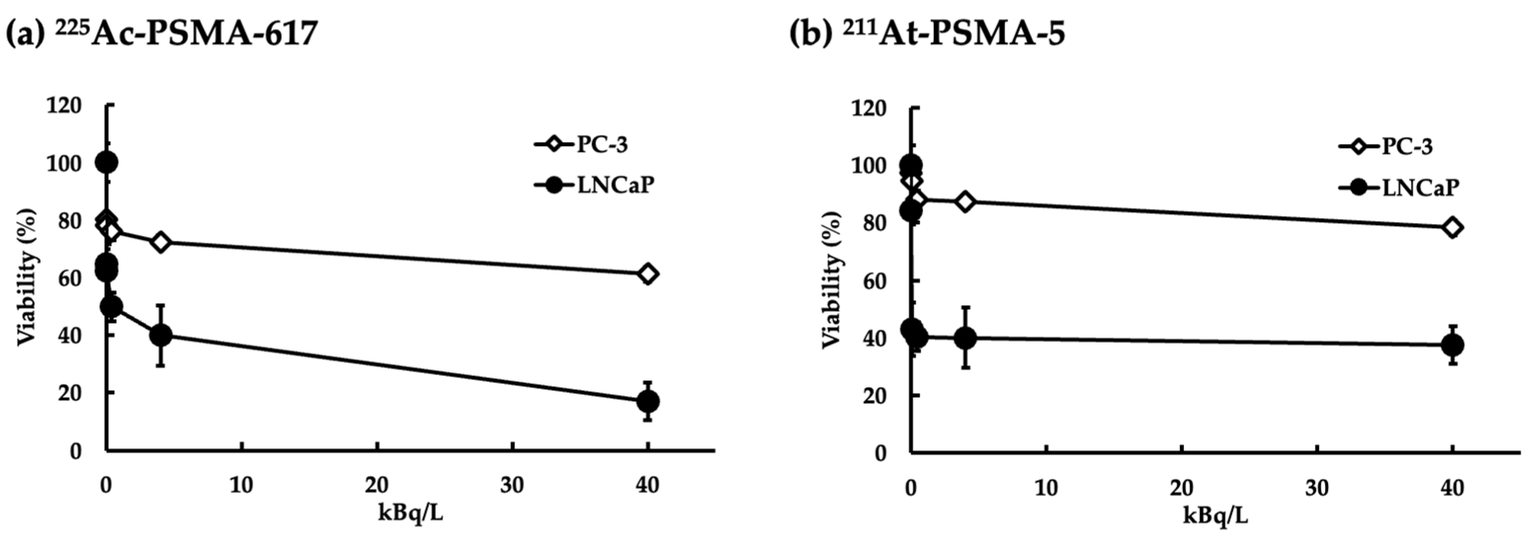
2.1. Evaluation of Effects on Cell Viability

We seeded the cell line LNCaP, which is known for its high PSMA expression, and the cell line PC3, characterized by low PSMA expression, at a density of 1 × 10⁴ cells/well in a 96-well plate. The cells were treated with 225Ac-PSMA-617 or 211At-PSMA-5 for 3 days, and cell viability was evaluated. The toxicity of the labeled PSMA was more pronounced in LNCaP cells than in PC3 cells (Figure 1). The 225Ac-PSMA-617 nuclide had a stronger effect on cell viability than 211At-PSMA-5; however, above a certain concentration, cell viability did not decrease in a dose-dependent manner.

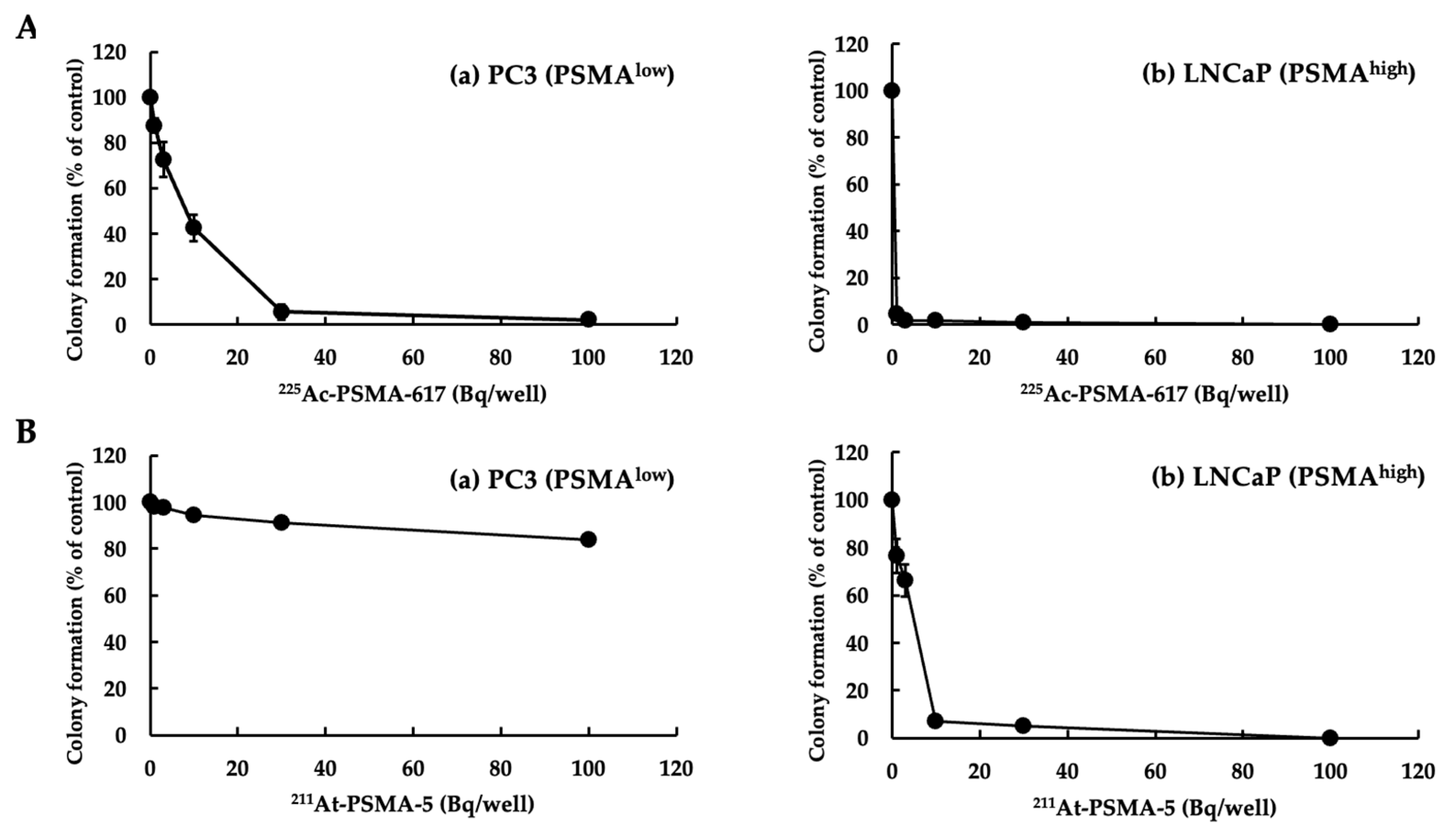
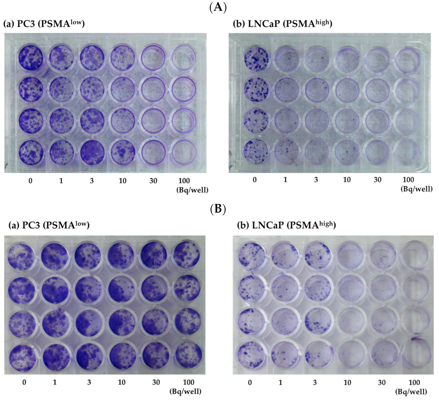
2.2. Evaluation of Effect on Replication

We performed a colony formation assay to assess the extent to which 225Ac-PSMA-617 and 211At-PSMA-5 affected the replication ability of the cells. Figure 2 shows a numerical graph based on the photographs shown in Figure 3. Cytotoxicity occurred in both PC3 (PSMAlow) and LNCaP (PSMAhigh) cells in a concentration-dependent manner (Figure 2A and Figure 3A). This effect appeared to be stronger in cells with high PSMA expression. When the cells were treated with 225Ac-PSMA-617, dose-dependent inhibition of replication was observed in both PC3 and LNCaP cells. After treatment with 211At-PSMA-5, no effect was observed in PC3 cells (Figure 2B and Figure 3B).

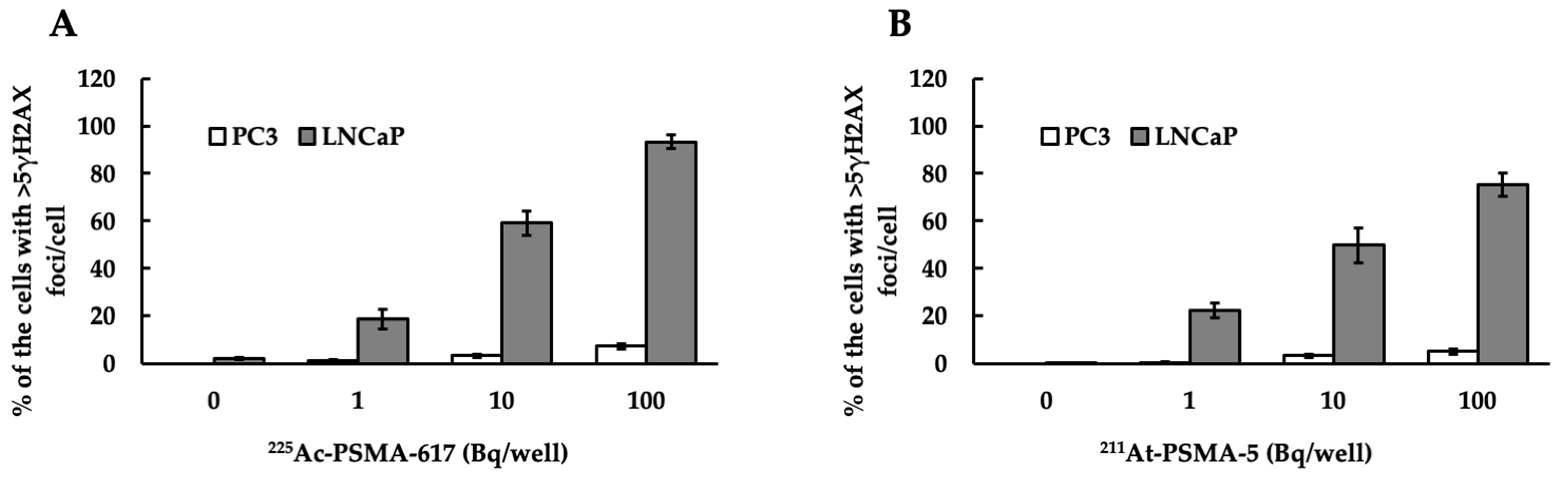
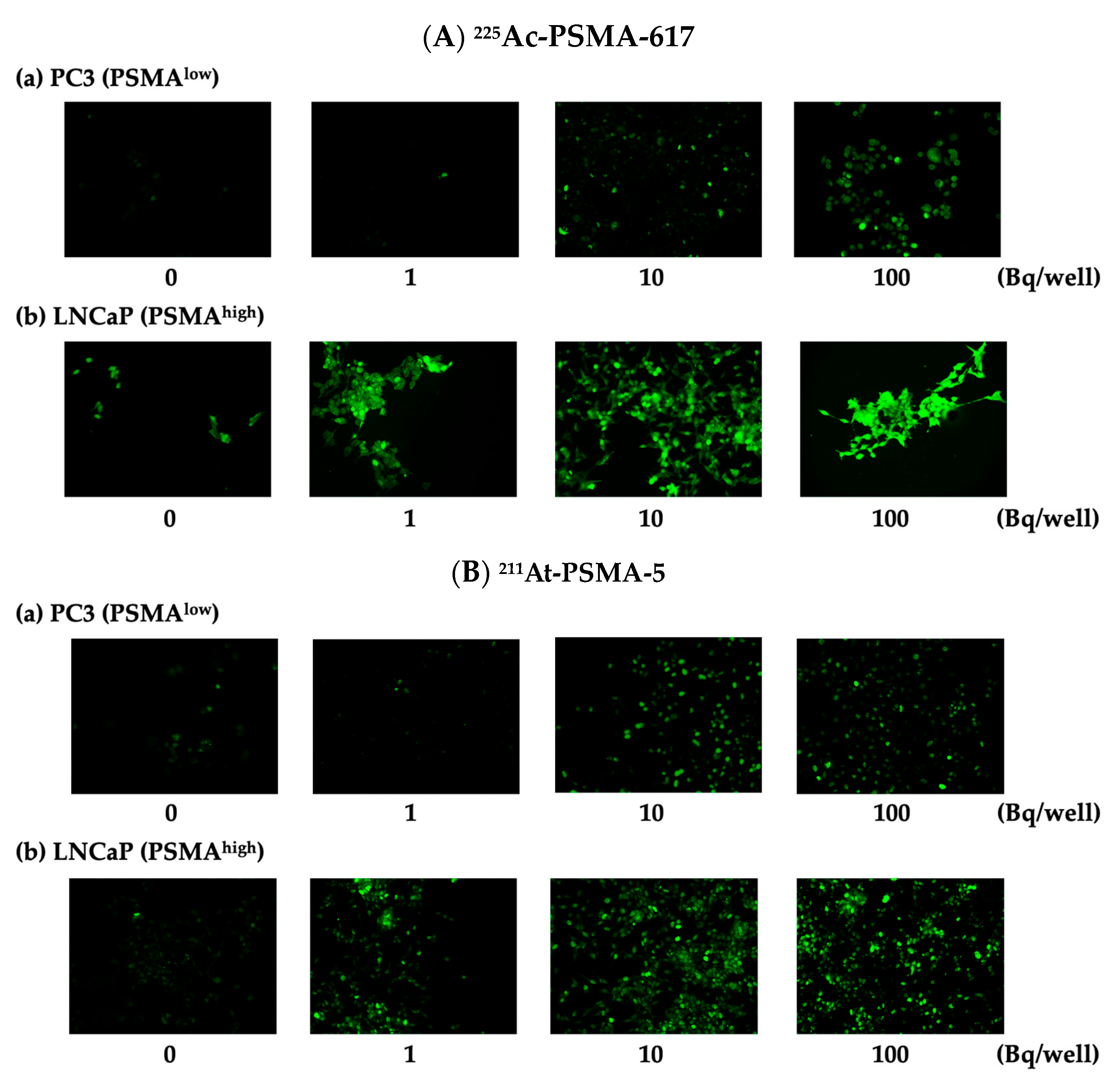
2.3. Evaluation of Cytotoxicity

DNA double strand breaks (DSBs) were observed in both PC3 (PSMAlow) and LNCaP (PSMAhigh) cells (Figure 4). Figure 5 depicts a numerical graph based on the photographs shown in Figure 4. Green fluorescence indicates the presence of γH2AX, a marker of DSB. Focies of γH2AX were strongly induced by 225Ac-PSMA-617 (Figure 5A), and many focies were induced by 211At-PSMA-5 (Figure 5B). When comparing their effects on (a) PC3 and (b) LNCaP cells, more foci were found in the LNCaP cells, indicating that many DSBs appeared in the LNCaP cells.

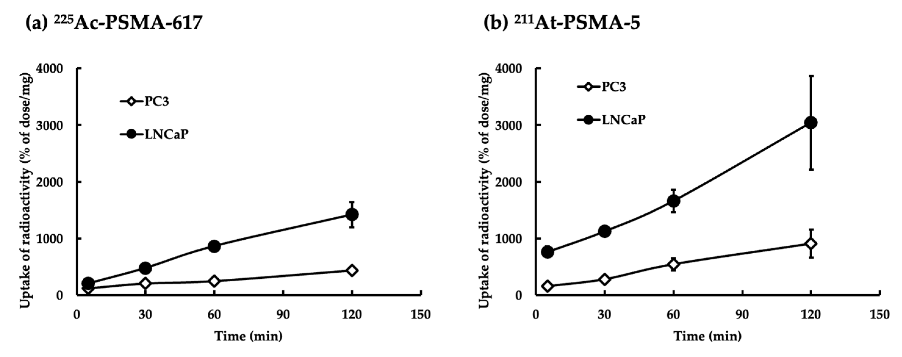
2.4. Uptake of 225Ac-PSMA-617 or 211At-PSMA-5

The 225Ac-PSMA-617 nuclide had a low background of intracellular uptake and was hardly taken up by PC3 (PSMAlow) cells (Figure 6a). In contrast, 211At-PSMA-5 was taken up in a certain amount by PC3 cells and large amounts of uptake were observed in LNCaP (PSMAhigh) cells (Figure 6b).

2.5. Inhibition of Unlabeled Chemicals

The inhibition of uptake by unlabeled chemicals using LNCaP cells (PSMAhigh) is shown in Figure 7. When we confirmed the presence of competitive inhibition by unlabeled chemicals for uptake at the same dose, it was clear that 211At-PSMA-5 (Figure 7b) was inhibited at a lower concentration than 225Ac-PSMA-617 (Figure 7a). The IC50 of 225Ac-PSMA-617 was 2.64 nM and that of 211At-PSMA-5 was 0.32 nM. Non-specific binding was evaluated using PC3 cells (PSMAlow). The effects of non-labeled chemicals on both the uptake of 225Ac-PSMA-617 (Figure 7c) and 211At-PSMA-5 (Figure 7d) in PC3 cells were thought to be minimal.

2.6. Stability of 225Ac-PSMA-617 and 211At-PSMA-5

The stability of 211At-PSMA5 was evaluated using HPLC, TLC, and electrophoresis. Its stability in blood and urine, along with the absence of metabolites and degradation products, was detected in vitro experiments. In in vivo experiments, slight (<1%) deastatination was observed. The same evaluations were conducted on 225Ac-PSMA-617, confirming that it was stable.

3. Discussion

In this study, 225Ac-PSMA-617 and 211At-PSMA-5 were compared in terms of their nuclide decay number (Bq) in order to focus on the performance of the labeled chemicals. This study revealed that 225Ac, which contains many α-particles, is highly cytotoxic, as was expected (Figure 1). The strong cytotoxic effect of 225Ac-PSMA-617 was reflected in the loss of replication ability (Figure 2 and Figure 3). At first glance, 225Ac-PSMA-617 seemed to have stronger cytotoxicity than 211At-PSMA-5. However, considering the AUC and α-ray emission ratio, it was difficult to say that 225Ac-PSMA-617 was more effective than 211At-PSMA-5 because there was a 25 to 83-fold difference even with the same radioactivity (Bq). We also compared γH2AX as an indicator of radiation-induced DNA damage (DNA double strand breaks, DSBs), and it became clear that 211At-PSMA-5 induces DNA damage at the same levels of 225Ac-PSMA-617 (Figure 4 and Figure 5). This was thought to be because 211At-PSMA-5 was taken up by cells in larger amounts and acted closer to the nucleus (Figure 6). Labeled chemicals appear to work more effectively as nuclear medicine therapeutics for intracellular uptake. It is interesting to note that more 211At-PSMA-5 was taken up by the cells even though both chemical structures are similar (Figure 8 and Figure 9). However, considering their structures, there seems to be a slight difference in fat solubility. Since 211At-PSMA-5 is slightly more lipophilic, it is presumed to have a higher affinity for the cell membrane (Figure 7). The correction for non-specific binding concerning IC50 should be strictly calculated using the inhibition value in an experimental system using PC3 cells (PSMAlow), Xenopus oocytes, or HEK293 cells overexpressing PSMA. However, for both 225Ac-PSMA-617 and 211At-PSMA-5, the ratio of PC3 to LNCaP cell uptake at 120 min did not change (approximately 30%) (Figure 6). Even when the background was subtracted at the same rate, the relationship between the IC50 of 225Ac-PSMA-617 and 211At-PSMA-5 was similar. Thus, the IC50 was calculated from the LNCaP value (Figure 7). The IC50 value of 225Ac-PSMA-617 was approximately eight-fold higher than that of 211At. The lipid solubility of chemicals is an important factor in nuclear medicine. This is because a certain degree of fat solubility improves tumors [18,19,20,21]. However, if fat solubility is too high, adsorption onto plastic experimental tools will be high. For example, an increase in the amount of adsorption on the purification column results in a poor collection yield. Additionally, when administered to animals, it has been observed that excretion from the intestinal tract increases and the amount excreted in feces increases. Screening for suitable compounds should consider both accumulation and excretion.
225Ac is an excellent therapeutic-emitting nuclide, but its current supply is limited. PSMA is also highly specific and only has a few side effects (caused by damage to the salivary and lacrimal glands, such as dry eye [22] and xerostomia [23]). Non-specific accumulation over a long period would induce side effects. If the ratio of non-specific accumulation is the same, the nuclide with a shorter half-time is less likely to induce side effects due to non-specific accumulation. The 211At-PSMA-5 nuclide requires significantly fewer chemicals for labeling than 225Ac-PSMA-617, and its cost of manufacturing is also lower. In this study, we attempted to compare two α-emitting nuclides used in TAT. We believe that our results suggest that, even though its half-life is much shorter than 225Ac, 211At can be sufficiently tolerated in clinical use by effectively utilizing its properties. If the half-life is short, the hospitalization period for nuclide decay can be shortened. This might reduce the burden on patients.
Although there are many clinical reports of 225Ac-PSMA-617 use in humans, there are only a few reports on animal experiments using 225Ac. Additionally, these reports were published later than the human reports. The 225Ac-PSMA-617 nuclide demonstrated high levels of accumulation in the liver. However, it is well known that 225Ac alone accumulates in the liver. There are doubts as to whether the results showing accumulation only in the liver can depict metastasis [24]. A previous study used a combination of the PD-L1 antibody and 225Ac-PSMA-617 in RM1-PGLS mice. Although it is used in combination with an immune checkpoint inhibitor, it has not been as effective as expected [25,26]. Rodent metabolism is different from that of primates, meaning that research results on rodents are not guaranteed to be applicable to humans. In contrast, 211At-PSMA-5 was investigated for imaging in cynomolgus monkeys by Watabe et al. Additionally, no acute inflammation was observed, and the side effects were expected to be minimal. Although clinical trials using 211At-PSMA-5 have not yet been conducted, animal studies have shown positive results [11]. Thus, we expect that future clinical trials of 211At-PSMA-5 will yield good results.
Irradiation to the target with high linear energy transfer (LET) radiation is highly lethal. Thus, resistance to this radiation is unlikely to occur. Therefore, selecting a specific and excellent molecular target, such as PSMA, is essential for nuclear medicine. To achieve a stable supply of nuclides, selecting optimal molecular targets, establishing chemicals that recognize and label these targets, and collaborating across departments, such as medicine, science, and nuclear physics, is necessary.
We also confirmed the utility of 225Ac in another molecular target, fibroblast activate protein α (FAPα) [27,28]. However, as previously mentioned, its supply is insufficient for clinical use [8]. Various research groups are attempting to find methods to produce it in large quantities; however, no satisfactory method has yet been established. Therefore, there is an urgent need to establish a method for producing 225Ac. Currently, we believe that the most promising method in Japan is the use of an electron linear accelerator. The production of 225Ac was conducted using 226Ra and a linear accelerator at Tohoku university [226Ra(γ, n)225Ra→225Ac]. In Japan, the National Institute for Quantum Science and Technology (QST) and Nihon Medi-physics, Co. Ltd. are also trying to produce it with the transmutation of 226Ra to obtain 225Ac [226Ra(p, 2n)225Ac] [29]. In Canada’s particle accelerator center (TRIUMF), a method of obtaining 225Ac through the nuclear spallation of thorium-232 [232Th(p, spall)225Ra→225Ac] is being attempted [30]. In contrast, 211At has a short half-life; therefore, it must be manufactured in large quantities. This must be performed efficiently because it uses an accelerator. It is desirable to create multiple bases within a country where supplies are needed. However, to transport them to places where transportation is difficult, it is necessary to create large quantities. In 2023, Haba et al. aimed to develop targeted irradiation technology that minimizes the loss of 211At due to radioactive decay and increases the production efficiency of 211At through high-intensity beam irradiation. Their results may be useful for the mass production and supply of 211At.
The half-life of 211At is 7.2 h and α decays to bismuth-207 (207Bi with a half-life of 32 years) with a probability of 41.8%. Therefore, with a probability of 58.2%, it can become polonium-211 (211Po) through electron capture decay. Since 211Po undergoes α decay immediately (with a half-life of 0.52 s) to the stable isotope lead-207 (207Pb), 211At emits α-particles with virtually 100% probability. On the other hand, there are many daughter nuclides of 225Ac, such as francium-221 (221Fr, with a half-life of 0.12 μ seconds), 217At (with a half-life of 20 milliseconds), 213Bi (with a half-life of 47 min), 213Po (with a half-life of 3.65 μ seconds), thallium-209 (209Tl, with a half-life of 1 h), 209Pb (with a half-life of 3.3 h), and 209Bi (stable), all of which have short lifetimes. Because none of these generate radon as daughter nuclide, which is a gas, post-administration management is easy. For practical use, it is very important that the nuclides are easily handled. In addition to the two nuclides discussed in this research, there are several other α-emitting nuclides that may be used for TAT, as shown in Table 1. However, various choices should be made with consideration of the balance between demand and supply.
We are currently developing nuclear medicine therapeutics using the 211At and 225Ac nuclides. During this period, we have observed the advantages and disadvantages of each nuclide. Initially, we thought that 225Ac-PSMA-617 would provide better results than 211At-PSMA-5 and we hoped that 211At-PSMA-5 would be the next choice. However, the performance of 211At-PSMA-5 was unexpectedly good. High-power accelerators are currently needed to obtain 211At. However, since it can be obtained using accelerators, it is easier to obtain than 225Ac, particularly in Japan. Although the availability of nuclides differs from country to country, it can be said that, at least in Japan, astatine has been shown to be extremely useful as a labeled nuclide for nuclear medicine therapeutics. We are currently conducting an investigator-initiated clinical trial of astatine-labeled chemicals (Na211At, targeting thyroid cancer) at Osaka University Hospital. By demonstrating the usefulness of nuclear medicine therapeutics, especially α-emitting nuclear medicine therapeutics, we hope that this will become a new choice for patients who are currently unable to undergo surgery or for whom existing drugs are not effective.
The most important step in the development of nuclear medicine is the selection of molecular targets. If expressed in normal tissues as well as in cancer tissues, nuclear medicine therapeutics can accumulate in normal tissues and damage them, causing side effects. Therefore, it is desirable to use molecular targets with a higher specificity. In the development of compounds, even for the same molecular target, selectivity can vary significantly depending on the structure of the compound. In addition, side effects can occur when the labeled nuclide is released from the compound. This is because 211At behaves in a similar fashion to iodine and 225Ac accumulates in the bones and liver [31]. Consideration is also required in order to ensure safety. Key considerations in the clinical application of 225Ac-PSMA-617 include liver uptake, which is assumed to result from the decay products of 225Ac-PSMA-617. DTPA is an abbreviation for diethylenetriamine penta-acetic acid, a chelate compound with affinity for metals, and is a type of chemical protective agent against radiation damage. For example, Techne® DTPA Kits (PDRadiopharma, Inc., Tokyo, Japan) or Indium (111In) DTPA Injections (Nihon Medi-Physics Co., Ltd., Tokyo, Japan) exist. This protective agent has the function of removing radioactive materials from the body and is said to be most effective in excreting radioisotopes. DTPA may be added to the formulation of 225Ac-PSMA-617 to prevent uptake in the liver (faster renal clearance). In the case of 211At-PSMA-5, the properties of the element are different from 225Ac; therefore, it may be necessary to use a different drug.
The functions of PSMA itself in cancer tissues are gradually being elucidated. For example, research conducted by Watanabe, et al. revealed the existence of PSMA-positive tumor endothelial cells in human prostate tumors, which enhance tumor angiogenesis in prostate cancer tissues [32]. The elucidation of the role of PSMA in cancer tissues has supported its importance as a molecular target. On the other hand, reports on the structure and dynamics of chemicals are also being considered [33]. We are also conducting studies, but because the in vivo environment and the stability of compounds are interrelated, the results are often not what we expected. We hope that similar studies conducted by various groups will clarify this relationship.

4. Materials and Methods

The 211At nuclide was acquired from RIKEN through a supply platform for short-lived radioisotopes. The 211At nuclide was produced according to the 209Bi(α, 2n)211At reaction and was separated from the Bi target using the dry distillation method. The separated 211At was then dissolved in pure water [11]. The 225Ac nuclide was produced mainly by members of Tohoku University [34,35]. Experiments after isolation were conducted at Osaka University. The intensity of the nuclides were measured using a germanium semiconductor detector (BE2020, Canberra, Mirion Technologies, Inc., Atlanta, GA. USA) and a γ-counter (Wizard2 2480, PerkinElmer, Inc., Shelton, CT, USA). The samples treated with 225Ac were maintained until radiative equilibrium was reached before the measurements were taken, according to a previous study [31].

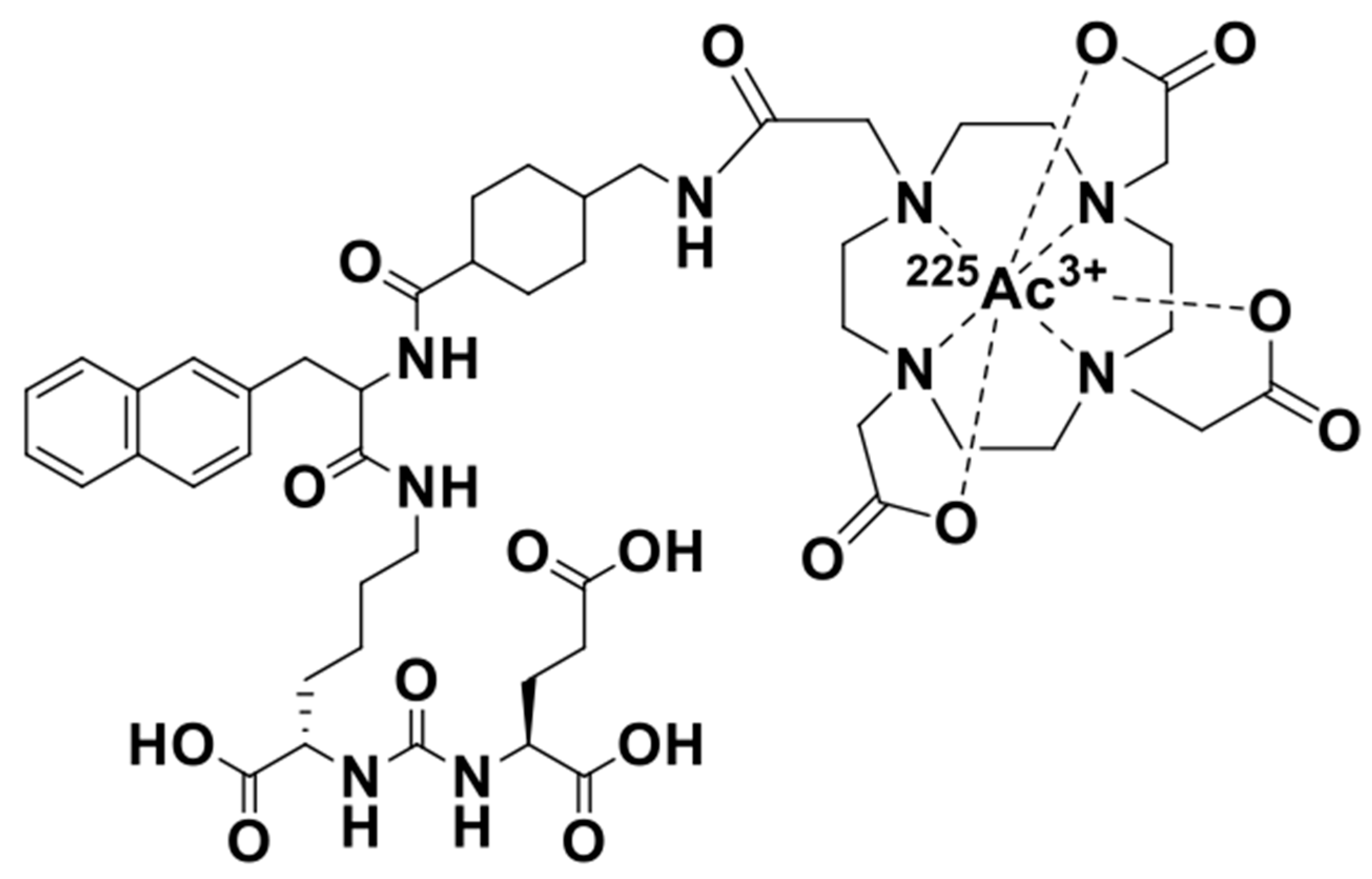
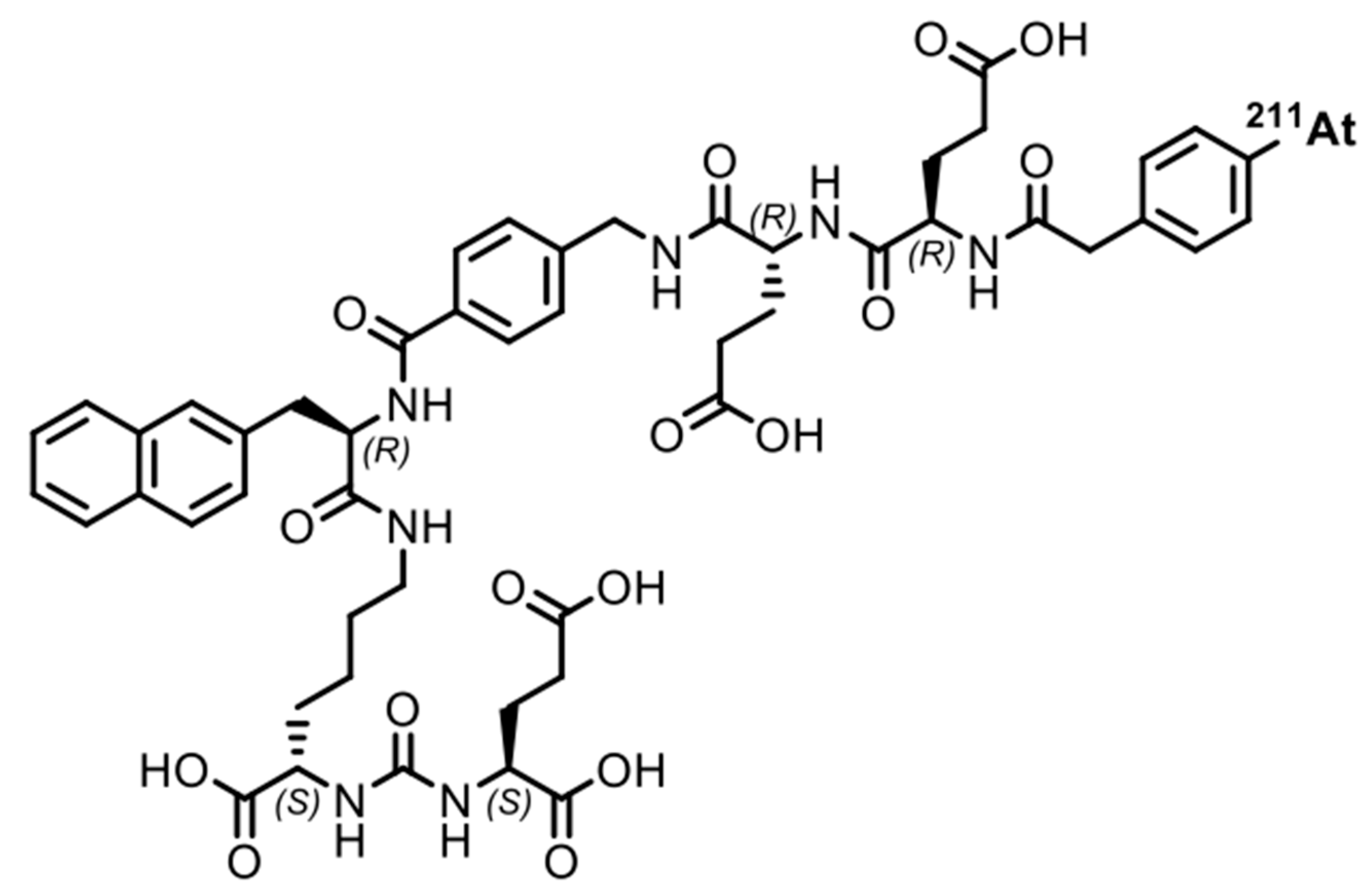
4.1. Structure and Preparation of 225Ac-PSMA-617 and 211At-PSMA-5

The PSMA-selected chemicals were labeled with each nuclide using previously reported procedures. These structures are shown in Figure 8 and Figure 9. The PSMA-5 precursor was synthesized at the Peptide Institute. Inc. (Osaka, Japan) for the Shirakami Reaction. The labeling method is explained in detail in the next section. To evaluate the quality of the labeled chemicals, we used a previously reported method.

4.2. Nuclide Production and Chemical Labeling

4.2.1. Production and PSMA-5 Labeling of 211At

PSMA-5 was used as a highly selective PSMA compound to label with 211At. This compound has been previously reported by Watabe et al. We determined this compound to be optimal based on the labeling efficiency and in vitro and in vivo experimental results [11]. The method we used for labeling PSMA-5 was the “borono group-astatine exchange reaction”, also known as the “Shirakami reaction” [36].

4.2.2. Production and PSMA-617 Labeling of 225Ac

The labeling method for 225Ac was established according to a previous study [37]. The 225Ac used for labeling was separated at the Tohoku University Institute for Materials Research and was then transported to Osaka University for use. PSMA-617 was dissolved in DMSO (1 mg/mL) and a 10% DMSO aqueous solution was prepared. This solution was mixed with 0.2 M AcONH4 and 10% Ascorbic acid and was incubated in 80 °C for 2 h. After measuring the dose using a Curie meter (ICG-8; ALOKA, Ltd., Tokyo, Japan), the quality was confirmed using electrophoresis and it was then used in experiments.

4.3. Cell Culture

The PC3 and LNCaP cells were obtained from RIKEN and ATCC cell banks, respectively. The cells were maintained in RPMI1640 (Fujifilm Wako Pure Chemical, Osaka, Japan) supplemented with 10% heat-inactivated fetal bovine serum (Thermo Fisher Scientific, Waltham, MA, USA) and 1% penicillin-streptomycin (Fujifilm Wako Pure Chemical). Sodium pyruvate (Fujifilm Wako Pure Chemical) was added to the LNCaP culture medium at a concentration of 1%. The cells were maintained using trypsin-EDTA (Fujifilm Wako Pure Chemical), according to standard methods. Both cell lines were in the logarithmic growth phase at the time of experimental preparation.

4.4. Evaluation of Cell Viability

Two days before treatment, the cells were seeded in 1 × 10⁴ cells/mL in 96-well culture plates. The cell numbers were measured using TC-20TM (Bio-Rad Laboratories, Inc., Hercules, CA, USA). After 1 h of treatment, the cells were cultured for three days. Cell viability was evaluated using a cell counting kit-8 (Dojin, Kumamoto, Japan), according to the manufacturer’s protocol. The absorbance was measured at 450 nm using a MultiSkan FC (Thermo Fisher Scientific).

4.5. Colony Formation Assay

Both LNCaP (PSMAhigh) and PC3 (PSMAlow) cells were seeded in 24-well plates and treated with labeled PSMAs at various concentrations. After 1 h of treatment, the cells were peeled and seeded at 1000 cells per well. Colony formation was observed for two weeks. After observation, the cells were fixed with 1% crystal violet solution. Colony formation was calculated by analyzing the coverage ratio of cells based on photographs using ImageJ software (https://imagej.net/downloads, accessed on 1 April 2023) [38].

4.6. Evaluation of DNA Double Strand Breaks

The cells were seeded in a culture chamber (WATSON, Tokyo, Japan). After treatment with labeled chemicals, the cells were fixed and did the immunofluorescence staining, according to a previously reported protocol [37]. The cells were observed using a BZ-X810 microscope (KEYENCE, Osaka, Japan). The fluorescence intensities were analyzed using ImageJ software [38].

4.7. Uptake of 225Ac-PSMA-617 and 211At-PSMA-5

Both LNCaP (PSMAhigh) and PC3 (PSMAlow) cells were collected after 5, 30, 60, and 120 min of treatment, washed with PBS (-) three times, lysed using 0.1 N NaOH solutions, and collected into microtubes. The sample counts were measured using a γ-counter and their counts were corrected for the amounts of cell protein, according to a previous published protocol [39]. We also measured the protein amounts in the cell suspension using a protein assay BCA kit (Fujifilm Wako Pure Chemical), according to the manufacture’s protocol. The absorbance was measured at 570 nm using a MultiSkan FC.

4.8. Inhibition Assay

Based on previous research [40], the LNCaP cells were simultaneously treated with unlabeled chemicals (PSMA-617 or PSMA-5). For the inhibition assay, the cell uptake experiments and procedures were the same, except for treatment with the compound as an inhibitor. The amount of unlabeled compound added was more than 100 times that of the labeled compound.

5. Conclusions

Let us consider the respective absorbed doses of 211At-PSMA-5 and 225Ac-PSMA617 in the cell experiments. Since the cell treatment conditions were the same, it can be assumed that the masses of cells were the same. In this case, the energy value and the absorbed dose was proportional. In other words, it is thought that comparisons can be made in becquerel, considering the half-life. For example, assuming that there is no washout from the cells, considering the area under curve (AUC) for 3 days, 225Ac-PSMA-617 is about 6.3 times more powerful than 211At-PSMA-5. Considering that 225Ac emits four α rays, it is about 25 times more powerful. However, the amount of uptake itself is halved (Figure 6), which is approximately 12 times as much. There is no more than a 10-fold difference in their effects on the cells (Figure 4). Therefore, it can be said that PSMA-5 is superior as a compound. It might be better to consider the stability of PSMA-5 in more detail, but we consider that this stability might be increased by inducing three unnatural amino acids (Figure 9). PSMA-5 is expected to be sufficient for clinical use.
It might be said that 211At-PSMA-5 is more easily taken up by LNCaP cells than 225Ac-PSMA-617, suggesting that PSMA-5 might be superior as a compound (Figure 6 and Figure 7). On the other hand, whether it is better in clinical practice may depend on the balance between effectiveness and potential side effects, aside from the availability of the nuclide. Although more studies may be needed, our findings indicate the possibility of completing treatment regimens faster because of the shorter half-life of 211At-PSMA-5 for repeated administration compared to 225Ac-PSMA-617.

6. Patents

Patent No. JP7237366B2, JP7232527B2.

Author Contributions

Conceptualization, K.K.-N. and Y.S.; methodology, K.K.-N. and Y.S.; 211At production, X.Y. and H.H.; 225Ac production, K.S., H.K. and K.T.; nuclide separation, K.O. and A.T.; labeling chemicals, Y.S.; formal analysis, K.K.-N. and Y.K.; investigation, K.K.-N. and Y.S.; resources, J.C. and F.L.G.; data curation, K.K.-N. and Y.S.; writing—original draft preparation, K.K.-N.; writing—review and editing, K.K.-N.; visualization, K.K.-N.; supervision, T.W.; project administration, K.K.-N., Y.S. and K.F.; funding acquisition, T.W. All authors have read and agreed to the published version of the manuscript.

Funding

This research was supported by AMED under Grant Number JP23ym0126091. This work was also performed under the U-233 cooperation project between JAEA and the GIMRT Collaborative Research Program of the Institute for Materials Research, Tohoku University (Proposal No. 202212-RDKGE-0046).

Institutional Review Board Statement

Not applicable.

Informed Consent Statement

Not applicable.

Data Availability Statement

Raw data were generated at Osaka University. Derived data supporting the findings of this study are available from the corresponding author [K.K.-N.] on request.

Acknowledgments

Thank you very much to Hitoshi Egawa, Takanori Kobayashi, and Atsuko Saito-Kawakami for their technical support.

Conflicts of Interest

The authors declare no conflict of interest.

References

  1. Radchenko, V.; Morgenstern, A.; Jalilian, A.R.; Ramogida, C.F.; Cutler, C.; Duchemin, C.; Hoehr, C.; Haddad, F.; Bruchertseifer, F.; Gausemel, H.; et al. Production and Supply of α-Particle-Emitting Radionuclides for Targeted α-Therapy. J. Nucl. Med. 2021, 62, 1495–1503. [Google Scholar] [CrossRef] [PubMed]
  2. Jang, A.; Kendi, A.T.; Johnson, G.B.; Halfdanarson, T.R.; Sartor, O. Targeted Alpha-Particle Therapy: A Review of Current Trials. Int. J. Mol. Sci. 2023, 24, 11626. [Google Scholar] [CrossRef] [PubMed]
  3. Seifert, R.; Alberts, I.L.; Afshar-Oromieh, A.; Rahbar, K. Prostate Cancer Theranostics: PSMA Targeted Therapy. PET Clin. 2021, 16, 391–396. [Google Scholar] [CrossRef] [PubMed]
  4. Boinapally, S.; Alati, S.; Jiang, Z.; Yan, Y.; Lisok, A.; Singh, R.; Lofland, G.; Minn, I.; Hobbs, R.F.; Pomper, M.G.; et al. Preclinical Evaluation of a New Series of Albumin-Binding 177Lu-Labeled PSMA-Based Low-Molecular-Weight Radiotherapeutics. Molecules 2023, 28, 6158. [Google Scholar] [CrossRef]
  5. Alati, S.; Singh, R.; Pomper, M.G.; Rowe, S.P.; Banerjee, S.R. Preclinical Development in Radiopharmaceutical Therapy for Prostate Cancer. Semin. Nucl. Med. 2023, 53, 663–686. [Google Scholar] [CrossRef]
  6. Siegel, R.L.; Miller, K.D.; Wagle, N.S.; Jemal, A. Cancer statistics, 2023. CA Cancer J. Clin. 2023, 73, 17–48. [Google Scholar] [CrossRef]
  7. Feuerecker, B.; Kratochwil, C.; Ahmadzadehfar, H.; Morgenstern, A.; Eiber, M.; Herrmann, K.; Pomykala, K.L. Clinical Translation of Targeted α-Therapy: An Evolution or a Revolution? J. Nucl. Med. 2023, 64, 685–692. [Google Scholar] [CrossRef]
  8. Robertson, A.K.H.; Caterina, F.R.; Paul, S.; Valery, R. Development of 225Ac Radiopharmaceuticals: TRIUMF Perspectives and Experiences. Curr. Radiopharm. 2018, 11, 156–172. [Google Scholar] [CrossRef]
  9. Patell, K.; Kurian, M.; Garcia, J.A.; Mendiratta, P.; Barata, P.C.; Jia, A.Y.; Spratt, D.E.; Brown, J.R. Lutetium-177 PSMA for the treatment of metastatic castrate resistant prostate cancer: A systematic review. Expert Rev. Anticancer Ther. 2023, 23, 731–744. [Google Scholar] [CrossRef]
  10. Hofman, M.S.; Emmett, L.; Sandhu, S.; Iravani, A.; Joshua, A.M.; Goh, J.C.; Pattison, D.A.; Tan, T.H.; Kirkwood, I.D.; Ng, S.; et al. [177Lu]Lu-PSMA-617 versus cabazitaxel in patients with metastatic castration-resistant prostate cancer (TheraP): A randomised, open-label, phase 2 trial. Lancet 2021, 397, 797–804. [Google Scholar] [CrossRef]
  11. Watabe, T.; Kaneda-Nakashima, K.; Shirakami, Y.; Kadonaga, Y.; Ooe, K.; Wang, Y.; Haba, H.; Toyoshima, A.; Cardinale, J.; Giesel, F.L.; et al. Targeted α-therapy using astatine (211At)-labeled PSMA1, 5, and 6: A preclinical evaluation as a novel compound. Eur. J. Nucl. Med. Mol. Imaging 2023, 50, 849–858. [Google Scholar] [CrossRef]
  12. Langbein, T.; Weber, W.A.; Eiber, M. Future of Theranostics: An Outlook on Precision Oncology in Nuclear Medicine. J. Nucl. Med. 2019, 60 (Suppl. S2), 13S–19S. [Google Scholar] [CrossRef] [PubMed]
  13. Rizzo, A.; Annunziata, S.; Salvatori, M. Side effects of theragnostic agents currently employed in clinical practice. Q. J. Nucl. Med. Mol. Imaging 2021, 65, 315–326. [Google Scholar] [CrossRef] [PubMed]
  14. Zoi, V.; Giannakopoulou, M.; Alexiou, G.A.; Bouziotis, P.; Thalasselis, S.; Tzakos, A.G.; Fotopoulos, A.; Papadopoulos, A.N.; Kyritsis, A.P.; Sioka, C. Nuclear Medicine and Cancer Theragnostics: Basic Concepts. Diagnostics 2023, 13, 3064. [Google Scholar] [CrossRef] [PubMed]
  15. Giammarile, F. Theranostic in Nuclear Medicine—The paradigm of NET. Hell. J. Nucl. Med. 2023, 26, 42–43. [Google Scholar]
  16. Ukon, N.; Higashi, T.; Hosono, M.; Kinuya, S.; Yamada, T.; Yanagida, S.; Namba, M.; Nakamura, Y. Manual on the proper use of meta-[211At] astato-benzylguanidine ([211At] MABG) injections in clinical trials for targeted alpha therapy (1st edition). Ann. Nucl. Med. 2022, 36, 695–709. [Google Scholar] [CrossRef] [PubMed]
  17. Report of Health Labour Sciences Research Grant (19IA1004). Available online: https://niph.repo.nii.ac.jp/records/2594 (accessed on 27 December 2023).
  18. Sugiyama, T.; Sadazuka, Y.; Miyagishima, A.; Nozawa, Y.; Nagasawa, K.; Ohnishi, N.; Yokoyama, T. Membrance transport on tumor cell and antitumor activity of doxorubicin and pirarubicin. Drug Deliv. Syst. 1998, 13, 35–40. [Google Scholar] [CrossRef]
  19. Tanaka, K.; Kunimatsu, T.; Shimakura, J.; Hanada, M. Development of Miriplatin, a Novel Antitumor Platinum for Hepatocellular Carcinoma. Sumitomo Kagaku 2011, 1, 11–12. [Google Scholar]
  20. Hama, S. Development of a novel nanocarrier focusing on the physicochemical properties of an anti-cancer therapy drug. Yakugaku Zasshi 2014, 134, 1049–1054. [Google Scholar] [CrossRef]
  21. Izci, M.; Maksoudian, C.; Manshian, B.B.; Soenen, S.J. The Use of Alternative Strategies for Enhanced Nanoparticle Delivery to Solid Tumors. Chem. Rev. 2021, 121, 1746–1803. [Google Scholar] [CrossRef]
  22. Hofman, M.S.; Violet, J.; Hicks, R.J.; Ferdinandus, J.; Thang, S.P.; Akhurst, T.; Iravani, A.; Kong, G.; Ravi Kumar, A.; Murphy, D.G.; et al. [177Lu]-PSMA-617 radionuclide treatment in patients with metastatic castration-resistant prostate cancer (LuPSMA trial): A single-centre, single-arm, phase 2 study. Lancet Oncol. 2018, 19, 825–833. [Google Scholar] [CrossRef] [PubMed]
  23. Feuerecker, B.; Tauber, R.; Knorr, K.; Heck, M.; Beheshti, A.; Seidl, C.; Bruchertseifer, F.; Pickhard, A.; Gafita, A.; Kratochwil, C.; et al. Activity and Adverse Events of Actinium-225-PSMA-617 in Advanced Metastatic Castration-resistant Prostate Cancer After Failure of Lutetium-177-PSMA. Eur. Urol. 2021, 79, 343–350. [Google Scholar] [CrossRef] [PubMed]
  24. Stuparu, A.D.; Meyer, C.A.L.; Evans-Axelsson, S.L.; Lückerath, K.; Wei, L.H.; Kim, W.; Poddar, S.; Mona, C.E.; Dahlbom, M.; Girgis, M.D.; et al. Targeted alpha therapy in a systemic mouse model of prostate cancer—A feasibility study. Theranostics 2020, 10, 2612–2620. [Google Scholar] [CrossRef]
  25. Czernin, J.; Current, K.; Mona, C.E.; Nyiranshuti, L.; Hikmat, F.; Radu, C.G.; Lückerath, K. Immune-Checkpoint Blockade Enhances 225Ac-PSMA617 Efficacy in a Mouse Model of Prostate Cancer. J. Nucl. Med. 2021, 62, 228–231. [Google Scholar] [CrossRef] [PubMed]
  26. Kelly, J.M.; Amor-Coarasa, A.; Ponnala, S.; Nikolopoulou, A.; Williams, C., Jr.; Thiele, N.A.; Schlyer, D.; Wilson, J.J.; DiMagno, S.G.; Babich, J.W. A Single Dose of 225Ac-RPS-074 Induces a Complete Tumor Response in an LNCaP Xenograft Model. J. Nucl. Med. 2019, 60, 649–655. [Google Scholar] [CrossRef] [PubMed]
  27. Watabe, T.; Liu, Y.; Kaneda-Nakashima, K.; Shirakami, Y.; Lindner, T.; Ooe, K.; Toyoshima, A.; Nagata, K.; Shimosegawa, E.; Haberkorn, U.; et al. Theranostics Targeting Fibroblast Activation Protein in the Tumor Stroma: 64Cu- and 225Ac-Labeled FAPI-04 in Pancreatic Cancer Xenograft Mouse Models. J. Nucl. Med. 2020, 61, 563–569. [Google Scholar] [CrossRef]
  28. Liu, Y.; Watabe, T.; Kaneda-Nakashima, K.; Shirakami, Y.; Naka, S.; Ooe, K.; Toyoshima, A.; Nagata, K.; Haberkorn, U.; Kratochwil, C.; et al. Fibroblast activation protein targeted therapy using [177Lu]FAPI-46 compared with [225Ac]FAPI-46 in a pancreatic cancer model. Eur. J. Nucl. Med. Mol. Imaging 2022, 49, 871–880. [Google Scholar] [CrossRef]
  29. Nagatsu, K.; Suzuki, H.; Fukada, M.; Ito, T.; Ichinose, J.; Honda, Y.; Minegishi, K.; Higashi, T.; Zhang, M.R. Cyclotron production of 225Ac from an electroplated 226Ra target. Eur. J. Nucl. Med. Mol. Imaging 2021, 49, 279–289. [Google Scholar] [CrossRef]
  30. Robertson, A.K.H.; McNeil, B.L.; Yang, H.; Gendron, D.; Perron, R.; Radchenko, V.; Zeisler, S.; Causey, P.; Schaffer, P. 232Th-sepallation-produced 225Ac with reduced 225Ac content. Inorg. Chem. 2020, 59, 12156–12165. [Google Scholar] [CrossRef]
  31. Kaneda-Nakashima, K.; Zhang, Z.; Nagata, K.; Shirasaki, K.; Kikunaga, H.; Yamamura, T.; Ooe, K.; Watabe, T.; Toyoshima, A.; Yoshimura, T.; et al. Dispersion of Alpha-Nuclides during Animal Experiments. Radiat. Safety Manag. 2021, 20, 29–38. [Google Scholar] [CrossRef]
  32. Watanabe, R.; Maekawa, M.; Kiyoi, T.; Kurata, M.; Miura, N.; Kikugawa, T.; Higashiyama, S.; Saika, T. PSMA-positive membranes secreted from prostate cancer cells have potency to transform vascular endothelial cells into an angiogenic state. Prostate 2021, 81, 1390–1401. [Google Scholar] [CrossRef]
  33. van der Gaag, S.; Bartelink, I.H.; Vis, A.N.; Burchell, G.L.; Oprea-Lager, D.E.; Hendrikse, H. Pharmacological Optimization of PSMA-Based Radioligand Therapy. Biomedicines 2022, 10, 3020. [Google Scholar] [CrossRef] [PubMed]
  34. Apostolidis, C.; Molinet, R.; Rasmussen, G.; Morgenstern, A. Production of Ac-225 from Th-229 for targeted alpha therapy. Anal. Chem. 2005, 77, 6288–6291. [Google Scholar] [CrossRef] [PubMed]
  35. Zielinska, B.; Apostolidis, C.; Bruchertseifer, F.; Morgenstern, A. An improved method for the production of Ac-225/Bi-213 from Th-229 for targeted alpha therapy. Solvent Extr. Ion Exch. 2007, 25, 339–349. [Google Scholar] [CrossRef]
  36. Shirakami, Y.; Watabe, T.; Obata, H.; Kaneda, K.; Ooe, K.; Liu, Y.; Teramoto, T.; Toyoshima, A.; Shinohara, A.; Shimosegawa, E.; et al. Synthesis of [211At]4-astato-L-phenylalanine by dihydroxyboryl-astatine substitution reaction in aqueous solution. Sci. Rep. 2021, 11, 12982. [Google Scholar] [CrossRef] [PubMed]
  37. Kratochwil, C.; Bruchertseifer, F.; Giesel, F.L.; Weis, M.; Verburg, F.A.; Mottaghy, F.; Kopka, K.; Apostolidis, C.; Haberkorn, U.; Morgenstern, A. 225Ac-PSMA-617 for PSMA-Targeted α-Radiation Therapy of Metastatic Castration-Resistant Prostate Cancer. J. Nucl. Med. 2016, 57, 1941–1944. [Google Scholar] [CrossRef]
  38. Abramoff, M.D.; Magalhaes, P.J.; Ram, S.J. Image Processing with ImageJ. Biophotonics Int. 2004, 11, 36–42. [Google Scholar]
  39. Khunweeraphong, N.; Nagamori, S.; Wiriyasermkul, P.; Nishinaka, Y.; Wongthai, P.; Ohgaki, R.; Tanaka, H.; Tominaga, H.; Sakurai, H.; Kanai, Y. Establishment of stable cell lines with high expression of heterodimers of human 4F2hc and human amino acid transporter LAT1 or LAT2 and delineation of their differential interaction with α-alkyl moieties. J. Pharmacol. Sci. 2012, 119, 368–380. [Google Scholar] [CrossRef]
  40. Cui, C.; Hanyu, M.; Hatori, A.; Zhang, Y.; Xie, L.; Ohya, T.; Fukada, M.; Suzuki, H.; Nagatsu, K.; Jiang, C.; et al. Synthesis and evaluation of [64Cu]PSMA-617 targeted for prostate-specific membrane antigen in prostate cancer. Am. J. Nucl. Med. Mol. Imaging 2017, 7, 40–52. [Google Scholar]
Figure 1. Cell viability following PSMA-targeted radioligand administration in LNCaP (PSMAhigh) and PC3 (PSMAlow) cell lines. Diamonds represent PC3 cell lines and circles represent LNCaP cell lines. The mean ± S.E of the triplicate results is shown.
Ijms 25 00933 g001
Figure 2. Colony formation (% of control). (A) (a) PC3 (PSMAlow) and (b) LNCaP (PSMAhigh) cells treated with 225Ac-PSMA-617. (B) (a) PC3 and (b) LNCaP cells treated with 211At-PSMA-5. The mean ± S.E of the triplicate results is shown.
Ijms 25 00933 g002
Figure 3. Cell images. (A) (a) PC3 (PSMAlow) and (b) LNCaP (PSMAhigh) cells treated with 225Ac-PSMA-617 and stained with crystal violet. (B) (a) PC3 and (b) LNCaP cells treated with 211At-PSMA-5 and stained with crystal violet.
Ijms 25 00933 g003
Figure 4. Percentage of cells with >5γH2AX foci/cells. (A) The white bar represents PC3 (PSMAlow) cells treated with 225Ac-PSMA-617, the gray bar represents LNCaP (PSMAhigh) cells treated with 225Ac-PSMA-617. (B) the white bar represents PC3 cells treated with 211At-PSMA-5, and the gray bar represents LNCaP cells treated with 211At-PSMA-5. The mean ± S.E of the triplicate results is shown.
Ijms 25 00933 g004
Figure 5. DSB induction of the cell lines. (A) (a) PC3 (PSMAlow) and (b) LNCaP (PSMAhigh) cells treated with 225Ac-PSMA-617. (B) (a) PC3 and (b) LNCaP cells treated with 211At-PSMA-5.
Ijms 25 00933 g005
Figure 6. Uptake of the cell lines. (a) PC3 (PSMAlow) and LNCaP (PSMAhigh) cells treated with 225Ac-PSMA-617. (b) PC3 and LNCaP cells treated with 211At-PSMA-5. The y axis indicates the uptake of radioactivity (% of dose/mg protein). The mean ± S.E of the triplicate results is shown.
Ijms 25 00933 g006
Figure 7. Inhibition of uptake of labeled PSMA in LNCaP (PSMAhgh) cells. Inhibition of (a) 225Ac-PSMA-617 and (b) 211At-PSMA-5 with unlabeled chemicals. The same experiment was performed in PC3 cells (PSMAlow). Inhibition of (c) 225Ac-PSMA-617 and (d) 211At-PSMA-5 with unlabeled chemicals.
Ijms 25 00933 g007
Figure 8. Structure of 225Ac-PSMA-617 radiotherapeutics.
Ijms 25 00933 g008
Figure 9. Structure of 211At-PSMA5 radiotherapeutics.
Ijms 25 00933 g009
Table 1. TAT Radionuclides.
NuclideHalf-LifeDecayStable
227Th/223Ra18.7 days/11.4 daysα, β207Pb
225Ac/213Bi10 days/45.6 minα, β209Bi
211At7.2 hα, EC207Pb
212Pb/212Bi10.6 h/60.6 minα, β208Pb
230U/226Th20.8 days/30.6 minα, β, EC206Pb
149Tb4.1 hα, EC145Nd
Th: thorium, Ra: radium, Tb: terbium, Nd: neodymium, Ac: actinium, Bi: bismuth, At: astatine, Pb: lead, U: uran.
Disclaimer/Publisher’s Note: The statements, opinions and data contained in all publications are solely those of the individual author(s) and contributor(s) and not of MDPI and/or the editor(s). MDPI and/or the editor(s) disclaim responsibility for any injury to people or property resulting from any ideas, methods, instructions or products referred to in the content.

