
Effects of Waterpipe Smoke Exposure on Experimentally Induced Chronic Kidney Disease in Mice
 and
and 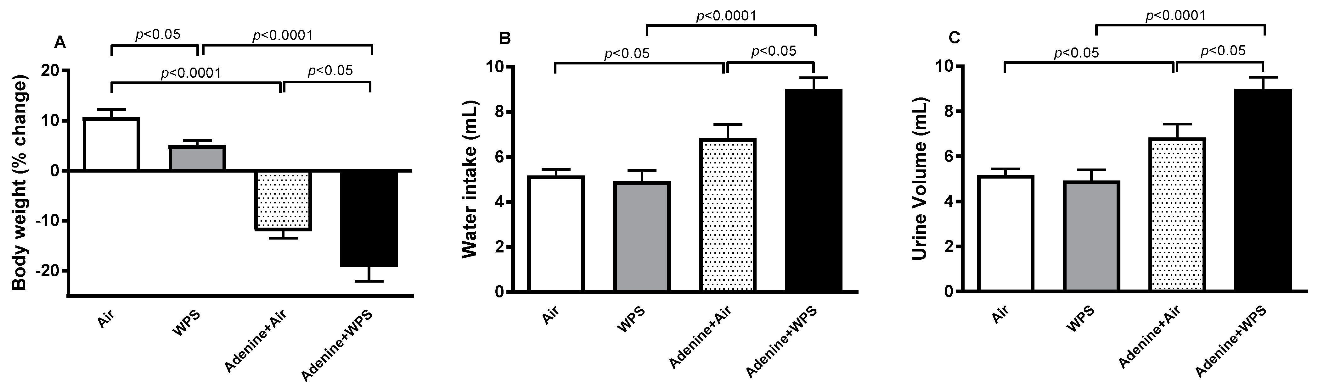
Abstract
:1. Introduction
2. Results
2.1. Body Weight, Water Intake and Urine Volume Following WPS Exposure in Mice with CKD
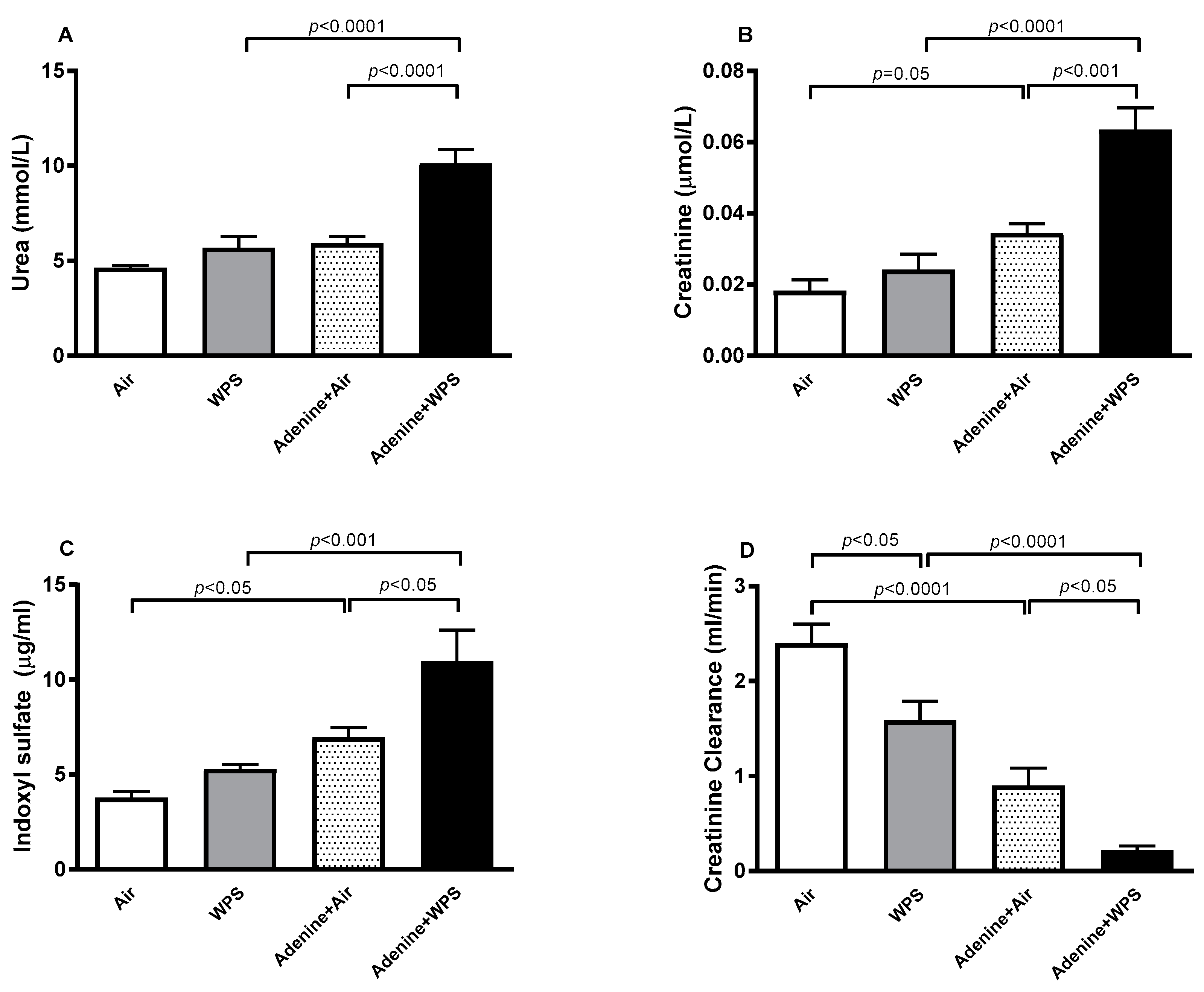
2.2. Urea, Creatine and Indoxyl Sulfate Concentrations in Plasma and Creatinine Clearance Following WPS Exposure in Mice with CKD
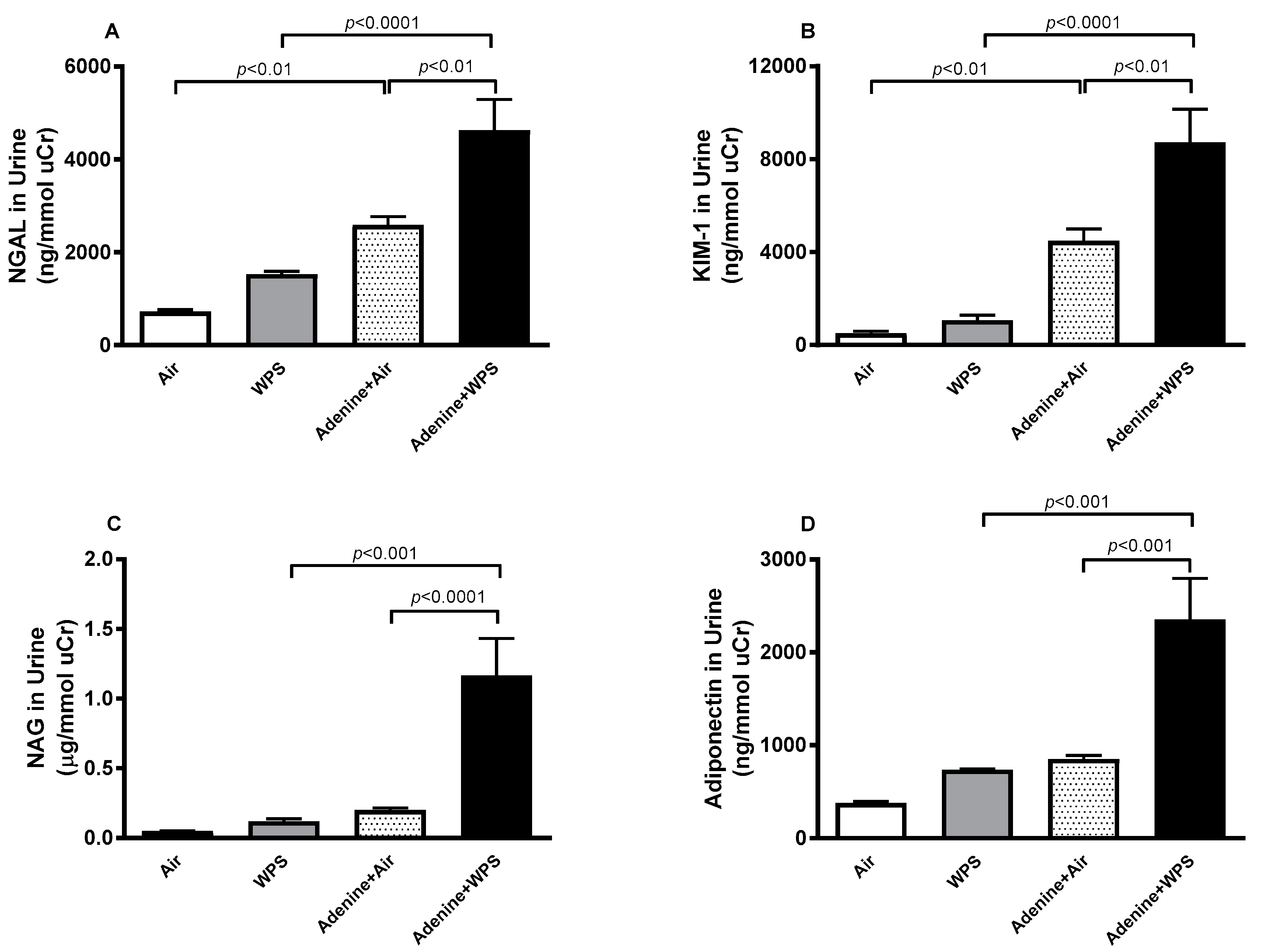
2.3. Neutrophil Gelatinase- Associated Lipocalin (NGAL), Kidney Injury Molecule-1 (KIM-1), N-acetyl-β-D-glucosaminidase (NAG) and Adiponectin in Urine Following WPS Exposure in Mice with CKD
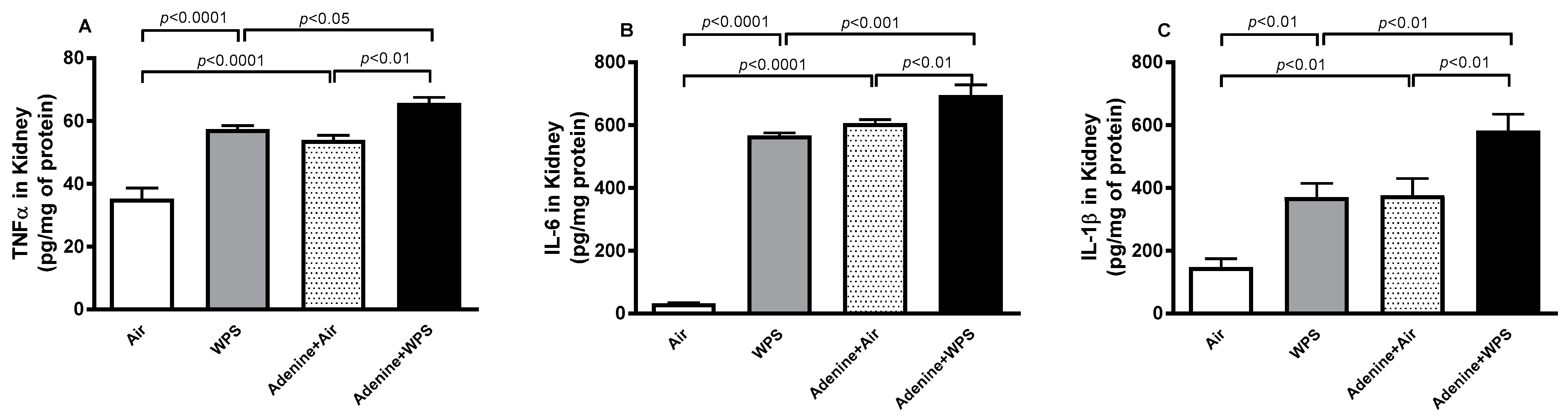
2.4. Tumor Necrosis Factor-α (TNFα), Interleukin (IL)−6 and IL-1β Concentrations in Kidney Tissue Following WPS Exposure in Mice with CKD
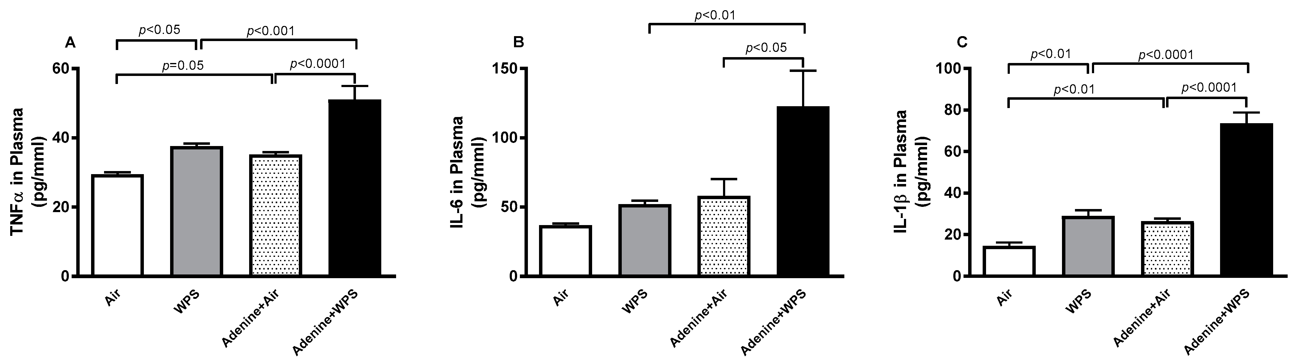
2.5. TNFα, IL-6 and IL-1β Concentrations in Plasma Following WPS Exposure in Mice with CKD
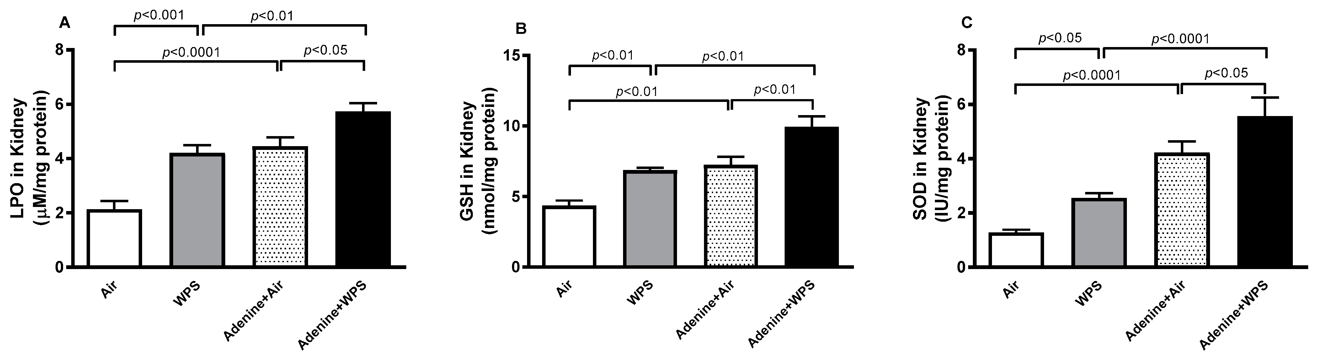
2.6. Lipid Peroxidation (LPO) and Reduced Glutathione (GSH) Concentrations and Superoxide Dismutase (SOD) Activity in Kidney Tissue Following WPS Exposure in Mice with CKD
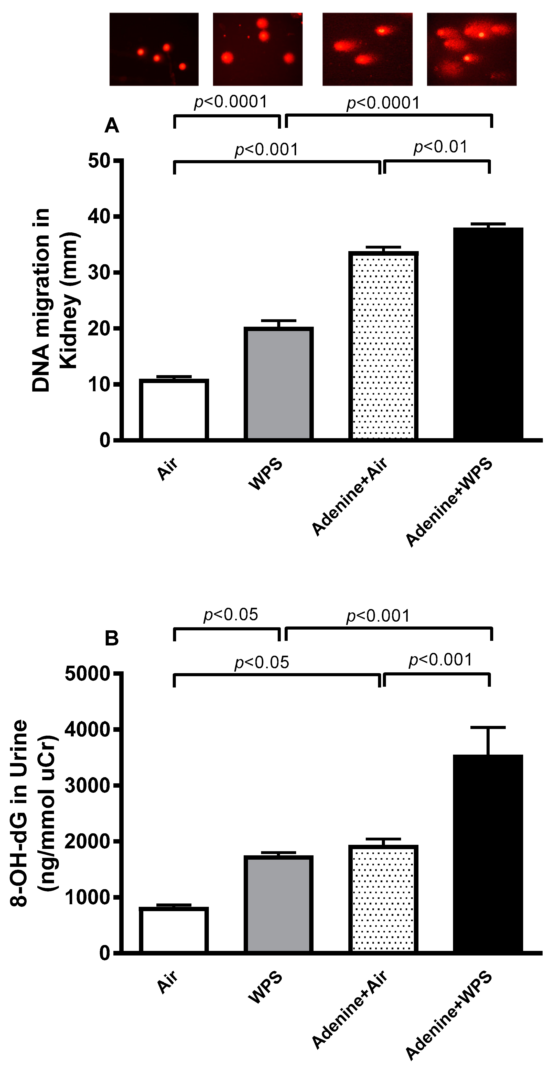
2.7. DNA Damage in Kidney Tissue and Urine Following WPS Exposure in Mice with CKD
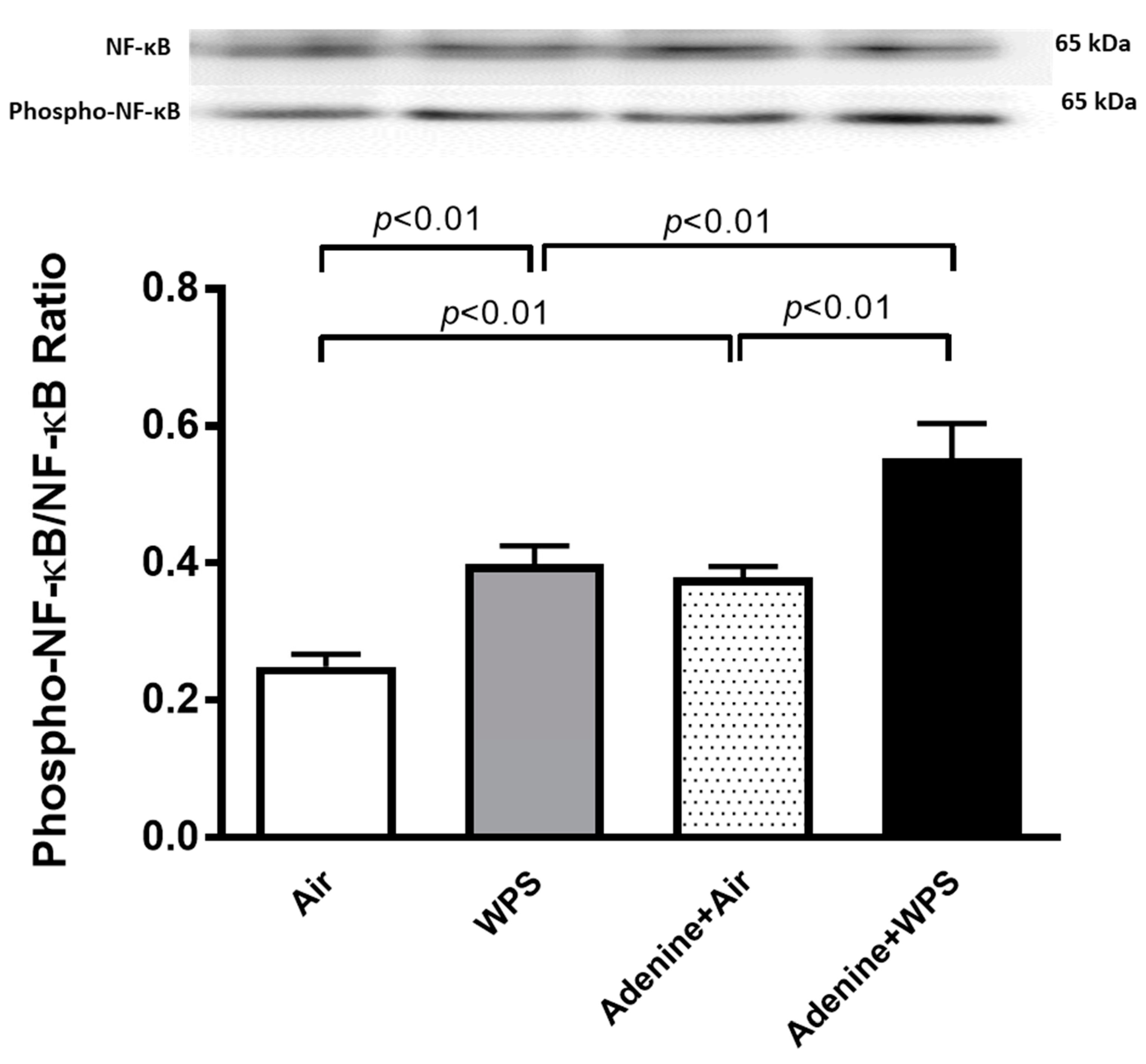
2.8. Total and Phosphorylated (Phospho) Nuclear Factor κappa B (NF-κB) Expression in Kidney Tissue Following WPS Exposure in Mice with CKD
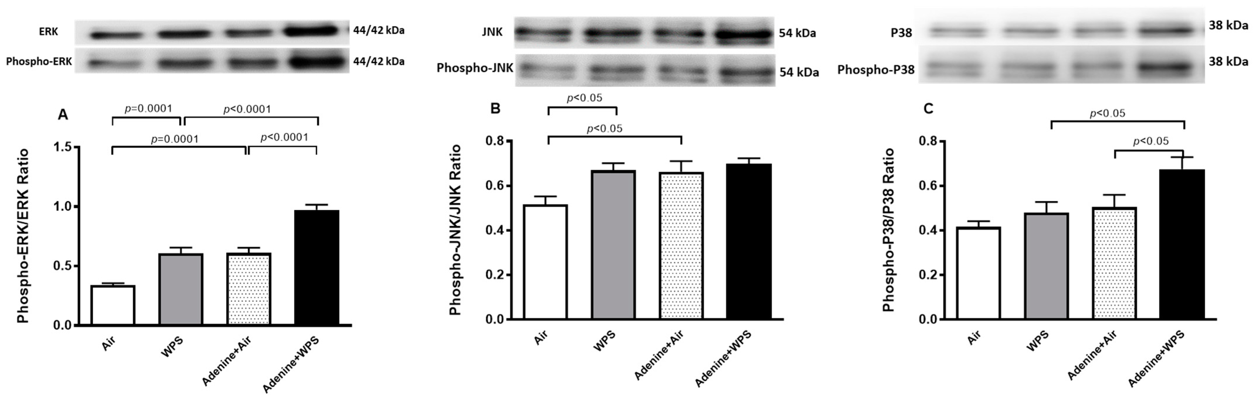
2.9. Total and Phosphorylated (Phospho) Extracellular Signal-Regulated Kinase (ERK), c-Jun NH2-Terminal Kinase (JNK), and p-38 Expression in Kidney Tissue Following WPS Exposure in Mice with CKD
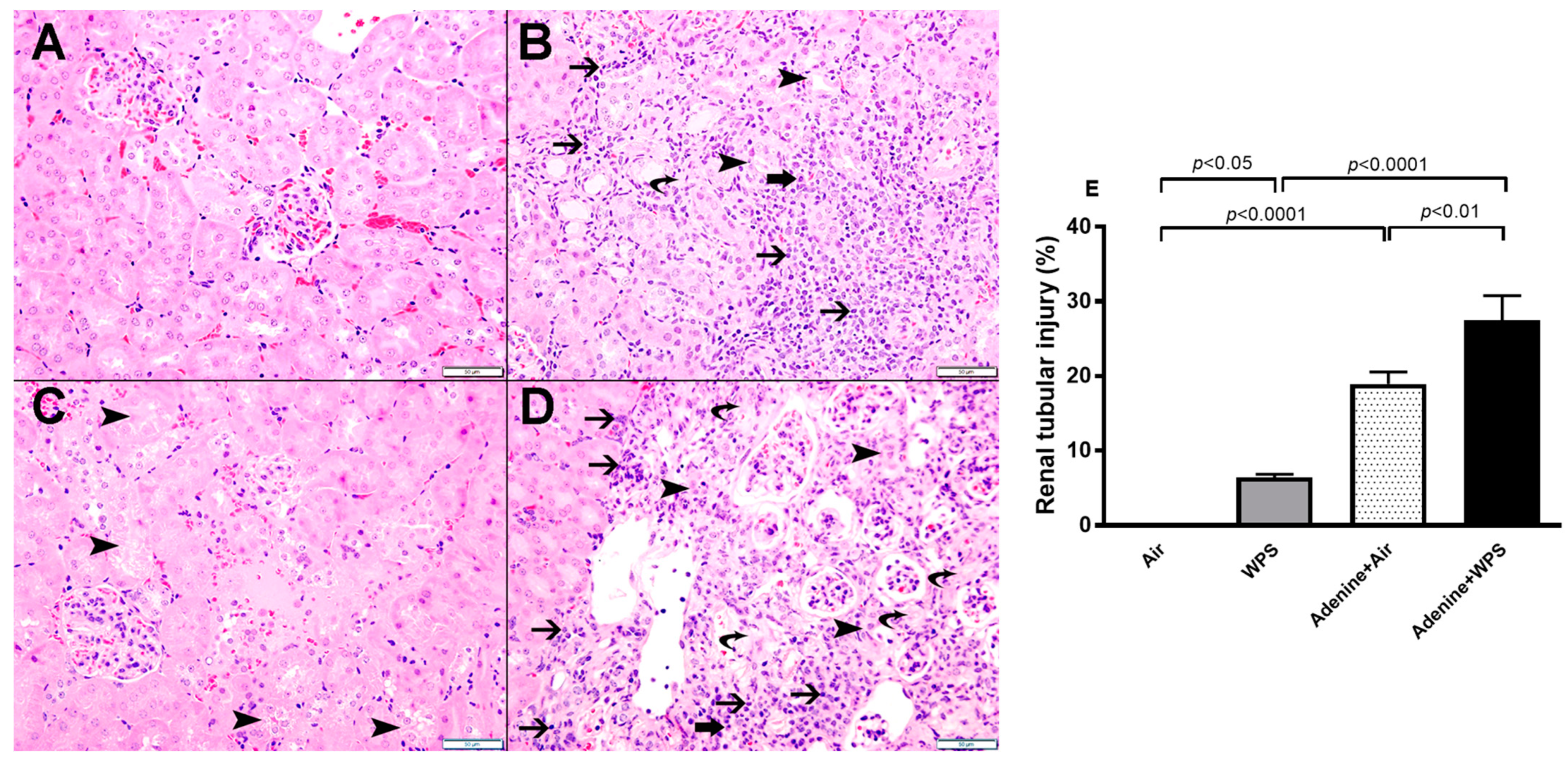
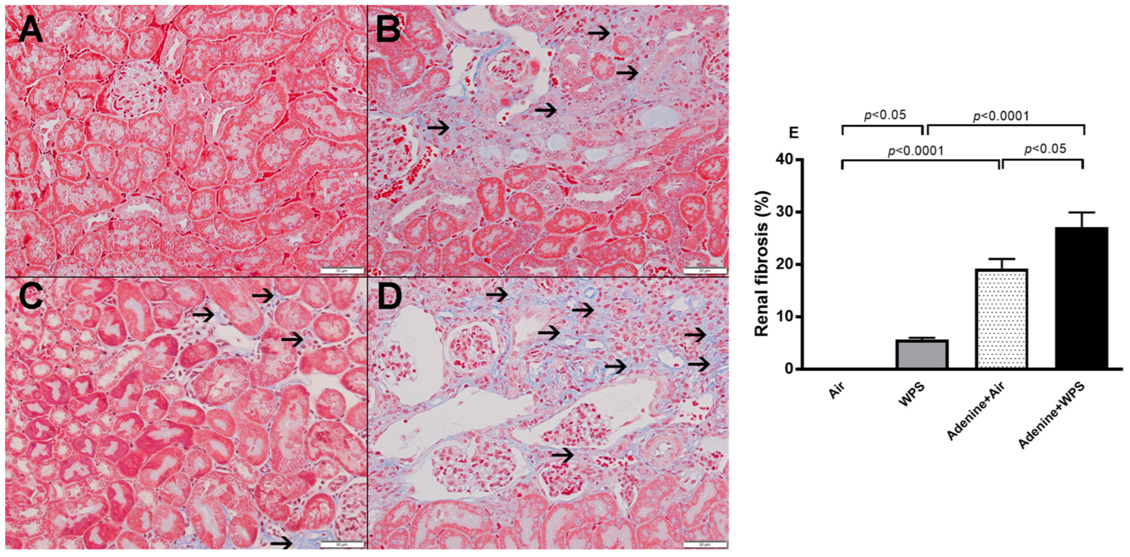
2.10. Renal Histology Following WPS Exposure in Mice with CKD
3. Discussion
4. Materials and Methods
4.1. Animals and WPS Exposure
- Air group: this control group was given standard feed and exposed to air only for 30 min, 5 times/week for four weeks.
- WPS group: this group was given standard feed and exposed to WPS for 30 min, 5 times/week for four weeks.
- Adenine + air group: this group was given the same standard diet in powdered form containing 0.15% adenine and exposed to air only for 30 min, 5 times/week for four weeks.
- Adenine + WPS group: this group was given the same standard diet in powdered form containing 0.15% adenine and exposed to WPS for 30 min, 5 times/week for four weeks.
4.2. Blood and Kidney Collection
4.3. Biochemical Tests in Plasma, Urine, Kidney Homogenate
4.4. Assessment of Kidney DNA Damage
4.5. Western Blot Analysis
4.6. Histopathological Analysis of the Kidneys
4.7. Statistics
5. Conclusions
Author Contributions
Funding
Institutional Review Board Statement
Informed Consent Statement
Data Availability Statement
Conflicts of Interest
References
- Jawad, M.; Charide, R.; Waziry, R.; Darzi, A.; Ballout, R.A.; Akl, E.A. The prevalence and trends of waterpipe tobacco smoking: A systematic review. PLoS ONE 2018, 13, e0192191. [Google Scholar] [CrossRef] [PubMed]
- Bhatnagar, A.; Maziak, W.; Eissenberg, T.; Ward, K.D.; Thurston, G.; King, B.A.; Sutfin, E.L.; Cobb, C.O.; Griffiths, M.; Goldstein, L.B.; et al. Water Pipe (Hookah) Smoking and Cardiovascular Disease Risk: A Scientific Statement From the American Heart Association. Circulation 2019, 139, e917–e936. [Google Scholar] [CrossRef] [PubMed]
- Khan, N.A.; Lawyer, G.; McDonough, S.; Wang, Q.; Kassem, N.O.; Kas-Petrus, F.; Ye, D.; Singh, K.P.; Rahman, I. Systemic biomarkers of inflammation, oxidative stress and tissue injury and repair among waterpipe, cigarette and dual tobacco smokers. Tob. Control 2020, 29, s102–s109. [Google Scholar] [CrossRef] [PubMed]
- Eissenberg, T.; Shihadeh, A. Waterpipe tobacco and cigarette smoking: Direct comparison of toxicant exposure. Am. J. Prev. Med. 2009, 37, 518–523. [Google Scholar] [CrossRef] [PubMed]
- Daher, N.; Saleh, R.; Jaroudi, E.; Sheheitli, H.; Badr, T.; Sepetdjian, E.; Al, R.M.; Saliba, N.; Shihadeh, A. Comparison of carcinogen, carbon monoxide, and ultrafine particle emissions from narghile waterpipe and cigarette smoking: Sidestream smoke measurements and assessment of second-hand smoke emission factors. Atmos. Environ. (1994) 2010, 44, 8–14. [Google Scholar] [CrossRef] [PubMed]
- Xia, J.; Wang, L.; Ma, Z.; Zhong, L.; Wang, Y.; Gao, Y.; He, L.; Su, X. Cigarette smoking and chronic kidney disease in the general population: A systematic review and meta-analysis of prospective cohort studies. Nephrol. Dial. Transplant. 2017, 32, 475–487. [Google Scholar] [CrossRef]
- Xu, X.; Nie, S.; Ding, H.; Hou, F.F. Environmental pollution and kidney diseases. Nat. Rev. Nephrol. 2018, 14, 313–324. [Google Scholar] [CrossRef]
- Yacoub, R.; Habib, H.; Lahdo, A.; Al Ali, R.; Varjabedian, L.; Atalla, G.; Kassis Akl, N.; Aldakheel, S.; Alahdab, S.; Albitar, S. Association between smoking and chronic kidney disease: A case control study. BMC Public Health 2010, 10, 731. [Google Scholar] [CrossRef]
- Miller, M.R.; Newby, D.E. Air pollution and cardiovascular disease: Car sick. Cardiovasc. Res. 2020, 116, 279–294. [Google Scholar] [CrossRef]
- Lv, J.C.; Zhang, L.X. Prevalence and Disease Burden of Chronic Kidney Disease. Adv. Exp. Med. Biol. 2019, 1165, 3–15. [Google Scholar] [CrossRef]
- Soule, E.K.; Lipato, T.; Eissenberg, T. Waterpipe tobacco smoking: A new smoking epidemic among the young? Curr. Pulmonol. Rep. 2015, 4, 163–172. [Google Scholar] [CrossRef] [PubMed]
- Rababa’h, A.M.; Sultan, B.B.; Alzoubi, K.H.; Khabour, O.F.; Ababneh, M.A. Exposure to waterpipe smoke induces renal functional and oxidative biomarkers variations in mice. Inhal. Toxicol. 2016, 28, 508–513. [Google Scholar] [CrossRef] [PubMed]
- Nemmar, A.; Beegam, S.; Yuvaraju, P.; Yasin, J.; Ali, B.H.; Adeghate, E. Nose-Only Water-Pipe Smoke Exposure in Mice Elicits Renal Histopathological Alterations, Inflammation, Oxidative Stress, DNA Damage, and Apoptosis. Front. Physiol. 2020, 11, 46. [Google Scholar] [CrossRef] [PubMed]
- Jain, R.B. Concentrations of serum cotinine across stages of glomerular function among US adult smokers and nonsmokers. Environ. Sci. Pollut. Res. 2020, 27, 34978–34986. [Google Scholar] [CrossRef] [PubMed]
- Deyo, D.; Hemingway, J.; Hughes, D.R. Identifying Patients With Undiagnosed Chronic Conditions: An Examination of Patient Costs Before Chronic Disease Diagnosis. J. Am. Coll. Radiol. 2015, 12, 1388–1394. [Google Scholar] [CrossRef] [PubMed]
- Kovesdy, C.P. Epidemiology of chronic kidney disease: An update 2022. Kidney Int. Suppl. 2022, 12, 7–11. [Google Scholar] [CrossRef] [PubMed]
- Boersma, P.; Black, L.I.; Ward, B.W. Prevalence of Multiple Chronic Conditions Among US Adults, 2018. Prev. Chronic Dis. 2020, 17, E106. [Google Scholar] [CrossRef]
- Cao, Y.; Chen, X.; Sun, H. Silencing of O-linked N-acetylglucosamine transferase ameliorates hypercalcemia-induced neurotoxicity in renal failure by regulating EZH2/KLF2/CXCL1 axis. Cell Death Dis. 2021, 12, 819. [Google Scholar] [CrossRef]
- Ali, B.H.; Al-Salam, S.; Al Za’abi, M.; Waly, M.I.; Ramkumar, A.; Beegam, S.; Al-Lawati, I.; Adham, S.A.; Nemmar, A. New model for adenine-induced chronic renal failure in mice, and the effect of gum acacia treatment thereon: Comparison with rats. J. Pharmacol. Toxicol. Methods 2013, 68, 384–393. [Google Scholar] [CrossRef]
- Kelly, J.T.; Su, G.; Zhang, L.; Qin, X.; Marshall, S.; González-Ortiz, A.; Clase, C.M.; Campbell, K.L.; Xu, H.; Carrero, J.J. Modifiable Lifestyle Factors for Primary Prevention of CKD: A Systematic Review and Meta-Analysis. J. Am. Soc. Nephrol. 2021, 32, 239–253. [Google Scholar] [CrossRef]
- Elihimas Júnior, U.F.; Elihimas, H.C.; Lemos, V.M.; Leão Mde, A.; Sá, M.P.; França, E.E.; Lemos, A.; Valente, L.M.; Markman Filho, B. Smoking as risk factor for chronic kidney disease: Systematic review. J. Bras. Nefrol. 2014, 36, 519–528. [Google Scholar] [CrossRef] [PubMed]
- Tsai, W.C.; Wu, H.Y.; Peng, Y.S.; Ko, M.J.; Wu, M.S.; Hung, K.Y.; Wu, K.D.; Chu, T.S.; Chien, K.L. Risk Factors for Development and Progression of Chronic Kidney Disease: A Systematic Review and Exploratory Meta-Analysis. Medicine 2016, 95, e3013. [Google Scholar] [CrossRef] [PubMed]
- Jo, W.; Lee, S.; Joo, Y.S.; Nam, K.H.; Yun, H.R.; Chang, T.I.; Kang, E.W.; Yoo, T.H.; Han, S.H.; Kang, S.W.; et al. Association of smoking with incident CKD risk in the general population: A community-based cohort study. PLoS ONE 2020, 15, e0238111. [Google Scholar] [CrossRef] [PubMed]
- Tohidi, M.; Hasheminia, M.; Mohebi, R.; Khalili, D.; Hosseinpanah, F.; Yazdani, B.; Nasiri, A.A.; Azizi, F.; Hadaegh, F. Incidence of chronic kidney disease and its risk factors, results of over 10 year follow up in an Iranian cohort. PLoS ONE 2012, 7, e45304. [Google Scholar] [CrossRef] [PubMed]
- Parizadeh, D.; Momenan, A.A.; Amouzegar, A.; Azizi, F.; Hadaegh, F. Tobacco Smoking: Findings from 20 Years of the Tehran Lipid and Glucose Study. Int. J. Endocrinol. Metab. 2018, 16, e84738. [Google Scholar] [CrossRef] [PubMed]
- Al-Sawalha, N.A.; AlSari, R.R.; Khabour, O.F.; Alzoubi, K.H. Influence of prenatal waterpipe tobacco smoke exposure on renal biomarkers in adult offspring rats. Inhal. Toxicol. 2019, 31, 171–179. [Google Scholar] [CrossRef]
- Huang, H.J.; Chou, C.L.; Sandar, T.T.; Liu, W.C.; Yang, H.C.; Lin, Y.C.; Zheng, C.M.; Chiu, H.W. Currently Used Methods to Evaluate the Efficacy of Therapeutic Drugs and Kidney Safety. Biomolecules 2023, 13, 1581. [Google Scholar] [CrossRef]
- Nemmar, A.; Karaca, T.; Beegam, S.; Yuvaraju, P.; Yasin, J.; Hamadi, N.K.; Ali, B.H. Prolonged Pulmonary Exposure to Diesel Exhaust Particles Exacerbates Renal Oxidative Stress, Inflammation and DNA Damage in Mice with Adenine-Induced Chronic Renal Failure. Cell. Physiol. Biochem. 2016, 38, 1703–1713. [Google Scholar] [CrossRef]
- Tumur, Z.; Niwa, T. Indoxyl Sulfate Inhibits Nitric Oxide Production and Cell Viability by Inducing Oxidative Stress in Vascular Endothelial Cells. Am. J. Nephrol. 2009, 29, 551–557. [Google Scholar] [CrossRef]
- Dou, L.; Jourde-Chiche, N.; Faure, V.; Cerini, C.; Berland, Y.; Dignat-George, F.; Brunet, P. The uremic solute indoxyl sulfate induces oxidative stress in endothelial cells. J. Thromb. Haemost. 2007, 5, 1302–1308. [Google Scholar] [CrossRef]
- Tan, X.; Cao, X.; Zou, J.; Shen, B.; Zhang, X.; Liu, Z.; Lv, W.; Teng, J.; Ding, X. Indoxyl sulfate, a valuable biomarker in chronic kidney disease and dialysis. Hemodial. Int. 2017, 21, 161–167. [Google Scholar] [CrossRef] [PubMed]
- McMahon, G.M.; Waikar, S.S. Biomarkers in nephrology: Core Curriculum 2013. Am. J. Kidney Dis. 2013, 62, 165–178. [Google Scholar] [CrossRef] [PubMed]
- Ali, B.H.; Al-Salam, S.; Al Suleimani, Y.; Al Kalbani, J.; Al Bahlani, S.; Ashique, M.; Manoj, P.; Al Dhahli, B.; Al Abri, N.; Naser, H.T.; et al. Curcumin Ameliorates Kidney Function and Oxidative Stress in Experimental Chronic Kidney Disease. Basic Clin. Pharmacol. Toxicol. 2018, 122, 65–73. [Google Scholar] [CrossRef] [PubMed]
- Gil, A.; Brod, V.; Awad, H.; Heyman, S.N.; Abassi, Z.; Frajewicki, V. Neutrophil gelatinase-associated lipocalin in a triphasic rat model of adenine-induced kidney injury. Ren. Fail. 2016, 38, 1448–1454. [Google Scholar] [CrossRef] [PubMed]
- Rysz, J.; Gluba-Brzózka, A.; Franczyk, B.; Jabłonowski, Z.; Ciałkowska-Rysz, A. Novel Biomarkers in the Diagnosis of Chronic Kidney Disease and the Prediction of Its Outcome. Int. J. Mol. Sci. 2017, 18, 1702. [Google Scholar] [CrossRef] [PubMed]
- Nemmar, A.; Karaca, T.; Beegam, S.; Yuvaraju, P.; Yasin, J.; Ali, B.H. Lung Oxidative Stress, DNA Damage, Apoptosis, and Fibrosis in Adenine-Induced Chronic Kidney Disease in Mice. Front. Physiol. 2017, 8, 896. [Google Scholar] [CrossRef] [PubMed]
- Rapa, S.F.; Di Iorio, B.R.; Campiglia, P.; Heidland, A.; Marzocco, S. Inflammation and Oxidative Stress in Chronic Kidney Disease-Potential Therapeutic Role of Minerals, Vitamins and Plant-Derived Metabolites. Int. J. Mol. Sci. 2019, 21, 263. [Google Scholar] [CrossRef] [PubMed]
- Touyz, R.M.; Rios, F.J.; Alves-Lopes, R.; Neves, K.B.; Camargo, L.L.; Montezano, A.C. Oxidative Stress: A Unifying Paradigm in Hypertension. Can. J. Cardiol. 2020, 36, 659–670. [Google Scholar] [CrossRef]
- Siti, H.N.; Kamisah, Y.; Kamsiah, J. The role of oxidative stress, antioxidants and vascular inflammation in cardiovascular disease (a review). Vasc. Pharmacol. 2015, 71, 40–56. [Google Scholar] [CrossRef]
- Birben, E.; Sahiner, U.M.; Sackesen, C.; Erzurum, S.; Kalayci, O. Oxidative stress and antioxidant defense. World Allergy Organ. J. 2012, 5, 9–19. [Google Scholar] [CrossRef]
- Schupp, N.; Stopper, H.; Heidland, A. DNA Damage in Chronic Kidney Disease: Evaluation of Clinical Biomarkers. Oxidative Med. Cell. Longev. 2016, 2016, 3592042. [Google Scholar] [CrossRef] [PubMed]
- Zhang, H.; Sun, S.C. NF-κB in inflammation and renal diseases. Cell Biosci. 2015, 5, 63. [Google Scholar] [CrossRef] [PubMed]
- Cuarental, L.; Sucunza-Sáenz, D.; Valiño-Rivas, L.; Fernandez-Fernandez, B.; Sanz, A.B.; Ortiz, A.; Vaquero, J.J.; Sanchez-Niño, M.D. MAP3K kinases and kidney injury. Nefrol. (Engl. Ed.) 2019, 39, 568–580. [Google Scholar] [CrossRef]
- Al Za’abi, M.; Ali, B.H.; Al Suleimani, Y.; Adham, S.A.; Ali, H.; Manoj, P.; Ashique, M.; Nemmar, A. The Effect of Metformin in Diabetic and Non-Diabetic Rats with Experimentally-Induced Chronic Kidney Disease. Biomolecules 2021, 11, 814. [Google Scholar] [CrossRef] [PubMed]
- Nemmar, A.; Al-Salam, S.; Beegam, S.; Zaaba, N.E.; Elzaki, O.; Ali, B.H. Waterpipe smoke inhalation potentiates cardiac oxidative stress, inflammation, mitochondrial dysfunction, apoptosis and autophagy in experimental hypertension. Biomed. Pharmacother. 2023, 158, 114144. [Google Scholar] [CrossRef] [PubMed]
- Diwan, V.; Brown, L.; Gobe, G.C. Adenine-induced chronic kidney disease in rats. Nephrology 2018, 23, 5–11. [Google Scholar] [CrossRef] [PubMed]
- Nakano, S.; Masuda, K.; Asanuma, T.; Nakatani, S. The effect of chronic renal failure on cardiac function: An experimental study with a rat model. J. Echocardiogr. 2016, 14, 156–162. [Google Scholar] [CrossRef] [PubMed]
- Nemmar, A.; Al-Salam, S.; Yuvaraju, P.; Beegam, S.; Yasin, J.; Ali, B.H. Chronic exposure to water-pipe smoke induces cardiovascular dysfunction in mice. Am. J. Physiol. Heart Circ. Physiol. 2017, 312, H329–H339. [Google Scholar] [CrossRef]
- Toukan, Y.; Hakim, F.; Bentur, Y.; Aharon-Peretz, J.; Elemy, A.; Gur, M.; Hanna, M.; Fisher, T.; Scherb, I.; Bentur, L. The Effect of a 30-Min Water-Pipe Smoking Session on Cognitive Measures and Cardio-Pulmonary Parameters. Nicotine Tob. Res. 2020, 22, 1347–1353. [Google Scholar] [CrossRef]
- Bentur, L.; Hellou, E.; Goldbart, A.; Pillar, G.; Monovich, E.; Salameh, M.; Scherb, I.; Bentur, Y. Laboratory and clinical acute effects of active and passive indoor group water-pipe (narghile) smoking. Chest 2014, 145, 803–809. [Google Scholar] [CrossRef]
- Hakim, F.; Hellou, E.; Goldbart, A.; Katz, R.; Bentur, Y.; Bentur, L. The acute effects of water-pipe smoking on the cardiorespiratory system. Chest 2011, 139, 775–781. [Google Scholar] [CrossRef] [PubMed]
- Zaaba, N.E.; Al-Salam, S.; Beegam, S.; Elzaki, O.; Yasin, J.; Nemmar, A. Catalpol Attenuates Oxidative Stress and Inflammation via Mechanisms Involving Sirtuin-1 Activation and NF-κB Inhibition in Experimentally-Induced Chronic Kidney Disease. Nutrients 2023, 15, 237. [Google Scholar] [CrossRef] [PubMed]
- de Souza, M.F.; Goncales, T.A.; Steinmetz, A.; Moura, D.J.; Saffi, J.; Gomez, R.; Barros, H.M. Cocaine induces DNA damage in distinct brain areas of female rats under different hormonal conditions. Clin. Exp. Pharmacol. Physiol. 2014, 41, 265–269. [Google Scholar] [CrossRef] [PubMed]
- Olive, P.L.; Banath, J.P.; Fjell, C.D. DNA strand breakage and DNA structure influence staining with propidium iodide using the alkaline comet assay. Cytometry 1994, 16, 305–312. [Google Scholar] [CrossRef]
- Kuchařová, M.; Hronek, M.; Rybáková, K.; Zadák, Z.; Štětina, R.; Josková, V.; Patková, A. Comet assay and its use for evaluating oxidative DNA damage in some pathological states. Physiol. Res. 2019, 68, 1–15. [Google Scholar] [CrossRef]
- Zhu, T.; Zhang, W.; Xiao, M.; Chen, H.; Jin, H. Protective role of andrographolide in bleomycin-induced pulmonary fibrosis in mice. Int. J. Mol. Sci. 2013, 14, 23581–23596. [Google Scholar] [CrossRef]


Disclaimer/Publisher’s Note: The statements, opinions and data contained in all publications are solely those of the individual author(s) and contributor(s) and not of MDPI and/or the editor(s). MDPI and/or the editor(s) disclaim responsibility for any injury to people or property resulting from any ideas, methods, instructions or products referred to in the content. |
© 2024 by the authors. Licensee MDPI, Basel, Switzerland. This article is an open access article distributed under the terms and conditions of the Creative Commons Attribution (CC BY) license (https://creativecommons.org/licenses/by/4.0/).
Share and Cite
Beegam, S.; Al-Salam, S.; Zaaba, N.E.; Elzaki, O.; Ali, B.H.; Nemmar, A. Effects of Waterpipe Smoke Exposure on Experimentally Induced Chronic Kidney Disease in Mice. Int. J. Mol. Sci. 2024, 25, 585. https://doi.org/10.3390/ijms25010585
Beegam S, Al-Salam S, Zaaba NE, Elzaki O, Ali BH, Nemmar A. Effects of Waterpipe Smoke Exposure on Experimentally Induced Chronic Kidney Disease in Mice. International Journal of Molecular Sciences. 2024; 25(1):585. https://doi.org/10.3390/ijms25010585
Chicago/Turabian StyleBeegam, Sumaya, Suhail Al-Salam, Nur Elena Zaaba, Ozaz Elzaki, Badreldin H. Ali, and Abderrahim Nemmar. 2024. "Effects of Waterpipe Smoke Exposure on Experimentally Induced Chronic Kidney Disease in Mice" International Journal of Molecular Sciences 25, no. 1: 585. https://doi.org/10.3390/ijms25010585