

Novel CRYGC Mutation in Conserved Ultraviolet-Protective Tryptophan (p.Trp131Arg) Is Linked to Autosomal Dominant Congenital Cataract

 , , and
Abstract
:1. Introduction
2. Materials and Methods
2.1. Patient
2.2. Genes of Interest
2.3. Exome Sequencing and Analysis
2.4. Segregation Analysis
3. Results
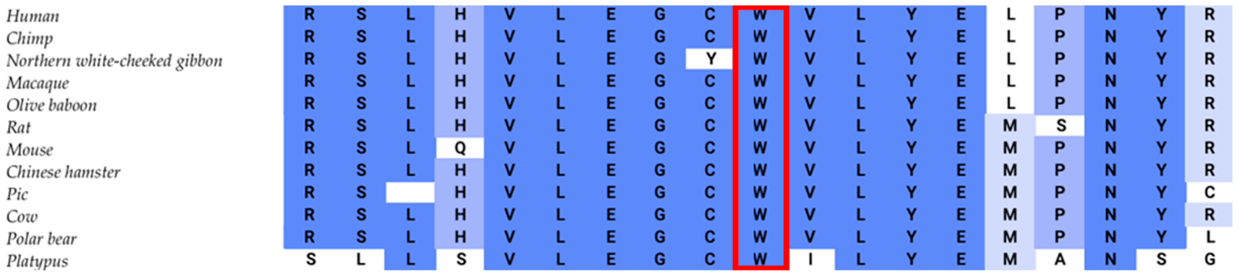
3.1. Case Presentation
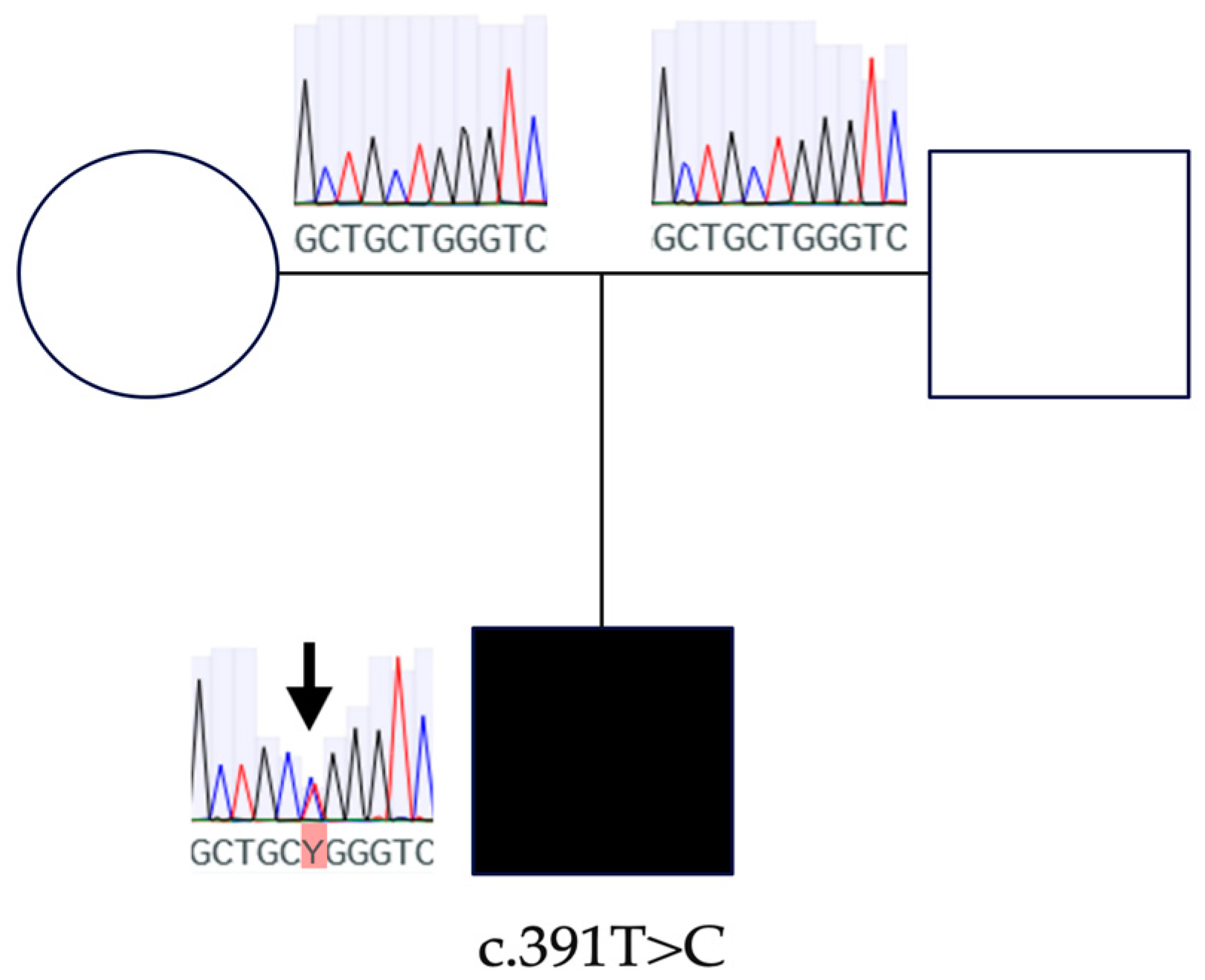
3.2. Segregation Analysis
4. Discussion
5. Conclusions
Supplementary Materials
Author Contributions
Funding
Institutional Review Board Statement
Informed Consent Statement
Data Availability Statement
Acknowledgments
Conflicts of Interest
References
- Jain, I.S.; Pillay, P.; Gangwar, D.N.; Dhir, S.P.; Kaul, V.K. Congenital Cataract: Etiology and Morphology. J. Pediatr. Ophthalmol. Strabismus 1983, 20, 238–242. [Google Scholar] [CrossRef]
- Wu, X.; Long, E.; Lin, H.; Liu, Y. Prevalence and Epidemiological Characteristics of Congenital Cataract: A Systematic Review and Meta-Analysis. Sci. Rep. 2016, 6, 28564. [Google Scholar] [CrossRef]
- Messina-Baas, O.; Cuevas-Covarrubias, S.A. Inherited Congenital Cataract: A Guide to Suspect the Genetic Etiology in the Cataract Genesis. Mol. Syndromol. 2017, 8, 58–78. [Google Scholar] [CrossRef]
- Sheeladevi, S.; Lawrenson, J.G.; Fielder, A.R.; Suttle, C.M. Global Prevalence of Childhood Cataract: A Systematic Review. Eye 2016, 30, 1160–1169. [Google Scholar] [CrossRef]
- Pichi, F.; Lembo, A.; Serafino, M.; Nucci, P. Genetics of Congenital Cataract. Dev. Ophthalmol. 2016, 57, 1–14. [Google Scholar] [CrossRef]
- Reddy, M.A.; Francis, P.J.; Berry, V.; Bhattacharya, S.S.; Moore, A.T. Molecular Genetic Basis of Inherited Cataract and Associated Phenotypes. Surv. Ophthalmol. 2004, 49, 300–315. [Google Scholar] [CrossRef]
- Yi, J.; Yun, J.; Li, Z.K.; Xu, C.T.; Pan, B.R. Epidemiology and Molecular Genetics of Congenital Cataracts. Int. J. Ophthalmol. 2011, 4, 422–432. [Google Scholar] [CrossRef]
- Berry, V.; Georgiou, M.; Fujinami, K.; Quinlan, R.; Moore, A.; Michaelides, M. Inherited Cataracts: Molecular Genetics, Clinical Features, Disease Mechanisms and Novel Therapeutic Approaches. Br. J. Ophthalmol. 2020, 104, 1331–1337. [Google Scholar] [CrossRef]
- Rechsteiner, D.; Issler, L.; Koller, S.; Lang, E.; Bähr, L.; Feil, S.; Rüegger, C.M.; Kottke, R.; Toelle, S.P.; Zweifel, N.; et al. Genetic Analysis in a Swiss Cohort of Bilateral Congenital Cataract. JAMA Ophthalmol. 2021, 139, 691–700. [Google Scholar] [CrossRef]
- Gillespie, R.L.; O’Sullivan, J.; Ashworth, J.; Bhaskar, S.; Williams, S.; Biswas, S.; Kehdi, E.; Ramsden, S.C.; Clayton-Smith, J.; Black, G.C.; et al. Personalized Diagnosis and Management of Congenital Cataract by Next-Generation Sequencing. Ophthalmology 2014, 121, 2124–2137.e2. [Google Scholar] [CrossRef]
- Tavtigian, S.V.; Deffenbaugh, A.M.; Yin, L.; Judkins, T.; Scholl, T.; Samollow, P.B.; De Silva, D.; Zharkikh, A.; Thomas, A. Comprehensive Statistical Study of 452 BRCA1 Missense Substitutions with Classification of Eight Recurrent Substitutions as Neutral. J. Med. Genet. 2006, 43, 295–305. [Google Scholar] [CrossRef]
- Jackson, D.; Malka, S.; Harding, P.; Palma, J.; Dunbar, H.; Moosajee, M. Molecular Diagnostic Challenges for Non-Retinal Developmental Eye Disorders in the United Kingdom. Am. J. Med. Genet. Part C Semin. Med. Genet. 2020, 184, 578–589. [Google Scholar] [CrossRef]
- Peng, Y.; Zheng, Y.; Deng, Z.; Zhang, S.; Tan, Y.; Hu, Z.; Tao, L.; Luo, Y. Case Report: A de Novo Variant of CRYGC Gene Associated with Congenital Cataract and Microphthalmia. Front. Genet. 2022, 13, 866246. [Google Scholar] [CrossRef]
- Shiels, A.; Hejtmancik, J.F. Molecular Genetics of Cataract. Prog. Mol. Biol. Transl. Sci. 2015, 134, 203–218. [Google Scholar] [CrossRef]
- Santana, A.; Waiswol, M. The Genetic and Molecular Basis of Congenital Cataract. Arq. Bras. Oftalmol. 2011, 74, 136–142. [Google Scholar] [CrossRef]
- Bloemendal, H.; De Jong, W.; Jaenicke, R.; Lubsen, N.H.; Slingsby, C.; Tardieu, A. Ageing and Vision: Structure, Stability and Function of Lens Crystallins. Prog. Biophys. Mol. Biol. 2004, 86, 407–485. [Google Scholar] [CrossRef]
- Zhao, W.J.; Xu, J.; Chen, X.J.; Liu, H.H.; Yao, K.; Yan, Y. Bin Effects of Cataract-Causing Mutations W59C and W151C on ΒB2-Crystallin Structure, Stability and Folding. Int. J. Biol. Macromol. 2017, 103, 764–770. [Google Scholar] [CrossRef]
- Chen, J.; Callis, P.R.; King, J. Mechanism of the Very Efficient Quenching of Tryptophan Fluorescence in Human ΓD- and ΓS-Crystallins: The γ-Crystallin Fold May Have Evolved to Protect Tryptophan Residues from Ultraviolet Photodamage. Biochemistry 2009, 48, 3708–3716. [Google Scholar] [CrossRef]
- Robman, L.; Taylor, H. External Factors in the Development of Cataract. Eye 2005, 19, 1074–1082. [Google Scholar] [CrossRef]
- Association, W.M. World Medical Association Declaration of Helsinki: Ethical Principles for Medical Research Involving Human Subjects. JAMA 2013, 310, 2191–2194. [Google Scholar] [CrossRef]
- Haug, P.; Koller, S.; Maggi, J.; Lang, E.; Feil, S.; Wlodarczyk, A.; Bähr, L.; Steindl, K.; Rohrbach, M.; Gerth-Kahlert, C.; et al. Whole Exome Sequencing in Coloboma/Microphthalmia: Identification of Novel and Recurrent Variants in Seven Genes. Genes 2021, 12, 65. [Google Scholar] [CrossRef]
- Lang, E.; Koller, S.; Bähr, L.; Töteberg-Harms, M.; Atac, D.; Roulez, F.; Bahr, A.; Steindl, K.; Feil, S.; Berger, W.; et al. Exome Sequencing in a Swiss Childhood Glaucoma Cohort Reveals CYP1B1 and FOXC1 Variants as Most Frequent Causes. Transl. Vis. Sci. Technol. 2020, 9, 47. [Google Scholar] [CrossRef]
- Richards, S.; Aziz, N.; Bale, S.; Bick, D.; Das, S.; Gastier-Foster, J.; Grody, W.W.; Hegde, M.; Lyon, E.; Spector, E.; et al. Standards and Guidelines for the Interpretation of Sequence Variants: A Joint Consensus Recommendation of the American College of Medical Genetics and Genomics and the Association for Molecular Pathology. Genet. Med. Off. J. Am. Coll. Med. Genet. 2015, 17, 405–424. [Google Scholar] [CrossRef]
- Blundell, T.; Lindley, P.; Miller, L.; Moss, D.; Slingsby, C.; Turnell, B.; Wistow, G. The molecular structure and stability of the eye lens: X-ray Analysis of y-Crystallin II. Nature. 1981, 289, 773. [Google Scholar] [CrossRef]
- Héon, E.; Priston, M.; Schorderet, D.F.; Billingsley, G.D.; Girard, P.O.; Lubsen, N.; Munier, F.L. The Gamma-Crystallins and Human Cataracts: A Puzzle Made Clearer. Am. J. Hum. Genet. 1999, 65, 1261–1267. [Google Scholar] [CrossRef]
- Berry, V.; Ionides, A.; Pontikos, N.; Georgiou, M.; Yu, J.; Ocaka, L.A.; Moore, A.T.; Quinlan, R.A.; Michaelides, M. The Genetic Landscape of Crystallins in Congenital Cataract. Orphanet J. Rare Dis. 2020, 15, 333. [Google Scholar] [CrossRef]
- Astiazarán, M.C.; García-Montaño, L.A.; Sánchez-Moreno, F.; Matiz-Moreno, H.; Zenteno, J.C. Next Generation Sequencing-Based Molecular Diagnosis in Familial Congenital Cataract Expands the Mutational Spectrum in Known Congenital Cataract Genes. Am. J. Med. Genet. A 2018, 176, 2637–2645. [Google Scholar] [CrossRef]
- Jiao, X.; Viswanathan, M.; Bobrova, N.F.; Romanova, T.V.; Hejtmancik, J.F. Molecular Genetic Analysis of Ukrainian Families with Congenital Cataracts. Child 2022, 10, 51. [Google Scholar] [CrossRef]
- Zhang, J.; Sun, D.; Wang, Y.; Mu, W.; Peng, Y.; Mi, D. Identification of a novel CRYGC mutation in a pedigree affected with congenital cataracts. Zhonghua Yixue Yichuanxue Zazhi = Chin. J. Med. Genet. 2019, 36, 697–700. [Google Scholar] [CrossRef]
- Fu, C.; Xu, J.; Yang, X.; Chen, X.; Yao, K. Cataract-Causing Mutations L45P and Y46D Impair the Thermal Stability of ΓC-Crystallin. Biochem. Biophys. Res. Commun. 2021, 539, 70–76. [Google Scholar] [CrossRef]
- Zhong, Z.; Wu, Z.; Han, L.; Chen, J. Novel Mutations in CRYGC Are Associated with Congenital Cataracts in Chinese Families. Sci. Rep. 2017, 7, 189. [Google Scholar] [CrossRef]
- Kumar, M.; Agarwal, T.; Khokhar, S.; Kumar, M.; Kaur, P.; Roy, T.S.; Dada, R. Mutation Screening and Genotype Phenotype Correlation of α-Crystallin, γ-Crystallin and GJA8 Gene in Congenital Cataract. Mol. Vis. 2011, 17, 693–707. [Google Scholar]
- Sun, Z.; Zhou, Q.; Li, H.; Yang, L.; Wu, S.; Sui, R. Mutations in Crystallin Genes Result in Congenital Cataract Associated with Other Ocular Abnormalities. Mol. Vis. 2017, 23, 977–986. [Google Scholar]
- Karahan, M.; Demirtaş, A.A.; Erdem, S.; Ava, S.; Tekeş, S.; Keklikçi, U. Crystalline Gene Mutations in Turkish Children with Congenital Cataracts. Int. Ophthalmol. 2021, 41, 2847–2852. [Google Scholar] [CrossRef]
- Moon, D.; Park, H.W.; Surl, D.; Won, D.; Lee, S.T.; Shin, S.; Choi, J.R.; Han, J. Precision Medicine through Next-Generation Sequencing in Inherited Eye Diseases in a Korean Cohort. Genes 2022, 13, 27. [Google Scholar] [CrossRef]
- Li, J.; Leng, Y.; Han, S.; Yan, L.; Lu, C.; Luo, Y.; Zhang, X.; Cao, L. Clinical and Genetic Characteristics of Chinese Patients with Familial or Sporadic Pediatric Cataract. Orphanet J. Rare Dis. 2018, 13, 94. [Google Scholar] [CrossRef]
- Li, D.; Wang, S.; Ye, H.; Tang, Y.; Qiu, X.; Fan, Q.; Rong, X.; Liu, X.; Chen, Y.; Yang, J.; et al. Distribution of Gene Mutations in Sporadic Congenital Cataract in a Han Chinese Population. Mol. Vis. 2016, 22, 589–598. [Google Scholar]
- Li, X.-Q.; Cai, H.-C.; Zhou, S.-Y.; Yang, J.-H.; Xi, Y.-B.; Gao, X.-B.; Zhao, W.-J.; Li, P.; Zhao, G.-Y.; Tong, Y.; et al. A Novel Mutation Impairing the Tertiary Structure and Stability of ΓC-Crystallin (CRYGC) Leads to Cataract Formation in Humans and Zebrafish Lens. Hum. Mutat. 2012, 33, 391–401. [Google Scholar] [CrossRef]
- Xi, Y.-B.; Chen, X.-J.; Zhao, W.-J.; Yan, Y.-B. Congenital Cataract-Causing Mutation G129C in ΓC-Crystallin Promotes the Accumulation of Two Distinct Unfolding Intermediates That Form Highly Toxic Aggregates. J. Mol. Biol. 2015, 427, 2765–2781. [Google Scholar] [CrossRef]
- Prokudin, I.; Simons, C.; Grigg, J.R.; Storen, R.; Kumar, V.; Phua, Z.Y.; Smith, J.; Flaherty, M.; Davila, S.; Jamieson, R. V Exome Sequencing in Developmental Eye Disease Leads to Identification of Causal Variants in GJA8, CRYGC, PAX6 and CYP1B1. Eur. J. Hum. Genet. 2014, 22, 907–915. [Google Scholar] [CrossRef]
- Fan, F.; Luo, Y.; Wu, J.; Gao, C.; Liu, X.; Mei, H.; Zhou, X. The Mutation Spectrum in Familial versus Sporadic Congenital Cataract Based on Next-Generation Sequencing. BMC Ophthalmol. 2020, 20, 361. [Google Scholar] [CrossRef]
- Ma, A.S.; Grigg, J.R.; Ho, G.; Prokudin, I.; Farnsworth, E.; Holman, K.; Cheng, A.; Billson, F.A.; Martin, F.; Fraser, C.; et al. Sporadic and Familial Congenital Cataracts: Mutational Spectrum and New Diagnoses Using Next-Generation Sequencing. Hum. Mutat. 2016, 37, 371–384. [Google Scholar] [CrossRef]
- Santhiya, S.T.; Shyam Manohar, M.; Rawlley, D.; Vijayalakshmi, P.; Namperumalsamy, P.; Gopinath, P.M.; Löster, J.; Graw, J. Novel Mutations in the Gamma-Crystallin Genes Cause Autosomal Dominant Congenital Cataracts. J. Med. Genet. 2002, 39, 352–358. [Google Scholar] [CrossRef]
- Gonzalez-Huerta, L.M.; Messina-Baas, O.M.; Cuevas-Covarrubias, S.A. A Family with Autosomal Dominant Primary Congenital Cataract Associated with a CRYGC Mutation: Evidence of Clinical Heterogeneity. Mol. Vis. 2007, 13, 1333–1338. [Google Scholar]
- Devi, R.R.; Yao, W.; Vijayalakshmi, P.; Sergeev, Y.V.; Sundaresan, P.; Hejtmancik, J.F. Crystallin Gene Mutations in Indian Families with Inherited Pediatric Cataract. Mol. Vis. 2008, 14, 1157–1170. [Google Scholar]
- Yao, K.; Jin, C.; Zhu, N.; Wang, W.; Wu, R.; Jiang, J.; Shentu, X. A Nonsense Mutation in CRYGC Associated with Autosomal Dominant Congenital Nuclear Cataract in a Chinese Family. Mol. Vis. 2008, 14, 1272–1276. [Google Scholar]
- Kandaswamy, D.K.; Vasantha, K.; Graw, J.; Santhiya, S.T. A Novel CRYGC E128* Mutation Underlying an Autosomal Dominant Nuclear Cataract in a South Indian Kindred. Ophthalmic Genet. 2020, 41, 556–562. [Google Scholar] [CrossRef]
- Patel, N.; Anand, D.; Monies, D.; Maddirevula, S.; Khan, A.O.; Algoufi, T.; Alowain, M.; Faqeih, E.; Alshammari, M.; Qudair, A.; et al. Novel Phenotypes and Loci Identified through Clinical Genomics Approaches to Pediatric Cataract. Hum. Genet. 2017, 136, 205–225. [Google Scholar] [CrossRef]
- Reis, L.M.; Tyler, R.C.; Muheisen, S.; Raggio, V.; Salviati, L.; Han, D.P.; Costakos, D.; Yonath, H.; Hall, S.; Power, P.; et al. Whole Exome Sequencing in Dominant Cataract Identifies a New Causative Factor, CRYBA2, and a Variety of Novel Alleles in Known Genes. Hum. Genet. 2013, 132, 761–770. [Google Scholar] [CrossRef]
- Taylan Sekeroglu, H.; Karaosmanoglu, B.; Taskiran, E.Z.; Simsek Kiper, P.O.; Alikasifoglu, M.; Boduroglu, K.; Coskun, T.; Utine, G.E. Molecular Etiology of Isolated Congenital Cataract Using Next-Generation Sequencing: Single Center Exome Sequencing Data from Turkey. Mol. Syndromol. 2020, 11, 302–308. [Google Scholar] [CrossRef]
- Zhang, L.; Fu, S.; Ou, Y.; Zhao, T.; Su, Y.; Liu, P. A Novel Nonsense Mutation in CRYGC Is Associated with Autosomal Dominant Congenital Nuclear Cataracts and Microcornea. Mol. Vis. 2009, 15, 276–282. [Google Scholar]
- Kessel, L.; Bach-Holm, D.; Al-Bakri, M.; Roos, L.; Lund, A.; Grønskov, K. Genetic Disease Is a Common Cause of Bilateral Childhood Cataract in Denmark. Ophthalmic Genet. 2021, 42, 650–658. [Google Scholar] [CrossRef]
- Guo, Y.; Su, D.; Li, Q.; Yang, Z.; Ma, Z.; Ma, X.; Zhu, S. A Nonsense Mutation of CRYGC Associated with Autosomal Dominant Congenital Nuclear Cataracts and Microcornea in a Chinese Pedigree. Mol. Vis. 2012, 18, 1874–1880. [Google Scholar]
- Zhuang, J.; Cao, Z.; Zhu, Y.; Liu, L.; Tong, Y.; Chen, X.; Wang, Y.; Lu, C.; Ma, X.; Yang, J. Mutation Screening of Crystallin Genes in Chinese Families with Congenital Cataracts. Mol. Vis. 2019, 25, 427–437. [Google Scholar]
- Ren, Z.; Li, A.; Shastry, B.S.; Padma, T.; Ayyagari, R.; Scott, M.H.; Parks, M.M.; Kaiser-Kupfer, M.I.; Hejtmancik, J.F. A 5-Base Insertion in the GammaC-Crystallin Gene Is Associated with Autosomal Dominant Variable Zonular Pulverulent Cataract. Hum. Genet. 2000, 106, 531–537. [Google Scholar] [CrossRef]
- Kondo, Y.; Saitsu, H.; Miyamoto, T.; Lee, B.J.; Nishiyama, K.; Nakashima, M.; Tsurusaki, Y.; Doi, H.; Miyake, N.; Kim, J.H.; et al. Pathogenic Mutations in Two Families with Congenital Cataract Identified with Whole-Exome Sequencing. Mol. Vis. 2013, 19, 384–389. [Google Scholar]
- Zhou, Z.; Zhao, L.; Guo, Y.; Zhuang, J.; Zhuo, N.; Chen, H.; Liu, J.; Wang, L. A Novel Mutation in CRYGC Mutation Associated with Autosomal Dominant Congenital Cataracts and Microcornea. Ophthalmol. Sci. 2022, 2, 100093. [Google Scholar] [CrossRef]
- Fernández-Alcalde, C.; Nieves-Moreno, M.; Noval, S.; Peralta, J.M.; Montaño, V.E.F.; Del Pozo, Á.; Santos-Simarro, F.; Vallespín, E. Molecular and Genetic Mechanism of Non-Syndromic Congenital Cataracts. Mutation Screening in Spanish Families. Genes 2021, 12, 580. [Google Scholar] [CrossRef]
- Wang, B.; Yu, C.; Xi, Y.B.; Cai, H.C.; Wang, J.; Zhou, S.; Zhou, S.; Wu, Y.; Yan, Y.B.; Ma, X.; et al. A Novel CRYGD Mutation (p.Trp43Arg) Causing Autosomal Dominant Congenital Cataract in a Chinese Family. Hum. Mutat. 2011, 32, 1939–1947. [Google Scholar] [CrossRef]
- Ji, F.; Jungs, J.; Koharudin, L.M.I.; Gronenborn, A.M. The Human W42R ΓD-Crystallin Mutant Structure Provides a Link between Congenital and Age-Related Cataracts. J. Biol. Chem. 2013, 288, 99–109. [Google Scholar] [CrossRef]
- Rao, S.; Chun, C.; Fan, J.; Kofron, J.M.; Yang, M.B.; Hegde, R.S.; Ferrara, N.; Copenhagen, D.R.; Lang, R.A. A Direct and Melanopsin-Dependent Fetal Light Response Regulates Mouse Eye Development. Nature 2013, 494, 243–246. [Google Scholar] [CrossRef]
- Pérez-Sánchez, A.; Barrajón-Catalán, E.; Herranz-López, M.; Micol, V. Nutraceuticals for Skin Care: A Comprehensive Review of Human Clinical Studies. Nutrients 2018, 10, 403. [Google Scholar] [CrossRef]
- Santhiya, S.T.; Kumar, G.S.; Sudhakar, P.; Gupta, N.; Klopp, N.; Illig, T.; Söker, T.; Groth, M.; Platzer, M.; Gopinath, P.M.; et al. Molecular Analysis of Cataract Families in India: New Mutations in the CRYBB2 and GJA3 Genes and Rare Polymorphisms. Mol. Vis. 2010, 16, 1837–1847. [Google Scholar]
- Xu, J.; Wang, H.; Wang, A.; Xu, J.; Fu, C.; Jia, Z.; Yao, K.; Chen, X. ΒB2 W151R Mutant Is Prone to Degradation, Aggregation and Exposes the Hydrophobic Side Chains in the Fourth Greek Key Motif. Biochim. Biophys. Acta-Mol. Basis Dis. 2021, 1867, 166018. [Google Scholar] [CrossRef]
- Chen, W.; Chen, X.; Hu, Z.; Lin, H.; Zhou, F.; Luo, L.; Zhang, X.; Zhong, X.; Yang, Y.; Wu, C.; et al. A Missense Mutation in CRYBB2 Leads to Progressive Congenital Membranous Cataract by Impacting the Solubility and Function of ΒB2-Crystallin. PLoS ONE 2013, 8, e81290. [Google Scholar] [CrossRef]
- Santhiya, S.T.; Manisastry, S.M.; Rawlley, D.; Malathi, R.; Anishetty, S.; Gopinath, P.M.; Vijayalakshmi, P.; Namperumalsamy, P.; Adamski, J.; Graw, J. Mutation Analysis of Congenital Cataracts in Indian Families: Identification of SNPs and a New Causative Allele in CRYBB2 Gene. Investig. Ophthalmol. Vis. Sci. 2004, 45, 3599–3607. [Google Scholar] [CrossRef]


| Gene | CRYGC |
| cDNA | NM_020989.4:c.391T>C |
| Predicted amino acid change | p.Trp131Arg |
| Zygosity | het |
| gnomAD | n/a |
| Mode of inheritance | ad |
| Region | Exon 3 |
| ACMG | pathogenic (PP3 + PM1 + PM2 + PS2) [23] |
| Exon/ Intron | cDNA | Amino Acid Change | Coding Effect | Protein Domain | Phenotype | Reference |
|---|---|---|---|---|---|---|
| Exon 2 | NM_020989.4:c.13A>C | p.Thr5Pro | missense | 1st Greek key | Coppock-like CC | Heon et al. (1999) [25]; Berry et al. (2020) [26] |
| Exon 2 | NM_020989.4:c.17T>C | p.Phe6Ser | missense | 1st Greek key | Lamellar CC | Astiazaran et al. (2018) [27] |
| Exon 2 | NM_020989.4:c.83C>T | p.Pro28Leu | missense | 1st Greek key | Nuclear CC + microphthalmos + nystagmus | Jiao, et al. (2022) [28] |
| Exon 2 | NM_020989.4:c.110G>C | p.Arg37Pro | missense | 1st Greek key | CC NFS | Zhang et al. (2019) [29] |
| Exon 2 | NM_020989.4:c.134T>C | p.Leu45Pro | missense | 2nd Greek key | Non-syndromic CC | Gillespie et al. (2014) [10]; Fu et al. (2021) [30] |
| Exon 2 | NM_020989.4:c.136T>G | p.Tyr46Asp | missense | 2nd Greek key | Nuclear CC | Zhong et al. (2017) [31]; Fu et al. (2021) [30] |
| Exon 2 | NM_020989.4:c.143G>A | p.Arg48His | missense | 2nd Greek key | Nuclear pulverulent CC; unilateral CC + optic disc coloboma | Kumar et al. (2011) [32]; Sun et al. (2017) [33] |
| Exon 2 | NM_020989.4: c.164A>G | p.Gln55Arg | missense | 2nd Greek key | CC NFS | Karahan et al. (2021) [34] |
| Exon 2 | NM_020989.4:c.173T>C | p.Leu58Pro | missense | 2nd Greek key | CC NFS | Moon et al. (2021) [35] |
| Exon 2 | NM_020989.4:c.233C>T | p.Ser78Phe | missense | 2nd Greek key | CC + microcornea | Li et al. (2018) [36] |
| Exon 3 | NM_020989.4:c.280G>A | p.Glu94Lys | missense | 3rd Greek key | Unilateral total CC | Li et al. (2016) [37] |
| Exon 3 | NM_020989.4:c.385G>T | p.Gly129Cys | missense | 4th Greek key | CC NFS | Li et al. (2012) [38]; Xi et al. (2015) [39] |
| Exon 3 | NM_020989.4:c.497C>T | p.Ser166Phe | missense | 4th Greek key | Nuclear CC + microphthalmos | Prokudin et al. (2014) [40]; Zhong et al. (2017) [31]; Fan et al. (2020) [41]; Ma et al. (2016) [42] |
| Exon 3 | NM_020989.4:c.502C>T | p.Arg168Trp | missense | 4th Greek key | Lamellar/nuclear CC + peripupillary iris atrophy, nystagmus, | Santhiya et al. (2022) [43]; Gonzaez-Huerta et al. (2007) [44]; Devi et al. (2008) [45] |
| Exon 3 | NM_020989.4:c.327C>A | p.Cys109Ter | nonsense | 3rd Greek key | Nuclear CC | Yao et al. (2008) [46] |
| Exon 3 | NM_020989.4:c.337C>T | p.Gln113Ter | nonsense | 3rd Greek key | Nuclear CC | Li et al. (2016) [37] |
| Exon 3 | NM_020989.4:c.382G>T | p.Glu128Ter | nonsense | 3rd Greek key | Nuclear CC | Kandaswamy et al. (2020) [47] |
| Exon 3 | NM_020989.4:c.402C>G | p.Tyr134Ter | nonsense | 4th Greek key | CC NFS | Gillespie et al. (2014) [10] |
| Exon 3 | NM_020989.4:c.403G>T | p.Glu135Ter | nonsense | 4th Greek key | CC + microcornea | Patel et al. (2017) [48] |
| Exon 3 | NM_020989.4:c.417C>G | p.Tyr139Ter | nonsense | 4th Greek key | Total CC + microphthalmos | Reis et al. (2013) [49] |
| Exon 3 | NM_020989.4:c.417C>A | p.Tyr139Ter | nonsense | 4th Greek key | Nuclear CC + microcornea | Zhong et al. (2017) [31] |
| Exon 3 | NM_020989.4:c.432C>G | p.Tyr144Ter | nonsense | 4th Greek key | Nuclear CC | Zhong et al. (2017) [31]; Sun et al. (2017) [33]; Taylan Sekeroglu et al. (2020) [50] |
| Exon 3 | NM_020989.4:c.470G>A | p.Trp157Ter | nonsense | 4th Greek key | Nuclear CC + microcornea | Zhang et al. (2009) [51]; Kessel et al. (2021) [52] |
| Exon 3 | NM_020989.4:c.471G>A | p.Trp157Ter | nonsense | 4th Greek key | Nuclear CC + microcornea | Guo et al. (2012) [53] |
| Exon 3 | NM_020989.4:c.505A>T | p.Arg169Ter | nonsense | 4th Greek key | Nuclear CC | Zhong et al. (2017) [31] |
| Intron 1 | NM_020989.4:c.10-1G>A | splicing | CC NFS | Zhuang et al. (2019) [54] | ||
| Exon 2 | NM_020989.4:c.119_123dupGCGGC | p.Cys42AlafsTer63 | frameshift | 2nd Greek key | Zonular pulverulent CC | Ren et al. (2000) [55] |
| Exon 2 | NM_020989.4:c.124delT | p.Cys42AlafsTer61 | frameshift | 2nd Greek key | Total CC ± microphthalmos | Kondo et al. (2013) [56] |
| Exon 2 | NM_020989.4:c.130delA | p.Met44CysfsTer59 | frameshift | 2nd Greek key | Total CC + microcornea | Sun et al. (2017) [33] |
| Exon 2 | NM_020989.4:c.157_161dup-GCGGC | p.Gln55ValfsTer50 | frameshift | 2nd Greek key | CC NFS | Reis et al. (2013) [49] |
| Exon 2 | NM_020989.4:c.179delG | p.Arg60GlnfsTer43 | frameshift | 2nd Greek key | Nuclear CC | Berry et al. (2020) [26] |
| Exon 2 | NM_020989.4:c.192delC | p.Asp65ThrfsTer38 | frameshift | 2nd Greek key | CC NFS | Fan et al. (2020) [41] |
| Exon 2 | NM_020989.4:c.193delG | p.Asp65ThrfsTer38 | frameshift | 2nd Greek key | Nuclear CC | Zhong et al. (2017) [31] |
| Exon 3 | NM_020989.4:c.320_321del-AA | p.Glu107GlyfsTer56 | frameshift | 3rd Greek key | Total CC | Rechsteiner et al. (2021) [9] |
| Exon 3 | NM_020989.4:c.328_329del-CCinsT | p.Pro110SerfsTer37 | frameshift | 3rd Greek key | Lamellar CC | Ma et al. (2016) [42] |
| Exon 3 | NM_020989.4:c.386_389dup-GCTG | p.Cys130TrpfsTer35 | frameshift | 4th Greek key | Nuclear CC ± microphthalmos | Zhou et al. (2022) [57] |
| Exon 3 | NM_020989.4:c.394delG | p.Val132SerfsTer15 | frameshift | 4th Greek key | Total CC + microphthalmos | Peng et al. (2022) [13] |
| Exon 3 | NM_020989.4:c.423delG | p.Arg142GlyfsTer5 | frameshift | 4th Greek key | Nuclear CC | Zhong et al. (2017) [31] |
| Exon 3 | NM_020989.4:c.423dupG | p.Arg142AlafsTer22 | frameshift | 4th Greek key | Nuclear CC | Zhong et al. (2017) [31] |
| Exon 3 | NM_020989.4:c.425_432dup | p.Leu145GlyfsTer5 | frameshift | 4th Greek key | Nuclear CC + microphthalmos + iris malformations | Fernández-Alcalde et al. (2021) [58] |
| Exon 3 | NM_020989.4:c.438delG | p.Arg147GlyfsTer32 | frameshift | 4th Greek key | Nuclear CC | Fernandez-Alcade et al. (2021) [58] |
| Gene | Exon | cDNA | Amino Acid Change | Coding Effect | Protein Domain | Phenotype | Reference |
|---|---|---|---|---|---|---|---|
| CRYGD | Exon 2 | NM_006891.4:c.127T>C | p.Trp43Arg | missense | 2nd Greek Key | Nuclear CC | Wang et al. (2011) [59]; Ji et al. (2013) [60] |
| CRYBB2 | Exon 4 | NM_000496.3:c.177G>C | p.Trp59Arg | missense | 2nd Greek Key | Total CC | Santhiya et al. (2010) [63]; Zhao et al. (2017) [17] |
| CRYBB2 | Exon 6 | NM_000496.3:c.451T>C | p.Trp151Arg | missense | 4th Greek key | Progressive CC | Xu et al. (2021) [64] |
| CRYBB2 | Exon 6 | NM_000496.3:c.453G>C | p.Trp151Cys | missense | 4th Greek key | Progressive membranous CC | Chen et al. (2013) [65]; Zhao et al. (2017) [17] |
| CRYBB2 | Exon 6 | NM_000496.3:c.453G>T | p.Trp151Cys | missense | 4th Greek key | Nuclear CC | Santhiya et al. (2004) [66] |
Disclaimer/Publisher’s Note: The statements, opinions and data contained in all publications are solely those of the individual author(s) and contributor(s) and not of MDPI and/or the editor(s). MDPI and/or the editor(s) disclaim responsibility for any injury to people or property resulting from any ideas, methods, instructions or products referred to in the content. |
© 2023 by the authors. Licensee MDPI, Basel, Switzerland. This article is an open access article distributed under the terms and conditions of the Creative Commons Attribution (CC BY) license (https://creativecommons.org/licenses/by/4.0/).
Share and Cite
Delas, F.; Koller, S.; Feil, S.; Dacheva, I.; Gerth-Kahlert, C.; Berger, W. Novel CRYGC Mutation in Conserved Ultraviolet-Protective Tryptophan (p.Trp131Arg) Is Linked to Autosomal Dominant Congenital Cataract. Int. J. Mol. Sci. 2023, 24, 16594. https://doi.org/10.3390/ijms242316594
Delas F, Koller S, Feil S, Dacheva I, Gerth-Kahlert C, Berger W. Novel CRYGC Mutation in Conserved Ultraviolet-Protective Tryptophan (p.Trp131Arg) Is Linked to Autosomal Dominant Congenital Cataract. International Journal of Molecular Sciences. 2023; 24(23):16594. https://doi.org/10.3390/ijms242316594
Chicago/Turabian StyleDelas, Flora, Samuel Koller, Silke Feil, Ivanka Dacheva, Christina Gerth-Kahlert, and Wolfgang Berger. 2023. "Novel CRYGC Mutation in Conserved Ultraviolet-Protective Tryptophan (p.Trp131Arg) Is Linked to Autosomal Dominant Congenital Cataract" International Journal of Molecular Sciences 24, no. 23: 16594. https://doi.org/10.3390/ijms242316594