
Unraveling the Potential of miRNAs from CSCs as an Emerging Clinical Tool for Breast Cancer Diagnosis and Prognosis
 , , and
Abstract
:1. Introduction
2. miRNA Expression Related to BC Stem-like Phenotype
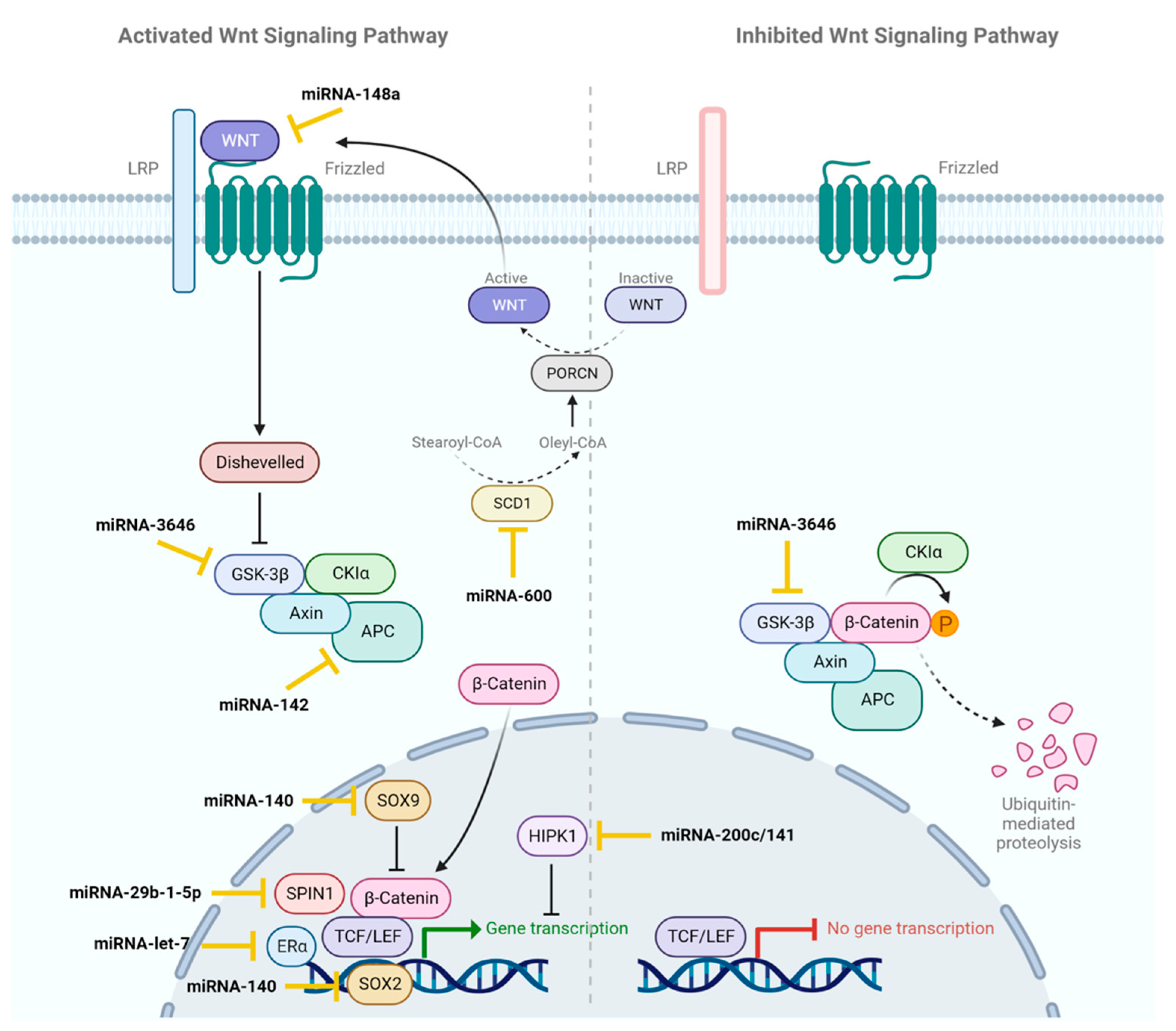
2.1. Wnt/β-Catenin
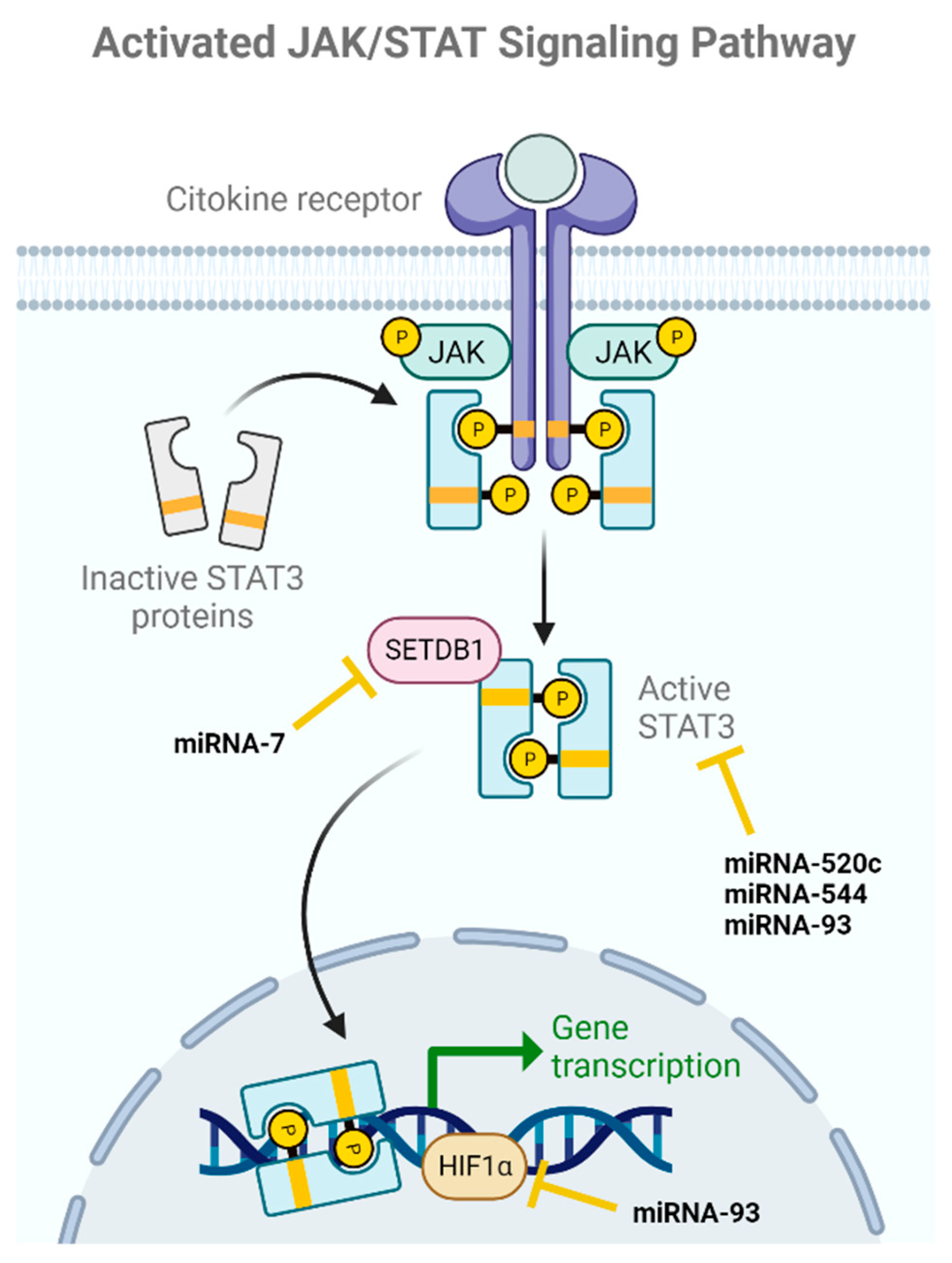
2.2. JAK/STAT
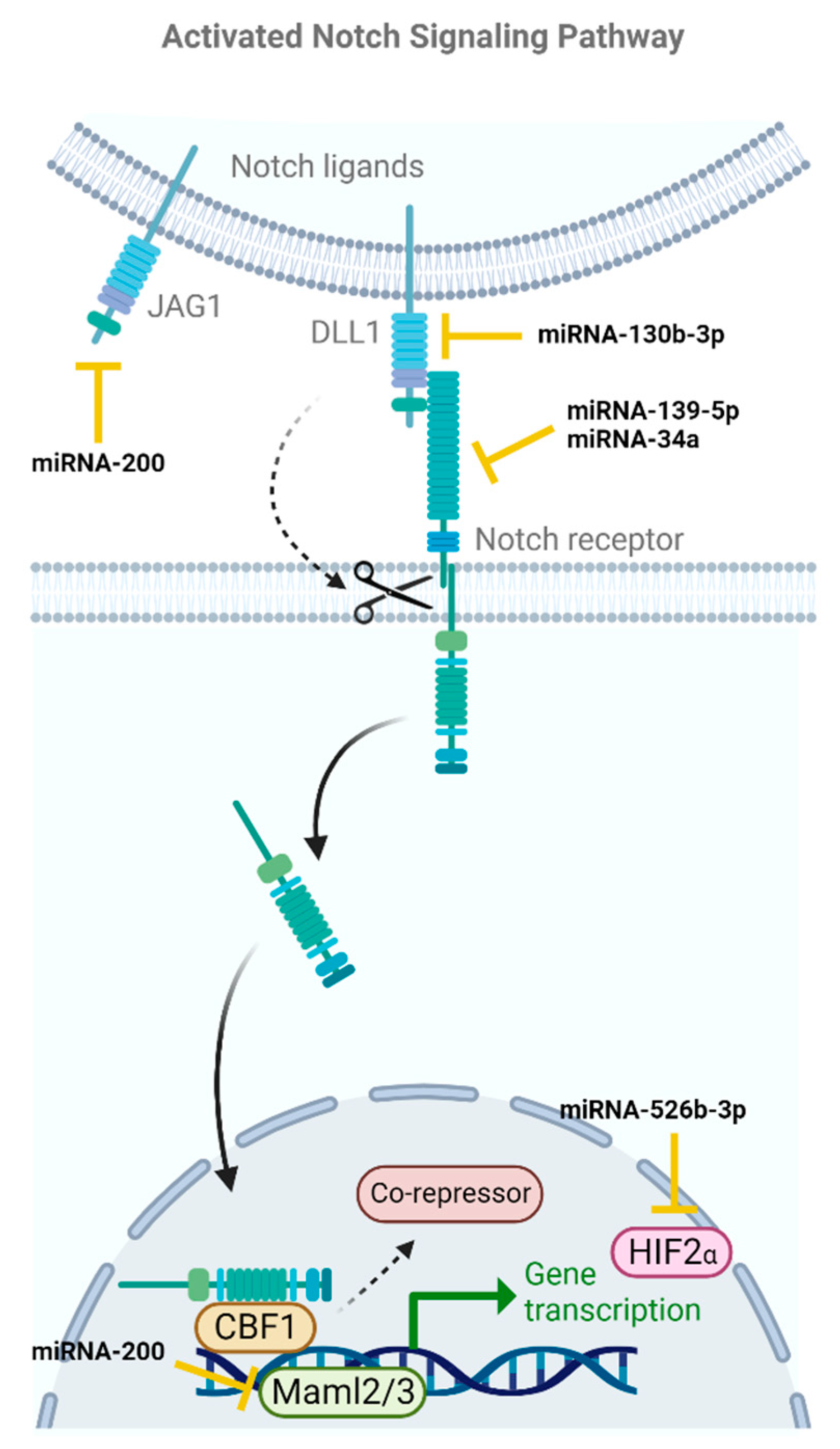
2.3. Notch
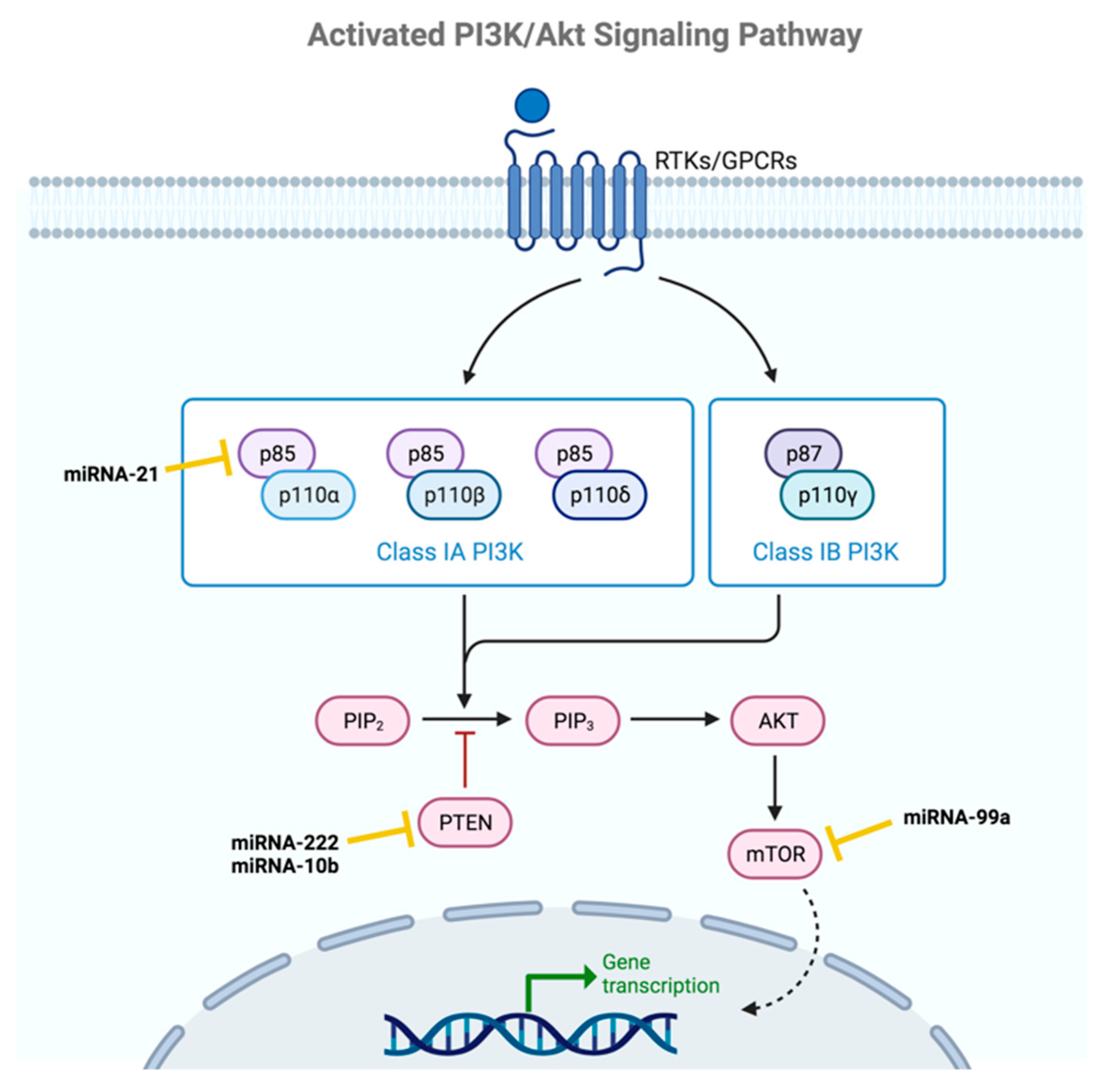
2.4. PI3K-AKT
2.5. TGF-β
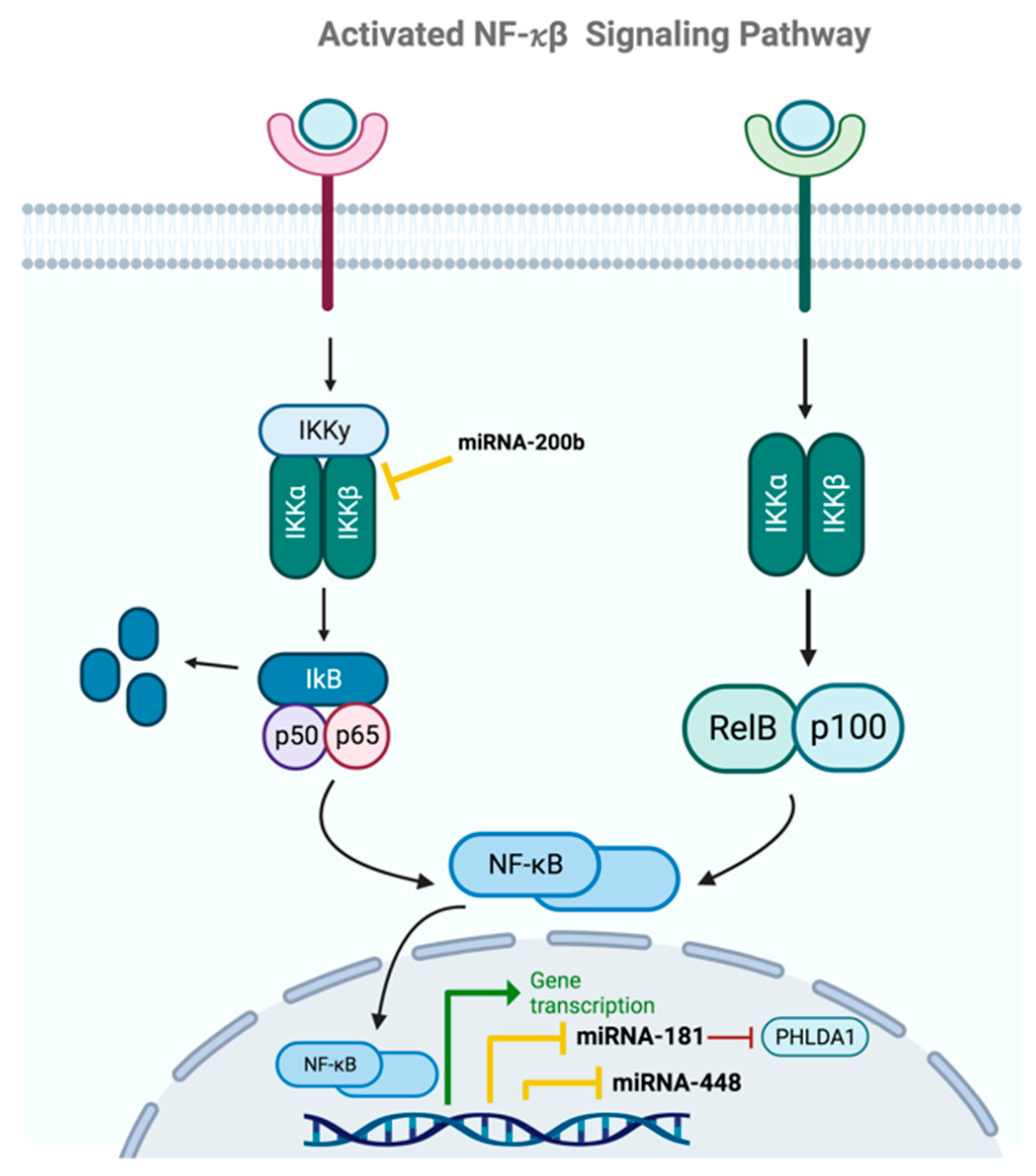
2.6. NF-κβ
3. miRNAs as Diagnostic Biomarkers in BCSCs
4. miRNA as Prognostic Biomarkers in BCSCs
5. miRNA and Chemo-Radioresistance in BCSCs

| MiRNA | MiRNA Acts as a Tumor Suppressor or Oncogenic miRNA | An Increase in Expression Causes | References |
|---|---|---|---|
| miRNA-378a-3p, miRNA-378d | oncogenic | chemoresistance | [114] |
| miRNA-205 | oncogenic | chemoresistance | [121] |
| miRNA-708-3p | tumor suppressor | chemosensitivity | [117] |
| miRNA-1207 | oncogenic | chemoresistance | [122] |
| miRNA-137 | tumor suppressor | chemosensitivity | [123] |
| miRNA-142-3p | tumor suppressor | radiosensitivity | [124] |
| miRNA-29b-3p | oncogenic | radiosensitivity | [125] |
| miRNA-5088-5p | tumor suppressor | radioresistance | [126] |
| Let-7 | tumor suppressor | radiosensitivity | [127] |
6. Clinical Trials Related to miRNAs and BCSCs
7. Future Perspectives
8. Conclusions
Author Contributions
Funding
Institutional Review Board Statement
Informed Consent Statement
Data Availability Statement
Acknowledgments
Conflicts of Interest
References
- Harbeck, N.; Gnant, M. Breast Cancer. Lancet 2017, 389, 1134–1150. [Google Scholar] [CrossRef] [PubMed]
- Barzaman, K.; Karami, J.; Zarei, Z.; Hosseinzadeh, A.; Kazemi, M.H.; Moradi-Kalbolandi, S.; Safari, E.; Farahmand, L. Breast Cancer: Biology, Biomarkers, and Treatments. Int. Immunopharmacol. 2020, 84, 106535. [Google Scholar] [CrossRef] [PubMed]
- Reinert, T.; Barrios, C.H. Optimal Management of Hormone Receptor Positive Metastatic Breast Cancer in 2016. Ther. Adv. Med. Oncol. 2015, 7, 304–320. [Google Scholar] [CrossRef]
- Wuerstlein, R.; Harbeck, N. Neoadjuvant Therapy for HER2-Positive Breast Cancer. RRCT 2017, 12, 81–92. [Google Scholar] [CrossRef] [PubMed]
- Swain, S.M.; Shastry, M.; Hamilton, E. Targeting HER2-Positive Breast Cancer: Advances and Future Directions. Nat. Rev. Drug Discov. 2023, 22, 101–126. [Google Scholar] [CrossRef]
- Lehmann, B.D.; Bauer, J.A.; Chen, X.; Sanders, M.E.; Chakravarthy, A.B.; Shyr, Y.; Pietenpol, J.A. Identification of Human Triple-Negative Breast Cancer Subtypes and Preclinical Models for Selection of Targeted Therapies. J. Clin. Investig. 2011, 121, 2750–2767. [Google Scholar] [CrossRef]
- Yin, L.; Duan, J.-J.; Bian, X.-W.; Yu, S. Triple-Negative Breast Cancer Molecular Subtyping and Treatment Progress. Breast Cancer Res. 2020, 22, 61. [Google Scholar] [CrossRef] [PubMed]
- Atashzar, M.R.; Baharlou, R.; Karami, J.; Abdollahi, H.; Rezaei, R.; Pourramezan, F.; Zoljalali Moghaddam, S.H. Cancer Stem Cells: A Review from Origin to Therapeutic Implications. J. Cell. Physiol. 2020, 235, 790–803. [Google Scholar] [CrossRef]
- Schwörer, S.; Vardhana, S.A.; Thompson, C.B. Cancer Metabolism Drives a Stromal Regenerative Response. Cell Metab. 2019, 29, 576–591. [Google Scholar] [CrossRef]
- Andrianifahanana, M.; Hernandez, D.M.; Yin, X.; Kang, J.; Jung, M.; Wang, Y.; Yi, E.S.; Roden, A.C.; Limper, A.H.; Leof, E.B. Profibrotic Up-regulation of Glucose Transporter 1 by TGF-β Involves Activation of MEK and Mammalian Target of Rapamycin Complex 2 Pathways. FASEB J. 2016, 30, 3733–3744. [Google Scholar] [CrossRef]
- Angelin, A.; Gil-de-Gómez, L.; Dahiya, S.; Jiao, J.; Guo, L.; Levine, M.H.; Wang, Z.; Quinn, W.J., III; Kopinski, P.K.; Wang, L.; et al. Foxp3 Reprograms T Cell Metabolism to Function in Low-Glucose, High-Lactate Environments. Cell Metab. 2017, 25, 1282–1293.e7. [Google Scholar] [CrossRef] [PubMed]
- Chuthapisith, S.; Eremin, J.; El-Sheemey, M.; Eremin, O. Breast Cancer Chemoresistance: Emerging Importance of Cancer Stem Cells. Surg. Oncol. 2010, 19, 27–32. [Google Scholar] [CrossRef] [PubMed]
- Filho, O.M.; Ignatiadis, M.; Sotiriou, C. Genomic Grade Index: An Important Tool for Assessing Breast Cancer Tumor Grade and Prognosis. Crit. Rev. Oncol./Hematol. 2011, 77, 20–29. [Google Scholar] [CrossRef]
- Cianfrocca, M.; Goldstein, L.J. Prognostic and Predictive Factors in Early-Stage Breast Cancer. Oncologist 2004, 9, 606–616. [Google Scholar] [CrossRef]
- Cuzick, J.; Dowsett, M.; Pineda, S.; Wale, C.; Salter, J.; Quinn, E.; Zabaglo, L.; Mallon, E.; Green, A.R.; Ellis, I.O.; et al. Prognostic Value of a Combined Estrogen Receptor, Progesterone Receptor, Ki-67, and Human Epidermal Growth Factor Receptor 2 Immunohistochemical Score and Comparison With the Genomic Health Recurrence Score in Early Breast Cancer. J. Clin. Oncol. 2011, 29, 4273–4278. [Google Scholar] [CrossRef]
- Ahn, S.K.; Moon, H.-G.; Ko, E.; Kim, H.S.; Shin, H.-C.; Kim, J.; You, J.M.; Han, W.; Noh, D.-Y. Preoperative Serum Tissue Polypeptide-Specific Antigen Is a Valuable Prognostic Marker in Breast Cancer. Int. J. Cancer 2013, 132, 875–881. [Google Scholar] [CrossRef]
- Morata-Tarifa, C.; Picon-Ruiz, M.; Griñan-Lison, C.; Boulaiz, H.; Perán, M.; Garcia, M.A.; Marchal, J.A. Validation of Suitable Normalizers for miR Expression Patterns Analysis Covering Tumour Heterogeneity. Sci. Rep. 2017, 7, 39782. [Google Scholar] [CrossRef] [PubMed]
- Hill, C.S. Transcriptional Control by the SMADs. Cold Spring Harb. Perspect. Biol. 2016, 8, a022079. [Google Scholar] [CrossRef]
- Yoon, S.-O.; Shin, S.; Karreth, F.A.; Buel, G.R.; Jedrychowski, M.P.; Plas, D.R.; Dedhar, S.; Gygi, S.P.; Roux, P.P.; Dephoure, N.; et al. Focal Adhesion- and IGF1R-Dependent Survival and Migratory Pathways Mediate Tumor Resistance to mTORC1/2 Inhibition. Mol. Cell 2017, 67, 512–527.e4. [Google Scholar] [CrossRef]
- Humphries, B.; Wang, Z.; Yang, C. MicroRNA Regulation of Breast Cancer Stemness. Int. J. Mol. Sci. 2021, 22, 3756. [Google Scholar] [CrossRef]
- El Helou, R.; Pinna, G.; Cabaud, O.; Wicinski, J.; Bhajun, R.; Guyon, L.; Rioualen, C.; Finetti, P.; Gros, A.; Mari, B.; et al. miR-600 Acts as a Bimodal Switch That Regulates Breast Cancer Stem Cell Fate through WNT Signaling. Cell Rep. 2017, 18, 2256–2268. [Google Scholar] [CrossRef] [PubMed]
- Jiang, Q.; He, M.; Ma, M.-T.; Wu, H.-Z.; Yu, Z.-J.; Guan, S.; Jiang, L.-Y.; Wang, Y.; Zheng, D.-D.; Jin, F.; et al. MicroRNA-148a Inhibits Breast Cancer Migration and Invasion by Directly Targeting WNT-1. Oncol. Rep. 2016, 35, 1425–1432. [Google Scholar] [CrossRef] [PubMed]
- Wolfson, B.; Eades, G.; Zhou, Q. Roles of microRNA-140 in Stem Cell-Associated Early Stage Breast Cancer. World J. Stem Cells 2014, 6, 591–597. [Google Scholar] [CrossRef]
- Zhang, X.; Zhong, S.; Xu, Y.; Yu, D.; Ma, T.; Chen, L.; Zhao, Y.; Chen, X.; Yang, S.; Wu, Y.; et al. MicroRNA-3646 Contributes to Docetaxel Resistance in Human Breast Cancer Cells by GSK-3β/β-Catenin Signaling Pathway. PLoS ONE 2016, 11, e0153194. [Google Scholar] [CrossRef]
- Isobe, T.; Hisamori, S.; Hogan, D.J.; Zabala, M.; Hendrickson, D.G.; Dalerba, P.; Cai, S.; Scheeren, F.; Kuo, A.H.; Sikandar, S.S.; et al. miR-142 Regulates the Tumorigenicity of Human Breast Cancer Stem Cells through the Canonical WNT Signaling Pathway. eLife 2014, 3, e01977. [Google Scholar] [CrossRef] [PubMed]
- Lv, C.; Li, F.; Li, X.; Tian, Y.; Zhang, Y.; Sheng, X.; Song, Y.; Meng, Q.; Yuan, S.; Luan, L.; et al. Author Correction: MiR-31 Promotes Mammary Stem Cell Expansion and Breast Tumorigenesis by Suppressing Wnt Signaling Antagonists. Nat. Commun. 2020, 11, 5308. [Google Scholar] [CrossRef] [PubMed]
- Drago-Ferrante, R.; Pentimalli, F.; Carlisi, D.; Blasio, A.D.; Saliba, C.; Baldacchino, S.; Degaetano, J.; Debono, J.; Caruana-Dingli, G.; Grech, G.; et al. Suppressive Role Exerted by microRNA-29b-1-5p in Triple Negative Breast Cancer through SPIN1 Regulation. Oncotarget 2017, 8, 28939–28958. [Google Scholar] [CrossRef]
- Chen, Y.; Wu, N.; Liu, L.; Dong, H.; Liu, X. microRNA-128-3p Overexpression Inhibits Breast Cancer Stem Cell Characteristics through Suppression of Wnt Signalling Pathway by Down-regulating NEK2. J. Cell. Mol. Medi 2020, 24, 7353–7369. [Google Scholar] [CrossRef]
- Liu, B.; Du, R.; Zhou, L.; Xu, J.; Chen, S.; Chen, J.; Yang, X.; Liu, D.; Shao, Z.; Zhang, L.; et al. miR-200c/141 Regulates Breast Cancer Stem Cell Heterogeneity via Targeting HIPK1/β-Catenin Axis. Theranostics 2018, 8, 5801–5813. [Google Scholar] [CrossRef]
- Sun, X.; Xu, C.; Tang, S.-C.; Wang, J.; Wang, H.; Wang, P.; Du, N.; Qin, S.; Li, G.; Xu, S.; et al. Let-7c Blocks Estrogen-Activated Wnt Signaling in Induction of Self-Renewal of Breast Cancer Stem Cells. Cancer Gene Ther. 2016, 23, 83–89. [Google Scholar] [CrossRef]
- Liu, Z.; Liu, H.; Desai, S.; Schmitt, D.C.; Zhou, M.; Khong, H.T.; Klos, K.S.; McClellan, S.; Fodstad, O.; Tan, M. miR-125b Functions as a Key Mediator for Snail-Induced Stem Cell Propagation and Chemoresistance. J. Biol. Chem. 2013, 288, 4334–4345. [Google Scholar] [CrossRef] [PubMed]
- Liu, T.; Hu, K.; Zhao, Z.; Chen, G.; Ou, X.; Zhang, H.; Zhang, X.; Wei, X.; Wang, D.; Cui, M.; et al. MicroRNA-1 down-Regulates Proliferation and Migration of Breast Cancer Stem Cells by Inhibiting the Wnt/β-Catenin Pathway. Oncotarget 2015, 6, 41638–41649. [Google Scholar] [CrossRef] [PubMed]
- Wang, N.; Wei, L.; Huang, Y.; Wu, Y.; Su, M.; Pang, X.; Ji, F.; Zhong, C.; Chen, T.; Li, B. miR520c Blocks EMT Progression of Human Breast Cancer Cells by Repressing STAT3. Oncol. Rep. 2017, 37, 1537–1544. [Google Scholar] [CrossRef] [PubMed]
- Zhu, Z.; Wang, S.; Zhu, J.; Yang, Q.; Dong, H.; Huang, J. MicroRNA-544 down-Regulates Both Bcl6 and Stat3 to Inhibit Tumor Growth of Human Triple Negative Breast Cancer. Biol. Chem. 2016, 397, 1087–1095. [Google Scholar] [CrossRef] [PubMed]
- Liu, S.; Patel, S.H.; Ginestier, C.; Ibarra, I.; Martin-Trevino, R.; Bai, S.; McDermott, S.P.; Shang, L.; Ke, J.; Ou, S.J.; et al. MicroRNA93 Regulates Proliferation and Differentiation of Normal and Malignant Breast Stem Cells. PLoS Genet. 2012, 8, e1002751. [Google Scholar] [CrossRef]
- Liu, Y.; Zhang, J.; Sun, X.; Li, M. EMMPRIN Down-Regulating miR-106a/b Modifies Breast Cancer Stem-like Cell Properties via Interaction with Fibroblasts Through STAT3 and HIF-1α. Sci. Rep. 2016, 6, 28329. [Google Scholar] [CrossRef]
- Zhang, H.; Cai, K.; Wang, J.; Wang, X.; Cheng, K.; Shi, F.; Jiang, L.; Zhang, Y.; Dou, J. MiR-7, Inhibited Indirectly by LincRNA HOTAIR, Directly Inhibits SETDB1 and Reverses the EMT of Breast Cancer Stem Cells by Downregulating the STAT3 Pathway. Stem Cells 2014, 32, 2858–2868. [Google Scholar] [CrossRef]
- Park, E.Y.; Chang, E.; Lee, E.J.; Lee, H.-W.; Kang, H.-G.; Chun, K.-H.; Woo, Y.M.; Kong, H.K.; Ko, J.Y.; Suzuki, H.; et al. Targeting of miR34a–NOTCH1 Axis Reduced Breast Cancer Stemness and Chemoresistance. Cancer Res. 2014, 74, 7573–7582. [Google Scholar] [CrossRef]
- Shui, Y.; Yu, X.; Duan, R.; Bao, Q.; Wu, J.; Yuan, H.; Ma, C. miR-130b-3p Inhibits Cell Invasion and Migration by Targeting the Notch Ligand Delta-like 1 in Breast Carcinoma. Gene 2017, 609, 80–87. [Google Scholar] [CrossRef]
- Zhang, H.D.; Sun, D.W.; Mao, L.; Zhang, J.; Jiang, L.H.; Li, J.; Wu, Y.; Ji, H.; Chen, W.; Wang, J.; et al. MiR-139-5p inhibits the biological function of breast cancer cells by targeting Notch1 and mediates chemosensitivity to docetaxel. Biochem. Biophys. Res. Commun. 2015, 465, 702–713. [Google Scholar] [CrossRef]
- Liu, J.-H.; Li, W.-T.; Yang, Y.; Qi, Y.-B.; Cheng, Y.; Wu, J.-H. MiR-526b-3p Attenuates Breast Cancer Stem Cell Properties and Chemoresistance by Targeting HIF-2α/Notch Signaling. Front. Oncol. 2021, 11, 696269. [Google Scholar] [CrossRef] [PubMed]
- Zhang, Y.; Bin, X.; Zhang, X. Effects of miRNAs on Functions of Breast Cancer Stem Cells and Treatment of Breast Cancer. OncoTargets Ther. 2018, 11, 4263–4270. [Google Scholar] [CrossRef]
- Shen, H.; Wang, D.; Li, L.; Yang, S.; Chen, X.; Zhou, S.; Zhong, S.; Zhao, J.; Tang, J. MiR-222 Promotes Drug-Resistance of Breast Cancer Cells to Adriamycin via Modulation of PTEN/Akt/FOXO1 Pathway. Gene 2017, 596, 110–118. [Google Scholar] [CrossRef] [PubMed]
- Li, B.; Lu, Y.; Yu, L.; Han, X.; Wang, H.; Mao, J.; Shen, J.; Wang, B.; Tang, J.; Li, C.; et al. miR-221/222 Promote Cancer Stem-like Cell Properties and Tumor Growth of Breast Cancer via Targeting PTEN and Sustained Akt/NF-κB/COX-2 Activation. Chem.-Biol. Interact. 2017, 277, 33–42. [Google Scholar] [CrossRef] [PubMed]
- Han, M.; Liu, M.; Wang, Y.; Chen, X.; Xu, J.; Sun, Y.; Zhao, L.; Qu, H.; Fan, Y.; Wu, C. Antagonism of miR-21 Reverses Epithelial-Mesenchymal Transition and Cancer Stem Cell Phenotype through AKT/ERK1/2 Inactivation by Targeting PTEN. PLoS ONE 2012, 7, e39520. [Google Scholar] [CrossRef]
- Yan, L.-X.; Liu, Y.-H.; Xiang, J.-W.; Wu, Q.-N.; Xu, L.-B.; Luo, X.-L.; Zhu, X.-L.; Liu, C.; Xu, F.-P.; Luo, D.-L.; et al. PIK3R1 Targeting by miR-21 Suppresses Tumor Cell Migration and Invasion by Reducing PI3K/AKT Signaling and Reversing EMT, and Predicts Clinical Outcome of Breast Cancer. Int. J. Oncol. 2016, 48, 471–484. [Google Scholar] [CrossRef]
- Bahena-Ocampo, I.; Espinosa, M.; Ceballos-Cancino, G.; Lizarraga, F.; Campos-Arroyo, D.; Schwarz, A.; Maldonado, V.; Melendez-Zajgla, J.; Garcia-Lopez, P. miR-10b Expression in Breast Cancer Stem Cells Supports Self-Renewal through Negative PTEN Regulation and Sustained AKT Activation. EMBO Rep. 2016, 17, 648–658. [Google Scholar] [CrossRef]
- Yang, Z.; Han, Y.; Cheng, K.; Zhang, G.; Wang, X. miR-99a Directly Targets the mTOR Signalling Pathway in Breast Cancer Side Population Cells. Cell Prolif. 2014, 47, 587–595. [Google Scholar] [CrossRef]
- Wang, Y.; Yu, Y.; Tsuyada, A.; Ren, X.; Wu, X.; Stubblefield, K.; Rankin-Gee, E.K.; Wang, S.E. Transforming Growth Factor-β Regulates the Sphere-Initiating Stem Cell-like Feature in Breast Cancer through miRNA-181 and ATM. Oncogene 2011, 30, 1470–1480. [Google Scholar] [CrossRef]
- Qian, P.; Banerjee, A.; Wu, Z.-S.; Zhang, X.; Wang, H.; Pandey, V.; Zhang, W.-J.; Lv, X.-F.; Tan, S.; Lobie, P.E.; et al. Loss of SNAIL Regulated miR-128-2 on Chromosome 3p22.3 Targets Multiple Stem Cell Factors to Promote Transformation of Mammary Epithelial Cells. Cancer Res. 2012, 72, 6036–6050. [Google Scholar] [CrossRef]
- Kastrati, I.; Canestrari, E.; Frasor, J. PHLDA1 Expression Is Controlled by an Estrogen Receptor-NFκB-miR-181 Regulatory Loop and Is Essential for Formation of ER+ Mammospheres. Oncogene 2015, 34, 2309–2316. [Google Scholar] [CrossRef]
- Wu, H.; Wang, G.; Wang, Z.; An, S.; Ye, P.; Luo, S. A Negative Feedback Loop between miR-200b and the Nuclear factor-κB Pathway via IKBKB/IKK -β in Breast Cancer Cells. FEBS J. 2016, 283, 2259–2271. [Google Scholar] [CrossRef]
- Mak, K.-K.; Wu, A.T.H.; Lee, W.-H.; Chang, T.-C.; Chiou, J.-F.; Wang, L.-S.; Wu, C.-H.; Huang, C.-Y.F.; Shieh, Y.-S.; Chao, T.-Y.; et al. Pterostilbene, a Bioactive Component of Blueberries, Suppresses the Generation of Breast Cancer Stem Cells within Tumor Microenvironment and Metastasis via Modulating NF-κB/microRNA 448 Circuit. Mol. Nutr. Food Res. 2013, 57, 1123–1134. [Google Scholar] [CrossRef] [PubMed]
- Pai, S.G.; Carneiro, B.A.; Mota, J.M.; Costa, R.; Leite, C.A.; Barroso-Sousa, R.; Kaplan, J.B.; Chae, Y.K.; Giles, F.J. Wnt/Beta-Catenin Pathway: Modulating Anticancer Immune Response. J. Hematol. Oncol. 2017, 10, 101. [Google Scholar] [CrossRef] [PubMed]
- Schwartz, D.M.; Bonelli, M.; Gadina, M.; O’Shea, J.J. Type I/II Cytokines, JAKs, and New Strategies for Treating Autoimmune Diseases. Nat. Rev. Rheumatol. 2016, 12, 25–36. [Google Scholar] [CrossRef]
- Deng, X.; Zhao, Y.; Wang, B. miR-519d-Mediated Downregulation of STAT3 Suppresses Breast Cancer Progression. Oncol. Rep. 2015, 34, 2188–2194. [Google Scholar] [CrossRef]
- Karamboulas, C.; Ailles, L. Developmental Signaling Pathways in Cancer Stem Cells of Solid Tumors. Biochim. Biophys. Acta (BBA)—Gen. Subj. 2013, 1830, 2481–2495. [Google Scholar] [CrossRef] [PubMed]
- Shen, S.-M.; Ji, Y.; Zhang, C.; Dong, S.-S.; Yang, S.; Xiong, Z.; Ge, M.-K.; Yu, Y.; Xia, L.; Guo, M.; et al. Nuclear PTEN Safeguards Pre-mRNA Splicing to Link Golgi Apparatus for Its Tumor Suppressive Role. Nat. Commun. 2018, 9, 2392. [Google Scholar] [CrossRef]
- Lee, Y.-R.; Chen, M.; Pandolfi, P.P. The Functions and Regulation of the PTEN Tumour Suppressor: New Modes and Prospects. Nat. Rev. Mol. Cell Biol. 2018, 19, 547–562. [Google Scholar] [CrossRef]
- Vazquez-Santillan, K.; Melendez-Zajgla, J.; Jimenez-Hernandez, L.; Martínez-Ruiz, G.; Maldonado, V. NF-κB Signaling in Cancer Stem Cells: A Promising Therapeutic Target? Cell Oncol. 2015, 38, 327–339. [Google Scholar] [CrossRef]
- Verzella, D.; Pescatore, A.; Capece, D.; Vecchiotti, D.; Ursini, M.V.; Franzoso, G.; Alesse, E.; Zazzeroni, F. Life, Death, and Autophagy in Cancer: NF-κB Turns up Everywhere. Cell Death Dis. 2020, 11, 210. [Google Scholar] [CrossRef]
- Soleimanpour, E.; Babaei, E.; Hosseinpour-Feizi, M.-A.; Montazeri, V. Circulating miR-21 and miR-155 as Potential Noninvasive Biomarkers in Iranian Azeri Patients with Breast Carcinoma. J. Cancer Res. Ther. 2019, 15, 1092–1097. [Google Scholar] [CrossRef] [PubMed]
- De Abreu, F.B.; Wells, W.A.; Tsongalis, G.J. The Emerging Role of the Molecular Diagnostics Laboratory in Breast Cancer Personalized Medicine. Am. J. Pathol. 2013, 183, 1075–1083. [Google Scholar] [CrossRef] [PubMed]
- Han, J.-G.; Jiang, Y.-D.; Zhang, C.-H.; Yang, Y.-M.; Pang, D.; Song, Y.-N.; Zhang, G.-Q. A Novel Panel of Serum miR-21/miR-155/miR-365 as a Potential Diagnostic Biomarker for Breast Cancer. Ann. Surg. Treat. Res. 2017, 92, 55–66. [Google Scholar] [CrossRef] [PubMed]
- Markou, A.; Sourvinou, I.; Vorkas, P.A.; Yousef, G.M.; Lianidou, E. Clinical Evaluation of microRNA Expression Profiling in Non Small Cell Lung Cancer. Lung Cancer 2013, 81, 388–396. [Google Scholar] [CrossRef]
- Itani, M.M.; Nassar, F.J.; Tfayli, A.H.; Talhouk, R.S.; Chamandi, G.K.; Itani, A.R.S.; Makoukji, J.; Boustany, R.-M.N.; Hou, L.; Zgheib, N.K.; et al. A Signature of Four Circulating microRNAs as Potential Biomarkers for Diagnosing Early-Stage Breast Cancer. Int. J. Mol. Sci. 2021, 22, 6121. [Google Scholar] [CrossRef]
- Bertoli, G.; Cava, C.; Castiglioni, I. MicroRNAs: New Biomarkers for Diagnosis, Prognosis, Therapy Prediction and Therapeutic Tools for Breast Cancer. Theranostics 2015, 5, 1122–1143. [Google Scholar] [CrossRef]
- Wang, P.-Y.; Gong, H.-T.; Li, B.-F.; Lv, C.-L.; Wang, H.-T.; Zhou, H.-H.; Li, X.-X.; Xie, S.-Y.; Jiang, B.-F. Higher Expression of Circulating miR-182 as a Novel Biomarker for Breast Cancer. Oncol. Lett. 2013, 6, 1681–1686. [Google Scholar] [CrossRef]
- Chiang, C.-H.; Hou, M.-F.; Hung, W.-C. Up-Regulation of miR-182 by β-Catenin in Breast Cancer Increases Tumorigenicity and Invasiveness by Targeting the Matrix Metalloproteinase Inhibitor RECK. Biochim. et Biophys. Acta (BBA)—Gen. Subj. 2013, 1830, 3067–3076. [Google Scholar] [CrossRef]
- Krishnan, K.; Steptoe, A.L.; Martin, H.C.; Wani, S.; Nones, K.; Waddell, N.; Mariasegaram, M.; Simpson, P.T.; Lakhani, S.R.; Gabrielli, B.; et al. MicroRNA-182-5p Targets a Network of Genes Involved in DNA Repair. RNA 2013, 19, 230–242. [Google Scholar] [CrossRef]
- Moskwa, P.; Buffa, F.M.; Pan, Y.; Panchakshari, R.; Gottipati, P.; Muschel, R.J.; Beech, J.; Kulshrestha, R.; Abdelmohsen, K.; Weinstock, D.M.; et al. miR-182-Mediated Downregulation of BRCA1 Impacts DNA Repair and Sensitivity to PARP Inhibitors. Mol. Cell 2011, 41, 210–220. [Google Scholar] [CrossRef] [PubMed]
- Swellam, M.; Zahran, R.F.K.; Ghonem, S.A.; Abdel-Malak, C. Serum MiRNA-27a as Potential Diagnostic Nucleic Marker for Breast Cancer. Arch. Physiol. Biochem. 2021, 127, 90–96. [Google Scholar] [CrossRef] [PubMed]
- Wang, W.; Luo, Y. MicroRNAs in Breast Cancer: Oncogene and Tumor Suppressors with Clinical Potential. J. Zhejiang Univ. Sci. B 2015, 16, 18–31. [Google Scholar] [CrossRef] [PubMed]
- Huang, Q.; Gumireddy, K.; Schrier, M.; Le Sage, C.; Nagel, R.; Nair, S.; Egan, D.A.; Li, A.; Huang, G.; Klein-Szanto, A.J.; et al. The microRNAs miR-373 and miR-520c Promote Tumour Invasion and Metastasis. Nat. Cell Biol. 2008, 10, 202–210. [Google Scholar] [CrossRef] [PubMed]
- Bakr, N.M.; Mahmoud, M.S.; Nabil, R.; Boushnak, H.; Swellam, M. Impact of Circulating miRNA-373 on Breast Cancer Diagnosis through Targeting VEGF and Cyclin D1 Genes. J. Genet. Eng. Biotechnol. 2021, 19, 84. [Google Scholar] [CrossRef]
- Abbas, M.A.; El Sayed, I.E.T.; Kamel Abdu-Allah, A.M.; Kalam, A.; Al-Sehemi, A.G.; Al-Hartomy, O.A.; Salah Abd El-rahman, M. Expression of MiRNA-29b and MiRNA-31 and Their Diagnostic and Prognostic Values in Egyptian Females with Breast Cancer. Non-Coding RNA Res. 2022, 7, 248–257. [Google Scholar] [CrossRef] [PubMed]
- Wang, C.; Bian, Z.; Wei, D.; Zhang, J. miR-29b Regulates Migration of Human Breast Cancer Cells. Mol. Cell Biochem. 2011, 352, 197–207. [Google Scholar] [CrossRef]
- Niu, T.; Zhang, W.; Xiao, W. MicroRNA Regulation of Cancer Stem Cells in the Pathogenesis of Breast Cancer. Cancer Cell Int. 2021, 21, 31. [Google Scholar] [CrossRef]
- Folini, M.; Gandellini, P.; Longoni, N.; Profumo, V.; Callari, M.; Pennati, M.; Colecchia, M.; Supino, R.; Veneroni, S.; Salvioni, R.; et al. miR-21: An Oncomir on Strike in Prostate Cancer. Mol. Cancer 2010, 9, 12. [Google Scholar] [CrossRef]
- Si, M.-L.; Zhu, S.; Wu, H.; Lu, Z.; Wu, F.; Mo, Y.-Y. miR-21-Mediated Tumor Growth. Oncogene 2007, 26, 2799–2803. [Google Scholar] [CrossRef]
- Zhu, S.; Wu, H.; Wu, F.; Nie, D.; Sheng, S.; Mo, Y.-Y. MicroRNA-21 Targets Tumor Suppressor Genes in Invasion and Metastasis. Cell Res. 2008, 18, 350–359. [Google Scholar] [CrossRef]
- Liu, J.; Mao, Q.; Liu, Y.; Hao, X.; Zhang, S.; Zhang, J. Analysis of miR-205 and miR-155 Expression in the Blood of Breast Cancer Patients. Chin. J. Cancer Res. 2013, 25, 46–54. [Google Scholar] [CrossRef]
- Li, X.; Zou, W.; Wang, Y.; Liao, Z.; Li, L.; Zhai, Y.; Zhang, L.; Gu, S.; Zhao, X. Plasma-based microRNA Signatures in Early Diagnosis of Breast Cancer. Mol. Gen. Gen. Med. 2020, 8, e1092. [Google Scholar] [CrossRef] [PubMed]
- Matamala, N.; Vargas, M.T.; González-Cámpora, R.; Miñambres, R.; Arias, J.I.; Menéndez, P.; Andrés-León, E.; Gómez-López, G.; Yanowsky, K.; Calvete-Candenas, J.; et al. Tumor MicroRNA Expression Profiling Identifies Circulating MicroRNAs for Early Breast Cancer Detection. Clin. Chem. 2015, 61, 1098–1106. [Google Scholar] [CrossRef] [PubMed]
- Chen, H.; Liu, H.; Zou, H.; Chen, R.; Dou, Y.; Sheng, S.; Dai, S.; Ai, J.; Melson, J.; Kittles, R.A.; et al. Evaluation of Plasma miR-21 and miR-152 as Diagnostic Biomarkers for Common Types of Human Cancers. J. Cancer 2016, 7, 490–499. [Google Scholar] [CrossRef] [PubMed]
- Khalighfard, S.; Alizadeh, A.M.; Irani, S.; Omranipour, R. Plasma miR-21, miR-155, miR-10b, and Let-7a as the Potential Biomarkers for the Monitoring of Breast Cancer Patients. Sci. Rep. 2018, 8, 17981. [Google Scholar] [CrossRef] [PubMed]
- Eissa, S.; Matboli, M.; Shehata, H.H. Breast Tissue–Based microRNA Panel Highlights microRNA-23a and Selected Target Genes as Putative Biomarkers for Breast Cancer. Transl. Res. 2015, 165, 417–427. [Google Scholar] [CrossRef]
- Xiao, S.; Zhu, H.; Luo, J.; Wu, Z.; Xie, M. miR-425-5p Is Associated with Poor Prognosis in Patients with Breast Cancer and Promotes Cancer Cell Progression by Targeting PTEN. Oncol. Rep. 2019, 42, 2550–2560. [Google Scholar] [CrossRef] [PubMed]
- Li, Y.; Zhang, D.; Wang, J. MicroRNA-373 Promotes Tumorigenesis of Renal Cell Carcinoma in Vitro and in Vivo. Mol. Med. Rep. 2017, 16, 7048–7055. [Google Scholar] [CrossRef]
- Bai, X.; Yang, M.; Xu, Y. MicroRNA-373 Promotes Cell Migration via Targeting Salt-Inducible Kinase 1 Expression in Melanoma. Exp. Ther. Med. 2018, 16, 4759–4764. [Google Scholar] [CrossRef]
- Saeidi, N.; Saeidi, G.; Kheirandish, S.; Rashkuiyeh, Z.Z.R.; Kalantar, S.M.; Sheikhha, M.H.; Firoozabadi, N.G. Evaluation of Circulating miRNA146a, miRNA155 and miRNA373 as Potential Biomarkers in Ovarian Cancer Detection. J. Mol. Genet. Med. 2018, 12, 3. [Google Scholar] [CrossRef]
- Nicolini, A.; Ferrari, P.; Duffy, M.J. Prognostic and Predictive Biomarkers in Breast Cancer: Past, Present and Future. Semin. Cancer Biol. 2018, 52, 56–73. [Google Scholar] [CrossRef]
- Fung, F.; Cornacchi, S.D.; Vanniyasingam, T.; Dao, D.; Thabane, L.; Simunovic, M.; Hodgson, N.; O’Brien, M.A.; Reid, S.; Heller, B.; et al. Predictors of 5-Year Local, Regional, and Distant Recurrent Events in a Population-Based Cohort of Breast Cancer Patients. Am. J. Surg. 2017, 213, 418–425. [Google Scholar] [CrossRef] [PubMed]
- Fan, J.; Dong, C.; Ma, B. Screening and Bioinformatics Analysis of MicroRNA Biomarkers in Triple-Negative Breast Cancer. Crit. Rev. Eukaryot. Gene Expr. 2023, 33, 29–37. [Google Scholar] [CrossRef] [PubMed]
- Duffy, M.J.; McDermott, E.W.; Crown, J. Use of Multiparameter Tests for Identifying Women with Early Breast Cancer Who Do Not Need Adjuvant Chemotherapy. Clin. Chem. 2017, 63, 804–806. [Google Scholar] [CrossRef]
- Farré, P.L.; Duca, R.B.; Massillo, C.; Dalton, G.N.; Graña, K.D.; Gardner, K.; Lacunza, E.; De Siervi, A. MiR-106b-5p: A Master Regulator of Potential Biomarkers for Breast Cancer Aggressiveness and Prognosis. Int. J. Mol. Sci. 2021, 22, 11135. [Google Scholar] [CrossRef]
- Liu, M.; Zhou, S.; Wang, J.; Zhang, Q.; Yang, S.; Feng, J.; Xu, B.; Zhong, S. Identification of Genes Associated with Survival of Breast Cancer Patients. Breast Cancer 2019, 26, 317–325. [Google Scholar] [CrossRef]
- Lu, J.; Tan, T.; Zhu, L.; Dong, H.; Xian, R. Hypomethylation Causes MIR21 Overexpression in Tumors. Mol. Ther.-Oncolytics 2020, 18, 47–57. [Google Scholar] [CrossRef]
- Wang, H.; Tan, Z.; Hu, H.; Liu, H.; Wu, T.; Zheng, C.; Wang, X.; Luo, Z.; Wang, J.; Liu, S.; et al. microRNA-21 Promotes Breast Cancer Proliferation and Metastasis by Targeting LZTFL1. BMC Cancer 2019, 19, 738. [Google Scholar] [CrossRef]
- Yu, X.; Liang, J.; Xu, J.; Li, X.; Xing, S.; Li, H.; Liu, W.; Liu, D.; Xu, J.; Huang, L.; et al. Identification and Validation of Circulating MicroRNA Signatures for Breast Cancer Early Detection Based on Large Scale Tissue-Derived Data. J. Breast Cancer 2018, 21, 363–370. [Google Scholar] [CrossRef]
- Pourteimoor, V.; Paryan, M.; Mohammadi-Yeganeh, S. microRNA as a Systemic Intervention in the Specific Breast Cancer Subtypes with C-MYC Impacts; Introducing Subtype-based Appraisal Tool. J. Cell. Physiol. 2018, 233, 5655–5669. [Google Scholar] [CrossRef] [PubMed]
- Guarnieri, A.L.; Towers, C.G.; Drasin, D.J.; Oliphant, M.U.J.; Andrysik, Z.; Hotz, T.J.; Vartuli, R.L.; Linklater, E.S.; Pandey, A.; Khanal, S.; et al. The miR-106b-25 Cluster Mediates Breast Tumor Initiation through Activation of NOTCH1 via Direct Repression of NEDD4L. Oncogene 2018, 37, 3879–3893. [Google Scholar] [CrossRef] [PubMed]
- Tan, W.; Liang, G.; Xie, X.; Jiang, W.; Tan, L.; Sanders, A.J.; Liu, Z.; Ling, Y.; Zhong, W.; Tian, Z.; et al. Incorporating MicroRNA into Molecular Phenotypes of Circulating Tumor Cells Enhances the Prognostic Accuracy for Patients with Metastatic Breast Cancer. Oncologist 2019, 24, e1044–e1054. [Google Scholar] [CrossRef]
- Wang, Z.; Li, T.-E.; Chen, M.; Pan, J.-J.; Shen, K.-W. miR-106b-5p Contributes to the Lung Metastasis of Breast Cancer via Targeting CNN1 and Regulating Rho/ROCK1 Pathway. Aging 2020, 12, 1867–1887. [Google Scholar] [CrossRef] [PubMed]
- Shen, S.; Song, Y.; Zhao, B.; Xu, Y.; Ren, X.; Zhou, Y.; Sun, Q. Cancer-Derived Exosomal miR-7641 Promotes Breast Cancer Progression and Metastasis. Cell Commun. Signal 2021, 19, 20. [Google Scholar] [CrossRef]
- Kim, J.; Yao, F.; Xiao, Z.; Sun, Y.; Ma, L. MicroRNAs and Metastasis: Small RNAs Play Big Roles. Cancer Metastasis Rev. 2018, 37, 5–15. [Google Scholar] [CrossRef]
- Iorio, M.V.; Ferracin, M.; Liu, C.-G.; Veronese, A.; Spizzo, R.; Sabbioni, S.; Magri, E.; Pedriali, M.; Fabbri, M.; Campiglio, M.; et al. MicroRNA Gene Expression Deregulation in Human Breast Cancer. Cancer Res. 2005, 65, 7065–7070. [Google Scholar] [CrossRef]
- Dwedar, F.I.; Shams-Eldin, R.S.; Nayer Mohamed, S.; Mohammed, A.F.; Gomaa, S.H. Potential Value of Circulatory microRNA10b Gene Expression and Its Target E-Cadherin as a Prognostic and Metastatic Prediction Marker for Breast Cancer. J. Clin. Lab. Anal. 2021, 35, e23887. [Google Scholar] [CrossRef]
- Zhang, J.; Yang, J.; Zhang, X.; Xu, J.; Sun, Y.; Zhang, P. MicroRNA-10b Expression in Breast Cancer and Its Clinical Association. PLoS ONE 2018, 13, e0192509. [Google Scholar] [CrossRef]
- Msheik, Z.S.; Nassar, F.J.; Chamandi, G.; Itani, A.R.; Gadaleta, E.; Chalala, C.; Alwan, N.; Nasr, R.R. miR-126 Decreases Proliferation and Mammosphere Formation of MCF-7 and Predicts Prognosis of ER+ Breast Cancer. Diagnostics 2022, 12, 745. [Google Scholar] [CrossRef]
- Rouigari, M.; Dehbashi, M.; Tabatabaeian, H.; Ghaedi, K.; Mohammadynejad, P.; Azadeh, M. Evaluation of the Expression Level and Hormone Receptor Association of miR-126 in Breast Cancer. Indian. J. Clin. Biochem. 2019, 34, 451–457. [Google Scholar] [CrossRef] [PubMed]
- Thomopoulou, K.; Papadaki, C.; Monastirioti, A.; Koronakis, G.; Mala, A.; Kalapanida, D.; Mavroudis, D.; Agelaki, S. MicroRNAs Regulating Tumor Immune Response in the Prediction of the Outcome in Patients with Breast Cancer. Front. Mol. Biosci. 2021, 8, 668534. [Google Scholar] [CrossRef] [PubMed]
- Gong, C.; Tan, W.; Chen, K.; You, N.; Zhu, S.; Liang, G.; Xie, X.; Li, Q.; Zeng, Y.; Ouyang, N.; et al. Prognostic Value of a BCSC-Associated MicroRNA Signature in Hormone Receptor-Positive HER2-Negative Breast Cancer. eBioMedicine 2016, 11, 199–209. [Google Scholar] [CrossRef]
- Yang, Q.; Zhao, S.; Shi, Z.; Cao, L.; Liu, J.; Pan, T.; Zhou, D.; Zhang, J. Chemotherapy-Elicited Exosomal miR-378a-3p and miR-378d Promote Breast Cancer Stemness and Chemoresistance via the Activation of EZH2/STAT3 Signaling. J. Exp. Clin. Cancer Res. 2021, 40, 120. [Google Scholar] [CrossRef]
- Rycaj, K.; Tang, D.G. Cancer Stem Cells and Radioresistance. Int. J. Radiat. Biol. 2014, 90, 615–621. [Google Scholar] [CrossRef] [PubMed]
- Hanahan, D.; Weinberg, R.A. Hallmarks of Cancer: The Next Generation. Cell 2011, 144, 646–674. [Google Scholar] [CrossRef]
- Lee, J.-W.; Guan, W.; Han, S.; Hong, D.-K.; Kim, L.-S.; Kim, H. MicroRNA-708-3p Mediates Metastasis and Chemoresistance through Inhibition of Epithelial-to-Mesenchymal Transition in Breast Cancer. Cancer Sci. 2018, 109, 1404–1413. [Google Scholar] [CrossRef]
- Pokharel, D.; Padula, M.P.; Lu, J.F.; Jaiswal, R.; Djordjevic, S.P.; Bebawy, M. The Role of CD44 and ERM Proteins in Expression and Functionality of P-Glycoprotein in Breast Cancer Cells. Molecules 2016, 21, 290. [Google Scholar] [CrossRef]
- Samuel, P.; Fabbri, M.; Carter, D.R.F. Mechanisms of Drug Resistance in Cancer: The Role of Extracellular Vesicles. Proteomics 2017, 17, 1600375. [Google Scholar] [CrossRef]
- Mao, L.; Li, J.; Chen, W.; Cai, Y.; Yu, D.; Zhong, S.; Zhao, J.; Zhou, J.; Tang, J. Exosomes Decrease Sensitivity of Breast Cancer Cells to Adriamycin by Delivering microRNAs. Tumor Biol. 2016, 37, 5247–5256. [Google Scholar] [CrossRef]
- Zhao, Y.; Jin, L.-J.; Zhang, X.-Y. Exosomal miRNA-205 Promotes Breast Cancer Chemoresistance and Tumorigenesis through E2F1. Aging 2021, 13, 18498–18514. [Google Scholar] [CrossRef]
- Hou, X.; Niu, Z.; Liu, L.; Guo, Q.; Li, H.; Yang, X.; Zhang, X. miR-1207-5p Regulates the Sensitivity of Triple-negative Breast Cancer Cells to Taxol Treatment via the Suppression of LZTS1 Expression. Oncol. Lett. 2019, 17, 990–998. [Google Scholar] [CrossRef]
- Cheng, S.; Huang, Y.; Lou, C.; He, Y.; Zhang, Y.; Zhang, Q. FSTL1 Enhances Chemoresistance and Maintains Stemness in Breast Cancer Cells via Integrin Β3/Wnt Signaling under miR-137 Regulation. Cancer Biol. Ther. 2019, 20, 328–337. [Google Scholar] [CrossRef] [PubMed]
- Troschel, F.M.; Böhly, N.; Borrmann, K.; Braun, T.; Schwickert, A.; Kiesel, L.; Eich, H.T.; Götte, M.; Greve, B. miR-142-3p Attenuates Breast Cancer Stem Cell Characteristics and Decreases Radioresistance in Vitro. Tumour Biol. 2018, 40, 101042831879188. [Google Scholar] [CrossRef] [PubMed]
- Pan, D.; Du, Y.; Li, R.; Shen, A.; Liu, X.; Li, C.; Hu, B. miR-29b-3p Increases Radiosensitivity in Stemness Cancer Cells via Modulating Oncogenes Axis. Front. Cell Dev. Biol. 2021, 9, 741074. [Google Scholar] [CrossRef] [PubMed]
- Seok, H.J.; Choi, J.Y.; Yi, J.M.; Bae, I.H. Targeting miR-5088-5p Attenuates Radioresistance by Suppressing Slug. Noncoding RNA Res. 2023, 8, 164–173. [Google Scholar] [CrossRef]
- Sun, H.; Ding, C.; Zhang, H.; Gao, J. Let-7 miRNAs Sensitize Breast Cancer Stem Cells to Radiation-Induced Repression through Inhibition of the Cyclin D1/Akt1/Wnt1 Signaling Pathway. Mol. Med. Rep. 2016, 14, 3285–3292. [Google Scholar] [CrossRef]
- Wang, T.; Fahrmann, J.F.; Lee, H.; Li, Y.-J.; Tripathi, S.C.; Yue, C.; Zhang, C.; Lifshitz, V.; Song, J.; Yuan, Y.; et al. JAK/STAT3-Regulated Fatty Acid β-Oxidation Is Critical for Breast Cancer Stem Cell Self-Renewal and Chemoresistance. Cell Metab. 2018, 27, 136–150.e5. [Google Scholar] [CrossRef]
- Dar, A.A.; Majid, S.; De Semir, D.; Nosrati, M.; Bezrookove, V.; Kashani-Sabet, M. miRNA-205 Suppresses Melanoma Cell Proliferation and Induces Senescence via Regulation of E2F1 Protein. J. Biol. Chem. 2011, 286, 16606–16614. [Google Scholar] [CrossRef]
- Salajegheh, A.; Vosgha, H.; Md Rahman, A.; Amin, M.; Smith, R.A.; Lam, A.K.-Y. Modulatory Role of miR-205 in Angiogenesis and Progression of Thyroid Cancer. J. Mol. Endocrinol. 2015, 55, 183–196. [Google Scholar] [CrossRef]
- Gregory, P.A.; Bert, A.G.; Paterson, E.L.; Barry, S.C.; Tsykin, A.; Farshid, G.; Vadas, M.A.; Khew-Goodall, Y.; Goodall, G.J. The miR-200 Family and miR-205 Regulate Epithelial to Mesenchymal Transition by Targeting ZEB1 and SIP1. Nat. Cell Biol. 2008, 10, 593–601. [Google Scholar] [CrossRef] [PubMed]
- Muratsu-Ikeda, S.; Nangaku, M.; Ikeda, Y.; Tanaka, T.; Wada, T.; Inagi, R. Downregulation of miR-205 Modulates Cell Susceptibility to Oxidative and Endoplasmic Reticulum Stresses in Renal Tubular Cells. PLoS ONE 2012, 7, e41462. [Google Scholar] [CrossRef] [PubMed]
- Xiao, Y.; Humphries, B.; Yang, C.; Wang, Z. MiR-205 Dysregulations in Breast Cancer: The Complexity and Opportunities. Non-Coding RNA 2019, 5, 53. [Google Scholar] [CrossRef] [PubMed]
- Wu, G.; Liu, A.; Zhu, J.; Lei, F.; Wu, S.; Zhang, X.; Ye, L.; Cao, L.; He, S. MiR-1207 Overexpression Promotes Cancer Stem Cell-like Traits in Ovarian Cancer by Activating the Wnt/β-Catenin Signaling Pathway. Oncotarget 2015, 6, 28882–28894. [Google Scholar] [CrossRef]
- Zhao, Y.; Li, Y.; Lou, G.; Zhao, L.; Xu, Z.; Zhang, Y.; He, F. MiR-137 Targets Estrogen-Related Receptor Alpha and Impairs the Proliferative and Migratory Capacity of Breast Cancer Cells. PLoS ONE 2012, 7, e39102. [Google Scholar] [CrossRef]
- Hosseini Mojahed, F.; Aalami, A.H.; Pouresmaeil, V.; Amirabadi, A.; Qasemi Rad, M.; Sahebkar, A. Clinical Evaluation of the Diagnostic Role of MicroRNA-155 in Breast Cancer. Int. J. Genom. 2020, 2020, 9514831. [Google Scholar] [CrossRef]
- Müller, V.; Gade, S.; Steinbach, B.; Loibl, S.; Von Minckwitz, G.; Untch, M.; Schwedler, K.; Lübbe, K.; Schem, C.; Fasching, P.A.; et al. Changes in Serum Levels of miR-21, miR-210, and miR-373 in HER2-Positive Breast Cancer Patients Undergoing Neoadjuvant Therapy: A Translational Research Project within the Geparquinto Trial. Breast Cancer Res. Treat. 2014, 147, 61–68. [Google Scholar] [CrossRef]
- Grimaldi, A.M.; Nuzzo, S.; Condorelli, G.; Salvatore, M.; Incoronato, M. Prognostic and Clinicopathological Significance of MiR-155 in Breast Cancer: A Systematic Review. Int. J. Mol. Sci. 2020, 21, 5834. [Google Scholar] [CrossRef]
- Xu, J.; Wu, K.-J.; Jia, Q.-J.; Ding, X.-F. Roles of miRNA and lncRNA in Triple-Negative Breast Cancer. J. Zhejiang Univ. Sci. B 2020, 21, 673–689. [Google Scholar] [CrossRef]
- Zheng, S.-R.; Guo, G.-L.; Zhang, W.; Huang, G.-L.; Hu, X.-Q.; Zhu, J.; Huang, Q.-D.; You, J.; Zhang, X.-H. Clinical Significance of miR-155 Expression in Breast Cancer and Effects of miR-155 ASO on Cell Viability and Apoptosis. Oncol. Rep. 2012, 27, 1149–1155. [Google Scholar] [CrossRef]
- Kozomara, A.; Birgaoanu, M.; Griffiths-Jones, S. miRBase: From microRNA Sequences to Function. Nucleic Acids Res. 2019, 47, D155–D162. [Google Scholar] [CrossRef] [PubMed]
- Chen, L.; Heikkinen, L.; Wang, C.; Yang, Y.; Sun, H.; Wong, G. Trends in the Development of miRNA Bioinformatics Tools. Brief. Bioinform. 2019, 20, 1836–1852. [Google Scholar] [CrossRef] [PubMed]
- Aparicio-Puerta, E.; Gómez-Martín, C.; Giannoukakos, S.; Medina, J.M.; Marchal, J.A.; Hackenberg, M. mirnaQC: A Webserver for Comparative Quality Control of miRNA-Seq Data. Nucleic Acids Res. 2020, 48, W262–W267. [Google Scholar] [CrossRef] [PubMed]
- Aparicio-Puerta, E.; Gómez-Martín, C.; Giannoukakos, S.; Medina, J.M.; Scheepbouwer, C.; García-Moreno, A.; Carmona-Saez, P.; Fromm, B.; Pegtel, M.; Keller, A.; et al. sRNAbench and sRNAtoolbox 2022 Update: Accurate miRNA and sncRNA Profiling for Model and Non-Model Organisms. Nucleic Acids Res. 2022, 50, W710–W717. [Google Scholar] [CrossRef] [PubMed]
- Liu, T.; Zhang, Q.; Zhang, J.; Li, C.; Miao, Y.-R.; Lei, Q.; Li, Q.; Guo, A.-Y. EVmiRNA: A Database of miRNA Profiling in Extracellular Vesicles. Nucleic Acids Res. 2019, 47, D89–D93. [Google Scholar] [CrossRef]


| BCSCs-Associated Pathway | MiRNA | MiRNA Acts as a Tumor Suppressor or Oncogenic miRNA | Target | References |
|---|---|---|---|---|
| Wnt/β-catenin | miRNA-600 | tumor suppressor | SCD1 | [21] |
| miRNA-148a | tumor suppressor | WNT-1 | [22] | |
| miRNA-140 | tumor suppressor | SOX2/SOX9 | [23] | |
| miRNA-3646 | oncogenic | G3SK | [24] | |
| miRNA-142 | oncogenic | APC | [25] | |
| miRNA-31 | tumor suppressor | DKK1 | [26] | |
| miRNA-29b | tumor suppressor | SPIN1 | [27] | |
| miRNA-128-3p | tumor suppressor | NEK2 | [28] | |
| miRNA-200c/141 | tumor suppressor | HIPK1 | [29] | |
| Let-7 | tumor suppressor | ERα | [30] | |
| miRNA-125b | oncogenic | CK2-a | [31] | |
| miRNA-1 | tumor suppressor | FZD7 | [32] | |
| JACK/STAT | miRNA-520c | tumor suppressor | STAT3 | [33] |
| miRNA-544 | tumor suppressor | STAT3 | [34] | |
| miRNA-93 | tumor suppressor | STAT3 | [35] | |
| miRNA-106a/b | tumor suppressor | STAT3 | [36] | |
| miRNA-7 | tumor suppressor | SETDB1 | [37] | |
| Notch | miRNA-34a | tumor suppressor | Notch1 | [38] |
| miRNA-130b-3p | tumor suppressor | DLL1 | [39] | |
| miRNA-139-5p | tumor suppressor | Notch1 | [40] | |
| miRNA-526b-3p | tumor suppressor | HIF-2α/Notch | [41] | |
| miRNA-200 family | tumor suppressor | JAG1/Maml2,3 | [42] | |
| PI3K/Akt | miRNA-221/222 | oncogenic | PTEN | [43,44] |
| miRNA-21 | oncogenic | P85α | [45,46] | |
| miRNA-10b | oncogenic | PTEN | [47] | |
| miRNA-99a | tumor suppressor | mTOR | [48] | |
| TGF-β | miRNA-181 | oncogenic | ATM | [49] |
| miRNA-128-2 | tumor suppressor | SNAIL | [50] | |
| NF-κβ | miRNA-181 | tumor suppressor | PHLDA1 | [51] |
| miRNA-200b | tumor suppressor | IKK-B | [52] | |
| miRNA-448 | tumor suppressor | NFKB | [53] |
| MiRNA | MiRNA Acts as a Tumor Suppressor or Oncogenic miRNA | Target | References |
|---|---|---|---|
| miRNA-182 | Oncogenic | BRCA1 | [68,69,70,71] |
| miRNA-27a | Oncogenic | FOXO1 | [72,73] |
| miRNA-373 | Oncogenic | CD44, VEGF | [74,75] |
| miRNA-29b, miRNA-31 | Oncogenic | PTEN | [26,76,77,78] |
| miRNA-21, miRNA-155 | Oncogenic | various | [62,79,80,81,82] |
| miRNA-21, miRNA-155, miRNA-365 | Oncogenic and tumor suppressor | various | [64] |
| miRNA-21, miRNA-155, miRNA-23a, miRNA-425-5p, miRNA-139-5p | Oncogenic and tumor suppressor | PTEN, FOXM1 | [66,83,84,85,86,87,88] |
| MiRNA | MiRNA Acts as a Tumor Suppressor or Oncogenic miRNA | Target | References |
|---|---|---|---|
| miRNA-21-5p, miRNA-106b-5p | Oncogenic | GAB1, GNG12, HBP1, SESN1 | [96,97,98,99,100,101,102,103,104] |
| miRNA-7641 | Oncogenic | [105] | |
| miRNA-10b | Oncogenic | E-cad | [89,106,107,108,109] |
| miRNA-126 | Tumor suppressor | [110,111,112] | |
| miRNA-21, miRNA-30c, miRNA-181a, miRNA-181c, miRNA-125b, miRNA-7, miRNA-200a, miRNA-135b, miRNA-22 and miRNA-200c | Oncogenic and tumor suppressor | [113] |
| Clinical Trial Title | Clinical Trial ID/References | miRNAs Studied | Clinical Trial Status |
|---|---|---|---|
| Clinical Evaluation of the Diagnostic Role of MicroRNA-155 in Breast Cancer | [136] | miRNA-155 | Finished |
| Aberrant Expression of MicroRNA for Diagnosis of Breast Cancer | NCT04720508 | miRNA-373, miRNA-425-5p | Not yet recruiting |
| Circulating microRNA 21 Expression Level Before and After Neoadjuvant Systemic Therapy in Breast Carcinoma | NCT05151224 | miRNA-21 | Not yet recruiting |
| Diagnostic and Prognostic Value of MicroRNA in Breast Cancer Patients | NCT04778202 | miRNA-125-5p, miRNA-143-3p | Not yet recruiting |
| Changes in serum levels of miR-21, miR-210, and miR-373 in HER2-positive breast cancer patients undergoing neoadjuvant therapy: a translational research project within the Geparquinto trial | (NCT00567554) [137] | miRNA-21, miRNA-210, miRNA-373 | finished |
Disclaimer/Publisher’s Note: The statements, opinions and data contained in all publications are solely those of the individual author(s) and contributor(s) and not of MDPI and/or the editor(s). MDPI and/or the editor(s) disclaim responsibility for any injury to people or property resulting from any ideas, methods, instructions or products referred to in the content. |
© 2023 by the authors. Licensee MDPI, Basel, Switzerland. This article is an open access article distributed under the terms and conditions of the Creative Commons Attribution (CC BY) license (https://creativecommons.org/licenses/by/4.0/).
Share and Cite
Nogueras Pérez, R.; Heredia-Nicolás, N.; de Lara-Peña, L.; López de Andrés, J.; Marchal, J.A.; Jiménez, G.; Griñán-Lisón, C. Unraveling the Potential of miRNAs from CSCs as an Emerging Clinical Tool for Breast Cancer Diagnosis and Prognosis. Int. J. Mol. Sci. 2023, 24, 16010. https://doi.org/10.3390/ijms242116010
Nogueras Pérez R, Heredia-Nicolás N, de Lara-Peña L, López de Andrés J, Marchal JA, Jiménez G, Griñán-Lisón C. Unraveling the Potential of miRNAs from CSCs as an Emerging Clinical Tool for Breast Cancer Diagnosis and Prognosis. International Journal of Molecular Sciences. 2023; 24(21):16010. https://doi.org/10.3390/ijms242116010
Chicago/Turabian StyleNogueras Pérez, Raquel, Noelia Heredia-Nicolás, Laura de Lara-Peña, Julia López de Andrés, Juan Antonio Marchal, Gema Jiménez, and Carmen Griñán-Lisón. 2023. "Unraveling the Potential of miRNAs from CSCs as an Emerging Clinical Tool for Breast Cancer Diagnosis and Prognosis" International Journal of Molecular Sciences 24, no. 21: 16010. https://doi.org/10.3390/ijms242116010