
Characterization of Potential Melanoma Predisposition Genes in High-Risk Brazilian Patients
 , and
, and
Abstract
:1. Introduction
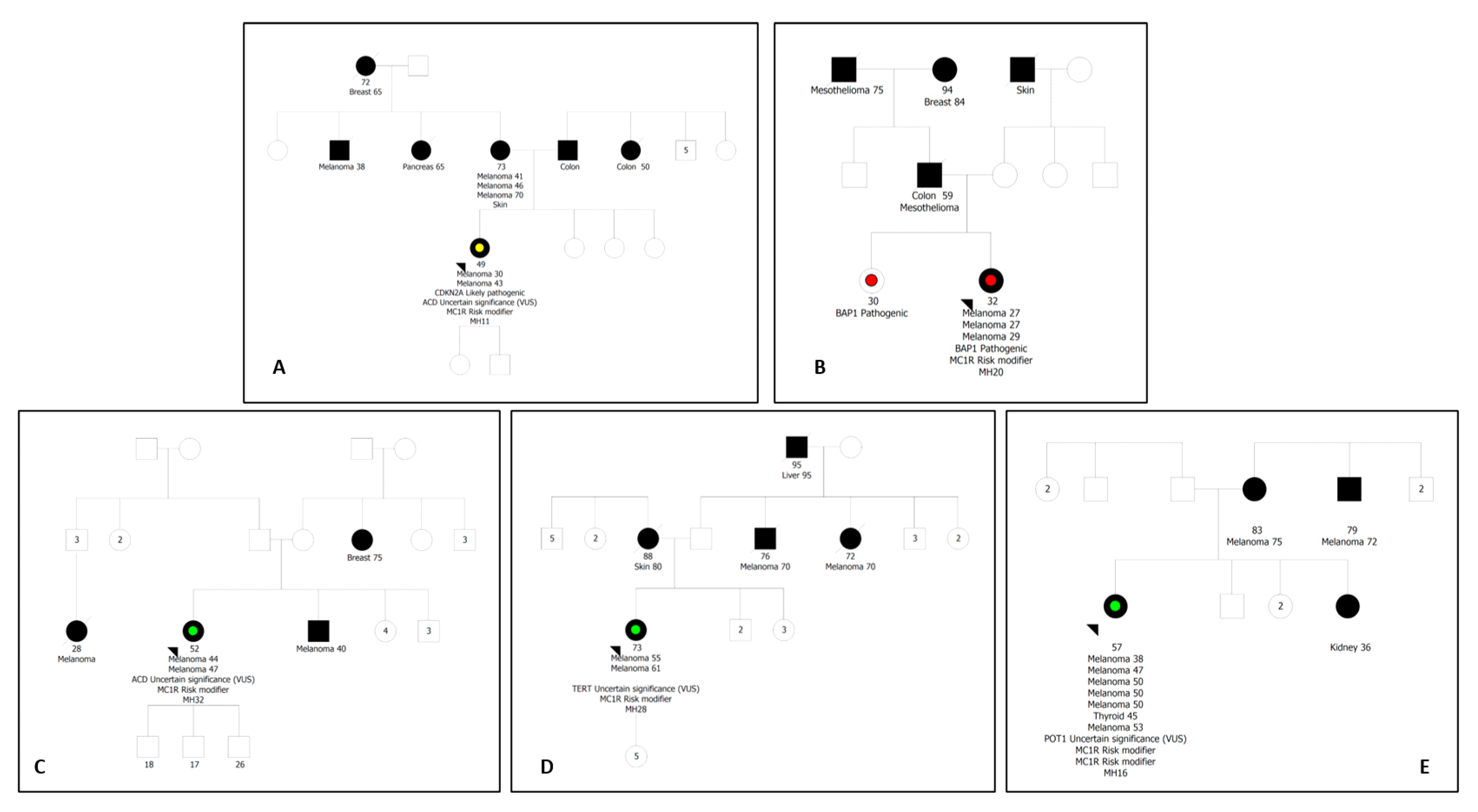
2. Results
3. Discussion
4. Materials and Methods
4.1. Patients’ Selection
4.2. Germline Genetic Analysis
4.3. Statistical Analysis
5. Conclusions
Author Contributions
Funding
Institutional Review Board Statement
Informed Consent Statement
Data Availability Statement
Acknowledgments
Conflicts of Interest
References
- Berwick, M.; Erdei, E.; Hay, J. Melanoma epidemiology and public health. Dermatol. Clin. 2009, 27, 205–214. [Google Scholar] [CrossRef] [PubMed]
- Psaty, E.L.; Scope, A.; Halpern, A.C.; Marghoob, A.A. Defining the patient at high risk for melanoma. Int. J. Dermatol. 2010, 49, 362–376. [Google Scholar] [CrossRef] [PubMed]
- Read, J.; Wadt, K.A.; Hayward, N.K. Melanoma genetics. J. Med. Genet. 2016, 53, 1–14. [Google Scholar] [CrossRef] [PubMed]
- Hansen, C.B.; Wadge, L.M.; Lowstuter, K.; Boucher, K.; Leachman, S.A. Clinical germline genetic testing for melanoma. Lancet Oncol. 2004, 5, 314–319. [Google Scholar] [CrossRef]
- Leachman, S.A.; Carucci, J.; Kohlmann, W.; Banks, K.C.; Asgari, M.M.; Bergman, W.; Bianchi-Scarrà, G.; Brentnall, T.; Paillerets, B.B.-D.; Bruno, W.; et al. Selection criteria for genetic assessment of patients with familial melanoma. J. Am. Acad Dermatol. 2009, 61, 677.e1–677.e14. [Google Scholar] [CrossRef]
- Auroy, S.; Avril, M.-F.; Chompret, A.; Pham, D.; Goldstein, A.M.; Bianchi-Scarrà, G.; Frebourg, T.; Joly, P.; Spatz, A.; Rubino, C.; et al. Sporadic multiple primary melanoma cases: CDKN2A germline mutations with a founder effect. Genes Chromosomes Cancer 2001, 32, 195–202. [Google Scholar] [CrossRef]
- Aoude, L.G.; Wadt, K.A.; Pritchard, A.L.; Hayward, N.K. Genetics of familial melanoma: 20 years after CDKN2A. Pigment Cell Melanoma Res. 2015, 28, 148–160. [Google Scholar] [CrossRef]
- de Snoo, F.A.; Hayward, N.K. Cutaneous melanoma susceptibility and progression genes. Cancer Lett. 2005, 230, 153–186. [Google Scholar] [CrossRef]
- Goldstein, A.M.; Chan, M.; Harland, M.; Hayward, N.K.; Demenais, F.; Bishop, D.T.; Azizi, E.; Bergman, W.; Bianchi-Scarra, G.; Bruno, W.; et al. Features associated with germline CDKN2A mutations: A GenoMEL study of melanoma-prone families from three continents. J. Med. Genet. 2007, 44, 99–106. [Google Scholar] [CrossRef]
- Hansson, J. Familial melanoma. Surg. Clin. N. Am. 2008, 88, 897–916. [Google Scholar] [CrossRef]
- Rossi, M.; Pellegrini, C.; Cardelli, L.; Ciciarelli, V.; Di Nardo, L.; Fargnoli, M.C. Familial Melanoma: Diagnostic and Management Implications. Dermatol. Pract. Concept. 2019, 9, 10–16. [Google Scholar] [CrossRef] [PubMed]
- Bottillo, I.; La Starza, R.; Radio, F.C.; Molica, C.; Pedace, L.; Pierini, T.; De Bernardo, C.; Stingeni, L.; Bargiacchi, S.; Paiardini, A.; et al. A novel germline mutation in CDK4 codon 24 associated to familial melanoma. Clin. Genet. 2018, 93, 934–935. [Google Scholar] [CrossRef] [PubMed]
- Bruno, W.; Dalmasso, B.; Barile, M.; Andreotti, V.; Elefanti, L.; Colombino, M.; Vanni, I.; Allavena, E.; Barbero, F.; Passoni, E.; et al. Predictors of germline status for hereditary melanoma: 5 years of multi-gene panel testing within the Italian Melanoma Intergroup. ESMO Open 2022, 7, 100525. [Google Scholar] [CrossRef] [PubMed]
- Potrony, M.; Badenas, C.; Aguilera, P.; Puig-Butille, J.A.; Carrera, C.; Malvehy, J.; Puig, S. Update in genetic susceptibility in melanoma. Ann. Transl. Med. 2015, 3, 210. [Google Scholar] [CrossRef]
- Bertolotto, C.; The French Familial Melanoma Study Group; Lesueur, F.; Giuliano, S.; Strub, T.; de Lichy, M.; Bille, K.; Dessen, P.; D’hayer, B.; Mohamdi, H.; et al. Corrigendum: A SUMOylation-defective MITF germline mutation predisposes to melanoma and renal carcinoma. Nature 2016, 531, 126. [Google Scholar] [CrossRef]
- Aoude, L.G.; Pritchard, A.L.; Robles-Espinoza, C.D.; Wadt, K.; Harland, M.; Choi, J.; Gartside, M.; Quesada, V.; Johansson, P.; Palmer, J.M.; et al. Nonsense mutations in the shelterin complex genes ACD and TERF2IP in familial melanoma. J. Natl. Cancer Inst. 2014, 107, dju408. [Google Scholar] [CrossRef]
- Horn, S.; Figl, A.; Rachakonda, P.S.; Fischer, C.; Sucker, A.; Gast, A.; Kadel, S.; Moll, I.; Nagore, E.; Hemminki, K.; et al. TERT promoter mutations in familial and sporadic melanoma. Science 2013, 339, 959–961. [Google Scholar] [CrossRef]
- Robles- Espinoza, C.D.; Harland, M.; Ramsay, A.J.; Aoude, L.G.; Quesada, V.; Ding, Z.; Pooley, K.A.; Pritchard, A.L.; Tiffen, J.C.; Petljak, M.; et al. POT1 loss-of-function variants predispose to familial melanoma. Nat. Genet. 2014, 46, 478–481. [Google Scholar] [CrossRef]
- Puig, S.; Potrony, M.; Cuellar, F.; Puig-Butille, J.A.; Carrera, C.; Aguilera, P.; Nagore, E.; Garcia-Casado, Z.; Requena, C.; Kumar, R.; et al. Characterization of individuals at high risk of developing melanoma in Latin America: Bases for genetic counseling in melanoma. Genet. Med. 2016, 18, 727–736. [Google Scholar] [CrossRef]
- Bakos, L.; Masiero, N.; Bakos, R.; Burttet, R.; Wagner, M.; Benzano, D. European ancestry and cutaneous melanoma in Southern Brazil. J. Eur. Acad Dermatol. Venereol. 2009, 23, 304–307. [Google Scholar] [CrossRef]
- Grazziotin, T.; Rey, M.; Bica, C.; Pinto, L.; Bonamigo, R.; Puig-Butille, J.; Cuellar, F.; Puig, S. Genetic variations of patients with familial or multiple melanoma in Southern Brazil. J. Eur. Acad Dermatol. Venereol. 2013, 27, e179–e185. [Google Scholar] [CrossRef]
- Sung, H.; Ferlay, J.; Siegel, R.L.; Laversanne, M.; Soerjomataram, I.; Jemal, A.; Bray, F. Global Cancer Statistics 2020: GLOBOCAN Estimates of Incidence and Mortality Worldwide for 36 Cancers in 185 Countries. CA Cancer J. Clin. 2021, 71, 209–249. [Google Scholar] [CrossRef]
- Ministério da Saúde. Instituto Nacional de Câncer José Alencar Gomes da Silva. Estimativa 2020: Incidência de Câncer No Brasil. Rio de Janeiro: INCA; 2019. [Última Modificação: 07/07/2022]. Available online: https://www.inca.gov.br/estimativa/estado-capital/brasil (accessed on 12 March 2023).
- Ashton-Prolla, P.; Bakos, L.; Jr, G.J.; Giugliani, R.; Azevedo, S.J.; Hogg, D. Clinical and molecular characterization of patients at risk for hereditary melanoma in southern Brazil. J. Investig. Dermatol. 2008, 128, 421–425. [Google Scholar] [CrossRef]
- de Ávila, A.L.R.; Krepischi, A.C.V.; Moredo, L.F.; Aguiar, T.F.M.; da Silva, F.C.; de Sá, B.C.S.; de Nóbrega, A.F.; Achatz, M.I.W.; Duprat, J.P.; Landman, G.; et al. Germline CDKN2A mutations in Brazilian patients of hereditary cutaneous melanoma. Fam. Cancer 2014, 13, 645–649. [Google Scholar] [CrossRef]
- Huber, J.; Ramos, E.S. The P48T germline mutation and polymorphism in the CDKN2A gene of patients with melanoma. Braz. J. Med. Biol. Res. 2006, 39, 237–241. [Google Scholar] [CrossRef]
- Pramio, D.T.; Aguiar, T.; de Araújo, E.S.S.; Moredo, L.; Sá, B.C.S.D.; Achatz, M. Low frequency of germline TERT and MITF mutations in Brazilian melanoma prone patients. Hered. Genet. 2014, 3, 3. [Google Scholar] [CrossRef]
- Aguilera, P.; Malvehy, J.; Carrera, C.; Palou, J.; Puig-Butillé, J.A.; Alòs, L.; Badenas, C.; Puig, S. Clinical and Histopathological Characteristics between Familial and Sporadic Melanoma in Barcelona, Spain. J. Clin. Exp. Dermatol. Res. 2014, 5, 231. [Google Scholar] [CrossRef] [PubMed]
- Márquez-Rodas, I.; González, M.M.; Nagore, E.; Gómez-Fernández, C.; Avilés-Izquierdo, J.A.; Maldonado-Seral, C.; Soriano, V.; Majem-Tarruella, M.; Palomar, V.; Maseda, R.; et al. Spanish Multidisciplinary Group of Melanoma (GEM). Frequency and characteristics of familial melanoma in Spain: The FAM-GEM-1 Study. PLoS ONE 2015, 10, e0124239. [Google Scholar] [CrossRef] [PubMed]
- Bainvoll, L.; Mandelbaum, R.S.; Worswick, S.D.; Matsuo, K. Anatomical location of melanoma: A gender-specific analysis. J. Surg. Oncol. 2023, 127, 203–206. [Google Scholar] [CrossRef] [PubMed]
- Bruno, W.; Pastorino, L.; Ghiorzo, P.; Andreotti, V.; Martinuzzi, C.; Menin, C.; Elefanti, L.; Stagni, C.; Vecchiato, A.; Rodolfo, M.; et al. Multiple primary melanomas (MPMs) and criteria for genetic assessment: MultiMEL, a multicenter study of the Italian Melanoma Intergroup. J. Am. Acad Dermatol. 2016, 74, 325–332. [Google Scholar] [CrossRef]
- Huerta, C.; Garcia-Casado, Z.; Bañuls, J.; Moragon, M.; Oliver, V.; Unamuno, B.; Requena, C.; Kumar, R.; Nagore, E. Characteristics of Familial Melanoma in Valencia, Spain, Based on the Presence of CDKN2A Mutations and MC1R Variants. Acta Derm. Venereol. 2018, 98, 512–516. [Google Scholar] [CrossRef]
- Pellegrini, C.; Maturo, M.G.; Martorelli, C.; Suppa, M.; Antonini, A.; Kostaki, D.; Verna, L.; Landi, M.T.; Peris, K.; Fargnoli, M.C. Characterization of melanoma susceptibility genes in high-risk patients from Central Italy. Melanoma Res. 2017, 27, 258–267. [Google Scholar] [CrossRef] [PubMed]
- de Sá, B.C.S.; de Macedo, M.P.; Torrezan, G.T.; Braga, J.C.T.; Fidalgo, F.; Moredo, L.F.; Lellis, R.; Duprat, J.P.; Carraro, D.M. BAP1 tumor predisposition syndrome case report: Pathological and clinical aspects of BAP1-inactivated melanocytic tumors (BIMTs), including dermoscopy and confocal microscopy. BMC Cancer 2019, 19, 1077. [Google Scholar] [CrossRef]
- Shen, E.; Xiu, J.; Lopez, G.Y.; Bentley, R.; Jalali, A.; Heimberger, A.B.; Bainbridge, M.N.; Bondy, M.L.; Walsh, K.M. POT1 mutation spectrum in tumour types commonly diagnosed among POT1-associated hereditary cancer syndrome families. J. Med. Genet. 2020, 57, 664–670. [Google Scholar] [CrossRef] [PubMed]
- Potrony, M.; Puig-Butille, J.; Ribera-Sola, M.; Iyer, V.; Robles-Espinoza, C.; Aguilera, P.; Carrera, C.; Malvehy, J.; Badenas, C.; Landi, M.; et al. POT1 germline mutations but not TERT promoter mutations are implicated in melanoma susceptibility in a large cohort of Spanish melanoma families. Br. J. Dermatol. 2019, 181, 105–113. [Google Scholar] [CrossRef] [PubMed]
- Wilson, T.L.-S.; Hattangady, N.; Lerario, A.M.; Williams, C.; Koeppe, E.; Quinonez, S.; Osborne, J.; Cha, K.B.; Else, T. A new POT1 germline mutation-expanding the spectrum of POT1-associated cancers. Fam. Cancer 2017, 16, 561–566. [Google Scholar] [CrossRef]
- Demenais, F.; Mohamdi, H.; Chaudru, V.; Goldstein, A.M.; Bishop, J.A.N.; Bishop, D.T.; Kanetsky, P.A.; Hayward, N.K.; Gillanders, E.; Elder, D.E.; et al. Association of MC1R variants and host phenotypes with melanoma risk in CDKN2A mutation carriers: A GenoMEL study. J. Natl. Cancer Inst. 2010, 102, 1568–1583. [Google Scholar] [CrossRef]
- Fidalgo, F.; Torrezan, G.T.; de Sá, B.C.S.; Barros, B.D.d.F.; Moredo, L.F.; Valieris, R.; de Souza, S.J.; Duprat, J.P.; Krepischi, A.C.V.; Carraro, D.M. Family-based whole-exome sequencing identifies rare variants potentially related to cutaneous melanoma predisposition in Brazilian melanoma-prone families. PLoS ONE 2022, 17, e0262419. [Google Scholar] [CrossRef]
- Slominski, R.M.; Sarna, T.; Płonka, P.M.; Raman, C.; Brożyna, A.A.; Slominski, A.T. Melanoma, Melanin, and Melanogenesis: The Yin and Yang Relationship. Front Oncol. 2022, 12, 842496. [Google Scholar] [CrossRef]
- Çakır, A.; Elcin, G.; Kilickap, S.; Gököz, Ö.; Taskiran, Z.E.; Celik, I. Phenotypic and Genetic Features that Differ Between Hereditary and Sporadic Melanoma: Results of a Preliminary Study from a Single Center from Turkey. Dermatol. Pract. Concept. 2023, 13, e2023146. [Google Scholar] [CrossRef]
- Tagliabue, E.; Gandini, S.; Bellocco, R.; Maisonneuve, P.; Newton-Bishop, J.; Polsky, D.; Lazovich, D.; Kanetsky, P.; Ghiorzo, P.; Gruis, N.; et al. MC1R variants as melanoma risk factors independent of at-risk phenotypic characteristics: A pooled analysis from the M-SKIP project. Cancer Manag. Res. 2018, 10, 1143–1154. [Google Scholar] [CrossRef] [PubMed]
- Williams, P.F.; Olsen, C.M.; Hayward, N.K.; Whiteman, D.C. Melanocortin 1 receptor and risk of cutaneous melanoma: A meta-analysis and estimates of population burden. Int. J. Cancer 2011, 129, 1730–1740. [Google Scholar] [CrossRef] [PubMed]
- Swope, V.B.; Abdel-Malek, Z.A. MC1R: Front and Center in the Bright Side of Dark Eumelanin and DNA Repair. Int. J. Mol. Sci. 2018, 19, 2667. [Google Scholar] [CrossRef] [PubMed]
- Duffy, D.; Lee, K.; Jagirdar, K.; Pflugfelder, A.; Stark, M.; McMeniman, E.; Soyer, H.; Sturm, R. High naevus count and MC1R red hair alleles contribute synergistically to increased melanoma risk. Br. J. Dermatol. 2019, 181, 1009–1016. [Google Scholar] [CrossRef] [PubMed]
- de Torre, C.; Garcia-Casado, Z.; Martínez-Escribano, J.A.; Botella-Estrada, R.; Bañuls, J.; Oliver, V.; Mercader, P.; Azaña, J.M.; Frias, J.; Nagore, E. Influence of loss of function MC1R variants in genetic susceptibility of familial melanoma in Spain. Melanoma Res. 2010, 20, 342–348. [Google Scholar] [CrossRef]
- Richards, S.; Aziz, N.; Bale, S.; Bick, D.; Das, S.; Gastier-Foster, J.; Grody, W.W.; Hegde, M.; Lyon, E.; Spector, E.; et al. Standards and guidelines for the interpretation of sequence variants: A joint consensus recommendation of the American College of Medical Genetics and Genomics and the Association for Molecular Pathology. Genet. Med. 2015, 17, 405–424. [Google Scholar] [CrossRef]
- Hu, H.-H.; Benfodda, M.; Dumaz, N.; Gazal, S.; Descamps, V.; Bourillon, A.; Basset-Seguin, N.; Riffault, A.; Ezzedine, K.; Bagot, M.; et al. A large French case-control study emphasizes the role of rare Mc1R variants in melanoma risk. Biomed. Res. Int. 2014, 2014, 925716. [Google Scholar] [CrossRef]
- Naslavsky, M.S.; Yamamoto, G.L.; de Almeida, T.F.; Ezquina, S.A.M.; Sunaga, D.Y.; Pho, N.; Bozoklian, D.; Sandberg, T.O.M.; Brito, L.A.; Lazar, M.; et al. Exomic variants of an elderly cohort of Brazilians in the ABraOM database. Hum. Mutat. 2017, 38, 751–763. [Google Scholar] [CrossRef]


| FM | MPM | p | |
|---|---|---|---|
| Age at diagnosis (years) mean | 43.5 | 35.6 | 0.047 |
| (Range) | (15–69) | (25–49) | |
| Total nevus count median | 74 | 145 | 0.04 |
| (Range) | (4–348) | (23–334) | |
| n (%) | n (%) | ||
| Sex | M10/F16 | M8/F3 | |
| Phototype | |||
| I/II | 22 (84.6) | 11 (100) | 0.296 |
| III/IV | 4 (15.4) | 0 | |
| Eye color | |||
| Blue/green | 15 (57.7) | 5 (45.5) | 0.748 |
| Brown/black | 11 (42.3) | 6 (54.5) | |
| Hair color | |||
| Red/blond | 16 (61.5) | 6 (54.5) | 0.728 |
| Brown/black | 10 (38.5) | 5 (45.5) | |
| Freckles density (back) | |||
| Low | 7 (26.9) | 3 (27.3) | 1.00 |
| High | 19 (73.1) | 8 (72.7) | |
| Childhood sunburn | |||
| Yes | 22 (84.6) | 11 (100) | 0.296 |
| No | 4 (15.4) | 0 | |
| Adulthood sunburn | |||
| Yes | 13 (50.0) | 9 (81.8) | 0.141 |
| No | 13 (50.0) | 2 (72.7) | |
| AMS | |||
| Yes | 8 (30.8) | 7 (63.6) | 0.08 |
| No | 18 (69.2) | 4 (36.4) | |
| Nevus count | |||
| ≤100 | 16 (61.5) | 3 (27.3) | 0.122 |
| >100 | 10 (38.5) | 8 (72.7) | |
| Tumors features | n (%) | n (%) | |
| Breslow thickness | |||
| In situ | 57 (53.8) | 19 (45.2) | |
| ≤1 mm | 46 (43.4) | 21 (50.0) | 0.56 |
| >1 mm | 3 (2.8) | 2 (4.8) | |
| Histopathological subtype | |||
| Superficial spreading | 90 (94.7) | 37 (97.4) | |
| Nodular | 1 (1.1) | 0 (0) | |
| Lentigo maligna | 1 (1.1) | 0 (0) | 1.0 |
| Acral lentiginous | 2 (2.1) | 1 (2.6) | |
| Others | 1 (1.1) | 0 (0) | |
| Nevus-associated melanoma | |||
| Yes | 52 (58.4) | 23 (67.6) | 0.46 |
| No | 37 (41.6) | 11 (32.4) | |
| Anatomical site | |||
| Head and neck | 3 (2.8) | 3 (7.1) | 0.04 |
| Trunk | 50 (47.2) | 28 (66.7) | |
| Upper limbs | 18 (17.0) | 5 (11.9) | |
| Lower limbs | 35 (33.0) | 6 (14.3) |
| Case | Gene | Type | RefSeq (NM) | HGVS cDNA; Protein | dbSNP | MAF (gnomAD/ABraOM) | ClinVar | Varsome/Franklin | In Silico Predictors Benign (b)/Pathogenic (p) | Revel | ACMG |
|---|---|---|---|---|---|---|---|---|---|---|---|
| MH11 | CDKN2A | missense | NM_000077.4 | c.71G>C; p.(Arg24Pro) | rs104894097 | 0.000017/ na | P(9); PP(1) | P/LP | 8 b/4 p | 0.4 | P |
| MH11 | ACD | missense | NM_001082486.2 | c.1255G>A; p.(Glu419Lys) | na | na | na | LB/VUS | 11 b/1 p | 0.04 | VUS |
| MH16 | POT1 | missense | NM_015450.2 | c.318T>G; p.(Phe106Leu) | na | na | na | VUS/VUS | 2 b/11 p | 0.71 | VUS |
| MH20 | BAP1 | Frameshift deletion | NM_004656.3 | c.1265delG; p.(Gly422Glufs*8) | na | na | na | P/LP | 0 b/1 p | na | P |
| MH28 | TERT | Inframe deletion | NM_198253.2 | c.1323_1325delGGA; p.(Glu441del) | rs377639087 | 0.00164/0.00329 | B/LB(12); VUS(3); P(1) | VUS/B | 1 b/0 p | na | VUS |
| MH32 | ACD | missense | NM_001082486.2 | c.962C>T; p.(Ser321Leu) | rs374925782 | 0.000072/0.00164 | VUS(4) | LB/VUS | 8 b/4 p | 0.07 | VUS |
| Patient | n° M | Sex | n° Nevi | Group | High-Penetrance Variant | MC1R Variants | MC1R Allele Type |
|---|---|---|---|---|---|---|---|
| MH01 | 19 | M | 209 | FM | None | p.Gln23Ter | u/0 |
| MH02 | 3 | M | 4 | FM | None | p.Arg163Gln p.Val92Met) | r/r |
| MH03 | 1 | F | 58 | FM | None | p.Arg151Cys | R/R * |
| MH04 | 3 | M | 78 | MPM | None | p.Arg151Cys p.Val60Leu | R/r |
| MH05 | 3 | F | 23 | MPM | None | p.Val60Leu | r/0 |
| MH06 | 2 | F | 75 | FM | None | p.Arg151Cys | R/0 |
| MH07 | 4 | M | 247 | MPM | None | wt | 0/0 |
| MH08 | 4 | F | 97 | FM | None | p.Asp294His p.Val60Leu | R/r |
| MH09 | 1 | M | 30 | FM | None | p.Asp84Glu | R/0 |
| MH10 | 1 | M | 118 | FM | None | p.Arg160Trp | R/0 |
| MH11 | 2 | F | 65 | FM | CDKN2A (P) ACD (VUS) | p.Val60Leu | r/0 |
| MH12 | 4 | M | 203 | MPM | None | p.Val60Leu | r/0 |
| MH14 | 3 | M | 62 | MPM | None | p.Arg160Trp | R/0 |
| MH15 | 3 | M | 190 | MPM | None | wt | 0/0 |
| MH16 | 6 | F | 110 | FM | POT1 (VUS) | p.Arg151Cys p.Ser83Leu | R/u |
| MH17 | 2 | M | 167 | FM | None | wt | 0/0 |
| MH18 | 3 | M | 117 | FM | None | p.Val60Leu p.Ile264Val | r/u |
| MH19 | 2 | F | 46 | FM | None | wt | 0/0 |
| MH20 | 3 | F | 109 | MPM | BAP1 (P) | p.Arg160Trp | R/0 |
| MH21 | 4 | M | 145 | MPM | None | p.Arg163Gln | r/0 |
| MH22 | 2 | M | 90 | FM | None | p.Arg160Trp | R/0 |
| MH23 | 6 | M | 59 | FM | None | p.Arg151Cys | R/0 |
| MH24 | 6 | F | 118 | FM | None | p.Arg160Trp p.Ile155Thr | R/r |
| MH25 | 2 | F | 14 | FM | None | p.Arg151Cys | R/0 |
| MH26 | 5 | F | 178 | FM | None | p.Arg151Cys | R/0 |
| MH27 | 2 | F | 19 | FM | None | p.Arg160Trp | R/0 |
| MH28 | 2 | F | 24 | FM | TERT (VUS) | p.Thr272Met | u/0 |
| MH29 | 28 | F | 348 | FM | None | p.Arg142His p.Arg151Cys | R/R |
| MH30 | 2 | F | 70 | FM | None | p.Val60Leu | r/r * |
| MH31 | 1 | M | 18 | FM | None | p.Arg160Trp | R/0 |
| MH32 | 2 | F | 7 | FM | ACD (VUS) | p.Val92Met | r/0 |
| MH33 | 4 | M | 316 | FM | None | p.Arg151Cys | R/0 |
| MH34 | 5 | F | 176 | MPM | None | p.Arg142His p.Val60Leu | R/r |
| MH35 | 6 | M | 334 | MPM | None | p.Arg151Cys p.Arg163Gln | R/r |
| MH36 | 1 | F | 73 | FM | None | p.Arg163Gln | r/0 |
| MH37 | 2 | F | 109 | FM | None | wt | 0/0 |
| MH38 | 4 | M | 145 | MPM | None | p.Val60Leu p.Gly89Ar | r/u |
| MC1R Variant | Cohort (37 Patients) | ABraOM Control Group (609) | Statistics | |||||||
|---|---|---|---|---|---|---|---|---|---|---|
| HZ | HT | AF | HZ | HT | AF | OR | 95% CI | p-Value | ||
| p.D84E | R | 0 | 1 | 1.4% | 0 | 2 | 0.2% | 10.93 | 0.98–122.52 | 0.052 |
| p.R142H | R | 0 | 2 | 2.7% | 0 | 1 | 0.1% | 43.73 | 3.90–490.07 | 0.002 |
| p.R151C | R | 1 | 9 | 14.9% | 0 | 26 | 2.1% | 9.25 | 4.34–19.74 | <0.001 |
| p.R160W | R | 0 | 7 | 9.5% | 1 | 29 | 2.5% | 4.94 | 2.08–11.74 | <0.001 |
| p.D294H | R | 0 | 1 | 1.4% | 0 | 21 | 1.7% | 1.04 | 0.14–7.89 | 0.969 |
| Total R | 1 | 20 | 29.7% | 1 | 79 | 6.7% | 5.94 | 3.44–10.26 | <0.001 | |
| p.V60L | r | 1 | 8 | 13.5% | 5 | 107 | 9.6% | 1.41 | 0.70–2.84 | 0.333 |
| p.V92M | r | 0 | 2 | 2.7% | 1 | 46 | 3.9% | 0.69 | 0.16–2.91 | 0.611 |
| p.I155T | r | 0 | 1 | 1.4% | 0 | 7 | 0.6% | 2.36 | 0.29–19.52 | 0.425 |
| p.R163Q | r | 0 | 4 | 5.4% | 10 | 84 | 8.5% | 0.64 | 0.23–1.79 | 0.390 |
| Total r | 1 | 15 | 23.0% | 16 | 244 | 22.7% | 1.02 | 0.59–1.79 | 0.924 | |
| p.Q23* | u | 0 | 1 | 1.4% | 0 | 2 | 0.2% | 8.54 | 0.77–95.33 | 0.081 |
| p.G89R | u | 0 | 1 | 1.4% | 0 | 1 | 0.1% | 17.08 | 1.06–275.97 | 0.046 |
| p.I264V | u | 0 | 1 | 1.4% | 0 | 2 | 0.2% | 8.54 | 0.77–95.33 | 0.081 |
| Total u | 0 | 3 | 4.1% | 0 | 5 | 0.4% | 10.25 | 2.40–43.75 | 0.002 | |
Disclaimer/Publisher’s Note: The statements, opinions and data contained in all publications are solely those of the individual author(s) and contributor(s) and not of MDPI and/or the editor(s). MDPI and/or the editor(s) disclaim responsibility for any injury to people or property resulting from any ideas, methods, instructions or products referred to in the content. |
© 2023 by the authors. Licensee MDPI, Basel, Switzerland. This article is an open access article distributed under the terms and conditions of the Creative Commons Attribution (CC BY) license (https://creativecommons.org/licenses/by/4.0/).
Share and Cite
Soares de Sá, B.C.; Moredo, L.F.; Torrezan, G.T.; Fidalgo, F.; de Araújo, É.S.S.; Formiga, M.N.; Duprat, J.P.; Carraro, D.M. Characterization of Potential Melanoma Predisposition Genes in High-Risk Brazilian Patients. Int. J. Mol. Sci. 2023, 24, 15830. https://doi.org/10.3390/ijms242115830
Soares de Sá BC, Moredo LF, Torrezan GT, Fidalgo F, de Araújo ÉSS, Formiga MN, Duprat JP, Carraro DM. Characterization of Potential Melanoma Predisposition Genes in High-Risk Brazilian Patients. International Journal of Molecular Sciences. 2023; 24(21):15830. https://doi.org/10.3390/ijms242115830
Chicago/Turabian StyleSoares de Sá, Bianca Costa, Luciana Facure Moredo, Giovana Tardin Torrezan, Felipe Fidalgo, Érica Sara Souza de Araújo, Maria Nirvana Formiga, João Pereira Duprat, and Dirce Maria Carraro. 2023. "Characterization of Potential Melanoma Predisposition Genes in High-Risk Brazilian Patients" International Journal of Molecular Sciences 24, no. 21: 15830. https://doi.org/10.3390/ijms242115830