Article

Modulation of the Gut Microbiota by the Plantaricin-Producing Lactiplantibacillus plantarum D13, Analysed in the DSS-Induced Colitis Mouse Model

1
Laboratory for Antibiotic, Enzyme, Probiotic and Starter Culture Technologies, Department of Biochemical Engineering, Faculty of Food Technology and Biotechnology, University of Zagreb, Pierottijeva 6, 10000 Zagreb, Croatia
2
Department of Animal Physiology, Faculty of Science, University of Zagreb, Rooseveltov trg 6, 10000 Zagreb, Croatia
*
Author to whom correspondence should be addressed.
Int. J. Mol. Sci. 2023, 24(20), 15322; https://doi.org/10.3390/ijms242015322
Submission received: 3 October 2023 / Revised: 10 October 2023 / Accepted: 16 October 2023 / Published: 18 October 2023
(This article belongs to the Special Issue Gut Microbiota and Metabolic Diseases 2.0)

Abstract

:
Lactiplantibacillus plantarum D13 shows antistaphylococcal and antilisterial activity, probably due to the synthesis of a presumptive bacteriocin with antibiofilm capacity released in the cell-free supernatant (CFS), whose inhibitory effect is enhanced by cocultivation with susceptible strains. An in silico analysis of the genome of strain D13 confirmed the pln gene cluster. Genes associated with plantaricin biosynthesis, structure, transport, antimicrobial activity, and immunity of strain D13 were identified. Furthermore, the predicted homology-based 3D structures of the cyclic conformation of PlnE, PlnF, PlnJ, and PlnK revealed that PlnE and PlnK contain two helices, while PlnF and PlnJ contain one and two helices, respectively. The potential of the strain to modulate the intestinal microbiota in healthy or dextran sulphate sodium (DSS)-induced colitis mouse models was also investigated. Strain D13 decreased the disease activity index (DAI) and altered the gut microbiota of mice with DSS-induced colitis by increasing the ratio of beneficial microbial species (Allobaculum, Barnesiella) and decreasing those associated with inflammatory bowel disease (Candidatus Saccharimonas). This suggests that strain D13 helps to restore the gut microbiota after DSS-induced colitis, indicating its potential for further investigation as a probiotic strain for the prevention and treatment of colitis.

1. Introduction

The gut microbiota represents a complex ecosystem that inhabits the digestive tract and lives in symbiosis with the host. Dysbiosis of the gut microbiota is associated with many diseases, primarily those related to the disruption of intestinal dysfunction, such as inflammatory bowel disease (IBD), represented by ulcerative colitis and Crohn’s disease, and irritable bowel syndrome (IBS) [1,2]. The link between the composition of the intestinal microbiota and the development of disease has been extensively studied [1,2]. This has led to the development of microbial-based and other strategies to alter its composition, such as manipulation with prebiotics (mainly dietary fibers), probiotics, synbiotics, parabiotics, and/or postbiotics and equivalent synthetic products that positively influence the immunological response and inflammation [3]. Lactiplantibacillus plantarum (formerly named Lactobacillus plantarum) (Lb. plantarum) is one of the most extensively studied lactic acid bacteria (LAB), ubiquitous in the various microbiomes of fermented foods and in the human microbiome [4,5,6]. In addition, certain strains isolated from human saliva or the gastrointestinal tract (GIT) are considered important for health-promoting host–microbe interactions [7]. In both aspects of application, either as a functional starter or probiotic culture, the potential biosynthesis of bacteriocins is attractive as it ensures a competitive advantage and eliminates undesirable contaminating strains that may be present in food or gut microbiomes. Bacteriocin-producing probiotic strains have the ability to modulate the gut microbiota by conferring resistance to pathogen colonisation. Namely, these beneficial bacteria can inhibit the colonisation of pathogenic bacteria by synthesising antimicrobials such as lactic acid, thus lowering the luminal pH, or by producing bacteriocins, thus inhibiting pathogens, as well as through competing for intestinal sites and nutrients, forming coaggregates and finally eliminating undesirable species. This is the reason that bacteriocins are not only recognised as antimicrobial peptides but also as signalling or colonising molecules. These functions, which may be synergistic, are the basis for the establishment of a producer strain in the GIT, its immunomodulatory activity or its direct competitive exclusion of pathogens, and its ability to biomodulate the composition of the gut microbiota [8]. Another functional aspect of bacteriocins is their activity against biofilms of foodborne pathogens and multidrug-resistant bacteria, which is important in the context of food safety and antibiotic resistance, as resistant bacteria in biofilms can withstand a thousand-fold higher concentration of antibiotics compared to planktonic cells [9]. Therefore, bacteriocins can be used as potential natural alternatives to effectively inhibit biofilm formation and disrupt already formed biofilms, either alone or in combination with drugs other than antibiotic antagonists [10]. Advances in the characterisation of bacteriocins have sparked interest in the industrial application of these molecules either as biopreservatives or as new-generation antimicrobials [11]. The aim of this study was to examine, among selected autochthonous Lb. plantarum strains, those with the most pronounced antibacterial activity and to focus on their potential for the synthesis of bacteriocins. Next, we explored the potential of bacteriocin-producing Lb. plantarum to beneficially modulate the composition of the gut microbiota in mice with DSS-induced colitis.

2. Results

2.1. Selection of Lb. plantarum Strains with Potential Plantaricin Production

The potential bacteriocin activity of twelve Lb. plantarum strains was tested in vitro by an agar spot assay against the common Gram-positive pathogens S. aureus ATCC® 25923 and L. monocytogenes ATCC® 19111. The analysed strains of Lb. plantarum showed antistaphylococcal and antilisterial activity. The inhibitory effect was more pronounced against Staphylococcus aureus ATCC® 25923 than against Listeria monocytogenes ATCC® 19111, since all strains of Lb. plantarum displayed strong antistaphylococcal inhibition, while the antilisterial activity was characterised as medium and strong, depending on the strain (Supplementary Table S1). Based on the results, six Lb. plantarum strains (D13, M5, SF15C, ZG1C, M92C, L4) with the most prominent antibacterial activity were selected for the further characterisation of potential plantaricin production. Since the bacteriocins of the class II, which includes plantaricins, are active in both acidic and neutral environments but can be inactivated by proteases, such as trypsin from the pancreas and pepsin from the stomach, cell-free supernatants (CFSs) of Lb. plantarum strains were exposed to proteinase K, pepsin, and pancreatin and a temperature of 100 °C for 30 min. These specific treatments resulted in a partial reduction in the antistaphylococcal and antilisterial activity of Lb. plantarum D13 and SF15C, respectively (p < 0.05), indicating the proteinaceous nature of the antimicrobial component (Supplementary Table S2), whereby the D13 strain displayed the stronger inhibition. The protein nature of the antimicrobial compound in the CFSs of the M92C strain was assumed, since the proteolytic enzymes applied partially abolished the inhibitory activity against both test microorganisms (p < 0.05). Furthermore, pancreatin treatment significantly (p < 0.05) impacted the decrease in the antibacterial activity of the CFSs of strains M5 and ZG1C against L. monocytogenes ATCC® 19111, and, in the same manner, pepsin treatment reduced the antimicrobial potential of the CFS of strain ZG1C.
Next, the selected Lb. plantarum strains were screened for the presence of pln loci by PCR using gene-specific primers. According to the results, the plantaricin-related genes plnA, plnEF, and plnJ were detected with an expected amplicon size of 450 bp, 428 bp, and 475 bp, respectively (Supplementary Figure S1). The genes plnNC8, plnS, and plnW were not amplified and the negative control did not result in amplification, confirming the absence of contamination or non-specific amplification. Based on the results of the microbiological and genomic analysis, the strain Lb. plantarum D13 was selected for the further characterisation of plantaricin biosynthesis. The molecular identity of the PCR amplicons of strain D13 was confirmed by sequencing and aligned with the nucleotide sequences deposited in NCBI. The sequences of the amplicons showed a high level of sequence identity (>98.2%), indicating that the genome harbours a pln gene cluster (Supplementary Table S3).
One approach to characterising bacteriocins is to stimulate their synthesis by coculturing a producer strain with a sensitive bacterium. Therefore, Lb. plantarum D13 was cocultivated with L. monocytogenes ATCC® 19111 and S. aureus ATCC® 25923 to assess the potential of coculture-inducible plantaricin production. After 24 h, the number of culturable cells of L. monocytogenes ATCC® 19111 decreased significantly from 8.93 (±0.13) to 5.97 (±0.25) log (CFU/mL). The inhibitory effect was also present, although less pronounced, when D13 was cocultured with S. aureus® 25923 (from 9.05 (±0.07) to 7.39 (±0.12) log (CFU/mL)) (Figure 1a). During the cocultivation experiments, the inhibitory activity of D13 was determined by the agar spot assay method. The induction of plantaricin activity was enhanced during the exponential growth phase in coculture and persisted throughout the stationary growth phase, compared to the activity when the D13 strain was grown in monoculture (Figure 1b). The antimicrobial activity of the acids produced by the metabolism of the Lb. plantarum D13 strain was eliminated by monitoring the pH values during the cocultivation, which corresponded to the values measured in the monoculture of the test microorganisms, and additionally by testing their inhibition using the determined lactic acid concentrations in coculture (8.1 and 6.3 g/L with L. monocytogenes ATCC® 19111 and S. aureus ATCC® 25923, respectively) at the end of the experiment. Considering the monitored pH values and the stronger antimicrobial activity of CFSs compared to lactic acid, it can be speculated that the inhibitory effect of the Lb. plantarum D13 strain during cocultivation is the result of possible plantaricin activity.

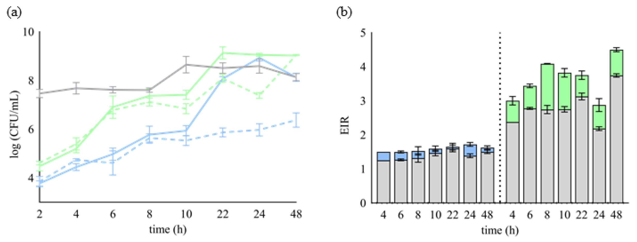
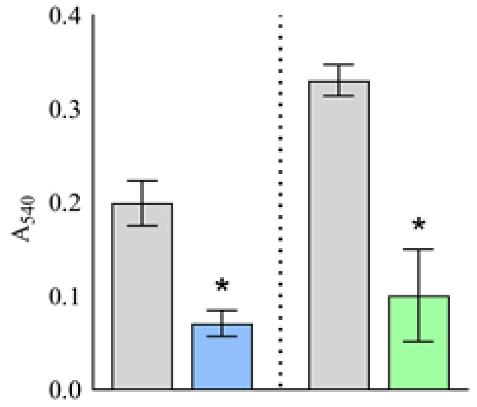
2.2. Putative Plantaricins as Agents against Biofilm Formation

Foodborne biofilms are of particular concern to the food industry, and S. aureus and L. monocytogenes are among the most significant biofilm formers [10]. Here, we investigated whether the CFS of Lb. plantarum D13 could inhibit biofilm formation by L. monocytogenes ATCC® 19111 and S. aureus ATCC® 25923. When a bacterial suspension of L. monocytogenes ATCC® 19111 was treated simultaneously with the CFS of Lb. plantarum D13, biofilm formation was significantly (p < 0.001) reduced compared to the control, i.e., the bacterial suspension alone, and the inhibition rate of biofilm formation was 64.59 (±6.94)% (Figure 2). A similar effect was obtained against S. aureus ATCC® 25923, where the inhibition rate of biofilm formation was 69.58 (±14.97)%. Therefore, it can be speculated that the putative plantaricins in CFS possesses a certain ability to inhibit the biofilm formation of these two pathogen representatives. Given the demonstrated antibiofilm and antimicrobial activity of Lb. plantarum D13, we can suggest its potential ability to eradicate an already formed biofilm in the microenvironment against susceptible bacterial species (Figure 1 and Figure 2).

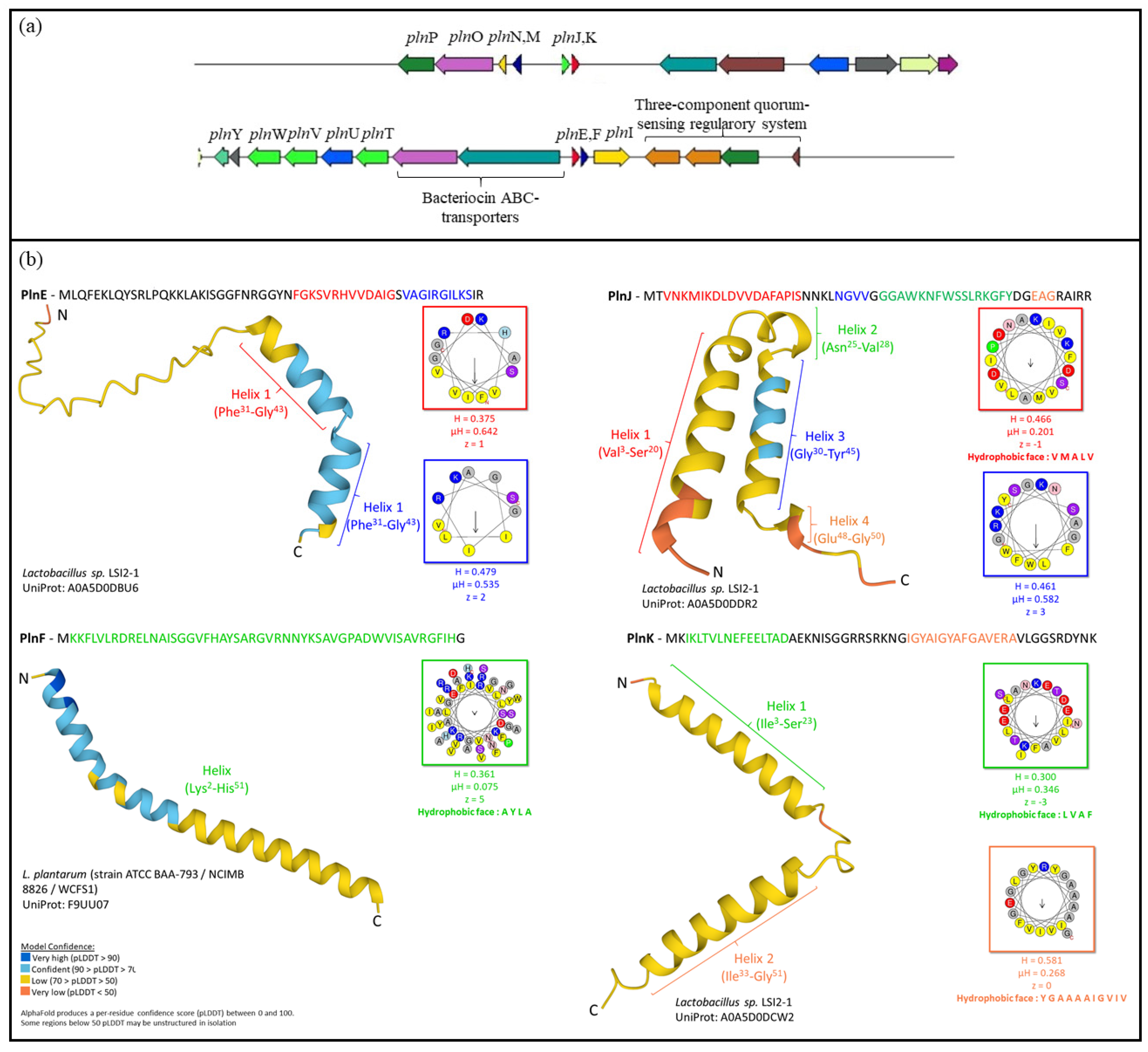
2.3. In Silico Characterisation of Plantaricin-Related Genes

The whole genome of Lb. plantarum D13 was sequenced to confirm the identification of the strain and to identify potential bacteriocin cluster genes that may be related to the observed antibacterial phenotype. The genome size of D13 is 3.34 Mb and it has GC content of 44.4%. The threshold in finding the corresponding genes was set at a region size of 16,000 bp, 15 regions, and an e-value of 1 × 10⁻²⁰ for the selection of pinned coding sequences. In total, 22 genes related to planataricin synthesis were found in contigs 7, 12, and 14. The genes showed high similarity to those of the Lb. plantarum WCFS1 genome, with the exception of the immunity protein PlnP, which is located upstream of the plnMNOP operon on contig 14. The Lb. plantarum D13 genome contains five plantaricin-specific operons on contigs 7 and 12, including the regulator operon plnABCD, the plantaricin operons plnEFI, plnJKLR, and plnMNOP, and the transport operon plnGHTUVW, which are required for the production of the class IIb bacteriocins, plantaricin plnEF, plnJK, and plnA (Figure 3a). Genes associated with the biosynthesis, structure, transport, and antimicrobial activity of bacteriocins and the immunity of the producer strain are listed in Supplementary Table S4. The alpha helices in the bacteriocin structure determine its function. Thus, the predictions of the cyclic conformation of PlnE, PlnF, PlnJ, and PlnK were performed in silico, according to previous structural studies of the respective plantaricins available in the AlphaFold Protein Structure Database, with a threshold of 100% sequence similarity. Since their structures share 100% amino acid identity with the respective peptides of other strains with characterised plantaricin antimicrobial activity, it is presumed that their function could be similar. According to the resulting homology-based three-dimensional structures, PlnE and PlnK contain two helices, while PlnF and PlnJ contain one and two helices, respectively. HeliQuest analyses of the α-helices of the amino acid sequences of the putative two-peptide plantaricins PlnEF and PlnJK revealed the presence of hydrophobic residues of PlnF, PlnJ (Helix 1), and PlnK (Helix 1 and 2), which are in line with the hydrophobicity, hydrophobic moment, and net charge disclosed (Figure 3b).

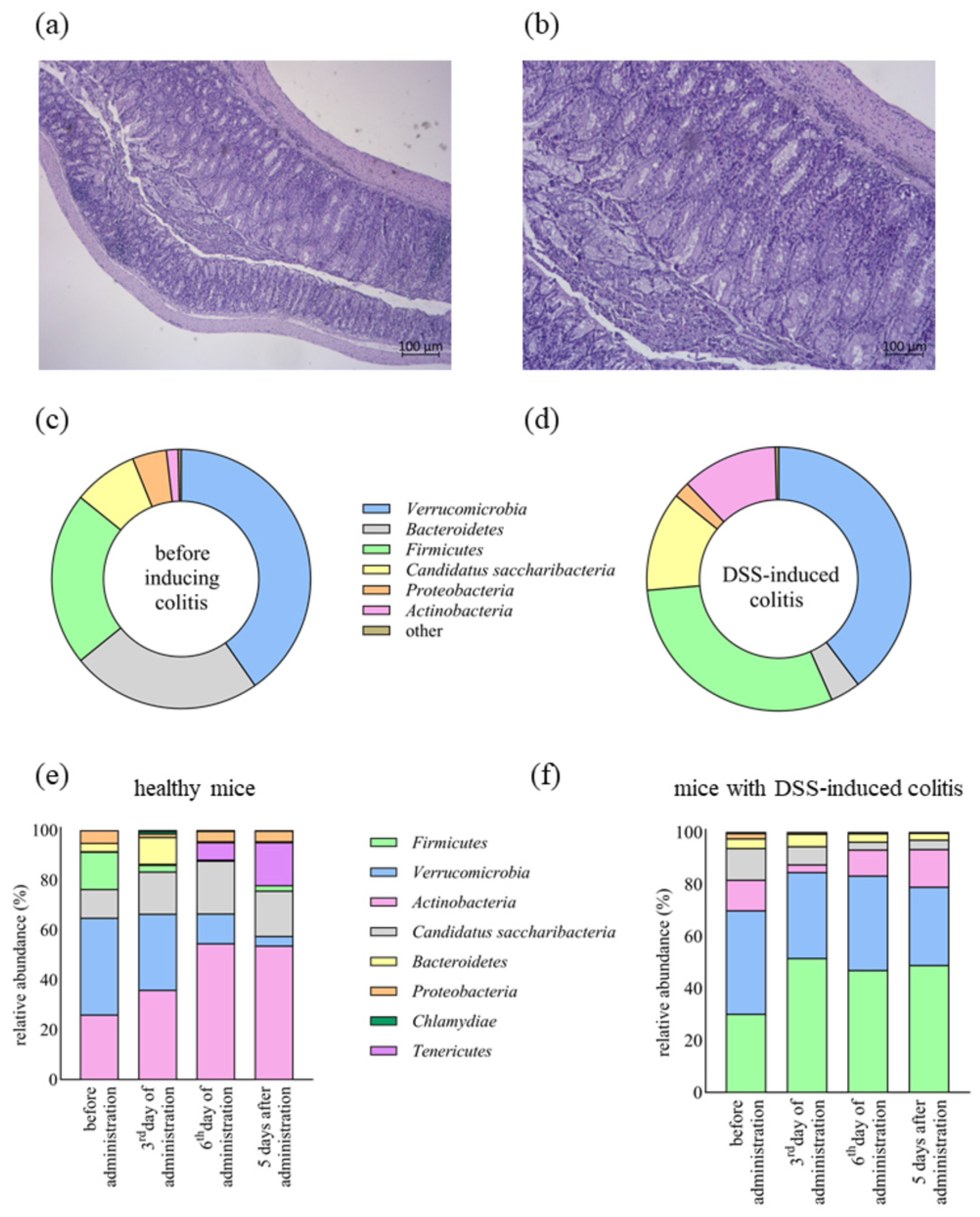
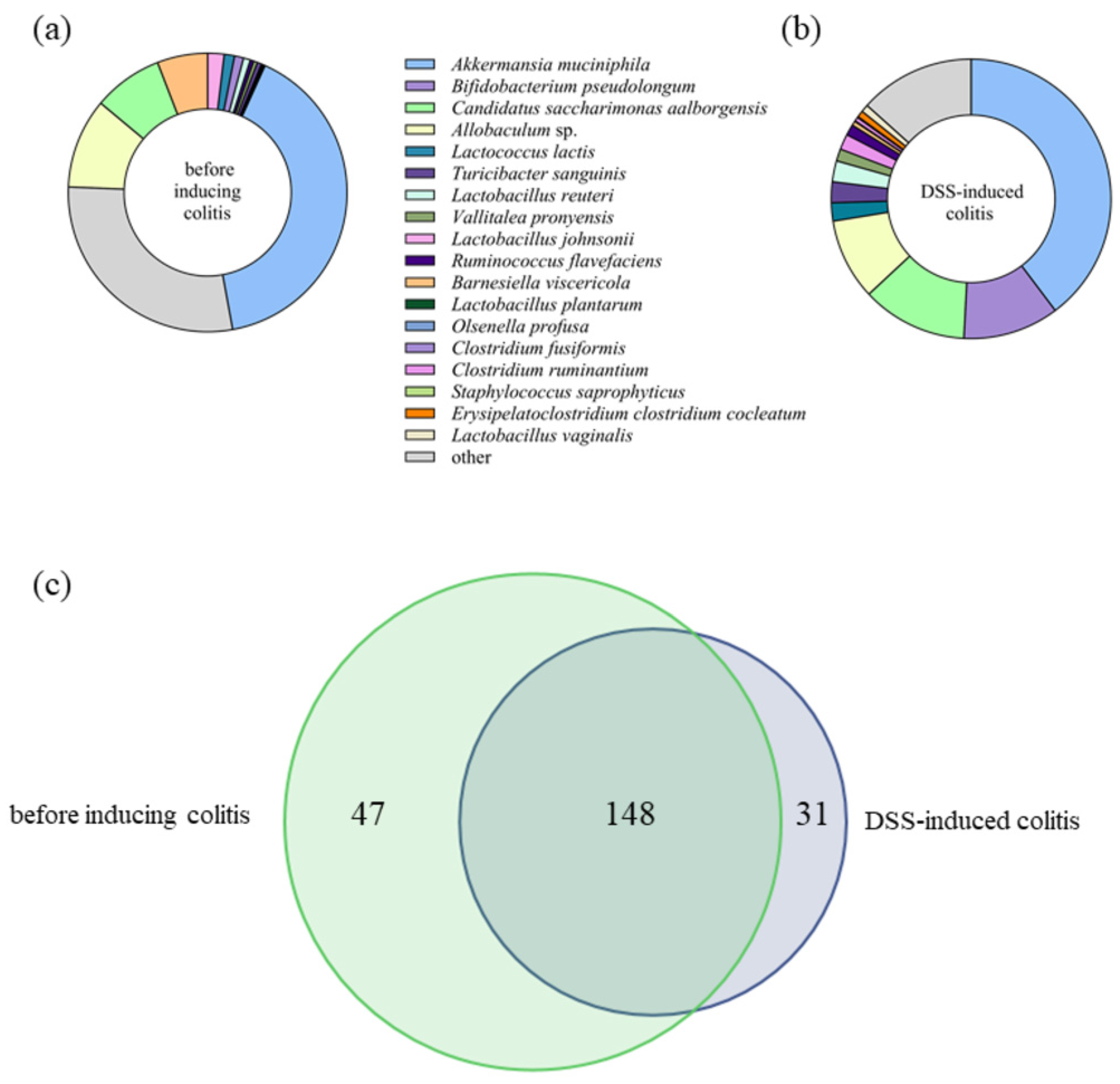
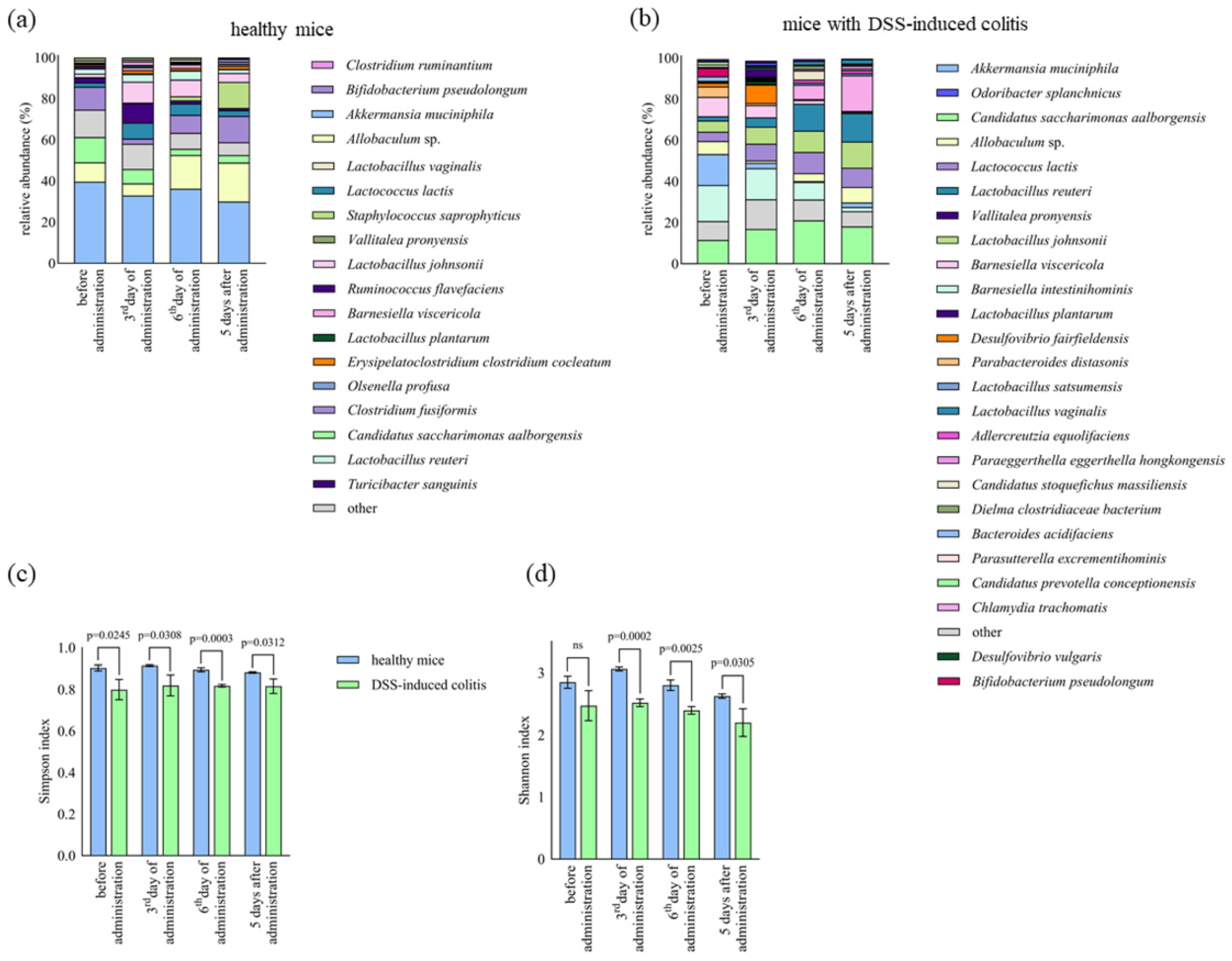
2.4. The Impact of Lb. plantarum D13 on the Composition of the Gut Microbiome of Mice with DSS-Induced Colitis

Interventional strategies to restore dysbiosis in IBD and to treat it suggest the use of probiotics, of which the use of Lactobacillus spp. is highly recommended. Lb. plantarum D13 has previously shown significant aggregation capacity and the ability to adhere to the extracellular matrix proteins collagen and laminin, but also to the Caco-2 cell line [12], which is important for probiotic activity.
Therefore, and considering its plantarcin production, we investigated the ability of plantaricin-producing Lb. plantarum D13 to alter the composition of the gut microbiota in a mouse model of IBD, particularly in DSS-induced colitis in mice. After the induction of colitis and during the administration of the strain Lb. plantarum D13, the disease activity index (DAI) score was determined to monitor the development of colitis and its severity. After DSS-induced colitis, the DAI score was 8.00 ± 1.09 on the sacrifice day, whereas it decreased to 6.00 ± 1.20 after the administration of strain D13, implying a possible positive influence of strain Lb. plantarum D13 on the slowing of disease progression and possible recovery (Figure 4a,b). Moreover, a difference in weight loss was observed between the control group with DSS-induced colitis and the group treated with the Lb. plantarum D13 strain (9.09 (±0.63)% vs. 6.25 (±0.41)%).
In order to determine whether Lb. plantarum D13 improves tissue regeneration and reduces inflammatory cell infiltration, distal colon tissue was analysed to evaluate ulcerative lesions through histopathological score analysis. Before the administration of Lb. plantarum D13, haematoxylin and eosin (HE) analysis revealed partially pronounced mucus hypersecretion with a moderately preserved epithelium and glandular architecture. Intramucosal multiplied granulation tissue with more abundant lymphocytic infiltration and foci of high-grade dysplasia were also visible (Figure 4b). The group treated with Lb. plantarum D13 showed slightly less mucosal damage. Foci of superficial ulceration were visible, but the architecture of the crypts within the intestinal mucosa was maintained and initial ischemic changes were visible. Visibly increased granulation (inflammatory tissue) was seen in the area of the mucosa with moderately abundant lymphocytic infiltrates. The application of Lb. plantarum D13 reduced inflammatory cell infiltration in DSS-induced colitis but did not lead to complete mucosal recovery (Figure 4a). The histological score before the administration of Lb. plantarum D13 was 16.17 ± 1.83 points (range 13–18), while, in the DSS group treated with Lb. plantarum D13, it was 12.5 ± 2.25 (p < 0.05) (range 10–16).
In addition, healthy mice were fed the D13 strain, and 16S rRNA gene analysis of the gut microbiota of the healthy group and the DSS-induced colitis group identified a total of 10 different phyla, 133 bacterial genera, and 226 bacterial species. At the phylum level, Verrucomicrobia, Bacteroidetes, Firmicutes, Candidatus saccharibacteria, Proteobacteria, and Actinobacteria dominated, accounting for more than 99% of the total microbial composition (Figure 4c,d). In the DSS-induced colitis group, the relative abundance of Bacteroidetes was significantly (p < 0.01) reduced (23.81 (±2.82)% to 3.67 (±1.86)%), while the abundances (p < 0.05) of Firmicutes (21.66 (±2.97)% to 30.22 (±2.94)%) and Actinobacteria (1.46 (±0.40)% to 11.76 (±0.69)%) significantly increased (Figure 4d). After Lb. plantarum D13 administration in a model of DSS-induced colitis and in healthy mice, the relative composition of Firmicutes (p < 0.01) increased from 30.22 (±2.94)% to 47.016 (±0.628)% and 26.06 (±4.10)% to 54.63 (±4.18)%, respectively (Figure 4e,f). After D13 treatment, the level of Candidatus saccharibacteria was significantly reduced (p < 0.01). In healthy mice, Lb. plantarum D13 treatment caused a significant (p < 0.05) decrease in the respective levels of Verrucomicrobia and Bacteroidetes of 15.01 (±6.34)% to 0.42 (±0.03)% and 38.83 (±4.85)% to 11.96 (±2.54)% (Figure 4e). This effect persisted in both cases 5 days after administration with strain D13 (Figure 4e,f). At the species level, a pairwise comparison of the gut microbiota of mice before and after the induction of colitis revealed that treatment with DSS decreased (p < 0.05) the abundance of Barnesiella viscericola, Barnesiella intestinihominis, Bacteroides acidifaciens, Parasutterella excrementihominis, and Parabacteroides distasonis. Conversely, the gut microbiota of the DSS-induced colitis group showed increased relative abundance of Bifidobacterium pseudolongum, Turicibacter sanguinis, Lactobacillus reuteri, Vallitalea pronyensis, Ruminococcus flavefaciens, Clostridium ruminantium, Staphylococcus saprophyticus, Erysipelatoclostridium clostridium cocleatum, and Lactobacillus vaginalis (Figure 5a,b). Of the total 226 species-level operational taxonomic units (OTUs) found in this study, only 148 (65.5%) were found in both groups, while 47 (20.8%) and 31 (13.7%) were found before and after inducing colitis, respectively, supporting the hypothesis of species depletion and consequent reduced biodiversity in the DSS-induced colitis model (Figure 5c). The most abundant species (over 1% of relative abundance) when D13 was administered to healthy mice (Figure 6a) and to mice with DSS-induced colitis (Figure 6b) comprised 26 and 18 species, respectively. The significantly higher relative abundance of Lb. plantarum species was observed on day 3 and day 6 of administration in the healthy and DSS-induced mice, respectively. Administration of Lb. plantarum D13 enriched the amount (p < 0.05) of Allobaculum, Lactococcus lactis, Barnesiella viscericola, and Olsenella profuse and depleted Candidatus Saccharimonas aalborgensis and Ruminococcus flavefaciens in the group of mice with DSS-induced colitis. Treatment with Lb. plantarum D13 significantly (p < 0.01) increased the abundance of Lactobacillus, both in healthy and DSS-induced colitis models, from 8.33 (±2.28)% to 26.25 (±5.28)% and 5.32 (±0.89)% to 14.08 (±1.568)%, respectively.
The microbial diversity of the faecal samples during the administration of Lb. plantarum D13 was estimated from the observed OTUs via the Simpson (Figure 6c) and Shannon indices (Figure 6d). During the administration of the Lb. plantarum D13 strain, the α-diversity in the form of the Simpson and Shannon indices was significantly increased in the model of healthy mice compared to mice with DSS-induced colitis (p < 0.05). Since these index values directly reflect microbial diversity, the results suggest a significant loss of microbial richness in the group of mice with DSS-induced colitis (p < 0.05) (Figure 6c,d). This could be due to dysbiosis caused by colitis, where DSS reduced the richness of bacterial species in faecal samples.

3. Discussion

Lactobacillus spp. produce a range of macromolecules, including bacteriocins, exopolysaccharides (EPSs), surface layer proteins (Slps), and proteinases, which may contribute to the application of producer strains in the development of innovative functional foods or as dietary supplements and even live biotherapeutic products [13,14,15]. Bacteriocins, Slps, and EPSs promote competitive pathogen exclusion, with bacteriocins or biopeptides produced by proteinases exhibiting direct antimicrobial activity [4,16]. The bacteriocins produced by Lb. plantarum, termed plantaricins, are generally classified as bacteriocins of the class II and comprise a broad class of bacteriocins with a range of bactericidal/bacteriostatic modes of action [17]. Since these bacteriocins primarily act against Gram-positive bacteria, we aimed to determine whether the antilisterial and antistaphylococcal activity of these strains was related to the bacteriocin-producing phenotype. Therefore, the experimental model was based on testing the antimicrobial activity against L. monocytogenes, the most common foodborne pathogen, and S. aureus, a model human pathogen commonly isolated as methicillin-resistant Staphylococcus aureus (MRSA) [18]. In this study, the phenotype of bacteriocin production in Lb. plantarum strains was analysed by considering a group of twelve autochthonous strains. Six of these, which showed significant inhibitory activity against acid-tolerant L. monocytogens ATCC® 19111 and S. aureus ATCC® 25923, were selected for further PCR analysis of the plantaricin structural genes. PCR-amplified genomic fragments revealed three pln genes of interest, which were then sequenced. Furthermore, in vitro studies of CFSs confirmed the presence of a proteinaceous substance, particularly in strain Lb. plantarum D13. Strain D13 was previously studied in terms of the techno-functional properties of probiotics, such as survival under stress conditions and colonisation potential (cell surface hydrophobicity; capacity for aggregation and coaggregation; adhesion to mucin, Caco-2 cells, and subepithelial extracellular matrix proteins) [15]. In addition, this strain exhibited caseinolytic activity and, when used as a functional starter culture in the consortium, increased content of peptides with potential bioactive activity was obtained in the dried fresh cheese produced [5,16].
The characterisation of bacteriocins is the most challenging among the antimicrobial substances of LAB, as they are synthesised in very low concentrations. The biosynthesis of bacteriocins is strain-specific and can be optimised via the cultivation conditions and cocultivation with susceptible bacteria. Therefore, to stimulate the antimicrobial activity and synthesis of potential plantaricin, the cocultivation of Lb. plantarum D13 with susceptible strains L. monocytogenes ATCC® 19111 and S. aureus ATCC® 25923 was performed. Lb. plantarum D13 showed strong activity against L. monocytogenes ATCC® 19111, which is consistent with the antimicrobial spectrum of plantaricins inhibiting Listeria spp. The induction of bacteriocinogenic activity was observed during the exponential growth phase. This is consistent with Wu et al. [19], who suggested that the increased bacteriocin activity and expression of genes related to bacteriocins were induced by the cocultivation of Lb. plantarum RUB1 with L. monocytogenes ATCC® 19111 or S. aureus ATCC® 6538. The antibacterial activity was weaker against S. aureus ATCC® 25923, which can be attributed to the fact that the bacteriocin activity is stronger against related bacterial strains, such as L. monocytogenes ATCC® 19111, than against other Gram-positive bacterial strains.
Next, we studied the capacity of the D13 strain to abolish biofilms formed by two pathogens in vitro. Namely, one of the approaches to suppressing the biofilms of pathogens in the food industry is the use of bacteriocins as an alternative to antibiotics [10]. Therefore, we evaluated the potential of strain D13 to inhibit biofilm formation. The CFS of strain Lb. plantarum D13 effectively inhibited the biofilms of both pathogens. Similar findings were reported by Zhao et al. [20], where the inhibition rate of S. aureus ATCC® 25923 biofilm formation was 22.60% and 52.39%, respectively, when plantaricin 827 was tested. These results provide the basis for the further investigation of the potential of the putative plantaricin from the CFS strain Lb. plantarum D13 as an antibiofilm agent.
In addition, the whole genome of Lb. plantarum D13 was sequenced and annotated. Genome mining analysis was performed to characterise the biosynthetic gene clusters encoding plantaricin. Comparative genome analyses of Lb. plantarum of different origins divided the strains into three lineages, A, B, and C, which differ in the presence of genes encoding bacteriocins, with PlnJK present in lineage A, while PlnEF is present in lineage A or C [21]. The production of some bacteriocins has been shown to be regulated by quorum sensing mediated by an induction peptide (IP), a membrane-bound sensor (histidine protein kinase; HPK), and a cytoplasmic response regulator (RR). This is consistent with our results, where a three-component quorum-sensing regulatory system was found in the genome of the D13 strain, together with genes encoding plantaricin A and the two-peptide plantaricins PlnEF and PlnJK, which were also found in other Lb. plantarum strains, namely LP965, EL3, L28, BL1, and SF9C [17,22,23]. Genes responsible for the production and putative biochemical function of these antimicrobials have also been deciphered, as have ABC transporters responsible for the secretion of bacteriocins and bacteriocin immunity proteins (PlnI, PlnP and PlnL, PlnM). plnY and plnO, encoding biosynthesis proteins, and plnT, plnU, plnV, and plnW, encoding integral membrane proteins with the domain of the CAAX family of membrane-bound proteases, were identified. PlnW is a two-peptide bacteriocin that inhibits many strains of Gram-positive bacteria, whereas the function of the other three plantaricins has not yet been investigated. To fully understand the underlying mechanisms of bacteriocins, it is crucial to elucidate the structure, which will allow us to understand the molecular specificity of a given bacteriocin through structure–function analyses [17,24]. The predicted 3D homology structure of plantaricins EF and JK showed that they form amphiphilic α-helices with hydrophobic residues. This is consistent with the mode of action in which the bacteriocins of the class II bind to the membrane of the target cell via the N-terminal region due to their hydrophobic nature, while the C-terminal region forms an amphiphilic helical structure that allows them to penetrate the membrane, leading to the depolarisation and death of a susceptible microorganism. PlnE forms two α-helices, whereas PlnF forms one long helix. This is consistent with published findings that the two α-helix-like regions of PlnE are separated by a flexible GXXXG motif and are both amphiphilic, while the C-terminal part of the plnF helix is amphiphilic and the N-terminal part is polar. The hydrophobicity, hydrophobic moment, and net charge of plantaricins could be crucial for their antimicrobial activity. Accordingly, bacteriocins with more positive charge accelerate the binding of the α-helix to negatively charged phospholipids in the cell membrane through a stronger inter-charge interaction. Moreover, higher hydrophobicity improves penetration into the cell membrane, which accelerates bacterial cell disintegration, while the hydrophobic moment is used to quantify the amphipathicity of peptides [25].
Another functional feature of the use of Lb. plantarum and its plantaricin is the potential modulation of the intestinal microbiota and the resulting influence on intestinal disorders [26]. IBD is the most common intestinal disease, for which treatment with probiotic bacteria is often recommended [27]. IBD is associated with dysbiosis of the gut microbiota, which may lead to the development or exacerbation of the disease. Given its antimicrobial activity, we aimed to assess the capacity of Lb. plantarum D13 to restore the imbalanced intestinal microbiota of mice with DSS-induced colitis and potentially alleviate colitis symptoms. The most abundant phyla in the gut microbiota are Firmicutes, Bacteroidetes, Actinobacteria, and Proteobacteria, and changes in their distribution lead to dysbiosis. In our study, after the onset of colitis, dysbiosis was observed through a significant decrease in the relative abundance of Bacteroidetes and an increase in Firmicutes and Actinobacteria. The effect of Lb. plantarum D13 on the gut microbiota of DSS-induced mice was evident, not only in terms of the abundance of Lb. plantarum species but also in terms of the abundance of bacterial species associated with its beneficial effect. Namely, bacteria of the species Allobaculum produce butyric acid and have a positive effect on the expression of the protein ANGPTL4, which is a key regulator of lipid metabolism [28]. Barnesiella intestinihominis and Barnesiella viscericola are species associated with beneficial effects, such as protection against colitis. Olsenella spp. produce short-chain fatty acids, acetate and lactate [29], which play an important role in metabolic homeostasis and overall gut health. Conversely, the administration of Lb. plantarum D13 decreased the levels of Candidatus Saccharimonas, which causes inflammatory diseases of the intestinal mucosa, and Ruminococcus flavefaciens, which is associated with the risk of developing obesity and whose increased presence in the intestinal microbiome correlates with the development of IBS [30]. Our results imply that Lb. plantarum D13 contributes to the restoration of the gut microbiome after DSS-induced colitis, indicating the potential for further investigation as a probiotic strain for the prevention and treatment of colitis.

4. Materials and Methods

4.1. Bacterial Strains and Cultivation Conditions

All Lb. plantarum strains and test microorganisms used in this study were deposited in the Culture Collection of the Laboratory for Antibiotic, Enzyme, Probiotic and Starter Culture Technologies, University of Zagreb Faculty of Food Technology and Biotechnology (CIM-FFTB), in de Man–Rogosa–Sharpe (MRS; Difco, Detroit, MI, USA) and Brain Heart Infusion (BHI; Biolife, Milano, Italy) broth, respectively, supplemented with 15% (v/v) glycerol (Sigma-Aldrich, Saint Louis, MO, USA), and were maintained as frozen stock at −80 °C (Eppendorf, Hamburg, Germany) (Table 1). Lb. plantarum strains were isolated from various autochthonous fermented food microbiomes in the Laboratory for Antibiotic, Enzyme, Probiotic and Starter Culture Technologies.
S. aureus ATCC® 25923 and L. monocytogenes ATCC® 19111 were used to test the antimicrobial activity of the selected Lb. plantarum strains. Prior to each experimental trial, the strains were transferred from the −80 °C stock culture and subcultured twice in an appropriate growth medium under the growth conditions listed in Table 1.

4.2. Characterisation of the Antimicrobial Activity and the pln Loci

The antimicrobial activity of the Lb. plantarum strains, against S. aureus ATCC® 25923™ and L. monocytogenes ATCC® 19111, was determined by an agar spot test according to Butorac et al. [17]. The proteinaceous feature of the plantaricins was tested by treating the CFSs with 1 mg/mL proteinase K (Invitrogen, Carlsbad, CA, USA), pepsin (AppliChem, Darmstadt, Germany), and pancreatin (AppliChem, Darmstadt, Germany) for 2 h at 37 °C and heating the samples at 100 °C/30 min, with antimicrobial activity determined by the agar spot test method, against S. aureus ATCC® 25923 and L. monocytogenes ATCC® 19111, according to Butorac et al. [17]. PCR screening of plantaricin structural genes was performed according to Butorac et al. [17]. The PCR products were sequenced by Macrogen (Amsterdam, The Netherlands). Sequences were aligned to the NCBI nt database using the BLASTn algorithm v. 2.11.0 (https://blast.ncbi.nlm.nih.gov/Blast.cgi, accessed on 2 May 2023) [31]. The potential to induce plantaricin biosynthesis of Lb. plantarum D13 was investigated by performing cocultivation experiments according to Butorac et al. [17]. CFSs were obtained by centrifugation of the overnight grown medium at 13,000 rpm for 10 min, and the supernatants were filtered through a Millipore filter (Sigma-Aldrich, Saint Louis, MO, USA) with a pore size of 0.22 µm. The pH values of CFSs were measured using a pH meter (SI Analytics, Mainz, Germany) and titratable acidity by titration with 0.1 M NaOH (Kemika, Zagreb, Croatia).

4.3. Inhibition of Biofilm Formation

The influence of the CFS of the selected Lb. plantarum D13 strain on the biofilm formation of L. monocytogenes ATCC® 19111 and S. aureus ATCC® 25923 was performed in vitro as described by Bazargani and Rohloff [32], with slight modifications. The foodborne pathogenic strains tested were grown overnight in BHI broth and the suspension was adjusted to correspond to a McFarland turbidity of 0.5 (Biolife, Milan, Italy). Then, 100 µL of the bacterial suspension was inoculated onto a sterile 96-well microtitre plate (Corning Inc., Corning, New York, NY, USA). The bacterial suspensions were treated with 100 µL of the CFS of the strain Lb. plantarum D13. Pathogen growth in the BHI medium served as a control. The inoculated microtitre plate was incubated at 37 °C for 24 h under aerobic conditions. After incubation, the medium was removed and the wells were washed three times with phosphate-buffered saline (pH = 7.4) (FFTB, Zagreb, Croatia). Then, 100 µL of crystal violet (Thermo Fisher Scientific, Waltham, MA, USA) was added to each well and they were incubated at room temperature for 30 min. The dye was removed and then the wells were washed 5 times with sterile distilled water and decolorised with 200 µL of 96% ethanol (Merck Milipore, Darmstadt, Germany) for 10 min at room temperature. Finally, 100 µL of the ethanol–dye suspension from each sample was transferred to a different 96-well microplate where the optical density (OD) was measured at 540 nm using the Infinite F Plex microplate reader (Tecan, Grödig, Austria). The inhibition ratio was calculated as follows:
I% = (1 − A/A0) × 100%
where A and A0 represent the absorbance of the CFS-treated bacterial suspension and the bacterial suspension, respectively.

4.4. Genome Sequencing and Analysis of the pln Loci

The strain Lb. plantarum D13, isolated from smoked fresh cheese, was identified by whole genome sequencing (WGS) at IGA Technology Services Srl (Udine, Italy). Genomic DNA was extracted using the Maxwell® DNA Cell Kit in the Maxwell® 16 Research System (Promega, Madison, WI, USA) according to the manufacturer’s recommendations. Sequencing of 16S rRNA variable regions V3–V4 was performed with the MiSeq 2500 instrument from Illumina (Illumina, San Diego, CA, USA) using the paired-end approach as described in Banić et al. [15]. The whole genome sequences of strain D13 were deposited in the NCBI database under BioProject PRJNA388578 (v. 1.0) with BioSample accession number SAMN07179270. CASAVA v. 1.8.2 was used for data processing. The annotation, distribution, and categorisation of sequenced genes were performed using the Rapid Annotation using Subsystem Technologies (RAST) SEED viewer (http://rast.nmpdr.org/, accessed on 15 February 2023) [33]. The identification of plantaricin-related genes and subsequent assigned functions, shown as clusters, was performed using Genome Browser. Three-dimensional structure prediction of the discovered plantaricins was performed using the AlphaFold Protein Structure Database (https://alphafold.ebi.ac.uk/, accessed on 4 April 2023) [34,35]. The HeliQuest web server was used to calculate the physicochemical properties and amino acid compositions of the α-helices in the discovered plantaricins (https://heliquest.ipmc.cnrs.fr/, accessed on 20 April 2023) [36].

4.5. In Vivo Animal Trial

4.5.1. Experimental Animals

Male and female C57BL/6 mice, aged two to three months and weighing 20 to 30 g, obtained from the Department of Animal Physiology, Faculty of Science, University of Zagreb, were used for this study. During the experiment, the animals were kept under standard conditions (temperature 25 (±3) °C, relative humidity 55 (±10)%, and 12 h of light and 12 h of darkness) in cages, separated by sex and feeding conditions. Prior to the experiment, the mice were fed a standard laboratory diet, and water was provided ad libitum. The food pellets were a certified standard diet for mice: 4RF21 (Mucedola, Settimo Milanese, Italy; batch no. 238603, shape 12 mm). The maintenance, housing, handling, and care of all experimental animals were performed according to the guidelines in force in the Republic of Croatia (Animal Welfare Act, OG 19/1999) and according to the Guide for the Care and Use of Laboratory Animals, DHHS Publ. # (NIH) 86-23. All guidelines are in accordance with the internationally accepted principles for the use and care of laboratory animals as found in the European Community guidelines (EEC Directive of 1986; 86/609/EEC). The Ethics Committee of the Faculty of Science (University of Zagreb, Croatia) approved the study (approval code: 251-58-10617-19-285).

4.5.2. DSS-Induced Colitis and Administration of Lb. plantarum D13 Strain

C57BL/6 mice received 3% DSS (36,000–50,000 Da) (MP Biomedicals, Santa Ana, CA, USA) in sterile water for 5 days to establish a colitis mouse model, followed by the administration of Lb. plantarum D13. Strain D13 was administered intragastrically daily for 6 consecutive days with 500 µL of a bacterial suspension suspended in saline at a final concentration of 109 CFU/mL. Faecal samples were collected before treatment with DSS, after treatment/before administration, on day 3 and day 6 of administration, and 5 days after the administration of Lb. plantarum D13. A healthy mouse model administrated strain D13 served as a control. DNA from the mice faecal samples collected at specific time points was isolated and analysed according to Butorac et al. [17]. In brief, total DNA from faecal mice samples was extracted using a Maxwell® DNA Tissue Kit (Promega, Madison, WI, USA) according to the manufacturer’s instructions. Samples were sequenced at Molecular Research LP (MRDNA, Shallowater, TX, USA) with the Illumina MiSeq platform using primers 341F (5′-CCTACGGGNGGCWGCAG-3′) and 518R (5′-ATTACCGCGGCTGCTGG-3′). No side effects were reported after administration. To assess whether colitis was induced, the DAI was determined separately for each animal using the measured parameters that corresponded to the clinical picture of ulcerative colitis in humans [37]. The following parameters were used for calculation: (a) weight loss (0 = none, 1 = 1–3% weight loss, 2 = 3–6% weight loss, 3 = 6–9% weight loss, and 4 = >9% weight loss); (b) stool consistency/diarrhoea (0 = normal, 2 = loose stool, 4 = watery diarrhoea); (c) bleeding (0 = no bleeding, 2 = mild bleeding, 4 = severe bleeding). The DAI is calculated as a total score, i.e., the sum of weight loss, diarrhoea, and bleeding, which can result in a DAI value from 0 (unchanged) to 12 (severe colitis). To assess histological alterations in the distal colon, the tissue samples were fixed in 4% paraformaldehyde (Gram Mol, Zagreb, Croatia), embedded in paraffin (Sigma-Aldrich Chemie GmbH, Taufkirchen, Germany), and cut into 5-μm-thick sections. The slides were then stained with haematoxylin (Merck, Darmstadt, Germany) and eosin (Acros Organics, Geel, Belgium) (HE) and analysed by a pathologist on a Zeiss Axio Star microscope (Carl Zeiss Microscopy GmbH, Jena, Germany). The pathohistological assessment was performed according to the work by Koelink et al. [38] and included the following parameters: the presence of ulcerations, inflammatory cells (such as neutrophils, macrophages, lymphocytes, and plasma cells), signs of oedema, crypt loss, surface epithelial cell hyperplasia, goblet cell reduction, and signs of epithelial regeneration. The evaluation of the degree of ulcerative lesions was performed using the mouse colitis histology index (MCHI), which includes eight histological components: inflammatory infiltrate, goblet cell loss, hyperplasia, crypt density, muscle thickness, submucosal infiltration, ulcerations, and crypt abscesses (all categorised from 0 to 3). The histopathological score was calculated based on the sum of the eight scores, ranging from 0 to 24: MCHI = 1 × goblet cell loss [four categories] + 2 × crypt density [three categories] + 2 × hyperplasia [four categories] + 3 × submucosal infiltrate [four categories].

4.6. Statistical Analysis

All experiments were performed in triplicate and results were reported as the mean of three independent trials ± standard deviation (SD). Statistical significance was appraised by analysis of variance (ANOVA). Pairwise differences between the mean values of groups were determined using Tukey’s honestly significant difference (HSD) test for post-analysis of variance pairwise comparisons (https://www.statskingdom.com/index.html, accessed on 1 September 2023) (Statistics Kingdom, 2017). Differences between groups were considered significant at p < 0.05. Microbiota diversity analysis was performed using the PAleontological STatistics (PAST) software, v. 4.13 [39]. Images were created using GraphPad Prism v.9.4.1 (GraphPad Software, San Diego, CA, USA).

5. Conclusions

In this study, we show that plantarcin-producing Lb. plantarum D13 has the probiotic potential to exclude S. aureus and L. monocytogenes and modulate the gut microbiota of healthy or DSS-induced colitis in mice by balancing the ratio of beneficial and commensal species. The biomodulation capacity is supported by the analysis of the specific antimicrobial activity and the characterisation of the putative D13 plantaricin by identifying the plantaricin biosynthesis cluster, predicting its 3D structure, and demonstrating its antibiofilm properties. Further studies are needed to decipher the underlying mechanisms related to the selected D13 strain and to translate these in vivo findings into precise molecular mechanisms. However, taken together, these results encourage future studies to characterise the use of plantaricin-producing Lb. plantarum D13 as a biomodulation agent to potentially combat colitis.

Supplementary Materials

The supporting information can be downloaded at: https://www.mdpi.com/article/10.3390/ijms242015322/s1.

Author Contributions

Conceptualisation, K.B., J.N., M.B., A.L.P. and B.K.; methodology, K.B., M.B., N.Č., N.O., D.O. and J.P.; validation, K.B., J.N., A.L.P. and M.B.; data processing, M.B., K.B., N.Č., N.O. and J.P.; writing—original draft preparation, K.B., B.K., J.N., J.Š. and A.L.P.; writing—review and editing, B.K., J.N. and A.L.P.; supervision, J.Š. and B.K. All authors have read and agreed to the published version of the manuscript.

Funding

This work was funded by the Croatian Science Foundation through projects IP-2014-09-7009 and IP-2019-04-2237. The authors also acknowledge the financial support of the University of Zagreb, Croatia. The authors are also thankful for the equipment that was procured with the financial support obtained through the project “Bioprospecting of the Adriatic Sea” (KK.01.1.1.01.0002), granted to the Scientific Centre of Excellence for Marine Bioprospecting (BioProCro) at the Ruđer Bošković Institute, Zagreb, Croatia.

Institutional Review Board Statement

This study was conducted in accordance with the guidelines in force in the Republic of Croatia (Animal Welfare Act, OG 19/1999) and in accordance with the Guide for the Care and Use of Laboratory Animals, DHHS Publ. # (NIH) 86-23. All guidelines for mice applicable in Croatia are in accordance with the internationally accepted principles for the use and care of laboratory animals as found in the European Community guidelines (EEC Directive of 1986; 86/609/EEC). The Ethics Committee of the Faculty of Science (University of Zagreb, Croatia) approved the study (approval code: 251-58-10617-19-285, approval date: 12 April 2019).

Informed Consent Statement

Not applicable.

Data Availability Statement

Not applicable.

Acknowledgments

The authors acknowledge Snježana Ramić from the Department of Pathology, University Cancer Hospital, Sestre Milosrdnice University Hospital Centre, for the histological preparation of the mouse colon tissue.

Conflicts of Interest

The authors declare no conflict of interest.

References

  1. Quaglio, A.E.V.; Grillo, T.G.; De Oliveira, E.C.S.; Di Stasi, L.C.; Sassaki, L.Y. Gut microbiota, inflammatory bowel disease and colorectal cancer. World J. Gastroenterol. 2022, 28, 4053–4060. [Google Scholar] [CrossRef] [PubMed]
  2. Bouter, K.E.; van Raalte, D.H.; Groen, A.K.; Nieuwdorp, M. Role of the gutmicrobiome in the pathogenesis of obesity and obesity-related met-abolic dysfunction. Gastroenterology 2017, 152, 1671–1678. [Google Scholar] [CrossRef]
  3. Martyniak, A.; Medyńska-Przęczek, A.; Wędrychowicz, A.; Skoczeń, S.; Tomasik, P.J. Prebiotics, Probiotics, Synbiotics, Paraprobiotics and Postbiotic Compounds in IBD. Biomolecules 2021, 11, 1903. [Google Scholar] [CrossRef] [PubMed]
  4. Banić, M.; Butorac, K.; Čuljak, N.; Leboš Pavunc, A.; Novak, J.; Bellich, B.; Kazazić, S.; Kazazić, S.; Cescutti, P.; Šušković, J.; et al. The Human Milk Microbiota Produces Potential Therapeutic Biomolecules and Shapes the Intestinal Microbiota of Infants. Int. J. Mol. Sci. 2022, 23, 14382. [Google Scholar] [CrossRef] [PubMed]
  5. Fuhren, J.; Rösch, C.; ten Napel, M.; Schols, H.A.; Kleerebezem, M. Synbiotic Matchmaking in Lactobacillus plantarum: Substrate Screening and Gene-Trait Matching to Characterize Strain-Specific Carbohydrate Utilization. Appl. Environ. Microb. 2020, 86, e01081-20. [Google Scholar] [CrossRef]
  6. Kumar, V.; Sheoran, P.; Gupta, A.; Yadav, J.; Kumar Tiwari, S. Antibacterial property of bacteriocin produced by Lactobacillus plantarum LD4 isolated from a fermented food. Ann. Microbiol. 2016, 66, 1431–1440. [Google Scholar] [CrossRef]
  7. van den Nieuwboer, M.; van Hemert, S.; Claassen, E.; de Vos, W.M. Lactobacillus plantarum WCFS1 and its host interaction: A dozen years after the genome. Microb. Biotechnol. 2016, 9, 452–465. [Google Scholar] [CrossRef]
  8. Dobson, A.; Cotter, P.D.; Ross, R.P.; Hill, C. Bacteriocin Production: A Probiotic Trait? Appl. Environ. Microbiol. 2021, 78, 1–6. [Google Scholar] [CrossRef]
  9. Trubenová, B.; Roizman, D.; Moter, A.; Rolff, J.; Regoes, R.R. Population genetics, biofilm recalcitrance, and antibiotic resistance evolution. Trends Microbiol. 2022, 30, 841–852. [Google Scholar] [CrossRef]
  10. Duraisamy, S.; Balakrishnan, S.; Ranjith, S.; Husain, F.; Sathyan, A.; Peter, A.S.; Prahalathan, C.; Kumarasamy, A. Bacteriocin—A potential antimicrobial peptide towards disrupting and preventing biofilm formation in the clinical and environmental locales. Environ. Sci. Pollut. Res. 2020, 27, 44922–44936. [Google Scholar] [CrossRef]
  11. Soltani, S.; Hammami, R.; Cotter, P.D.; Rebuffat, S.; Said, L.B.; Gaudreau, H.; Bédard, F.; Biron, E.; Drider, D.; Fliss, I. Bacteriocins as a new generation of antimicrobials: Toxicity aspects and regulations. FEMS Microbiol. Rev. 2021, 45, 039. [Google Scholar] [CrossRef] [PubMed]
  12. Uroić, K.; Novak, J.; Hynönen, U.; Pietilä, T.E.; Leboš Pavunc, A.; Kant, R.; Kos, B.; Palva, A.; Šušković, J. The role of S-layer in adhesive and immunomodulating properties of probiotic starter culture Lactobacillus brevis D6 isolated from artisanal smoked fresh cheese. LWT-Food Sci. Technol. 2016, 69, 623–632. [Google Scholar] [CrossRef]
  13. Martín, R.; Langella, P. Emerging Health Concepts in the Probiotics Field: Streamlining the Definitions. Front. Microbiol. 2019, 10, 1047. [Google Scholar] [CrossRef]
  14. Vinderola, G.; Sanders, M.E.; Salminen, S. The Concept of Postbiotics. Foods 2022, 11, 1077. [Google Scholar] [CrossRef] [PubMed]
  15. Banić, M.; Uroić, K.; Leboš Pavunc, A.; Novak, J.; Zorić, K.; Durgo, K.; Petković, H.; Jamnik, P.; Kazazić, S.; Kazazić, S.; et al. Characterization of S-layer proteins of potential probiotic starter culture Lactobacillus brevis SF9B isolated from sauerkraut. LWT-Food Sci. Technol. 2018, 93, 257–267. [Google Scholar] [CrossRef]
  16. Novak, J.; Butorac, K.; Leboš Pavunc, A.; Banić, M.; Butorac, A.; Lepur, A.; Oršolić, N.; Tonković, K.; Bendelja, K.; Čuljak, N.; et al. A lactic acid bacteria consortium impacted the content of casein-derived biopeptides in dried fresh cheese. Molecules 2022, 27, 160. [Google Scholar] [CrossRef]
  17. Butorac, K.; Banić, M.; Novak, J.; Leboš Pavunc, A.; Uroić, K.; Durgo, K.; Oršolić, N.; Kukolj, M.; Radović, S.; Scalabrin, S.; et al. The functional capacity of plantaricin-producing Lactobacillus plantarum SF9C and S-layer-carrying Lactobacillus brevis SF9B to withstand gastrointestinal transit. Microb. Cell Fact. 2020, 19, 106. [Google Scholar] [CrossRef]
  18. Liu, F.; Rajabi, S.; Shi, C.; Afifirad, G.; Omidi, N.; Kouhsari, E.; Azizian, K. Antibacterial activity of recently approved antibiotics against methicillin-resistant Staphylococcus aureus (MRSA) strains: A systematic review and meta-analysis. Ann. Clin. Microbiol. Antimicrob. 2022, 21, 37. [Google Scholar] [CrossRef]
  19. Wu, A.; Fu, Y.; Kong, L.; Shen, Q.; Liu, M.; Zeng, X.; Wu, Z.; Guo, Y.; Pan, D. Production of a class IIb bacteriocin with broad-spectrum antimicrobial activity in Lactiplantibacillus plantarum RUB1. Probiotics Antimicrob. Prot. 2021, 13, 1820–1832. [Google Scholar] [CrossRef]
  20. Zhao, D.; Wang, Q.; Lu, F.; Bie, X.; Zhao, H.; Lu, Z.; Lu, Y. A novel plantaricin 827 effectively inhibits Staphylococcus aureus and extends shelf life of skim milk. LWT-Food Sci. Technol. 2022, 154, 112849. [Google Scholar] [CrossRef]
  21. Choi, S.; Baek, M.-G.; Chung, M.-J.; Lim, S.; Yi, H. Distribution of bacteriocin genes in the lineages of Lactiplantibacillus plantarum. Sci. Rep. 2021, 11, 20063. [Google Scholar] [CrossRef] [PubMed]
  22. Gholamzadeh, M.; Gharajeh, N.H.; Hejazi, M.A. Genetic and in silico analysis of plantaricin EFI locus in indigenous isolates of lactobacillus plantarum. Biotechnol. Prog. 2019, 35, e2773. [Google Scholar] [CrossRef] [PubMed]
  23. Heeney, D.D.; Yarov-Yarovoy, V.; Marco, M.L. Sensitivity to the two peptide bacteriocin plantaricin EF is dependent on CorC, a membrane-bound, magnesium/cobalt efflux protein. Microbiologyopen 2019, 8, e827. [Google Scholar] [CrossRef] [PubMed]
  24. Tenea, G.N. Decoding the gene variants of two native probiotic Lactiplantibacillus plantarum strains through whole-genome resequencing: Insights into bacterial adaptability to stressors and antimicrobial strength. Genes 2022, 13, 443. [Google Scholar] [CrossRef] [PubMed]
  25. Jiang, Y.; Mei, C.; Huang, X.; Gu, Q.; Song, D. Antibacterial activity and mechanism of a bacteriocin derived from the valine-cecropin A(1–8)-plantaricin ZJ5(1–18) hybrid peptide against Escherichia coli O104. Food Biophys. 2020, 15, 442–451. [Google Scholar] [CrossRef]
  26. Pu, J.; Hang, S.; Liu, M.; Chen, Z.; Xiong, J.; Li, Y.; Wu, H.; Zhao, X.; Liu, S.; Gu, Q.; et al. A class IIb bacteriocin plantaricin NC8 modulates gut microbiota of different enterotypes in vitro. Front. Nutr. 2022, 9, 877948. [Google Scholar] [CrossRef]
  27. de Moreno de LeBlanc, A.; Levit, R.; Savoy de Giori, G.; LeBlanc, J.G. Vitamin producing lactic acid bacteria as complementary treatments for intestinal infammation. Anti-Inflamm. Anti-Allergy Agents Med. Chem. 2018, 17, 50–56. [Google Scholar] [CrossRef]
  28. Zibin, Z.; Wentao, L.; Ying, R.; Xiaoqiong, L.; Shenjun, Z.; Hua, Y.; Yingping, Y. Allobaculum involves in the modulation of intestinal ANGPTLT4 expression in mice treated by high-fat diet. Front. Nutr. 2021, 8, 690138. [Google Scholar]
  29. Li, W.; Zhang, K.; Yang, H. Pectin alleviates high fat (lard) diet-induced nonalcoholic fatty liver disease in mice: Possible role of short-chain fatty acids and gut microbiota regulated by pectin. J. Agric. Food Chem. 2018, 66, 8015–8025. [Google Scholar] [CrossRef]
  30. Gryaznova, M.; Dvoretskaya, Y.; Burakova, I.; Syromyatnikov, M.; Popov, E.; Kokina, A.; Mikhaylov, E.; Popov, V. Dynamics of changes in the gut microbiota of healthy mice fed with lactic acid bacteria and Bifidobacteria. Microorganisms 2022, 10, 1020. [Google Scholar] [CrossRef]
  31. Altschul, S.F.; Madden, T.L.; Schäffer, A.A.; Zhang, J.; Zhang, Z.; Miller, W.; Lipman, D.J. Gapped BLAST and PSI-BLAST: A new generation of protein database search programs. Nucleic Acids Res. 1997, 25, 3389–3402. [Google Scholar] [CrossRef] [PubMed]
  32. Bazargani, M.M.; Rohloff, J. Antibiofilm activity of essential oils and plant extracts against Staphylococcus aureus and Escherichia coli biofilms. Food Control 2016, 61, 156–164. [Google Scholar] [CrossRef]
  33. Overbeek, R.; Olson, R.; Pusch, G.D.; Olsen, G.J.; Davis, J.J.; Dis, T.; Edwards, R.A.; Gerdes, S.; Parrello, B.; Shukla, M.; et al. The seed and the rapid annotation of microbial genomes using subsystems technology (RAST). Nucleic Acids Res. 2014, 42, 206–214. [Google Scholar] [CrossRef] [PubMed]
  34. Jumper, J.; Evans, R.; Pritzel, A.; Pritzel, A.; Green, T.; Figurnov, M.; Ronneberger, O.; Tunyasuvunakool, K.; Bates, R.; Žídek, A.; et al. Highly accurate protein structure prediction with AlphaFold. Nature 2021, 596, 583–589. [Google Scholar] [CrossRef] [PubMed]
  35. Varadi, M.; Anyango, S.; Deshpande, M.; Nair, S.; Natassia, C.; Yordanova, G.; Yuan, D.; Stroe, O.; Wood, G.; Laydon, A.; et al. AlphaFold Protein Structure Database: Massively expanding the structural coverage of protein-sequence space with high-accuracy models. Nucleic Acids Res. 2022, 50, 439–444. [Google Scholar] [CrossRef] [PubMed]
  36. Gautier, R.; Douguet, D.; Antonny, B.; Drin, G. HELIQUEST: A web server to screen sequences with specific a-helical properties. Bioinformatics 2008, 24, 2101–2102. [Google Scholar] [CrossRef]
  37. Lan, D.; Niu, J.; Miao, J.; Dong, X.; Wang, H.; Yang, G.; Wang, K.; Miao, Y. Expression of guanylate cyclase-C, guanylin and uroguanylin is downregulated proportionally to the ulcerative colitis disease activity index. Sci. Rep. 2016, 6, 25034. [Google Scholar] [CrossRef]
  38. Koelink, P.J.; Wildenberg, M.E.; Stitt, L.W.; Feagan, B.G.; Koldijk, M.; van‘t Wout, A.B.; Atreya, R.; Vieth, M.; Brandse, J.F.; Duijst, S.; et al. Development of reliable, valid and responsive scoring systems for endoscopy and histology in animal models for inflammatory bowel disease. J. Crohn’s Colitis 2018, 12, 794–803. [Google Scholar] [CrossRef]
  39. Hammer, Ø.; Harper, D.A.T.; Ryan, P.D. PAST: Paleontological statistics software package for education and data analysis. Palaeontol. Electron. 2001, 4, 9. [Google Scholar]
Figure 1. Growth inhibition (log (CFU/mL)) of L. monocytogenes ATCC® 19111™ (− −) and S. aureus ATCC® 25923™ (− −) during 48 h of co-cultivation with Lb. plantarum D13. Growth curves of Lb. plantarum D13 (), L. monocytogenes ATCC® 19111™ (), and S. aureus ATCC® 25923™ () alone (a). The corresponding values of the effective inhibition ratio (EIR) for the test microorganisms. The lower grey part of the bars represents the EIR of Lb. plantarum D13 alone (), and the upper part of the bar represents the EIR of Lb. plantarum D13 towards L. monocytogenes ATCC® 19111™ () and S. aureus ATCC® 25923™ (), during 48 h of cocultivation (b). Results are given as mean ± SD of three independent experiments.
Ijms 24 15322 g001
Figure 2. Effects of cell-free supernatant (CFS) of Lb. plantarum D13 on biofilm formation (A540) of L. monocytogenes ATCC® 19111 () and S. aureus ATCC® 25923 (). * Significant difference (p < 0.001) from the untreated control of the respective test microorganisms ().
Ijms 24 15322 g002
Figure 3. Mosaic pln gene cluster of Lb. plantarum D13 strain (a). The 3D homology structure prediction of PlnE, PlnF, PlnJ, and PlnK of strain D13 using AlphaFold Protein Structure Database. The source microorganisms with a sequence similarity threshold of 100% and the respective UniProt ID are indicated, and the helical-wheel representation of regions that may form an α-helix were analysed with HeliQuest, where a one-letter code is used for one amino acid; yellow—hydrophobic residues; purple—serine and threonine residues; dark blue—basic residues; red—acidic residues; pink—asparagine and glutamine residues; grey—alanine and glycine residues; light blue—histidine residues; green—proline residues. H—hydrophobicity; μH—hydrophobic moment; z—net charge (calculated at pH = 7.4, assuming that histidine is neutral and that the N-terminal amino group and the C-terminal carboxyl group of the sequence are uncharged) (b).
Ijms 24 15322 g003
Figure 4. In situ dysplasia of colonic mucosa in mice with DSS -induced colitis, with (a) and without (b) application of Lb. plantarum D13. Analysis of the composition of the gut microbiota at the phylum level before (c) and after (d) DSS-induced colitis in mice, during different phases of administration of Lb. plantarum D13 (3rd and 6th day), and on 5th day after the end of administration in healthy mice (e) and mice with DSS-induced colitis (f). Phyla with an average prevalence of less than 1% are classified as other.
Ijms 24 15322 g004
Figure 5. The relative abundance (%) of bacterial species of the gut microbiota before (a) and after DSS-induced colitis in mice (b). Venn diagram of shared and unique bacterial operational taxonomic units (OTUs) among groups before and after DSS-induced colitis (c). Species with an average representation of less than 1% are classified as other.
Ijms 24 15322 g005
Figure 6. Relative abundance (%) of species before, during (3rd and 6th day), and after (5 day) administration of Lb. plantarum D13 in healthy mice (a) and mice with DSS-induced colitis (b). Alpha diversity of operational taxonomic units (OTUs) using Simpson (c) and Shannon indices (d), between healthy and DSS-induced mice during administration of Lb. plantarum D13. Species with an average representation of less than 1% are classified as other. ns—not significant.
Ijms 24 15322 g006
Table 1. Bacterial strains, cultivation conditions, and isolation sources used in this study.
Bacterial StrainCultivation ConditionsSource
Lb. plantarum D4MRS, 37 °C, microaerophilicSmoked fresh cheese
Lb. plantarum D5MRS, 37 °C, microaerophilicSmoked fresh cheese
Lb. plantarum D7MRS, 37 °C, microaerophilicSmoked fresh cheese
Lb. plantarum D13MRS, 37 °C, microaerophilicSmoked fresh cheese
Lb. plantarum M4MRS, 37 °C, microaerophilicDried fresh cheese
Lb. plantarum M5MRS, 37 °C, microaerophilicDried fresh cheese
Lb. plantarum MA2MRS, 37 °C, microaerophilicDried fresh cheese
Lb. plantarum MA3MRS, 37 °C, microaerophilicDried fresh cheese
Lb. plantarum SF15CMRS, 37 °C, microaerophilicSauerkraut
Lb. plantarum ZG1CMRS, 37 °C, microaerophilicSauerkraut
Lb. plantarum M92CMRS, 37 °C, microaerophilicFermented milk
Lb. plantarum L4MRS, 37 °C, microaerophilicSilage
S. aureus ATCC® 25923BHI, 37 °C, aerobicATCC 1
L. monocytogenes ATCC® 19111BHI, 37 °C, aerobicATCC 1
1 ATCC—American Type Culture Collection.
Disclaimer/Publisher’s Note: The statements, opinions and data contained in all publications are solely those of the individual author(s) and contributor(s) and not of MDPI and/or the editor(s). MDPI and/or the editor(s) disclaim responsibility for any injury to people or property resulting from any ideas, methods, instructions or products referred to in the content.

