
The Role of microRNAs in Epigenetic Regulation of Signaling Pathways in Neurological Pathologies
 ,
,
Abstract
:1. Introduction
2. Role of miRNAs in Epigenetic Regulation of Genome Translation
3. Methods for Assessing the Pool and miRNA Expression
4. The Role of MicroRNAs in Signaling Pathways of Alzheimer’s-Type Neurodegeneration
5. Regulation of Signaling Pathways in Ischemia and Reperfusion Injury of Nerve Cells Involving microRNAs
6. Peculiarities of Epigenetic Regulation in BBB Dysfunction
7. Conclusions
Author Contributions
Funding
Institutional Review Board Statement
Informed Consent Statement
Data Availability Statement
Conflicts of Interest
Abbreviations
| AIM2 | Interferon-inducible protein AIM2 (absent in melanoma 2) |
| AREG | Amphiregulin |
| BCL2L14 | Apoptosis facilitator Bcl-2-like protein 14 is a protein that, in humans, is encoded by the BCL2L14 gene |
| BCL2L13 | A protein which, in humans, is encoded by the BCL2L13 gene on chromosome 22 |
| BBB | Blood–brain barrier |
| CCL1 | Chemokine (C-C motif) ligand 1 |
| DNA | Deoxyribonucleic acid |
| DLGAP4 | Disks large-associated protein 4 (DAP-4) |
| DUSP14 | Dual-specificity Phosphatase 14 |
| FOXO | Forkhead box protein O1 (FOXO1), also known as forkhead in rhabdomyosarcoma (FKHR) |
| FBXO3 | F-box protein 3 |
| GTP | Guanosine triphosphate |
| HDAC2 | Histone deacetylase 2 |
| HSP70 | The 70-kilodalton heat shock proteins |
| HSP90 | The 90-kilodalton heat shock proteins |
| Il-1β | Interleukin-1 beta |
| Il-6 | Interleukin 6 |
| IGF1R | Insulin-like growth factor 1 receptor |
| IGFBP3 | Insulin-like growth factor-binding protein 3 |
| LNA | Locked nucleic acid |
| MAPK 1 | Mitogen-activated protein kinase 1 |
| MEG3 | Maternally expressed 3 imprinted long non-coding RNA gene |
| MEF2C | Myocyte enhancer factor 2C |
| MMP9 | Matrix metallopeptidase 9 |
| MAP3K2 | Mitogen-activated protein Kinase 2 |
| mRNA | Messenger RNA |
| Nrf2/ARE | Nuclear factor erythroid 2-related factor 2 |
| OGD | Oxygen–glucose deprivation |
| PCR | Polymerase chain reaction |
| qRT-PCR | Quantitative reverse transcription polymerase chain reaction |
| RNA | Ribonucleic acid |
| RPL34-AS1 | Ribosomal protein L34 |
| RISC/microRNP | RNA-induced silencing complex/miR–ribonucleoprotein complex |
| RNase | Ribonuclease |
| SOX9 | SRY (sex-determining region Y)-Box |
| SIRT1 | (Silent mating type information regulation 2 homolog) 1 |
| SIRT6 | Stress-responsive protein deacetylase and mono-ADP ribosyl transferase enzyme encoded by the SIRT6 gene |
| siRNAs | Small interfering RNAs |
| TRAF3 | TNF receptor-associated factor |
| TRBP | Transactivation response RNA binding protein |
| TNF-α | Tumor necrosis factor |
| YTHDF1 | YTH domain family; member 1 is a protein that, in humans, is encoded by the YTHDF1 gene |
References
- Lu, T.X.; Rothenberg, M.E. MicroRNA. J. Allergy Clin. Immunol. 2018, 141, 1202–1207. [Google Scholar] [CrossRef]
- Morselli, M.; Dieci, G. Epigenetic regulation of human non-coding RNA gene transcription. Biochem. Soc. Trans. 2022, 50, 723–736. [Google Scholar] [CrossRef] [PubMed]
- Mohr, A.M.; Mott, J.L. Overview of microRNA biology. Semin. Liver Dis. 2015, 35, 3–11. [Google Scholar] [CrossRef]
- Nachtigall, P.G.; Bovolenta, L.A. Computational Detection of MicroRNA Targets. Methods Mol. Biol. 2022, 2257, 187–209. [Google Scholar] [PubMed]
- Ho, P.T.B.; Clark, I.M.; Le, L.T.T. MicroRNA-Based Diagnosis and Therapy. Int. J. Mol. Sci. 2022, 23, 7167. [Google Scholar] [CrossRef] [PubMed]
- Rupaimoole, R.; Slack, F.J. MicroRNA therapeutics: Towards a new era for the management of cancer and other diseases. Nat. Rev. Drug Discov. 2017, 16, 203–222. [Google Scholar] [CrossRef] [PubMed]
- Marques, S.; Outeiro, T.F. Epigenetics in Parkinson’s and Alzheimer’s diseases. Epigenetics Dev. Dis. 2013, 61, 507–525. [Google Scholar]
- Neal, M.; Richardson, J.R. Epigenetic regulation of astrocyte function in neuroinflammation and neurodegeneration. Biochim. Biophys. Acta Mol. Basis Dis. 2018, 1864, 432–443. [Google Scholar] [CrossRef]
- Ghosh, P.; Saadat, A. Neurodegeneration and epigenetics: A review. Neurologia 2023, 38, e62–e68. [Google Scholar] [CrossRef]
- Aigner, A. Applications of RNA interference: Current state and prospects for siRNA-based strategies in vivo. Appl. Microbiol. Biotechnol. 2007, 76, 9–21. [Google Scholar] [CrossRef]
- Aksenenko, M.B.; Komina, A.V.; Palkina, N.V.; Averchuk, A.S.; Rybnikov, Y.A.; Dyhno, Y.A.; Ruksha, T.G. Transcriptomic analysis of melanoma cells extracted from different sites of the primary tumor. Sib. J. Oncol. 2018, 17, 59–66. [Google Scholar] [CrossRef]
- Kurakula, H.; Vaishnavi, S.; Sharif, M.Y.; Ellipilli, S. Emergence of Small Interfering RNA-Based Gene Drugs for Various Diseases. ACS Omega 2023, 8, 20234–20250. [Google Scholar] [CrossRef] [PubMed]
- Lee, Y.; Kim, M.; Han, J.; Yeom, K.H.; Lee, S.; Baek, S.H.; Kim, V.N. MicroRNA genes are transcribed by RNA polymerase II. EMBO J. 2004, 23, 4051–4060. [Google Scholar] [CrossRef] [PubMed]
- Mraz, M.; Dolezalova, D.; Plevova, K.; Stano Kozubik, K.; Mayerova, V.; Cerna, K.; Musilova, K.; Tichy, B.; Pavlova, S.; Borsky, M.; et al. MicroRNA-650 expression is influenced by immunoglobulin gene rearrangement and affects the biology of chronic lymphocytic leukemia. Blood 2012, 119, 2110–2113. [Google Scholar] [CrossRef] [PubMed]
- Gatti da Silva, G.H.; Coltri, P.P. A Reporter Assay to Analyze Intronic microRNA Maturation in Mammalian Cells. J. Vis. Exp. 2022, 184, e63498. [Google Scholar]
- Wong, A.C.H.; Rasko, J.E.J. Splice and Dice: Intronic microRNAs, Splicing and Cancer. Biomedicines 2021, 9, 1268. [Google Scholar] [CrossRef]
- Wakiyama, M.; Ogami, K.; Iwaoka, R.; Aoki, K.; Hoshino, S.I. MicroRNP-mediated translational activation of nonadenylated mRNAs in a mammalian cell-free system. Genes Cells 2018, 23, 332–344. [Google Scholar] [CrossRef]
- Nakanishi, K. Anatomy of RISC: How do small RNAs and chaperones activate Argonaute proteins? Wiley Interdiscip. Rev. RNA 2016, 7, 637–660. [Google Scholar] [CrossRef]
- Iwakawa, H.O.; Tomari, Y. Life of RISC: Formation, action, and degradation of RNA-induced silencing complex. Mol. Cell 2022, 82, 30–43. [Google Scholar] [CrossRef]
- Jin, S.; Zhan, J.; Zhou, Y. Argonaute proteins: Structures and their endonuclease activity. Mol. Biol. Rep. 2021, 48, 4837–4849. [Google Scholar] [CrossRef]
- Kobayashi, H.; Tomari, Y. RISC assembly: Coordination between small RNAs and Argonaute proteins. Biochim. Biophys. Acta 2016, 1859, 71–81. [Google Scholar] [CrossRef]
- Bibel, B.; Elkayam, E.; Silletti, S.; Komives, E.A.; Joshua-Tor, L. Target binding triggers hierarchical phosphorylation of human Argonaute-2 to promote target release. Elife 2022, 11, e76908. [Google Scholar] [CrossRef]
- Warnement, C.M.; Cismowski, M.J.; Rogers, L.K. Optimizing miR-29 measurements in biobanked, heparinized samples. Life Sci. 2019, 238, 116894. [Google Scholar] [CrossRef] [PubMed]
- Ingle, R.G.; Fang, W.J. An Overview of the Stability and Delivery Challenges of Commercial Nucleic Acid Therapeutics. Pharmaceutics 2023, 15, 1158. [Google Scholar] [CrossRef] [PubMed]
- Low, S.S.; Ji, D.; Chai, W.S.; Liu, J.; Khoo, K.S.; Salmanpour, S.; Karimi, F.; Deepanraj, B.; Show, P.L. Recent Progress in Nanomaterials Modified Electrochemical Biosensors for the Detection of MicroRNA. Micromachines. Micromachines 2021, 12, 1409. [Google Scholar] [CrossRef] [PubMed]
- Torres, A.G.; Fabani, M.M.; Vigorito, E.; Gait, M.J. MicroRNA fate upon targeting with anti-miRNA oligonucleotides as revealed by an improved Northern-blot-based method for miRNA detection. RNA 2011, 17, 933–943. [Google Scholar] [CrossRef] [PubMed]
- Tian, T.; Wang, J.; Zhou, X. A review: microRNA detection methods. Org. Biomol. Chem. 2015, 13, 2226–2238. [Google Scholar] [CrossRef] [PubMed]
- Trinh, M.P.; Carballo, J.G.; Adkins, G.B.; Guo, K.; Zhong, W. Physical and chemical template-blocking strategies in the exponential amplification reaction of circulating microRNAs. Anal. Bioanal. Chem. 2020, 412, 2399–2412. [Google Scholar] [CrossRef]
- de Ferronato, G.A.; Cerezetti, M.B.; Bridi, A.; Prado, C.M.; Dos Santos, G.; Bastos, N.M.; da Rosa, P.M.S.; Ferst, J.G.; da Silveira, J.C. MicroRNA Profiling Using a PCR-Based Method. Methods Mol. Biol. 2023, 2595, 159–170. [Google Scholar]
- Komori, M.; Komiya, K.; Shirakawa, T.; Morikawa, T.J.; Yoshimura, T. Measurement of microRNA with isothermal DNA amplification on fully automated immunoassay analyzers. Anal. Bioanal. Chem. 2019, 411, 3789–3800. [Google Scholar] [CrossRef]
- Liu, H.; Li, L.; Duan, L. High specific and ultrasensitive isothermal detection of microRNA by padlock probe-based exponential rolling circle amplification. Anal. Chem. 2013, 85, 7941–7947. [Google Scholar] [CrossRef] [PubMed]
- Deng, R.; Tang, L.; Tian, Q.; Wang, Y.; Lin, L.; Li, J. Toehold-initiated rolling circle amplification for visualizing individual microRNAs in situ in single cells. Angew. Chem. 2014, 126, 2421–2425. [Google Scholar] [CrossRef]
- Aziz, N.B.; Mahmudunnabi, R.G.; Umer, M.; Sharma, S.; Rashid, M.A.; Alhamhoom, Y.; Shim, Y.B.; Salomon, C.; Shiddiky, M.J.A. MicroRNAs in ovarian cancer and recent advances in the development of microRNA-based biosensors. Analyst 2020, 145, 2038–2057. [Google Scholar] [CrossRef] [PubMed]
- Martino, S.; Tammaro, C.; Misso, G.; Falco, M.; Scrima, M.; Bocchetti, M.; Rea, I.; De Stefano, L.; Caraglia, M. microRNA Detection via Nanostructured Biochips for Early Cancer Diagnostics. Int. J. Mol. Sci. 2023, 24, 7762. [Google Scholar] [CrossRef] [PubMed]
- Withanage, M.H.H.; Liang, H.; Zeng, E. RNA-Seq Experiment and Data Analysis. Methods Mol. Biol. 2022, 2418, 405–424. [Google Scholar]
- Yaylak, B.; Akgül, B. Experimental MicroRNA Detection Methods. Methods Mol. Biol. 2022, 2257, 33–55. [Google Scholar]
- Takegawa-Araki, T.; Yasukawa, K.; Iwazaki, N.; Utsugi, M.; Furukawa, H.; Kumagai, S.; Sawamoto, H.; Obika, S. 2’-N-Alkylaminocarbonyl-2’-amino-LNA: Synthesis, duplex stability, nuclease resistance, and in vitro anti-microRNA activity. Bioorg. Med. Chem. 2023, 78, 117148. [Google Scholar] [CrossRef]
- Langner, H.K.; Jastrzebska, K.; Caruthers, M.H. Synthesis and Characterization of Thiophosphoramidate Morpholino Oligonucleotides and Chimeras. J. Am. Chem. Soc. 2020, 142, 16240–16253. [Google Scholar] [CrossRef]
- Javanmard, S.H.; Vaseghi, G.; Ghasemi, A.; Rafiee, L.; Ferns, G.A.; Esfahani, H.N.; Nedaeinia, R. Therapeutic inhibition of microRNA-21 (miR-21) using locked-nucleic acid (LNA)-anti-miR and its effects on the biological behaviors of melanoma cancer cells in preclinical studies. Cancer Cell Int. 2020, 20, 384. [Google Scholar] [CrossRef]
- Smith, C.I.E.; Zain, R. Therapeutic Oligonucleotides: State of the Art. Annu. Rev. Pharmacol. Toxicol. 2019, 59, 605–630. [Google Scholar] [CrossRef]
- Bhattarai, U.; Hsieh, W.C.; Yan, H.; Guo, Z.F.; Shaikh, A.Y.; Soltani, A.; Song, Y.; Ly, D.H.; Liang, F.S. Bifunctional small molecule-oligonucleotide hybrid as microRNA inhibitor. Bioorg. Med. Chem. 2020, 28, 115394. [Google Scholar] [CrossRef] [PubMed]
- Graham, H.; Chandler, D.J.; Dunbar, S.A. The genesis and evolution of bead-based multiplexing. Methods 2019, 158, 2–11. [Google Scholar] [CrossRef] [PubMed]
- Cai, S.; Pataillot-Meakin, T.; Shibakawa, A.; Ren, R.; Bevan, C.L.; Ladame, S.; Ivanov, A.P.; Edel, J.B. Single-molecule amplification-free multiplexed detection of circulating microRNA cancer biomarkers from serum. Nat. Commun. 2021, 12, 3515. [Google Scholar] [CrossRef] [PubMed]
- Xu, Y.; Zhang, P.; Yi, F.; Wei, J.; Du, Q. Rainbowarray Microsphere-Based Gene Detection Assay. SLAS Technol. 2020, 25, 258–266. [Google Scholar] [CrossRef]
- Buhagiar, A.; Seria, E.; Borg, M.; Borg, J.; Ayers, D. Overview of microRNAs as liquid biopsy biomarkers for colorectal cancer sub-type profiling and chemoresistance. Cancer Drug Resist. 2021, 4, 934–945. [Google Scholar] [CrossRef]
- Ohtsuka, M.; Iwamoto, K.; Naito, A.; Imasato, M.; Hyuga, S.; Nakahara, Y.; Mikamori, M.; Furukawa, K.; Moon, J.; Asaoka, T.; et al. Circulating MicroRNAs in Gastrointestinal Cancer. Cancers 2021, 13, 3348. [Google Scholar] [CrossRef]
- Ionescu, R.F.; Cretoiu, S.M. MicroRNAs as monitoring markers for right-sided heart failure and congestive hepatopathy. J. Med. Life 2021, 14, 142–147. [Google Scholar] [CrossRef] [PubMed]
- Wu, Y.; Li, Q.; Zhang, R.; Dai, X.; Chen, W.; Xing, D. Circulating microRNAs: Biomarkers of disease. Clin. Chim. Acta 2021, 516, 46–54. [Google Scholar] [CrossRef]
- Pozniak, T.; Shcharbin, D.; Bryszewska, M. Circulating microRNAs in Medicine. Int. J. Mol. Sci. 2022, 23, 3996. [Google Scholar] [CrossRef]
- Metintaş, M.; Ak, G.; Özbayer, C.; Boğar, F.; Metintaş, S. Serum Expression Levels of Certain miRNAs in Predicting Diagnosis, Prognosis, and Response to Chemotherapy in Malignant Pleural Mesothelioma. Balkan Med. J. 2022, 39, 246–254. [Google Scholar] [CrossRef]
- Takizawa, S.; Matsuzaki, J.; Ochiya, T. Circulating microRNAs: Challenges with their use as liquid biopsy biomarkers. Cancer Biomark. 2022, 35, 1–9. [Google Scholar] [CrossRef] [PubMed]
- Franco, S.; Pluvinet, R.; Sanchez-Herrero, J.F.; Sumoy, L.; Martinez, M.A. Rapid and accurate quantification of isomiRs by RT-qPCR. Sci. Rep. 2022, 12, 17220. [Google Scholar] [CrossRef] [PubMed]
- Deng, R.; Zhang, K.; Li, J. Isothermal Amplification for MicroRNA Detection: From the Test Tube to the Cell. Acc. Chem. Res. 2017, 50, 1059–1068. [Google Scholar] [CrossRef] [PubMed]
- Gines, G.; Menezes, R.; Xiao, W.; Rondelez, Y.; Taly, V. Emerging isothermal amplification technologies for microRNA biosensing: Applications to liquid biopsies. Mol. Asp. Med. 2020, 72, 100832. [Google Scholar] [CrossRef] [PubMed]
- Ye, J.; Xu, M.; Tian, X.; Cai, S.; Zeng, S. Research advances in the detection of miRNA. J. Pharm. Anal. 2019, 9, 217–226. [Google Scholar] [CrossRef]
- Beta, M.; Krishnakumar, S.; Elchuri, S.V.; Salim, B.; Narayanan, J. A comparative fluorescent beacon-based method for serum microRNA quantification. Anal. Sci. 2015, 31, 231–235. [Google Scholar] [CrossRef]
- Chan, H.N.; Ho, S.L.; He, D.; Li, H.W. Direct and sensitive detection of circulating miRNA in human serum by ligase-mediated amplification. Talanta 2020, 206, 120217. [Google Scholar] [CrossRef]
- Masud, M.K.; Umer, M.; Hossain, M.S.A.; Yamauchi, Y.; Nguyen, N.T.; Shiddiky, M.J.A. Nanoarchitecture Frameworks for Electrochemical MiRNA Detection. Trends Biochem. Sci. 2019, 44, 433–452. [Google Scholar] [CrossRef]
- Vilímová, I.; Chourpa, I.; David, S.; Soucé, M.; Hervé-Aubert, K. Two-step formulation of magnetic nanoprobes for microRNA capture. RSC Adv. 2022, 12, 7179–7188. [Google Scholar] [CrossRef]
- Wu, Y.; Huang, J.; Yang, X.; Yang, Y.; Quan, K.; Xie, N.; Li, J.; Ma, C.; Wang, K. Gold Nanoparticle Loaded Split-DNAzyme Probe for Amplified miRNA Detection in Living Cells. Anal. Chem. 2017, 89, 8377–8383. [Google Scholar] [CrossRef]
- Remsburg, C.; Konrad, K.; Sampilo, N.F.; Song, J.L. Analysis of microRNA functions. Methods Cell Biol. 2019, 151, 323–334. [Google Scholar]
- Li, Y.; Zhang, Z. Computational Biology in microRNA. Wiley Interdiscip. Rev. RNA 2015, 6, 435–452. [Google Scholar] [CrossRef] [PubMed]
- de Amorim, T.S.; Pedro, D.L.F.; Paschoal, A.R. MicroRNA Databases and Tools. Methods Mol. Biol. 2022, 2257, 131–166. [Google Scholar]
- Tomasello, L.; Cluts, L.; Croce, C.M. Experimental Validation of MicroRNA Targets: Mutagenesis of Binding Regions. Methods Mol. Biol. 2019, 1970, 331–339. [Google Scholar] [PubMed]
- Yu, D.; Chen, S.; Li, D.; Knox, B.; Guo, L.; Ning, B. FREMSA: A Method That Provides Direct Evidence of the Interaction between microRNA and mRNA. Methods Mol. Biol. 2020, 2102, 557–566. [Google Scholar] [PubMed]
- Mortazavi, S.S.; Bahmanpour, Z.; Daneshmandpour, Y.; Roudbari, F.; Sheervalilou, R.; Kazeminasab, S.; Emamalizadeh, B. An updated overview and classification of bioinformatics tools for MicroRNA analysis, which one to choose? Comput. Biol. Med. 2021, 134, 104544. [Google Scholar] [CrossRef] [PubMed]
- Ragini Sakshi Mani, I.; Singh, V. Applications of bioinformatics in epigenetics. Prog. Mol. Biol. Transl. Sci. 2023, 198, 1–13. [Google Scholar]
- Takousis, P.; Sadlon, A.; Schulz, J.; Wohlers, I.; Dobricic, V.; Middleton, L.; Lill, C.M.; Perneczky, R.; Bertram, L. Differential expression of microRNAs in Alzheimer’s disease brain, blood, and cerebrospinal fluid. Alzheimer’s Dement. 2019, 15, 1468–1477. [Google Scholar] [CrossRef]
- Swarbrick, S.; Wragg, N.; Ghosh, S.; Stolzing, A. Systematic Review of miRNA as Biomarkers in Alzheimer’s Disease. Mol. Neurobiol. 2019, 56, 6156–6167. [Google Scholar] [CrossRef]
- Nunomura, A.; Perry, G. RNA and Oxidative Stress in Alzheimer’s Disease: Focus on microRNAs. Oxidative Med. Cell. Longev. 2020, 2020, 2638130. [Google Scholar] [CrossRef]
- Walgrave, H.; Zhou, L.; De Strooper, B.; Salta, E. The promise of microRNA-based therapies in Alzheimer’s disease: Challenges and perspectives. Mol. Neurodegener. 2021, 16, 76. [Google Scholar] [PubMed]
- Mayo, S.; Benito-León, J.; Peña-Bautista, C.; Baquero, M.; Cháfer-Pericás, C. Recent Evidence in Epigenomics and Proteomics Biomarkers for Early and Minimally Invasive Diagnosis of Alzheimer’s and Parkinson’s Diseases. Curr. Neuropharmacol. 2021, 19, 1273–1303. [Google Scholar] [CrossRef]
- Liu, S.; Fan, M.; Zheng, Q.; Hao, S.; Yang, L.; Xia, Q.; Qi, C.; Ge, J. MicroRNAs in Alzheimer’s disease: Potential diagnostic markers and therapeutic targets. Biomed. Pharmacother. 2022, 148, 112681. [Google Scholar] [CrossRef] [PubMed]
- Silvestro, S.; Bramanti, P.; Mazzon, E. Role of miRNAs in Alzheimer’s Disease and Possible Fields of Application. Int. J. Mol. Sci. 2019, 20, 3979. [Google Scholar] [CrossRef] [PubMed]
- Yuen, S.C.; Liang, X.; Zhu, H.; Jia, Y.; Leung, S.W. Prediction of differentially expressed microRNAs in blood as potential biomarkers for Alzheimer’s disease by meta-analysis and adaptive boosting ensemble learning. Alzheimer’s Res. Ther. 2021, 13, 126. [Google Scholar]
- Chang, C.H.; Lin, C.H.; Lane, H.Y. Machine Learning and Novel Biomarkers for the Diagnosis of Alzheimer’s Disease. Int. J. Mol. Sci. 2021, 22, 2761. [Google Scholar] [CrossRef]
- Xu, A.; Kouznetsova, V.L.; Tsigelny, I.F. Alzheimer’s Disease Diagnostics Using MiRNA Biomarkers and Machine Learning. J. Alzheimer’s Dis. 2022, 86, 841–859. [Google Scholar] [CrossRef]
- Shokhirev, M.N.; Johnson, A.A. An integrative machine-learning meta-analysis of high-throughput omics data identifies age-specific hallmarks of Alzheimer’s disease. Ageing Res. Rev. 2022, 81, 101721. [Google Scholar] [CrossRef]
- Chiricosta, L.; D’Angiolini, S.; Gugliandolo, A.; Mazzon, E. Artificial Intelligence Predictor for Alzheimer’s Disease Trained on Blood Transcriptome: The Role of Oxidative Stress. Int. J. Mol. Sci. 2022, 23, 5237. [Google Scholar] [CrossRef]
- Akhter, R. Circular RNA and Alzheimer’s Disease. Adv. Exp. Med. Biol. 2018, 1087, 239–243. [Google Scholar]
- Ma, N.; Tie, C.; Yu, B.; Zhang, W.; Wan, J. Identifying lncRNA-miRNA-mRNA networks to investigate Alzheimer’s disease pathogenesis and therapy strategy. Aging 2020, 12, 2897–2920. [Google Scholar] [CrossRef] [PubMed]
- Gowda, P.; Reddy, P.H.; Kumar, S. Deregulated mitochondrial microRNAs in Alzheimer’s disease: Focus on synapse and mitochondria. Ageing Res. Rev. 2022, 73, 101529. [Google Scholar] [PubMed]
- Su, L.; Li, R.; Zhang, Z.; Liu, J.; Du, J.; Wei, H. Identification of altered exosomal microRNAs and mRNAs in Alzheimer’s disease. Ageing Res. Rev. 2022, 73, 101497. [Google Scholar] [CrossRef]
- Garcia, G.; Pinto, S.; Ferreira, S.; Lopes, D.; Serrador, M.J.; Fernandes, A.; Vaz, A.R.; Mendonça, A.; Edenhofer, F.; Malm, T.; et al. Emerging Role of miR-21-5p in Neuron-Glia Dysregulation and Exosome Transfer Using Multiple Models of Alzheimer’s Disease. Cells 2022, 11, 3377. [Google Scholar] [CrossRef]
- Ryazanova, M.V.; Averchuk, A.S.; Novikova, S.V.; Salmina, A.B. Molecular mechanisms of angiogenesis: Brain is in the focus. Opera Med. Physiol. 2022, 9, 54–72. [Google Scholar]
- Tregub, P.P.; Averchuk, A.S.; Baranich, T.I.; Ryazanova, M.V.; Salmina, A.B. Physiological and Pathological Remodeling of Cerebral Microvessels. Int. J. Mol. Sci. 2022, 23, 12683. [Google Scholar] [CrossRef]
- Da Mesquita, S.; Louveau, A.; Vaccari, A.; Smirnov, I.; Cornelison, R.C.; Kingsmore, K.M.; Contarino, C.; Onengut-Gumuscu, S.; Farber, E.; Raper, D.; et al. Functional aspects of meningeal lymphatics in ageing and Alzheimer’s disease. Nature 2018, 560, 185–191. [Google Scholar] [CrossRef]
- Ihezie, S.A.; Mathew, I.E.; McBride, D.W.; Dienel, A.; Blackburn, S.L.; Thankamani Pandit, P.K. Epigenetics in blood-brain barrier disruption. Fluids Barriers CNS 2021, 18, 17. [Google Scholar] [PubMed]
- Neag, M.A.; Mitre, A.O.; Burlacu, C.C.; Inceu, A.I.; Mihu, C.; Melincovici, C.S.; Bichescu, M.; Buzoianu, A.D. miRNA Involvement in Cerebral Ischemia-Reperfusion Injury. Front. Neurosci. 2022, 16, 901360. [Google Scholar]
- Van den Hove, D.L.; Kompotis, K.; Lardenoije, R.; Kenis, G.; Mill, J.; Steinbusch, H.W.; Lesch, K.P.; Fitzsimons, C.P.; De Strooper, B.; Rutten, B.P. Epigenetically regulated microRNAs in Alzheimer’s disease. Neurobiol. Aging 2014, 35, 731–745. [Google Scholar] [CrossRef]
- Stoccoro, A.; Coppedè, F. Role of epigenetics in Alzheimer’s disease pathogenesis. Neurodegener. Dis. Manag. 2018, 8, 181–193. [Google Scholar] [CrossRef] [PubMed]
- Nikolac Perkovic, M.; Videtic Paska, A.; Konjevod, M.; Kouter, K.; Svob Strac, D.; Nedic Erjavec, G.; Pivac, N. Epigenetics of Alzheimer’s Disease. Biomolecules 2021, 11, 195. [Google Scholar] [CrossRef] [PubMed]
- Bryzgalov, L.O.; Korbolina, E.E.; Merkulova, T.I. Exploring the Genetic Predisposition to Epigenetic Changes in Alzheimer’s Disease. Int. J. Mol. Sci. 2023, 24, 7955. [Google Scholar] [CrossRef] [PubMed]
- Coneys, R.; Wood, I.C. Alzheimer’s disease: The potential of epigenetic treatments and current clinical candidates. Neurodegener. Dis. Manag. 2020, 10, 543–558. [Google Scholar] [CrossRef]
- Pandey, D.; Pal, T.; Sharma, A.; Flora, S. Potential Epigenetic Targets for Combating Alzheimer’s Disease. Mini Rev. Med. Chem. 2021, 21, 1527–1540. [Google Scholar] [CrossRef] [PubMed]
- Coppedè, F. Epigenetic regulation in Alzheimer’s disease: Is it a potential therapeutic target? Expert Opin. Ther. Targets 2021, 25, 283–298. [Google Scholar] [CrossRef] [PubMed]
- Hajjo, R.; Sabbah, D.A.; Abusara, O.H.; Al Bawab, A.Q. A Review of the Recent Advances in Alzheimer’s Disease Research and the Utilization of Network Biology Approaches for Prioritizing Diagnostics and Therapeutics. Diagnostics 2022, 12, 2975. [Google Scholar] [CrossRef]
- Yao, X.; Yao, R.; Yi, J.; Huang, F. Upregulation of miR-496 decreases cerebral ischemia/reperfusion injury by negatively regulating BCL2L14. Neurosci. Lett. 2019, 696, 197–205. [Google Scholar] [CrossRef]
- Jiang, D.; Sun, X.; Wang, S.; Man, H. Upregulation of miR-874-3p decreases cerebral ischemia/reperfusion injury by directly targeting BMF and BCL2L13. Biomed. Pharmacother. 2019, 117, 108941. [Google Scholar] [CrossRef]
- Liu, E.; Sun, H.; Wu, J.; Kuang, Y. MiR-92b-3p regulates oxygen and glucose deprivation-reperfusion-mediated apoptosis and inflammation by targeting TRAF3 in PC12 cells. Exp. Physiol. 2020, 105, 1792–1801. [Google Scholar] [CrossRef]
- Wang, J.; Xu, Z.; Chen, X.; Li, Y.; Chen, C.; Wang, C.; Zhu, J.; Wang, Z.; Chen, W.; Xiao, Z.; et al. MicroRNA-182-5p attenuates cerebral ischemia-reperfusion injury by targeting Toll-like receptor 4. Biochem. Biophys. Res. Commun. 2018, 505, 677–684. [Google Scholar] [CrossRef] [PubMed]
- Chen, Z.; Yang, J.; Zhong, J.; Luo, Y.; Du, W.; Hu, C.; Xia, H.; Li, Y.; Zhang, J.; Li, M.; et al. MicroRNA-193b-3p alleviates focal cerebral ischemia and reperfusion-induced injury in rats by inhibiting 5-lipoxygenase expression. Exp. Neurol. 2020, 327, 113223. [Google Scholar] [CrossRef] [PubMed]
- Zhang, M.; Zhu, Y.; Wei, M.; Liu, H. Neuroprotective effects of miR-30c on rats with cerebral ischemia/reperfusion injury by targeting SOX9. Pathol. Res. Pract. 2020, 216, 153271. [Google Scholar] [CrossRef] [PubMed]
- Zhang, H.; Li, M.; Liang, J.; Li, M.; Sun, X. Long Non-coding RNA PVT1 Inhibits miR-30c-5p to Upregulate Rock2 to Modulate Cerebral Ischemia/Reperfusion Injury Through MAPK Signaling Pathway Activation. Mol. Neurobiol. 2021, 58, 6032–6048. [Google Scholar] [CrossRef]
- Yu, Y.; Zhang, X.; Han, Z.; Zhao, W.; Zhang, L. Expression and regulation of miR-449a and AREG in cerebral ischemic injury. Metab. Brain Dis. 2019, 34, 821–832. [Google Scholar] [CrossRef]
- Chai, Z.; Gong, J.; Zheng, P.; Zheng, J. Inhibition of miR-19a-3p decreases cerebral ischemia/reperfusion injury by targeting IGFBP3 in vivo and in vitro. Biol. Res. 2020, 53, 17. [Google Scholar] [CrossRef]
- Wei, W.; Li, H.; Deng, Y.; Zheng, X.; Zhou, Y.; Xue, X. The combination of Alisma and Atractylodes ameliorates cerebral ischaemia/reperfusion injury by negatively regulating astrocyte-derived exosomal miR-200a-3p/141-3p by targeting SIRT1. J. Ethnopharmacol. 2023, 313, 116597. [Google Scholar] [CrossRef]
- Ruan, Z.F.; Xie, M.; Gui, S.J.; Lan, F.; Wan, J.; Li, Y. MiR-370 accelerated cerebral ischemia reperfusion injury via targeting SIRT6 and regulating Nrf2/ARE signal pathway. Kaohsiung J. Med. Sci. 2020, 36, 741–749. [Google Scholar] [CrossRef]
- Gu, C.; Mo, W.; Wang, K.; Gao, M.; Chen, J.; Zhang, F.; Shen, J. Exosomal miR-370-3p increases the permeability of blood-brain barrier in ischemia/reperfusion stroke of brain by targeting MPK1. Aging 2023, 15, 1931–1943. [Google Scholar] [CrossRef]
- Liang, J.; Wang, Q.; Li, J.Q.; Guo, T.; Yu, D. Long non-coding RNA MEG3 promotes cerebral ischemia-reperfusion injury through increasing pyroptosis by targeting miR-485/AIM2 axis. Exp. Neurol. 2020, 325, 113139. [Google Scholar] [CrossRef]
- Shi, Y.; Li, K.; Xu, K.; Liu, Q.H. MiR-155-5p accelerates cerebral ischemia-reperfusion injury via targeting DUSP14 by regulating NF-κB and MAPKs signaling pathways. Eur. Rev. Med. Pharmacol. Sci. 2020, 24, 1408–1419. [Google Scholar] [PubMed]
- Shi, Y.; Li, Z.; Li, K.; Xu, K. miR-155-5p accelerates cerebral ischemia-reperfusion inflammation injury and cell pyroptosis via DUSP14/ TXNIP/NLRP3 pathway. Acta Biochim. Pol. 2022, 69, 787–793. [Google Scholar] [CrossRef]
- Zhang, L.; Liu, C.; Huang, C.; Xu, X.; Teng, J. miR-155 Knockdown Protects against Cerebral Ischemia and Reperfusion Injury by Targeting MafB. BioMed Res. Int. 2020, 2020, 6458204. [Google Scholar] [CrossRef] [PubMed]
- Zhang, M.; Wang, J.; Li, J.; Kong, F.; Lin, S. miR-101-3p improves neuronal morphology and attenuates neuronal apoptosis in ischemic stroke in young mice by downregulating HDAC9. Transl. Neurosci. 2023, 14, 20220286. [Google Scholar] [CrossRef] [PubMed]
- Fan, W.; Qin, Y.; Tan, J.; Li, B.; Liu, Y.; Rong, J.; Shi, W.; Yu, B. RGD1564534 represses NLRP3 inflammasome activity in cerebral injury following ischemia-reperfusion by impairing miR-101a-3p-mediated Dusp1 inhibition. Exp. Neurol. 2023, 359, 114266. [Google Scholar] [CrossRef]
- Ye, X.; Fang, H.; Feng, Y.; Qian, H.; Wu, Z.; Chen, S. MiR-10b-3p Protects Cerebral I/R Injury through Targeting Programmed Cell Death 5 (PDCD5). Crit. Rev. Eukaryot. Gene Expr. 2021, 31, 85–98. [Google Scholar] [CrossRef]
- Sun, K.; Zhang, J.; Yang, Q.; Zhu, J.; Zhang, X.; Wu, K.; Li, Z.; Xie, W.; Luo, X. MiR-10b-3p alleviates cerebral ischemia/reperfusion injury by targeting Krüppel-like factor 5 (KLF5). Pflügers Arch. -Eur. J. Physiol. 2022, 474, 343–353. [Google Scholar] [CrossRef]
- Yang, L.; Wang, L.; Wang, J.; Liu, P. Long non-coding RNA Gm11974 aggravates oxygen-glucose deprivation-induced injury via miR-122-5p/SEMA3A axis in ischaemic stroke. Metab. Brain Dis. 2021, 36, 2059–2069. [Google Scholar] [CrossRef]
- Huang, P.; Wei, S.; Ren, J.; Tang, Z.; Guo, M.; Situ, F.; Zhang, D.; Zhu, J.; Xiao, L.; Xu, J.; et al. MicroRNA-124-3p alleviates cerebral ischaemia-induced neuroaxonal damage by enhancing Nrep expression. J. Stroke Cerebrovasc. Dis. 2023, 32, 106949. [Google Scholar] [CrossRef]
- Zhang, K.L.; Li, S.M.; Hou, J.Y.; Hong, Y.H.; Chen, X.X.; Zhou, C.Q.; Wu, H.; Zheng, G.H.; Zeng, C.T.; Wu, H.D.; et al. Elabela, a Novel Peptide, Exerts Neuroprotective Effects Against Ischemic Stroke Through the APJ/miR-124-3p/CTDSP1/AKT Pathway. Cell. Mol. Neurobiol. 2023, 43, 2989–3003. [Google Scholar] [CrossRef]
- Zhang, R.; Zhou, W.; Yu, Z.; Yang, L.; Liu, G.; Yu, H.; Zhou, Q.; Min, Z.; Zhang, C.; Wu, Q.; et al. miR-1247-3p mediates apoptosis of cerebral neurons by targeting caspase-2 in stroke. Brain Res. 2019, 1714, 18–26. [Google Scholar] [CrossRef]
- Yan, Q.; Sun, S.Y.; Yuan, S.; Wang, X.Q.; Zhang, Z.C. Inhibition of microRNA-9-5p and microRNA-128-3p can inhibit ischemic stroke-related cell death in vitro and in vivo. IUBMB Life 2020, 72, 2382–2390. [Google Scholar] [CrossRef] [PubMed]
- Hou, H.; Wang, Y.; Yang, L.; Wang, Y. Exosomal miR-128-3p reversed fibrinogen-mediated inhibition of oligodendrocyte progenitor cell differentiation and remyelination after cerebral ischemia. CNS Neurosci. Ther. 2023, 29, 1405–1422. [Google Scholar] [CrossRef] [PubMed]
- Zhang, H.; Wang, X.; Chen, W.; Yang, Y.; Wang, Y.; Wan, H.; Zhu, Z. Danhong injection alleviates cerebral ischemia-reperfusion injury by inhibiting autophagy through miRNA-132-3p/ATG12 signal axis. J. Ethnopharmacol. 2023, 300, 115724. [Google Scholar] [CrossRef] [PubMed]
- Yang, X.; Xu, J.; Lan, S.; Tong, Z.; Chen, K.; Liu, Z.; Xu, S. Exosomal miR-133a-3p Derived from BMSCs Alleviates Cerebral Ischemia-Reperfusion Injury via Targeting DAPK2. Int. J. Nanomed. 2023, 18, 65–78. [Google Scholar] [CrossRef]
- Dai, Q.; Ma, Y.; Xu, Z.; Zhang, L.; Yang, H.; Liu, Q.; Wang, J. Downregulation of circular RNA HECTD1 induces neuroprotection against ischemic stroke through the microRNA-133b/TRAF3 pathway. Life Sci. 2021, 264, 118626. [Google Scholar] [CrossRef]
- Chi, W.; Meng, F.; Li, Y.; Li, P.; Wang, G.; Cheng, H.; Han, S.; Li, J. Impact of microRNA-134 on neural cell survival against ischemic injury in primary cultured neuronal cells and mouse brain with ischemic stroke by targeting HSPA12B. Brain Res. 2014, 1592, 22–33. [Google Scholar] [CrossRef]
- Wang, C.; Hu, F. Long noncoding RNA SOX2OT silencing alleviates cerebral ischemia-reperfusion injury via miR-135a-5p-mediated NR3C2 inhibition. Brain Res. Bull. 2021, 173, 193–202. [Google Scholar] [CrossRef]
- Yi, M.; Li, Y.; Wang, D.; Zhang, Q.; Yang, L.; Yang, C. KCNQ1OT1 Exacerbates Ischemia-Reperfusion Injury Through Targeted Inhibition of miR-140-3P. Inflammation 2020, 43, 1832–1845. [Google Scholar] [CrossRef]
- Zhang, Y.; Su, Q.; Xia, W.; Jia, K.; Meng, D.; Wang, X.; Ni, X.; Su, Z. MiR-140–3p directly targets Tyro3 to regulate OGD/R-induced neuronal injury through the PI3K/Akt pathway. Brain Res. Bull. 2023, 192, 93–106. [Google Scholar]
- Liu, T.; Feng, J.; Sun, Z.; He, M.; Sun, L.; Dong, S.; Guo, Z.; Zhang, G. Inhibition of miR-141-3p attenuates apoptosis of neural stem cells via targeting PBX1 to regulate PROK2 transcription in MCAO mice. Cell Cycle 2023, 22, 403–418. [Google Scholar] [CrossRef]
- Meng, S.; Wang, B.; Li, W. LncRNA MALAT1 improves cerebral ischemia-reperfusion injury and cognitive dysfunction by regulating miR-142-3p/SIRT1 axis. Int. J. Neurosci. 2023, 133, 740–753. [Google Scholar] [CrossRef]
- Li, J.; Ma, L. MiR-142-3p Attenuates Oxygen Glucose Deprivation/Reoxygenation-Induced Injury by Targeting FBXO3 in Human Neuroblastoma SH-SY5Y Cells. World Neurosurg. 2020, 136, e149–e157. [Google Scholar] [CrossRef] [PubMed]
- Wang, N.; Zhang, L.; Lu, Y.; Zhang, M.; Zhang, Z.; Wang, K.; Lv, J. Down-regulation of microRNA-142-5p attenuates oxygen-glucose deprivation and reoxygenation-induced neuron injury through up-regulating Nrf2/ARE signaling pathway. Biomed. Pharmacother. 2017, 89, 1187–1195. [Google Scholar] [CrossRef] [PubMed]
- Wang, S.; Liu, Z. Inhibition of microRNA-143-3p Attenuates Cerebral Ischemia/Reperfusion Injury by Targeting FSTL1. Neuromol. Med. 2021, 23, 500–510. [Google Scholar] [CrossRef] [PubMed]
- Li, Y.; Zhao, Y.; Cheng, M.; Qiao, Y.; Wang, Y.; Xiong, W.; Yue, W. Suppression of microRNA-144-3p attenuates oxygen-glucose deprivation/reoxygenation-induced neuronal injury by promoting Brg1/Nrf2/ARE signaling. J. Biochem. Mol. Toxicol. 2018, 32, e22044. [Google Scholar] [CrossRef]
- Zhou, H.; Zhou, J.; Teng, H.; Yang, H.; Qiu, J.; Li, X. MiR-145 enriched exosomes derived from bone marrow-derived mesenchymal stem cells protects against cerebral ischemia-reperfusion injury through downregulation of FOXO1. Biochem. Biophys. Res. Commun. 2022, 632, 92–99. [Google Scholar] [CrossRef]
- Du, Y.; Ma, X.; Ma, L.; Li, S.; Zheng, J.; Lv, J.; Cui, L.; Lv, J. Inhibition of microRNA-148b-3p alleviates oxygen-glucose deprivation/reoxygenation-induced apoptosis and oxidative stress in HT22 hippocampal neuron via reinforcing Sestrin2/Nrf2 signalling. Clin. Exp. Pharmacol. Physiol. 2020, 47, 561–570. [Google Scholar] [CrossRef]
- Wang, X.; Xu, Q.; Wang, S. Overexpression of miR-149-5p Attenuates Cerebral Ischemia/Reperfusion (I/R) Injury by Targeting Notch2. Neuromol. Med. 2022, 24, 279–289. [Google Scholar] [CrossRef] [PubMed]
- Zhang, A.; Qian, Y.; Qian, J. MicroRNA-152-3p protects neurons from oxygen-glucose-deprivation/reoxygenation-induced injury through upregulation of Nrf2/ARE antioxidant signaling by targeting PSD-93. Biochem. Biophys. Res. Commun. 2019, 517, 69–76. [Google Scholar] [CrossRef]
- Li, Y.; Peng, B.; Li, Y.; Huang, A.; Peng, Y.; Yu, Q.; Li, Y. MiR-203a-3p/153-3p improves cognitive impairments induced by ischemia/reperfusion via blockade of SRC-mediated MAPK signaling pathway in ischemic stroke. Chem. Biol. Interact. 2022, 358, 109900. [Google Scholar] [CrossRef] [PubMed]
- Wang, H.J.; Tang, X.L.; Huang, G.; Li, Y.B.; Pan, R.H.; Zhan, J.; Wu, Y.K.; Liang, J.F.; Bai, X.X.; Cai, J. Long Non-Coding KCNQ1OT1 Promotes Oxygen-Glucose-Deprivation/Reoxygenation-Induced Neurons Injury Through Regulating MIR-153-3p/FOXO3 Axis. J. Stroke Cerebrovasc. Dis. 2020, 29, 105126. [Google Scholar] [CrossRef] [PubMed]
- Zha, H.; Miao, W.; Rong, W.; Wang, A.; Jiang, W.; Liu, R.; Liu, L.; Wang, Y. Remote ischaemic perconditioning reduces the infarct volume and improves the neurological function of acute ischaemic stroke partially through the miR-153-5p/TLR4/p65/IkBa signalling pathway. Folia Neuropathol. 2021, 59, 335–349. [Google Scholar] [CrossRef]
- Li, S.; Zhu, P.; Wang, Y.; Huang, S.; Wu, Z.; He, J.; Hu, X.; Wang, Y.; Yuan, Y.; Zhao, B.; et al. miR-181a targets PTEN to mediate the neuronal injury caused by oxygen-glucose deprivation and reoxygenation. Metab. Brain Dis. 2023, 38, 2077–2091. [Google Scholar] [CrossRef] [PubMed]
- Song, X.; Xue, Y.; Cai, H. Down-Regulation of miR-181a-5p Prevents Cerebral Ischemic Injury by Upregulating En2 and Activating Wnt/β-catenin Pathway. J. Stroke Cerebrovasc. Dis. 2021, 30, 105485. [Google Scholar] [CrossRef]
- Song, H.; Zhang, X.; Chen, R.; Miao, J.; Wang, L.; Cui, L.; Ji, H.; Liu, Y. Cortical Neuron-Derived Exosomal MicroRNA-181c-3p Inhibits Neuroinflammation by Downregulating CXCL1 in Astrocytes of a Rat Model with Ischemic Brain Injury. Neuroimmunomodulation 2019, 26, 217–233. [Google Scholar] [CrossRef]
- Li, S.; Chen, S.; Wang, Y.; Hu, X.; Wang, Y.; Wu, Z.; Huang, S.; He, J.; Deng, F.; Zhao, B.; et al. Direct targeting of DOCK4 by miRNA-181d in oxygen-glucose deprivation/reoxygenation-mediated neuronal injury. Lipids Health Dis. 2023, 22, 34. [Google Scholar] [CrossRef]
- Zhang, T.; Tian, C.; Wu, J.; Zhang, Y.; Wang, J.; Kong, Q.; Mu, L.; Sun, B.; Ai, T.; Wang, Y.; et al. MicroRNA-182 exacerbates blood-brain barrier (BBB) disruption by downregulating the mTOR/FOXO1 pathway in cerebral ischemia. FASEB J. 2020, 34, 13762–13775. [Google Scholar] [CrossRef]
- Alhadidi, Q.M.; Xu, L.; Sun, X.; Althobaiti, Y.S.; Almalki, A.; Alsaab, H.O.; Stary, C.M. MiR-182 Inhibition Protects Against Experimental Stroke in vivo and Mitigates Astrocyte Injury and Inflammation in vitro via Modulation of Cortactin Activity. Neurochem. Res. 2022, 47, 3682–3696. [Google Scholar] [CrossRef]
- Ding, W.; Gu, Q.; Liu, M.; Zou, J.; Sun, J.; Zhu, J. Astrocytes-derived exosomes pre-treated by berberine inhibit neuroinflammation after stroke via miR-182-5p/Rac1 pathway. Int. Immunopharmacol. 2023, 118, 110047. [Google Scholar] [CrossRef]
- Wang, W.; Hu, Y.; Zhang, Y. FTX Attenuates Cerebral Ischemia-Reperfusion Injury by Inhibiting Apoptosis and Oxidative Stress via miR-186-5p/MDM4 Pathway. Neurotox. Res. 2022, 40, 542–552. [Google Scholar] [CrossRef]
- Ren, Z.; Hu, Y.; Guo, D.; Guan, Z.; Chen, L.; He, J.; Yu, W. Increased miR-187-3p expression after cerebral ischemia/reperfusion induces apoptosis via initiation of endoplasmic reticulum stress. Neurosci. Lett. 2021, 759, 135947. [Google Scholar] [CrossRef]
- Fan, W.; Rong, J.; Shi, W.; Liu, W.; Wang, J.; Tan, J.; Yu, B.; Tong, J. GATA6 Inhibits Neuronal Autophagy and Ferroptosis in Cerebral ischemia-reperfusion Injury Through a miR-193b/ATG7 axis-dependent Mechanism. Neurochem. Res. 2023, 48, 2552–2567. [Google Scholar] [CrossRef] [PubMed]
- Li, F.; Liang, J.; Tong, H.; Zhu, S.; Tang, D. Inhibition of microRNA-199a-5p ameliorates oxygen-glucose deprivation/reoxygenation-induced apoptosis and oxidative stress in HT22 neurons by targeting Brg1 to activate Nrf2/HO-1 signalling. Clin. Exp. Pharmacol. Physiol. 2020, 47, 1020–1029. [Google Scholar] [CrossRef] [PubMed]
- Zhang, G.; Li, T.; Chang, X.; Xing, J. Long Noncoding RNA SNHG14 Promotes Ischemic Brain Injury via Regulating miR-199b/AQP4 Axis. Neurochem. Res. 2021, 46, 1280–1290. [Google Scholar] [CrossRef]
- Ge, X.L.; Wang, J.L.; Liu, X.; Zhang, J.; Liu, C.; Guo, L. Inhibition of miR-19a protects neurons against ischemic stroke through modulating glucose metabolism and neuronal apoptosis. Cell. Mol. Biol. Lett. 2019, 24, 37. [Google Scholar] [CrossRef] [PubMed]
- Zhou, F.; Wang, Y.K.; Zhang, C.G.; Wu, B.Y. miR-19a/b-3p promotes inflammation during cerebral ischemia/reperfusion injury via SIRT1/FoxO3/SPHK1 pathway. J. Neuroinflamm. 2021, 18, 122. [Google Scholar] [CrossRef] [PubMed]
- Gao, N.; Tang, H.; Gao, L.; Tu, G.L.; Luo, H.; Xia, Y. LncRNA H19 Aggravates Cerebral Ischemia/Reperfusion Injury by Functioning as a ceRNA for miR-19a-3p to Target PTEN. Neuroscience 2020, 437, 117–129. [Google Scholar] [CrossRef]
- Zhang, N.; Yang, L.; Wen, J.; Cui, H. miR-200b-3p antagomir inhibits neuronal apoptosis in oxygen-glucose deprivation (OGD) model through regulating β-TrCP. Brain Res. 2023, 1800, 148192. [Google Scholar] [CrossRef] [PubMed]
- Li, B.; Huang, Z.; Meng, J.; Yu, W.; Yang, H. MiR-202-5p attenuates neurological deficits and neuronal injury in MCAO model rats and OGD-induced injury in Neuro-2a cells by targeting eIF4E-mediated induction of autophagy and inhibition of Akt/GSK-3β pathway. Mol. Cell. Probes. 2020, 51, 101497. [Google Scholar] [CrossRef]
- Xie, X.; Cao, Y.; Dai, L.; Zhou, D. Bone marrow mesenchymal stem cell-derived exosomal lncRNA KLF3-AS1 stabilizes Sirt1 protein to improve cerebral ischemia/reperfusion injury via miR-206/USP22 axis. Mol. Med. 2023, 29, 3. [Google Scholar] [CrossRef] [PubMed]
- Sampath, D.; Branyan, T.E.; Markowsky, K.G.; Gunda, R.; Samiya, N.; Obenaus, A.; Sohrabji, F. Sex differences in cognitive impairment after focal ischemia in middle-aged rats and the effect of iv miR-20a-3p treatment. Neurobiol. Aging 2023, 129, 168–177. [Google Scholar] [CrossRef] [PubMed]
- Branyan, T.E.; Selvamani, A.; Park, M.J.; Korula, K.E.; Kosel, K.F.; Srinivasan, R.; Sohrabji, F. Functional Assessment of Stroke-Induced Regulation of miR-20a-3p and Its Role as a Neuroprotectant. Transl. Stroke Res. 2022, 13, 432–448. [Google Scholar] [CrossRef]
- Liu, N.; Fan, Y.; Li, Y.; Zhang, Y.; Li, J.; Wang, Y.; Wang, Z.; Liu, Y.; Li, Y.; Kang, Z.; et al. OL-FS13 Alleviates Cerebral Ischemia-reperfusion Injury by Inhibiting miR-21-3p Expression. Curr. Neuropharmacol. 2023. [Google Scholar] [CrossRef]
- Peng, Z.; Li, M.; Tan, X.; Xiang, P.; Wang, H.; Luo, Y.; Yang, Y.; Huang, H.; Chen, Z.; Xia, H.; et al. miR-211-5p alleviates focal cerebral ischemia-reperfusion injury in rats by down-regulating the expression of COX2. Biochem. Pharmacol. 2020, 177, 113983. [Google Scholar] [CrossRef] [PubMed]
- Tian, Y.S.; Zhong, D.; Liu, Q.Q.; Zhao, X.L.; Sun, H.X.; Jin, J.; Wang, H.N.; Li, G.Z. Upregulation of miR-216a exerts neuroprotective effects against ischemic injury through negatively regulating JAK2/STAT3-involved apoptosis and inflammatory pathways. J. Neurosurg. 2018, 130, 977–988. [Google Scholar] [CrossRef]
- Cao, Y.; Liu, H.; Zhang, J.; Dong, Y. Circular RNA cZNF292 silence alleviates OGD/R-induced injury through up-regulation of miR-22 in rat neural stem cells (NSCs). Artif. Cells Nanomed. Biotechnol. 2020, 48, 594–601. [Google Scholar] [CrossRef]
- Kui, L.; Li, Z.; Wang, G.; Li, X.; Zhao, F.; Jiao, Y. CircPDS5B Reduction Improves Angiogenesis Following Ischemic Stroke by Regulating MicroRNA-223-3p/NOTCH2 Axis. Neurol. Genet. 2023, 9, e200074. [Google Scholar] [CrossRef]
- Zhao, Y.; Gan, Y.; Xu, G.; Hua, K.; Liu, D. Exosomes from MSCs overexpressing microRNA-223-3p attenuate cerebral ischemia through inhibiting microglial M1 polarization mediated inflammation. Life Sci. 2020, 260, 118403. [Google Scholar] [CrossRef]
- Wei, X.Y.; Zhang, T.Q.; Suo, R.; Qu, Y.Y.; Chen, Y.; Zhu, Y.L. Long non-coding RNA RPL34-AS1 ameliorates oxygen-glucose deprivation-induced neuronal injury via modulating miR-223-3p/IGF1R axis. Hum. Cell 2022, 35, 1785–1796. [Google Scholar] [CrossRef]
- Zhang, Y.; Liu, J.; Su, M.; Wang, X.; Xie, C. Exosomal microRNA-22-3p alleviates cerebral ischemic injury by modulating KDM6B/BMP2/BMF axis. Stem Cell Res. Ther. 2021, 12, 111. [Google Scholar] [CrossRef] [PubMed]
- Zhang, H.S.; Ouyang, B.; Ji, X.Y.; Liu, M.F. Gastrodin Alleviates Cerebral Ischaemia/Reperfusion Injury by Inhibiting Pyroptosis by Regulating the lncRNA NEAT1/miR-22-3p Axis. Neurochem. Res. 2021, 46, 1747–1758. [Google Scholar] [CrossRef] [PubMed]
- Deng, Y.; Ma, G.; Dong, Q.; Sun, X.; Liu, L.; Miao, Z.; Gao, F. Overexpression of miR-224-3p alleviates apoptosis from cerebral ischemia reperfusion injury by targeting FIP200. J Cell. Biochem. 2019, 120, 17151–17158. [Google Scholar] [CrossRef] [PubMed]
- Huo, H.; Hu, C.; Lu, Y.; Zhou, J.; Mai, Z. Silencing of circCDC14A prevents cerebral ischemia-reperfusion injury via miR-23a-3p/CXCL12 axis. J. Biochem. Mol. Toxicol. 2022, 36, e22982. [Google Scholar] [CrossRef]
- Kuai, F.; Zhou, L.; Zhou, J.; Sun, X.; Dong, W. Long non-coding RNA THRIL inhibits miRNA-24-3p to upregulate neuropilin-1 to aggravate cerebral ischemia-reperfusion injury through regulating the nuclear factor κB p65 signaling. Aging 2021, 13, 9071–9084. [Google Scholar] [CrossRef]
- Di, G.; Yang, X.; Cheng, F.; Liu, H.; Xu, M. CEBPA-AS1 Knockdown Alleviates Oxygen-Glucose Deprivation/Reperfusion-Induced Neuron Cell Damage by the MicroRNA 24-3p/BOK Axis. Mol. Cell. Biol. 2021, 41, e0006521. [Google Scholar] [CrossRef]
- Li, Y.; Zhang, J.K.; Yu, Z.T.; Jiang, J.W.; Tang, H.; Tu, G.L.; Xia, Y. LncRNA XIST Exacerbates Oxygen-Glucose Deprivation/Reoxygenation-Induced Cerebral Injury Through the miR-25-3p/TRAF3 Axis. Mol. Neurobiol. 2023. [Google Scholar] [CrossRef]
- Wei, R.; Zhang, L.; Hu, W.; Wu, J.; Zhang, W. Long non-coding RNA AK038897 aggravates cerebral ischemia/reperfusion injury via acting as a ceRNA for miR-26a-5p to target DAPK1. Exp. Neurol. 2019, 314, 100–110. [Google Scholar] [CrossRef]
- Li, Z.; Li, J.; Tang, N. Long noncoding RNA Malat1 is a potent autophagy inducer protecting brain microvascular endothelial cells against oxygen-glucose deprivation/reoxygenation-induced injury by sponging miR-26b and upregulating ULK2 expression. Neuroscience 2017, 354, 1–10. [Google Scholar] [CrossRef]
- Xiao, Y.; Zheng, S.; Duan, N.; Li, X.; Wen, J. MicroRNA-26b-5p alleviates cerebral ischemia-reperfusion injury in rats via inhibiting the N-myc/PTEN axis by downregulating KLF10 expression. Hum. Exp. Toxicol. 2021, 40, 1250–1262. [Google Scholar] [CrossRef]
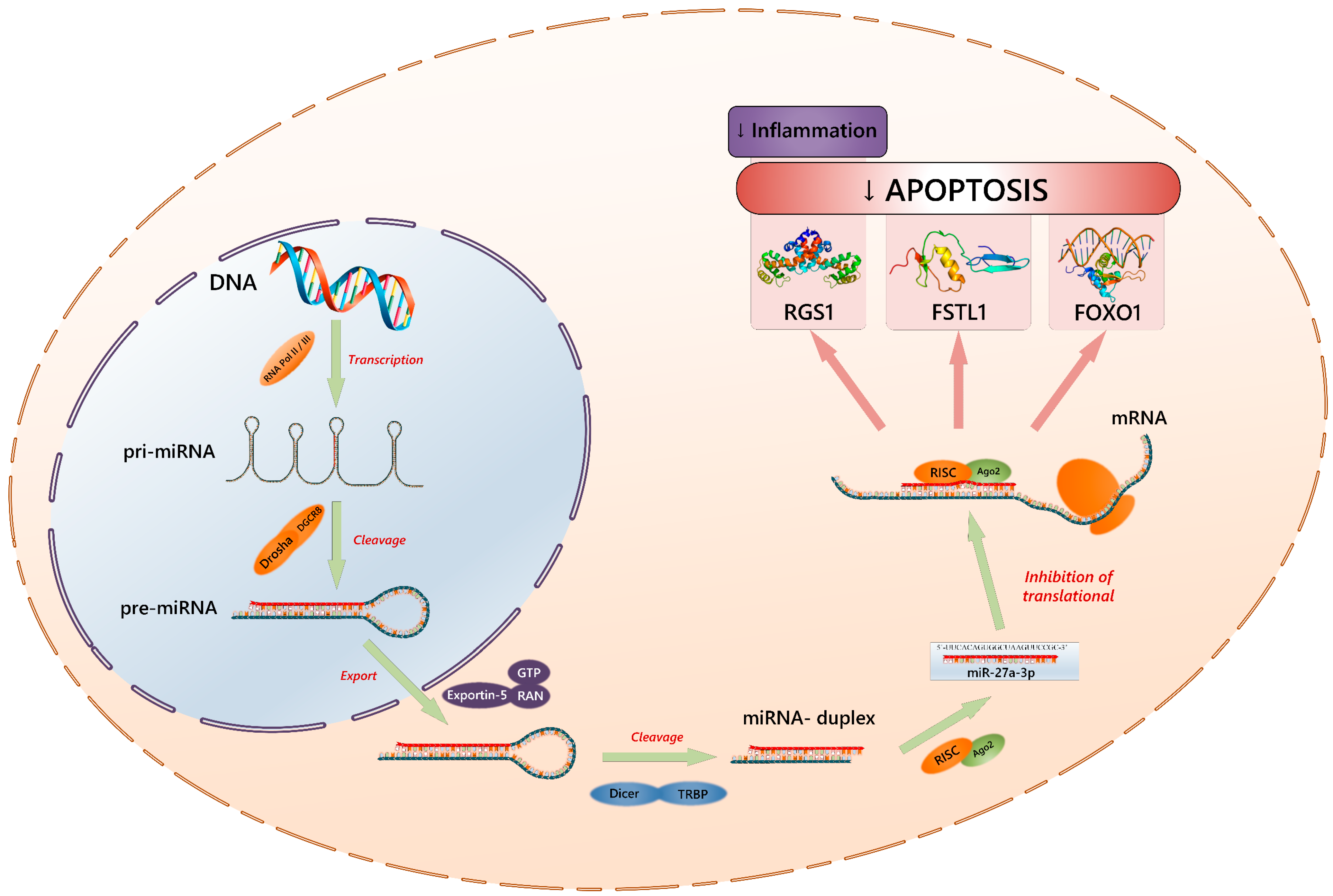
- Li, W.; Zhu, Q.; Xu, X.; Hu, X. MiR-27a-3p suppresses cerebral ischemia-reperfusion injury by targeting FOXO1. Aging 2021, 13, 11727–11737. [Google Scholar] [CrossRef] [PubMed]
- Zhang, Z.; He, J.; Wang, B. Circular RNA circ_HECTD1 regulates cell injury after cerebral infarction by miR-27a-3p/FSTL1 axis. Cell Cycle 2021, 20, 914–926. [Google Scholar] [CrossRef]
- Li, J.; Peng, L.; Bai, W.; Peng, P.; Chen, W.; Yang, W.; Shao, J. Biliverdin Protects Against Cerebral Ischemia/Reperfusion Injury by Regulating the miR-27a-3p/Rgs1 Axis. Neuropsychiatr. Dis Treat. 2021, 17, 1165–1181. [Google Scholar] [CrossRef]
- Gao, X.Z.; Zhang, Z.X.; Han, G.L. MiR-29a-3p Enhances the Viability of Rat Neuronal Cells that Injured by Oxygen-Glucose Deprivation/Reoxygenation Treatment Through Targeting TNFRSF1A and Regulating NF-κB Signaling Pathway. J. Stroke Cerebrovasc. Dis. 2020, 29, 105210. [Google Scholar] [CrossRef] [PubMed]
- Zhang, Z.; Wang, N.; Zhang, Y.; Zhao, J.; Lv, J. Downregulation of microRNA-302b-3p relieves oxygen-glucose deprivation/re-oxygenation induced injury in murine hippocampal neurons through up-regulating Nrf2 signaling by targeting fibroblast growth factor 15/19. Chem. Biol. Interact. 2019, 309, 108705. [Google Scholar] [CrossRef]
- Zhou, X.; Wang, Z.; Xu, B.; Ji, N.; Meng, P.; Gu, L.; Li, Y. Long non-coding RNA NORAD protects against cerebral ischemia/reperfusion injury induced brain damage, cell apoptosis, oxidative stress and inflammation by regulating miR-30a-5p/YWHAG. Bioengineered 2021, 12, 9174–9188. [Google Scholar] [CrossRef] [PubMed]
- Tao, H.; Dong, L.; Shan, X.; Li, L.; Chen, H. MicroRNA-32-3p facilitates cerebral ischemia/reperfusion injury through inhibiting Cab39/AMPK. Int. Immunopharmacol. 2023, 121, 110504. [Google Scholar] [CrossRef]
- Yi, S.; Zhang, C.; Li, N.; Fu, Y.; Li, H.; Zhang, J. miR-325-3p Protects Neurons from Oxygen-Glucose Deprivation and Reoxygenation Injury via Inhibition of RIP3. Dev. Neurosci. 2020, 42, 83–93. [Google Scholar] [CrossRef]
- Wang, Y.; Pan, W.; Wang, Y.; Chen, S. MicroRNA-32-5p attenuates cerebral ischemia/reperfusion injuries by modulating the phosphatase and tensin homologous protein. Metab. Brain Dis. 2021, 36, 2495–2504. [Google Scholar] [CrossRef]
- Yan, F.; Wang, P.; Yang, X.; Wang, F. Long non-coding RNA HOXA11-AS regulates ischemic neuronal death by targeting miR-337-3p/YBX1 signaling pathway: Protective effect of dexmedetomidine. Aging 2023, 15, 2797–2811. [Google Scholar] [CrossRef]
- Yu, Z.; Zhu, M.; Shu, D.; Zhang, R.; Xiang, Z.; Jiang, A.; Liu, S.; Zhang, C.; Yuan, Q.; Hu, X. LncRNA PEG11as aggravates cerebral ischemia/reperfusion injury after ischemic stroke through miR-342-5p/PFN1 axis. Life Sci. 2023, 313, 121276. [Google Scholar] [CrossRef] [PubMed]
- Zhu, H.; Zhang, Y.; Zhu, Y. MiR-342-5p protects neurons from cerebral ischemia induced-apoptosis through regulation of Akt/NF-κB pathways by targeting CCAR2. J. Stroke Cerebrovasc. Dis. 2023, 32, 106901. [Google Scholar] [CrossRef] [PubMed]
- Ye, X.; Song, H.; Hu, H.; Zhou, C.; Chen, Q.; Hong, L.; Huang, M.; Zhu, H. MiR-361-3p alleviates cerebral ischemia-reperfusion injury by targeting NACC1 through the PINK1/Parkin pathway. J. Mol. Histol. 2022, 53, 357–367. [Google Scholar] [CrossRef] [PubMed]
- Jia, Y.; Liu, J.; Hu, H.; Duan, Q.; Chen, J.; Li, L. MiR-363-3p attenuates neonatal hypoxic-ischemia encephalopathy by targeting DUSP5. Neurosci. Res. 2021, 171, 103–113. [Google Scholar] [CrossRef]
- Yang, B.; Zang, L.; Cui, J.; Wei, L. Circular RNA TTC3 regulates cerebral ischemia-reperfusion injury and neural stem cells by miR-372-3p/TLR4 axis in cerebral infarction. Stem Cell Res. Ther. 2021, 12, 125. [Google Scholar] [CrossRef]
- Shen, J.; Han, Q.; Li, W.; Chen, X.; Lu, J.; Zheng, J.; Xue, S. miR-383-5p Regulated by the Transcription Factor CTCF Affects Neuronal Impairment in Cerebral Ischemia by Mediating Deacetylase HDAC9 Activity. Mol. Neurobiol. 2022, 59, 6307–6320. [Google Scholar] [CrossRef]
- He, Z.; Zhao, Y.; Zhu, Y.; Wang, W.; Liu, X.; Lu, F. Interfering TUG1 Attenuates Cerebrovascular Endothelial Apoptosis and Inflammatory injury After Cerebral Ischemia/Reperfusion via TUG1/miR-410/FOXO3 ceRNA Axis. Neurotox. Res. 2022, 40, 1–13. [Google Scholar] [CrossRef]
- Xu, J.; Huang, X.; Liu, S.; Chen, D.; Xie, Y.; Zhao, Z. The protective effects of lncRNA ZFAS1/miR-421-3p/MEF2C axis on cerebral ischemia-reperfusion injury. Cell Cycle 2022, 21, 1915–1931. [Google Scholar] [CrossRef]
- Zheng, L.; Tang, X.; Lu, M.; Sun, S.; Xie, S.; Cai, J.; Zan, J. microRNA-421-3p prevents inflammatory response in cerebral ischemia/reperfusion injury through targeting m6A Reader YTHDF1 to inhibit p65 mRNA translation. Int. Immunopharmacol. 2020, 88, 106937. [Google Scholar] [CrossRef]
- Gan, C.; Ouyang, F. Exosomes Released from Bone-Marrow Stem Cells Ameliorate Hippocampal Neuronal Injury Through transferring miR-455-3p. J. Stroke Cerebrovasc. Dis. 2022, 31, 106142. [Google Scholar] [CrossRef]
- Fan, Y.; Wei, L.; Zhang, S.; Song, X.; Yang, J.; He, X.; Zheng, X. LncRNA SNHG15 Knockdown Protects Against OGD/R-Induced Neuron Injury by Downregulating TP53INP1 Expression via Binding to miR-455-3p. Neurochem. Res. 2021, 46, 1019–1030. [Google Scholar] [CrossRef]
- Chen, X.; Zhang, S.; Shi, P.; Su, Y.; Zhang, D.; Li, N. MiR-485-5p Promotes Neuron Survival through Mediating Rac1/Notch2 Signaling Pathway after Cerebral Ischemia/Reperfusion. Curr. Neurovasc. Res. 2020, 17, 259–266. [Google Scholar] [CrossRef] [PubMed]
- Zheng, H.; Zhang, G.; Liu, G.; Wang, L. Up-regulation of lncRNA NEAT1 in cerebral ischemic stroke promotes activation of astrocytes by modulation of miR-488-3p/RAC1. Exp. Brain Res. 2023, 241, 395–406. [Google Scholar] [CrossRef] [PubMed]
- Zhou, L.; Yang, W.; Yao, E.; Li, H.; Wang, J.; Wang, K.; Zhong, X.; Peng, Z.; Huang, X. MicroRNA-488-3p Regulates Neuronal Cell Death in Cerebral Ischemic Stroke Through Vacuolar Protein Sorting 4B (VPS4B). Neuropsychiatr. Dis. Treat. 2021, 17, 41–55. [Google Scholar] [CrossRef] [PubMed]
- Jia, T.; Wang, M.; Yan, W.; Wu, W.; Shen, R. Upregulation of miR-489-3p Attenuates Cerebral Ischemia/Reperfusion Injury by Targeting Histone Deacetylase 2 (HDAC2). Neuroscience 2022, 484, 16–25. [Google Scholar] [CrossRef]
- Song, L.; Mu, L.; Wang, H. MicroRNA-489-3p aggravates neuronal apoptosis and oxidative stress after cerebral ischemia-reperfusion injury. Bioengineered 2022, 13, 14047–14056. [Google Scholar] [CrossRef]
- Sun, L.; Ji, D.; Zhi, F.; Fang, Y.; Zhu, Z.; Ni, T.; Zhu, Q.; Bao, J. MiR-494-3p Upregulation Exacerbates Cerebral Ischemia Injury by Targeting Bhlhe40. Yonsei Med. J. 2022, 63, 389–398. [Google Scholar] [CrossRef]
- Yin, K.J.; Deng, Z.; Huang, H.; Hamblin, M.; Xie, C.; Zhang, J.; Chen, Y.E. miR-497 regulates neuronal death in mouse brain after transient focal cerebral ischemia. Neurobiol. Dis. 2010, 38, 17–26. [Google Scholar] [CrossRef]
- Shan, W.; Ge, H.; Chen, B.; Huang, L.; Zhu, S.; Zhou, Y. Upregulation of miR-499a-5p Decreases Cerebral Ischemia/Reperfusion Injury by Targeting PDCD4. Cell. Mol. Neurobiol. 2022, 42, 2157–2170. [Google Scholar] [CrossRef]
- Wang, H.; Yang, H.; Chang, M.; Sun, F.; Qi, H.; Li, X. Long non-coding RNA TTTY15 sponges miR-520a-3p to exacerbate neural apoptosis induced by cerebral ischemia/reperfusion via targeting IRF9 both in vivo and in vitro. Biomed. J. 2023, 46, 100530. [Google Scholar] [CrossRef]
- Shi, Y.; Yi, Z.; Zhao, P.; Xu, Y.; Pan, P. MicroRNA-532-5p protects against cerebral ischemia-reperfusion injury by directly targeting CXCL1. Aging 2021, 13, 11528–11541. [Google Scholar] [CrossRef] [PubMed]
- Wei, L.; Peng, Y.; Yang, X.J.; Zhou, P. Knockdown of long non-coding RNA RMRP protects cerebral ischemia-reperfusion injury via the microRNA-613/ATG3 axis and the JAK2/STAT3 pathway. Kaohsiung J. Med. Sci. 2021, 37, 468–478. [Google Scholar] [CrossRef] [PubMed]
- Gao, J.D.; Li, R.J.; Ma, P.L.; Yu, L.L.; Li, J.T.; Tian, H.T. Knockdown of lncRNA HCP5 protects against cerebral ischemia/reperfusion injury by regulating miR-652-3p. J. Biol. Regul. Homeost. Agents 2020, 34, 893–900. [Google Scholar]
- Zhang, X.; Feng, Y.; Li, J.; Zheng, L.; Shao, Y.; Zhu, F.; Sun, X. MicroRNA-665-3p attenuates oxygen-glucose deprivation-evoked microglial cell apoptosis and inflammatory response by inhibiting NF-κB signaling via targeting TRIM8. Int. Immunopharmacol. 2020, 85, 106650. [Google Scholar] [CrossRef]
- Kong, L.Y.; Li, Y.; Rao, D.Y.; Wu, B.; Sang, C.P.; Lai, P.; Ye, J.S.; Zhang, Z.X.; Du, Z.M.; Yu, J.J.; et al. miR-666-3p Mediates the Protective Effects of Mesenchymal Stem Cell-derived Exosomes Against Oxygen-glucose Deprivation and Reoxygenation- induced Cell Injury in Brain Microvascular Endothelial Cells via Mitogen-activated Protein Kinase Pathway. Curr. Neurovasc. Res. 2021, 18, 20–77. [Google Scholar] [CrossRef]
- Xu, H.; Nie, B.; Liu, L.; Zhang, C.; Zhang, Z.; Xu, M.; Mei, Y. Curcumin Prevents Brain Damage and Cognitive Dysfunction During Ischemic-reperfusion Through the Regulation of miR-7-5p. Curr. Neurovasc. Res. 2019, 16, 441–454. [Google Scholar] [CrossRef]
- Hu, X.; Ma, F.; Cheng, Z.; Zeng, S.; Shen, R.; Li, X.; Hu, J.; Jin, Z.; Cheng, J. LncRNA PEG11as silencing sponges miR-874-3p to alleviate cerebral ischemia stroke via regulating autophagy in vivo and in vitro. Aging 2022, 14, 5177–5194. [Google Scholar] [CrossRef] [PubMed]
- Shen, J.; Li, G.; Zhu, Y.; Xu, Q.; Zhou, H.; Xu, K.; Huang, K.; Zhan, R.; Pan, J. Foxo1-induced miR-92b down-regulation promotes blood-brain barrier damage after ischaemic stroke by targeting NOX4. J. Cell. Mol. Med. 2021, 25, 5269–5282. [Google Scholar] [CrossRef]
- Zhang, G.; Guo, J.; Zeng, J.; Zhang, X.; Chen, R.; Wang, G.; Liang, W. LncRNA SNHG14 is beneficial to oxygen glucose deprivation/reoxygenation-induced neuro-2a cell injury via mir-98-5p sequestration-caused BCL2L13 upregulation. Metab. Brain Dis. 2022, 37, 2005–2016. [Google Scholar] [CrossRef]
- Qi, D.; Wang, W.; Zhang, Y.; Zhang, T. MiR-99b regulates cerebral ischemia neuronal injury through targeting IGF1R. Panminerva Medica 2023, 65, 30–36. [Google Scholar] [CrossRef]
- Huang, Y.; Tang, J.; Li, X.; Long, X.; Huang, Y.; Zhang, X. miR-92b-3p Exerts Neuroprotective Effects on Ischemia/Reperfusion-Induced Cerebral Injury via Targeting NOX4 in a Rat Model. Oxidative Med. Cell. Longev. 2022, 2022, 3494262. [Google Scholar] [CrossRef] [PubMed]
- McConnell, H.L.; Mishra, A. Cells of the Blood-Brain Barrier: An Overview of the Neurovascular Unit in Health and Disease. Methods Mol. Biol. 2022, 2492, 3–24. [Google Scholar]
- Ago, T. The neurovascular unit in health and ischemic stroke. Nihon Rinsho. 2016, 74, 583–588. (In Japanese) [Google Scholar] [PubMed]
- Pivoriūnas, A.; Verkhratsky, A. Astrocyte-Endotheliocyte Axis in the Regulation of the Blood-Brain Barrier. Neurochem. Res. 2021, 46, 2538–2550. [Google Scholar] [CrossRef] [PubMed]
- Guo, X.; Liu, R.; Jia, M.; Wang, Q.; Wu, J. Ischemia Reperfusion Injury Induced Blood Brain Barrier Dysfunction and the Involved Molecular Mechanism. Neurochem. Res. 2023, 48, 2320–2334. [Google Scholar] [CrossRef] [PubMed]
- Cai, Z.; Qiao, P.F.; Wan, C.Q.; Cai, M.; Zhou, N.K.; Li, Q. Role of Blood-Brain Barrier in Alzheimer’s Disease. J. Alzheimer’s Dis. 2018, 63, 1223–1234. [Google Scholar] [CrossRef]
- Sweeney, M.D.; Sagare, A.P.; Zlokovic, B.V. Blood-brain barrier breakdown in Alzheimer disease and other neurodegenerative disorders. Nat. Rev. Neurol. 2018, 14, 133–150. [Google Scholar] [CrossRef]
- Sweeney, M.D.; Zhao, Z.; Montagne, A.; Nelson, A.R.; Zlokovic, B.V. Blood-Brain Barrier: From Physiology to Disease and Back. Physiol. Rev. 2019, 99, 21–78. [Google Scholar] [CrossRef]
- Andjelkovic, A.V.; Stamatovic, S.M.; Phillips, C.M.; Martinez-Revollar, G.; Keep, R.F. Modeling blood-brain barrier pathology in cerebrovascular disease in vitro: Current and future paradigms. Fluids Barriers CNS 2020, 17, 44. [Google Scholar] [CrossRef]
- Chekhonin, V.P.; Lebedev, S.V.; Dmitrieva, T.B.; Blinov, D.V.; Gurina, O.I.; Semenova, A.V.; Volodin, N.N. Enzyme immunoassay of NSE and GFAP as the criterion of dynamic evaluation of the rat blood-brain barrier in perinatal hypoxic ischemic injury of the CNS. Bull. Exp. Biol. Med. 2003, 136, 261–265. [Google Scholar] [CrossRef]
- Chalbot, S.; Zetterberg, H.; Blennow, K.; Fladby, T.; Andreasen, N.; Grundke-Iqbal, I.; Iqbal, K. Blood-cerebrospinal fluid barrier permeability in Alzheimer’s disease. J. Alzheimer’s Dis. 2011, 25, 505–515. [Google Scholar] [CrossRef] [PubMed]
- Helgudottir, S.S.; Routhe, L.J.; Burkhart, A.; Jonsson, K.; Pedersen, I.S.; Lichota, J.; Moos, T. Epigenetic Regulation of Ferroportin in Primary Cultures of the Rat Blood-Brain Barrier. Mol. Neurobiol. 2020, 57, 3526–3539. [Google Scholar] [CrossRef] [PubMed]
- Sun, P.; Hamblin, M.H.; Yin, K.J. Non-coding RNAs in the regulation of blood-brain barrier functions in central nervous system disorders. Fluids Barriers CNS 2022, 19, 27. [Google Scholar] [CrossRef] [PubMed]
- Sookoian, S.; Gianotti, T.F.; Burgueño, A.L.; Pirola, C.J. Fetal metabolic programming and epigenetic modifications: A systems biology approach. Pediatr. Res. 2013, 73, 531–542. [Google Scholar] [CrossRef] [PubMed]
- Yang, K.; Zeng, L.; Ge, A.; Wang, S.; Zeng, J.; Yuan, X.; Mei, Z.; Wang, G.; Ge, J. A systematic review of the research progress of non-coding RNA in neuroinflammation and immune regulation in cerebral infarction/ischemia-reperfusion injury. Front. Immunol. 2022, 13, 930171. [Google Scholar] [CrossRef] [PubMed]
- Meng, Q.; Yang, G.; Yang, Y.; Ding, F.; Hu, F. Protective effects of histone deacetylase inhibition by Scriptaid on brain injury in neonatal rat models of cerebral ischemia and hypoxia. Int. J. Clin. Exp. Pathol. 2020, 13, 179–191. [Google Scholar]
- Jambhekar, A.; Dhall, A.; Shi, Y. Roles and regulation of histone methylation in animal development. Nat. Rev. Mol. Cell Biol. 2019, 20, 625–641, Erratum in Nat. Rev. Mol. Cell Biol. 2020, 21, 59. [Google Scholar] [CrossRef]
- Churilova, A.V.; Gluschenko, T.S.; Rybnikova, E.A.; Samoilov, M.O. The Effect of Histone Deacetylase Inhibitor on the Expression Level of Glucococrticoid Receptor in Rat Forebrain under Hypoxia. Cell Tissue Biol. 2019, 13, 79–84. [Google Scholar] [CrossRef]
- Deniz, B.F.; Confortim, H.D.; Miguel, P.M.; Bronauth, L.; Fernandes, I.R.; Muotri, A.R.; Pereira, L.O. High gestational folic acid supplementation prevents hypoxia-ischemia-induced caspase-3 augmenting without changing synapsin and H3 methylation levels in the rat hippocampus. Int. J. Dev. Neurosci. 2021, 81, 510–519. [Google Scholar] [CrossRef]
- Paz, A.A.; González-Candia, A. Potential pharmacological target of tight junctions to improve the BBB permeability in neonatal Hypoxic-Ischemic encephalopathy Diseases. Biochem. Pharmacol. 2023, 207, 115356. [Google Scholar] [CrossRef]
- Yang, C.; Hawkins, K.E.; Doré, S.; Candelario-Jalil, E. Neuroinflammatory mechanisms of blood-brain barrier damage in ischemic stroke. Am. J. Physiol. Cell Physiol. 2019, 316, C135–C153. [Google Scholar] [CrossRef] [PubMed]
- Shen, G.; Ma, Q. MicroRNAs in the Blood-Brain Barrier in Hypoxic-Ischemic Brain Injury. Curr. Neuropharmacol. 2020, 18, 1180–1186. [Google Scholar] [CrossRef] [PubMed]
- Fan, F.; Yang, J.; Xu, Y.; Guan, S. MiR-539 Targets MMP-9 to Regulate the Permeability of Blood-Brain Barrier in Ischemia/Reperfusion Injury of Brain. Neurochem. Res. 2018, 43, 2260–2267. [Google Scholar] [CrossRef]
- Yao, X.; Wang, Y.; Zhang, D. microRNA-21 Confers Neuroprotection Against Cerebral Ischemia-Reperfusion Injury and Alleviates Blood-Brain Barrier Disruption in Rats via the MAPK Signaling Pathway. J. Mol. Neurosci. 2018, 65, 43–53. [Google Scholar] [CrossRef]
- Zuo, X.; Lu, J.; Manaenko, A.; Qi, X.; Tang, J.; Mei, Q.; Xia, Y.; Hu, Q. MicroRNA-132 attenuates cerebral injury by protecting blood-brain-barrier in MCAO mice. Exp. Neurol. 2019, 316, 12–19. [Google Scholar] [CrossRef] [PubMed]
- Deng, L.; Zhang, J.; Chen, S.; Wu, Y.; Fan, X.; Zuo, T.; Hu, Q.; Jiang, L.; Yang, S.; Dong, Z. miR-671-5p Upregulation Attenuates Blood-Brain Barrier Disruption in the Ischemia Stroke Model Via the NF-кB/MMP-9 Signaling Pathway. Mol. Neurobiol. 2023, 60, 3824–3838. [Google Scholar] [CrossRef]
- Bai, Y.; Zhang, Y.; Han, B.; Yang, L.; Chen, X.; Huang, R.; Wu, F.; Chao, J.; Liu, P.; Hu, G.; et al. Circular RNA DLGAP4 Ameliorates Ischemic Stroke Outcomes by Targeting miR-143 to Regulate Endothelial-Mesenchymal Transition Associated with Blood-Brain Barrier Integrity. J. Neurosci. 2018, 38, 32–50. [Google Scholar] [CrossRef]
- Huang, L.; Ma, Q.; Li, Y.; Li, B.; Zhang, L. Inhibition of microRNA-210 suppresses pro-inflammatory response and reduces acute brain injury of ischemic stroke in mice. Exp. Neurol. 2018, 300, 41–50. [Google Scholar] [CrossRef]
- Ponnusamy, V.; Yip, P.K. The role of microRNAs in newborn brain development and hypoxic ischaemic encephalopathy. Neuropharmacology 2019, 149, 55–65. [Google Scholar] [CrossRef]
- Ma, Q.; Dasgupta, C.; Li, Y.; Huang, L.; Zhang, L. MicroRNA-210 Suppresses Junction Proteins and Disrupts Blood-Brain Barrier Integrity in Neonatal Rat Hypoxic-Ischemic Brain Injury. Int. J. Mol. Sci. 2017, 18, E1356. [Google Scholar] [CrossRef]
- Wang, Y.; Wang, M.D.; Xia, Y.P.; Gao, Y.; Zhu, Y.Y.; Chen, S.C.; Mao, L.; He, Q.W.; Yue, Z.Y.; Hu, B. MicroRNA-130a regulates cerebral ischemia-induced blood-brain barrier permeability by targeting Homeobox A5. FASEB J. 2018, 32, 935–944. [Google Scholar] [CrossRef] [PubMed]
- Pan, J.; Qu, M.; Li, Y.; Wang, L.; Zhang, L.; Wang, Y.; Tang, Y.; Tian, H.L.; Zhang, Z.; Yang, G.Y. MicroRNA-126-3p/-5p Overexpression Attenuates Blood-Brain Barrier Disruption in a Mouse Model of Middle Cerebral Artery Occlusion. Stroke 2020, 51, 619–627. [Google Scholar] [CrossRef] [PubMed]
- Bernstein, D.L.; Zuluaga-Ramirez, V.; Gajghate, S.; Reichenbach, N.L.; Polyak, B.; Persidsky, Y.; Rom, S. miR-98 reduces endothelial dysfunction by protecting blood-brain barrier (BBB) and improves neurological outcomes in mouse ischemia/reperfusion stroke model. J. Cereb. Blood Flow Metab. 2020, 40, 1953–1965. [Google Scholar] [CrossRef] [PubMed]
- Ebrahimi, V.; Rastegar-Moghaddam, S.H.; Mohammadipour, A. Therapeutic Potentials of MicroRNA-126 in Cerebral Ischemia. Mol. Neurobiol. 2023, 60, 2062–2069. [Google Scholar] [CrossRef] [PubMed]
- Li, F.; Zhou, F.; Yang, B. MicroRNA152-3p Protects Against Ischemia/Reperfusion-Induced Bbb Destruction Possibly Targeting the MAP3K2/JNK/c-Jun Pathway. Neurochem. Res. 2023, 48, 1293–1304. [Google Scholar] [CrossRef]
- Hu, H.; Hone, E.A.; Provencher, E.A.P.; Sprowls, S.A.; Farooqi, I.; Corbin, D.R.; Sarkar, S.N.; Hollander, J.M.; Lockman, P.R.; Simpkins, J.W.; et al. MiR-34a Interacts with Cytochrome c and Shapes Stroke Outcomes. Sci. Rep. 2020, 10, 3233. [Google Scholar] [CrossRef]
- Yin, K.J.; Deng, Z.; Hamblin, M.; Xiang, Y.; Huang, H.; Zhang, J.; Jiang, X.; Wang, Y.; Chen, Y.E. Peroxisome proliferator-activated receptor delta regulation of miR-15a in ischemia-induced cerebral vascular endothelial injury. J. Neurosci. 2010, 30, 6398–6408. [Google Scholar] [CrossRef]
- Wan, Y.; Jin, H.J.; Zhu, Y.Y.; Fang, Z.; Mao, L.; He, Q.; Xia, Y.P.; Li, M.; Li, Y.; Chen, X.; et al. MicroRNA-149-5p regulates blood-brain barrier permeability after transient middle cerebral artery occlusion in rats by targeting S1PR2 of pericytes. FASEB J. 2018, 32, 3133–3148. [Google Scholar] [CrossRef]
- Perales, G.; Westenskow, M.; Gutierrez, R.; Caldwell, K.K.; Allan, A.M.; Gardiner, A.S. MicroRNA-150-5p is upregulated in the brain microvasculature during prenatal alcohol exposure and inhibits the angiogenic factor Vezf1. Alcohol. Clin. Exp. Res. 2022, 46, 1953–1966. [Google Scholar] [CrossRef]


| microRNA | Target Protein/Signaling Pathway | Direction of Gene Expression | Ischemia/OGD Effect | Ref. | Biological Effect of RNA Interference |
|---|---|---|---|---|---|
| miR-101-3p | ↓ HDAC9 | ↑ | ↓ apoptosis ↑ morphology of neurons | [114] | Positive |
| miR-101a-3p | ↑ Dusp1 | ↓ | ↓ apoptosis | [115] | Negative |
| miR-10b-3p | ↓ PDCD5 | ↑ | ↓ apoptosis | [116] | Positive |
| ↓ KLF5 | ↑ | ↓ apoptosis ↓ swelling ↓ inflammation | [117] | Positive | |
| miR-122-5p | ↓ SEMA3A | ↑ | ↓ apoptosis ↓ oxidative stress | [118] | Positive |
| miR-124-3p | ↓ Nrep/Rnf38 | ↑ | ↓ apoptosis | [119] | Positive |
| ↓ CTDSP1/AKT | ↑ | ↓ apoptosis ↓ axon damage | [120] | Positive | |
| miR-1247-3p | ↓ Caspase-2 | ↑ | ↓ apoptosis | [121] | Positive |
| miR-128-3p * | ↑ FOXO/Relaxin | ↓ | ↓ apoptosis | [122] | Negative |
| ↓ ACVR1/BMP | ↑ | ↓ demyelination ↑ differentiation | [123] | Positive | |
| miR-132-3p | ↓ ATG12 ↑ p-PI3K/p-AKT/mTOR | ↑ | ↓ oxidative stress ↓ autophagy | [124] | Positive |
| miR-133a-3p | ↓ DAPK1 ↑ p-Akt и p-mTOR | ↑ | ↓ apoptosis ↓ autophagy | [125] | Positive |
| miR-133b | ↓ TRAF3/NF-κB | ↑ | ↓ apoptosis | [126] | Positive |
| miR-134 | ↓ HSPA12B | ↓ | ↓ apoptosis | [127] | Negative |
| miR-135a-5p | ↓ NR3C2 | ↑ | ↓ apoptosis ↓ oxidative stress ↓ inflammation | [128] | Positive |
| miR-140-3p * | ↑ HIF-1α | ↑ | ↓ apoptosis ↓ oxidative stress ↓ inflammation | [129] | Positive |
| ↓ Tyro3/PI3K/Akt ↓ Bax/Caspase-3 ↑ Bcl-2 | ↑ | ↑ apoptosis ↑ oxidative stress | [130] | Negative | |
| miR-141-3p | ↑ SIRT1/ZO-1/Occludin/Claudin-5/CD31 ↓ p-NF-κB/IL-1β/TNF-α/GFAP | ↓ | ↓ inflammation | [107] | Negative |
| ↑ PBX1/PROK2 | ↓ | ↓ apoptosis | [131] | Negative | |
| miR-142-3p * | ↑ SIRT1/SOD/Catalase ↓ TNF-α/IL-6/IL-1β/ROS/MDA | ↓ | ↓ apoptosis | [132] | Negative |
| ↓ FBXO3 | ↑ | ↓ apoptosis ↓ inflammation | [133] | Positive | |
| miR-142-5p | ↑ Nrf2/ARE | ↓ | ↓ oxidative stress | [134] | Negative |
| miR-143-3p | ↑ FSTL1 ↓ Bax/Caspase-3 ↑ Bcl-2 | ↓ | ↓ apoptosis | [135] | Negative |
| miR-144-3p | ↑ Brg1/Nrf2/ARE | ↓ | ↓ apoptosis ↓ oxidative stress | [136] | Negative |
| miR-145 | ↓ FOXO1 | ↑ | ↓ apoptosis ↓ oxidative stress ↓ inflammation | [137] | Positive |
| miR-148b-3p | ↑ Sestrin2/Nrf2 | ↓ | ↓ apoptosis ↓ oxidative stress | [138] | Negative |
| miR-149-5p | ↓ Notch2 | ↑ | ↓ apoptosis ↓ inflammation | [139] | Positive |
| miR-152-3p | ↓ PSD-93 ↑ Nrf2/ARE | ↑ | ↓ apoptosis ↓ oxidative stress | [140] | Positive |
| miR-153-3p | ↓ SRC/MAPK | ↑ | ↓ apoptosis ↓ oxidative stress ↓ inflammation | [141] | Positive |
| ↓ FOXO3 | ↑ | ↓ apoptosis | [142] | Positive | |
| miR-153-5p | ↓ TLR4/p65/IkBa | ↑ | ↓ apoptosis | [143] | Positive |
| miR-155 | ↑ MafB ↓ IL-1β/IL-6/TNF-α ↓ iNOS/COX-2 | ↓ | ↓ apoptosis ↓ inflammation | [113] | Negative |
| miR-155-5p | ↑ DUSP14/TXNIP/NLRP3 | ↓ | ↓ inflammation ↓ pyroptosis | [112] | Negative |
| ↑ DUSP14/NF-kB/MAPKs | ↓ | ↓ apoptosis | [111] | Negative | |
| miR-181a | ↑ PTEN | ↓ | ↓ apoptosis ↓ oxidative stress | [144] | Negative |
| miR-181a-5p | ↑ En2/Wnt/β-catenin | ↓ | ↓ apoptosis | [145] | Negative |
| miR-181c-3p | ↓ CXCL1 | ↑ | ↓ inflammation | [146] | Positive |
| miR-181d | ↑ DOCK4 | ↓ | ↓ apoptosis ↓ inflammation | [147] | Negative |
| miR-182 | ↑ mTOR/FOXO1/Bcl-2/Bax | ↓ | ↓ apoptosis | [148] | Negative |
| ↑ cortactin | ↓ | ↓ mitochondrial dysfunction ↓ inflammation | [149] | Negative | |
| miR-182-5p | ↓ TLR4 | ↑ | ↓ inflammation | [101] | Positive |
| ↓ Rac1 | ↑ | ↓ inflammation | [150] | Positive | |
| miR-186-5p | ↑ MDM4 | ↓ | ↓ apoptosis ↓ oxidative stress | [151] | Negative |
| miR-187-3p | ↓ GRP78/Seipin | ↑ | ↑ apoptosis ↑ endoplasmic reticulum stress | [152] | Negative |
| miR-193b | ↓ ATG7 | ↑ | ↓ autophagy ↓ ferroptosis | [153] | Positive |
| miR-193b-3p | ↓ 5-lipoxigenase | ↑ | ↓ inflammation | [102] | Positive |
| miR-199a-5p | ↑ Brg1/Nrf2/HO-1 | ↓ | ↓ apoptosis ↓ oxidative stress | [154] | Negative |
| miR-199b | ↓ AQP4 | ↑ | ↓ oxidative stress ↓ inflammation | [155] | Positive |
| miR-19a | ↑ ADIPOR2 | ↓ | ↓ apoptosis ↑ glucose metabolism | [156] | Negative |
| miR-19a-3p * | ↑ FOXO3/SPHK1/NF-kB p65 ↓ SIRT1 | ↑ | ↑ inflammation | [157] | Negative |
| ↑ IGFBP3 | ↓ | ↓ apoptosis ↓ inflammation | [106] | Negative | |
| ↓ PTEN/PI3K/AKT | ↑ | ↓ apoptosis ↓ oxidative stress | [158] | Positive | |
| miR-19b-3p | ↑ FOXO3/SPHK1/NF-kB p65 ↓ SIRT1 | ↑ | ↑ inflammation | [157] | Negative |
| miR-200a-3p | ↑ SIRT1/ZO-1/Occludin/Claudin-5/CD31 ↓ p-NF-κB/IL-1β/TNF-α/GFAP | ↓ | ↓ inflammation | [107] | Negative |
| miR-200b-3p | ↑ β-TrCP | ↓ | ↓ apoptosis | [159] | Negative |
| miR-202-5p | ↓ eIF4E/ ↑ Akt/GSK-3β | ↑ | ↓ apoptosis ↓ autophagy | [160] | Positive |
| miR-203a-3p | ↓ SRC/MAPK | ↑ | ↓ apoptosis ↓ oxidative stress ↓ inflammation | [141] | Positive |
| miR-206 | ↑ USP22/Sirt1 | ↓ | ↓ apoptosis ↓ inflammation | [161] | Negative |
| miR-20a-3p | - | ↑ | ↓ cognitive dysfunction | [162] | Positive |
| ↓ MMP, IL-17A | ↑ | ↓ mitochondrial dysfunction ↓ inflammation | [163] | Positive | |
| miR-21-3p | ↑ CAMKK2/AMPK/Nrf-2 | ↓ | ↓ oxidative stress ↓ inflammation | [164] | Negative |
| miR-211-5p | ↓ COX2 ↓ PGD2/PGE2/TNF-α/IL-1β | ↑ | ↓ apoptosis ↓ inflammation | [165] | Positive |
| miR-216a | ↓ JAK2/STAT3 ↓ iNOS и MMP-9/TNF-α и IL-1β | ↑ | ↓ apoptosis ↓ inflammation | [166] | Positive |
| miR-22 | ↑ Wnt/β-catenin and PKC/ERK | ↑ | ↓ apoptosis | [167] | Positive |
| miR-223-3p * | ↓ NOTCH2 | ↑ | ↑ angiogenesis | [168] | Positive |
| ↓ CysLT 2 R | ↑ | ↓ inflammation | [169] | Positive | |
| ↑ IGF1R | ↓ | ↓ apoptosis ↑ glucose metabolism | [170] | Negative | |
| miR-22-3p | ↓ KDM6B/BMP2/BMF | ↑ | ↓ apoptosis | [171] | Positive |
| ↓ IL-1β, IL-18/Caspase-1 | ↑ | ↓ pyroptosis ↓ inflammation | [172] | Positive | |
| miR-224-3p | ↓ FIP200 | ↑ | ↓ apoptosis | [173] | Positive |
| miR-23a-3p | ↓ CXCL12 | ↑ | ↓ apoptosis | [174] | Positive |
| miR-24-3p | ↑ NRP1/NF-κB p65 | ↓ | ↑ apoptosis ↑ inflammation | [175] | Positive |
| ↓ BOK | ↑ | ↓ apoptosis ↓ oxidative stress | [176] | Positive | |
| miR-25-3p | ↓ TRAF3 | ↑ | ↓ apoptosis ↓ inflammation | [177] | Positive |
| miR-26a-5p | ↓ DAPK1 | ↑ | ↓ apoptosis | [178] | Positive |
| miR-26b | ↑ ULK2 | ↓ | ↑ autophagy | [179] | Positive |
| miR-26b-5p | ↓ KLF10/N-myc/PTEN | ↑ | ↓ apoptosis ↓ inflammation | [180] | Positive |
| miR-27a-3p | ↓ FOXO1/p27 Kip1 | ↑ | ↓ apoptosis | [181] | Positive |
| ↓ FSTL1 | ↑ | ↓ apoptosis | [182] | Positive | |
| ↓ Rgs1 | ↑ | ↓ apoptosis ↓ inflammation | [183] | Positive | |
| miR-29a-3p | ↓ NF-κB | ↑ | ↓ apoptosis | [184] | Positive |
| miR-302b-3p | ↑ FGF15/Nrf2/ARE | ↓ | ↓ apoptosis ↓ oxidative stress | [185] | Negative |
| miR-30a-5p | ↑ YWHAG | ↓ | ↓ apoptosis ↓ oxidative stress ↓ inflammation | [186] | Negative |
| miR-30c | ↓ SOX9/MAPK | ↑ | ↓ apoptosis | [103] | Positive |
| miR-30c-5p | ↑ Rock2/MAPK | ↓ | ↓ apoptosis | [104] | Negative |
| miR-32-3p | ↓ Cab39/AMPK | ↑ | ↑ apoptosis ↑ oxidative stress | [187] | Negative |
| miR-325-3p | ↓ RIP3 | ↑ | ↓ apoptosis | [188] | Positive |
| miR-32-5p | ↓ PTEN/PI3K/AKT | ↑ | ↓ cell necrosis | [189] | Positive |
| miR-337-3p | ↑ YBX1 | ↓ | ↓ apoptosis | [190] | Negative |
| miR-342-5p | ↑ PFN1 | ↓ | ↑ apoptosis | [191] | Positive |
| CCAR2/Akt/NF-κB | ↑ | ↓ apoptosis | [192] | Positive | |
| miR-361-3p | ↑ NACC1/PINK1/Parkin | ↑ | ↓ apoptosis ↓ oxidative stress | [193] | Positive |
| miR-363-3p | ↓ DUSP5 | ↑ | ↓ cell necrosis | [194] | Positive |
| miR-370 | ↓ SIRT6/Nrf2/ARE | ↑ | ↓ apoptosis ↓ oxidative stress ↓ inflammation | [108] | Positive |
| miR-370-3p | ↓ MAPK1 | ↑ | ↑ BBB permeability | [109] | Negative |
| miR-372-3p | ↓ TLR4 | ↑ | ↓ apoptosis | [195] | Positive |
| miR-383-5p | ↓ HDAC9 | ↑ | ↓ apoptosis ↓ endoplasmic reticulum stress | [196] | Positive |
| miR-410 | ↓ FOXO3 | ↑ | ↓ apoptosis ↓ oxidative stress | [197] | Positive |
| miR-421-3p * | ↑ MEF2C | ↓ | ↓ apoptosis | [198] | Negative |
| ↓ YTHDF1/NF-κB p65 | ↑ | ↓ inflammation | [199] | Positive | |
| miR-449a | ↓ AREG/EGFR/PI3K/Akt | ↑ | ↓ apoptosis | [105] | Positive |
| miR-455-3p | ↓ PDCD7 | ↑ | ↓ apoptosis | [200] | Positive |
| ↓ TP53INP1 | ↑ | ↓ apoptosis ↓ oxidative stress ↓ inflammation | [201] | Positive | |
| miR-485 | ↑ AIM2 | ↓ | ↑ pyroptosis | [110] | Positive |
| miR-485-5p | ↓ Rac1/Notch2 | ↑ | ↓ apoptosis ↓ inflammation | [202] | Positive |
| miR-488-3p | ↓ RAC1 | ↑ | ↓ apoptosis ↓ inflammation | [203] | Positive |
| ↓ VPS4B | ↑ | ↓ cell necrosis | [204] | Positive | |
| miR-489-3p * | ↓ HDAC2 | ↑ | ↓ apoptosis | [205] | Positive |
| ↓ SIRT1 | ↑ | ↑ apoptosis ↑ oxidative stress | [206] | Negative | |
| miR-494-3p | ↓ Bhlhe40 | ↑ | ↑ apoptosis ↑ oxidative stress | [207] | Negative |
| miR-496 | ↓ BCL2L14 | ↑ | ↓ apoptosis | [98] | Positive |
| miR-497 | ↑ bcl-2/bcl-w | ↓ | ↓ apoptosis | [208] | Negative |
| miR-499a-5p | ↓ PDCD4 | ↑ | ↓ apoptosis | [209] | Positive |
| miR-520a-3p | ↓ IRF9 | ↑ | ↓ apoptosis | [210] | Positive |
| miR-532-5p | ↓ CXCL1/CXCR2/NF-κB | ↑ | ↓ apoptosis ↓ oxidative stress ↓ inflammation | [211] | Positive |
| miR-613 | ↓ ATG3 | ↑ | ↓ apoptosis | [212] | Positive |
| miR-652-3p | ↑ Bcl-2 ↓ Bax | ↑ | ↓ apoptosis | [213] | Positive |
| miR-665-3p | ↓ TRIM8/NF-κB | ↑ | ↓ apoptosis ↓ inflammation | [214] | Positive |
| miR-666-3p | ↓ MAPK1 | ↑ | ↓ apoptosis | [215] | Positive |
| miR-7-5p | ↑ RelA p65 | ↓ | ↓ apoptosis ↓ oxidative stress ↓ inflammation | [216] | Negative |
| miR-874 | ↓ BMF/BCL2L13 | ↑ | ↓ apoptosis | [99] | Positive |
| miR-874-3p | ↓ ATG16L1 | ↑ | ↓ apoptosis | [217] | Positive |
| miR-92b | ↑ NOX4 | ↓ | ↑ BBB damage | [218] | Positive |
| miR-92b-3p | ↓ TRAF3 | ↑ | ↓ apoptosis ↓ mitochondrial dysfunction ↓ inflammation | [100] | Positive |
| ↓ NOX4 | ↑ | ↓ apoptosis ↓ oxidative stress ↓ inflammation | [100] | Positive | |
| miR-9-5p | ↑ FOXO/Relaxin | ↓ | ↓ apoptosis | [122] | Negative |
| miR-98-5p | ↓ BCL2L13 | ↑ | ↓ apoptosis ↓ oxidative stress ↓ inflammation ↓ endoplasmic reticulum stress | [219] | Positive |
| miR-99b | ↓ IGF1R | ↑ | ↓ apoptosis | [220] | Positive |
Disclaimer/Publisher’s Note: The statements, opinions and data contained in all publications are solely those of the individual author(s) and contributor(s) and not of MDPI and/or the editor(s). MDPI and/or the editor(s) disclaim responsibility for any injury to people or property resulting from any ideas, methods, instructions or products referred to in the content. |
© 2023 by the authors. Licensee MDPI, Basel, Switzerland. This article is an open access article distributed under the terms and conditions of the Creative Commons Attribution (CC BY) license (https://creativecommons.org/licenses/by/4.0/).
Share and Cite
Tregub, P.P.; Ibrahimli, I.; Averchuk, A.S.; Salmina, A.B.; Litvitskiy, P.F.; Manasova, Z.S.; Popova, I.A. The Role of microRNAs in Epigenetic Regulation of Signaling Pathways in Neurological Pathologies. Int. J. Mol. Sci. 2023, 24, 12899. https://doi.org/10.3390/ijms241612899
Tregub PP, Ibrahimli I, Averchuk AS, Salmina AB, Litvitskiy PF, Manasova ZS, Popova IA. The Role of microRNAs in Epigenetic Regulation of Signaling Pathways in Neurological Pathologies. International Journal of Molecular Sciences. 2023; 24(16):12899. https://doi.org/10.3390/ijms241612899
Chicago/Turabian StyleTregub, Pavel P., Irada Ibrahimli, Anton S. Averchuk, Alla B. Salmina, Peter F. Litvitskiy, Zaripat Sh. Manasova, and Inga A. Popova. 2023. "The Role of microRNAs in Epigenetic Regulation of Signaling Pathways in Neurological Pathologies" International Journal of Molecular Sciences 24, no. 16: 12899. https://doi.org/10.3390/ijms241612899