
Enzymatic Synthesis of 2-Chloropurine Arabinonucleosides with Chiral Amino Acid Amides at the C6 Position and an Evaluation of Antiproliferative Activity In Vitro
 ,
,  , , , ,
  and
and
Abstract
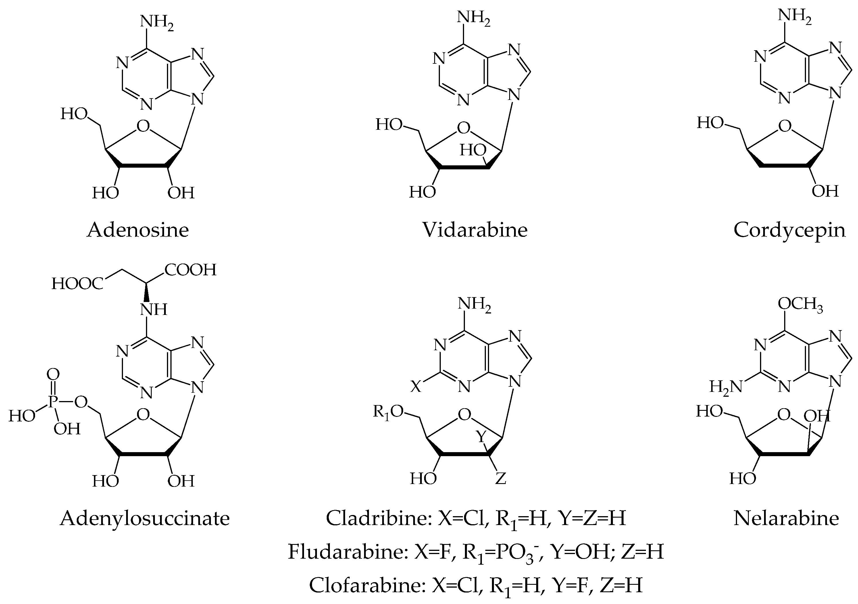
:1. Introduction
2. Results and Discussion
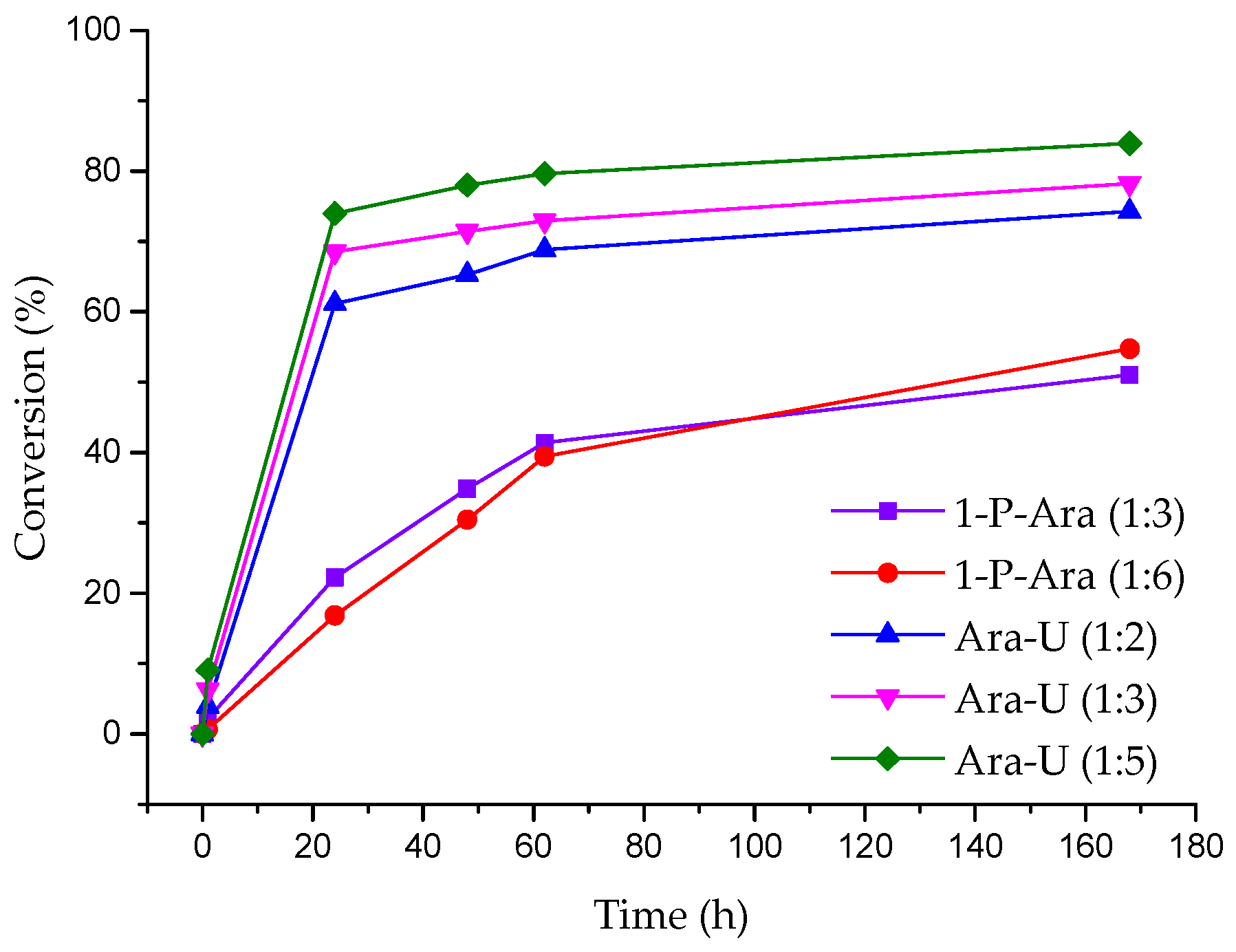
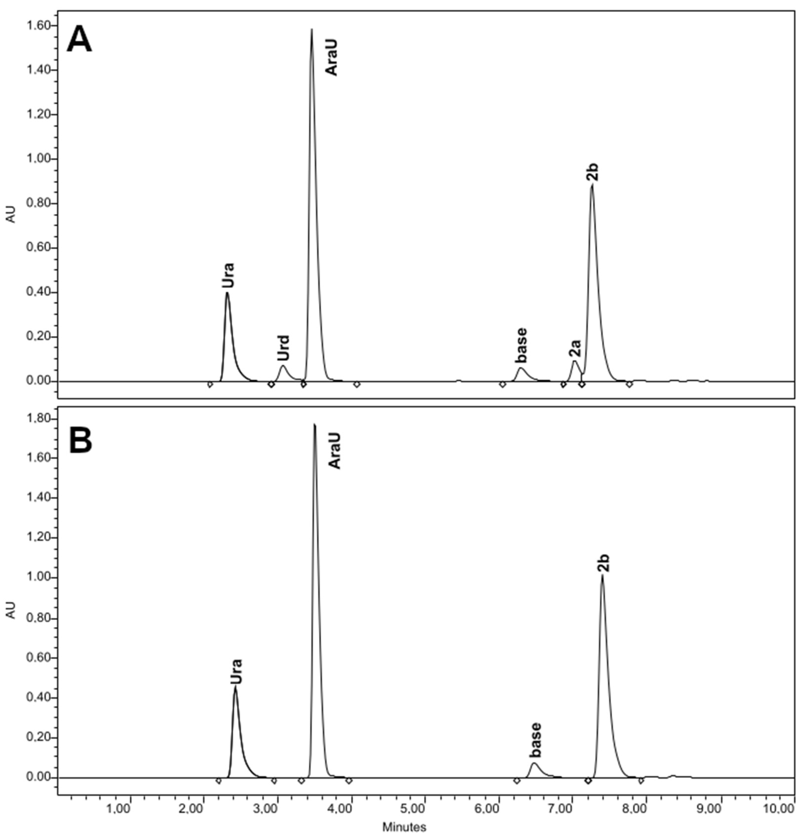
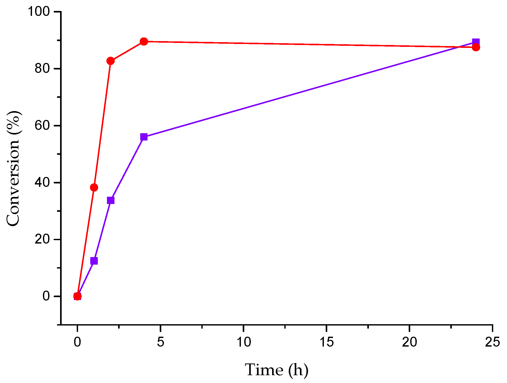
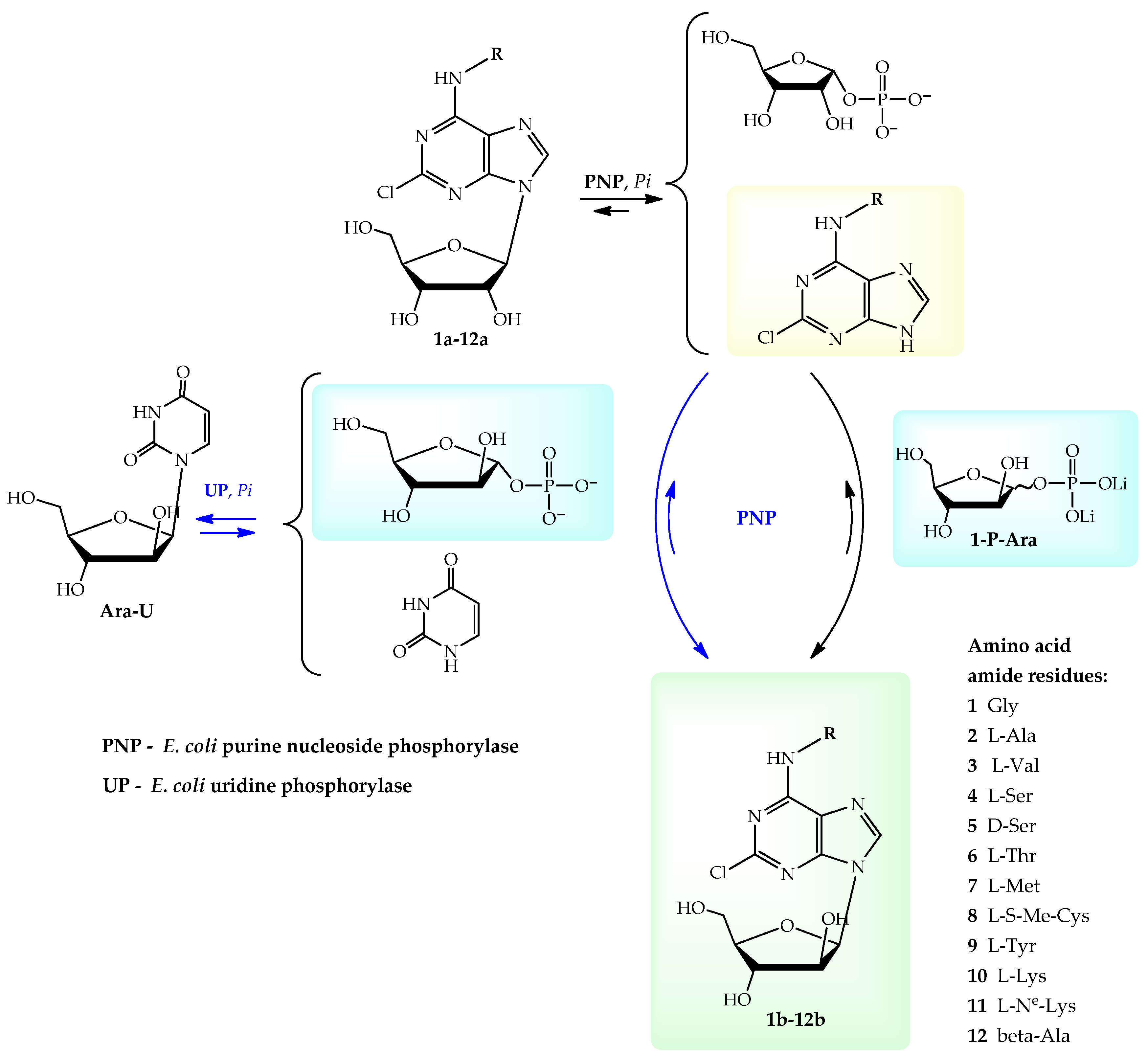
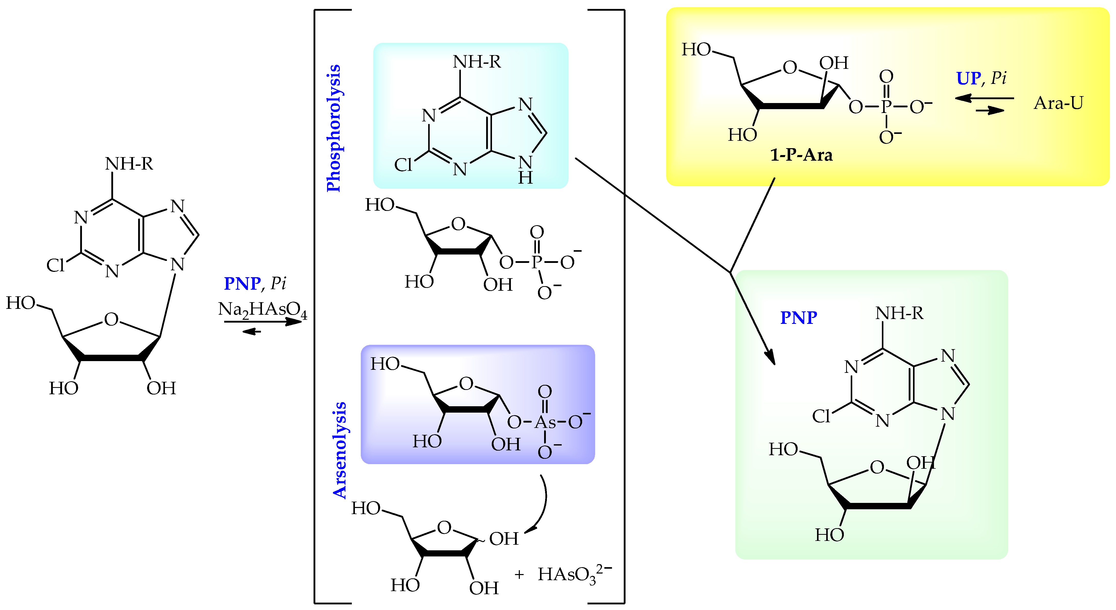
2.1. Synthesis of Arabinonucleosides
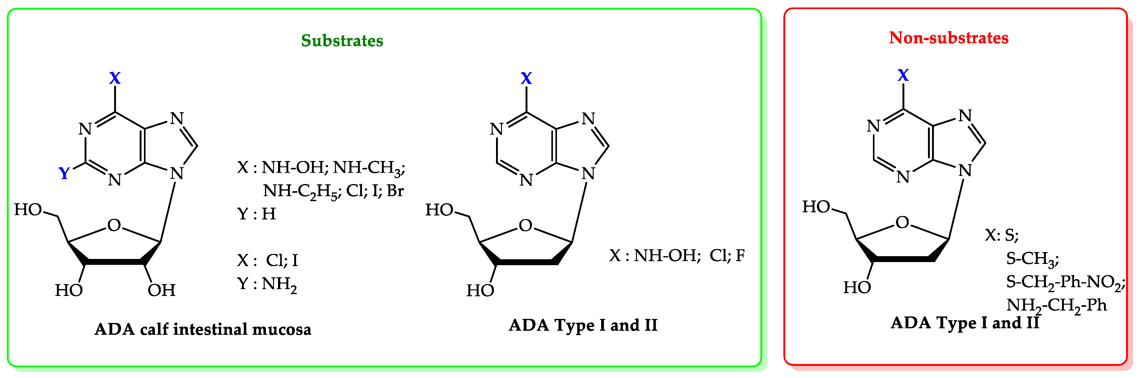
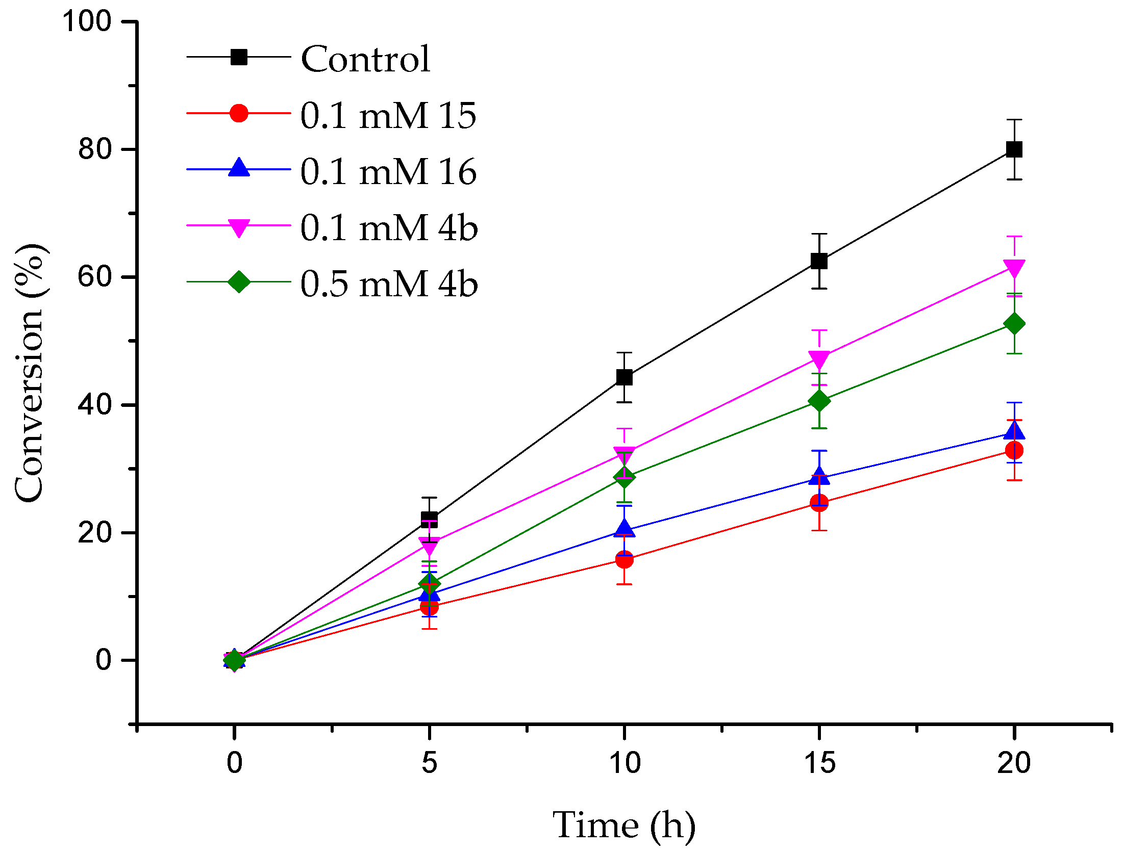
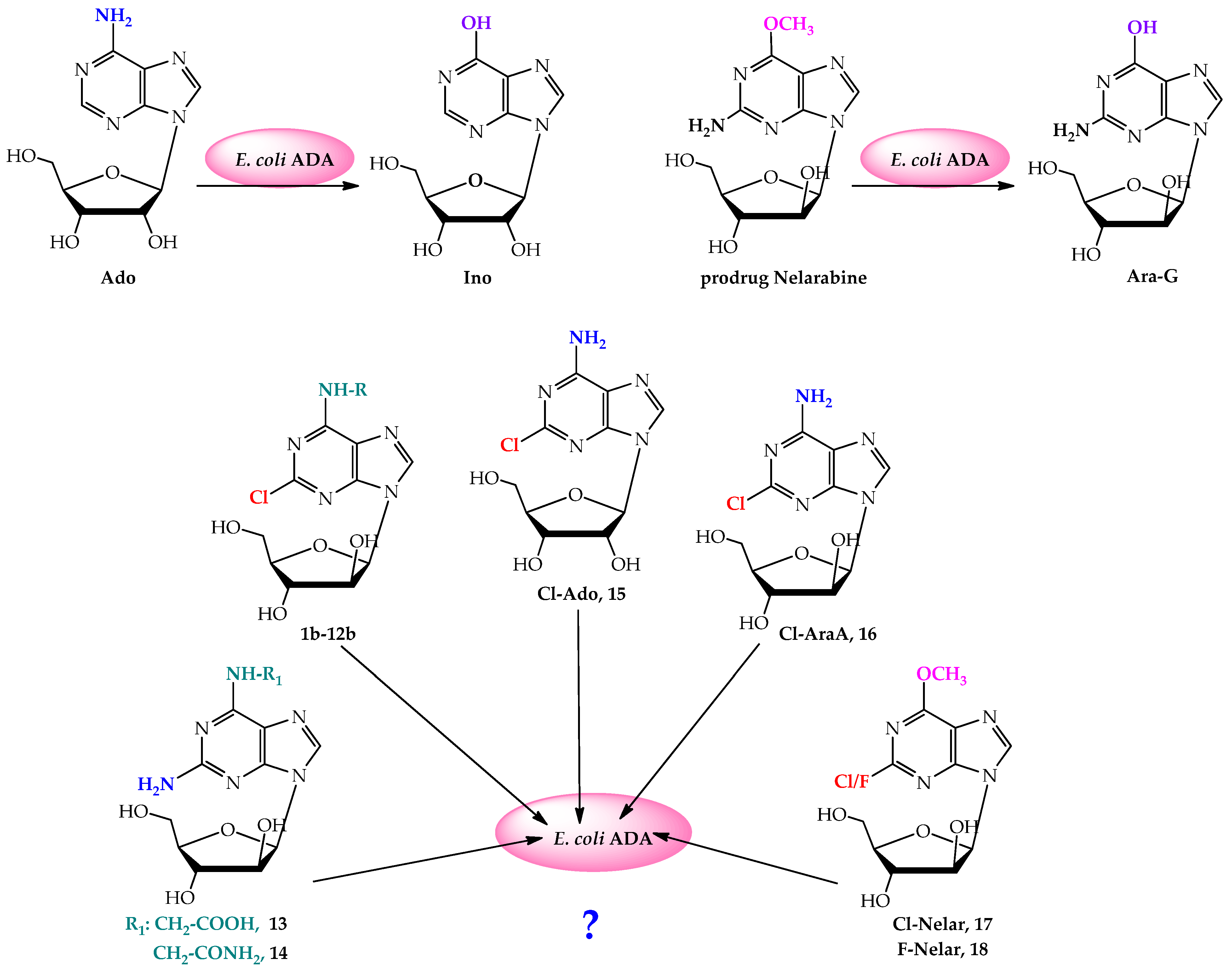
2.2. The ADA Substrate Specificity
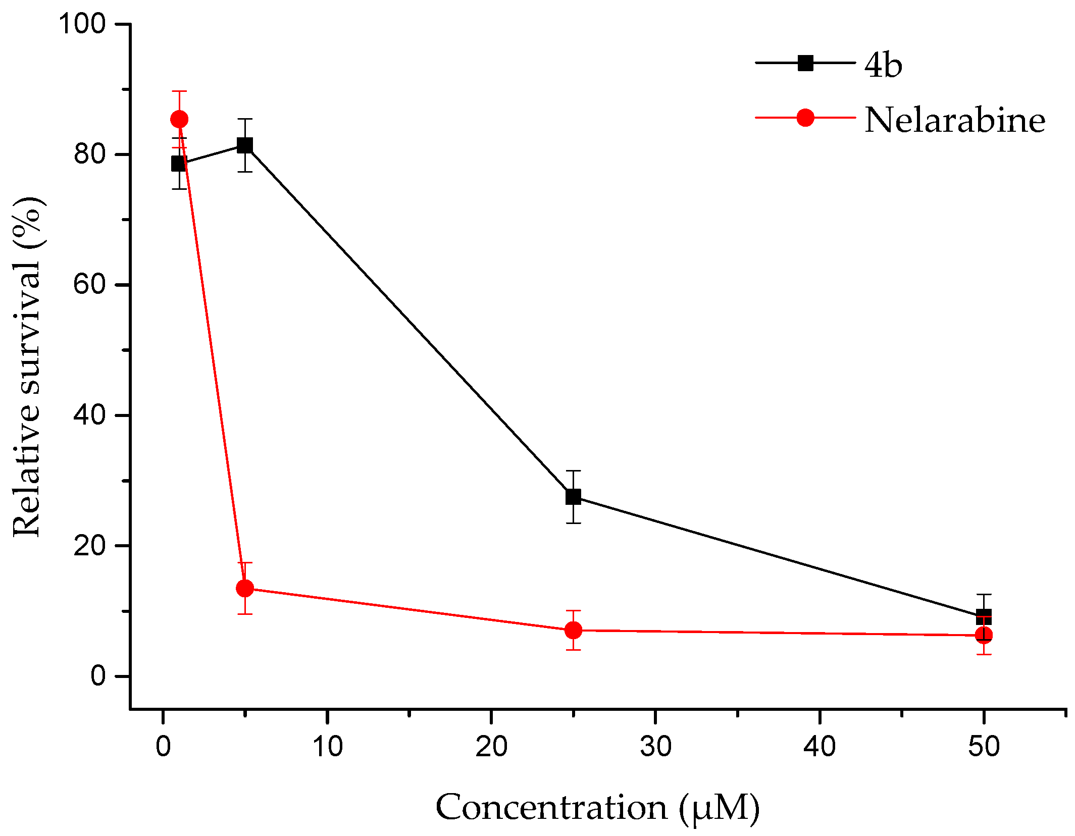
2.3. The Influence of the Synthesized Nucleosides on U937 Cell Survival
3. Materials and Methods
3.1. General Procedures
3.2. Enzymatic Reactions
3.3. The ADA Substrate Specificity
3.4. Inhibition of E. coli ADA
3.5. Biological Assay
4. Conclusions
Supplementary Materials
Author Contributions
Funding
Institutional Review Board Statement
Informed Consent Statement
Data Availability Statement
Conflicts of Interest
References
- Aurelio, L.; Baltos, J.-A.; Ford, L.; Nguyen, A.T.N.; Jörg, M.; Devine, S.M.; Valant, C.; White, P.J.; Christopoulos, A.; May, L.T.; et al. A Structure–Activity Relationship Study of Bitopic N6-Substituted Adenosine Derivatives as Biased Adenosine A1 Receptor Agonists. J. Med. Chem. 2018, 61, 2087–2103. [Google Scholar] [CrossRef]
- Jacobson, K.A.; Gao, Z.-G.; Paoletta, S.; Kiselev, E.; Chakraborty, S.; Jayasekara, P.S.; Balasubramanian, R.; Tosh, D.K. John daly lecture: Structure-guided drug design for adenosine and P2Y receptors. Comput. Struct. Biotechnol. J. 2015, 13, 286–298. [Google Scholar] [CrossRef]
- Madi, L.; Ochaion, A.; Rath-Wolfson, L.; Bar-Yehuda, S.; Erlanger, A.; Ohana, G.; Harish, A.; Merimski, O.; Barer, F.; Fishman, P. The A3 adenosine receptor is highly expressed in tumor versus normal cells: Potential target for tumor growth inhibition. Clin. Cancer Res. 2004, 10, 4472–4479. [Google Scholar] [CrossRef]
- Gessi, S.; Merighi, S.; Varani, K.; Cattabriga, E.; Benini, A.; Mirandola, P.; Leung, E.; Mac Lennan, S.; Feo, C.; Baraldi, S.; et al. Adenosine receptors in colon carcinoma tissues and colon tumoral cell lines: Focus on the A3 adenosine subtype. J. Cell. Physiol. 2007, 211, 826–836. [Google Scholar] [CrossRef] [PubMed]
- Fleysher, M.H.; Bernacki, R.J.; Bullard, G.A. Some short-chain N6-substituted adenosine analogs with antitumor properties. J. Med. Chem. 1980, 23, 1448–1452. [Google Scholar] [CrossRef] [PubMed]
- Krasnov, V.P.; Vigorov, A.Y.; Musiyak, V.V.; Nizova, I.A.; Gruzdev, D.A.; Matveeva, T.V.; Levit, G.L.; Kravchenko, M.A.; Skornyakov, S.N.; Bekker, O.B.; et al. Synthesis and antimycobacterial activity of N -(2-aminopurin-6-yl) and N -(purin-6-yl) amino acids and dipeptides. Bioorg. Med. Chem. Lett. 2016, 26, 2645–2648. [Google Scholar] [CrossRef]
- Musiyak, V.V.; Gruzdev, D.A.; Kravchenko, M.A.; Vakhrusheva, D.V.; Levit, G.L.; Krasnov, V.P.; Charushin, V.N. Synthesis and antimycobacterial activity of purine conjugates with (S)-lysine and (S)-ornithine. Mendeleev Commun. 2019, 29, 11–13. [Google Scholar] [CrossRef]
- Krasnov, V.P.; Vigorov, A.Y.; Gruzdev, D.A.; Levit, G.L.; Demin, A.M.; Nizova, I.A.; Tumashov, A.A.; Sadretdinova, L.S.; Gorbunov, E.B.; Charushin, V.N. Synthesis of enantiomers of N-(2-aminopurin-6-yl)amino acids. Russ. Chem. Bull. 2015, 64, 2106–2113. [Google Scholar] [CrossRef]
- Ward, D.N.; Wade, J.; Walborg, E.F.; Osdene, T.S. The synthesis of N-(6-Purinyl)amino acids. amino acids with a single reactive amino group1a. J. Org. Chem. 1961, 26, 5000–5005. [Google Scholar] [CrossRef]
- Letham, D.S.; Young, H. The synthesis and cytokinin activities of N-(purin-6-yl)amino acids. Phytochemistry 1971, 10, 23–28. [Google Scholar] [CrossRef]
- Matsubara, S.; Fujii, T.; Nishitani, T. Cytokinin Activities of N-(Purin-6-yl) amino Acids, N-(Purin-6-yl) peptides and Related Compounds (A. NATURAL SCIENCE). Sci. Rep Kyoto Prefect. Univ. Nat. Sci. Living Sci. 1988, 39, 1–6. [Google Scholar]
- Iwamura, H.; Yada, M.; Koshimizu, K.; Matsubara, S. Synthesis and comparative cytokinin activities of N-(Purin-6-yl)-d- and -l-amino acid methyl esters. Chem. Biol. Technol. Agric. 1978, 42, 1009–1014. [Google Scholar] [CrossRef]
- Ottria, R.; Casati, S.; Manzocchi, A.; Baldoli, E.; Mariotti, M.; Maier, J.A.; Ciuffreda, P. Synthesis and evaluation of in vitro anticancer activity of some novel isopentenyladenosine derivatives. Bioorg. Med. Chem. 2010, 18, 4249–4254. [Google Scholar] [CrossRef] [PubMed]
- Lapponi, M.J.; Rivero, C.W.; Zinni, M.A.; Britos, C.N.; Trelles, J.A. New developments in nucleoside analogues biosynthesis: A review. J. Mol. Catal. B Enzym. 2016, 133, 218–233. [Google Scholar] [CrossRef]
- Rottenberg, M.E.; Masocha, W.; Ferella, M.; Petitto-Assis, F.; Goto, H.; Kristensson, K.; McCaffrey, R.; Wigzell, H. Treatment of African trypanosomiasis with cordycepin and adenosine deaminase inhibitors in a mouse model. J. Infect. Dis. 2005, 192, 1658–1665. [Google Scholar] [CrossRef] [PubMed]
- Cristalli, G.; Vittori, S.; Eleuteri, A.; Grifantini, M.; Volpini, R.; Lupidi, G.; Capolongo, L.; Pesenti, E. Purine and 1-deazapurine ribonucleosides and deoxyribonucleosides: Synthesis and biological activity. J. Med. Chem. 1991, 34, 2226–2230. [Google Scholar] [CrossRef]
- Robak, P.; Robak, T. Older and new purine nucleoside analogs for patients with acute leukemias. Cancer Treat. Rev. 2013, 39, 851–861. [Google Scholar] [CrossRef]
- Robak, T.; Lech-Maranda, E.; Korycka, A.; Robak, E. Purine nucleoside analogs as immunosuppressive and antineoplastic agents: Mechanism of action and clinical activity. Curr. Med. Chem. 2006, 13, 3165–3189. [Google Scholar] [CrossRef]
- Vodnala, S.K.; Lundbäck, T.; Yeheskieli, E.; Sjöberg, B.; Gustavsson, A.L.; Svensson, R.; Olivera, G.C.; Eze, A.A.; de Koning, H.P.; Hammarström, L.G.; et al. Structure-activity relationships of synthetic cordycepin analogues as experimental therapeutics for African trypanosomiasis. J. Med. Chem. 2013, 56, 9861–9873. [Google Scholar] [CrossRef]
- Chassy, B.M.; Suhadolnik, R.J. Adenosine Aminohydrolase: Binding and hydrolysis of 2- and 6-substituted purine ribonucleosides and 9-substituted adenine nucleosides. J. Biol. Chem. 1967, 242, 3655–3658. [Google Scholar] [CrossRef]
- Robins, M.J.; Basom, G.L. Nucleic acid related compounds. 8. direct conversion of 2′-Deoxyinosine to 6-Chloropurine 2′-Deoxyriboside and selected 6-substituted deoxynucleosides and their evaluation as substrates of adenosine deaminase. Can. J. Chem. 1973, 51, 3161–3169. [Google Scholar] [CrossRef]
- Pospísilová, H.; Sebela, M.; Novák, O.; Frébort, I. Hydrolytic cleavage of N6-substituted adenine derivatives by eukaryotic adenine and adenosine deaminases. Biosci. Rep. 2008, 28, 335–347. [Google Scholar] [CrossRef]
- Gandhi, V.; Keating, M.J.; Bate, G.; Kirkpatrick, P. Nelarabine. Nat. Rev. Drug Discov. 2006, 5, 17–18. [Google Scholar] [CrossRef]
- Berzin, V.B.; Dorofeeva, E.V.; Leonov, V.N.; Miroshnikov, A.I. The preparative method for 2-fluoroadenosine synthesis. Russ. J. Bioorg. Chem. 2009, 35, 210–214. [Google Scholar] [CrossRef] [PubMed]
- Berzin, V.B.; Dorofeeva, E.V.; Leonov, V.N.; Lutonina, O.L.; Miroshnikov, A.I. Method of production of 2-chloroadenosine. Patent RU № 23,246,98C1, 20 May 2008. [Google Scholar]
- Montgomery, J.; Hewson, K. Nucleosides of 2-Fluoroadenine. J. Med. Chem. 1969, 12, 498–504. [Google Scholar] [CrossRef] [PubMed]
- Yu, X.-J.; Li, G.-X.; Qi, X.-X.; Deng, Y.-Q. Stereoselective synthesis of 9-β-d-arabianofuranosyl guanine and 2-amino-9-(β-d-arabianofuranosyl)purine. Bioorg. Med. Chem. Lett. 2005, 15, 683–685. [Google Scholar] [CrossRef]
- Glaudemans, C.P.J.; Fletcher, H.G. Syntheses with partially benzylated sugars. III.1 A simple pathway to a “cis- Nucleoside”, 9-β-D-Arabinofuranosyladenine (Spongoadenosine). J. Org. Chem. 1963, 28, 3004–3006. [Google Scholar] [CrossRef]
- Tuncbilek, M.; Kucukdumlu, A.; Guven, E.B.; Altiparmak, D.; Cetin-Atalay, R. Synthesis of novel 6-substituted amino-9-(β-d-ribofuranosyl)purine analogs and their bioactivities on human epithelial cancer cells. Bioorg. Med. Chem. Lett. 2018, 28, 235–239. [Google Scholar] [CrossRef] [PubMed]
- Utley, L.M.; Maldonado, J.; Awad, A.M. A practical synthesis of xylo- and arabinofuranoside precursors by diastereoselective reduction using Corey-Bakshi-Shibata catalyst. Nucleosides Nucleotides Nucleic Acids 2018, 37, 20–34. [Google Scholar] [CrossRef]
- Chattopadhyaya, J.B.; Reese, C.B. A synthesis of purine arabinosides. Nucleic Acids Res. 1978, 5, s67–s72. [Google Scholar] [CrossRef]
- Koszalka, G.W.; Averett, D.R.; Fyfe, J.A.; Roberts, G.B.; Spector, T.; Biron, K.; Krenitsky, T.A. 6-N-substituted derivatives of adenine arabinoside as selective inhibitors of varicella-zoster virus. Antimicrob. Agents Chemother. 1991, 35, 1437–1443. [Google Scholar] [CrossRef]
- Hanrahan, J.R.; Hutchinson, D.W. The enzymatic synthesis of antiviral agents. J. Biotechnol. 1992, 23, 193–210. [Google Scholar] [CrossRef] [PubMed]
- Konstantinova, I.D.; Antonov, K.V.; Fateev, I.V.; Miroshnikov, A.I.; Stepchenko, V.A.; Baranovsky, A.V.; Mikhailopulo, I.A. A Chemo-enzymatic synthesis of β-d-Arabinofuranosyl purine nucleosides. Synthesis 2011, 2011, 1555–1560. [Google Scholar] [CrossRef]
- Berzina, M.Y.; Eletskaya, B.Z.; Kayushin, A.L.; Dorofeeva, E.V.; Lutonina, O.I.; Fateev, I.V.; Paramonov, A.S.; Kostromina, M.A.; Zayats, E.A.; Abramchik, Y.A.; et al. Synthesis of 2-chloropurine ribosides with chiral amino acid amides at C6 and their evaluation as A1 adenosine receptor agonists. Bioorg. Chem. 2022, 126, 105878. [Google Scholar] [CrossRef]
- Esipov, R.S.; Gurevich, A.I.; Chuvikovsky, D.V.; Chupova, L.A.; Muravyova, T.I.; Miroshnikov, A.I. Overexpression of Escherichia coli genes encoding nucleoside phosphorylases in the pET/Bl21(DE3) system yields active recombinant enzymes. Protein Expr. Purif. 2002, 24, 56–60. [Google Scholar] [CrossRef] [PubMed]
- Hassan, A.E.; Abou-Elkhair, R.A.; Riordan, J.M.; Allan, P.W.; Parker, W.B.; Khare, R.; Waud, W.R.; Montgomery, J.A.; Secrist, J.A. Synthesis and evaluation of the substrate activity of C-6 substituted purine ribosides with E. coli purine nucleoside phosphorylase: Palladium mediated cross-coupling of organozinc halides with 6-chloropurine nucleosides. Eur. J. Med. Chem. 2012, 47, 167–174. [Google Scholar] [CrossRef] [PubMed]
- Mikhailopulo, I.A.; Miroshnikov, A.I. New trends in nucleoside biotechnology. Acta Nat. 2010, 2, 36–59. [Google Scholar] [CrossRef] [PubMed]
- Wempen, I.; Fox, J.J. [11] Synthesis of nucleoside derivatives by conversion from preformed nucleosides. Methods Enzymol. 1967, 12, 76–93. [Google Scholar] [CrossRef]
- Schramm, V.L. [13] Enzymatic transition-state analysis and transition-state analogs. Methods Enzym. 1999, 308, 301–355. [Google Scholar] [CrossRef]
- Kline, P.C.; Schramm, V.L. Purine nucleoside phosphorylase. Catalytic mechanism and transition-state analysis of the arsenolysis reaction. Biochemistry 1993, 32, 13212–13219. [Google Scholar] [CrossRef]
- Schramm, V.L. Enzymatic transition state theory and transition state analogue design. J. Biol. Chem. 2007, 282, 28297–28300. [Google Scholar] [CrossRef]
- Konstantinova, I.D.; Fateev, I.V.; Miroshnikov, A.I. The arsenolysis reaction in the biotechnological method of synthesis of modified purine β-D-arabinonucleosides. Russ. J. Bioorg. Chem. 2016, 42, 372–380. [Google Scholar] [CrossRef]
- Konstantinova, I.D.; Fateev, I.V.; Miroshnikov, A.I. Method for Production of Purine Nucleosides of β-D-Arabinofuranose Series. Patent RU № 262,402,3C2, 30 June 2017. [Google Scholar]
- Fateev, I.V.; Kostromina, M.A.; Abramchik, Y.A.; Eletskaya, B.Z.; Mikheeva, O.O.; Lukoshin, D.D.; Zayats, E.A.; Berzina, M.Y.; Dorofeeva, E.V.; Paramonov, A.S.; et al. Multi-enzymatic cascades in the synthesis of modified nucleosides: Comparison of the thermophilic and mesophilic pathways. Biomolecules 2021, 11, 586. [Google Scholar] [CrossRef] [PubMed]
- Fateev, I.V.; Antonov, K.V.; Konstantinova, I.D.; Muravyova, T.I.; Seela, F.; Esipov, R.S.; Miroshnikov, A.I.; Mikhailopulo, I.A. The chemoenzymatic synthesis of clofarabine and related 2’-deoxyfluoroarabinosyl nucleosides: The electronic and stereochemical factors determining substrate recognition by E. coli nucleoside phosphorylases. Beilstein J. Org. Chem. 2014, 10, 1657–1669. [Google Scholar] [CrossRef] [PubMed]
- Eletskaya, B.Z.; Gruzdev, D.A.; Krasnov, V.P.; Levit, G.L.; Kostromina, M.A.; Paramonov, A.S.; Kayushin, A.L.; Muzyka, I.S.; Muravyova, T.I.; Esipov, R.S.; et al. Enzymatic synthesis of novel purine nucleosides bearing a chiral benzoxazine fragment. Chem. Biol. Drug Des. 2019, 93, 605–616. [Google Scholar] [CrossRef]


| Compound | Amino Acid Amide Residue | R | Conversion into Nucleoside, % (HPLC), Azure Route | Nucleoside Yield (%), Azure Route |
|---|---|---|---|---|
| 1b | Gly | -CH-CONH2 | 82.88 | 63 |
| 2b | L-Ala | -CH(CH3)-CONH2 | 81.44 | 50 |
| 3b | L-Val | -CH(CH3)2-CONH2 | 69.15 | 46 |
| 4b | L-Ser | -CH(CH2OH)-CONH2 | 90.60 | 81 |
| 5b | D-Ser | -CH(CH2OH)-CONH2 | 78.10 | 77 |
| 6b | L-Thr | -CH(CH(OH)CH3)-CONH2 | 80.57 | 60 |
| 7b | L-Met | -CH(CH2-CH2-S-CH3)-CONH2 | 91.51 | 88 |
| 8b | L-S-Me-Cys | -CH(CH2-S-CH3)-CONH2 | 80.39 | 75 |
| 9b | L-Tyr | -CH(CH2-C6H4-OH)-CONH2 | 94.83 | 68 |
| 10b | L-Lys | -CH(CH2-CH2-CH2-CH2-NH2)-CONH2 | 96.85 | 92 |
| 11b | Nε-Lys | -CH2-CH2-CH2-CH2-CH(NH2)-CONH2 | 71.76 | 52 |
| 12b | β-Ala | -CH2-CH2-CONH2 | 70.02 | 63 |
| Peak Name | Ura | Urd | Ara-U | base | 2a | 2b |
|---|---|---|---|---|---|---|
| RT (min) | 2.312 | 3.062 | 3.454 | 6.285 | 7.014 | 7.248 |
| % Area before arsenate adding | 13.25 | 2.84 | 44.72 | 3.00 | 2.87 | 33.32 |
| % Area after arsenate adding | 15.09 | - | 44.00 | 3.57 | - | 37.34 |
| Compound | Acceptor mol wt. | Donor mol wt. | Substrates | Reaction Volume, mL | PNP, Units (A) b | UP, Units (B) b | Volume, ml (20 mM Na2HAsO4) | Reaction Time, h | Conversion of Base into Nucleoside (HPLC Data), % | Eluent c % | |
|---|---|---|---|---|---|---|---|---|---|---|---|
| Acceptor mg (mmol) | Donor mg (mmol) | ||||||||||
| 1b | 1a 359.09 | Ara-U 244.2 | 75 (0.21) | 260 (1.06) | 104 | 162 (776) | 94 (88) | 0.208 | 196 | 82.88 | 50 |
| 2b | 2a 373.10 | 80 (0.21) | 260 (1.06) | 106 | 165 (770) | 95 (89) | 0.212 | 196 | 81.44 | 50 | |
| 3b | 3a 401.13 | 80 (0.20) | 244 (1.00) | 100 | 156 (782) | 90 (90) | 0.200 | 336 | 69.15 | 70 | |
| 4b | 4a 389.10 | 80 (0.21) | 251 (1.03) | 103 | 161 (783) | 93 (90) | 0.206 | 216 | 90.60 | 50 | |
| 5b | 5a 389.10 | 100 (0.26) | 313 (1.28) | 128 | 200 (778) | 115 (90) | 0.256 | 504 | 78.10 | 50 | |
| 6b | 6a 403.11 | 100 (0.25) | 300 (1.23) | 75 | 115 (464) | 70 (57) | 0.150 | 196 | 80.57 | 70 | |
| 7b | 7a 433.11 | 34 (0.08) | 90 (0.37) | 20 | 133 (1694) | 90 (244) | 0.040 | 168 | 91.51 | 70 | |
| 8b | 8a 419.09 | 40 (0.10) | 120 (0.49) | 40 | 61 (639) | 41 (83) | 0.080 | 196 | 80.39 | 70 | |
| 9b | 9a 465.13 | 100 (0.21) | 260 (1.06) | 60 | 90 (419) | 54 (51) | 0.120 | 196 | 94.83 | 70 | |
| 10b | 10a 430.16 | 15 (0.03) | 41 (0.17) | 50 | 76 (2179) | 45 (268) | 0.100 | 168 | 96.85 | 70 | |
| 11b | 11a 430.16 | 100 (0.23) | 286 (1.17) | 117 | 91 (391) | 166 (142) | 0.234 | 196 | 71.76 | 70 | |
| 12b | 12a 373.10 | 80 (0.21) | 261 (1.07) | 107 | 167 (779) | 96 (90) | 0.214 | 168 | 70.02 | 50 | |
| Compound | Yield (%), (mg) | Purity (%) | RT, min | [α]D25 | UV λmax, nm (ε) | HRMS: m/z Calcd [M+H]+ | HRMS: m/z Found [M+H]+ |
|---|---|---|---|---|---|---|---|
| 1b | 63 (43) | 98.69 | 9.50 a | 16.8 (c 0.25, H2O/DMSO 1:1) | 268 (14,900), 212 (18,800) | 227.0442 (base) 359.0865 | 227.0458 359.0894 |
| 2b | 50 (40) | 99.72 | 7.47 a | 40.4 (c 0.5, H2O) | 270 (15,200) 213 (18,600) | 241.0599 (base) 373.1021 | 241.0592 373.1015 |
| 3b | 46 (37) | 96.63 | 9.24 a | 20.8 (c 0.5, H2O) | 270 (16,800) 213 (18,700) | 269.0912 (base) 401.1334 | 269.0889 401.1335 |
| 4b | 81 (58) | 99.27 | 6.15 a | 43.6 (c 0.5, H2O) | 270 (17,900) 212 (23,600) | 257.0548 (base) 389.0971 | 257.0541 389.1007 |
| 5b | 77 (77) | 95.48 | 5.76 a | −20.4 (c 0.5, H2O) | 268 (16,600) 212 (20,400) | 257.0548 (base) 389.0971 | 257.0541 389.1007 |
| 6b | 60 (60) | 95.75 | 5.68 b | 76.0 (c 1.0, H2O/DMSO) | 269 (21,000) 213 (24,800) | 271.0704 (base) 403.1127 | 271.0695 403.1107 |
| 7b | 88 (30) | 98.20 | 7.35 b | 54.0 (c 1.0, H2O/DMSO) | 268 (19,400) 212 (23,200) | 301.0632 (base) 433.1055 | 301.0636 433.1061 |
| 8b | 75 (21) | 91.20 | 7.01 b | 80.0 (c 1.0, H2O/DMSO) | 270 (16,700) 212 (18,700) | 287.0476 (base) 419.0898 | 287.0457 419.08703 |
| 9b | 68 (68) | 94.38 | 6.91 b | 46.0 (c 1.0, H2O/DMSO) | 271 (19,200) 213 (25,400) | 333.0861 (base) 465.1284 | 333.0851 465.1269 |
| 10b | 92 (10) | 98.42 | 6.63 b | - | 270 (15,300) 213 (17,800) | 298.1177 (base) 430.1600 | 298.1161 430.1581 |
| 11b | 52 (52) | 95.60 | 5.35 b | 4.8 (c 1.0, H2O/DMSO) | 270 (15,400) 213 (17,800) | 298.1177 (base) 430.1600 | 298.1161 430.1581 |
| 12b | 63 (50) | 99.13 | 7.24 b | −4.8 (c 0.25, H2O/DMSO 1:1) | 270 (16,500) 213 (19,400) | 241.0599 base 373.1021 | 241.0603 373.1043 |
| Inhibitor | Ki, mM |
|---|---|
| 2-chloradenosine 15 | 0.078 ± 0.012 |
| 2-chloro-arabinoadenosine 16 | 0.13 ± 0.02 |
| 4b | 1.2 ± 0.2 |
| Compound | U937 Cell Survival, % | IC50, µM | |||
|---|---|---|---|---|---|
| 1.00 µM | 5.00 µM | 25.00 µM | 50.00 µM | ||
| 1b | 79 ± 2.3 | 83 ± 2.4 | 64 ± 3.2 | 60 ± 3.0 | >50 |
| 2b | 73 ± 3.6 | 90 ± 3.6 | 71 ± 2.8 | 68 ± 2.0 | >50 |
| 3b | 81 ± 3.2 | 85 ± 2.5 | 69 ± 2.7 | 65 ± 1.9 | >50 |
| 4b | 78 ± 3.1 | 81 ± 4.0 | 27 ± 1.1 | 9 ± 0.2 | 16.0 |
| 5b | 80 ± 4.0 | 76 ± 3.8 | 58 ± 2.3 | 48 ± 1.4 | 50 |
| 6b | 98 ± 4.9 | 90 ± 2.7 | 85 ± 2.5 | 79 ± 2.3 | >50 |
| 7b | 100 ± 4.0 | 105 ± 5.2 | 90 ± 3.6 | 76 ± 2.2 | >50 |
| 8b | 85 ± 3.4 | 83 ± 2.4 | 70 ± 2.1 | 56 ± 1.7 | ≈50 |
| 9b | 88 ± 4.4 | 82 ± 4.1 | 76 ± 2.3 | 70 ± 2.8 | >50 |
| 10b | 74 ± 2.2 | 71 ± 2.1 | 69 ± 3.4 | 61 ± 1.8 | >50 |
| 11b | 84 ± 4.2 | 89 ± 4.4 | 84 ± 4.2 | 79 ± 2.3 | >50 |
| 12b | 100 ± 4.0 | 82 ± 3.2 | 71 ± 2.1 | 69 ± 2.0 | >50 |
| 13 | 98 ± 4.0 | 87 ± 2.6 | 75 ± 3.0 | 63 ± 1.8 | >50 |
| 14 | 94 ± 2.8 | 77 ± 2.3 | 69 ± 3.4 | 50 ± 2.0 | 50 |
| 15 | 83 ± 3.3 | 80 ± 2.4 | 47 ± 1.9 | 27 ± 1.1 | 22.0 |
| 17 | 92 ± 3.6 | 88 ± 4.4 | 80 ± 4.0 | 73 ± 2.1 | >50 |
| 18 | 98 ± 3.9 | 93 ± 3.7 | 84 ± 2.5 | 76 ± 3.8 | >50 |
| Nelarabine | 85 ± 2.5 | 13 ± 0.6 | 7 ± 0.3 | 6 ± 0.1 | 3.3 |
Disclaimer/Publisher’s Note: The statements, opinions and data contained in all publications are solely those of the individual author(s) and contributor(s) and not of MDPI and/or the editor(s). MDPI and/or the editor(s) disclaim responsibility for any injury to people or property resulting from any ideas, methods, instructions or products referred to in the content. |
© 2023 by the authors. Licensee MDPI, Basel, Switzerland. This article is an open access article distributed under the terms and conditions of the Creative Commons Attribution (CC BY) license (https://creativecommons.org/licenses/by/4.0/).
Share and Cite
Eletskaya, B.Z.; Berzina, M.Y.; Fateev, I.V.; Kayushin, A.L.; Dorofeeva, E.V.; Lutonina, O.I.; Zorina, E.A.; Antonov, K.V.; Paramonov, A.S.; Muzyka, I.S.; et al. Enzymatic Synthesis of 2-Chloropurine Arabinonucleosides with Chiral Amino Acid Amides at the C6 Position and an Evaluation of Antiproliferative Activity In Vitro. Int. J. Mol. Sci. 2023, 24, 6223. https://doi.org/10.3390/ijms24076223
Eletskaya BZ, Berzina MY, Fateev IV, Kayushin AL, Dorofeeva EV, Lutonina OI, Zorina EA, Antonov KV, Paramonov AS, Muzyka IS, et al. Enzymatic Synthesis of 2-Chloropurine Arabinonucleosides with Chiral Amino Acid Amides at the C6 Position and an Evaluation of Antiproliferative Activity In Vitro. International Journal of Molecular Sciences. 2023; 24(7):6223. https://doi.org/10.3390/ijms24076223
Chicago/Turabian StyleEletskaya, Barbara Z., Maria Ya. Berzina, Ilya V. Fateev, Alexei L. Kayushin, Elena V. Dorofeeva, Olga I. Lutonina, Ekaterina A. Zorina, Konstantin V. Antonov, Alexander S. Paramonov, Inessa S. Muzyka, and et al. 2023. "Enzymatic Synthesis of 2-Chloropurine Arabinonucleosides with Chiral Amino Acid Amides at the C6 Position and an Evaluation of Antiproliferative Activity In Vitro" International Journal of Molecular Sciences 24, no. 7: 6223. https://doi.org/10.3390/ijms24076223