Review

Brugada Syndrome: From Molecular Mechanisms and Genetics to Risk Stratification

by
Irene Paula Popa
1,
Dragomir N. Șerban
2,
Minela Aida Mărănducă
2,
Ionela Lăcrămioara Șerban
2,
Bogdan Ionel Tamba
3,* and
Ionuț Tudorancea
1,2
1
Cardiology Clinic, “St. Spiridon” County Clinical Emergency Hospital, 700111 Iași, Romania
2
Department of Physiology, “Grigore T. Popa” University of Medicine and Pharmacy, 700115 Iași, Romania
3
Department of Pharmacology, “Grigore T. Popa” University of Medicine and Pharmacy, 700115 Iași, Romania
*
Author to whom correspondence should be addressed.
Int. J. Mol. Sci. 2023, 24(4), 3328; https://doi.org/10.3390/ijms24043328
Submission received: 30 September 2022 / Revised: 13 January 2023 / Accepted: 6 February 2023 / Published: 7 February 2023
(This article belongs to the Special Issue Heart Diseases: From Molecular Basis to Therapy)

Abstract

:
Brugada syndrome (BrS) is a rare hereditary arrhythmia disorder, with a distinctive ECG pattern, correlated with an increased risk of ventricular arrhythmias and sudden cardiac death (SCD) in young adults. BrS is a complex entity in terms of mechanisms, genetics, diagnosis, arrhythmia risk stratification, and management. The main electrophysiological mechanism of BrS requires further research, with prevailing theories centered on aberrant repolarization, depolarization, and current-load match. Computational modelling, pre-clinical, and clinical research show that BrS molecular anomalies result in excitation wavelength (k) modifications, which eventually increase the risk of arrhythmia. Although a mutation in the SCN5A (Sodium Voltage-Gated Channel Alpha Subunit 5) gene was first reported almost two decades ago, BrS is still currently regarded as a Mendelian condition inherited in an autosomal dominant manner with incomplete penetrance, despite the recent developments in the field of genetics and the latest hypothesis of additional inheritance pathways proposing a more complex mode of inheritance. In spite of the extensive use of the next-generation sequencing (NGS) technique with high coverage, genetics remains unexplained in a number of clinically confirmed cases. Except for the SCN5A which encodes the cardiac sodium channel NaV1.5, susceptibility genes remain mostly unidentified. The predominance of cardiac transcription factor loci suggests that transcriptional regulation is essential to the Brugada syndrome’s pathogenesis. It appears that BrS is a multifactorial disease, which is influenced by several loci, each of which is affected by the environment. The primary challenge in individuals with a BrS type 1 ECG is to identify those who are at risk for sudden death, researchers propose the use of a multiparametric clinical and instrumental strategy for risk stratification. The aim of this review is to summarize the latest findings addressing the genetic architecture of BrS and to provide novel perspectives into its molecular underpinnings and novel models of risk stratification.

1. Introduction

Brugada syndrome (BrS) is a relatively rare hereditary arrhythmia disorder that was initially documented in 1992 [1]. Despite the latest hypothesis of additional inheritance pathways, BrS is still considered a Mendelian condition inherited in an autosomal dominant manner [2]. It is considered responsible for 4% of all sudden cardiac death (SCD) and nearly 20% of SCD in individuals with structurally normal hearts [3]. Arrhythmias, such as ventricular tachycardia (VT), ventricular fibrillation (VF), bradycardia, or atrioventricular node conduction dysfunction, are common in patients with BrS, with males being more prone than females to experience these heart rhythm disturbances [4]. BrS was previously presumed to be a primary electrical disorder of the structurally normal heart (6,7), specifically, an idiopathic VF produced by aberrant electrophysiologic activity in the right ventricular epicardium [5]. Nevertheless, structural anomalies in patients’ right ventricle outflow tract (RVOT) imply that Brugada syndrome may be part of a broader spectrum of right ventricular cardiomyopathies [6]. The main electrophysiological mechanism of BrS requires further research, with prevailing theories centered on aberrant repolarization, depolarization, and current-load match. Computational modelling, pre-clinical, and clinical research show that BrS molecular anomalies result in excitation wavelength (k) alterations, which eventually increase the risk of arrhythmia (10). Genetic variants have been discovered in 11–28% of Brugada syndrome patients, with a significant percentage involving the SCN5A (Sodium Voltage-Gated Channel Alpha Subunit 5) gene [7]. Several different genes have been postulated to induce BrS, but their implications are strongly contested [8,9]. Understanding the genetic aetiology of BrS is essential for determining asymptomatic genetic carriers at risk for SCD and stratifying the probability of future arrhythmic incidents [10]. The lack of a connection between genotype and phenotype in BrS-affected families emphasizes the complexity of this arrhythmogenic disorder and indicates that the onset and prognosis of BrS are probably a consequence of a mixture of various gene mutations and external conditions encompassing a delicate balance of ion currents [11,12]. The primary challenge in individuals with a BrS type 1 ECG is to identify those who are at risk for sudden death, researchers proposing the use of a multiparametric clinical and instrumental strategy for risk stratification.

2. Clinical Characteristics and Diagnostic Criteria of BrS

BrS is commonly diagnosed during the fifth decade of life, with an average age of 41 ± 15 years; however, it has also been reported in pediatric and geriatric patients [13]. Although many asymptomatic individuals with a Brugada ECG are fortuitously discovered, a minority of patients with BrS experience symptoms, such as nocturnal agonal breathing, syncope, and/or palpitations, which may progress to ventricular arrhythmias or SCD [13]. Supraventricular arrhythmias, including atrial fibrillation, atrial flutter, AV nodal re-entry tachycardia (AVNRT), and pre-excitation syndromes (Wolf–Parkinson–White syndrome) [14], may occur in up to 20% of BrS patients. In relation to the condition, increased sinus node recovery time and sino-atrial conduction time [15] together with delayed atrial conduction and atrial standstill have been described [16]. The development of supraventricular arrhythmias is strongly linked with ventricular inducibility. Among the Brugada syndrome patients with an indication for an implantable cardioverter-defibrillator (ICD) 27% exhibit supraventricular arrhythmias, compared to 13% of patients without an ICD indication, suggesting a more severe disease course in patients with spontaneous atrial arrhythmias [17].
After the initial expert consensus from 2002 regarding the diagnostic criteria for BrS [18], updates have been made in successive reports released in 2005 [19] and 2013 [20], along with technical electrocardiogram (ECG) standards in 2012 [21] and treatment guidelines in 2017 [22]. The latest expert consensus assessment, published in 2016 [23], introduced the Shanghai Score System, which assigns points based on ECG characteristics, clinical and family history, and genetic results. Up until now, the accuracy of the Shanghai Score System has been evaluated by one single-center retrospective study [24], which indicated that malignant cardiac events exclusively appeared in patients with scores that met diagnostic criteria. A type 1 ECG spontaneously occurring or being in a febrile state or following a drug provocation test, together with a Shanghai Score of 3.5 points, is necessary for the diagnosis of BrS. Despite a Shanghai Score of 3.5 in the lack of a type 1 ECG, BrS is not diagnosed. Nevertheless, it is considered that a type 1 ECG solely, whether spontaneous or documented during a provocation test, is positive for BrS. Given the relative difficulty and absence of supporting evidence in its prognostic value, the Shanghai Score System has not yet achieved universal recognition; therefore, a large percentage of medical centers continue to diagnose BrS merely on a type 1 ECG in the absence of particular adjustable variables [25].
There are three Brugada ECG patterns described: type 1 ECG is characterized by a coved ST elevation of ≥2 mm in V1-V3, followed by negative T waves, type 2 ECG has >2 mm of saddleback-shaped ST elevation, and type 3 has the morphology of either type 1 or type 2, but with <2 mm of ST-segment elevation [26]. Sodium channel blockers, such as ajmaline, procainamide, flecainide, disopyramide, propafenone, and pilsicainide, are beneficial in diagnosing BrS in circumstances when ST-segment elevation is not pathognomonic under baseline instances [27,28,29,30]. A negative INa-block test does not, however, rule out a latent type of BrS (for example, the negative predictive value of the flecainide test is 36%) [30,31]. Ajmaline and pilsicainide appear to be far more efficient than flecainide [32] or procainamide [28] in uncovering BrS. Brugada syndrome might also be revealed by bradycardia, a fever condition, or vagotonic drugs [27,33,34,35,36]. Positioning the right precordial leads at a higher position (one or two intercostal spaces above standard) might improve the sensitivity of the ECG for identifying the Brugada phenotype in certain individuals, both with and without a drug provocation test [37,38,39]. Various acute and chronic disorders can cause an ECG morphology mimicking BrS, which needs to be differentiated from Brugada syndrome. Among these, acute coronary events, pericarditis, myocarditis, pulmonary embolism, metabolic diseases, altered ion balance, dissecting aortic aneurysm, thiamine deficit, electric shock, and several drugs are the most frequently reported acute conditions [40]. Left ventricular hypertrophy, athlete’s heart, right bundle branch block (RBBB), pectus excavatum, septal hypertrophy, arrhythmogenic right ventricular cardiomyopathy/dysplasia (ARVC/D), autonomic nervous system abnormalities, Duchenne-dystrophy, Friedreich’s ataxia, mediastinal tumor, and Chagas disease are among the most frequently reported persistent causes mimicking the ECG morphology of Brs [21,41].

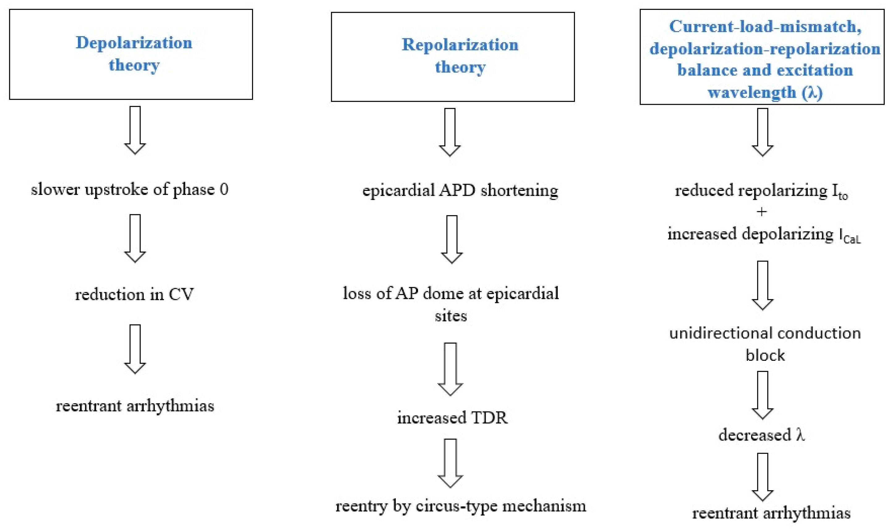
3. Pathophysiology of BrS

The need for a better comprehension of pathophysiological processes in BrS stems from the unanswered concerns about diagnosis, risk stratification for arrhythmia, and treatment [42]. The three prevailing electrophysiological theories behind BrS are centered on altered depolarization, repolarization, and mismatched current load [26].

3.1. Depolarization Theory

The conduction velocity (CV) of propagating cardiac action potentials (AP) includes both sodium channel activation resulting in cellular depolarization and gap junction conduction throughout subsequent cardiomyocytes. Any impairment to the adequate formation or propagation of action potentials may result in conduction abnormalities and arrhythmogenesis [43]. Almost one-fourth of BrS cases have been linked to genetic variants in the SCN5A gene that result in a reduced inward current during phase 0 [44]. It has been established that the slower upstroke during phase 0 and the subsequent delay in action potential formation have a significant impact on ventricular arrhythmogenesis in BrS. Martini and colleagues reported fibrotic lesions in the right ventricles, which may explain the right bundle branch block and the ST segment elevation on the electrocardiogram [45]. Several research addressing the disruption of SCN5A in animal models have indicated that mice with a targeted disruption of SCN5A had a lower conduction velocity in interstitial fibrosis [46,47,48].
There is a strong correlation between ECG phenotype and RVOT abnormalities. In individuals with BrS, an aberrant delayed potential was measured from the epicardium of the RVOT via an electrode placed into the conus branch of the right coronary artery (RCA) [49]. Furthermore, patients with a normal ECG at baseline may have BrS-type ECG characteristics during an acute myocardial infarction involving the right coronary artery [50]. Recent research that studied panoramic ventricular mapping in humans revealed electrogram lengthening and fragmentation, implying a decreased conduction velocity and an increased scattering [51]. According to the subtype, abnormal depolarization may influence the pathophysiology to various degrees. In instances of BrS when impaired calcium or potassium function is detected, repolarization anomalies might have a major impact, since the currents regulated by these channels play an important role in the plateau phase of the cardiac action potential instead of the depolarizing period [26].

3.2. Repolarization Theory

According to the repolarization hypothesis, the BrS phenotype is mainly caused by variable action potential duration (APD) shortening throughout the myocardial wall. Loss-of-function SCN5A genetic variants may have antagonistic effects on the rapid and delayed inactivation of Na+ channels, leading to various repolarization outcomes [52]. At slow cardiac rates, disturbances in rapid inactivation induce a persistent Na+ current, which lengthens repolarization. However, the intermediate kinetic component of slow inactivation is increased, delaying Na+ channel restoration, decreasing Na+ current, and shortening APD at high heart rates. In panoramic mapping research on BrS patients, the same biphasic pattern of APD, consisting of delaying succeeded by shortening, was described [51]. APD measured values from the epicardium (particularly the RVOT epicardium) were shorter than those acquired from the endocardium attributable to a considerably higher transient outward current in the epicardium (Ito). This is demonstrated by the more pronounced loss of dome-shaped AP morphology observed in the epicardium. Originally postulated by Yan and Antzelevitch in 1999, this is considered to explain re-entry through a phase 2 re-entrant pathway. Phase 2 re-entry involves electrotonic interactions and the diffusion of epicardial sites with an AP dome to sites in which this dome is lacking [53]. This might be the culprit of the R-on-T phenomenon, which results in an extrasystolic action potential prone to triggering ventricular arrhythmias [54]. ECG ST segment elevation and T-wave inversion are induced by rapid and inverse repolarization gradients.
Elevated Tpeak–Tend, a repolarization ECG indicator, is present in BrS patients and is related to a greater risk of arrhythmias or sudden cardiac death [55]. In addition, electrogram readings have shown a mixture of high repolarization gradient and delayed repolarization at the RVOT [51]. Ventricular tachycardia and ventricular fibrillation are probably triggered by impaired repolarization in individuals with BrS owing to calcium or potassium current imbalances. In addition, a re-entrant mechanism has been hypothesized as a consequence of Ca2+ channel mutations that lead to a loss of function [56]. Furthermore, the restoration theory suggests that a slope of the APD restitution curve greater than one is responsible for the appearance of repolarization alternans. APD alternans may generate high gradients in repolarization and refractoriness, as well as unidirectional conduction block and re-entry [57].

3.3. Current-Load-Mismatch, Depolarization-Repolarization Balance and Excitation Wavelength (λ)

In 2010, Hoogendijk and colleagues proposed the current-to-load mismatch subepicardial phenomena as the source of ventricular arrhythmias in individuals with BrS. Subepicardial excitation failure or deferred activation by current-to-load mismatch was discovered to be caused by a decrease in sodium current owing to channel malfunction or pore size. Computational model simulations also revealed that a disturbance in the homeostasis between the inward and outward currents might influence excitation, resulting in consequent ST-segment elevation [58,59]. In an explanted human heart model, it was shown that only the lack of local excitation was connected with ST-elevation, and not deferred activation or early repolarization [60]. Thus, by adjusting the Ito or ICa to adapt to the decreased sodium current, the degree of ST-elevation will be lowered [58]. To replicate comparable ST-elevation circumstances in pseudo-ECG recordings, conduction block, and excitatory failure were triggered by inhibiting sodium channels with ajmaline [59].
BrS patients have cardiac structural anomalies, particularly in the right ventricle (RV) and RVOT, which contribute to current-load-mismatch and excitation failure. This was subsequently validated by Ten Sande and colleagues through cardiac activation mapping, demonstrating that anatomical abnormalities in the subepicardial of the RV and RVOT are the most probable source of conduction alterations and ST-segment elevation [61]. This structural–electrophysiological connection is consistent with the presence of ventricular arrhythmias in BrS patients in their thirties when cardiac interstitial fibrosis is more pronounced [62]. Other research hypothesized that current-to-load mismatches at discontinuities might cause a level of conduction block, which would account for the RBBB morphology seen in BrS patients. Additionally, it should be emphasized that discontinuities are often related to depolarization anomalies in BrS arrhythmias [63]. This also interferes with action potential repolarization and recovery to produce the excitation (λ) determined by the product of effective refractory period (ERP) and conduction velocity. Both in pre-clinical animal models and BrS patients, a lower λ value has been linked with a higher probability of re-entrant arrhythmias [64,65].
Various sources of evidence support each of the postulated Brugada syndrome mechanisms, as most disorders arise and manifest via many processes. Although there is research that supports the different theories discussed previously, the far more convincing evidence is derived from human research of the heart and supports the repolarization theory. Ischemia-based models are derived from depolarization models; however, ischemia does not directly contribute to the condition. Furthermore, models for the depolarization theory illustrate the repolarization hypothesis. Arrhythmia and mutations in the Na+, K+ and Ca2+ channels are compatible with the repolarization theory. Research that modifies the Na+ channels does not give credence to the depolarization theory [66]. Excitation failure by current-to-load mismatch induces ST-segment elevation, which is regulated by INa, Ito, and ICaL, comparable to ST-segment elevation in Brugada patients. Thus, the current-to-load mismatch could explain the pathophysiology behind Brugada syndrome [59]. The three electrophysiological theories behind BrS are summarized in Figure 1.

4. Genetics of BrS

Following the initial discovery of two symptomatic siblings in 1992, familial inheritance was hypothesized based on the original description of the BrS [1]. Based on SCD and persistent ST elevation within a two-generation family, Kobayashi et al. validated the inheritance of the condition [67]. Multiple studies further confirmed the genetic basis of BrS [68,69,70]. Probst and collaborators investigated 13 families with BrS-ECGs+ in which, at least five family members had the hereditary SCN5A variant. This correlation was found to be challenging, as BrS-ECGs+ were identified in just 18% of mutation carriers at baseline and in 61% following drug testing, while 8 participants exhibited BrS-ECGs+ but did not carry the familial variant. These data may indicate that the presence of an SCN5A mutation is not a determinant of BrS-ECG+ occurrence. The genetic background provides varying susceptibilities to the impact of a single sodium channel loss-of-function mutation, being potent enough to induce a BrS-ECG+ irrespective of an SCN5A mutation [70]. Despite the fact that genetics remain unidentified in a number of clinically proven cases, irrespective of the extensive implementation of next-generation sequencing (NGS) with high coverage (at least 100×), BrS is still regarded as a genetic condition [71]. Genetic variants in at least 26 distinct genes have priorly been assumed to be responsible [72]; however, the deleterious role of all genes except SCN5A has lately been contested [8], several genetic variants, such as polymorphisms and non-genetic variants, including fever, are being considered to influence the disease’s expression [73]. In addition, numerous genetic tests that identify genetic variants in these genes provide findings of unclear relevance. The basis of this ambiguity is frequently the assumption that all instances of BrS are inherited via an autosomal dominant Mendelian pathway. This concept limits the geneticist from evaluating a potential cumulative effect from both common and rare genetic variants since there can only be one mutation based on the prior premise. Therefore, the importance of cumulative genetic variants within a single person in the underlying disease manifestation is a topic of consideration [74]. Various genetic variants within a gene might be accountable for a variety of phenotypes [75], even within the same family [76,77], making connections between genotype and phenotype particularly challenging.
Sodium channel mutations. The first genetic mutation related to BrS was described in 1998 [7] regarding the sodium channel protein type V subunit α gene (SCN5A), which encodes the alpha subunit of the voltage-gated NaV1.5 cardiac sodium channel, responsible for the modulation of the rapid sodium current in myocytes. Ever since, around 500 sporadic mutations in more than 40 genes have been described as presumably being related to BrS. These genes mainly encode sodium, potassium, and calcium channels, as well as proteins related to them [78]. The discovery of additional genes that may be linked with BrS has allowed genetic screening in clinical assessment. Nonetheless, an extensive study of all probable BrS-associated genes revealed a disorder mutation in less than 40% of confirmed cases, keeping the genetic origin of the condition undiscovered in the vast majority of families [79]. Currently, SCN5A is the primary gene linked with BrS, and roughly 30% of documented cases are imputable to one of the gene’s more than 350 genetic variants [80]. Copy number variations (CNVs) are further infrequent mutations which might induce BrS. Less than ten CNVs possibly related to BrS have been identified so far, and they are all found in the SCN5A gene [81,82,83]. In 2011, Eastaugh et al. described the case of the first BrS patient with a significant rearrangement involving the deletion of exons 9 and 10 of SCN5A. Standard sequencing omitted this rearrangement, which was uncovered via a quantitative method. Additionally, it was hypothesized that it would lead to haploinsufficiency. These results prompted the researchers to propose that the analysis of CNVs in SCN5A be included as a regular feature of genetic testing in BrS patients [81]. According to Mademont–Soler et al. in the largest screening conducted in 2016 for CNVs in SCN5A in genotype-negative BrS patients and assessment of their prevalence in BrS-associated minor genes, even though such rearrangements in SCN5A are not particularly widespread among BrS patients, they are identified at least in several of the instances who test negative for disease-causing variants in BrS-related genes utilizing standard sequencing techniques [82].
A plethora of studies have investigated the possibility of modulation of SCN5A genetic variants by common polymorphisms [84]. A genetic component which connects all SCN5A-related cases remains to be identified, presumably because BrS is a distinctive ECG pattern induced by several variables and not a singular mutation, a theory based on the observation that sodium channel blockers do not induce the BrS ECG pattern in healthy persons or all SCN5A mutation carriers [60]. In a prior genome-wide association study (GWAS) performed on 312 patients with BrS, three common susceptibility variants were discovered, with two alleles on chromosome 3 and one allele on chromosome 6 located near the genes SCN5A, SCN10A, and HEY2, thus indicating an intricate genetic architecture. Results suggested that susceptibility to BrS may be caused by common genetic variations in sodium channel genes or the transcriptional regulator HEY2 [74]. One study analyzed the connection between polygenic risk scores, which are considered predictive indicators for the outcomes of ajmaline testing, and the decelerating of cardiac conduction induced by ajmaline. The premise of the study was that a weighted mixture of certain prevalent genetic variations forecasts the individual reaction to sodium-channel blockage. The research established that rare coding variants in SCN5A and common non-coding variants in the SCN5ASCN10A locus impact not only the baseline ECG, but additionally the cardiac electrical response to sodium-channel blockage. The research outcomes may have relevance in two clinical situations: single-nucleotide polymorphisms (SNP) genotyping can be utilized to determine pre-test likelihood when conducting drug testing in individuals suspected of having BrS and genotyping ahead to the prescription of sodium-channel blocking action medications. Consequently, it is necessary to acknowledge that the significance of common variations in disease susceptibility is far more extensive than initially assumed [85].
In a comprehensive genome-wide association meta-analysis including 2820 unrelated BrS cases and 10,001 controls, a number of 12 loci (10 new) were reported as being correlated with BrS. The eight independent association signals at the SCN5A–SCN10A locus indicate the significance of impaired sodium channel activity in the BrS’s susceptibility, whilst the eight loci containing cardiac transcription factor genes imply transcriptional regulation as a major component of BrS’s pathogenesis [86]. In BrS pathogenesis, functional analyses of MAPRE2 indicate a novel mechanism of NaV1.5 regulation through the microtubule network. The UK Biobank findings reveal a genetic overlap among the BrS and cardiac electrical characteristics and prevalent diseases in the overall population. Polygenic risk score results reinforce the theory that the disorder threshold in BrS patients is attained by various contributions of rare SCN5A genetic variants, common risk alleles, and sodium channel inhibition [86].
The latest research has been centered on identifying the phenotypic consequences of various polymorphisms within the SCN5A gene [72,75,87,88,89,90,91,92] and searching for other genes implicated in this multi-causal disease [80,90,93]. One such study established the similarities in phenotype amongst individuals with SCN10A genetic variants, in comparison with SCN5A genetic variants, which included a personal history of cardiac arrest/syncope, spontaneous BrS ECG pattern, family history of sudden death, and arrhythmic substrate [90]. This is compatible with functional research conducted on human-induced pluripotent stem cell-derived cardiomyocytes, wherein single-cell phenotypic characteristics of BrS were detected in cells from a patient with SCN10A gene variations [94]. The results of a multicenter analysis in which potential genes, including SCN10A, were sequenced did not indicate a significant involvement for SCN10A variations as monogenic aetiology of BrS [95]. Nevertheless, research by Hu and colleagues revealed SCN10A as a key susceptibility gene for BrS, detecting SCN10A genetic variants in 25 of 150 probands (17%), indicating that this gene plays a significant part in BrS [96]. Research on the effects of the SCN10A gene on cardiac conduction [97] and the autonomic nervous system [98] highlights the significance of this gene. Despite their function being debatable, the sodium channel genes SCN1B, SCN2B, and SCN3B have been linked to BrS and are present in the BrS diagnostic panels [8].
Non-coding variants. The functional impact of variants in coding areas is simple to estimate, given that the genetic code is already identified, a substantial quantity of exome data is accessible, and a thorough understanding of Mendelian illnesses exists [99]. Nevertheless, it is challenging to estimate the impact of variants in non-coding areas. These noncoding variants are present in cis-regulatory elements (CREs), including enhancers, promoters, and insulators, in addition to areas which encode non-coding RNAs (ncRNAs) [100]. A significant percentage of non-coding variants associated with cardiovascular illnesses or traits have been identified through GWAS [101]. GWAS performed on ECG traits and cardiac conduction abnormalities have emphasized the connection between the SCN5A–SCN10A locus and QRS duration and PR interval, indicating a role in cardiac conduction and heart function [97,102]. Notably, the majority of variants detected in this research are found in non-coding areas. It is well established that SCN5A variants are correlated to cardiac arrhythmias. Nevertheless, the discovery of SCN10A as a substantial risk area for ECG traits proved noteworthy [103,104,105]. SCN10A encodes the alpha subunit of the sodium channel Nav1.8, which participates in cardiac electrophysiology; however, the molecular basis continues to remain debatable, particularly in terms of its expression in cardiomyocytes [106]. Several studies have established that NaV1.8 participates in the late cardiac sodium current [107,108] and that it may be relevant in arrhythmogenesis [109]. Nonetheless, a recent study revealed that rabbit and hiPSC-CM cardiac cells lack functional NaV1.8 channels [110]. Van den Boogaard and colleagues’ research identified an additional potential function of SCN10A non-coding variants in conduction abnormalities. Specifically, they demonstrated that the SCN10A intronic variation rs6801957, which was found in a GWAS as being related to cardiac conduction dysfunction, is localized in an enhancer area that regulates the expression of the SCN5A gene [111,112]. Through the investigation of a super-enhancer cluster downstream of SCN5A, Man and colleagues have subsequently reaffirmed the importance of the SCN5A–SCN10A locus in the regulation of gene expression and architecture. Intriguingly, they discovered that adequate SCN5A expression and cardiac conduction need the interaction of all regulatory elements in the locus [113]. Future research should determine if the functional impact of these variants is associated with the dysregulation of SCN5A expression via the involvement of SCN10A as an enhancer, the function of NaV1.8 in intracardiac neurons that modulate cardiac electrical activity, or both [114].
Sodium channel-associated. The activity of sodium channels may be altered by circumstances other than mutations in the channel’s coding gene. In analysis assessing the impact of KV4.3 overexpression on NaV1.5 current and subsequent sodium channel availability, the results revealed that certain variables, such as the gain-of-function of the KV4.3 protein encoded by the KCND3 gene, clearly influenced the current of the NaV1.5 protein [115]. Post-translational changes, including a malfunction in the splicing process [116] or trafficking [117], or a change in phosphorylation, methylation, or acetylation [118], might underlie abnormalities in the functionality of the channel encoded by SCN5A in the lack of polymorphisms in this gene. RANGRF (RAN Guanine Nucleotide Release Factor), commonly known as MOG1, modulates the development and functioning of the NaV1.5 cardiac sodium channel in individuals by augmenting the expression of NaV1.5 at the cell membrane, thereby raising sodium current density [119]. The GPD1L gene, which encodes a protein similar to glycerol-3-phosphate dehydrogenase-1 and is presently considered part of BrS’s diagnostic panels, modulates NaV1.5 as well [120]. The LRRC10 gene, a transcriptional target of Nkx2.5, which modulates the ion channel proteins expressed by the SCN5A, CACNA1C, and KCNH2 genes, is believed to be linked to BrS [121].
Calcium channel mutations. Considering the significance of calcium for the cardiac action potential, the key function of calcium currents in BrS is presumably underestimated. Calcium is essential to excitation–contraction coupling, connecting the electrical signal recorded by ECG that characterizes BrS to mechanical malfunction, especially ventricular fibrillation and decreased contractility, observed in BrS. Furthermore, the BrS phenotype is influenced by non-genetic variants, including a rise in vagal activity or body temperature, which are proven to affect calcium signaling. In addition, the BrS phenotype may be altered in patients by isoproterenol, which increases calcium transportation via L-type calcium channels, ryanodine receptors, and SERCA (sarcoendoplasmic reticulum calcium transport ATPase) [122]. CACNA1C genetic variants are responsible for around 6.6% of BrS cases, CACNB2b genetic variants for roughly 4.8%, and CACNA2D1 genetic variants are rare [123,124]. TRPM4 (Transient Receptor Potential Cation Channel Subfamily M Member 4) gene is responsible for approximately 6% of BrS cases [125], in an autosomal recessive form instead of an autosomal dominant manner [126]. According to one study, the PKP2 gene, which encodes plakophilin-2 and is located on desmosomes between intercalary discs, is related to roughly 2.5% of BrS cases [127].
Potassium channel mutations. Several cases of BrS have been connected to polymorphisms in potassium channel-encoding genes. The two most prevalent genes are the ABCC9 (ATP Binding Cassette Subfamily C Member 9) and KCNH2 (Potassium Voltage-Gated Channel Subfamily H Member 2), which account for 4–5% and 1–2% of instances, respectively. Rarely, BrS patients are linked to HCN4, KCND2, KCND3, and KCNJ8 genes [71].
Overlap Syndromes. Considering that SCN5A genetic variants are present across many cardiogenetic diseases, it is not unexpected to identify an overlap between BrS and other diseases. For instance, BrS may be detected in the proband, whereas long QT syndrome, epilepsy, febrile seizures, or total bundle branch block may be observed in family members [128,129,130,131]. Multiple studies have reported an overlap between arrhythmogenic right ventricular dysplasia/cardiomyopathy (ARVD/C) and BrS [132], and the explanation might include cell–cell junctions [133]. ARVC and BrS may both develop from mutations in the connexome, with the resulting phenotype dependent on the nature of the mutation [134,135]. PKP2 could be an essential gene in this respect since genetic variants in PKP2 are associated with impairment of desmosomal integrity, and sodium current deficit, and can be discovered in BrS patients [127,136]. The occurrence of ARVC in BrS patients has been linked to an increased risk of arrhythmias [137]. The genetics of families with overlap syndromes should be thoroughly studied, since the genetic underpinnings may be distinct from those of families in whom BrS is the sole documented feature. This is an additional illustration of the necessity for tailored therapy and family specific BrS genetic analysis [138].

5. Human Induced Pluripotent Stem Cell Models

Despite the fact that animal models and primary cell lines represent the most widespread approaches for preclinical drug testing at present, there are substantial issues with them. As a potential substitute for animal models and primary cells, human induced pluripotent stem cell-derived cardiomyocytes (hiPSC-CMs) provide several benefits, including a human genome, fundamental characteristics of human CMs, and the expression of certain cardiac-specific genes [139]. To date, only several BrS models based on induced pluripotent stem cells (iPSCs) have been reported. In 2014, Cerrone et al. reported the first iPSC-based model having a rather rare mutation in PKP2 that caused sodium current deficits [127]. In this research, hiPSC-CMs obtained from a patient with a PKP2 deficit exhibited significantly decreased INa, which was rectified by transfection of wild-type PKP2, but not BrS-related PKP2, indicating that PKP2 genetic variants may represent a molecular substrate of BrS [127]. In 2016, Liang et al. developed patient-specific hiPSC-CMs from two patients with Brs having two genetic variants in the SCN5A gene and demonstrated a decrease in INa and maximum upstroke velocity of the action potentials in comparison to healthy subjects hiPSC-CMs [140]. Furthermore to SCN5A and PKP2, the Ibrahim Akin research team generated in 2019 BrS-hiPSC-CM models with genetic variants in SCN1B and SCN10A [94,141].
RRAD is a new gene related to Brugada syndrome susceptibility discovered by a whole-exome sequencing study [142]. In comparison to control cells from a healthy sibling, iPSC-derived cardiomyocytes obtained from the affected person had a significant drop in INa and a modest reduction in ICaL. In addition, over 70% of these cells had cytoskeletal abnormalities, as indicated by an abnormally spherical shape, and a decreased number of focal adhesion sites [143].The genes associated with Brugada syndrome and other inherited arrhythmia diseases are summarized in Table 1.

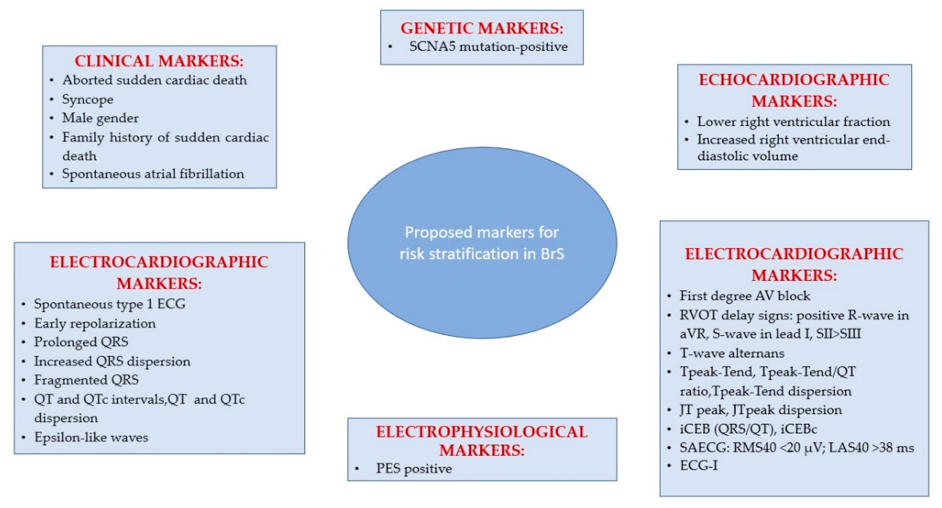
6. Arrhythmias and Sudden Cardiac Death Risk Stratification in BrS

It is widely acknowledged that individuals with symptomatic BrS have a higher probability of further arrhythmic events [13,144]. In contrast, a post-mortem analysis indicated that 72% of BrS-related SCDs happened in asymptomatic subjects. Moreover, 68% of this cohort would have been classified as low risk according to the Second Expert Consensus Conference on BrS [19]. Asymptomatic BrS patients have an annual incidence rate for arrhythmic events of between 0.5 and 1% [145,146,147]. Approximately 50% of the arrhythmic events will be represented by ventricular fibrillation despite any warning signs [148]. Even though it is challenging to quantify the progression of arrhythmic risk over the long term, considering a 1% yearly event rate, a 10% event rate at a 10-year follow-up in apparently healthy individuals is exceedingly high. Assessing the variables linked with a higher incidence of ventricular arrhythmias and SCD in BrS is a difficult undertaking [149]. Considering that implantable cardioverter-defibrillators (ICD) are linked with a 45% lifetime risk of complications [150], the option to implant them should be made sparingly. In fact, even if the development of subcutaneous ICDs may minimize the incidence of transvenous lead complications over the long term, there continues to be morbidity related to incorrect device therapies and the risk of infection with frequent generator replacements throughout time. It is consequently of the utmost significance to adequately stratify the risk of BrS patients, especially asymptomatic ones [151].
The primary challenge in individuals with a BrS type 1 ECG is to identify those who are at risk for sudden death and those who are not [144,152,153]. Several researchers have proposed identifying individuals with BrS type 1 ECG who are at risk for SD using a multiparametric clinical and instrumental strategy, including a family history of SD, syncope of presumed cardiac origin, a positive electrophysiology study, etc. Their findings indicate that individuals with several risk factors are at the greatest risk [154,155].
Clinical Markers. Research has shown that a history of aborted SCD is correlated with the greatest risk of subsequent arrhythmic episodes, resulting in an important prognostic significance [145,146,147]. The prevalence of arrhythmic events (sustained ventricular arrhythmia or adequate ICD therapy or SCD) was 13.5% per year in patients with a history of SCD, 3.2% per year in individuals with syncope, and 1% per year in asymptomatic patients, according to a meta-analysis [156]. Patients with a history of aborted SCD had an arrhythmia risk of 35% at 4 years [145,157], 44% at 7 years [147], and 48% at 10 years [156]. Various studies, as well as a meta-analysis, have shown that a history of syncope is related to a higher risk of subsequent arrhythmic episodes [145,146,158,159]. Priori’s team has established, nevertheless, that the correlation between syncope and transient ST segment elevation, and not a history of syncope solely, has the highest predictive value for identifying those at high risk [160]. Patients diagnosed with aborted SCD had a poor prognosis, according to Kamakura et al., whereas those asymptomatic or presenting with sole syncope had a favorable prognosis regardless of their ECG pattern [157]. Conte et al. established, nonetheless, that individuals with a history of syncope experience the same clinical course as asymptomatic patients. Specifically, throughout a long-term follow-up period of 83.8 ± 57.3 months, 11% of patients with syncope and 13% of the asymptomatic ones received adequate shocks [147]. This discrepancy may express the failure to distinguish between arrhythmic and neurally mediated syncope. Sacher et al. conducted a prospective analysis of the features of BrS patients who had suffered a syncope to establish if it could be distinguished as a syncope of arrhythmic vs. non-arrhythmic origin. Arrhythmic syncope was diagnosed in patients with no prodromal symptoms and no identified cause, who were unresponsive for no more than one minute and regained consciousness promptly. Non-arrhythmic syncope was diagnosed in patients with consciousness loss that lasted more than one minute, who had neurocardiogenic symptoms, and who did not suffer substantial physical trauma [161]. In Conte’s research, following the implantation of an ICD, 21 patients (11.9%) reported syncope. A number of five of them experienced neurally mediated syncope. In eight patients who experienced recurring syncope after ICD implantation, the percentage of ventricular pacing was less than one per cent, and no ventricular arrhythmias were observed. This may clarify why certain individuals with a history of unexplained syncope exhibit a favorable outcome [162].
Most research has shown that a family history of SCD does not predict future arrhythmic episodes in either symptomatic (3.3%) or asymptomatic (0.5%) individuals [145]. In contrast, Kamakura et al. reported that a family history of SCD occurring before age 45 is an independent risk factor for a poor outcome. Delise et al. showed that a family history of SCD could only be predictive when combined with other risk factors [154]. Regarding the more affected sex, studies have shown that the male gender has been related to a worse clinical outcome, according to a prior meta-analysis including 1545 individuals [159]. In the FINGER registry, the male gender was also linked with a lesser duration of time to the first incident, but with no statistical significance (mean incident rate per year, 3.0% for males and 0.9% for females) [145]. Additionally, in Conte’s research, men were almost three times more likely to get suitable shocks during follow-up [147]. According to the S Priori group, women with a BrS ECG pattern should not be considered a low-risk category as it was not found a significant predominance of male incidents (13%) compared to females (9%) [160].
BrS patients have an increased incidence of atrial fibrillation than the overall population of the same age. In a cohort of 560 BrS patients, 9% exhibited atrial fibrillation or atrial flutter [163]. 18% of 176 BrS patients with an ICD had paroxysmal AF throughout the course of 83.8 ± 57.3 months of follow-up [147]. There is a correlation between spontaneous atrial fibrillation and an increased frequency of syncopal episodes (60.0% vs. 22.2%) and confirmed ventricular fibrillation (40.0% vs. 14.2%). Sieira et al. showed that asymptomatic BrS patients with a history of sinus node dysfunction are eight times more likely to develop subsequent arrhythmic episodes [164]. The schematic representation of the proposed markers for risk stratification in Brugada syndrome is depicted in Figure 2.
Electrocardiographic Markers. A meta-analysis concluded that patients with spontaneous type 1 ECG pattern had a three to four times higher risk of incidents than those with a drug-induced ECG pattern. Since they have a poor prognosis as reported in various studies, identifying BrS patients with spontaneous type 1 ECG pattern is, therefore, mandatory [165]. In addition to the spontaneous type 1 ECG pattern, a variety of other ECG characteristics have been suggested for risk stratification, among which QRS fragmentation looks to be the most compelling of all ECG indicators. The fragmented QRS complex, which reflects a rise in conduction dispersion, might cause a unidirectional block [166,167,168], while broad QRS, which reflects a decrease in conduction velocity, will lower the excitation wavelength [169]. Both are risk factors for re-entrant arrhythmias. One study has shown that QRS-fragmentation is more prevalent in BrS patients with VF (85%) and syncope (50%) than in those without symptoms (34%) [170], an aspect that was validated by the PRELUDE trial, which demonstrated that QRS-fragmentation is an independent predictor of subsequent arrhythmias [146]. Furthermore, Tokioka et al. have established that the occurrence of QRS-fragmentation increases five times the risk of arrhythmic episodes [171].
Other ECG markers independently linked to SCD or adequate ICD therapy are first-degree atrioventricular block [172], prolonged QRS duration in leads II, V2, and V6 [173,174], and a prolonged QTc interval >460 ms in lead V2 [175]. Based on the results of preclinical research, it was determined that conduction anomalies must be included in risk indicators to improve their predictive ability [176,177]. For instance, the index of Cardiac Electrophysiological Balance (iCEB), provided by QT/QRS, is a substitute marker of λ, and its application has resulted in more accurate risk stratification [178,179].
Multiple ECG repolarization indicators for stratifying arrhythmic risk in the BrS population have been explored [180]. Tpeak–Tend interval, considered a measure of transmural scattering of repolarization [175], has been associated with malignant ventricular arrhythmias, although research on swine models revealed that it is a measure of global dispersion of repolarization rather than transmural [181]. The Tpeak–Tend interval through lead V1 to lead V4, the threshold value and the distribution throughout all precordial leads were substantially greater in symptomatic patients (aborted SCD, proper ICD treatment, syncope) than in asymptomatic patients. A value of the Tpeak–Tend interval of >100 ms was independently associated with arrhythmic episodes [172].
A negative T-wave in lead V1 has also been linked to a worse prognosis [182]. Furthermore, the development of T-wave alternans following treatment with pilsicainide was predictive of spontaneous VF [183]. In a comprehensive study of 166 BrS patients who underwent exercise testing, ST-segment elevation was reported in 57% of individuals with BrS, with ST elevation appearing during early recovery in 93 patients, while in two patients it appeared during the effort phase of the exercise and in five individuals the BrS ECG pattern was revealed by the exercise. Ventricular arrhythmias, including ventricular tachycardia and numerous premature ventricular contractions, were developed in three patients, all of them happening during the first phase of recovery and they resolved spontaneously [184]. Makimoto et al. found that ST-segment elevation during early recovery was distinctive in BrS patients and was substantially related to a greater cardiac event rate, particularly in patients with a prior episode of syncope or in asymptomatic individuals [185]. In contrast, Amin et al. were unable to demonstrate substantial differences among symptomatic and asymptomatic BrS patients in terms of ECG characteristics and their alterations throughout the exercise [186]. It has been found that an inferolateral early repolarization pattern is associated with arrhythmic episodes [187,188]. Tokioka et al. have demonstrated that the coupling of QRS-fragmentation and early repolarization pattern facilitates the assessment of individuals at high risk [171]. In prior research involving 290 persons with BrS, an early repolarization pattern characterized as notched or slurred J-point elevation primarily in lateral leads was found in 12% of the participants (35 individuals). Nonetheless, the appearance of an early repolarization pattern was not related to arrhythmic episodes throughout the course of this research [189]. After the follow-up, the aVR sign, defined as R wave ≥ 0.3 mV or R/q ≥ 0.75 in lead aVR, was linked to arrhythmic episodes [190].
The signal-averaged ECG (SAECG) uses three orthogonal leads aligned in the X, Y, and Z planes to quantify cardiac electrical activity and the software algorithms reduce noise and expose low-voltage signals [191]. Late potentials (LPs) are low voltage indicators of delayed conduction and have been associated with a higher risk for malignant cardiac events in BrS patients meeting the following conditions: RMS40 (root mean square voltage of the QRS complex terminal 40 ms) <20 µV and LAS40 (duration of low amplitude signals <40 µV of the QRS complex) >38 ms [192]. Ajiro et al. demonstrated that symptomatic individuals with BrS had considerably lower RMS40, greater LAS40, and longer filtered QRS duration than asymptomatic individuals [193]. Daily variations in ECG and SAECG features might be used to differentiate across BrS patients at high and low risk [194].
Electrocardiographic imaging (ECG-I) has shown significant conduction delays in the RVOT, and the administration of ajmaline exacerbates these abnormalities [51]. The extent and/or region of delay could be an additional valuable indicator for predicting risk, an ECG-I strategy to risk prediction has been suggested in an exercise stress-based pilot research [195].
Echocardiographic Markers. In one study, two echocardiographic parameters, a lower right ventricular ejection fraction and an increased right ventricular end-diastolic volume were linked with a history of syncope or SCD [196]. Applying tissue velocity imaging, Van Malderen et al. found a correlation between a history of malignant incidents and a prolonged right ventricular ejection delay [197].
Genetic Markers. Even though a genetic mutation in the SCN5A gene was not related to an increased risk of subsequent arrhythmic episodes in several prior studies [145,146,147] suggesting that genetic testing was a valuable diagnostic indicator but not effective for risk stratification, recent studies propose the SCN5A genetic status as a prognostic indicator for BrS patients [198,199]. Specifically, mutation carriers of SCN5A had more significant epicardial electrical anomalies and a more severe clinical phenotype. At least in a subset of patients, the mutant SCN5A gene works more as a phenotypic modulator than a true Mendelian dominant cause of the exhibited phenotype, thereby putting into doubt the autosomal dominant inheritance of BrS. This also holds for variations of “unknown significance” (VUS), which are often considered “benign”, but several of these VUS are subsequently categorized as pathogenic. Given that the oligogenic hypothesis is expected to be adopted in the near future, it is anticipated that additional VUS, particularly those in the SCN5A gene, will eventually be proven to be pathogenic [138].
Electrophysiological Markers. There are contradictory findings on the prognostic significance of electrophysiology studies (EPS) in asymptomatic BrS patients. Programmed ventricular stimulation (PVS) is an invasive electrophysiological procedure that involves direct stimulation of the RV apex and RVOT, with 1–3 premature extra stimuli following a specific number of impulses with a predetermined cycle length [200]. In the multinational SABRUS (Survey on Arrhythmic Events in Brugada Syndrome) research, VF inducibility throughout PVS was linked with a comparable time to the onset of the first arrhythmic episode as individuals with an SCN5A mutation and a family history of SCD, but longer compared to those with spontaneous type 1 ECG or syncope [201]. In contrast, in the PRELUDE (PRogrammed ELectrical stimUlation preDictive valuE) [146] and FINGER (France, Italy, Netherlands, Germany) [145] registries, a negative EPS was not related to a lower risk of an arrhythmic episode. In addition, there is controversy around the positive predictive value of EPS, since numerous studies have demonstrated that VT/VF inducibility is predictive of future arrhythmic episodes [202]; however, other research [145,147] does not validate the application of EPS for risk stratification. In one study, patients with VF inducibility had an event-related hazard ratio of 8.3. The event-free survival at one year was 99.0% for the non-inducible group and 89.0% for the inducible one and 100.0% for the asymptomatic patients without EPS inducibility [164].
Faucher et al. conducted a meta-analysis of 13 studies examining the prognostic significance of EPS in BrS patients based on their clinical manifestation. VF inducibility was linked with a nonsignificant increased risk of arrhythmic episodes throughout follow-up across the entire cohort of BrS patients. In contrast, the induction of sustained ventricular arrhythmia was substantially and uniformly related to a higher risk of arrhythmic episodes throughout follow-up in patients who suffered syncope [156]. Sroubek et al. conducted a meta-analysis to assess the significance of PVS in the risk stratification of BrS patients. Inducibility of VT or VF was related to a two- to three-times higher risk of malignant cardiac events. Nonetheless, patients with no inducible arrhythmias also suffered ventricular episodes throughout follow-up [203]. PVS is presently classed as a class IIb recommendation, where it may be applied for risk stratification in asymptomatic individuals with a spontaneous type 1 ECG [204].
Multiparametric Risk Stratification Scores. The link between these indicators and the value of their combination has not been thoroughly investigated. The Shanghai [24] and Sieira [205] scores have been recently postulated, to identify Brugada syndrome and to stratify the risk of SD in individuals with a BrS type 1 ECG. Overall, a gain in the cumulative score was related to a rise in the number of patients suffering life-threatening arrhythmias, showing that patients with a higher score exhibited an increased risk for VT/VF. Most of the patients with a high score had clinical manifestations, a family history of sudden death, and positive genetic testing. A score of 3.50 was the deciding factor with nearly 50% of patients classified into this group. Combining a spontaneous type 1 ECG pattern with either a strong clinical or family history, but not both, yielded a score of 4 to 5 points. The coupling of a spontaneous and/or drug-induced type 1 ECG with a major clinical and family history was assigned a score of ≥5.50. The event-free survival curves indicated that patients with 3.5 points had a moderate risk for VT/VF, those with 4 to 5 points had a high risk, and those with ≥5.5 points had the greatest risk. Thus, the prognosis was reliant on the cumulative score, and the incidence rate increased as the score ascended [24]. Probst et al. [206] analyzed a considerably wide group (1613 patients) across an extended period (mean 6.5 years), with patients divided into two groups: 477 with an ICD and 1136 without. In the first case, the outcome was the detection of rapid ventricular arrhythmias by the device or the delivery of adequate shocks. In the second case, the outcome was SD or aborted SD. In a multicenter study [207], the significance of 16 hypothesized risk variables for VA/SCD in BrS was assessed in one of the biggest BrS cohorts yet reported. PAR syncope and a spontaneous type 1 Brugada ECG pattern continue to be independent predictors of VA/SCD, consistent with the results of a significant number of BrS research. In this study, the identification of ER and a type 1 Brugada ECG pattern in peripheral leads, which had initially only been evaluated in a small number of studies, was discovered to be independent predictors of VA/SCD.

7. Conclusions

Current knowledge of the genetic and molecular pathogenesis of Brugada syndrome is constrained by a lack of a gold standard, being a challenging pathology regarding the diagnostic, arrhythmia risk stratification and management. Nonetheless, extensive and in-depth research about the genetic and molecular underpinnings continues to provide new perspectives. This encompasses the growing recognition of an underlying structural substrate’s importance. For the attending physician, identifying BrS patients at high risk of arrhythmia and SCD is a difficult task. Given the paucity of data, it is of the utmost importance to stratify risk using all applicable tools/modalities that have proven prognostic value. Thus, the diagnostic result may be improved if the present instruments are used adequately. Multiparametric scores tend to enhance risk stratification, while single risk variables have limited predictive relevance. Therefore, the elaboration and validation of a model risk score that integrates various clinical, genetic, electrocardiographic, and electrophysiologic markers, as well as environmental factors may enhance the prediction of arrhythmia and sudden cardiac death and could improve therapy management. Focusing on technologies that can enhance iPSC-CM maturity and achieve desirable engineered cardiac tissues, as well as standardizing experimental procedures and conducting more in-depth (functional) analyses of the obtained iPSC-CM models, may enable bridging the gap across model and clinical practice.

Author Contributions

Conceptualization, I.P.P., B.I.T. and I.T.; methodology I.P.P.; writing—original draft preparation I.P.P. and I.T.; writing—review and editing: I.P.P., M.A.M., I.L.Ș. and D.N.Ș.; visualization I.T. and B.I.T.; supervision I.L.Ș., D.N.Ș. and B.I.T. All authors have read and agreed to the published version of the manuscript.

Funding

This research received no external funding.

Institutional Review Board Statement

Not applicable.

Informed Consent Statement

Not applicable.

Data Availability Statement

Not applicable.

Conflicts of Interest

The authors declare no conflict of interest.

References

  1. Brugada, P.; Brugada, J. Right bundle branch block, persistent ST segment elevation and sudden cardiac death: A distinct clinical and electrocardiographic syndrome. A multicenter report. J. Am. Coll. Cardiol. 1992, 20, 1391–1396. [Google Scholar] [CrossRef]
  2. Juang, J.-M.; Huang, S.K.S. Brugada syndrome—An under-recognized electrical disease in patients with sudden cardiac death. Cardiology 2004, 101, 157–169. [Google Scholar] [CrossRef] [PubMed]
  3. Khan, A.; Mittal, S.; Sherrid, M.V. Current review of Brugada syndrome: From epidemiology to treatment. Anadolu Kardiyol. Derg. 2009, 9 (Suppl. 2), 12–16. [Google Scholar] [PubMed]
  4. Sendfeld, F.; Selga, E.; Scornik, F.S.; Pérez, G.J.; Mills, N.L.; Brugada, R. Experimental Models of Brugada syndrome. Int. J. Mol. Sci. 2019, 20, 2123. [Google Scholar] [CrossRef] [PubMed]
  5. Gussak, I.; Antzelevitch, C.; Bjerregaard, P.; Towbin, J.A.; Chaitman, B.R. The Brugada syndrome: Clinical, electrophysiologic and genetic aspects. J. Am. Coll. Cardiol. 1999, 33, 5–15. [Google Scholar] [CrossRef] [PubMed]
  6. Wilde, A.A.M.; Amin, A.S. Clinical Spectrum of SCN5A Mutations: Long QT Syndrome, Brugada Syndrome, and Cardiomyopathy. JACC Clin. Electrophysiol. 2018, 4, 569–579. [Google Scholar] [CrossRef]
  7. Chen, Q.; Kirsch, G.E.; Zhang, D.; Brugada, R.; Brugada, J.; Brugada, P.; Potenza, D.; Moya, A.; Borggrefe, M.; Breithardt, G.; et al. Genetic basis and molecular mechanism for idiopathic ventricular fibrillation. Nature 1998, 392, 293–296. [Google Scholar] [CrossRef]
  8. Hosseini, S.M.; Kim, R.; Udupa, S.; Costain, G.; Jobling, R.; Liston, E.; Jamal, S.M.; Szybowska, M.; Morel, C.F.; Bowdin, S.; et al. Reappraisal of Reported Genes for Sudden Arrhythmic Death: Evidence-Based Evaluation of Gene Validity for Brugada Syndrome. Circulation 2018, 138, 1195–1205. [Google Scholar] [CrossRef]
  9. London, B. Letter by London Regarding Article, “Reappraisal of Reported Genes for Sudden Arrhythmic Death: Evidence-Based Evaluation of Gene Validity for Brugada Syndrome”. Circulation 2019, 139, 1758–1759. [Google Scholar] [CrossRef]
  10. Garcia, J.; Tahiliani, J.; Johnson, N.M.; Aguilar, S.; Beltran, D.; Daly, A.; Decker, E.; Haverfield, E.; Herrera, B.; Murillo, L.; et al. Clinical genetic testing for the cardiomyopathies and arrhythmias: A systematic framework for establishing clinical validity and addressing genotypic and phenotypic heterogeneity. Front. Cardiovasc. Med. 2016, 3, 20. [Google Scholar] [CrossRef]
  11. Kline, J.; Costantini, O. Inherited cardiac arrhythmias and channelopathies. Med. Clin. N. Am. 2019, 103, 809–820. [Google Scholar] [CrossRef] [PubMed]
  12. Skinner, J.R.; Winbo, A.; Abrams, D.; Vohra, J.; Wilde, A.A. Channelopathies that lead to sudden cardiac death: Clinical and genetic aspects. Heart Lung Circ. 2019, 28, 22–30. [Google Scholar] [CrossRef] [PubMed]
  13. Antzelevitch, C.; Patocskai, B. Brugada syndrome: Clinical, genetic, molecular, cellular, and ionic aspects. Curr. Probl. Cardiol. 2016, 41, 7–57. [Google Scholar] [CrossRef] [PubMed]
  14. Eckardt, L.; Kirchhof, P.; Johna, R.; Haverkamp, W.; Breithardt, G.; Borggrefe, M. Wolff-Parkinson-White syndrome associated with Brugada syndrome. Pacing Clin. Electrophysiol. 2001, 24, 1423–1424. [Google Scholar] [CrossRef]
  15. Morita, H.; Fukushima-Kusano, K.; Nagase, S.; Miyaji, K.; Hiramatsu, S.; Banba, K.; Nishii, N.; Watanabe, A.; Kakishita, M.; Takenaka-Morita, S.; et al. Sinus node function in patients with Brugada-type ECG. Circ. J. 2004, 68, 473–476. [Google Scholar] [CrossRef]
  16. Takehara, N.; Makita, N.; Kawabe, J.; Sato, N.; Kawamura, Y.; Kitabatake, A.; Kikuchi, K. A cardiac sodium channel mutation identified in Brugada syndrome associated with atrial standstill. J. Intern. Med. 2004, 255, 137–142. [Google Scholar] [CrossRef]
  17. Bordachar, P.; Reuter, S.; Garrigue, S.; Caï, X.; Hocini, M.; Jaïs, P.; Haïssaguerre, M.; Clementy, J. Incidence, clinical implications and prognosis of atrial arrhythmias in Brugada syndrome. Eur. Heart J. 2004, 25, 879–884. [Google Scholar] [CrossRef] [PubMed]
  18. Wilde, A.A.M.; Antzelevitch, C.; Borggrefe, M.; Brugada, J.; Brugada, R.; Brugada, P.; Corrado, D.; Hauer, R.N.W.; Kass, R.S.; Nademanee, K.; et al. Proposed diagnostic criteria for the Brugada syndrome: Consensus report. Circulation 2002, 106, 2514–2519. [Google Scholar] [CrossRef]
  19. Antzelevitch, C.; Brugada, P.; Borggrefe, M.; Brugada, J.; Brugada, R.; Corrado, D.; Gussak, I.; LeMarec, H.; Nademanee, K.; Perez Riera, A.R.; et al. Brugada syndrome: Report of the second consensus conference: Endorsed by the Heart Rhythm Society and the European Heart Rhythm Association. Circulation 2005, 111, 659–670. [Google Scholar] [CrossRef]
  20. Priori, S.G.; Wilde, A.A.; Horie, M.; Cho, Y.; Behr, E.R.; Berul, C.; Blom, N.; Brugada, J.; Chiang, C.-E.; Huikuri, H.; et al. HRS/EHRA/APHRS expert consensus statement on the diagnosis and management of patients with inherited primary arrhythmia syndromes: Document endorsed by HRS, EHRA, and APHRS in May 2013 and by ACCF, AHA, PACES, and AEPC in June 2013. Heart Rhythm 2013, 10, 1932–1963. [Google Scholar] [CrossRef]
  21. Bayés de Luna, A.; Brugada, J.; Baranchuk, A.; Borggrefe, M.; Breithardt, G.; Goldwasser, D.; Lambiase, P.; Riera, A.P.; Garcia-Niebla, J.; Pastore, C.; et al. Current electrocardiographic criteria for diagnosis of Brugada pattern: A consensus report. J. Electrocardiol. 2012, 45, 433–442. [Google Scholar] [CrossRef] [PubMed]
  22. Al-Khatib, S.M.; Stevenson, W.G.; Ackerman, M.J.; Bryant, W.J.; Callans, D.J.; Curtis, A.B.; Deal, B.J.; Dickfeld, T.; Field, M.E.; Fonarow, G.C.; et al. 2017 AHA/ACC/HRS guideline for management of patients with ventricular arrhythmias and the prevention of sudden cardiac death: Executive summary: A report of the american college of cardiology/american heart association task force on clinical practice guidelines and the heart rhythm society. Circulation 2018, 138, e210–e271. [Google Scholar] [CrossRef] [PubMed]
  23. Antzelevitch, C.; Yan, G.-X.; Ackerman, M.J.; Borggrefe, M.; Corrado, D.; Guo, J.; Gussak, I.; Hasdemir, C.; Horie, M.; Huikuri, H.; et al. J-Wave syndromes expert consensus conference report: Emerging concepts and gaps in knowledge. Europace 2017, 19, 665–694. [Google Scholar] [CrossRef] [PubMed]
  24. Kawada, S.; Morita, H.; Antzelevitch, C.; Morimoto, Y.; Nakagawa, K.; Watanabe, A.; Nishii, N.; Nakamura, K.; Ito, H. Shanghai score system for diagnosis of brugada syndrome: Validation of the score system and system and reclassification of the patients. JACC Clin. Electrophysiol. 2018, 4, 724–730. [Google Scholar] [CrossRef]
  25. Walia, J.; Steinberg, C.; Laksman, Z. Brugada syndrome: Updated perspectives. RRCC 2019, 10, 19–32. [Google Scholar] [CrossRef]
  26. Li, K.H.C.; Lee, S.; Yin, C.; Liu, T.; Ngarmukos, T.; Conte, G.; Yan, G.-X.; Sy, R.W.; Letsas, K.P.; Tse, G. Brugada syndrome: A comprehensive review of pathophysiological mechanisms and risk stratification strategies. Int. J. Cardiol. Heart Vasc. 2020, 26, 100468. [Google Scholar] [CrossRef]
  27. Brugada, R.; Brugada, J.; Antzelevitch, C.; Kirsch, G.E.; Potenza, D.; Towbin, J.A.; Brugada, P. Sodium channel blockers identify risk for sudden death in patients with ST-segment elevation and right bundle branch block but structurally normal hearts. Circulation 2000, 101, 510–515. [Google Scholar] [CrossRef]
  28. Shimizu, W.; Antzelevitch, C.; Suyama, K.; Kurita, T.; Taguchi, A.; Aihara, N.; Takaki, H.; Sunagawa, K.; Kamakura, S. Effect of sodium channel blockers on ST segment, QRS duration, and corrected QT interval in patients with Brugada syndrome. J. Cardiovasc. Electrophysiol. 2000, 11, 1320–1329. [Google Scholar] [CrossRef]
  29. Hong, K.; Brugada, J.; Oliva, A.; Berruezo-Sanchez, A.; Potenza, D.; Pollevick, G.D.; Guerchicoff, A.; Matsuo, K.; Burashnikov, E.; Dumaine, R.; et al. Value of electrocardiographic parameters and ajmaline test in the diagnosis of Brugada syndrome caused by SCN5A mutations. Circulation 2004, 110, 3023–3027. [Google Scholar] [CrossRef]
  30. Meregalli, P.G.; Ruijter, J.M.; Hofman, N.; Bezzina, C.R.; Wilde, A.A.M.; Tan, H.L. Diagnostic value of flecainide testing in unmasking SCN5A-related Brugada syndrome. J. Cardiovasc. Electrophysiol. 2006, 17, 857–864. [Google Scholar] [CrossRef]
  31. Gavrielatos, G.; Letsas, K.P.; Pappas, L.K.; Efremidis, M.; Sideris, A.; Kardaras, F. Sensitivity and specificity of sodium channel blocking test in the diagnosis of Brugada syndrome. Int. J. Cardiol. 2010, 141, e31–e33. [Google Scholar] [CrossRef]
  32. Wolpert, C.; Echternach, C.; Veltmann, C.; Antzelevitch, C.; Thomas, G.P.; Spehl, S.; Streitner, F.; Kuschyk, J.; Schimpf, R.; Haase, K.K.; et al. Intravenous drug challenge using flecainide and ajmaline in patients with Brugada syndrome. Heart Rhythm 2005, 2, 254–260. [Google Scholar] [CrossRef]
  33. Brugada, P.; Brugada, J.; Brugada, R. Arrhythmia induction by antiarrhythmic drugs. Pacing Clin. Electrophysiol. 2000, 23, 291–292. [Google Scholar] [CrossRef] [PubMed]
  34. Antzelevitch, C.; Brugada, R. Fever and Brugada syndrome. Pacing Clin. Electrophysiol. 2002, 25, 1537–1539. [Google Scholar] [CrossRef]
  35. Junttila, M.J.; Gonzalez, M.; Lizotte, E.; Benito, B.; Vernooy, K.; Sarkozy, A.; Huikuri, H.V.; Brugada, P.; Brugada, J.; Brugada, R. Induced Brugada-type electrocardiogram, a sign for imminent malignant arrhythmias. Circulation 2008, 117, 1890–1893. [Google Scholar] [CrossRef]
  36. Adler, A.; Topaz, G.; Heller, K.; Zeltser, D.; Ohayon, T.; Rozovski, U.; Halkin, A.; Rosso, R.; Ben-Shachar, S.; Antzelevitch, C.; et al. Fever-induced Brugada pattern: How common is it and what does it mean? Heart Rhythm 2013, 10, 1375–1382. [Google Scholar] [CrossRef]
  37. Sangwatanaroj, S.; Prechawat, S.; Sunsaneewitayakul, B.; Sitthisook, S.; Tosukhowong, P.; Tungsanga, K. New electrocardiographic leads and the procainamide test for the detection of the Brugada sign in sudden unexplained death syndrome survivors and their relatives. Eur. Heart J. 2001, 22, 2290–2296. [Google Scholar] [CrossRef]
  38. Teijeiro, R.; Garro, H.A.; Acunzo, R.S.; Albino, E.; Chiale, P.A. Recording of high V1-V3 precordial leads may be essential to the diagnosis of Brugada syndrome during the ajmaline test. J. Cardiovasc. Pharmacol. Ther. 2006, 11, 153–155. [Google Scholar] [CrossRef] [PubMed]
  39. Márquez, M.F.; Allende, R.; Morales, J.L. Unmasking the Brugada syndrome with high parasternal leads. Europace 2007, 9, 1216. [Google Scholar] [CrossRef] [PubMed]
  40. Baranchuk, A.; Nguyen, T.; Ryu, M.H.; Femenía, F.; Zareba, W.; Wilde, A.A.M.; Shimizu, W.; Brugada, P.; Pérez-Riera, A.R. Brugada phenocopy: New terminology and proposed classification. Ann. Noninvasive Electrocardiol. 2012, 17, 299–314. [Google Scholar] [CrossRef]
  41. Arce, M.; Riera, A.R.P.; Femenía, F.; Baranchuk, A. Brugada electrocardiographic phenocopy in a patient with chronic Chagasic cardiomyopathy. Cardiol. J. 2010, 17, 525–527. [Google Scholar]
  42. Gourraud, J.-B.; Barc, J.; Thollet, A.; Le Scouarnec, S.; Le Marec, H.; Schott, J.-J.; Redon, R.; Probst, V. The Brugada Syndrome: A Rare Arrhythmia Disorder with Complex Inheritance. Front. Cardiovasc. Med. 2016, 3, 9. [Google Scholar] [CrossRef]
  43. Tse, G.; Yeo, J.M. Conduction abnormalities and ventricular arrhythmogenesis: The roles of sodium channels and gap junctions. Int. J. Cardiol. Heart Vasc. 2015, 9, 75–82. [Google Scholar] [CrossRef]
  44. Kyndt, F.; Probst, V.; Potet, F.; Demolombe, S.; Chevallier, J.C.; Baro, I.; Moisan, J.P.; Boisseau, P.; Schott, J.J.; Escande, D.; et al. Novel SCN5A mutation leading either to isolated cardiac conduction defect or Brugada syndrome in a large French family. Circulation 2001, 104, 3081–3086. [Google Scholar] [CrossRef]
  45. Martini, B.; Nava, A.; Thiene, G.; Buja, G.F.; Canciani, B.; Scognamiglio, R.; Daliento, L.; Dalla Volta, S. Ventricular fibrillation without apparent heart disease: Description of six cases. Am. Heart J. 1989, 118, 1203–1209. [Google Scholar] [CrossRef]
  46. Leoni, A.-L.; Gavillet, B.; Rougier, J.-S.; Marionneau, C.; Probst, V.; Le Scouarnec, S.; Schott, J.-J.; Demolombe, S.; Bruneval, P.; Huang, C.L.H.; et al. Variable Na(v)1.5 protein expression from the wild-type allele correlates with the penetrance of cardiac conduction disease in the Scn5a(+/-) mouse model. PLoS ONE 2010, 5, e9298. [Google Scholar] [CrossRef]
  47. Schweizer, P.A.; Fink, T.; Yampolsky, P.; Seyler, C.; Fabritz, L.; Kirchhof, P.; Becker, R.; Koenen, M.; Katus, H.A.; Thomas, D. Generation and characterization of SCN5A loss-of-function mutant mice modeling human brugada syndrome. Eur. Heart J. 2013, 34, 4556. [Google Scholar] [CrossRef]
  48. Boukens, B.J.; Sylva, M.; de Gier-de Vries, C.; Remme, C.A.; Bezzina, C.R.; Christoffels, V.M.; Coronel, R. Reduced sodium channel function unmasks residual embryonic slow conduction in the adult right ventricular outflow tract. Circ. Res. 2013, 113, 137–141. [Google Scholar] [CrossRef] [PubMed]
  49. Nagase, S.; Kusano, K.F.; Morita, H.; Fujimoto, Y.; Kakishita, M.; Nakamura, K.; Emori, T.; Matsubara, H.; Ohe, T. Epicardial electrogram of the right ventricular outflow tract in patients with the brugada syndrome. J. Am. Coll. Cardiol. 2002, 39, 1992–1995. [Google Scholar] [CrossRef] [PubMed]
  50. Kataoka, H. Electrocardiographic patterns of the Brugada syndrome in right ventricular infarction/ischemia. Am. J. Cardiol. 2000, 86, 1056. [Google Scholar] [CrossRef] [PubMed]
  51. Zhang, J.; Sacher, F.; Hoffmayer, K.; O’Hara, T.; Strom, M.; Cuculich, P.; Silva, J.; Cooper, D.; Faddis, M.; Hocini, M.; et al. Cardiac electrophysiological substrate underlying the ECG phenotype and electrogram abnormalities in Brugada syndrome patients. Circulation 2015, 131, 1950–1959. [Google Scholar] [CrossRef] [Green Version]
  52. Veldkamp, M.W.; Viswanathan, P.C.; Bezzina, C.; Baartscheer, A.; Wilde, A.A.; Balser, J.R. Two distinct congenital arrhythmias evoked by a multidysfunctional Na(+) channel. Circ. Res. 2000, 86, E91–E97. [Google Scholar] [CrossRef]
  53. Maoz, A.; Christini, D.J.; Krogh-Madsen, T. Dependence of phase-2 reentry and repolarization dispersion on epicardial and transmural ionic heterogeneity: A simulation study. Europace 2014, 16, 458–465. [Google Scholar] [CrossRef] [PubMed]
  54. Yan, G.X.; Antzelevitch, C. Cellular basis for the Brugada syndrome and other mechanisms of arrhythmogenesis associated with ST-segment elevation. Circulation 1999, 100, 1660–1666. [Google Scholar] [CrossRef] [PubMed]
  55. Maury, P.; Sacher, F.; Gourraud, J.-B.; Pasquié, J.-L.; Raczka, F.; Bongard, V.; Duparc, A.; Mondoly, P.; Sadron, M.; Chatel, S.; et al. Increased Tpeak-Tend interval is highly and independently related to arrhythmic events in Brugada syndrome. Heart Rhythm 2015, 12, 2469–2476. [Google Scholar] [CrossRef] [PubMed]
  56. Antzelevitch, C.; Pollevick, G.D.; Cordeiro, J.M.; Casis, O.; Sanguinetti, M.C.; Aizawa, Y.; Guerchicoff, A.; Pfeiffer, R.; Oliva, A.; Wollnik, B.; et al. Loss-of-function mutations in the cardiac calcium channel underlie a new clinical entity characterized by ST-segment elevation, short QT intervals, and sudden cardiac death. Circulation 2007, 115, 442–449. [Google Scholar] [CrossRef] [PubMed]
  57. Pastore, J.M.; Girouard, S.D.; Laurita, K.R.; Akar, F.G.; Rosenbaum, D.S. Mechanism linking T-wave alternans to the genesis of cardiac fibrillation. Circulation 1999, 99, 1385–1394. [Google Scholar] [CrossRef] [PubMed]
  58. Hoogendijk, M.G.; Potse, M.; Linnenbank, A.C.; Verkerk, A.O.; den Ruijter, H.M.; van Amersfoorth, S.C.M.; Klaver, E.C.; Beekman, L.; Bezzina, C.R.; Postema, P.G.; et al. Mechanism of right precordial ST-segment elevation in structural heart disease: Excitation failure by current-to-load mismatch. Heart Rhythm 2010, 7, 238–248. [Google Scholar] [CrossRef] [PubMed]
  59. Hoogendijk, M.G.; Potse, M.; Vinet, A.; de Bakker, J.M.T.; Coronel, R. ST segment elevation by current-to-load mismatch: An experimental and computational study. Heart Rhythm 2011, 8, 111–118. [Google Scholar] [CrossRef]
  60. Hoogendijk, M.G.; Opthof, T.; Postema, P.G.; Wilde, A.A.M.; de Bakker, J.M.T.; Coronel, R. The Brugada ECG pattern: A marker of channelopathy, structural heart disease, or neither? Toward a unifying mechanism of the Brugada syndrome. Circ. Arrhythm. Electrophysiol. 2010, 3, 283–290. [Google Scholar] [CrossRef]
  61. Ten Sande, J.N.; Coronel, R.; Conrath, C.E.; Driessen, A.H.G.; de Groot, J.R.; Tan, H.L.; Nademanee, K.; Wilde, A.A.M.; de Bakker, J.M.T.; van Dessel, P.F.H.M. ST-Segment Elevation and Fractionated Electrograms in Brugada Syndrome Patients Arise From the Same Structurally Abnormal Subepicardial RVOT Area but Have a Different Mechanism. Circ. Arrhythm. Electrophysiol. 2015, 8, 1382–1392. [Google Scholar] [CrossRef] [PubMed]
  62. Coronel, R.; Casini, S.; Koopmann, T.T.; Wilms-Schopman, F.J.G.; Verkerk, A.O.; de Groot, J.R.; Bhuiyan, Z.; Bezzina, C.R.; Veldkamp, M.W.; Linnenbank, A.C.; et al. Right ventricular fibrosis and conduction delay in a patient with clinical signs of Brugada syndrome: A combined electrophysiological, genetic, histopathologic, and computational study. Circulation 2005, 112, 2769–2777. [Google Scholar] [CrossRef]
  63. Postema, P.G.; van Dessel, P.F.H.M.; de Bakker, J.M.T.; Dekker, L.R.C.; Linnenbank, A.C.; Hoogendijk, M.G.; Coronel, R.; Tijssen, J.G.P.; Wilde, A.A.M.; Tan, H.L. Slow and discontinuous conduction conspire in Brugada syndrome: A right ventricular mapping and stimulation study. Circ. Arrhythm. Electrophysiol. 2008, 1, 379–386. [Google Scholar] [CrossRef]
  64. Robyns, T.; Lu, H.R.; Gallacher, D.J.; Garweg, C.; Ector, J.; Willems, R.; Janssens, S.; Nuyens, D. Evaluation of Index of Cardio-Electrophysiological Balance (iCEB) as a New Biomarker for the Identification of Patients at Increased Arrhythmic Risk. Ann. Noninvasive Electrocardiol. 2016, 21, 294–304. [Google Scholar] [CrossRef] [PubMed]
  65. Tse, G.; Yan, B.P. Traditional and novel electrocardiographic conduction and repolarization markers of sudden cardiac death. Europace 2017, 19, 712–721. [Google Scholar] [CrossRef]
  66. Wilde, A.A.M.; Postema, P.G.; Di Diego, J.M.; Viskin, S.; Morita, H.; Fish, J.M.; Antzelevitch, C. The pathophysiological mechanism underlying Brugada syndrome: Depolarization versus repolarization. J. Mol. Cell. Cardiol. 2010, 49, 543–553. [Google Scholar] [CrossRef]
  67. Kobayashi, T.; Shintani, U.; Yamamoto, T.; Shida, S.; Isshiki, N.; Tanaka, T.; Ohmoto, Y.; Kitamura, M.; Kato, S.; Misaki, M. Familial occurrence of electrocardiographic abnormalities of the Brugada-type. Intern. Med. 1996, 35, 637–640. [Google Scholar] [CrossRef] [PubMed]
  68. Garg, A.; Finneran, W.; Feld, G.K. Familial sudden cardiac death associated with a terminal QRS abnormality on surface 12-lead electrocardiogram in the index case. J. Cardiovasc. Electrophysiol. 1998, 9, 642–647. [Google Scholar] [CrossRef]
  69. Brugada, P.; Brugada, R.; Brugada, J. Sudden death in patients and relatives with the syndrome of right bundle branch block, ST segment elevation in the precordial leads V(1)to V(3)and sudden death. Eur. Heart J. 2000, 21, 321–326. [Google Scholar] [CrossRef] [PubMed]
  70. Probst, V.; Wilde, A.A.M.; Barc, J.; Sacher, F.; Babuty, D.; Mabo, P.; Mansourati, J.; Le Scouarnec, S.; Kyndt, F.; Le Caignec, C.; et al. SCN5A mutations and the role of genetic background in the pathophysiology of Brugada syndrome. Circ. Cardiovasc. Genet. 2009, 2, 552–557. [Google Scholar] [CrossRef]
  71. Monasky, M.M.; Micaglio, E.; Ciconte, G.; Pappone, C. Brugada syndrome: Oligogenic or mendelian disease? Int. J. Mol. Sci. 2020, 21, 1687. [Google Scholar] [CrossRef] [PubMed] [Green Version]
  72. Micaglio, E.; Monasky, M.M.; Ciconte, G.; Vicedomini, G.; Conti, M.; Mecarocci, V.; Giannelli, L.; Giordano, F.; Pollina, A.; Saviano, M.; et al. Novel SCN5A frameshift mutation in brugada syndrome associated with complex arrhythmic phenotype. Front. Genet. 2019, 10, 547. [Google Scholar] [CrossRef] [PubMed]
  73. Marangoni, S.; Di Resta, C.; Rocchetti, M.; Barile, L.; Rizzetto, R.; Summa, A.; Severi, S.; Sommariva, E.; Pappone, C.; Ferrari, M.; et al. A Brugada syndrome mutation (p.S216L) and its modulation by p.H558R polymorphism: Standard and dynamic characterization. Cardiovasc. Res. 2011, 91, 606–616. [Google Scholar] [CrossRef]
  74. Bezzina, C.R.; Barc, J.; Mizusawa, Y.; Remme, C.A.; Gourraud, J.-B.; Simonet, F.; Verkerk, A.O.; Schwartz, P.J.; Crotti, L.; Dagradi, F.; et al. Common variants at SCN5A-SCN10A and HEY2 are associated with Brugada syndrome, a rare disease with high risk of sudden cardiac death. Nat. Genet. 2013, 45, 1044–1049. [Google Scholar] [CrossRef]
  75. Monasky, M.M.; Micaglio, E.; Giachino, D.; Ciconte, G.; Giannelli, L.; Locati, E.T.; Ramondini, E.; Cotugno, R.; Vicedomini, G.; Borrelli, V.; et al. Genotype-Phenotype Correlation in a Family with Brugada Syndrome Harboring the Novel p.Gln371* Nonsense Variant in the SCN5A Gene. Int. J. Mol. Sci. 2019, 20, 5522. [Google Scholar] [CrossRef]
  76. Zaklyazminskaya, E.; Dzemeshkevich, S. The role of mutations in the SCN5A gene in cardiomyopathies. Biochim. Biophys. Acta 2016, 1863, 1799–1805. [Google Scholar] [CrossRef] [PubMed]
  77. Li, W.; Yin, L.; Shen, C.; Hu, K.; Ge, J.; Sun, A. SCN5A variants: Association with cardiac disorders. Front. Physiol. 2018, 9, 1372. [Google Scholar] [CrossRef] [PubMed]
  78. Campuzano, O.; Sarquella-Brugada, G.; Fernandez-Falgueras, A.; Cesar, S.; Coll, M.; Mates, J.; Arbelo, E.; Perez-Serra, A.; Del Olmo, B.; Jordá, P.; et al. Genetic interpretation and clinical translation of minor genes related to Brugada syndrome. Hum. Mutat. 2019, 40, 749–764. [Google Scholar] [CrossRef]
  79. Coppola, G.; Corrado, E.; Curnis, A.; Maglia, G.; Oriente, D.; Mignano, A.; Brugada, P. Update on brugada syndrome 2019. Curr. Probl. Cardiol. 2021, 46, 100454. [Google Scholar] [CrossRef]
  80. Brugada, J.; Campuzano, O.; Arbelo, E.; Sarquella-Brugada, G.; Brugada, R. Present Status of Brugada Syndrome: JACC State-of-the-Art Review. J. Am. Coll. Cardiol. 2018, 72, 1046–1059. [Google Scholar] [CrossRef]
  81. Eastaugh, L.J.; James, P.A.; Phelan, D.G.; Davis, A.M. Brugada syndrome caused by a large deletion in SCN5A only detected by multiplex ligation-dependent probe amplification. J. Cardiovasc. Electrophysiol. 2011, 22, 1073–1076. [Google Scholar] [CrossRef]
  82. Mademont-Soler, I.; Pinsach-Abuin, M.L.; Riuró, H.; Mates, J.; Pérez-Serra, A.; Coll, M.; Porres, J.M.; Del Olmo, B.; Iglesias, A.; Selga, E.; et al. Large genomic imbalances in brugada syndrome. PLoS ONE 2016, 11, e0163514. [Google Scholar] [CrossRef] [PubMed]
  83. Sonoda, K.; Ohno, S.; Ozawa, J.; Hayano, M.; Hattori, T.; Kobori, A.; Yahata, M.; Aburadani, I.; Watanabe, S.; Matsumoto, Y.; et al. Copy number variations of SCN5A in Brugada syndrome. Heart Rhythm 2018, 15, 1179–1188. [Google Scholar] [CrossRef] [PubMed]
  84. Lizotte, E.; Junttila, M.J.; Dube, M.P.; Hong, K.; Benito, B.; DE Zutter, M.; Henkens, S.; Sarkozy, A.; Huikuri, H.V.; Towbin, J.; et al. Genetic modulation of brugada syndrome by a common polymorphism. J. Cardiovasc. Electrophysiol. 2009, 20, 1137–1141. [Google Scholar] [CrossRef] [PubMed]
  85. Tadros, R.; Tan, H.L.; ESCAPE-NET Investigators; El Mathari, S.; Kors, J.A.; Postema, P.G.; Lahrouchi, N.; Beekman, L.; Radivojkov-Blagojevic, M.; Amin, A.S.; et al. Predicting cardiac electrical response to sodium-channel blockade and Brugada syndrome using polygenic risk scores. Eur. Heart J. 2019, 40, 3097–3107. [Google Scholar] [CrossRef] [PubMed]
  86. Barc, J.; Tadros, R.; Glinge, C.; Chiang, D.Y.; Jouni, M.; Simonet, F.; Jurgens, S.J.; Baudic, M.; Nicastro, M.; Potet, F.; et al. Genome-wide association analyses identify new Brugada syndrome risk loci and highlight a new mechanism of sodium channel regulation in disease susceptibility. Nat. Genet. 2022, 54, 232–239. [Google Scholar] [CrossRef]
  87. Monasky, M.M.; Micaglio, E.; Ciconte, G.; Benedetti, S.; Di Resta, C.; Vicedomini, G.; Borrelli, V.; Ghiroldi, A.; Piccoli, M.; Anastasia, L.; et al. Genotype/phenotype relationship in a consanguineal family with brugada syndrome harboring the R1632C missense variant in the SCN5A gene. Front. Physiol. 2019, 10, 666. [Google Scholar] [CrossRef] [PubMed]
  88. Micaglio, E.; Monasky, M.M.; Ciconte, G.; Vicedomini, G.; Conti, M.; Mecarocci, V.; Giannelli, L.; Giordano, F.; Pollina, A.; Saviano, M.; et al. SCN5A nonsense mutation and NF1 frameshift mutation in a family with brugada syndrome and neurofibromatosis. Front. Genet. 2019, 10, 50. [Google Scholar] [CrossRef]
  89. Micaglio, E.; Monasky, M.M.; Resta, N.; Bagnulo, R.; Ciconte, G.; Giannelli, L.; Locati, E.T.; Vicedomini, G.; Borrelli, V.; Ghiroldi, A.; et al. Novel SCN5A p.W697X Nonsense Mutation Segregation in a Family with Brugada Syndrome. Int. J. Mol. Sci. 2019, 20, 4920. [Google Scholar] [CrossRef]
  90. Monasky, M.M.; Micaglio, E.; Vicedomini, G.; Locati, E.T.; Ciconte, G.; Giannelli, L.; Giordano, F.; Crisà, S.; Vecchi, M.; Borrelli, V.; et al. Comparable clinical characteristics in Brugada syndrome patients harboring SCN5A or novel SCN10A variants. Europace 2019, 21, 1550–1558. [Google Scholar] [CrossRef] [PubMed]
  91. Yeates, L.; Ingles, J.; Gray, B.; Singarayar, S.; Sy, R.W.; Semsarian, C.; Bagnall, R.D. A balanced translocation disrupting SCN5A in a family with Brugada syndrome and sudden cardiac death. Heart Rhythm 2019, 16, 231–238. [Google Scholar] [CrossRef] [PubMed]
  92. Yagihara, N.; Watanabe, H.; Barnett, P.; Duboscq-Bidot, L.; Thomas, A.C.; Yang, P.; Ohno, S.; Hasegawa, K.; Kuwano, R.; Chatel, S.; et al. Variants in the SCN5A promoter associated with various arrhythmia phenotypes. J. Am. Heart Assoc. 2016, 5, 3644. [Google Scholar] [CrossRef] [PubMed]
  93. Pappone, C.; Monasky, M.M.; Ciconte, G. Epicardial ablation in genetic cardiomyopathies: A new frontier. Eur. Heart J. Suppl. 2019, 21, B61–B66. [Google Scholar] [CrossRef]
  94. El-Battrawy, I.; Albers, S.; Cyganek, L.; Zhao, Z.; Lan, H.; Li, X.; Xu, Q.; Kleinsorge, M.; Huang, M.; Liao, Z.; et al. A cellular model of Brugada syndrome with SCN10A variants using human-induced pluripotent stem cell-derived cardiomyocytes. Europace 2019, 21, 1410–1421. [Google Scholar] [CrossRef] [PubMed]
  95. Behr, E.R.; Savio-Galimberti, E.; Barc, J.; Holst, A.G.; Petropoulou, E.; Prins, B.P.; Jabbari, J.; Torchio, M.; Berthet, M.; Mizusawa, Y.; et al. Role of common and rare variants in SCN10A: Results from the Brugada syndrome QRS locus gene discovery collaborative study. Cardiovasc. Res. 2015, 106, 520–529. [Google Scholar] [CrossRef]
  96. Hu, D.; Barajas-Martínez, H.; Pfeiffer, R.; Dezi, F.; Pfeiffer, J.; Buch, T.; Betzenhauser, M.J.; Belardinelli, L.; Kahlig, K.M.; Rajamani, S.; et al. Mutations in SCN10A are responsible for a large fraction of cases of Brugada syndrome. J. Am. Coll. Cardiol. 2014, 64, 66–79. [Google Scholar] [CrossRef]
  97. Chambers, J.C.; Zhao, J.; Terracciano, C.M.N.; Bezzina, C.R.; Zhang, W.; Kaba, R.; Navaratnarajah, M.; Lotlikar, A.; Sehmi, J.S.; Kooner, M.K.; et al. Genetic variation in SCN10A influences cardiac conduction. Nat. Genet. 2010, 42, 149–152. [Google Scholar] [CrossRef]
  98. Qi, B.; Wei, Y.; Chen, S.; Zhou, G.; Li, H.; Xu, J.; Ding, Y.; Lu, X.; Zhao, L.; Zhang, F.; et al. Nav1.8 channels in ganglionated plexi modulate atrial fibrillation inducibility. Cardiovasc. Res. 2014, 102, 480–486. [Google Scholar] [CrossRef]
  99. Lek, M.; Karczewski, K.J.; Minikel, E.V.; Samocha, K.E.; Banks, E.; Fennell, T.; O’Donnell-Luria, A.H.; Ware, J.S.; Hill, A.J.; Cummings, B.B.; et al. Analysis of protein-coding genetic variation in 60,706 humans. Nature 2016, 536, 285–291. [Google Scholar] [CrossRef]
  100. Sakabe, N.J.; Savic, D.; Nobrega, M.A. Transcriptional enhancers in development and disease. Genome Biol. 2012, 13, 238. [Google Scholar] [CrossRef]
  101. GWAS Catalog. Available online: https://www.ebi.ac.uk/gwas/home (accessed on 12 January 2023).
  102. Sotoodehnia, N.; Isaacs, A.; de Bakker, P.I.W.; Dörr, M.; Newton-Cheh, C.; Nolte, I.M.; van der Harst, P.; Müller, M.; Eijgelsheim, M.; Alonso, A.; et al. Common variants in 22 loci are associated with QRS duration and cardiac ventricular conduction. Nat. Genet. 2010, 42, 1068–1076. [Google Scholar] [CrossRef] [PubMed] [Green Version]
  103. Akopian, A.N.; Sivilotti, L.; Wood, J.N. A tetrodotoxin-resistant voltage-gated sodium channel expressed by sensory neurons. Nature 1996, 379, 257–262. [Google Scholar] [CrossRef] [PubMed]
  104. Benn, S.C.; Costigan, M.; Tate, S.; Fitzgerald, M.; Woolf, C.J. Developmental expression of the TTX-resistant voltage-gated sodium channels Nav1.8 (SNS) and Nav1.9 (SNS2) in primary sensory neurons. J. Neurosci. 2001, 21, 6077–6085. [Google Scholar] [CrossRef] [PubMed]
  105. Fukuoka, T.; Noguchi, K. Comparative study of voltage-gated sodium channel α-subunits in non-overlapping four neuronal populations in the rat dorsal root ganglion. Neurosci. Res. 2011, 70, 164–171. [Google Scholar] [CrossRef] [PubMed]
  106. Verkerk, A.O.; Remme, C.A.; Schumacher, C.A.; Scicluna, B.P.; Wolswinkel, R.; de Jonge, B.; Bezzina, C.R.; Veldkamp, M.W. Functional Nav1.8 channels in intracardiac neurons: The link between SCN10A and cardiac electrophysiology. Circ. Res. 2012, 111, 333–343. [Google Scholar] [CrossRef]
  107. Stroud, D.M.; Yang, T.; Bersell, K.; Kryshtal, D.O.; Nagao, S.; Shaffer, C.; Short, L.; Hall, L.; Atack, T.C.; Zhang, W.; et al. Contrasting Nav1.8 Activity in Scn10a-/- Ventricular Myocytes and the Intact Heart. J. Am. Heart Assoc. 2016, 5, 2946. [Google Scholar] [CrossRef]
  108. Yang, T.; Atack, T.C.; Stroud, D.M.; Zhang, W.; Hall, L.; Roden, D.M. Blocking Scn10a channels in heart reduces late sodium current and is antiarrhythmic. Circ. Res. 2012, 111, 322–332. [Google Scholar] [CrossRef]
  109. Dybkova, N.; Ahmad, S.; Pabel, S.; Tirilomis, P.; Hartmann, N.; Fischer, T.H.; Bengel, P.; Tirilomis, T.; Ljubojevic, S.; Renner, A.; et al. Differential regulation of sodium channels as a novel proarrhythmic mechanism in the human failing heart. Cardiovasc. Res. 2018, 114, 1728–1737. [Google Scholar] [CrossRef]
  110. Casini, S.; Marchal, G.A.; Kawasaki, M.; Nariswari, F.A.; Portero, V.; van den Berg, N.W.E.; Guan, K.; Driessen, A.H.G.; Veldkamp, M.W.; Mengarelli, I.; et al. Absence of Functional Nav1.8 Channels in Non-diseased Atrial and Ventricular Cardiomyocytes. Cardiovasc. Drugs Ther. 2019, 33, 649–660. [Google Scholar] [CrossRef]
  111. van den Boogaard, M.; Smemo, S.; Burnicka-Turek, O.; Arnolds, D.E.; van de Werken, H.J.G.; Klous, P.; McKean, D.; Muehlschlegel, J.D.; Moosmann, J.; Toka, O.; et al. A common genetic variant within SCN10A modulates cardiac SCN5A expression. J. Clin. Investig. 2014, 124, 1844–1852. [Google Scholar] [CrossRef]
  112. van den Boogaard, M.; Wong, L.Y.E.; Tessadori, F.; Bakker, M.L.; Dreizehnter, L.K.; Wakker, V.; Bezzina, C.R.; ’t Hoen, P.A.C.; Bakkers, J.; Barnett, P.; et al. Genetic variation in T-box binding element functionally affects SCN5A/SCN10A enhancer. J. Clin. Investig. 2012, 122, 2519–2530. [Google Scholar] [CrossRef] [PubMed] [Green Version]
  113. Man, J.C.K.; Mohan, R.A.; van den Boogaard, M.; Hilvering, C.R.E.; Jenkins, C.; Wakker, V.; Bianchi, V.; de Laat, W.; Barnett, P.; Boukens, B.J.; et al. An enhancer cluster controls gene activity and topology of the SCN5A-SCN10A locus in vivo. Nat. Commun. 2019, 10, 4943. [Google Scholar] [CrossRef] [PubMed]
  114. Pérez-Agustín, A.; Pinsach-Abuin, M.L.; Pagans, S. Role of Non-Coding Variants in Brugada Syndrome. Int. J. Mol. Sci. 2020, 21, 8556. [Google Scholar] [CrossRef] [PubMed]
  115. Portero, V.; Wilders, R.; Casini, S.; Charpentier, F.; Verkerk, A.O.; Remme, C.A. KV4.3 expression modulates nav1.5 sodium current. Front. Physiol. 2018, 9, 178. [Google Scholar] [CrossRef] [PubMed]
  116. Gütter, C.; Benndorf, K.; Zimmer, T. Characterization of N-terminally mutated cardiac Na(+) channels associated with long QT syndrome 3 and Brugada syndrome. Front. Physiol. 2013, 4, 153. [Google Scholar] [CrossRef] [PubMed]
  117. Moreau, A.; Keller, D.I.; Huang, H.; Fressart, V.; Schmied, C.; Timour, Q.; Chahine, M. Mexiletine differentially restores the trafficking defects caused by two brugada syndrome mutations. Front. Pharmacol. 2012, 3, 62. [Google Scholar] [CrossRef] [PubMed]
  118. Marionneau, C.; Abriel, H. Regulation of the cardiac Na+ channel NaV1.5 by post-translational modifications. J. Mol. Cell. Cardiol. 2015, 82, 36–47. [Google Scholar] [CrossRef]
  119. Chakrabarti, S.; Wu, X.; Yang, Z.; Wu, L.; Yong, S.L.; Zhang, C.; Hu, K.; Wang, Q.K.; Chen, Q. MOG1 rescues defective trafficking of Na(v)1.5 mutations in Brugada syndrome and sick sinus syndrome. Circ. Arrhythm. Electrophysiol. 2013, 6, 392–401. [Google Scholar] [CrossRef]
  120. Valdivia, C.R.; Ueda, K.; Ackerman, M.J.; Makielski, J.C. GPD1L links redox state to cardiac excitability by PKC-dependent phosphorylation of the sodium channel SCN5A. Am. J. Physiol. Heart Circ. Physiol. 2009, 297, H1446–H1452. [Google Scholar] [CrossRef]
  121. Huang, L.; Tang, S.; Chen, Y.; Zhang, L.; Yin, K.; Wu, Y.; Zheng, J.; Wu, Q.; Makielski, J.C.; Cheng, J. Molecular pathological study on LRRC10 in sudden unexplained nocturnal death syndrome in the Chinese Han population. Int. J. Legal Med. 2017, 131, 621–628. [Google Scholar] [CrossRef]
  122. Monasky, M.M.; Pappone, C.; Piccoli, M.; Ghiroldi, A.; Micaglio, E.; Anastasia, L. Calcium in brugada syndrome: Questions for future research. Front. Physiol. 2018, 9, 1088. [Google Scholar] [CrossRef] [Green Version]
  123. Nielsen, M.W.; Holst, A.G.; Olesen, S.-P.; Olesen, M.S. The genetic component of Brugada syndrome. Front. Physiol. 2013, 4, 179. [Google Scholar] [CrossRef]
  124. Juang, J.-M.J.; Horie, M. Genetics of Brugada syndrome. J. Arrhythm. 2016, 32, 418–425. [Google Scholar] [CrossRef]
  125. Launay, P.; Fleig, A.; Perraud, A.L.; Scharenberg, A.M.; Penner, R.; Kinet, J.P. TRPM4 is a Ca2+-activated nonselective cation channel mediating cell membrane depolarization. Cell 2002, 109, 397–407. [Google Scholar] [CrossRef]
  126. Janin, A.; Bessière, F.; Georgescu, T.; Chanavat, V.; Chevalier, P.; Millat, G. TRPM4 mutations to cause autosomal recessive and not autosomal dominant Brugada type 1 syndrome. Eur. J. Med. Genet. 2019, 62, 103527. [Google Scholar] [CrossRef] [PubMed]
  127. Cerrone, M.; Lin, X.; Zhang, M.; Agullo-Pascual, E.; Pfenniger, A.; Chkourko Gusky, H.; Novelli, V.; Kim, C.; Tirasawadichai, T.; Judge, D.P.; et al. Missense mutations in plakophilin-2 cause sodium current deficit and associate with a Brugada syndrome phenotype. Circulation 2014, 129, 1092–1103. [Google Scholar] [CrossRef] [PubMed]
  128. Parisi, P.; Oliva, A.; Coll Vidal, M.; Partemi, S.; Campuzano, O.; Iglesias, A.; Pisani, D.; Pascali, V.L.; Paolino, M.C.; Villa, M.P.; et al. Coexistence of epilepsy and Brugada syndrome in a family with SCN5A mutation. Epilepsy Res. 2013, 105, 415–418. [Google Scholar] [CrossRef]
  129. Sandorfi, G.; Clemens, B.; Csanadi, Z. Electrical storm in the brain and in the heart: Epilepsy and Brugada syndrome. Mayo Clin. Proc. 2013, 88, 1167–1173. [Google Scholar] [CrossRef] [PubMed]
  130. Veltmann, C.; Barajas-Martinez, H.; Wolpert, C.; Borggrefe, M.; Schimpf, R.; Pfeiffer, R.; Cáceres, G.; Burashnikov, E.; Antzelevitch, C.; Hu, D. Further insights in the most common SCN5A mutation causing overlapping phenotype of long QT syndrome, brugada syndrome, and conduction defect. J. Am. Heart Assoc. 2016, 5, 3379. [Google Scholar] [CrossRef]
  131. Camacho Velásquez, J.L.; Rivero Sanz, E.; Velazquez Benito, A.; Mauri Llerda, J.A. Epilepsia y síndrome de Brugada. Neurología 2017, 32, 58–60. [Google Scholar] [CrossRef]
  132. Peters, S.; Trümmel, M.; Denecke, S.; Koehler, B. Results of ajmaline testing in patients with arrhythmogenic right ventricular dysplasia-cardiomyopathy. Int. J. Cardiol. 2004, 95, 207–210. [Google Scholar] [CrossRef] [PubMed]
  133. Ben-Haim, Y.; Asimaki, A.; Behr, E.R. Brugada syndrome and arrhythmogenic cardiomyopathy: Overlapping disorders of the connexome? Europace 2021, 23, 653–664. [Google Scholar] [CrossRef] [PubMed]
  134. Agullo-Pascual, E.; Cerrone, M.; Delmar, M. Arrhythmogenic cardiomyopathy and Brugada syndrome: Diseases of the connexome. FEBS Lett. 2014, 588, 1322–1330. [Google Scholar] [CrossRef]
  135. Corrado, D.; Zorzi, A.; Cerrone, M.; Rigato, I.; Mongillo, M.; Bauce, B.; Delmar, M. Relationship between arrhythmogenic right ventricular cardiomyopathy and brugada syndrome: New insights from molecular biology and clinical implications. Circ. Arrhythm. Electrophysiol. 2016, 9, e003631. [Google Scholar] [CrossRef] [PubMed]
  136. Cerrone, M.; Delmar, M. Desmosomes and the sodium channel complex: Implications for arrhythmogenic cardiomyopathy and Brugada syndrome. Trends Cardiovasc. Med. 2014, 24, 184–190. [Google Scholar] [CrossRef] [PubMed]
  137. Scheirlynck, E.; Chivulescu, M.; Lie, Ø.H.; Motoc, A.; Koulalis, J.; de Asmundis, C.; Sieira, J.; Chierchia, G.-B.; Brugada, P.; Cosyns, B.; et al. Worse prognosis in brugada syndrome patients with arrhythmogenic cardiomyopathy features. JACC Clin. Electrophysiol. 2020, 6, 1353–1363. [Google Scholar] [CrossRef] [PubMed]
  138. Monasky, M.M.; Micaglio, E.; Locati, E.T.; Pappone, C. Evaluating the use of genetics in brugada syndrome risk stratification. Front. Cardiovasc. Med. 2021, 8, 652027. [Google Scholar] [CrossRef]
  139. Zhu, K.; Bao, X.; Wang, Y.; Lu, T.; Zhang, L. Human induced pluripotent stem cell (hiPSC)-derived cardiomyocyte modelling of cardiovascular diseases for natural compound discovery. Biomed. Pharmacother. 2023, 157, 113970. [Google Scholar] [CrossRef]
  140. Liang, P.; Sallam, K.; Wu, H.; Li, Y.; Itzhaki, I.; Garg, P.; Zhang, Y.; Vermglinchan, V.; Lan, F.; Gu, M.; et al. Patient-Specific and Genome-Edited Induced Pluripotent Stem Cell-Derived Cardiomyocytes Elucidate Single-Cell Phenotype of Brugada Syndrome. J. Am. Coll. Cardiol. 2016, 68, 2086–2096. [Google Scholar] [CrossRef]
  141. El-Battrawy, I.; Müller, J.; Zhao, Z.; Cyganek, L.; Zhong, R.; Zhang, F.; Kleinsorge, M.; Lan, H.; Li, X.; Xu, Q.; et al. Studying Brugada Syndrome With an SCN1B Variants in Human-Induced Pluripotent Stem Cell-Derived Cardiomyocytes. Front. Cell Dev. Biol. 2019, 7, 261. [Google Scholar] [CrossRef]
  142. Blok, M.; Boukens, B.J. Mechanisms of arrhythmias in the brugada syndrome. Int. J. Mol. Sci. 2020, 21, 7051. [Google Scholar] [CrossRef] [PubMed]
  143. Belbachir, N.; Portero, V.; Al Sayed, Z.R.; Gourraud, J.-B.; Dilasser, F.; Jesel, L.; Guo, H.; Wu, H.; Gaborit, N.; Guilluy, C.; et al. RRAD mutation causes electrical and cytoskeletal defects in cardiomyocytes derived from a familial case of Brugada syndrome. Eur. Heart J. 2019, 40, 3081–3094. [Google Scholar] [CrossRef] [PubMed]
  144. Mizusawa, Y.; Wilde, A.A.M. Brugada syndrome. Circ. Arrhythm. Electrophysiol. 2012, 5, 606–616. [Google Scholar] [CrossRef] [PubMed]
  145. Probst, V.; Veltmann, C.; Eckardt, L.; Meregalli, P.G.; Gaita, F.; Tan, H.L.; Babuty, D.; Sacher, F.; Giustetto, C.; Schulze-Bahr, E.; et al. Long-term prognosis of patients diagnosed with Brugada syndrome: Results from the FINGER Brugada Syndrome Registry. Circulation 2010, 121, 635–643. [Google Scholar] [CrossRef] [PubMed]
  146. Priori, S.G.; Gasparini, M.; Napolitano, C.; Della Bella, P.; Ottonelli, A.G.; Sassone, B.; Giordano, U.; Pappone, C.; Mascioli, G.; Rossetti, G.; et al. Risk stratification in Brugada syndrome: Results of the PRELUDE (PRogrammed ELectrical stimUlation preDictive valuE) registry. J. Am. Coll. Cardiol. 2012, 59, 37–45. [Google Scholar] [CrossRef] [PubMed]
  147. Conte, G.; Sieira, J.; Ciconte, G.; de Asmundis, C.; Chierchia, G.-B.; Baltogiannis, G.; Di Giovanni, G.; La Meir, M.; Wellens, F.; Czapla, J.; et al. Implantable cardioverter-defibrillator therapy in Brugada syndrome: A 20-year single-center experience. J. Am. Coll. Cardiol. 2015, 65, 879–888. [Google Scholar] [CrossRef]
  148. Viskin, S.; Rogowski, O. Asymptomatic Brugada syndrome: A cardiac ticking time-bomb? Europace 2007, 9, 707–710. [Google Scholar] [CrossRef]
  149. Letsas, K.P.; Georgopoulos, S.; Vlachos, K.; Karamichalakis, N.; Liatakis, I.; Korantzopoulos, P.; Liu, T.; Efremidis, M.; Sideris, A. Brugada syndrome:risk stratification and management. J. Atr. Fibrillation 2016, 9, 1413. [Google Scholar] [CrossRef]
  150. Hamilton, R.M. Implantable devices in young patients: Hitting the reset button on risk versus benefit. Heart Rhythm 2016, 13, 455–456. [Google Scholar] [CrossRef]
  151. Honarbakhsh, S.; Providencia, R.; Lambiase, P.D. Risk stratification in brugada syndrome: Current status and emerging approaches. Arrhythm. Electrophysiol. Rev. 2018, 7, 79–83. [Google Scholar] [CrossRef]
  152. Viskin, S.; Rosso, R.; Friedensohn, L.; Havakuk, O.; Wilde, A.A.M. Everybody has Brugada syndrome until proven otherwise? Heart Rhythm 2015, 12, 1595–1598. [Google Scholar] [CrossRef]
  153. Anselm, D.D.; Evans, J.M.; Baranchuk, A. Brugada phenocopy: A new electrocardiogram phenomenon. World J. Cardiol. 2014, 6, 81–86. [Google Scholar] [CrossRef] [PubMed]
  154. Delise, P.; Allocca, G.; Marras, E.; Giustetto, C.; Gaita, F.; Sciarra, L.; Calo, L.; Proclemer, A.; Marziali, M.; Rebellato, L.; et al. Risk stratification in individuals with the Brugada type 1 ECG pattern without previous cardiac arrest: Usefulness of a combined clinical and electrophysiologic approach. Eur. Heart J. 2011, 32, 169–176. [Google Scholar] [CrossRef] [PubMed]
  155. Okamura, H.; Kamakura, T.; Morita, H.; Tokioka, K.; Nakajima, I.; Wada, M.; Ishibashi, K.; Miyamoto, K.; Noda, T.; Aiba, T.; et al. Risk stratification in patients with Brugada syndrome without previous cardiac arrest–prognostic value of combined risk factors. Circ. J. 2015, 79, 310–317. [Google Scholar] [CrossRef] [PubMed]
  156. Fauchier, L.; Isorni, M.A.; Clementy, N.; Pierre, B.; Simeon, E.; Babuty, D. Prognostic value of programmed ventricular stimulation in Brugada syndrome according to clinical presentation: An updated meta-analysis of worldwide published data. Int. J. Cardiol. 2013, 168, 3027–3029. [Google Scholar] [CrossRef] [PubMed]
  157. Kamakura, S.; Ohe, T.; Nakazawa, K.; Aizawa, Y.; Shimizu, A.; Horie, M.; Ogawa, S.; Okumura, K.; Tsuchihashi, K.; Sugi, K.; et al. Long-term prognosis of probands with Brugada-pattern ST-elevation in leads V1-V3. Circ. Arrhythm. Electrophysiol. 2009, 2, 495–503. [Google Scholar] [CrossRef]
  158. Brugada, J.; Brugada, R.; Antzelevitch, C.; Towbin, J.; Nademanee, K.; Brugada, P. Long-term follow-up of individuals with the electrocardiographic pattern of right bundle-branch block and ST-segment elevation in precordial leads V1 to V3. Circulation 2002, 105, 73–78. [Google Scholar] [CrossRef] [PubMed]
  159. Gehi, A.K.; Duong, T.D.; Metz, L.D.; Gomes, J.A.; Mehta, D. Risk stratification of individuals with the Brugada electrocardiogram: A meta-analysis. J. Cardiovasc. Electrophysiol. 2006, 17, 577–583. [Google Scholar] [CrossRef]
  160. Priori, S.G.; Napolitano, C.; Gasparini, M.; Pappone, C.; Della Bella, P.; Giordano, U.; Bloise, R.; Giustetto, C.; De Nardis, R.; Grillo, M.; et al. Natural history of Brugada syndrome: Insights for risk stratification and management. Circulation 2002, 105, 1342–1347. [Google Scholar] [CrossRef]
  161. Sacher, F.; Arsac, F.; Wilton, S.B.; Derval, N.; Denis, A.; de Guillebon, M.; Ramoul, K.; Bordachar, P.; Ritter, P.; Hocini, M.; et al. Syncope in Brugada syndrome patients: Prevalence, characteristics, and outcome. Heart Rhythm 2012, 9, 1272–1279. [Google Scholar] [CrossRef]
  162. Letsas, K.P.; Efremidis, M.; Gavrielatos, G.; Filippatos, G.S.; Sideris, A.; Kardaras, F. Neurally mediated susceptibility in individuals with Brugada-type ECG pattern. Pacing Clin. Electrophysiol. 2008, 31, 418–421. [Google Scholar] [CrossRef] [PubMed]
  163. Giustetto, C.; Drago, S.; Demarchi, P.G.; Dalmasso, P.; Bianchi, F.; Masi, A.S.; Carvalho, P.; Occhetta, E.; Rossetti, G.; Riccardi, R.; et al. Risk stratification of the patients with Brugada type electrocardiogram: A community-based prospective study. Europace 2009, 11, 507–513. [Google Scholar] [CrossRef]
  164. Sieira, J.; Conte, G.; Ciconte, G.; de Asmundis, C.; Chierchia, G.-B.; Baltogiannis, G.; Di Giovanni, G.; Saitoh, Y.; Irfan, G.; Casado-Arroyo, R.; et al. Prognostic value of programmed electrical stimulation in Brugada syndrome: 20 years experience. Circ. Arrhythm. Electrophysiol. 2015, 8, 777–784. [Google Scholar] [CrossRef] [PubMed]
  165. Letsas, K.P.; Liu, T.; Shao, Q.; Korantzopoulos, P.; Giannopoulos, G.; Vlachos, K.; Georgopoulos, S.; Trikas, A.; Efremidis, M.; Deftereos, S.; et al. Meta-Analysis on Risk Stratification of Asymptomatic Individuals With the Brugada Phenotype. Am. J. Cardiol. 2015, 116, 98–103. [Google Scholar] [CrossRef] [PubMed]
  166. Meng, L.; Letsas, K.P.; Baranchuk, A.; Shao, Q.; Tse, G.; Zhang, N.; Zhang, Z.; Hu, D.; Li, G.; Liu, T. Meta-analysis of Fragmented QRS as an Electrocardiographic Predictor for Arrhythmic Events in Patients with Brugada Syndrome. Front. Physiol. 2017, 8, 678. [Google Scholar] [CrossRef]
  167. Rattanawong, P.; Riangwiwat, T.; Prasitlumkum, N.; Limpruttidham, N.; Kanjanahattakij, N.; Chongsathidkiet, P.; Vutthikraivit, W.; Chung, E.H. Baseline fragmented QRS increases the risk of major arrhythmic events in Brugada syndrome: Systematic review and meta-analysis. Ann. Noninvasive Electrocardiol. 2018, 23, e12507. [Google Scholar] [CrossRef]
  168. Morita, H.; Watanabe, A.; Morimoto, Y.; Kawada, S.; Tachibana, M.; Nakagawa, K.; Nishii, N.; Ito, H. Distribution and prognostic significance of fragmented QRS in patients with brugada syndrome. Circ. Arrhythm. Electrophysiol. 2017, 10, 4765. [Google Scholar] [CrossRef]
  169. Morita, H.; Watanabe, A.; Kawada, S.; Miyamoto, M.; Morimoto, Y.; Nakagawa, K.; Nishii, N.; Nakamura, K.; Ito, H. Identification of electrocardiographic risk markers for the initial and recurrent episodes of ventricular fibrillation in patients with Brugada syndrome. J. Cardiovasc. Electrophysiol. 2018, 29, 107–114. [Google Scholar] [CrossRef]
  170. Morita, H.; Kusano, K.F.; Miura, D.; Nagase, S.; Nakamura, K.; Morita, S.T.; Ohe, T.; Zipes, D.P.; Wu, J. Fragmented QRS as a marker of conduction abnormality and a predictor of prognosis of Brugada syndrome. Circulation 2008, 118, 1697–1704. [Google Scholar] [CrossRef]
  171. Tokioka, K.; Kusano, K.F.; Morita, H.; Miura, D.; Nishii, N.; Nagase, S.; Nakamura, K.; Kohno, K.; Ito, H.; Ohe, T. Electrocardiographic parameters and fatal arrhythmic events in patients with Brugada syndrome: Combination of depolarization and repolarization abnormalities. J. Am. Coll. Cardiol. 2014, 63, 2131–2138. [Google Scholar] [CrossRef]
  172. Maury, P.; Rollin, A.; Sacher, F.; Gourraud, J.-B.; Raczka, F.; Pasquié, J.-L.; Duparc, A.; Mondoly, P.; Cardin, C.; Delay, M.; et al. Prevalence and prognostic role of various conduction disturbances in patients with the Brugada syndrome. Am. J. Cardiol. 2013, 112, 1384–1389. [Google Scholar] [CrossRef]
  173. Junttila, M.J.; Brugada, P.; Hong, K.; Lizotte, E.; DE Zutter, M.; Sarkozy, A.; Brugada, J.; Benito, B.; Perkiomaki, J.S.; Mäkikallio, T.H.; et al. Differences in 12-lead electrocardiogram between symptomatic and asymptomatic Brugada syndrome patients. J. Cardiovasc. Electrophysiol. 2008, 19, 380–383. [Google Scholar] [CrossRef] [PubMed]
  174. Takagi, M.; Yokoyama, Y.; Aonuma, K.; Aihara, N.; Hiraoka, M.; Japan Idiopathic Ventricular Fibrillation Study (J-IVFS) Investigators. Clinical characteristics and risk stratification in symptomatic and asymptomatic patients with brugada syndrome: Multicenter study in Japan. J. Cardiovasc. Electrophysiol. 2007, 18, 1244–1251. [Google Scholar] [CrossRef] [PubMed]
  175. Castro Hevia, J.; Antzelevitch, C.; Tornés Bárzaga, F.; Dorantes Sánchez, M.; Dorticós Balea, F.; Zayas Molina, R.; Quiñones Pérez, M.A.; Fayad Rodríguez, Y. Tpeak-Tend and Tpeak-Tend dispersion as risk factors for ventricular tachycardia/ventricular fibrillation in patients with the Brugada syndrome. J. Am. Coll. Cardiol. 2006, 47, 1828–1834. [Google Scholar] [CrossRef]
  176. Tse, G. Novel conduction-repolarization indices for the stratification of arrhythmic risk. J. Geriatr. Cardiol. 2016, 13, 811–812. [Google Scholar] [CrossRef] [PubMed]
  177. Tse, G. (Tpeak-Tend)/QRS and (Tpeak-Tend)/(QT × QRS): Novel markers for predicting arrhythmic risk in Brugada syndrome. EP Eur. 2017, 19, 696. [Google Scholar] [CrossRef]
  178. Lu, H.R.; Yan, G.-X.; Gallacher, D.J. A new biomarker--index of cardiac electrophysiological balance (iCEB)--plays an important role in drug-induced cardiac arrhythmias: Beyond QT-prolongation and Torsades de Pointes (TdPs). J. Pharmacol. Toxicol. Methods 2013, 68, 250–259. [Google Scholar] [CrossRef] [PubMed]
  179. Tse, G. Both transmural dispersion of repolarization and of refractoriness are poor predictors of arrhythmogenicity: A role for iCEB (QT/QRS)? J. Geriatr. Cardiol. 2016, 13, 813–814. [Google Scholar] [CrossRef]
  180. Asvestas, D.; Tse, G.; Baranchuk, A.; Bazoukis, G.; Liu, T.; Saplaouras, A.; Korantzopoulos, P.; Goga, C.; Efremidis, M.; Sideris, A.; et al. High risk electrocardiographic markers in Brugada syndrome. IJC Heart Vasc. 2018, 18, 58–64. [Google Scholar] [CrossRef]
  181. Xia, Y.; Liang, Y.; Kongstad, O.; Liao, Q.; Holm, M.; Olsson, B.; Yuan, S. In vivo validation of the coincidence of the peak and end of the T wave with full repolarization of the epicardium and endocardium in swine. Heart Rhythm 2005, 2, 162–169. [Google Scholar] [CrossRef]
  182. Miyamoto, A.; Hayashi, H.; Makiyama, T.; Yoshino, T.; Mizusawa, Y.; Sugimoto, Y.; Ito, M.; Xue, J.Q.; Murakami, Y.; Horie, M. Risk determinants in individuals with a spontaneous type 1 Brugada ECG. Circ. J. 2011, 75, 844–851. [Google Scholar] [CrossRef] [PubMed] [Green Version]
  183. Tada, T.; Kusano, K.F.; Nagase, S.; Banba, K.; Miura, D.; Nishii, N.; Watanabe, A.; Nakamura, K.; Morita, H.; Ohe, T. Clinical significance of macroscopic T-wave alternans after sodium channel blocker administration in patients with Brugada syndrome. J. Cardiovasc. Electrophysiol. 2008, 19, 56–61. [Google Scholar] [CrossRef] [PubMed]
  184. Masrur, S.; Memon, S.; Thompson, P.D. Brugada syndrome, exercise, and exercise testing. Clin. Cardiol. 2015, 38, 323–326. [Google Scholar] [CrossRef] [PubMed]
  185. Makimoto, H.; Nakagawa, E.; Takaki, H.; Yamada, Y.; Okamura, H.; Noda, T.; Satomi, K.; Suyama, K.; Aihara, N.; Kurita, T.; et al. Augmented ST-segment elevation during recovery from exercise predicts cardiac events in patients with Brugada syndrome. J. Am. Coll. Cardiol. 2010, 56, 1576–1584. [Google Scholar] [CrossRef]
  186. Amin, A.S.; de Groot, E.A.A.; Ruijter, J.M.; Wilde, A.A.M.; Tan, H.L. Exercise-induced ECG changes in Brugada syndrome. Circ. Arrhythm. Electrophysiol. 2009, 2, 531–539. [Google Scholar] [CrossRef]
  187. Kawata, H.; Morita, H.; Yamada, Y.; Noda, T.; Satomi, K.; Aiba, T.; Isobe, M.; Nagase, S.; Nakamura, K.; Fukushima Kusano, K.; et al. Prognostic significance of early repolarization in inferolateral leads in Brugada patients with documented ventricular fibrillation: A novel risk factor for Brugada syndrome with ventricular fibrillation. Heart Rhythm 2013, 10, 1161–1168. [Google Scholar] [CrossRef]
  188. Sarkozy, A.; Chierchia, G.-B.; Paparella, G.; Boussy, T.; De Asmundis, C.; Roos, M.; Henkens, S.; Kaufman, L.; Buyl, R.; Brugada, R.; et al. Inferior and lateral electrocardiographic repolarization abnormalities in Brugada syndrome. Circ. Arrhythm. Electrophysiol. 2009, 2, 154–161. [Google Scholar] [CrossRef]
  189. Letsas, K.P.; Sacher, F.; Probst, V.; Weber, R.; Knecht, S.; Kalusche, D.; Haïssaguerre, M.; Arentz, T. Prevalence of early repolarization pattern in inferolateral leads in patients with Brugada syndrome. Heart Rhythm 2008, 5, 1685–1689. [Google Scholar] [CrossRef]
  190. Babai Bigi, M.A.; Aslani, A.; Shahrzad, S. aVR sign as a risk factor for life-threatening arrhythmic events in patients with Brugada syndrome. Heart Rhythm 2007, 4, 1009–1012. [Google Scholar] [CrossRef]
  191. Steinberg, J.S.; Berbari, E.J. The signal-averaged electrocardiogram: Update on clinical applications. J. Cardiovasc. Electrophysiol. 1996, 7, 972–988. [Google Scholar] [CrossRef]
  192. Huang, Z.; Patel, C.; Li, W.; Xie, Q.; Wu, R.; Zhang, L.; Tang, R.; Wan, X.; Ma, Y.; Zhen, W.; et al. Role of signal-averaged electrocardiograms in arrhythmic risk stratification of patients with Brugada syndrome: A prospective study. Heart Rhythm 2009, 6, 1156–1162. [Google Scholar] [CrossRef] [PubMed]
  193. Ajiro, Y.; Hagiwara, N.; Kasanuki, H. Assessment of markers for identifying patients at risk for life-threatening arrhythmic events in Brugada syndrome. J. Cardiovasc. Electrophysiol. 2005, 16, 45–51. [Google Scholar] [CrossRef] [PubMed]
  194. Tatsumi, H.; Takagi, M.; Nakagawa, E.; Yamashita, H.; Yoshiyama, M. Risk stratification in patients with Brugada syndrome: Analysis of daily fluctuations in 12-lead electrocardiogram (ECG) and signal-averaged electrocardiogram (SAECG). J. Cardiovasc. Electrophysiol. 2006, 17, 705–711. [Google Scholar] [CrossRef] [PubMed]
  195. Leong, K.M.W.; Ng, F.S.; Yao, C.; Roney, C.; Taraborrelli, P.; Linton, N.W.F.; Whinnett, Z.I.; Lefroy, D.C.; Davies, D.W.; Boon Lim, P.; et al. ST-Elevation Magnitude Correlates With Right Ventricular Outflow Tract Conduction Delay in Type I Brugada ECG. Circ. Arrhythm. Electrophysiol. 2017, 10, 5107. [Google Scholar] [CrossRef]
  196. Rudic, B.; Schimpf, R.; Veltmann, C.; Doesch, C.; Tülümen, E.; Schoenberg, S.O.; Borggrefe, M.; Papavassiliu, T. Brugada syndrome: Clinical presentation and genotype-correlation with magnetic resonance imaging parameters. Europace 2016, 18, 1411–1419. [Google Scholar] [CrossRef] [PubMed]
  197. Van Malderen, S.C.H.; Kerkhove, D.; Theuns, D.A.M.J.; Weytjens, C.; Droogmans, S.; Tanaka, K.; Daneels, D.; Van Dooren, S.; Meuwissen, M.; Bonduelle, M.; et al. Prolonged right ventricular ejection delay identifies high risk patients and gender differences in Brugada syndrome. Int. J. Cardiol. 2015, 191, 90–96. [Google Scholar] [CrossRef]
  198. Ciconte, G.; Monasky, M.M.; Santinelli, V.; Micaglio, E.; Vicedomini, G.; Anastasia, L.; Negro, G.; Borrelli, V.; Giannelli, L.; Santini, F.; et al. Brugada syndrome genetics is associated with phenotype severity. Eur. Heart J. 2021, 42, 1082–1090. [Google Scholar] [CrossRef] [PubMed]
  199. Pappone, C.; Ciconte, G.; Micaglio, E.; Monasky, M.M. Common modulators of Brugada syndrome phenotype do not affect SCN5A prognostic value. Eur. Heart J. 2021, 42, 1273–1274. [Google Scholar] [CrossRef]
  200. Kossaify, A.; Refaat, M. Programmed ventricular stimulation--indications and limitations: A comprehensive update and review. Hellenic J. Cardiol. 2013, 54, 39–46. [Google Scholar] [PubMed]
  201. Milman, A.; Hochstadt, A.; Andorin, A.; Gourraud, J.-B.; Sacher, F.; Mabo, P.; Kim, S.-H.; Conte, G.; Arbelo, E.; Kamakura, T.; et al. Time-to-first appropriate shock in patients implanted prophylactically with an implantable cardioverter-defibrillator: Data from the Survey on Arrhythmic Events in BRUgada Syndrome (SABRUS). Europace 2019, 21, 796–802. [Google Scholar] [CrossRef]
  202. Brugada, P.; Brugada, R.; Mont, L.; Rivero, M.; Geelen, P.; Brugada, J. Natural history of Brugada syndrome: The prognostic value of programmed electrical stimulation of the heart. J. Cardiovasc. Electrophysiol. 2003, 14, 455–457. [Google Scholar] [CrossRef] [PubMed]
  203. Sroubek, J.; Probst, V.; Mazzanti, A.; Delise, P.; Hevia, J.C.; Ohkubo, K.; Zorzi, A.; Champagne, J.; Kostopoulou, A.; Yin, X.; et al. Programmed ventricular stimulation for risk stratification in the brugada syndrome: A pooled analysis. Circulation 2016, 133, 622–630. [Google Scholar] [CrossRef] [PubMed]
  204. Zeppenfeld, K.; Tfelt-Hansen, J.; de Riva, M.; Winkel, B.G.; Behr, E.R.; Blom, N.A.; Charron, P.; Corrado, D.; Dagres, N.; de Chillou, C.; et al. 2022 ESC Guidelines for the management of patients with ventricular arrhythmias and the prevention of sudden cardiac death. Eur. Heart J. 2022, 43, 3997–4126. [Google Scholar] [CrossRef] [PubMed]
  205. Sieira, J.; Conte, G.; Ciconte, G.; Chierchia, G.-B.; Casado-Arroyo, R.; Baltogiannis, G.; Di Giovanni, G.; Saitoh, Y.; Juliá, J.; Mugnai, G.; et al. A score model to predict risk of events in patients with Brugada Syndrome. Eur. Heart J. 2017, 38, 1756–1763. [Google Scholar] [CrossRef] [PubMed]
  206. Probst, V.; Goronflot, T.; Anys, S.; Tixier, R.; Briand, J.; Berthome, P.; Geoffroy, O.; Clementy, N.; Mansourati, J.; Jesel, L.; et al. Robustness and relevance of predictive score in sudden cardiac death for patients with Brugada syndrome. Eur. Heart J. 2021, 42, 1687–1695. [Google Scholar] [CrossRef] [PubMed]
  207. Honarbakhsh, S.; Providencia, R.; Garcia-Hernandez, J.; Martin, C.A.; Hunter, R.J.; Lim, W.Y.; Kirkby, C.; Graham, A.J.; Sharifzadehgan, A.; Waldmann, V.; et al. A Primary Prevention Clinical Risk Score Model for Patients With Brugada Syndrome (BRUGADA-RISK). JACC Clin. Electrophysiol. 2021, 7, 210–222. [Google Scholar] [CrossRef] [PubMed]
Figure 1. Schematic representation of the three electrophysiological theories behind BrS; AP (action potential); APD (action potential duration); CV (conduction delay); Ito (transient outward potassium current); ICaL (L-type Ca2+ current); TDR (transmural dispersion of repolarization).
Ijms 24 03328 g001
Figure 2. Schematic representation of the proposed markers for risk stratification in Brugada syndrome; BrS (Brugada syndrome); SCN5A (sodium channel protein type V subunit α gene); RVOT (right ventricle outflow tract); PES (programmed electrical stimulation); AV (atrioventricular); iCEB (index of Cardiac Electrophysiological Balance); SAECG (signal-averaged ECG); RMS40 (root mean square voltage of the QRS complex terminal 40 ms); LAS40 (duration of low amplitude signals <40 µV of the QRS complex); ECG-I (electrocardiographic imaging).
Ijms 24 03328 g002
Table 1. Genes associated with Brugada syndrome (BrS) and other inherited arrhythmia diseases [124].
INHERITED CARDIAC DISEASESGENES ASSOCIATED
LQTSCN5A
SCN1B
CACNA1C
CACNB2b
CACNA2D1
SQTCACNA1C
CACNB2b
CACNA2D1
SSS/PCCDSCN5A
SCN1B
SCN2B
SCN3B
SCN10A
RANGRF
GPD1-L
SLMAP
ERSCACNA1C
CACNB2b
CACNA2D1
ABCC9
KCND3
KCNE3
KCNE5
KCNJ8
KCNH2
HCN4
ARVCPKP2
TRPM4
HEY2
ARVC (arrhythmogenic right ventricular cardiomyopathy); ERS (early repolarization syndrome); LQT (long-QT syndrome); PCCD (progressive cardiac conduction defect); SQT (short-QT syndrome); SSS (sick sinus syndrome).
Disclaimer/Publisher’s Note: The statements, opinions and data contained in all publications are solely those of the individual author(s) and contributor(s) and not of MDPI and/or the editor(s). MDPI and/or the editor(s) disclaim responsibility for any injury to people or property resulting from any ideas, methods, instructions or products referred to in the content.

