
Precision Medicine in Glaucoma: Artificial Intelligence, Biomarkers, Genetics and Redox State

 ,
,
Abstract
:1. Introduction
2. Method of Literature Search
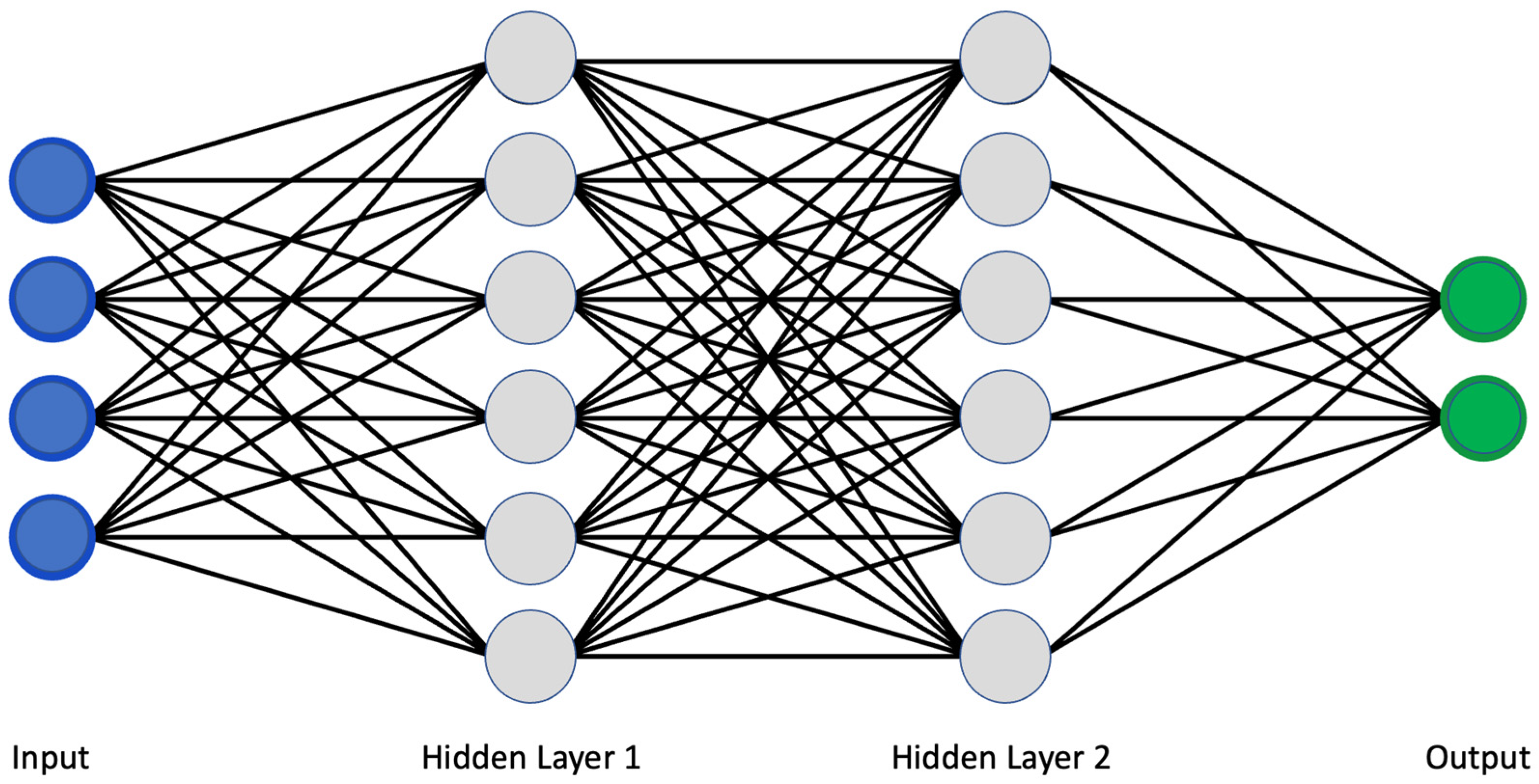
3. Artificial Intelligence in Glaucoma
3.1. Fundus Photography
3.2. Optical Coherence Tomography
3.3. Standard Automatic Perimetry
4. Biomarkers and Precision Medicine in Glaucoma
4.1. Proteins and Hormones as Biomarkers in Glaucoma
4.2. Metabolic Biomarkers in Glaucoma
4.3. Antibodies as Biomarkers in Glaucoma
4.4. Exosomes in Glaucoma
5. Genetics and Precision Medicine in Glaucoma
6. NAD+/NADH Redox State and Glaucoma
7. Conclusions
Author Contributions
Funding
Conflicts of Interest
Abbreviations
References
- Fry, L.E.; Fahy, E.; Chrysostomou, V.; Hui, F.; Tang, J.; van Wijngaarden, P.; Petrou, S.; Crowston, J.G. The coma in glaucoma: Retinal ganglion cell dysfunction and recovery. Prog. Retin. Eye Res. 2018, 65, 77–92. [Google Scholar] [CrossRef] [PubMed]
- Biomarkers Definitions Working Group; Atkinson, A.J., Jr.; Colburn, W.A.; DeGruttola, V.G.; DeMets, D.L.; Downing, G.J.; Hoth, D.F.; Oates, J.A.; Peck, C.C.; Spilker, B.A.; et al. Biomarkers and surrogate endpoints: Preferred definitions and conceptual framework. Clin. Pharmacol. Ther. 2001, 69, 89–95. [Google Scholar] [CrossRef]
- Strimbu, K.; Tavel, J.A. What Are Biomarkers? Curr. Opin. HIV AIDS 2010, 5, 463–466. [Google Scholar] [CrossRef] [PubMed]
- Drucker, E.; Krapfenbauer, K. Pitfalls and limitations in translation from biomarker discovery to clinical utility in predictive and personalised medicine. EPMA J. 2013, 4, 7–10. [Google Scholar] [CrossRef] [PubMed]
- Sumit, S. A Comprehensive Guide to Convolutional Neural Networks—The ELI5 Way. Available online: https://towardsdatascience.com/a-comprehensive-guide-to-convolutional-neural-networks-the-eli5-way-3bd2b1164a53 (accessed on 11 November 2022).
- Nuzzi, R.; Boscia, G.; Marolo, P.; Ricardi, F. The Impact of Artificial Intelligence and Deep Learning in Eye Diseases: A Review. Front. Med. 2021, 8, 710329. [Google Scholar] [CrossRef]
- Ting, D.S.W.; Cheung, C.Y.-L.; Lim, G.; Tan, G.S.W.; Quang, N.D.; Gan, A.; Hamzah, H.; Garcia-Franco, R.; Yeo, I.Y.S.; Lee, S.Y.; et al. Development and Validation of a Deep Learning System for Diabetic Retinopathy and Related Eye Diseases Using Retinal Images from Multiethnic Populations with Diabetes. JAMA 2017, 318, 2211–2223. [Google Scholar] [CrossRef]
- Basodi, S.; Ji, C.; Zhang, H.; Pan, Y. Gradient amplification: An efficient way to train deep neural networks. Big Data Min. Anal. 2020, 3, 196–207. [Google Scholar] [CrossRef]
- Aqeel, A. Difference between AlexNet, VGGNet, ResNet, and Inception. Available online: https://towardsdatascience.com/the-w3h-of-alexnet-vggnet-resnet-and-inception-7baaaecccc96 (accessed on 11 November 2022).
- Quigley, H.A. The Size and Shape of the Optic Disc in Normal Human Eyes. Arch. Ophthalmol. 1990, 108, 51–57. [Google Scholar] [CrossRef]
- Varma, R.; Steinmann, W.C.; Scott, I.U. Expert Agreement in Evaluating the Optic Disc for Glaucoma. Ophthalmology 1992, 99, 215–221. [Google Scholar] [CrossRef]
- Li, Z.; He, Y.; Keel, S.; Meng, W.; Chang, R.T.; He, M. Efficacy of a Deep Learning System for Detecting Glaucomatous Optic Neuropathy Based on Color Fundus Photographs. Ophthalmology 2018, 125, 1199–1206. [Google Scholar] [CrossRef] [Green Version]
- Masumoto, H.; Tabuchi, H.; Nakakura, S.; Ishitobi, N.; Miki, M.; Enno, H. Deep-learning Classifier with an Ultrawide-field Scanning Laser Ophthalmoscope Detects Glaucoma Visual Field Severity. Eur. J. Gastroenterol. Hepatol. 2018, 27, 647–652. [Google Scholar] [CrossRef] [PubMed]
- Li, F.; Yan, L.; Wang, Y.; Shi, J.; Chen, H.; Zhang, X.; Jiang, M.; Wu, Z.; Zhou, K. Deep learning-based automated detection of glaucomatous optic neuropathy on color fundus photographs. Graefe Arch. Clin. Exp. Ophthalmol. 2020, 258, 851–867. [Google Scholar] [CrossRef] [PubMed]
- Hemelings, R.; Elen, B.; Barbosa-Breda, J.; Lemmens, S.; Meire, M.; Pourjavan, S.; Vandewalle, E.; Van De Veire, S.; Blaschko, M.B.; De Boever, P.; et al. Accurate prediction of glaucoma from colour fundus images with a convolutional neural network that relies on active and transfer learning. Acta Ophthalmol. 2020, 98, e94–e100. [Google Scholar] [CrossRef]
- Kolář, R.; Jan, J. Detection of Glaucomatous Eye via Color Fundus Images Using Fractal Dimensions. Radioengineering 2008, 17, 109–114. [Google Scholar]
- Nayak, J.; Acharya, U.R.; Bhat, P.S.; Shetty, N.; Lim, T.-C. Automated Diagnosis of Glaucoma Using Digital Fundus Images. J. Med. Syst. 2009, 33, 337–346. [Google Scholar] [CrossRef] [PubMed]
- Bock, R.; Meier, J.; Nyúl, L.G.; Hornegger, J.; Michelson, G. Glaucoma risk index: Automated glaucoma detection from color fundus images. Med. Image Anal. 2010, 14, 471–481. [Google Scholar] [CrossRef]
- Acharya, U.R.; Dua, S.; Du, X.; S, V.S.; Chua, C.K. Automated Diagnosis of Glaucoma Using Texture and Higher Order Spectra Features. IEEE Trans. Inf. Technol. Biomed. 2011, 15, 449–455. [Google Scholar] [CrossRef] [PubMed]
- Dua, S.; Acharya, U.R.; Chowriappa, P.; Sree, S.V. Wavelet-Based Energy Features for Glaucomatous Image Classification. IEEE Trans. Inf. Technol. Biomed. 2012, 16, 80–87. [Google Scholar] [CrossRef]
- Mookiah, M.R.K.; Acharya, U.R.; Lim, C.M.; Petznick, A.; Suri, J.S. Data mining technique for automated diagnosis of glaucoma using higher order spectra and wavelet energy features. Knowl. Based Syst. 2012, 33, 73–82. [Google Scholar] [CrossRef]
- Noronha, K.P.; Acharya, U.R.; Nayak, K.P.; Martis, R.J.; Bhandary, S.V. Automated classification of glaucoma stages using higher order cumulant features. Biomed. Signal Process. Control. 2014, 10, 174–183. [Google Scholar] [CrossRef]
- Acharya, U.R.; Ng, E.; Eugene, L.W.J.; Noronha, K.P.; Min, L.C.; Nayak, K.P.; Bhandary, S.V. Decision support system for the glaucoma using Gabor transformation. Biomed. Signal Process. Control. 2015, 15, 18–26. [Google Scholar] [CrossRef] [Green Version]
- Issac, A.; Sarathi, M.P.; Dutta, M.K. An adaptive threshold based image processing technique for improved glaucoma detection and classification. Comput. Methods Programs Biomed. 2015, 122, 229–244. [Google Scholar] [CrossRef] [PubMed]
- Raja, C.; Gangatharan, N. A Hybrid Swarm Algorithm for optimizing glaucoma diagnosis. Comput. Biol. Med. 2015, 63, 196–207. [Google Scholar] [CrossRef] [PubMed]
- Singh, A.; Dutta, M.K.; ParthaSarathi, M.; Uher, V.; Burget, R. Image processing based automatic diagnosis of glaucoma using wavelet features of segmented optic disc from fundus image. Comput. Methods Programs Biomed. 2016, 124, 108–120. [Google Scholar] [CrossRef] [PubMed]
- Acharya, U.R.; Bhat, S.; Koh, J.E.; Bhandary, S.V.; Adeli, H. A novel algorithm to detect glaucoma risk using texton and local configuration pattern features extracted from fundus images. Comput. Biol. Med. 2017, 88, 72–83. [Google Scholar] [CrossRef] [PubMed]
- Maheshwari, S.; Pachori, R.B.; Kanhangad, V.; Bhandary, S.V.; Acharya, U.R. Iterative variational mode decomposition based automated detection of glaucoma using fundus images. Comput. Biol. Med. 2017, 88, 142–149. [Google Scholar] [CrossRef]
- Raghavendra, U.; Bhandary, S.V.; Gudigar, A.; Acharya, U.R. Novel expert system for glaucoma identification using non-parametric spatial envelope energy spectrum with fundus images. Biocybern. Biomed. Eng. 2018, 38, 170–180. [Google Scholar] [CrossRef]
- Kausu, T.; Gopi, V.P.; Wahid, K.A.; Doma, W.; Niwas, S.I. Combination of clinical and multiresolution features for glaucoma detection and its classification using fundus images. Biocybern. Biomed. Eng. 2018, 38, 329–341. [Google Scholar] [CrossRef]
- Koh, J.E.; Ng, E.Y.; Bhandary, S.V.; Hagiwara, Y.; Laude, A.; Acharya, U.R. Automated retinal health diagnosis using pyramid histogram of visual words and Fisher vector techniques. Comput. Biol. Med. 2018, 92, 204–209. [Google Scholar] [CrossRef]
- Soltani, A.; Battikh, T.; Jabri, I.; Lakhoua, N. A new expert system based on fuzzy logic and image processing algorithms for early glaucoma diagnosis. Biomed. Signal Process. Control. 2018, 40, 366–377. [Google Scholar] [CrossRef]
- Fu, H.; Cheng, J.; Xu, Y.; Zhang, C.; Wong, D.W.K.; Liu, J.; Cao, X. Disc-Aware Ensemble Network for Glaucoma Screening from Fundus Image. IEEE Trans. Med. Imaging 2018, 37, 2493–2501. [Google Scholar] [CrossRef] [PubMed] [Green Version]
- Christopher, M.; Belghith, A.; Bowd, C.; Proudfoot, J.A.; Goldbaum, M.H.; Weinreb, R.N.; Girkin, C.A.; Liebmann, J.M.; Zangwill, L.M. Performance of Deep Learning Architectures and Transfer Learning for Detecting Glaucomatous Optic Neuropathy in Fundus Photographs. Sci. Rep. 2018, 8, 16685. [Google Scholar] [CrossRef] [PubMed]
- Chai, Y.; Liu, H.; Xu, J. Glaucoma diagnosis based on both hidden features and domain knowledge through deep learning models. Knowl. Based Syst. 2018, 161, 147–156. [Google Scholar] [CrossRef]
- Ahn, J.M.; Kim, S.; Ahn, K.-S.; Cho, S.-H.; Lee, K.B.; Kim, U.S. A deep learning model for the detection of both advanced and early glaucoma using fundus photography. PLoS ONE 2018, 13, e0207982. [Google Scholar] [CrossRef]
- Shibata, N.; Tanito, M.; Mitsuhashi, K.; Fujino, Y.; Matsuura, M.; Murata, H.; Asaoka, R. Development of a deep residual learning algorithm to screen for glaucoma from fundus photography. Sci. Rep. 2018, 8, 14665. [Google Scholar] [CrossRef] [PubMed]
- Mohamed, N.A.; Zulkifley, M.A.; Zaki, W.M.D.W.; Hussain, A. An automated glaucoma screening system using cup-to-disc ratio via Simple Linear Iterative Clustering superpixel approach. Biomed. Signal Process. Control. 2019, 53, 101454. [Google Scholar] [CrossRef]
- Bajwa, M.N.; Malik, M.I.; Siddiqui, S.A.; Dengel, A.; Shafait, F.; Neumeier, W.; Ahmed, S. Two-stage framework for optic disc localization and glaucoma classification in retinal fundus images using deep learning. BMC Med. Inform. Decis. Mak. 2019, 19, 153. [Google Scholar] [CrossRef]
- Liu, H.; Li, L.; Wormstone, I.M.; Qiao, C.; Zhang, C.; Liu, P.; Li, S.; Wang, H.; Mou, D.; Pang, R.; et al. Development and Validation of a Deep Learning System to Detect Glaucomatous Optic Neuropathy Using Fundus Photographs. JAMA Ophthalmol. 2019, 137, 1353–1360. [Google Scholar] [CrossRef]
- Al-Aswad, L.A.; Kapoor, R.; Chu, C.K.; Walters, S.; Gong, D.; Garg, A.; Gopal, K.; Patel, V.; Sameer, T.; Rogers, T.W.; et al. Evaluation of a Deep Learning System for Identifying Glaucomatous Optic Neuropathy Based on Color Fundus Photographs. Eur. J. Gastroenterol. Hepatol. 2019, 28, 1029–1034. [Google Scholar] [CrossRef]
- Asaoka, R.; Tanito, M.; Shibata, N.; Mitsuhashi, K.; Nakahara, K.; Fujino, Y.; Matsuura, M.; Murata, H.; Tokumo, K.; Kiuchi, Y. Validation of a Deep Learning Model to Screen for Glaucoma Using Images from Different Fundus Cameras and Data Augmentation. Ophthalmol. Glaucoma 2019, 2, 224–231. [Google Scholar] [CrossRef]
- Kim, M.; Han, J.C.; Hyun, S.H.; Janssens, O.; Van Hoecke, S.; Kee, C.; De Neve, W. Medinoid: Computer-Aided Diagnosis and Localization of Glaucoma Using Deep Learning †. Appl. Sci. 2019, 9, 3064. [Google Scholar] [CrossRef] [Green Version]
- Orlando, J.I.; Fu, H.; Breda, J.B.; van Keer, K.; Bathula, D.R.; Diaz-Pinto, A.; Fang, R.; Heng, P.-A.; Kim, J.; Lee, J.; et al. REFUGE Challenge: A unified framework for evaluating automated methods for glaucoma assessment from fundus photographs. Med. Image Anal. 2020, 59, 101570. [Google Scholar] [CrossRef] [PubMed]
- Phene, S.; Dunn, R.C.; Hammel, N.; Liu, Y.; Krause, J.; Kitade, N.; Schaekermann, M.; Sayres, R.; Wu, D.J.; Bora, A.; et al. Deep Learning and Glaucoma Specialists. Ophthalmology 2019, 126, 1627–1639. [Google Scholar] [CrossRef] [PubMed]
- Rogers, T.W.; Jaccard, N.; Carbonaro, F.; Lemij, H.G.; Vermeer, K.A.; Reus, N.; Trikha, S. Evaluation of an AI system for the automated detection of glaucoma from stereoscopic optic disc photographs: The European Optic Disc Assessment Study. Eye 2019, 33, 1791–1797. [Google Scholar] [CrossRef] [PubMed]
- Thompson, A.C.; Jammal, A.A.; Medeiros, F.A. A Deep Learning Algorithm to Quantify Neuroretinal Rim Loss from Optic Disc Photographs. Am. J. Ophthalmol. 2019, 201, 9–18. [Google Scholar] [CrossRef]
- Zhao, R.; Chen, X.; Liu, X.; Chen, Z.; Guo, F.; Li, S. Direct Cup-to-Disc Ratio Estimation for Glaucoma Screening via Semi-Supervised Learning. IEEE J. Biomed. Health Inform. 2019, 24, 1104–1113. [Google Scholar] [CrossRef]
- Zhao, R.; Li, S. Multi-indices quantification of optic nerve head in fundus image via multitask collaborative learning. Med. Image Anal. 2020, 60, 101593. [Google Scholar] [CrossRef]
- Schuman, J.; Hee, M.R.; Arya, A.V.; Pedut-Kloizman, T.; Puliafito, C.A.; Fujimoto, J.G.; Swanson, E.A. Optical coherence tomography: A new tool for glaucoma diagnosis. Curr. Opin. Ophthalmol. 1995, 6, 89–95. [Google Scholar] [CrossRef]
- Thompson, A.C.; Jammal, A.A.; Berchuck, S.I.; Mariottoni, E.; Medeiros, F.A. Assessment of a Segmentation-Free Deep Learning Algorithm for Diagnosing Glaucoma from Optical Coherence Tomography Scans. JAMA Ophthalmol. 2020, 138, 333–339. [Google Scholar] [CrossRef]
- Jammal, A.A.; Thompson, A.C.; Mariottoni, E.B.; Berchuck, S.I.; Urata, C.N.; Estrela, T.; Wakil, S.M.; Costa, V.P.; Medeiros, F.A. Human Versus Machine: Comparing a Deep Learning Algorithm to Human Gradings for Detecting Glaucoma on Fundus Photographs. Am. J. Ophthalmol. 2020, 211, 123–131. [Google Scholar] [CrossRef]
- Medeiros, F.A.; Jammal, A.A.; Thompson, A.C. From Machine to Machine. Ophthalmology 2019, 126, 513–521. [Google Scholar] [CrossRef] [PubMed]
- Medeiros, F.A.; Jammal, A.A.; Mariottoni, E.B. Detection of Progressive Glaucomatous Optic Nerve Damage on Fundus Photographs with Deep Learning. Ophthalmology 2021, 128, 383–392. [Google Scholar] [CrossRef] [PubMed]
- Asaoka, R.; Murata, H.; Hirasawa, K.; Fujino, Y.; Matsuura, M.; Miki, A.; Kanamoto, T.; Ikeda, Y.; Mori, K.; Iwase, A.; et al. Using Deep Learning and Transfer Learning to Accurately Diagnose Early Onset Glaucoma from Macular Optical Coherence Tomography Images. Am. J. Ophthalmol. 2019, 198, 136–145. [Google Scholar] [CrossRef] [PubMed]
- Wang, X.; Chen, H.; Ran, A.-R.; Luo, L.; Chan, P.P.; Tham, C.C.; Chang, R.T.; Mannil, S.S.; Cheung, C.Y.; Heng, P.-A. Towards multi-center glaucoma OCT image screening with semi-supervised joint structure and function multi-task learning. Med. Image Anal. 2020, 63, 101695. [Google Scholar] [CrossRef]
- Maetschke, S.; Antony, B.; Ishikawa, H.; Wollstein, G.; Schuman, J.; Garnavi, R. A feature agnostic approach for glaucoma detection in OCT volumes. PLoS ONE 2019, 14, e0219126. [Google Scholar] [CrossRef]
- Ran, A.R.; Cheung, C.Y.; Wang, X.; Chen, H.; Luo, L.-Y.; Chan, P.P.; Wong, M.O.M.; Chang, R.T.; Mannil, S.S.; Young, A.L.; et al. Detection of glaucomatous optic neuropathy with spectral-domain optical coherence tomography: A retrospective training and validation deep-learning analysis. Lancet Digit. Health 2019, 1, e172–e182. [Google Scholar] [CrossRef]
- Russakoff, D.B.; Mannil, S.S.; Oakley, J.D.; Ran, A.R.; Cheung, C.Y.; Dasari, S.; Riyazzuddin, M.; Nagaraj, S.; Rao, H.L.; Chang, D.; et al. A 3D Deep Learning System for Detecting Referable Glaucoma Using Full OCT Macular Cube Scans. Transl. Vis. Sci. Technol. 2020, 9, 12. [Google Scholar] [CrossRef]
- Fu, H.; Baskaran, M.; Xu, Y.; Lin, S.; Wong, D.W.K.; Liu, J.; Tun, T.A.; Mahesh, M.; Perera, S.A.; Aung, T. A Deep Learning System for Automated Angle-Closure Detection in Anterior Segment Optical Coherence Tomography Images. Am. J. Ophthalmol. 2019, 203, 37–45. [Google Scholar] [CrossRef]
- Xu, B.Y.; Chiang, M.; Chaudhary, S.; Kulkarni, S.; Pardeshi, A.A.; Varma, R. Deep Learning Classifiers for Automated Detection of Gonioscopic Angle Closure Based on Anterior Segment OCT Images. Am. J. Ophthalmol. 2019, 208, 273–280. [Google Scholar] [CrossRef]
- Lee, T.; Jammal, A.A.; Mariottoni, E.B.; Medeiros, F.A. Predicting Glaucoma Development with Longitudinal Deep Learning Predictions from Fundus Photographs. Am. J. Ophthalmol. 2021, 225, 86–94. [Google Scholar] [CrossRef]
- Muhammad, H.; Fuchs, T.J.; De Cuir, N.; De Moraes, C.G.; Blumberg, D.M.; Liebmann, J.M.; Ritch, R.; Hood, D.C. Hybrid Deep Learning on Single Wide-field Optical Coherence tomography Scans Accurately Classifies Glaucoma Suspects. Eur. J. Gastroenterol. Hepatol. 2017, 26, 1086–1094. [Google Scholar] [CrossRef] [PubMed]
- Lee, J.; Kim, Y.K.; Park, K.H.; Jeoung, J.W. Diagnosing Glaucoma with Spectral-Domain Optical Coherence Tomography Using Deep Learning Classifier. Eur. J. Gastroenterol. Hepatol. 2020, 29, 287–294. [Google Scholar] [CrossRef] [PubMed]
- Devalla, S.K.; Chin, K.S.; Mari, J.-M.; Tun, T.A.; Strouthidis, N.G.; Aung, T.; Thiéry, A.H.; Girard, M.J.A. A Deep Learning Approach to Digitally Stain Optical Coherence Tomography Images of the Optic Nerve Head. Investig. Opthalmol. Vis. Sci. 2018, 59, 63–74. [Google Scholar] [CrossRef]
- Mariottoni, E.B.; Jammal, A.A.; Urata, C.N.; Berchuck, S.I.; Thompson, A.C.; Estrela, T.; Medeiros, F.A. Quantification of Retinal Nerve Fibre Layer Thickness on Optical Coherence Tomography with a Deep Learning Segmentation-Free Approach. Sci. Rep. 2020, 10, 402. [Google Scholar] [CrossRef] [PubMed]
- Fu, H.; Xu, Y.; Lin, S.; Wong, D.W.K.; Baskaran, M.; Mahesh, M.; Aung, T.; Liu, J. Angle-Closure Detection in Anterior Segment OCT Based on Multilevel Deep Network. IEEE Trans. Cybern. 2020, 50, 3358–3366. [Google Scholar] [CrossRef]
- Hao, H.; Zhao, Y.; Fu, H.; Shang, Q.; Li, F.; Zhang, X.; Liu, J. Anterior Chamber Angles Classification in Anterior Segment OCT Images via Multi-Scale Regions Convolutional Neural Networks. In Proceedings of the 2019 41st Annual International Conference of the IEEE Engineering in Medicine and Biology Society (EMBC), Berlin, Germany, 23–27 July 2019; Volume 2019, pp. 849–852. [Google Scholar] [CrossRef]
- Asaoka, R.; Murata, H.; Iwase, A.; Araie, M. Detecting Preperimetric Glaucoma with Standard Automated Perimetry Using a Deep Learning Classifier. Ophthalmology 2016, 123, 1974–1980. [Google Scholar] [CrossRef]
- Elze, T.; Pasquale, L.R.; Shen, L.; Chen, T.C.; Wiggs, J.L.; Bex, P.J. Patterns of functional vision loss in glaucoma determined with archetypal analysis. J. R. Soc. Interface 2015, 12, 20141118. [Google Scholar] [CrossRef] [PubMed]
- Wang, M.; Shen, L.Q.; Pasquale, L.R.; Petrakos, P.; Formica, S.; Boland, M.V.; Wellik, S.R.; De Moraes, C.G.; Myers, J.S.; Saeedi, O.; et al. An Artificial Intelligence Approach to Detect Visual Field Progression in Glaucoma Based on Spatial Pattern Analysis. Investig. Opthalmol. Vis. Sci. 2019, 60, 365–375. [Google Scholar] [CrossRef]
- DeRoos, L.; Nitta, K.; Lavieri, M.S.; Van Oyen, M.P.; Kazemian, P.; Andrews, C.A.; Sugiyama, K.; Stein, J.D. Comparing Perimetric Loss at Different Target Intraocular Pressures for Patients with High-Tension and Normal-Tension Glaucoma. Ophthalmol. Glaucoma 2021, 4, 251–259. [Google Scholar] [CrossRef]
- Garcia, G.-G.P.; Lavieri, M.S.; Andrews, C.; Liu, X.; Van Oyen, M.P.; Kass, M.A.; Gordon, M.O.; Stein, J.D. Accuracy of Kalman Filtering in Forecasting Visual Field and Intraocular Pressure Trajectory in Patients with Ocular Hypertension. JAMA Ophthalmol. 2019, 137, 1416–1423. [Google Scholar] [CrossRef]
- Kazemian, P.; Lavieri, M.S.; Van Oyen, M.P.; Andrews, C.; Stein, J.D. Personalized Prediction of Glaucoma Progression Under Different Target Intraocular Pressure Levels Using Filtered Forecasting Methods. Ophthalmology 2018, 125, 569–577. [Google Scholar] [CrossRef] [PubMed]
- Kucur, S.; Holló, G.; Sznitman, R. A deep learning approach to automatic detection of early glaucoma from visual fields. PLoS ONE 2018, 13, e0206081. [Google Scholar] [CrossRef] [PubMed]
- Li, F.; Wang, Z.; Qu, G.; Song, D.; Yuan, Y.; Xu, Y.; Gao, K.; Luo, G.; Xiao, Z.; Lam, D.S.C.; et al. Automatic differentiation of Glaucoma visual field from non-glaucoma visual filed using deep convolutional neural network. BMC Med. Imaging 2018, 18, 1–7. [Google Scholar] [CrossRef] [PubMed]
- Berchuck, S.I.; Mukherjee, S.; Medeiros, F.A. Estimating Rates of Progression and Predicting Future Visual Fields in Glaucoma Using a Deep Variational Autoencoder. Sci. Rep. 2019, 9, 35. [Google Scholar] [CrossRef]
- Wen, J.C.; Lee, C.S.; Keane, P.A.; Xiao, S.; Rokem, A.S.; Chen, P.P.; Wu, Y.; Lee, A.Y. Forecasting future Humphrey Visual Fields using deep learning. PLoS ONE 2019, 14, e0214875. [Google Scholar] [CrossRef]
- Cueto, A.F.-V.; Álvarez, L.; García, M.; Álvarez-Barrios, A.; Artime, E.; Cueto, L.F.-V.; Coca-Prados, M.; González-Iglesias, H. Candidate Glaucoma Biomarkers: From Proteins to Metabolites, and the Pitfalls to Clinical Applications. Biology 2021, 10, 763. [Google Scholar] [CrossRef]
- Fiedorowicz, E.; Cieślińska, A.; Kuklo, P.; Grzybowski, A. Protein Biomarkers in Glaucoma: A Review. J. Clin. Med. 2021, 10, 5388. [Google Scholar] [CrossRef]
- Farkas, R.H.; Chowers, I.; Hackam, A.S.; Kageyama, M.; Nickells, R.W.; Otteson, D.C.; Duh, E.J.; Wang, C.; Valenta, D.F.; Gunatilaka, T.L.; et al. Increased Expression of Iron-Regulating Genes in Monkey and Human Glaucoma. Investig. Opthalmol. Vis. Sci. 2004, 45, 1410–1417. [Google Scholar] [CrossRef]
- Lin, S.-C.; Wang, S.Y.; Yoo, C.; Singh, K.; Lin, S.C. Association Between Serum Ferritin and Glaucoma in the South Korean Population. JAMA Ophthalmol. 2014, 132, 1414–1420. [Google Scholar] [CrossRef]
- Wang, J.; Fu, M.; Liu, K.; Wang, N.; Zhang, Z.; Zhou, M.; Xu, X. Matricellular Proteins Play a Potential Role in Acute Primary Angle Closure. Curr. Eye Res. 2018, 43, 771–777. [Google Scholar] [CrossRef]
- González-Iglesias, H.; Álvarez, L.; García, M.; Escribano, J.; Rodríguez-Calvo, P.P.; Fernández-Vega, L.; Coca-Prados, M. Comparative proteomic study in serum of patients with primary open-angle glaucoma and pseudoexfoliation glaucoma. J. Proteomics 2014, 98, 65–78. [Google Scholar] [CrossRef] [PubMed]
- Oddone, F.; Roberti, G.; Micera, A.; Busanello, A.; Bonini, S.; Quaranta, L.; Agnifili, L.; Manni, G. Exploring Serum Levels of Brain Derived Neurotrophic Factor and Nerve Growth Factor across Glaucoma Stages. PLoS ONE 2017, 12, e0168565. [Google Scholar] [CrossRef] [PubMed]
- Ghaffariyeh, A.; Honarpisheh, N.; Shakiba, Y.; Puyan, S.; Chamacham, T.; Zahedi, F.; Zarrineghbal, M. Brain-derived neurotrophic factor in patients with normal-tension glaucoma. Optometry 2009, 80, 635–638. [Google Scholar] [CrossRef]
- Ghaffariyeh, A.; Honarpisheh, N.; Heidari, M.H.; Puyan, S.; Abasov, F. Brain-Derived Neurotrophic Factor as a Biomarker in Primary Open-Angle Glaucoma. Optom. Vis. Sci. 2011, 88, 80–85. [Google Scholar] [CrossRef] [PubMed]
- Gupta, D.; Wen, J.C.; Huebner, J.L.; Stinnett, S.; Kraus, V.B.; Tseng, H.C.; Walsh, M. Cytokine biomarkers in tear film for primary open-angle glaucoma. Clin. Ophthalmol. 2017, 11, 411–416. [Google Scholar] [CrossRef]
- Guo, T.; Guo, L.; Fan, Y.; Fang, L.; Wei, J.; Tan, Y.; Chen, Y.; Fan, X. Aqueous humor levels of TGFβ2 and SFRP1 in different types of glaucoma. BMC Ophthalmol. 2019, 19, 170. [Google Scholar] [CrossRef]
- Li, S.; Zhang, H.; Shao, M.; Li, Y.; Song, Y.; Sun, X.; Cao, W. Association Between 17-β-Estradiol and Interleukin-8 and Visual Field Progression in Postmenopausal Women with Primary Angle Closure Glaucoma. Am. J. Ophthalmol. 2020, 217, 55–67. [Google Scholar] [CrossRef]
- Baker, A.E.; Brautigam, V.M.; Watters, J.J. Estrogen Modulates Microglial Inflammatory Mediator Production via Interactions with Estrogen Receptor β. Endocrinology 2004, 145, 5021–5032. [Google Scholar] [CrossRef]
- Czlonkowska, A.; Ciesielska, A.; Gromadzka, G.; Kurkowska-Jastrzębska, I. Gender Differences in Neurological Disease: Role of Estrogens and Cytokines. Endocrine 2006, 29, 243–256. [Google Scholar] [CrossRef]
- Canizales, L.; Rodriguez, L.; Rivera, C.; Martinez, A.; Mendez, F.; Castillo, A. Low-level expression of SOD1 in peripheral blood samples of patients diagnosed with primary open-angle glaucoma. Biomark. Med. 2016, 10, 1218–1223. [Google Scholar] [CrossRef]
- Mirzaei, M.; Gupta, V.; Deng, L.; Gupta, V.B.; Graham, S. Complement pathway in Alzheimer’s pathology and retinal neurodegenerative disorders—The road ahead. Neural Regen. Res. 2020, 15, 257–258. [Google Scholar] [CrossRef] [PubMed]
- Cha, Y.W.; Kim, S.T. Serum and aqueous humor levels of brain-derived neurotrophic factor in patients with primary open-angle glaucoma and normal-tension glaucoma. Int. Ophthalmol. 2021, 41, 3869–3875. [Google Scholar] [CrossRef] [PubMed]
- Lee, J.Y.; Kim, J.M.; Kim, I.T.; Yoo, C.K.; Won, Y.S.; Kim, J.H.; Kwon, H.S.; Park, K.H. Relationship between Plasma Homocysteine Level and Glaucomatous Retinal Nerve Fiber Layer Defect. Curr. Eye Res. 2017, 42, 918–923. [Google Scholar] [CrossRef] [PubMed]
- Lin, Z.; Huang, S.; Yu, H.; Sun, J.; Huang, P.; Zhong, Y. Analysis of Plasma Hydrogen Sulfide, Homocysteine, and L-Cysteine in Open-Angle Glaucoma Patients. J. Ocul. Pharmacol. Ther. 2020, 36, 649–657. [Google Scholar] [CrossRef]
- López-Riquelme, N.; Villalba, C.; Tormo, C.; Belmonte, A.; Fernandez, C.; Torralba, G.; Hernández, F. Endothelin-1 levels and biomarkers of oxidative stress in glaucoma patients. Int. Ophthalmol. 2015, 35, 527–532. [Google Scholar] [CrossRef]
- Leibovitzh, H.; Cohen, E.; Levi, A.; Kramer, M.; Shochat, T.; Goldberg, E.; Krause, I. Relationship between homocysteine and intraocular pressure in men and women. Medicine 2016, 95, e4858. [Google Scholar] [CrossRef]
- Baumane, K.; Ranka, R.; Laganovska, G. Association of NT-proANP Level in Plasma and Humor Aqueous with Primary Open-Angle Glaucoma. Curr. Eye Res. 2017, 42, 233–236. [Google Scholar] [CrossRef]
- Javadiyan, S.; Burdon, K.P.; Whiting, M.J.; Abhary, S.; Straga, T.; Hewitt, A.W.; Mills, R.; Craig, J. Elevation of Serum Asymmetrical and Symmetrical Dimethylarginine in Patients with Advanced Glaucoma. Investig. Opthalmol. Vis. Sci. 2012, 53, 1923–1927. [Google Scholar] [CrossRef]
- Glantzounis, G.K.; Tsimoyiannis, E.C.; Kappas, A.M.; Galaris, D.A. Uric Acid and Oxidative Stress. Curr. Pharm. Des. 2005, 11, 4145–4151. [Google Scholar] [CrossRef]
- Liu, W.; Guo, R.; Huang, D.; Ji, J.; Gansevoort, R.T.; Snieder, H.; Jansonius, N.M. Co-occurrence of chronic kidney disease and glaucoma: Epidemiology and etiological mechanisms. Surv. Ophthalmol. 2023, 68, 1–16. [Google Scholar] [CrossRef]
- Li, S.; Shao, M.; Li, D.; Tang, B.; Cao, W.; Sun, X. Association of serum uric acid levels with primary open-angle glaucoma: A 5-year case–control study. Acta Ophthalmol. 2019, 97, e356–e363. [Google Scholar] [CrossRef] [PubMed]
- Golubnitschaja, O.; Flammer, J. What Are the Biomarkers for Glaucoma? Surv. Ophthalmol. 2007, 52, S155–S161. [Google Scholar] [CrossRef] [PubMed]
- Kotikoski, H.; Moilanen, E.; Vapaatalo, H.; Aine, E. Biochemical markers of the L-arginine-nitric oxide pathway in the aqueous humour in glaucoma patients. Acta Ophthalmol. Scand. 2002, 80, 191–195. [Google Scholar] [CrossRef] [PubMed]
- Baudouin, C.; Garcher, C.; Haouat, N.; Bron, A.; Gastaud, P. Expression of Inflammatory Membrane Markers by Conjunctival Cells in Chronically Treated Patients with Glaucoma. Ophthalmology 1994, 101, 454–460. [Google Scholar] [CrossRef] [PubMed]
- Beutgen, V.M.; Perumal, N.; Pfeiffer, N.; Grus, F.H. Autoantibody Biomarker Discovery in Primary Open Angle Glaucoma Using Serological Proteome Analysis (SERPA). Front. Immunol. 2019, 10, 381. [Google Scholar] [CrossRef]
- Hohenstein-Blaul, N.V.T.U.; Kunst, S.; Pfeiffer, N.; Grus, F.H. Biomarkers for glaucoma: From the lab to the clinic. Eye 2017, 31, 225–231. [Google Scholar] [CrossRef]
- Grus, F.H.; Joachim, S.C.; Bruns, K.; Lackner, K.J.; Pfeiffer, N.; Wax, M.B. Serum Autoantibodies to α-Fodrin Are Present in Glaucoma Patients from Germany and the United States. Investig. Opthalmol. Vis. Sci. 2006, 47, 968–976. [Google Scholar] [CrossRef]
- Joachim, S.C.; Pfeiffer, N.; Grus, F.H. Autoantibodies in patients with glaucoma: A comparison of IgG serum antibodies against retinal, optic nerve, and optic nerve head antigens. Graefe Arch. Clin. Exp. Ophthalmol. 2005, 243, 817–823. [Google Scholar] [CrossRef] [PubMed]
- Joachim, S.C.; Wuenschig, D.; Pfeiffer, N.; Grus, F.H. IgG antibody patterns in aqueous humor of patients with primary open angle glaucoma and pseudoexfoliation glaucoma. Mol. Vis. 2007, 13, 1573–1579. [Google Scholar]
- Tezel, G.; Thornton, I.L.; Tong, M.G.; Luo, C.; Yang, X.; Cai, J.; Powell, D.W.; Soltau, J.B.; Liebmann, J.M.; Ritch, R. Immunoproteomic Analysis of Potential Serum Biomarker Candidates in Human Glaucoma. Investig. Opthalmol. Vis. Sci. 2012, 53, 8222–8231. [Google Scholar] [CrossRef]
- Schmelter, C.; Perumal, N.; Funke, S.; Bell, K.; Pfeiffer, N.; Grus, F.H. Peptides of the variable IgG domain as potential biomarker candidates in primary open-angle glaucoma (POAG). Hum. Mol. Genet. 2017, 26, 4451–4464. [Google Scholar] [CrossRef] [PubMed]
- Liu, J.; Jiang, F.; Jiang, Y.; Wang, Y.; Li, Z.; Shi, X.; Zhu, Y.; Wang, H.; Zhang, Z. Roles of Exosomes in Ocular Diseases. Int. J. Nanomed. 2020, 15, 10519–10538. [Google Scholar] [CrossRef] [PubMed]
- Lerner, N.; Avissar, S.; Beit-Yannai, E. Extracellular vesicles mediate signaling between the aqueous humor producing and draining cells in the ocular system. PLoS ONE 2017, 12, e0171153. [Google Scholar] [CrossRef] [PubMed]
- Lerner, N.; Schreiber-Avissar, S.; Beit-Yannai, E. Extracellular vesicle-mediated crosstalk between NPCE cells and TM cells result in modulation of Wnt signalling pathway and ECM remodelling. J. Cell. Mol. Med. 2020, 24, 4646–4658. [Google Scholar] [CrossRef] [PubMed]
- Stamer, W.; Hoffman, E.; Luther, J.; Hachey, D.; Schey, K. Protein profile of exosomes from trabecular meshwork cells. J. Proteomics 2011, 74, 796–804. [Google Scholar] [CrossRef] [PubMed]
- Mead, B.; Ahmed, Z.; Tomarev, S. Mesenchymal Stem Cell–Derived Small Extracellular Vesicles Promote Neuroprotection in a Genetic DBA/2J Mouse Model of Glaucoma. Investig. Opthalmol. Vis. Sci. 2018, 59, 5473–5480. [Google Scholar] [CrossRef]
- Pan, D.; Chang, X.; Xu, M.; Zhang, M.; Zhang, S.; Wang, Y.; Luo, X.; Xu, J.; Yang, X.; Sun, X. UMSC-derived exosomes promote retinal ganglion cells survival in a rat model of optic nerve crush. J. Chem. Neuroanat. 2019, 96, 134–139. [Google Scholar] [CrossRef]
- Liu, Y.; Allingham, R.R. Major review: Molecular genetics of primary open-angle glaucoma. Exp. Eye Res. 2017, 160, 62–84. [Google Scholar] [CrossRef]
- Khawaja, A.P.; Viswanathan, A.C. Are we ready for genetic testing for primary open-angle glaucoma? Eye 2018, 32, 877–883. [Google Scholar] [CrossRef]
- Sharma, R.; Grover, A. Myocilin-associated Glaucoma: A Historical Perspective and Recent Research Progress. Mol. Vis. 2021, 27, 480–493. [Google Scholar]
- Tamm, E.R. Myocilin and glaucoma: Facts and ideas. Prog. Retin. Eye Res. 2002, 21, 395–428. [Google Scholar] [CrossRef]
- Morissette, J.; Côté, G.; Anctil, J.L.; Plante, M.; Amyot, M.; Héon, E.; Trope, G.E.; Weissenbach, J.; Raymond, V. A common gene for juvenile and adult-onset primary open-angle glaucomas confined on chromosome 1q. Am. J. Hum. Genet. 1995, 56, 1431–1442. [Google Scholar] [PubMed]
- Scheetz, T.E.; Faga, B.; Ortega, L.; Roos, B.R.; Gordon, M.O.; Kass, M.A.; Wang, K.; Fingert, J.H. Glaucoma Risk Alleles in the Ocular Hypertension Treatment Study. Ophthalmology 2016, 123, 2527–2536. [Google Scholar] [CrossRef] [PubMed]
- Trikha, S.; Saffari, E.; Nongpiur, M.; Baskaran, M.; Ho, H.; Li, Z.; Tan, P.-Y.; Allen, J.; Khor, C.-C.; Perera, S.A.; et al. A Genetic Variant in TGFBR3-CDC7 Is Associated with Visual Field Progression in Primary Open-Angle Glaucoma Patients from Singapore. Ophthalmology 2015, 122, 2416–2422. [Google Scholar] [CrossRef] [PubMed]
- Whirl-Carrillo, M.; McDonagh, E.M.; Hebert, J.M.; Gong, L.; Sangkuhl, K.; Thorn, C.F.; Altman, R.B.; E Klein, T. Pharmacogenomics Knowledge for Personalized Medicine. Clin. Pharmacol. Ther. 2012, 92, 414–417. [Google Scholar] [CrossRef] [PubMed]
- McCarty, C.A.; Mukesh, B.N.; Kitchner, T.E.; Hubbard, W.C.; Wilke, R.A.; Burmester, J.K.; Patchett, R.B. Intraocular Pressure Response to Medication in a Clinical Setting: The Marshfield Clinic Personalized Medicine Research Project. Eur. J. Gastroenterol. Hepatol. 2008, 17, 372–377. [Google Scholar] [CrossRef] [PubMed]
- Sakurai, M.; Higashide, T.; Ohkubo, S.; Takeda, H.; Sugiyama, K. Association between genetic polymorphisms of the prostaglandin F2α receptor gene, and response to latanoprost in patients with glaucoma and ocular hypertension. Br. J. Ophthalmol. 2014, 98, 469–473. [Google Scholar] [CrossRef]
- Low, S.; Mohamed, R.; Davidson, A.; Papadopoulos, M.; Grassi, P.; Webster, A.R.; Black, G.C.; Foster, P.J.; Garway-Heath, D.F.; Bloom, P.A. A new paradigm for delivering personalised care: Integrating genetics with surgical interventions in BEST1 mutations. Eye 2020, 34, 577–583. [Google Scholar] [CrossRef]
- Jeong, S.; Patel, N.; Edlund, C.K.; Hartiala, J.; Hazelett, D.J.; Itakura, T.; Wu, P.-C.; Avery, R.L.; Davis, J.L.; Flynn, H.W.; et al. Identification of a Novel Mucin Gene HCG22 Associated With Steroid-Induced Ocular Hypertension. Investig. Opthalmol. Vis. Sci. 2015, 56, 2737–2748. [Google Scholar] [CrossRef]
- Fini, M.E.; Schwartz, S.G.; Gao, X.; Jeong, S.; Patel, N.; Itakura, T.; Price, M.O.; Price, F.W.; Varma, R.; Stamer, W.D. Steroid-induced ocular hypertension/glaucoma: Focus on pharmacogenomics and implications for precision medicine. Prog. Retin. Eye Res. 2017, 56, 58–83. [Google Scholar] [CrossRef]
- Patel, N.; Itakura, T.; González, J.M.; Schwartz, S.G.; Fini, M.E. GPR158, an Orphan Member of G Protein-Coupled Receptor Family C: Glucocorticoid-Stimulated Expression and Novel Nuclear Role. PLoS ONE 2013, 8, e57843. [Google Scholar] [CrossRef] [PubMed]
- Yu-Wai-Man, C.; Tagalakis, A.D.; Meng, J.; Bouremel, Y.; Lee, R.M.H.; Virasami, A.; Hart, S.L.; Khaw, P.T. Genotype-Phenotype Associations of IL6 and PRG4 With Conjunctival Fibrosis After Glaucoma Surgery. JAMA Ophthalmol. 2017, 135, 1147–1155. [Google Scholar] [CrossRef] [PubMed] [Green Version]
- Zimmermann, C.; Weger, M.; Faschinger, C.; Renner, W.; Mossböck, G. Role of Interleukin 6–174G>C Polymorphism in Primary Open-Angle Glaucoma. Eur. J. Ophthalmol. 2013, 23, 183–186. [Google Scholar] [CrossRef] [PubMed]
- Lin, K.-H.; Feng, S.-C.; Shen, Y.-C.; Wei, L.-C.; Liang, C.-Y.; Chang, C.-J.; Yang, Y.-Y.; Chiu, C.-H.; Wang, C.-Y. Interleukin-6(-174) Locus Polymorphism and Serum IL-6 Levels in Normal Tension Glaucoma. Ophthalmic Genet. 2014, 35, 255–257. [Google Scholar] [CrossRef]
- Fernando, O.; Tagalakis, A.D.; Awwad, S.; Brocchini, S.; Khaw, P.T.; Hart, S.L.; Yu-Wai-Man, C. Development of Targeted siRNA Nanocomplexes to Prevent Fibrosis in Experimental Glaucoma Filtration Surgery. Mol. Ther. 2018, 26, 2812–2822. [Google Scholar] [CrossRef]
- Megevand, G.S.; Bron, A.M. Personalising surgical treatments for glaucoma patients. Prog. Retin. Eye Res. 2021, 81, 100879. [Google Scholar] [CrossRef]
- McDonnell, F.; Irnaten, M.; Clark, A.F.; O’Brien, C.J.; Wallace, D.M. Hypoxia-Induced Changes in DNA Methyla-tion Alter RASAL1 and TGFβ1 Expression in Human Trabecular Meshwork Cells. PLoS ONE 2016, 11, e0153354. [Google Scholar] [CrossRef]
- Zhou, G.; Liu, B. Single nucleotide polymorphisms of metabolic syndrome-related genes in primary open angle glaucoma. Int. J. Ophthalmol. 2010, 3, 36–42. [Google Scholar] [CrossRef]
- Petriti, B.; Williams, P.; Lascaratos, G.; Chau, K.-Y.; Garway-Heath, D. Neuroprotection in Glaucoma: NAD+/NADH Redox State as a Potential Biomarker and Therapeutic Target. Cells 2021, 10, 1402. [Google Scholar] [CrossRef]
- Nzoughet, J.K.; de la Barca, J.M.C.; Guehlouz, K.; Leruez, S.; Coulbault, L.; Allouche, S.; Bocca, C.; Muller, J.; Amati-Bonneau, P.; Gohier, P.; et al. Nicotinamide Deficiency in Primary Open-Angle Glaucoma. Investig. Opthalmol. Vis. Sci. 2019, 60, 2509–2514. [Google Scholar] [CrossRef]
- Williams, P.A.; Harder, J.M.; Foxworth, N.E.; Cochran, K.E.; Philip, V.M.; Porciatti, V.; Smithies, O.; John, S.W.M. Vitamin B3 modulates mitochondrial vulnerability and prevents glaucoma in aged mice. Science 2017, 355, 756–760. [Google Scholar] [CrossRef] [PubMed]
- Hui, F.; Tang, J.; Williams, P.A.; McGuinness, M.B.; Hadoux, X.; Casson, R.J.; Coote, M.; Trounce, I.A.; Martin, K.R.; van Wijngaarden, P.; et al. Improvement in inner retinal function in glaucoma with nicotinamide (vitamin B3) supplementation: A crossover randomized clinical trial. Clin. Exp. Ophthalmol. 2020, 48, 903–914. [Google Scholar] [CrossRef] [PubMed]
- Salech, F.; Ponce, D.P.; Paula-Lima, A.C.; SanMartin, C.D.; Behrens, M.I. Nicotinamide, a Poly [ADP-Ribose] Polymerase 1 (PARP-1) Inhibitor, as an Adjunctive Therapy for the Treatment of Alzheimer’s Disease. Front. Aging Neurosci. 2020, 12, 255. [Google Scholar] [CrossRef] [PubMed]
- Avery, M.A.; Sheehan, A.E.; Kerr, K.S.; Wang, J.; Freeman, M.R. WldS requires Nmnat1 enzymatic activity and N16–VCP interactions to suppress Wallerian degeneration. J. Cell Biol. 2009, 184, 501–513. [Google Scholar] [CrossRef]
- Braidy, N.; Grant, R.; Adams, S.; Brew, B.; Guillemin, G.J. Mechanism for Quinolinic Acid Cytotoxicity in Human Astrocytes and Neurons. Neurotox. Res. 2009, 16, 77–86. [Google Scholar] [CrossRef]
- Neufeld, A.H.; Hernandez, M.R.; Gonzalez, M. Nitric Oxide Synthase in the Human Glaucomatous Optic Nerve Head. Arch. Ophthalmol. 1997, 115, 497–503. [Google Scholar] [CrossRef]
- Balaiya, S.; Ferguson, L.R.; Chalam, K.V. Evaluation of Sirtuin Role in Neuroprotection of Retinal Ganglion Cells in Hypoxia. Investig. Opthalmol. Vis. Sci. 2012, 53, 4315–4322. [Google Scholar] [CrossRef]
- Fea, A.M.; Novarese, C.; Caselgrandi, P.; Boscia, G. Glaucoma Treatment and Hydrogel: Current Insights and State of the Art. Gels 2022, 8, 510. [Google Scholar] [CrossRef]


| Author | Year | N. of Images | Structure | SEN | SPEC | ACC | AUC |
|---|---|---|---|---|---|---|---|
| Kolar et al. [16] | 2008 | 30 | FD | 93.80% | |||
| Nayak et al. [17] | 2009 | 61 | Morphological | 100% | 80% | 90% | |
| Bock et al. [18] | 2010 | 575 | Glaucoma Risk Index | 73% | 85% | 80% | |
| Acharya et al. [19] | 2011 | 60 | SVM | 91% | |||
| Dua et al. [20] | 2012 | 60 | DWT | 93.3% | |||
| Mookiah et al. [21] | 2012 | 60 | DWT, HOS | 86.7% | 93.3% | 93.3% | |
| Noronha et al. [22] | 2014 | 272 | Higher order cumulant features | 100% | 92% | 92.6% | |
| Acharya et al. [23] | 2015 | 510 | Gabor transform | 89.7% | 96.2% | 93.1% | |
| Isaac et al. [24] | 2015 | 67 | Cropped input image after segmentation | 100% | 90% | 94.1% | |
| Raja et al. [25] | 2015 | 158 | Hybrid PSO | 97.5% | 98.3% | 98.2% | |
| Singh et al. [26] | 2016 | 63 | Wavelet feature extraction | 100% | 90.9% | 94.7% | |
| Acharya et al. [27] | 2017 | 702 | kNN (K = 2) Glaucoma Risk index | 96.2% | 93.7% | 95.7% | |
| Maheshwari et al. [28] | 2017 | 488 | Variational mode decomposition | 93.6% | 95.9% | 94.7% | |
| Raghavendra et al. [29] | 2017 | 1000 | RT, MCT, GIST | 97.80% | 95.8% | 97% | |
| Ting et al. [7] | 2017 | 494,661 | VGGNet | 96.4% | 87.2% | 0.942 | |
| Kausu et al. [30] | 2018 | 86 | Wavelet feature extraction, Morphological | 98% | 97.1% | 97.7% | |
| Koh et al. [31] | 2018 | 2220 | Pyramid histogram of visual words and Fisher vector | 96.73% | 96.9% | 96.7% | |
| Soltani et al. [32] | 2018 | 104 | Randomized Hough transform | 97.8% | 94.8% | 96.1% | |
| Li et al. [12] | 2018 | 48,116 | Inception-v3 | 95.6% | 92% | 92% | 0.986 |
| Fu et al. [33] | 2018 | 8109 | Disc-aware ensemble network (DENet) | 85% | 84% | 84% | 0.918 |
| Raghavendra et al. [29] | 2018 | 1426 | Eighteen-layer CNN | 98% | 98.30% | 98% | |
| Christopher et al. [34] | 2018 | 14,822 | VGG6, Inception-v3, ResNet50 | 84–92% | 83–93% | 0.91–0.97 | |
| Chai et al. [35] | 2018 | 2000 | MB-NN | 92.33% | 90.9% | 91.5% | |
| Ahn et al. [36] | 2018 | 1542 | Inception-v3 Custom 3-layer CNN | 84.5% 87.9% | 0.93 0.94 | ||
| Shibata et al. [37] | 2018 | 3132 | ResNet-18 | 0.965 | |||
| Mohamed et al. [38] | 2019 | 166 | Simple Linear Iterative Clustering (SLIC) | 97.6% | 92.3% | 98.6% | |
| Bajwa et al. [39] | 2019 | 780 | R-CNN | 71.2% | 0.874 | ||
| Liu et al. [40] | 2019 | 241,032 | ResNet (local validation) | 96.2% | 97.7% | 0.996 | |
| Al-Aswad et al. [41] | 2019 | 110 | ResNet-50 | 83.7% | 88.2% | 0.926 | |
| Asaoka et al. [42] | 2019 | 3132 | ResNet-34 | 0.965 | |||
| ResNet-34 without augmentation | 0.905 | ||||||
| VGGI I | 0.955 | ||||||
| VGGI 6 | 0.964 | ||||||
| Inception-v3 | 0.957 | ||||||
| Kim et al. [43] | 2019 | 1903 | Inception-V4 | 92% | 98% | 93% | 0.99 |
| Orlando et al. [44] | 2019 | 1200 | Refuge Data Set | 85% | 97.6% | 0.982 | |
| Phene et al. [45] | 2019 | 86,618 | Inception-v3 | 80% | 90.2% | 0.945 | |
| Rogers et al. [46] | 2019 | 94 | ResNet-50 | 80.9% | 86.2% | 83.7% | 0.871 |
| Thompson et al. [47] | 2019 | 9282 | ResNet-34 | 0.945 | |||
| Hemelings et al. [15] | 2020 | 8433 | ResNet-50 | 99% | 93% | 0.996 | |
| Zhao et al. [48] | 2020 | 421 | MFPPNet | 0.90 | |||
| Li et al. [49] | 2020 | 26,585 | ResNet101 | 96% | 93% | 94.1% | 0.992 |
| Author | Year | Outcome Measures | Arch | SEN | SPEC | ACC | AUC | |
|---|---|---|---|---|---|---|---|---|
| OCT Fundus | Thompson et al. [47] | 2019 | 1. Global BMO-MRW prediction | ResNet34 | 0.945 | |||
| 2. Yes glaucoma vs. No glaucoma | ||||||||
| Medeiros et al. [53] | 2019 | 1. RNFL thickness prediction | ResNet34 | 80% | 83.7% | 0.944 | ||
| 2. Glaucoma vs. Suspect/healthy | ||||||||
| Jammal et al. [52] | 2020 | RNFL prediction | ResNet34 | 0.801 | ||||
| Lee et al. [62] | 2021 | RFNL prediction | M2M | |||||
| Medeiros et al. [54] | 2021 | Detection of RFNL thinning from fundus photos | CNN | |||||
| OCT 2D | Asaoka et al. [55] | 2019 | Early POAG vs. no POAG | Novel CNN | 80% | 83.3% | 0.937 | |
| Muhammad et al. [63] | 2017 | Early glaucoma vs. health/suspected eyes | CNN + transfer learning | 93.1% | 0.97 | |||
| Lee et al. [64] | 2020 | GON vs. No GON | CNN (NASNet) | 94.7% | 100% | 0.990 | ||
| Devalla et al. [65] | 2018 | Glaucoma vs. normal | Digital stain of RNFL | 92% | 99% | 94% | ||
| Wang et al. [56] | 2020 | Glaucoma vs. no glaucoma | CNN + transfer learning | 0.979 | ||||
| Thompson et al. [51] | 2020 | POAG vs. no glaucoma | ResNet34 | 95% | 81% | 0.96 | ||
| Pre-perimetric vs. no glaucoma | 95% | 70% | 0.92 | |||||
| Glaucoma with any VF loss (perimetric) vs. no glaucoma | 95% | 80% | 0.97 | |||||
| Mild VF loss vs. no glaucoma | 95% | 85% | 0.92 | |||||
| Moderate VF loss vs. no glaucoma | 95% | 93% | 0.99 | |||||
| Severe VF loss vs. no glaucoma | 95% | 98% | 0.99 | |||||
| Mariottoni et al. [66] | 2020 | Global RNFL thickness value | ResNet34 | |||||
| OCT 3D | Ran et al. [58] | 2019 | Yes GON vs. No GON | CNN (NASNet) | 89% | 96% | 91% | 0.969 |
| 78–90% | 86% | 86% | 0.893 | |||||
| Maetschke et al. [57] | 2019 | POAG vs. no POAG | Feature-agnostic CNN | 0.94 | ||||
| 0.92 | ||||||||
| Russakoff et al. [59] | 2020 | Referable glaucoma vs. non-referable glaucoma | gNet3D-CNN | 0.88 | ||||
| AS-OCT | Fu et al. [60] | 2019 | Open angle vs. Angle closure | VGG-16 + transfer learning | 90% | 92% | 0.96 | |
| Fu et al. [67] | 2019 | Open angle vs. Angle closure | CNN | 0.9619 | ||||
| Xu et al. [61] | 2019 | 1. Open angle vs. angle closure | CNN (ResNet18) + transfer learning | 0.928 | ||||
| 2. Yes/PACD vs. no PACD | 0.964 | |||||||
| Hao et al. [68] | 2019 | Open angle vs. Narrowed Angle vs. Angle closure | MSRCNN | 0.914 |
| Author | Year | Outcomes Mesures | Architecture | SEN | SPEC | ACC | AUC |
|---|---|---|---|---|---|---|---|
| Asaoka et al. [69] | 2016 | Pre-perimetric VFs vs. VFs in healthy eyes | FNN | 0.926 | |||
| Kucur et al. [75] | 2018 | Early glaucomatous VF loss vs. no glaucoma | CNN with Voronoi representation | ||||
| Li et al. [12] | 2018 | Glaucomatous VF loss vs. no glaucoma | VGG I 5 | 93% | 83% | 88% | 0.966 |
| Li et al. [76] | 2018 | Glaucoma vs. Healthy | VGG | 93% | 3% | 0.966 | |
| Berchuck et al. [77] | 2019 | Rates of VF progression compared to SAP MD; Prediction of future VF compared to point-wise regression predictions | Deep variational autoencoder | ||||
| Wen et al. [78] | 2019 | HFA points and Mean Deviation | CascadeNet-5 | ||||
| Kazemian et al. [74] | 2018 | Forecasting visual field progression | Kalman Filtering Forecasting | ||||
| Garcia et al. [73] | 2019 | Forecasting visual field progression | Kalman Filtering Forecasting | ||||
| DeRoos et al. [72] | 2021 | Forecasting visual field progression | Kalman Filtering Forecasting |
| Author | Year | Type of Glaucoma | Biomarker | Type | Mechanism | Sample |
|---|---|---|---|---|---|---|
| Farkas et al. [81] | 2004 | Glaucoma | Transferrin, ceruloplasmin, ferritin | Peptides and Amino Acids | Upregulation | Serum |
| Lin et al. [82] | 2014 | Glaucoma | Ferritin | Peptides and Amino Acids | Upregulation | Serum |
| Wang et al. [83] | 2018 | Acute primary angle closure (APAC), non-glaucomatous cataract | Main matricellular proteins (SPARC, thrombospondin-2, and osteopontin) | Peptides and Amino Acids | Upregulation | Aqueous humor |
| González-Iglesias et al. [84] | 2014 | POAG, PEXG | Panel of 17 most differentially altered proteins | Peptides and Amino Acids | Serum | |
| Ghaffariyeh et al. [95] | 2009 | POAG | Brain-derived neurotrophic factor (BDNF) | Peptides and Amino Acids | Downregulation | Serum |
| Ghaffariyeh et al. [95] | 2011 | NTG | Brain-derived neurotrophic factor (BDNF) | Peptides and Amino Acids | Downregulation | Tear |
| Oddone et al. [85] | 2017 | POAG | Brain-derived neurotrophic factor (BDNF) | Peptides and Amino Acids | Downregulation | Serum |
| Oddone et al. [85] | 2017 | POAG | Nerve growth factor (NGF) | Peptides and Amino Acids | Downregulation | Serum |
| Gupta et al. [88] | 2017 | POAG | Proinflammatory cytokines (IFNγ, IL-10, IL-12p70, IL-13, IL-1β, IL-2, IL-4, IL-6, IL-8, and TNFα) | Peptides and Amino Acids | Human tear samples | |
| Guo et al. [89] | 2019 | POAG, CACG, PACS, AACG | TGFβ2, SFRP1 | Peptides and Amino Acids | Aqueous humor | |
| Li et al. [90] | 2020 | PACG | 17-β-estradiol (E2), interleukin-8 (IL-8) | Peptides and Amino Acids | Serum | |
| Canizales et al. [93] | 2016 | POAG | Superoxide dismutase 1 (SOD1) | Hormones and enzymes | Peripheral blood | |
| Mirzaei et al. [96] | 2020 | Glaucoma | Complement pathway | Peptides and Amino Acids | Regulation | Peripheral blood |
| Author | Year | Type of Glaucoma | Biomarker | Type | Mechanism | Sample |
|---|---|---|---|---|---|---|
| Lee et al. [96] | 2017 | Glaucomatous RNFL Defect | Homocysteine (Hcy) | Peptides, Amino Acids | Upregulation | Plasma |
| Lin et al. [97] | 2020 | POAG, NTG, OHT | Homocysteine (Hcy) | Peptides, Amino Acids | Upregulation | Plasma |
| Lin et al. [97] | 2020 | POAG, NTG, OHT | L-cysteine (Cys) | Peptides, Amino Acids | Upregulation | Plasma |
| López-Riquelme et al. [98] | 2015 | POAG, NTG | Homocysteine (Hcy) | Peptides, Amino Acids | Upregulation | Serum |
| López-Riquelme et al. [98] | 2015 | POAG, NTG | Endothelin-1 (ET-1) | Peptides, Amino Acids | Upregulation | Serum |
| Leibovitzh et al. [99] | 2016 | Glaucoma | Homocysteine (Hcy) | Peptides, Amino Acids | Upregulation | Plasma |
| Baumane et al. [100] | 2017 | Glaucoma and cataract | N-terminal fragment of the proatrial natriuretic peptide (NT-proANP, 1–98) | Peptides, Amino Acids | Upregulation | Plasma and aqueous humor |
| Javadiyan et al. [101] | 2012 | Glaucoma | Asymmetric dimethylarginine (ADMA), a dimethylated isomeric derivative of the amino acid l-arginine | Peptides, Amino Acids | Upregulation | Serum |
| Javadiyan et al. [101] | 2012 | Glaucoma | Symmetric dimethylarginine (SDMA), a dimethylated isomeric derivative of the amino acid l-arginine | Peptides, Amino Acids | Upregulation | Serum |
| Li et al. [104] | 2019 | POAG | Uric acid | Serum | ||
| Golubnitschaja et al. [105] | 2007 | Glaucoma | Stress response, apoptosis, DNA repair, cell adhesion, tissue remodeling, transcription regulation, multi-drug resistance, and energy metabolism | Peptides, Amino Acids | Circulating leukocytes in serum | |
| Kotikoski et al. [106] | 2002 | Glaucoma | NOx (nitrite + nitrate), nitrite and cGMP | Peptides, Amino Acids | Serum and aqueous humor | |
| Baudouin et al. [107] | 1994 | POAG | Inflammatory antigens | Peptides, Amino Acids | Upregulation | Conjunctival antigens |
| Author | Year | Type of Glaucoma | Biomarker | Type | Sample |
|---|---|---|---|---|---|
| Hohenstein-Blaul et al. [109] | 2017 | POAG | Anti-GFAP, anti-γ-synuclein, and anti-myoglobin antibody as a control | Autoantibodies and Antibodies | Aqueous humor and tears |
| Beutgen et al. [108] | 2019 | POAG | Antibodies against trabecular meshwork | Autoantibodies and Antibodies | Serum |
| Grus et al. [110] | 2006 | POAG, NTG | IgG autoantibody, α-fodrin | Autoantibodies and Antibodies | Serum |
| Joachim et al. [111] | 2005 | POAG, NTG | IgG | Autoantibodies and Antibodies | Serum |
| Joachim et al. [112] | 2007 | POAG, PEX | IgG (heat shock protein 27, α-enolase, actin, and GAPDH) | Autoantibodies and Antibodies | Aqueous humor |
| Tezel et al. [113] | 2012 | POAG | IgG | Autoantibodies and Antibodies | Serum |
| Schmelter et al. [114] | 2017 | POAG | Autoantibody, IgG | Autoantibodies and Antibodies | Serum |
| Author | Year | Materials | Target | Results |
|---|---|---|---|---|
| Lerner et al. [116] | 2017 | Cultured non-pigmented ciliary epithelium (NPCE) cells | Wnt signaling protein expression in the TM cells | >2-fold decrease in the level of β-catenin in the cytosolic fraction |
| Lerner et al. [117] | 2020 | NPCE primary cells | Wnt proteins in a human primary trabecular meshwork (TM) cells | Diminished pGSK3β phosphorylation and decreased cytosolic levels of β-catenin in primary TM cells. At the molecular level, it downregulated the expression of positive GSKβ regulator-AKT protein but increased the levels of GSKβ negative regulator-PP2A protein in TM cells. |
| Stamer et al. [118] | 2011 | Primary cultures of human TM cell monolayers | TM exosomes have a characteristic exosome protein profile and contain unique proteins, including the glaucoma-causing protein, myocilin. | |
| Mead et al. [119] | 2018 | Bone marrow-derived stem cell (BMSC) small extracellular vesicles (sEV) and control fibroblast-derived sEV were intravitreally injected into 3-month-old DBA/2J mice once a month for 9 months. | Retinal ganglion cell (RGC) neuroprotection promoted by BMSC sEV | DBA/2J mice developed chronic ocular hypertension beginning at 6 months. The delivery of BMSC sEV, but not fibroblast sEV, provided significant neuroprotective effects for RBPMSþ RGC while significantly reducing the number of degenerating axons seen in the optic nerve. BMSC sEV significantly preserved RGC function in 6-month-old mice but provided no benefit at 9 and 12 months. BMSC sEV are an effective neuroprotective treatment in a chronic model of ocular hypertension |
| Pan et al. [120] | 2019 | Umbilical mesenchymal stem cells derived exosomes (UMSC-Exos) in a rat optic nerve crush (ONC) model | UMSC-Exos significantly promoted Brn3a+ RGCs survival in the retinal ganglion cell layer compared with PBS controls. UMSC-Exos also significantly promoted GFAP+ glia cell activation in retina and optic nerve. However, no increase in GAP43+ axon counts in the optic nerve was found after UMSC-Exos treatment. |
| Author | Year | Type of Glaucoma | Gene | Chromosome | Location | Function |
|---|---|---|---|---|---|---|
| Morisette et al. [125] | 1995 | Primary Open Angle Glaucoma | GLC1A | 1q23-q25 | DlS445, DlS416/D1S480 | Glycoprotein with a myosin-like domain, a leucine zipper region and an olfactomedin domain |
| Tamm et al. [124] | 2002 | Primary Open Angle Glaucoma | MYOC | 1q24.3 | Q368X | Glycoprotein with a myosin-like domain, a leucine zipper region and an olfactomedin domain |
| Scheetz et al. [126] | 2016 | Primary Open Angle Glaucoma | TMCO1 | 1q24.1 | Transmembrane protein | |
| Trikha et al. [127] | 2015 | Primary Open Angle Glaucoma | TGFBR3-CDC7 | 1p22.1 | rs1192415 | Cell-surface chondroitin sulfate/heparan sulfate proteoglycan |
| McCarty et al. [129] | 2008 | Primary Open Angle Glaucoma | ||||
| Sakurai et al. [130] | 2014 | Primary Open Angle Glaucoma, Normal Tension Glaucoma, Ocular Hypertension | Prostaglandin F2α (FP) receptor | 1p31.1 | rs12093097 | Prostaglandine receptor |
| Low et al. [131] | 2020 | Malignant Glaucoma | BEST1 | 11q12.3 | c.602 T > C, c.454 C > G, c.481 + 1 G > T, c.914 T > C | Transmembrane protein |
| Fini et al. [133] | 2017 | Steroid-induced Glaucoma | GPR158, HCG22 | 10p12.1 (GRP158), 6p21.33 (HCG22) | Cell-surface protein with seven transmembrane (7TM) domain | |
| Patel et al. [134] | 2013 | Steroid-induced Glaucoma | GRP158 | 10p12.1 | Cell-surface protein with seven transmembrane (7TM) domain | |
| Jeong et al. [132] | 2015 | Steroid-induced Glaucoma | HCG22 | 6p21.33 | ||
| Yu-Wai-Man et al. [135] | 2017 | Primary Open Angle Glaucoma | IL6, PRG4 | 7p15.3 (IL6); 1q31.1 (PRG4) | Cytokine and proteoglycan | |
| Zimmermann et al. [136] | 2013 | Primary Open Angle Glaucoma | IL-6 (IL-6–174G > C) | 7p21 | rs1800795 | Proinflammatory cytokine |
| Lin et al. [137] | 2014 | Normal Tension Glaucoma | IL-6 (IL-6–174G > C) | 7p21 | rs1800795 | Proinflammatory cytokine |
| Zhou and Liu [141] | 2010 | Primary Open Angle Glaucoma | IL-6 | 7p21 | rs1524107 | Proinflammatory cytokine |
| Fernando et al. [138] | 2018 | Primary Open Angle Glaucoma | siRNA nanocomplexes | RNAi induced by double-stranded small interfering RNA | ||
| McDonnel et al. [140] | 2016 | Primary human normal (NTM) with glaucomatous (GTM) cells; NTM cells under hypoxic conditions | TGFβ1, RASAL1 | 19q13.2 (TGFβ1), 12q24.13 (RASAL1) | Profibrotic factors |
| Author | Year | Disease | Biomarker | Type | Mechanism | Sample |
|---|---|---|---|---|---|---|
| Petriti et al. [142] | 2021 | POAG | NAD+/NADH redox state | Vitamin | Upregulation | Lymphocytes from blood |
| Kouassi Nzoughet et al. [143] | 2019 | POAG | Nicotinamide | Vitamin | Downregulation | Plasma |
| Williams et al. [144] | 2017 | POAG | Nicotinamide Adenine Dinucleotide | Vitamin | Downregulation | Plasma |
| Hui et al. [145] | 2020 | POAG | Nicotinamide | Vitamin | Downregulation | Plasma |
| Salech et al. [146] | 2020 | Alzheimer’s Disease | Nicotinamide | Vitamin | Downregulation | Cerebrospinal fluid, microglia |
| Avery et al. [147] | 2009 | Slow Wallerian degeneration | Nicotinamide mononucleotide adenylyltransferase | Enzyme | Upregulation | Axons |
| Braidly et al. [148] | 2009 | Brain diseases | Inducible NOS, neuronal NOS | Enzyme | Upregulation | Human brain cells |
| Neufeld et al. [149] | 1997 | POAG | NOS-1, NOS-2, NOS-3 | Enzyme | Upregulation | Optics nerve head |
| Balayage et al. [150] | 2012 | Glaucoma and optic neuropathy | SIRT1 | Histone deacetylase | Uperegulation | Retinal ganglion cell |
Disclaimer/Publisher’s Note: The statements, opinions and data contained in all publications are solely those of the individual author(s) and contributor(s) and not of MDPI and/or the editor(s). MDPI and/or the editor(s) disclaim responsibility for any injury to people or property resulting from any ideas, methods, instructions or products referred to in the content. |
© 2023 by the authors. Licensee MDPI, Basel, Switzerland. This article is an open access article distributed under the terms and conditions of the Creative Commons Attribution (CC BY) license (https://creativecommons.org/licenses/by/4.0/).
Share and Cite
Fea, A.M.; Ricardi, F.; Novarese, C.; Cimorosi, F.; Vallino, V.; Boscia, G. Precision Medicine in Glaucoma: Artificial Intelligence, Biomarkers, Genetics and Redox State. Int. J. Mol. Sci. 2023, 24, 2814. https://doi.org/10.3390/ijms24032814
Fea AM, Ricardi F, Novarese C, Cimorosi F, Vallino V, Boscia G. Precision Medicine in Glaucoma: Artificial Intelligence, Biomarkers, Genetics and Redox State. International Journal of Molecular Sciences. 2023; 24(3):2814. https://doi.org/10.3390/ijms24032814
Chicago/Turabian StyleFea, Antonio Maria, Federico Ricardi, Cristina Novarese, Francesca Cimorosi, Veronica Vallino, and Giacomo Boscia. 2023. "Precision Medicine in Glaucoma: Artificial Intelligence, Biomarkers, Genetics and Redox State" International Journal of Molecular Sciences 24, no. 3: 2814. https://doi.org/10.3390/ijms24032814