Article

The Effect of the Tau Protein on D. melanogaster Lifespan Depends on GSK3 Expression and Sex

by
Ekaterina R. Veselkina
1,†,
Mikhail V. Trostnikov
1,2,†,
Natalia V. Roshina
1,3 and
Elena G. Pasyukova
1,*
1
Institute of Molecular Genetics, National Research Centre “Kurchatov Institute”, 123182 Moscow, Russia
2
Skolkovo Institute of Science and Technology, 121205 Moscow, Russia
3
Vavilov Institute of General Genetics, Russian Academy of Sciences, 119991 Moscow, Russia
*
Author to whom correspondence should be addressed.
These authors contributed equally to this work.
Int. J. Mol. Sci. 2023, 24(3), 2166; https://doi.org/10.3390/ijms24032166
Submission received: 5 December 2022 / Revised: 16 January 2023 / Accepted: 18 January 2023 / Published: 21 January 2023
(This article belongs to the Special Issue Gender-Related Molecular Mechanism in Neurological Disorders)

Abstract

:
The microtubule-associated conserved protein tau has attracted significant attention because of its essential role in the formation of pathological changes in the nervous system, which can reduce longevity. The study of the effects caused by tau dysfunction and the molecular mechanisms underlying them is complicated because different forms of tau exist in humans and model organisms, and the changes in protein expression can be multidirectional. In this article, we show that an increase in the expression of the main isoform of the Drosophila melanogaster tau protein in the nervous system has differing effects on lifespan depending on the sex of individuals but has no effect on the properties of the nervous system, in particular, the synaptic activity and distribution of another microtubule-associated protein, Futsch, in neuromuscular junctions. Reduced expression of tau in the nervous system does not affect the lifespan of wild-type flies, but it does increase the lifespan dramatically shortened by overexpression of the shaggy gene encoding the GSK3 (Glycogen Synthase Kinase 3) protein kinase, which is one of the key regulators of tau phosphorylation levels. This effect is accompanied by the normalization of the Futsch protein distribution impaired by shaggy overexpression. The results presented in this article demonstrate that multidirectional changes in tau expression can lead to effects that depend on the sex of individuals and the expression level of GSK3.

1. Introduction

Understanding the basis of various pathologies of the nervous system and the associated shortening of lifespan requires the identification of the most significant molecular triggers of related diseases. One of the likely candidates for this role is the tau protein, which belongs to the family of microtubule-associated proteins (MAPs), provides microtubule stability in axons [1,2] and plays a role in the control of neurogenesis and synaptic plasticity [3,4]. The impaired function and/or aggregation of the tau protein is associated with the development of Alzheimer’s and Parkinson’s diseases [5], as well as a number of diseases combined in the group of tauopathies [6,7].
Of all the possible posttranslational modifications of tau, it is assumed that phosphorylation plays the most significant role in the formation of toxic insoluble aggregated forms [8]. Although tau activity is normally regulated by phosphorylation, its hyperphosphorylation across a number of epitopes appears to be closely associated with neurodegenerative diseases [9,10]. For example, among the eighty-five potential phosphorylation sites in one of the isoforms of normal human tau, about forty have been predicted to be associated with pathological changes [11,12]. Tau phosphorylation level plays a significant role in maintaining cytoskeleton stability, determining the level of tau association with microtubules and their assembly and stability, as well as axonal transport [13,14,15]. Changes in tau interaction with the cytoskeleton lead to the disruption of the synaptic transmission, behavior, and memory [16,17]. In Drosophila models with transgenic human tau, it has been demonstrated that learning and memory are affected by Ser238, Thr245, and Ser262 phosphorylation [18,19,20]. In this regard, the identification of key regulators that phosphorylate this protein is essential.
One of the most important proteins involved in tau phosphorylation is the protein kinase GSK3 (Glycogen Synthase Kinase 3). The modulation of GSK3 activity can both enhance and attenuate the effects caused by altered tau expression [21,22,23]. The negative effects of the GSK3 overexpression in the nervous system of mice can be reduced by tau knockout, while the GSK3 deficiency leads to the decreased phosphorylation of the tau protein [24,25]. In Drosophila, the negative consequences of the overexpression of one of the isoforms of human tau are greatly enhanced by the increased expression of the Drosophila shaggy (sgg) gene encoding GSK3 [26]. It has been demonstrated that this regulation can apparently be mediated by the participation of GSK3 in the noncanonical Wnt pathway [27]. However, a high level of tau phosphorylation is not the only cause of protein toxicity: both increased and decreased GSK3 activity against tau can lead to a pathological phenotype. Apparently, the level of phosphorylation determines the optimal pattern of tau association with microtubules, and its deviation in either direction can have negative consequences [28].
Among the model organisms used to study the molecular basis of neurodegenerative diseases, particularly tauopathies, one of the most popular is the fruit fly D. melanogaster. The nervous system of the fruit fly is much simpler than that of mammals, but, at the same time, Drosophila neuronal genes have a significant number of orthologs in higher vertebrates [29,30]. Despite differences in the number of repeats responsible for microtubule binding, as well as structural differences in the N-terminal end, the tau proteins of the fruit fly and human have a significant, about 66%, level of homology [31]. A similarity has also been demonstrated for the effects of the overexpression of the human and Drosophila tau genes: the increased expression of both in the model organism causes abnormalities in the structure of the visual system [32]. Nevertheless, there seem to be nonoverlapping functions between the Drosophila and human proteins. Although the increased expression of both variants studied in [32] resulted in similar eye changes, only the overexpression of the Drosophila protein under the conditions of the increased expression of par1 gene resulted in lethality. In the same paper, it was demonstrated that the overexpression of the human and fly tau proteins in the Drosophila genetic background has different effects on the proteome. It is also known that the complete replacement of the Drosophila tau gene with the human one affects some functions of the nervous system [33]. Differences in the phenotype caused by changes in the expression of the Drosophila tau gene and the human tau gene can be caused by differences in the species-specific structure and the function of the protein isoforms. Even different isoforms from the same species can differ functionally. In Drosophila, the activation of transgenes corresponding to the human tau gene with three or four microtubule-binding motifs affects the nervous system function in different ways [34]. Differences in the effects of human and fruit fly tau proteins can also be explained by differences in the number of microtubule-binding domains. The Drosophila tau has five of these, not three or four like the human one. The fifth repeat, which is absent in the human protein, is highly homologous to the C. elegans tau [31]. Differences in the structure of the human and fruit fly tau may play a role not only in the maintenance of the basic functions of the protein but also in its interaction with the main regulators.
The identification of risk factors for the development of neurodegenerative diseases allows their causes to be figured out and ways to combat them to be found. The analysis of genetic factors allows to understand the molecular basis of hereditary forms of diseases, and the frequency of alleles associated with the manifestation of the pathologies of the nervous system characterizes the premature death factor burden in the human population. The influence of genetic factors strongly depends on gender and age [35,36,37]. Age is most strongly associated with an increased risk of neurodegenerative diseases as well as with impaired proteostasis, mitochondrial dysfunction, and other changes associated with neurodegeneration [38,39,40]. It has been shown that the level of stress induced in the nervous tissue during aging is dependent on sex and the interaction of sex hormones with specific receptors in the nervous system [41]. Sex differences in the nervous system can be explained by the sex-chromosome dosage effect [42]. It is known that the risk of certain neurodegenerative diseases is higher in women, and in women with moderate cognitive impairment, the tau protein has a wider connectivity network between different brain regions [43,44]. In Drosophila, a number of studies have also demonstrated the role of sex, in particular, sex-dependent differences in tau expression levels in controlling the pathologies of the nervous system. For example, it has been shown that male and female transcriptomes differ after traumatic brain injury in Drosophila, associated with a significant risk of developing neurodegenerative pathologies [45]. Decreased tau expression affected motor function and transcription in a sex-dependent manner, but not all neurological pathologies that appeared after brain injury correlated with tau expression levels [45].
The effects of tau on the nervous system are complex and determined by a set of isoforms and interactions with other proteins, which, in turn, may depend on gender and other factors. This creates the need to analyze the functional role of individual tau isoforms and their interaction with the individual isoforms of partner proteins with regard to sex and age. Given the close relationship between the decline of the nervous system and aging, it is also important to understand how changes in the expression of individual tau isoforms affect lifespan.
In this work, we studied the effect of the increased and decreased expression of the D. melanogaster tau in neurons on lifespan and the properties of the nervous system. We demonstrated that the overexpression of the major tau isoform in the nervous system, which includes all four microtubule binding sites present in the human tau, affected the lifespan of females but not of males, and we did not find any dependence of this effect on the GSK3 protein kinase. In contrast, the effect of the overexpression of the major tau isoform on synaptic activity in females was observed only if the GSK3 target sites were changed and, consequently, the interaction of tau with the protein kinase was presumably disrupted. We showed that tau knockdown, similar to gene deletion [46], had no effect on lifespan, but in females it mitigated the negative effect of GSK3 overexpression on survival. These results suggest an interaction between tau and GSK3 in controlling lifespan. The knockdown of the tau gene affecting most tau isoforms also resulted in the restoration of the distribution of the MAP-related Futsch protein in neuromuscular junctions in females against the background of GSK3 overexpression. Our results may indicate a role for the mutual compensation of the functions of different MAPs, not only in controlling the properties of the nervous system but also in controlling lifespan. Taken together, all our findings indicate that the effects of tau on the nervous system and lifespan are not always correlated, but both depend on sex and a complex interaction between tau and GSK3.

2. Results and Discussion

To assess the effect of increased and decreased tau expression in neurons on lifespan and the properties of the nervous system, we used tau overexpression and knockdown. Fourteen tau isoforms are currently predicted (https://flybase.org/reports/FBgn0266579, accessed on 20 January 2023). We cloned and overexpressed cDNA corresponding to the RA transcript of tau, encoding the major neuronal tau isoform [46], which includes all four microtubule binding sites present in the human tau. To better understand the interaction between tau and GSK3, we also cloned and overexpressed the major isoform with mutations affecting three amino acids that are phosphorylated by GSK3. These sites for mutagenesis were selected based on the comparison of Drosophila and human tau: in humans, the matching phosphorylation sites were shown to be associated with neuronal pathologies [47,48,49]. To mask the selected phosphorylation sites, serine and threonine were replaced with alanine, which does not greatly alter the protein structure and cannot be phosphorylated. For tau knockdown affecting all tau transcripts except RE (JBrowse 3R:27636471..27660425 (flybase.org)), we used the line obtained from Bloomington Drosophila Stock Center USA), which was also used in [50], where a significant decrease in protein amount as a result of knockdown was shown.

2.1. Tau Affects Drosophila Life Span

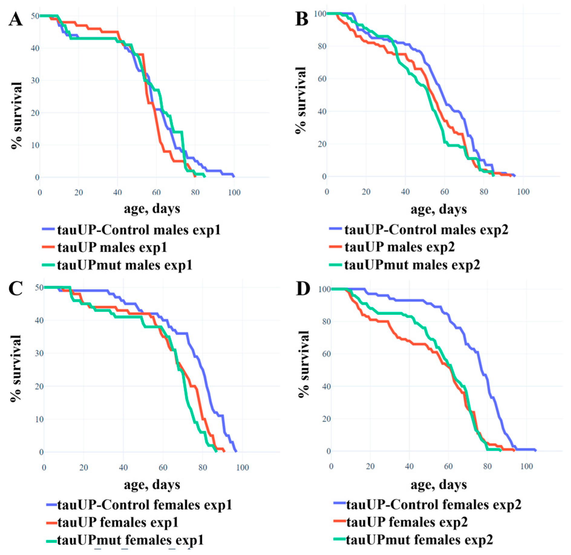
2.1.1. Overexpression of the RA Transcript of the Tau Gene Reduces the Lifespan of Females

To evaluate the effect of the increased expression of the D. melanogaster tau gene on lifespan, we used the transgenic lines with cDNA insertions encoding the normal RA transcript of the fruit fly tau gene (the tauUP line) or the RA transcript with mutations leading to amino acid substitutions in phosphorylation sites serine 103, threonine 151, and serine 343, which are targets of GSK3 protein kinase (the tauUPmut line). The tauUP-Control line with the same genetic background was used as a control. Transgene activation was induced in the nervous system at all stages of development using the pan-neuronal driver line NS. Trait evaluation was performed in two experiments, with sample sizes of 50 (Figure 1A,C) and 100 (Figure 1B,D) individuals per genotype/sex, respectively. In the first experiment, the overexpression of both tau transgenes resulted in a significant decrease in the mean lifespan of females (p = 0.006, Figure 1C) but not of males (p = 0.388, Figure 1A). In the second experiment with a larger sample, the negative effect was stronger in females (p < 0.001, Figure 1C) and a weak but statistically significant negative effect was detected in males (p = 0.016, Figure 1B). Such fluctuations between the results of repeated experiments are quite typical when the effects are virtually absent: in several subsequent experiments, the differences turned out to be significant or not at random [51]. In none of the experiments did the lifespan of individuals with the overexpression of the normal and mutant tau transgene differ significantly from each other.
Thus, we found that the overexpression of both normal and mutant RA transcripts of the tau gene significantly reduced the lifespan of females but had no or very little effect on male survival. This result indicates a specific and sex-dependent role of a particular tau gene transcript in the nervous system. Although the second experiment revealed a weak effect of the increased expression of the RA transcript on the lifespan of males, our results generally indicate its more important role in the nervous system of females. This may be due both to the sex-dependent function of the RA transcript itself and to the specifics of the posttranscriptional regulation of tau protein activity in the individuals of different sexes. These findings take on added importance in view of the fact that Alzheimer’s disease is significantly more common in women [52,53] and may indicate the possibility of a sex-dependent function of certain tau isoforms in humans.
Despite the proven role of GSK3 in phosphorylation and, consequently, the regulation of tau protein activity, we observed the same decrease in lifespan in females overexpressing both the normal RA transcript and the RA transcript with mutations masking epitopes for GSK3 phosphorylation. In order to gain a deeper understanding of this issue, we performed additional experiments.

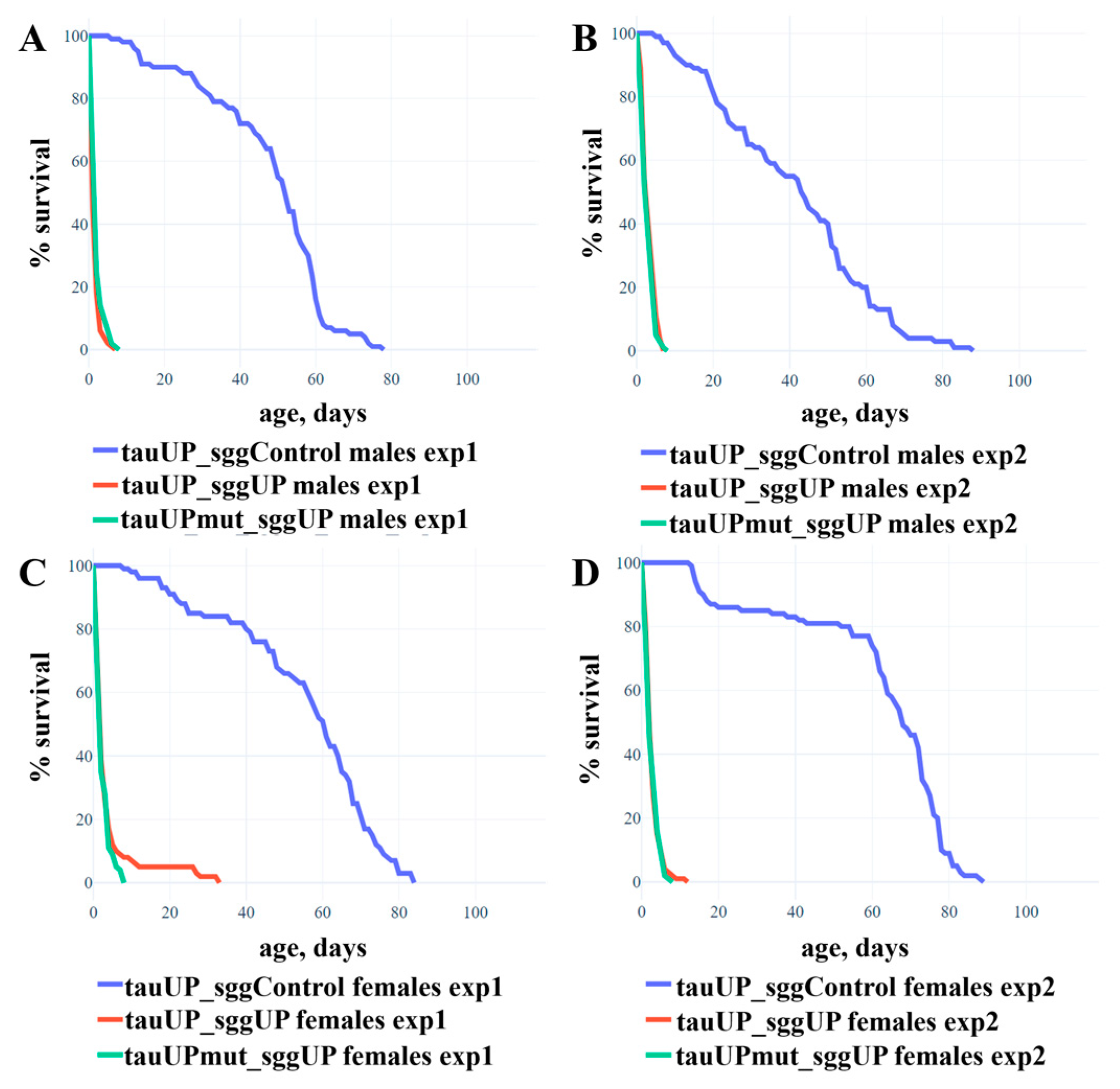
2.1.2. Overexpression of the RA Transcript of the Tau Gene Does Not Affect the Reduction in Lifespan Caused by the Overexpression of the RB Transcript of the Shaggy Gene

In [54], using the sggUP and sggUP-Control lines and the pan-neuronal driver elav, we demonstrated that the overexpression of the RB transcript of the shaggy gene encoding the GSK3 protein kinase of D. melanogaster in the nervous system severely reduces the lifespan of males and females (mean lifespan of males is ~2 days, females ~6 days). Since the tau protein is a target of GSK3, we can assume that a simultaneous increase in tau and shaggy expression will result in a non-additive enhancement of the effect in females, i.e., lethality. However, if the sites at which GSK3 phosphorylates tau are altered, the interaction of the proteins will be disrupted and there will be no enhancement of the effect. To test these assumptions, we used individuals whose genome contained two transgenes: one providing the increased expression of the normal or mutant RA transcript of the tau gene and the other providing the increased expression of the RB transcript of the shaggy gene (the lines tauUP_sggUP and tauUPmut_sggUP). The tauUP_sggUP-Control line with the same genetic background was used as a control. The transgene activation was induced in the nervous system at all the stages of development using the pan-neuronal driver line NS. In two repeated experiments, males and females with two activated transgenes were viable, and although their lifespan was greatly reduced compared to controls, it did not differ from the lifespan of males and females with an overexpression of the shaggy gene alone [54] (Figure 2; the mean lifespan of males was ~2 days, of females ~3 days). Differences in lifespan between males (or females) with an increased expression of the normal tau gene and shaggy gene and males (or females) with an increased expression of the mutant tau gene and shaggy gene were not found.
The result is consistent with the additive, independent effect of tau and shaggy on lifespan. Indeed, in males, we found no significant effect on the lifespan of the tau transgene overexpression itself, nor did the overexpression of this transgene change the effect of shaggy overexpression. In females, a slight decrease in lifespan as a result of the overexpression of the RA transcript of the tau gene was undetectable against a dramatic decrease in lifespan as a result of the overexpression of the RB transcript of the shaggy gene (~3 days versus ~6 days).
Overall, our experiments suggest that, while affecting lifespan separately, tau and GSK3 do not interact with each other. This result can be explained by several reasons. It is possible that, in the lifespan control pathway, tau is not a target of GSK3 at all. This assumption was confirmed both by the fact that the effect of tau and shaggy gene overexpression on lifespan was additive and by the fact that blocking the GSK3 target sites in the tau protein did not affect the observed effects. Perhaps tau is not targeted by GSK3 only at some stages of development, such as embryonic, when the expression of a number of genes, including shaggy, has been shown to be important in controlling the lifespan [54,55,56]. In part, our results can be explained by the fact that the level of phosphorylation of the mutant sites does not affect those properties of the tau isoform encoded by the RA transcript that are essential for its participation in lifespan control. Finally, in our experiments, we upregulated the expression of individual transcripts of both tau (RA transcript) and shaggy (RB transcript), so our results may indicate complex and non-obvious interactions between the individual forms of the respective proteins.

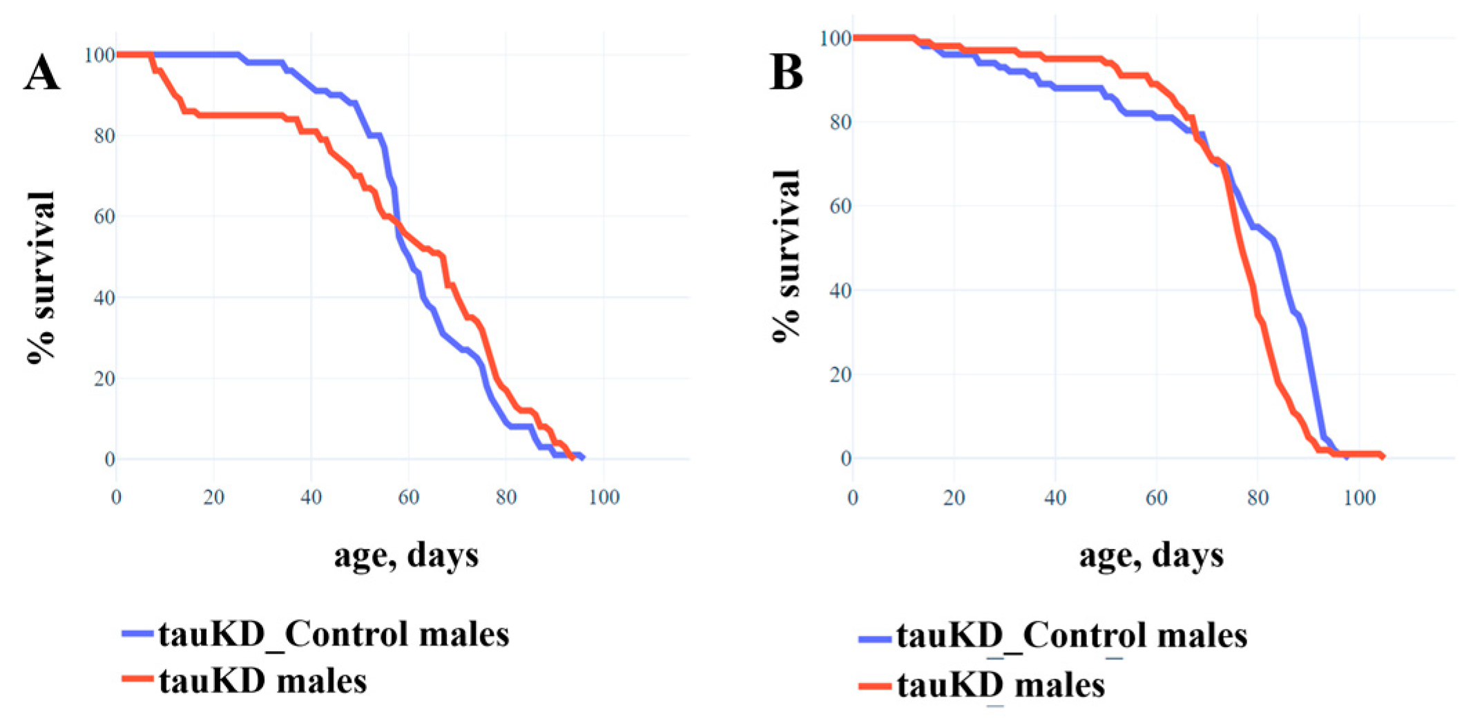
2.1.3. Knockdown of the Tau Gene Has No Effect on the Lifespan of Males and Females

It has previously been demonstrated that, in D. melanogaster, a decrease in tau function caused by the excision of certain exons, presumably encoding microtubule binding domains from the gene, has no effect on lifespan [46]. In this study, using the line tauKD, we evaluated the lifespan of males and females in which tau function was reduced as a result of transgene activity, causing the RNAi knockdown of most of the gene transcripts. The tauKD-Control line with the same genetic background was used as a control. Transgene activation was induced in the nervous system at all stages of development using the pan-neuronal driver line NS. Our data confirmed that a decrease in tau gene expression did not lead to a decrease in lifespan in either males or females (Figure 3A,B). It is worth noting that despite the statistical significance of the result in females (p = 0.011), there was no difference in mean lifespan between control and experimental cohorts (tauKD_Control ~75 days, tauKD ~74 days).
The absence of the effect of the tau gene dysfunction on survival revealed in this and in the earlier paper [46] may be related to the complex organization of this gene in D. melanogaster (http://flybase.org/reports/FBgn0266579, accessed on 20 January 2023). Fourteen tau transcripts have been described, and the functions of some of these may have been preserved when certain exons were excised, as described in [46]. As for the gene knockdown, according to the manufacturer, it does not affect all transcripts. For example, the RE transcript was not affected by either the exon excision or the RNAi knockdown and could compensate, at least with respect to lifespan, for the effect of the absence of other transcripts. In the future, a study of the functional specificity of minor tau transcripts could help to better characterize the function of the gene as a whole.

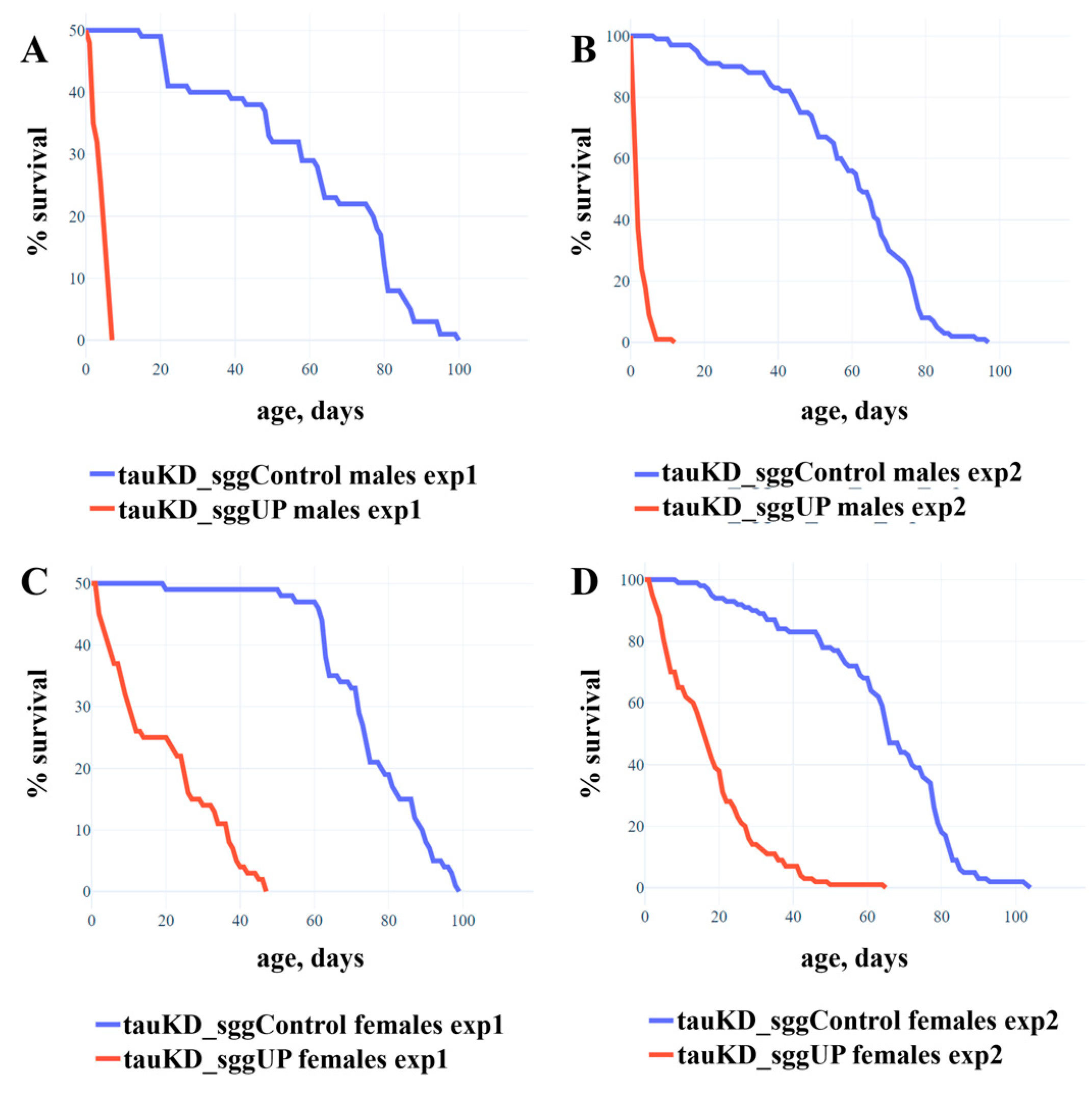
2.1.4. Knockdown of the Tau Gene Positively Affects the Lifespan of Females with the Overexpression of the RB Transcript of the shaggy Gene

The tau gene knockdown used in this work did not cause changes in the lifespan of males and females. This suggests that, since the tau protein is a target of GSK3, a decrease in tau caused by the knockdown of the tau gene would attenuate the severe negative effect of shaggy overexpression on lifespan. To test this assumption, we used individuals from the tauKD_sggUP line whose genome contained two transgenes: one provided the tau knockdown and the other provided the increased expression of the RB transcript of the shaggy gene. The tauKD_sggUP-Control line with the same genetic background was used as a control. Transgene activation was induced in the nervous system at all stages of development using the pan-neuronal driver line NS.
In two repeated experiments, the lifespan of males with decreased tau expression and increased shaggy expression was very low compared to controls (Figure 4A,B; mean lifespan ~3 days) and did not exceed that of males with increased shaggy expression ([54], mean lifespan ~2 days). In two repeated experiments, the lifespan of females with decreased tau expression and increased shaggy expression was greatly reduced compared to controls (Figure 4C,D; mean lifespan ~18 days) but increased three-fold compared to that of females with increased shaggy expression ([54], mean lifespan ~6 days).
Experiments showed that in males, decreasing the amount of tau protein did not mitigate the effect of excess GSK3 on lifespan. This result does not suggest that the tau protein is a target of GSK3 protein kinase in the male lifespan control pathway. However, in females, the decrease in tau protein mitigated the effects of excess GSK3 on lifespan, which, in accordance with our hypothesis, suggests that the tau protein is a target of GSK3 protein kinase in the female lifespan control pathway. Thus, this result shows for the first time that in females, tau and GSK3 interact in lifespan control. The causes of the differences in the control of the lifespan of individuals of different sexes remain unclear. In particular, it is unclear whether in males, tau and GSK3 do not interact in the lifespan control in this case. It cannot be excluded that, in males, tau levels are reduced less than in females and that the amount of remaining protein is sufficient for the full implementation of GSK3 effects on longevity. It is possible that in males, the minor PE tau isoform that was not affected by tau knockdown is more involved in the interaction with GSK3. The involvement of the minor isoform can also explain the fact that, even in females, the tau knockdown did not fully restore the lifespan reduced by GSK3 excess. However, it is likely that the latter fact can be explained by the existence of other GSK3 targets and non-tau lifespan control pathways in which the protein kinase is involved, since its range of interactions is known to be very broad [57].
Another question arises as to why the interaction between tau and GSK3 could be observed only with a simultaneous decrease in tau expression and increase in shaggy expression but not with a simultaneous increase in the expression of both genes. It is possible that a decrease in tau expression actually reduced the amount of phosphorylated protein to mediate the negative effect of GSK3 overexpression on lifespan, whereas an increase in the tau expression in our experiments did not result in a critical increase in target molecules. Given the selective effect of knockdown and overexpression on the individual transcripts of the two genes, it is also possible that the observed phenomena could be explained by a complex interplay between the different isoforms of the two proteins.

2.2. Tau Affects the Properties of the Nervous System

The observed changes in lifespan were detected as a result of transgene activation in the nervous system, which allowed us to hypothesize that sex-dependent changes in lifespan may be accompanied by specific changes in the nervous system. It is known that the level of the phosphorylation of tau protein by GSK3 protein kinase is one of the key factors in the development of pathological changes in the structure and function of nervous tissue [58,59]. Previously, we showed that alterations in GSK3 activity in all neurons caused pathological changes in the morphology and function of the neuromuscular junctions of Drosophila larvae [54]. We hypothesized that these effects are mediated by the influence of GSK3 on tau, and therefore alterations in the expression of tau protein itself may lead to similar changes in the nervous system.

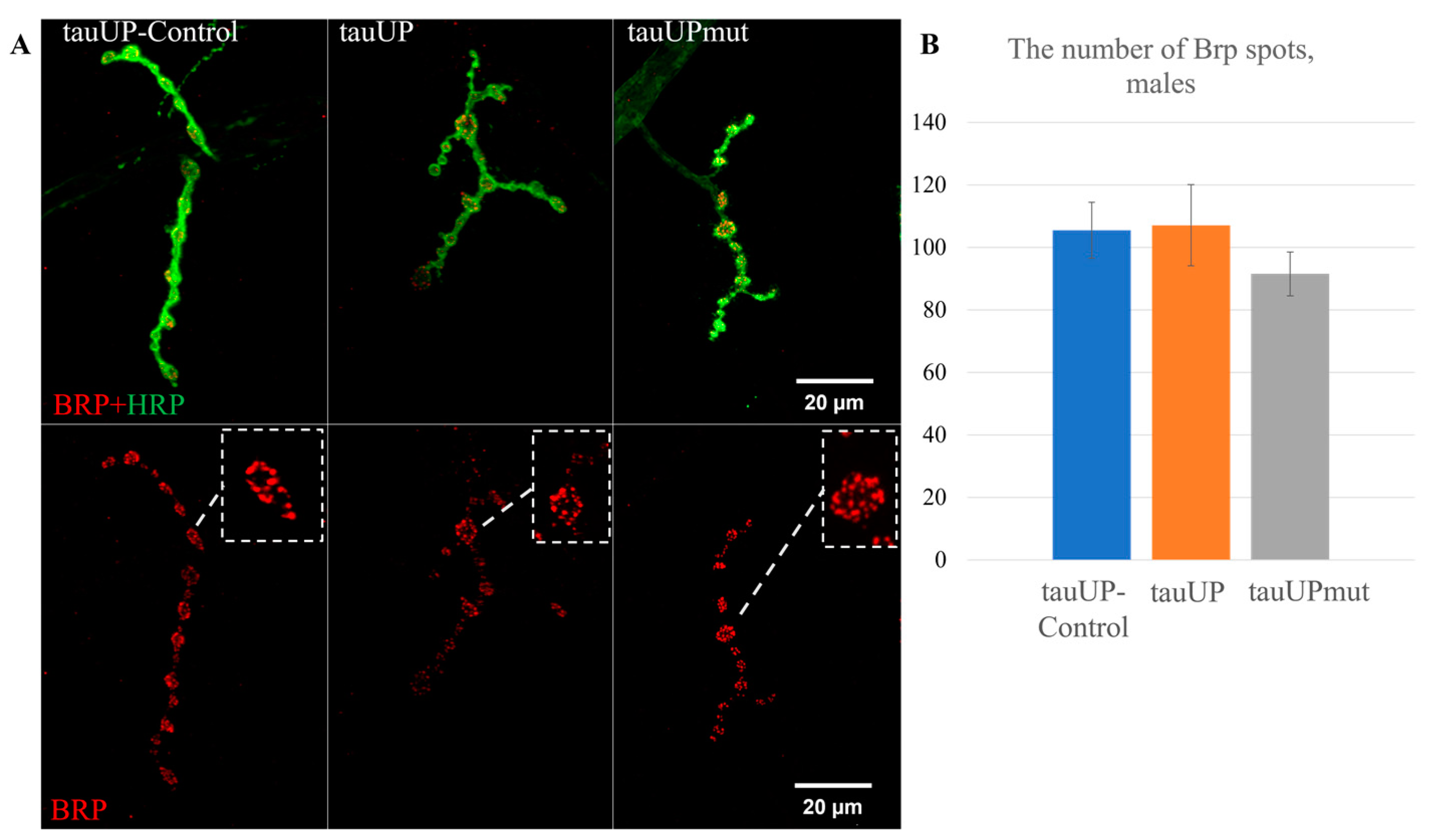
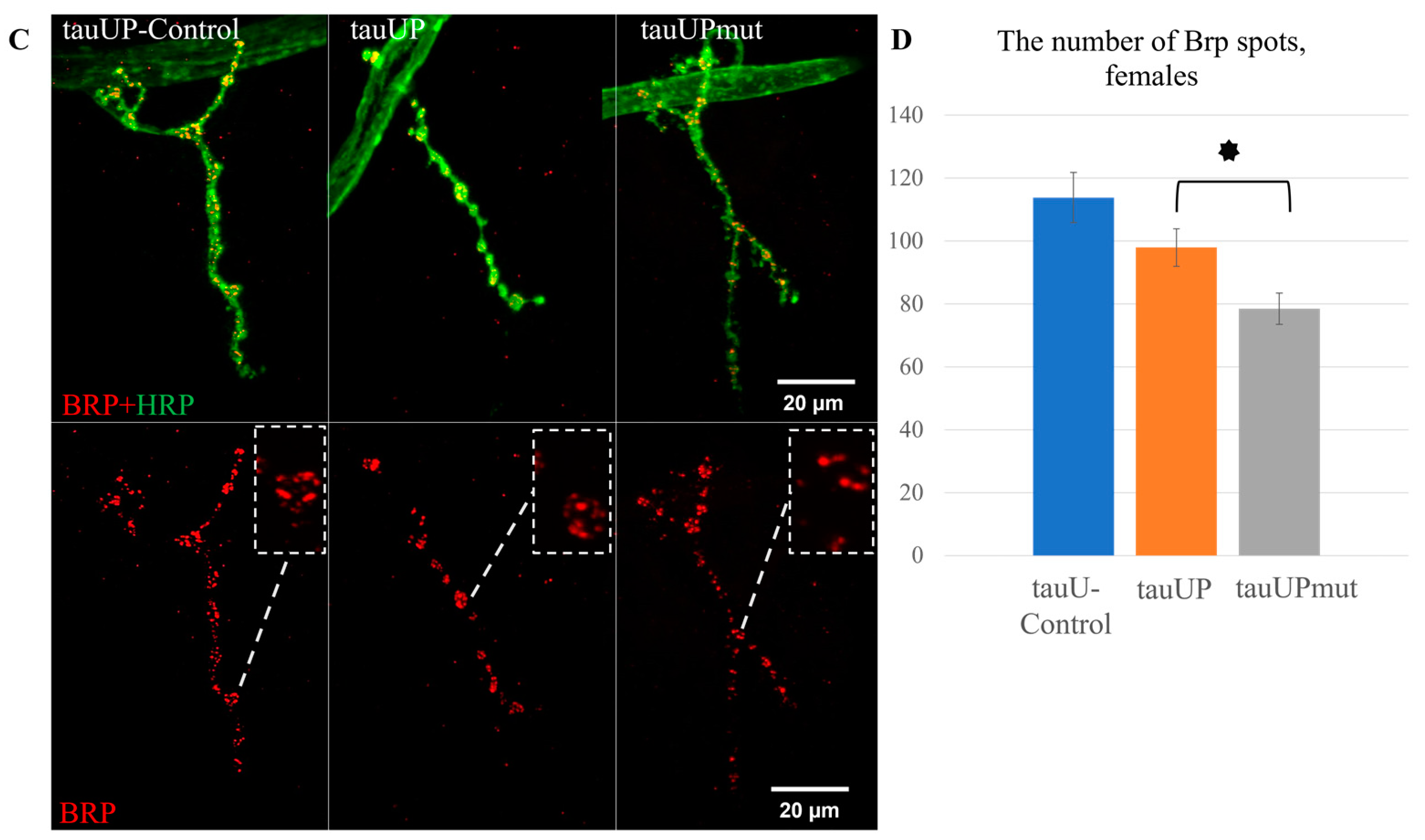
2.2.1. Overexpression of the RA Transcript of the Tau Gene Mutant for GSK3 Phosphorylation Sites Affects Synaptic Activity in Females

To test the hypothesis that a sex-dependent change in lifespan induced by increased tau expression may correlate with changes in synaptic activity, we estimated the number of Brp spots in synaptic contacts in the neuromuscular junctions of males and females with the overexpression of the normal RA transcript of the tau gene and with the overexpression of the RA transcript of the tau gene mutant for GSK3 phosphorylation sites using the tauUP, tauUPmut, and tauUP-Control lines with the same genetic background. Transgene activation was induced in the nervous system at all stages of development using the pan-neuronal driver line NS. The number of active zones of synaptic contacts characterized by the number of spots of Bruchpilot (BRP), a protein specific to active synaptic zones, is often used to indirectly evaluate synaptic activity [60,61].
In males with an activated transgene carrying a normal or mutant copy of the RA transcript of the tau gene, the average number of BRP spots in synaptic contacts did not differ significantly from the control values (Figure 5A,B). This result is consistent with the negligible effect on lifespan observed in males of this genotype described above. Contrary to our initial hypothesis, in females with an overexpression of the normal tau transcript, there was no significant change in the average number of BRP spots in synaptic contacts compared to the controls (Figure 5C,D). Therefore, a significant change, namely a decrease in the number of Brp spots in synaptic contacts in females with an overexpression of mutant tau (p = 0.035, Figure 5D), was all the more surprising.
In our experiments, the increased expression of the normal tau gene in the nervous system did not affect the number of Brp spots in synaptic contacts. These data are consistent with the results showing that overexpression of the human tau, although it causes the impairment of endo- and exocytosis, does not change the active zones of synaptic contacts in neuromuscular junctions [62]. However, the expression of an additional copy of tau encoding a protein with disrupted GSK3 phosphorylation sites led to the impairment of the properties of the nervous system, namely a decrease in synaptic activity evaluated by the number of BRP spots, and only in females. We have already mentioned the fact, important in this context, that some tauopathies, such as Alzheimer’s disease, are significantly more common in women [52,53].
We selected the sites for mutations in the tau gene based on the homology of Drosophila tau regions with human tau regions associated with the pathologies of the nervous system. The nervous system impairment we detected, however, was not due to the increase in tau phosphorylation, as is supposedly the case in the development of human tauopathies [47,48,49,63,64,65,66]. In Drosophila with impaired synaptic activity, the amount of tau protein was increased, but its phosphorylation level did not exceed that of the control. On the contrary, an additional protein with impaired phosphorylation sites should have been phosphorylated to a lesser degree than normal. Consequently, we are not discussing the effect of an increased amount but the effect of conformational changes of this excessive isoform. Thus, the epitopes of the Drosophila tau, which correspond to the epitopes of the human tau associated with the pathologies of the nervous system, are functionally important in Drosophila and are also relevant to the nervous system. However, in Drosophila and humans with an impaired nervous system, the level of phosphorylation of these epitopes is probably different.
Interestingly, the changes in lifespan caused by the overexpression of the normal Drosophila tau were not associated with changes in the number of Brp spots in synaptic contacts. The lack of a link between survival and the nervous system is also confirmed by the fact that lifespan changed equally in flies with unchanged and altered synaptic activity.

2.2.2. Overexpression of the RA Transcript of the Tau Gene Does Not Affect the Distribution of the Microtubule-Associated Futsch Protein

One of the possible causes of the impaired synaptic activity and disrupted transmission of components in the active zones of synaptic contacts may be a dysregulation of the axonal transport [17]. The tau protein belongs to a group of MAPs involved in the development of a wide range of neurodegenerative diseases [67,68]. In addition to tau, in Drosophila, other MAPs, such as Futsch, the human MAP1B homologue, are involved in the maintenance of cytoskeleton organization. Futsch colocalizes with microtubules and is required for cytoskeleton formation during axon growth and synaptogenesis. Futsch, like tau, is the target of phosphorylation by the protein kinase GSK3 [69]. Coordination in the shaping of certain cytoskeletal changes has been shown for the tau and Futsch proteins [70]; consequently, tau and Futsch can compensate for each other’s dysfunction. We analyzed the distribution of the Futsch protein in the neuromuscular junctions of males and females with an activated transgene encoding either the normal or the mutant RA transcript of the tau gene using the tauUP, tauUPmut, and tauUP-Control lines with the same genetic background. Transgene activation was induced in the nervous system at all stages of development using the pan-neuronal driver line NS. In none of the cases studied were males and females with tau overexpression found to differ significantly from individuals of the corresponding control genotypes in the distribution of the Futsch protein (Figure 6A,B).
The activation of expression of the transgene encoding the human tau gene in Drosophila is known to affect microtubules and, in particular, the Futsch protein [71]. However, in our model, the distribution of the MAP-related Futsch protein did not differ from the control in both sexes when either normal or mutant RA transcript of the tau gene was overexpressed. It is likely that the lack of effect may be due to the fact that different tau protein isoforms may interact differently with the Futsch protein.
The absence of irregularities in the Futsch distribution in individuals with an overexpression of mutant tau, demonstrated in this subsection of the results, suggests that the decrease in the number of Brp spots in synaptic contacts in animals of the same genotype described above was not related to changes in the cytoskeleton. It can be assumed that, in this case, the effect of the mutant tau overexpression on synaptic activity is not related to the canonical function of tau, which is to maintain microtubule stability.

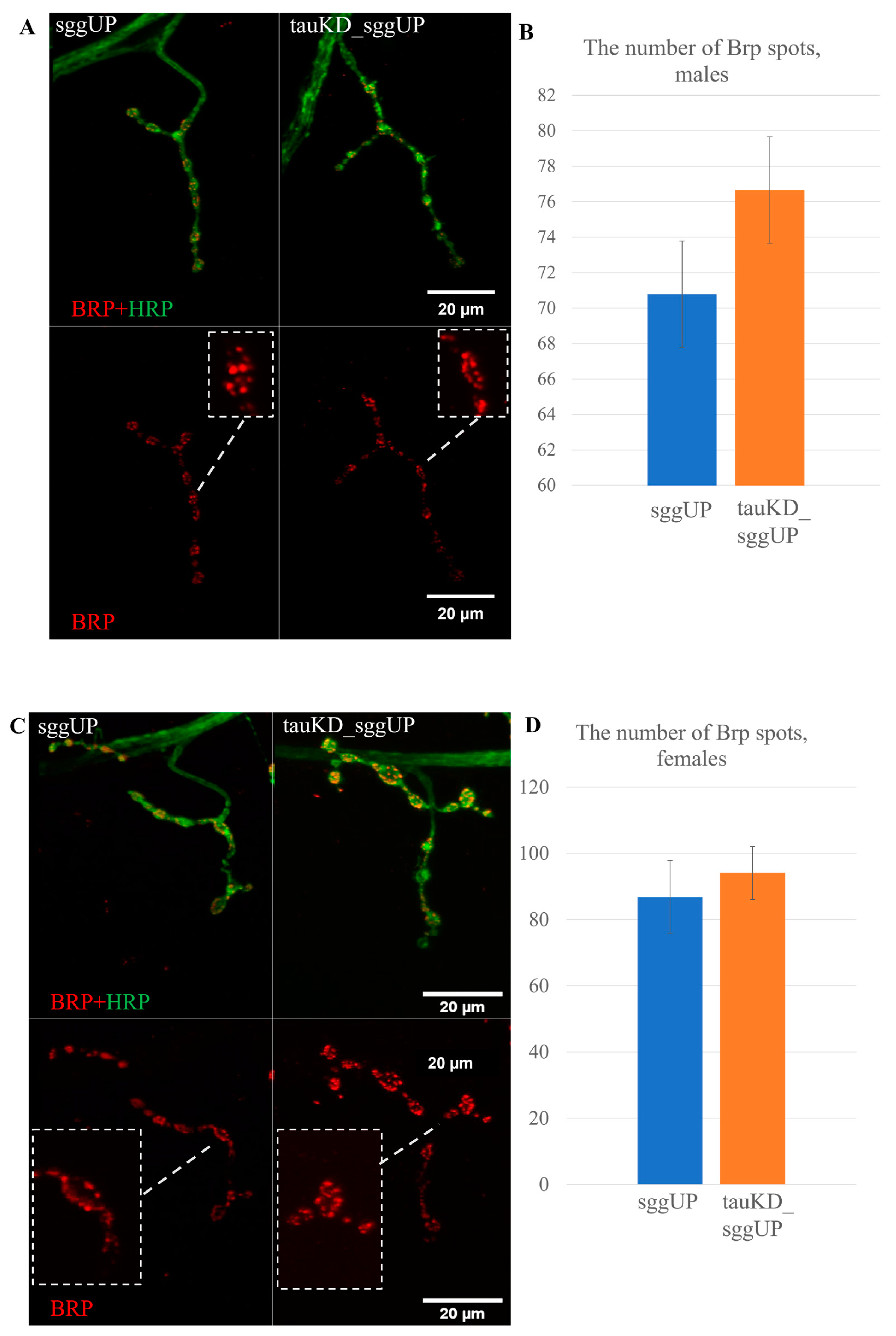
2.2.3. Knockdown of the Tau Gene Does Not Affect Synaptic Activity Reduced by the Overexpression of the RB Transcript of the Shaggy Gene

It was shown in [72] that the deletion of the tau gene in the fruit fly positively affected the number of zones of active synaptic contacts in neuromuscular junctions, reduced in models of neurodegenerative diseases caused by the expansion of trinucleotide repeats. We previously demonstrated the negative effect of the overexpression of the RB transcript of the shaggy gene in the Drosophila nervous system on the number of zones of active synaptic contacts [54]. We hypothesized that, in this case, the decrease in the amount of tau protein resulting from tau knockdown may also have a positive effect on the number of Brp spots in synaptic contacts. To test this assumption, we compared the number of Brp spots in synaptic contacts in individuals with a co-activation of the transgenes that decreased the expression of the tau gene and increased the expression of the RB transcript of the shaggy gene and in individuals with the activation of the transgene that increased the expression of the RB transcript of the shaggy gene using the tauKD_sggUP and sggUP lines. Transgene activation was induced in the nervous system at all stages of development using the pan-neuronal driver line NS. The overexpression of the shaggy gene decreased the number of Brp spots in synaptic contacts compared to controls, which is consistent with our earlier data. The knockdown of the tau gene in the background of shaggy overexpression did not result in a significant change in the number of Brp spots in synaptic contacts in either males or females (Figure 7A–D).
Knockdown of the tau gene in the nervous system, which does not affect the lifespan of males and females per se, positively affected the significantly reduced lifespan of females with a pan-neuronal increase in GSK3 expression. However, it did not affect the reduced number of Brp spots in the synaptic contacts of these females. This result further confirms that changes in lifespan and the properties of the nervous system do not always have common molecular causes. Our data may also indicate that tau is not the main target of GSK3 in the process of the formation of synaptic active zones, and changes in their number may occur due to alternative GSK3 targets. For example, it is known that the activation of GSK3 in mice reduces the expression of synapsin I, a protein of synaptic vesicles. Such a mechanism may underlie memory disorders associated with GSK3 activity [73]. At the same time, a comparison of our results with those demonstrated in [72] shows that the interaction of proteins is in fact based on the interaction of their different isoforms. The functions of the different protein isoforms may have been affected by the deletion [46] and knockdown of tau. It also cannot be ruled out that the decrease in tau expression caused by RNA interference may not have been sufficient to produce an effect comparable to that of the deletion of all exons with predicted microtubule binding sites.

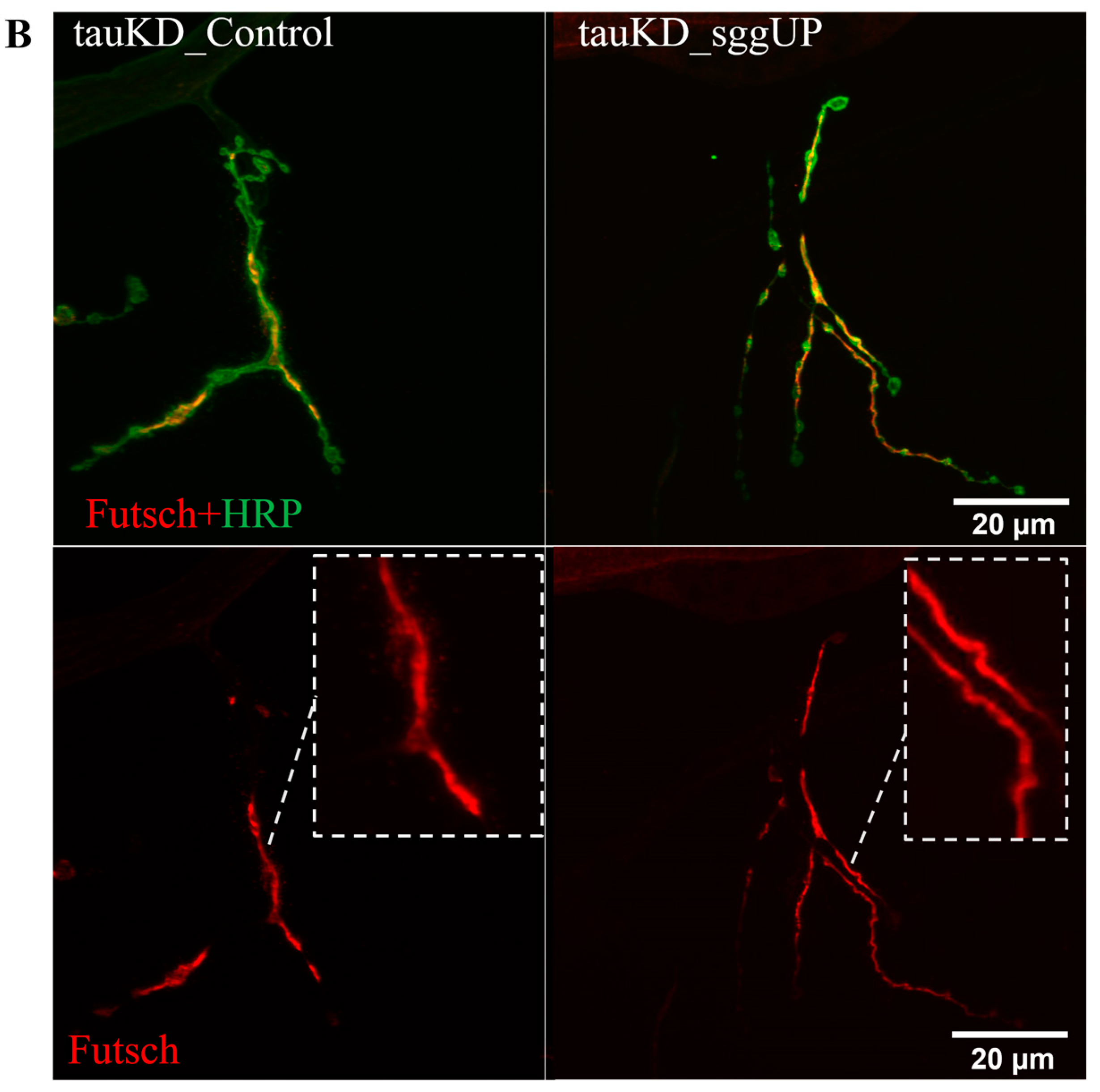
2.2.4. Knockdown of the Tau Gene Positively Affects the Distribution of the Microtubule-Associated Futsch Protein Impaired in Females with the Overexpression of the RB Transcript of the Shaggy Gene

Previously, we demonstrated that the increased expression of the RB transcript of the shaggy gene in the nervous system of male and female Drosophila altered the distribution of the Futsch protein in the synaptic boutons of neuromuscular junctions [54]. Since tau and Futsch can compensate for each other’s dysfunction [70], we analyzed the distribution of the Futsch protein in the neuromuscular junctions of males and females with activated transgenes causing the knockdown of tau gene and the overexpression of the RB transcript of the shaggy gene using the tauKD_sggUP and tauKD_Control lines. The transgene activation was induced in the nervous system at all stages of development using the pan-neuronal driver line NS. In males, the immunohistochemical staining of neuromuscular junctions revealed significant changes in the distribution of Futsch when compared with controls (Figure 8A). In females, no significant differences in the distribution of the Futsch protein from the control were detected (Figure 8B). Thus, a decrease in tau expression in females restored the disturbed distribution of Futsch caused by an increase in shaggy expression.
It remains unclear which molecular mechanism may be responsible for the effect of tau expression on the distribution of Futsch. It can be assumed that the phosphorylation level and stability of the Futsch protein, a direct target of the GSK3 protein kinase, may vary, depending on the total expression of the proteins of the MAP family. However, in this case, an increase rather than a decrease in the amount of tau against the background of GSK3 excess would lead to a compensatory effect, which we did not observe in our experiments. In [46], it was demonstrated that the tau deletion does not trigger compensatory mechanisms affecting the mRNA expression of other genes encoding MAPs, including Futsch; however, in that case, GSK3 expression remained normal. So far, data on the effect of reduced tau expression on the Futsch protein are very scarce, and this issue requires further study.
Of note, in females, tau knockdown against the background of increased shaggy expression leads to the compensation of the negative effect of GSK3 excess on both Futsch distribution and survival in females. Therefore, in this case, the change in tau and GSK3 expression has a coordinated effect on the properties of the nervous system and lifespan.

3. Materials and Methods

3.1. Lines and Crossings

The sggUP line, w [1118]; P{w + mC = UAS-sgg. B}MB5, containing the transgenic construct that encodes the normal PB form of GSK3 was used to provide sgg-RB overexpression upon induction; the co-isogenic sggUP-Control line, w [1118], without any transgenic construct, was used as a control line. The lines described in [74] were obtained from Bloomington Drosophila Stock Center, USA. Flies of the sggUP line have red eyes because w [1118] does not show up due to insertion, and flies of the sggControl line have white eyes, which has little or no effect on lifespan [75].
To obtain lines providing tau-RA overexpression, cDNA corresponding to this transcript was cloned into the pBID-UASC vector (Addgene, Watertown, MA, USA, https://www.addgene.org), which contains an attB site for the phi31 site-specific transformation of Drosophila embryos and a UAS enhancer sequences [55]. One clone with intact tau-RA sequences was sent to Evrogen (Moscow, Russia, https://evrogen.com) for in vitro mutagenesis at sites selected according to the assay described below. The original clone with the normal tau-RA sequence, the clone with the mutant tau-RA sequence, and the line y1 M{vas-int.Dm}ZH-2A w*; M{3xP3-RFP.attP’}ZH-51C with the second chromosome attP phi31 integration site [56] were then used for the transformation performed by BestGene, Inc. (Chino Hills, CA, USA., https://www.thebestgene.com/HomePage.do). In the transgenic lines obtained, most of the auxiliary sequences were excised from the inserted transgenes due to the induction of recombination between the loxP sites present in the pBID-UASC vector.
The tauUP and tauUPmut homozygous transgenic lines, y [1] M{vas-int.Dm}ZH-2A w*; M{3×P3-RFP.attP’}ZH-51C P{w + mC = UAS-tau.A}2M and y1 M{vas-int.Dm}ZH-2A w*; M{3×P3-RFP.attP’}ZH-51C P{w + mC = UAS-tau.A[mut]}2M, containing the transgenic constructs that encode the normal and mutant PA form of Tau, respectively, were used to provide tau-RA overexpression upon induction. The tauUP-Control line, y1 M{vas-int.Dm}ZH-2A w*; M{3xP3-RFP.attP’}ZH-51C, initially used for transformations, was used as a control line with the same genetic background. Flies of all three lines have yellow bodies and white eyes.
The tauKD line, y [1] v [1]; P{y[+t7.7] v[+t1.8] = TRiP.HMS02042}attP40, containing the transgenic construct that encodes dsRNA complementary to tau mRNA was used to provide an RNAi knockdown of endogenous fruit fly tau upon induction and the co-isogenic tauKD-Control line, y [1] v [1]; P{y[+t7.7] = CaryP}attP40, without the transgene encoding dsRNA was used as a control line, as suggested by the manufacturer (DRSC/TRiP Functional Genomics Resources, Boston, MA, USA., https://fgr.hms.harvard.edu/). Previously, the authors of [50] demonstrated a significant decrease in the amount of protein. Flies of the tauKD and the tauKD_Control lines have red eyes of various shades. The y [1] mutation, which was shown to be long lived [75], does not appear in either line.
To analyze the interaction between the tau and shaggy genes, we used standard substitution crosses with balancers to obtain lines with the first chromosome marked with the w [1118] mutation from the sggUP-Control line; the second chromosome with transgenes, providing tau overexpression or knockdown from the tauUP, tauUPmut, or tauKD lines; and the third chromosome providing shaggy overexpression from the sggUP line or the control third chromosome from the sggUP-Control line. In total, the following lines were obtained: tauUP_sggUP, tauUPmut_sggUP, and tauUP_sggUP-Control; tauKD_sggUP, and tauKD_sggUP-Control.
To induce the expression of the transgenic constructs, females from the pan-neuronal driver line NS, P{w[+mW.hs] = GawB}elav[C155] w [1118]; P{w[+mC] = UAS-Dcr-2. D}2, obtained from the Bloomington Drosophila Stock Center were crossed with males from the sggUP, tauUP, tauUP-mut, tauKD, tauUP_sggUP, tauUPmut_sggUP, tauKD_sggUP, or the corresponding control lines. In all tested individuals, including control flies, the same GAL4 driver was present in all experiments, which allowed us to account for the effect of the insertion of the transgene encoding GAL4 on the traits under study. The GAL4 transgene came from females, which also accounted for the maternal effect. In control flies, the chromosomes with the UAS transgenes were absent; instead, the original chromosomes without transgenes, which were used for insertion, were present. The attP phi31 integration site was initially embedded in these control chromosomes and further used for transgene insertions. Thus, the same disruption of the original chromosomal structure was present in both control and experimental lines. This design allowed us to account for the possible effect of the chromosomal disruption per se on lifespan and other phenotypes.
Flies were kept at 25 °C on a standard medium of semolina, sugar, raisins, yeast, and agar with nipagin, propionic acid, and streptomycin.

3.2. Tests for Wolbachia

All lines were tested for the presence of the Drosophila symbiont Wolbachia [76], using a quantitative PCR (MiniOpticon real-time PCR detection system, Bio-rad, (Hercules, CA, USA, https://www.bio-rad.com/)) with primers for the 16S rRNA gene, 5′-CATACCTATTCGAAGGGATAG-3′, and 5′-AGCTTCGAGTGAAACCAATTC-3′ [77].

3.3. Lifespan Assay

Lifespan was measured as previously described in [78]. Five virgin flies of the same genotype and sex, all collected on the same day from cultures with moderate density, were placed in replicate vials. Flies were transferred to vials with fresh food weekly. The number of dead flies was recorded daily. Experiments comparing fly lifespans were conducted simultaneously. Sample sizes were 50 to 100 flies per sex per genotype. The experiments with noteworthy results were repeated with an interval of approximately six months. The lifespan for each fly was estimated as the number of days alive from the day of eclosion to the day of death. Mean lifespans and survival curves were primarily used to characterize lifespan.

3.4. Immunostaining and Microscopy

Male and female third instar larvae ready for pupation were dissected in phosphate-buffered saline (PBS), fixed in 4% paraformaldehyde (Sigma-Aldrich, St. Louis, MO, USA, https://www.sigmaaldrich.com) at room temperature for 20 min, then washed in PBS (3 × 15 min). For immunostaining, preparations were blocked in 4% Normal Goat Serum for one hour at room temperature. Primary antibody incubation was performed overnight at 4 °C, washed in PBS (3 × 15 min), incubated in secondary antibodies for two hours, washed in PBS (5 × 10 min), and placed in a medium for immunofluorescence (VectaShield, Vector Labs, Newark, CA, USA, https://vectorlabs.com/). Neuromuscular junctions were analyzed in the fourth muscle of the third and fourth abdominal segments of larvae. A confocal laser scanning microscope (LSM 900, Zeiss, Jena, Germany, https://www.zeiss.com/), ImageJ 1.53 (http://rsb.info.nih.gov/ij/index.html) and ZEN Blue 3.6 (Zeiss) software were used. Sample sizes were 10 to 15 specimens per genotype per experiment.
The following primary antibodies were used: mouse anti-BRP (mAb NC82, 1:200; Developmental Studies Hybridoma Bank (DSHB, Iowa, IA, USA, https://dshb.biology.uiowa.edu/)) against Bruchpilot (BRP), a protein specific to active synaptic zones and required for vesicle release [60,61]; mouse anti-Futsch (mAb 22C10, 1:200; DSHB) against the microtubule-associated protein Futsch [79]; and Alexa Fluor 488-conjugated goat anti-HRP (1:500, Jackson ImmunoResearch, West Grove, PA, USA, https://www.jacksonimmuno.com/), against Horseradish Peroxidase (HRP), a widely used marker of presynaptic membranes [80]. Goat anti-mouse Cy3 conjugated (1:500, Jackson ImmunoResearch) were used as the secondary antibodies. Antibodies obtained from the DSHB were developed under the auspices of the NICHD and maintained by the University of Iowa, Department of Biology, Iowa City, IA 52242. The mean number of BRP spots in neuromuscular junctions was used to indirectly characterize synaptic activity. Futsch phenotype was qualitatively rated by the protein distribution uniformity inside of neuromuscular junctions.

3.5. Phosphorylation Sites and Mutagenesis

For the mutagenesis of GSK3-associated phosphorylation sites in the D. melanogaster tau protein, we chose the PA isoform because it corresponds to the major transcript of the tau gene [81] and includes all four microtubule binding sites that exist in the human tau. We compared the sequence of the PA isoform of the fruit fly tau (https://flybase.org/reports/FBtr0344741, accessed on 20 January 2023) with the longest 2N4R isoform of the human tau (https://www.ncbi.nlm.nih.gov/protein/NP_005901.2, accessed on 20 January 2023).
The analysis of the available data allowed us to identify a number of phosphorylation sites that were potentially associated with pathological changes in the human tau protein. Of these, we selected the following amino acid residues that are phosphorylated by the GSK3 beta protein kinase: S199, Thr231, and S 396 [47,49,66,82]. Next, using a multiple sequence alignment server (http://tcoffee.crg.cat, accessed on 20 January 2023), the amino acid sequence surrounding the selected human tau phosphorylation site was aligned with the corresponding amino acid sequence of D. melanogaster. The alignment resulted in the following matches:
S199 H. sapiens—S103 D. melanogaster.
Thr231 H. sapiens—Thr151 D. melanogaster.
S396 H. sapiens—S343 D. melanogaster.
The significance of the selected phosphorylation sites in the fruit fly tau protein was predicted using the bioinformatic resource NetPhos (https://services.healthtech.dtu.dk/service.php?NetPhos-3.1, accessed on 20 January 2023, predictive confidence threshold > 0.5). In the course of cloning the Drosophila tau gene (see details in Section 3.1), in order to mask the target phosphorylation sites, serine and threonine in the three selected sites were replaced with alanine, which should not greatly alter the protein structure and cannot be phosphorylated.

3.6. Statistical Analyses

To compare the control and mutant genotypes, Student’s t-test and the nonparametric, distribution-free Kruskal–Wallis test were used for the analyses of the number of Brp spots in neuromuscular junctions. These two tests gave consistent results, so only the results of the Kruskal–Wallis test are reported here. The standard descriptive statistical analysis of lifespan [83,84] was performed to determine the mean lifespan and its accompanying variances, standard deviations, and standard errors; the median, minimum, and maximum lifespans; and the lifespans of the lower and upper quartiles and the 10 and 90 percentiles (Tables S1–S4, Supplementary Materials). Survival curves were estimated using the Kaplan–Meier procedure. The nonparametric, distribution-free Mann–Whitney test and the Kolmogorov–Smirnov test were used to evaluate the statistical significance of the difference between the survival curves.

4. Conclusions

Lifespan is one of the most important quantitative characteristics of living organisms. Closely associated with it, aging is accompanied by a multitude of functional changes in various tissues and organ systems [85]. The nervous system plays a key role in communication between other body systems and regulates the response to many external and internal factors. In this context, the increase in nervous system dysfunction observed at a later age can lead not only to neurodegenerative diseases [86,87], but also to aging, thus reducing lifespan. At the molecular level, the relationship between the dysfunctions of the nervous system and decreased longevity is manifested in the fact that the same genes and proteins control both processes.
Among the considerable number of proteins associated with the occurrence of diseases of the nervous system, the tau protein plays an important role [6]. Note, however, that studies on this protein have yielded many contradictory results. There is evidence that the level of tau activity can both promote and be a marker of nervous system diseases and play a protective role [88,89,90]. The revealed contradictions could be due to the fact that, in a number of cases, tau properties were studied without taking into account specific protein isoforms, the genetic environment, and the sex of individuals. As for the effect of tau on lifespan, it has not been sufficiently characterized [46].
The data we obtained show that both an increase and a decrease in tau protein expression can affect lifespan and the properties of the nervous system and thus, in general, confirm the common genetic control of these traits (Figure 9). However, our results suggest that changes in lifespan and the specific properties of the nervous system as a result of impaired tau expression may be dissociated. In our model systems, changes in synaptic activity, which usually serve as a marker of the functional state of the nervous system, proved to be unrelated to changes in lifespan. In general, the observed pattern of effects depended on the direction of changes in tau expression, its interaction with other proteins, and the sex of the flies (Figure 9).
We confirmed the earlier data [46] that the loss of tau function does not lead to a change in lifespan. The lack of vital functions of tau in Drosophila may be partially related to the overlapping functions of other proteins belonging to the MAP family, such as Futsch. We showed that the effect of tau depletion depends on the expression level of one of the main tau regulators, the protein kinase GSK3. The reduction in tau expression mitigated the strong negative effect of GSK3 overexpression, not only on lifespan but also on Futsch properties. Thus, the mutual compensation of the functions of different MAPs may be important both for the control of nervous system properties and for the determination of longevity. Taken together, the data presented demonstrate the importance of the interaction of the tau protein with protein kinases, which phosphorylate it and thus regulate its activity, and with MAPs, which can compensate its functions.
We demonstrated that the effect of tau on lifespan and the properties of the nervous system depend on sex. On the one hand, this dependence can be explained by the fact that tau and/or GSK3 are exposed to proteins that directly regulate sex determination or to any sex-dependent general factors related, for example, to the insulin regulatory pathway [91]. On the other hand, this dependence can be explained by the specific functions of the individual isoforms of tau and GSK3 proteins. For example, the effect of the expression level of the RA transcript of the tau gene only on the lifespan of females may indicate the specific role of this particular isoform in individuals of that sex. Previously, we had also demonstrated that the effect of the increased expression of various shaggy transcripts on lifespan depends both on the tissue where it is altered and on sex [54]. Our results suggest the need for close attention regarding the specific roles of individual tau isoforms and the individual isoforms of its main regulators.
It is important to mention that the results obtained were based on changes in tau expression throughout the nervous system. The activity of the GSK3 protein kinase, one of the main regulators of tau, is known to be determined by phosphorylation. In the nervous system, the level of the phosphorylation of the key epitopes of GSK3 involved in the control of its activity depends on the types of neurons, as well as on the sex and age of individuals [92]. Previously, we demonstrated that changing the activity of the main isoform of GSK3 in motor and dopaminergic neurons led to a sex-dependent increase in lifespan, while changing its activity in other types of neurons had a negative effect [54,93]. These data suggest that the nature of the effects of GSK3 on its main targets in the nervous system, such as the tau protein, may also depend on neuronal type in a sex-dependent manner. In turn, different tau isoforms may also have specific functions in different types of neurons. Further study on the role of the expression levels of individual tau isoforms in different types of neurons will help to significantly expand our understanding of the patterns of neurodegenerative diseases and the control of lifespan.

Supplementary Materials

The following supporting information can be downloaded at: https://www.mdpi.com/article/10.3390/ijms24032166/s1.

Author Contributions

Conceptualization: E.G.P., M.V.T., E.R.V.; methodology: E.R.V., M.V.T., N.V.R.; formal analysis: M.V.T., E.R.V., E.G.P.; investigation: E.R.V., N.V.R., M.V.T.; writing—original draft preparation: M.V.T., E.R.V.; writing—review and editing: E.R.V., M.V.T., N.V.R., E.G.P.; visualization: E.R.V., M.V.T.; supervision: E.G.P.; funding acquisition: E.R.V. All authors have read and agreed to the published version of the manuscript.

Funding

This work was supported by the Russian State Budget and by the Russian Foundation for Basic Research grant #18-34-00934-мoл_a for E.R.V.

Institutional Review Board Statement

Not applicable.

Informed Consent Statement

Not applicable.

Data Availability Statement

Not applicable.

Acknowledgments

We thank Elena Fefelova for assistance in handling and transferring fly cultures. Stocks obtained from the Bloomington Drosophila Stock Center (NIH P40OD018537) were used in this study, and we express our sincere gratitude to the Center and its personnel for their continuous support. We also thank Addgene for providing the pBID-UASC plasmid, Evrogen for mutagenesis in vitro, and BestGene, Inc., for creating transgenic flies. The equipment of the Shared Research Center of the Institute of Molecular Genetics of the National Research Center “Kurchatov Institute” was used for this work.

Conflicts of Interest

The authors declare that the research was conducted in the absence of any commercial or financial relationships that could be construed as a potential conflict of interest.

References

  1. Barbier, P.; Zejneli, O.; Martinho, M.; Lasorsa, A.; Belle, V.; Smet-Nocca, C.; Tsvetkov, P.O.; Devred, F.; Landrieu, I. Role of Tau as a Microtubule-Associated Protein: Structural and Functional Aspects. Front. Aging Neurosci. 2019, 11, 204. [Google Scholar] [CrossRef] [PubMed] [Green Version]
  2. Kadavath, H.; Hofele, R.V.; Biernat, J.; Kumar, S.; Tepper, K.; Urlaub, H.; Mandelkow, E.; Zweckstetter, M. Tau Stabilizes Microtubules by Binding at the Interface between Tubulin Heterodimers. Proc. Natl. Acad. Sci. USA 2015, 112, 7501–7506. [Google Scholar] [CrossRef] [PubMed] [Green Version]
  3. Colonna, M.; Wang, Y. TREM2 Variants: New Keys to Decipher Alzheimer Disease Pathogenesis. Nat. Rev. Neurosci. 2016, 17, 201–207. [Google Scholar] [CrossRef]
  4. Kent, S.A.; Spires-Jones, T.L.; Durrant, C.S. The Physiological Roles of Tau and Aβ: Implications for Alzheimer’s Disease Pathology and Therapeutics. Acta Neuropathol. 2020, 140, 417–447. [Google Scholar] [CrossRef]
  5. Peng, C.; Trojanowski, J.Q.; Lee, V.M.-Y. Protein Transmission in Neurodegenerative Disease. Nat. Rev. Neurol. 2020, 16, 199–212. [Google Scholar] [CrossRef] [PubMed]
  6. Holper, S.; Watson, R.; Yassi, N. Tau as a Biomarker of Neurodegeneration. Int. J. Mol. Sci. 2022, 23, 7307. [Google Scholar] [CrossRef]
  7. Zhang, Y.; Wu, K.-M.; Yang, L.; Dong, Q.; Yu, J.-T. Tauopathies: New Perspectives and Challenges. Mol. Neurodegener. 2022, 17, 28. [Google Scholar] [CrossRef] [PubMed]
  8. Wang, Y.; Mandelkow, E. Tau in Physiology and Pathology. Nat. Rev. Neurosci. 2015, 17, 22–35. [Google Scholar] [CrossRef]
  9. Morris, M.; Knudsen, G.M.; Maeda, S.; Trinidad, J.C.; Ioanoviciu, A.; Burlingame, A.L.; Mucke, L. Tau Post-Translational Modifications in Wild-Type and Human Amyloid Precursor Protein Transgenic Mice. Nat. Neurosci. 2015, 18, 1183–1189. [Google Scholar] [CrossRef]
  10. Martin, L.; Latypova, X.; Terro, F. Post-Translational Modifications of Tau Protein: Implications for Alzheimer’s Disease. Neurochem. Int. 2011, 58, 458–471. [Google Scholar] [CrossRef]
  11. Hanger, D.P.; Byers, H.L.; Wray, S.; Leung, K.-Y.; Saxton, M.J.; Seereeram, A.; Reynolds, C.H.; Ward, M.A.; Anderton, B.H. Novel Phosphorylation Sites in Tau from Alzheimer Brain Support a Role for Casein Kinase 1 in Disease Pathogenesis. J. Biol. Chem. 2007, 282, 23645–23654. [Google Scholar] [CrossRef] [PubMed] [Green Version]
  12. Hanger, D.P.; Anderton, B.H.; Noble, W. Tau Phosphorylation: The Therapeutic Challenge for Neurodegenerative Disease. Trends Mol. Med. 2009, 15, 112–119. [Google Scholar] [CrossRef]
  13. Kanaan, N.M.; Morfini, G.; Pigino, G.; LaPointe, N.E.; Andreadis, A.; Song, Y.; Leitman, E.; Binder, L.I.; Brady, S.T. Phosphorylation in the Amino Terminus of Tau Prevents Inhibition of Anterograde Axonal Transport. Neurobiol. Aging 2012, 33, 826.e15–826.e30. [Google Scholar] [CrossRef] [PubMed] [Green Version]
  14. Fischer, D.; Mukrasch, M.D.; Biernat, J.; Bibow, S.; Blackledge, M.; Griesinger, C.; Mandelkow, E.; Zweckstetter, M. Conformational Changes Specific for Pseudophosphorylation at Serine 262 Selectively Impair Binding of Tau to Microtubules. Biochemistry 2009, 48, 10047–10055. [Google Scholar] [CrossRef] [PubMed]
  15. Kiris, E.; Ventimiglia, D.; Sargin, M.E.; Gaylord, M.R.; Altinok, A.; Rose, K.; Manjunath, B.S.; Jordan, M.A.; Wilson, L.; Feinstein, S.C. Combinatorial Tau Pseudophosphorylation: Markedly Different Regulatory Effects on Microtubule Assembly and Dynamic Instability than the Sum of the Individual Parts. J. Biol. Chem. 2011, 286, 14257–14270. [Google Scholar] [CrossRef] [Green Version]
  16. Biundo, F.; Del Prete, D.; Zhang, H.; Arancio, O.; D’Adamio, L. A Role for Tau in Learning, Memory and Synaptic Plasticity. Sci. Rep. 2018, 8, 3184. [Google Scholar] [CrossRef] [PubMed] [Green Version]
  17. Sydow, A.; Van der Jeugd, A.; Zheng, F.; Ahmed, T.; Balschun, D.; Petrova, O.; Drexler, D.; Zhou, L.; Rune, G.; Mandelkow, E.-M.; et al. Tau-Induced Defects in Synaptic Plasticity, Learning, and Memory Are Reversible in Transgenic Mice after Switching Off the Toxic Tau Mutant. J. Neurosci. 2011, 31, 2511–2525. [Google Scholar] [CrossRef] [Green Version]
  18. Kosmidis, S.; Grammenoudi, S.; Papanikolopoulou, K.; Skoulakis, E.M.C. Differential Effects of Tau on the Integrity and Function of Neurons Essential for Learning in Drosophila. J. Neurosci. 2010, 30, 464–477. [Google Scholar] [CrossRef] [Green Version]
  19. Papanikolopoulou, K.; Kosmidis, S.; Grammenoudi, S.; Skoulakis, E.M.C. Phosphorylation Differentiates Tau-Dependent Neuronal Toxicity and Dysfunction. Biochem. Soc. Trans. 2010, 38, 981–987. [Google Scholar] [CrossRef] [Green Version]
  20. Papanikolopoulou, K.; Skoulakis, E.M.C. Temporally Distinct Phosphorylations Differentiate Tau-Dependent Learning Deficits and Premature Mortality in Drosophila. Hum. Mol. Genet. 2015, 24, 2065–2077. [Google Scholar] [CrossRef]
  21. Povellato, G.; Tuxworth, R.I.; Hanger, D.P.; Tear, G. Modification of the Drosophila Model of In Vivo Tau Toxicity Reveals Protective Phosphorylation by GSK3β. Biol. Open 2014, 3, 1–11. [Google Scholar] [CrossRef] [PubMed] [Green Version]
  22. Rankin, C.A.; Sun, Q.; Gamblin, T.C. Tau Phosphorylation by GSK-3β Promotes Tangle-like Filament Morphology. Mol. Neurodegener. 2007, 2, 12. [Google Scholar] [CrossRef] [PubMed] [Green Version]
  23. Avila, J.; León-Espinosa, G.; García, E.; García-Escudero, V.; Hernández, F.; DeFelipe, J. Tau Phosphorylation by GSK3 in Different Conditions. Int. J. Alzheimer’s Dis. 2012, 2012, 578373. [Google Scholar] [CrossRef] [PubMed] [Green Version]
  24. Amaral, A.C.; Perez-Nievas, B.G.; Siao Tick Chong, M.; Gonzalez-Martinez, A.; Argente-Escrig, H.; Rubio-Guerra, S.; Commins, C.; Muftu, S.; Eftekharzadeh, B.; Hudry, E.; et al. Isoform-Selective Decrease of Glycogen Synthase Kinase-3-Beta (GSK-3β) Reduces synaptic Tau Phosphorylation, Transcellular Spreading, and Aggregation. Iscience 2021, 24, 102058. [Google Scholar] [CrossRef]
  25. Gómez de Barreda, E.; Pérez, M.; Gómez Ramos, P.; de Cristobal, J.; Martín-Maestro, P.; Morán, A.; Dawson, H.N.; Vitek, M.P.; Lucas, J.J.; Hernández, F.; et al. Tau-Knockout Mice Show Reduced GSK3-Induced Hippocampal Degeneration and Learning Deficits. Neurobiol. Dis. 2010, 37, 622–629. [Google Scholar] [CrossRef]
  26. Gistelinck, M.; Lambert, J.-C.; Callaerts, P.; Dermaut, B.; Dourlen, P. Drosophila Models of Tauopathies: What Have We Learned? Int. J. Alzheimer’s Dis. 2012, 2012, 970980. [Google Scholar] [CrossRef] [Green Version]
  27. Jackson, G.R.; Wiedau-Pazos, M.; Sang, T.-K.; Wagle, N.; Brown, C.A.; Massachi, S.; Geschwind, D.H. Human Wild-Type Tau Interacts with Wingless Pathway Components and Produces Neurofibrillary Pathology in Drosophila. Neuron 2002, 34, 509–519. [Google Scholar] [CrossRef]
  28. Chatterjee, S.; Sang, T.-K.; Lawless, G.M.; Jackson, G.R. Dissociation of Tau Toxicity and Phosphorylation: Role of GSK-3β, MARK and Cdk5 in a Drosophila Model. Hum. Mol. Genet. 2009, 18, 164–177. [Google Scholar] [CrossRef] [Green Version]
  29. Pereanu, W.; Shy, D.; Hartenstein, V. Morphogenesis and Proliferation of the Larval Brain Glia in Drosophila. Dev. Biol. 2005, 283, 191–203. [Google Scholar] [CrossRef] [Green Version]
  30. Pandey, U.B.; Nichols, C.D. Human Disease Models in Drosophila Melanogaster and the Role of the Fly in Therapeutic Drug Discovery. Pharmacol. Rev. 2011, 63, 411–436. [Google Scholar] [CrossRef]
  31. Heidary, G.; Fortini, M.E. Identification and Characterization of the Drosophila Tau Homolog. Mech. Dev. 2001, 108, 171–178. [Google Scholar] [CrossRef] [PubMed]
  32. Chen, X.; Li, Y.; Huang, J.; Cao, D.; Yang, G.; Liu, W.; Lu, H.; Guo, A. Study of Tauopathies by Comparing Drosophila and Human Tau in Drosophila. Cell Tissue Res. 2007, 329, 169–178. [Google Scholar] [CrossRef] [PubMed]
  33. Law, A.D.; Cassar, M.; Long, D.M.; Chow, E.S.; Giebultowicz, J.M.; Venkataramanan, A.; Strauss, R.; Kretzschmar, D. FTD-Associated Mutations in Tau Result in a Combination of Dominant and Recessive Phenotypes. Neurobiol. Dis. 2022, 170, 105770. [Google Scholar] [CrossRef]
  34. Sealey, M.A.; Vourkou, E.; Cowan, C.M.; Bossing, T.; Quraishe, S.; Grammenoudi, S.; Skoulakis, E.M.C.; Mudher, A. Distinct Phenotypes of Three-Repeat and Four-Repeat Human Tau in a Transgenic Model of Tauopathy. Neurobiol. Dis. 2017, 105, 74–83. [Google Scholar] [CrossRef] [Green Version]
  35. Azam, S.; Haque, M.E.; Balakrishnan, R.; Kim, I.-S.; Choi, D.-K. The Ageing Brain: Molecular and Cellular Basis of Neurodegeneration. Front. Cell Dev. Biol. 2021, 9, 683459. [Google Scholar] [CrossRef]
  36. Therriault, J.; Pascoal, T.A.; Benedet, A.L.; Tissot, C.; Savard, M.; Chamoun, M.; Lussier, F.; Kang, M.S.; Berzgin, G.; Wang, T.; et al. Frequency of Biologically-Defined Alzheimer Disease in Relation to Age, Sex, APOEE4 and Cognitive Impairment. Neurology 2021, 96, e975–e985. [Google Scholar] [CrossRef]
  37. Pinares-Garcia, P.; Stratikopoulos, M.; Zagato, A.; Loke, H.; Lee, J. Sex: A Significant Risk Factor for Neurodevelopmental and Neurodegenerative Disorders. Brain Sci. 2018, 8, 154. [Google Scholar] [CrossRef] [PubMed] [Green Version]
  38. López-Otín, C.; Blasco, M.A.; Partridge, L.; Serrano, M.; Kroemer, G. The Hallmarks of Aging. Cell 2013, 153, 1194–1217. [Google Scholar] [CrossRef] [PubMed] [Green Version]
  39. Liu, B.; McNally, S.; Kilpatrick, J.I.; Jarvis, S.P.; O’Brien, C.J. Aging and Ocular Tissue Stiffness in Glaucoma. Surv. Ophthalmol. 2018, 63, 56–74. [Google Scholar] [CrossRef]
  40. Marques-Aleixo, I.; Beleza, J.; Sampaio, A.; Stevanović, J.; Coxito, P.; Gonçalves, I.; Ascensão, A.; Magalhães, J. Preventive and Therapeutic Potential of Physical Exercise in Neurodegenerative Diseases. Antioxidants Redox Signal. 2021, 34, 674–693. [Google Scholar] [CrossRef]
  41. Chowen, J.A.; Garcia-Segura, L.M. Role of Glial Cells in the Generation of Sex Differences in Neurodegenerative Diseases and Brain Aging. Mech. Ageing Dev. 2021, 196, 111473. [Google Scholar] [CrossRef] [PubMed]
  42. Lentini, E.; Kasahara, M.; Arver, S.; Savic, I. Sex Differences in the Human Brain and the Impact of Sex Chromosomes and Sex Hormones. Cereb. Cortex 2013, 23, 2322–2336. [Google Scholar] [CrossRef] [PubMed] [Green Version]
  43. Shokouhi, S.; Taylor, W.D.; Albert, K.; Kang, H.; Newhouse, P.A.; Initiative, T.A.D.N. In Vivo Network Models Identify Sex Differences in the Spread of Tau Pathology across the Brain. Alzheimer’s Dement. Diagn. Assess. Dis. Monit. 2020, 12, e12016. [Google Scholar] [CrossRef] [PubMed]
  44. Buckley, R.F.; Mormino, E.C.; Rabin, J.S.; Hohman, T.J.; Landau, S.; Hanseeuw, B.J.; Jacobs, H.I.L.; Papp, K.V.; Amariglio, R.E.; Properzi, M.J.; et al. Sex Differences in the Association of Global Amyloid and Regional Tau Deposition Measured by Positron Emission Tomography in Clinically Normal Older Adults. JAMA Neurol. 2019, 76, 542–551. [Google Scholar] [CrossRef] [Green Version]
  45. Shah, E.J.; Gurdziel, K.; Ruden, D.M. Sex-Differences in Traumatic Brain Injury in the Absence of Tau in Drosophila. Genes 2021, 12, 917. [Google Scholar] [CrossRef]
  46. Burnouf, S.; Grönke, S.; Augustin, H.; Dols, J.; Gorsky, M.K.; Werner, J.; Kerr, F.; Alic, N.; Martinez, P.; Partridge, L. Deletion of Endogenous Tau Proteins is not Detrimental in Drosophila. Sci. Rep. 2016, 6, 23102. [Google Scholar] [CrossRef] [Green Version]
  47. Glodzik, L.; de Santi, S.; Tsui, W.H.; Mosconi, L.; Zinkowski, R.; Pirraglia, E.; Wang, H.Y.; Li, Y.; Rich, K.E.; Zetterberg, H.; et al. Phosphorylated Tau 231, Memory Decline and Medial Temporal Atrophy in Normal Elders. Neurobiol. Aging 2011, 32, 2131–2141. [Google Scholar] [CrossRef] [Green Version]
  48. Alonso, A.D.; Cohen, L.S. Our Tau Tales from Normal to Pathological Behavior. J. Alzheimer’s Dis. 2018, 64, S507–S516. [Google Scholar] [CrossRef]
  49. Li, T.; Paudel, H.K. Glycogen Synthase Kinase 3β Phosphorylates Alzheimer’s Disease-Specific Ser396 of Microtubule-Associated Protein Tau by a Sequential Mechanism. Biochemistry 2006, 45, 3125–3133. [Google Scholar] [CrossRef]
  50. Vourkou, E.; Paspaliaris, V.; Bourouliti, A.; Zerva, M.-C.; Prifti, E.; Papanikolopoulou, K.; Skoulakis, E.M.C. Differential Effects of Human Tau Isoforms to Neuronal Dysfunction and Toxicity in the Drosophila CNS. Int. J. Mol. Sci. 2022, 23, 12985. [Google Scholar] [CrossRef]
  51. Krementsova, A.V.; Roshina, N.V.; Tsybul’Ko, E.A.; Rybina, O.Y.; Symonenko, A.V.; Pasyukova, E.G. Reproducible Effects of the Mitochondria-Targeted Plastoquinone Derivative SkQ1 on Drosophila melanogaster Lifespan under Different Experimental Scenarios. Biogerontology 2012, 13, 595–607. [Google Scholar] [CrossRef] [PubMed]
  52. Fan, C.C.; Banks, S.J.; Thompson, W.K.; Chen, C.-H.; McEvoy, L.K.; Tan, C.H.; Kukull, W.; Bennett, D.A.; Farrer, L.A.; Mayeux, R.; et al. Sex-Dependent Autosomal Effects on Clinical Progression of Alzheimer’s disease. Brain 2020, 143, 2272–2280. [Google Scholar] [CrossRef]
  53. Oveisgharan, S.; Arvanitakis, Z.; Yu, L.; Farfel, J.; Schneider, J.A.; Bennett, D.A. Sex Differences in Alzheimer’s Disease and Common Neuropathologies of Aging. Acta Neuropathol. 2018, 136, 887–900. [Google Scholar] [CrossRef] [PubMed]
  54. Trostnikov, M.V.; Roshina, N.V.; Boldyrev, S.V.; Veselkina, E.R.; Zhuikov, A.A.; Krementsova, A.V.; Pasyukova, E.G. Disordered Expression of Shaggy, the Drosophila Gene Encoding a Serine-Threonine Protein Kinase GSK3, Affects the Lifespan in a Transcript-, Stage-, and Tissue-Specific Manner. Int. J. Mol. Sci. 2019, 20, 2200. [Google Scholar] [CrossRef] [PubMed] [Green Version]
  55. Symonenko, A.V.; Roshina, N.V.; Krementsova, A.V.; Pasyukova, E.G. Reduced Neuronal Transcription of Escargot, the Drosophila Gene Encoding a Snail-Type Transcription Factor, Promotes Longevity. Front. Genet. 2018, 9, 151. [Google Scholar] [CrossRef] [Green Version]
  56. Rybina, O.Y.; Schelkunov, M.I.; Veselkina, E.R.; Sarantseva, S.V.; Krementsova, A.V.; Vysokikh, M.Y.; Melentev, P.A.; Volodina, M.A.; Pasyukova, E.G. Knockdown of the Neuronal Gene Lim3 at the Early Stages of Development Affects Mitochondrial Function and Lifespan in Drosophila. Mech. Ageing Dev. 2019, 181, 29–41. [Google Scholar] [CrossRef] [PubMed]
  57. Patel, P.; Woodgett, J.R. Glycogen Synthase Kinase 3: A Kinase for All Pathways? Curr. Top. Dev. Biol. 2017, 123, 277–302. [Google Scholar] [CrossRef]
  58. Noble, W.; Hanger, D.P.; Miller, C.C.J.; Lovestone, S. The Importance of Tau Phosphorylation for Neurodegenerative Diseases. Front. Neurol. 2013, 4, 83. [Google Scholar] [CrossRef] [Green Version]
  59. Gao, Y.-L.; Wang, N.; Sun, F.-R.; Cao, X.-P.; Zhang, W.; Yu, J.-T. Tau in Neurodegenerative Disease. Ann. Transl. Med. 2018, 6, 175. [Google Scholar] [CrossRef]
  60. Wagh, D.A.; Rasse, T.M.; Asan, E.; Hofbauer, A.; Schwenkert, I.; Dürrbeck, H.; Buchner, S.; Dabauvalle, M.-C.; Schmidt, M.; Qin, G.; et al. Bruchpilot, a Protein with Homology to ELKS/CAST, Is Required for Structural Integrity and Function of Synaptic Active Zones in Drosophila. Neuron 2006, 49, 833–844. [Google Scholar] [CrossRef]
  61. Kittel, R.J.; Wichmann, C.; Rasse, T.M.; Fouquet, W.; Schmidt, M.; Schmid, A.; Wagh, D.A.; Pawlu, C.; Kellner, R.R.; Willig, K.I.; et al. Bruchpilot Promotes Active Zone Assembly, Ca2+ Channel Clustering, and Vesicle Release. Science 2006, 312, 1051–1054. [Google Scholar] [CrossRef] [PubMed] [Green Version]
  62. Chee, F.C.; Mudher, A.; Cuttle, M.F.; Newman, T.A.; MacKay, D.; Lovestone, S.; Shepherd, D. Over-Expression of Tau Results in Defective Synaptic Transmission in Drosophila Neuromuscular Junctions. Neurobiol. Dis. 2005, 20, 918–928. [Google Scholar] [CrossRef] [PubMed]
  63. Mondragón-Rodríguez, S.; Perry, G.; Luna-Muñoz, J.; Acevedo-Aquino, M.C.; Williams, S. Phosphorylation of Tau Protein at Sites Ser(396-404) is One of the Earliest Events in Alzheimer’s Disease and Down Syndrome. Neuropathol. Appl. Neurobiol. 2014, 40, 121–135. [Google Scholar] [CrossRef] [PubMed]
  64. Maurage, C.-A.; Sergeant, N.; Ruchoux, M.-M.; Hauw, J.-J.; Delacourte, A. Phosphorylated Serine 199 of Microtubule-Associated Protein Tau is a Neuronal Epitope Abundantly Expressed in Youth and an Early Marker of Tau Pathology. Acta Neuropathol. 2003, 105, 89–97. [Google Scholar] [CrossRef] [PubMed]
  65. Wang, J.-Z.; Grundke-Iqbal, I.; Iqbal, K. Kinases and Phosphatases and Tau Sites Involved in Alzheimer Neurofibrillary Degeneration. Eur. J. Neurosci. 2007, 25, 59–68. [Google Scholar] [CrossRef] [PubMed] [Green Version]
  66. Alonso, A.D.; Cohen, L.S.; Corbo, C.; Morozova, V.; ElIdrissi, A.; Phillips, G.; Kleiman, F.E. Hyperphosphorylation of Tau Associates With Changes in Its Function Beyond Microtubule Stability. Front. Cell. Neurosci. 2018, 12, 338. [Google Scholar] [CrossRef] [Green Version]
  67. Sferra, A.; Nicita, F.; Bertini, E. Microtubule Dysfunction: A Common Feature of Neurodegenerative Diseases. Int. J. Mol. Sci. 2020, 21, 7354. [Google Scholar] [CrossRef]
  68. Dubey, J.; Ratnakaran, N.; Koushika, S.P. Neurodegeneration and Microtubule Dynamics: Death by a Thousand Cuts. Front. Cell. Neurosci. 2015, 9, 343. [Google Scholar] [CrossRef] [Green Version]
  69. Gögel, S.; Wakefield, S.; Tear, G.; Klämbt, C.; Gordon-Weeks, P.R. The Drosophila Microtubule Associated Protein Futsch is Phosphorylated by Shaggy/Zeste-White 3 at an Homologous GSK3β Phosphorylation Site in MAP1B. Mol. Cell. Neurosci. 2006, 33, 188–199. [Google Scholar] [CrossRef]
  70. da Cruz, A.B.; Schwärzel, M.; Schulze, S.; Niyyati, M.; Heisenberg, M.; Kretzschmar, D. Disruption of the MAP1B-related Protein FUTSCH Leads to Changes in the Neuronal Cytoskeleton, Axonal Transport Defects, and Progressive Neurodegeneration in Drosophila. Mol. Biol. Cell 2005, 16, 2433–2442. [Google Scholar] [CrossRef]
  71. Xiong, Y.; Zhao, K.; Wu, J.; Xu, Z.; Jin, S.; Zhang, Y.Q. HDAC6 Mutations Rescue Human Tau-Induced Microtubule Defects in Drosophila. Proc. Natl. Acad. Sci. USA 2013, 110, 4604–4609. [Google Scholar] [CrossRef] [Green Version]
  72. Wen, X.; An, P.; Li, H.; Zhou, Z.; Sun, Y.; Wang, J.; Ma, L.; Lu, B. Tau Accumulation via Reduced Autophagy Mediates GGGGCC Repeat Expansion-Induced Neurodegeneration in Drosophila Model of ALS. Neurosci. Bull. 2020, 36, 1414–1428. [Google Scholar] [CrossRef] [PubMed]
  73. Zhu, L.-Q.; Wang, S.-H.; Liu, D.; Yin, Y.-Y.; Tian, Q.; Wang, X.-C.; Wang, Q.; Chen, J.-G.; Wang, J.-Z. Activation of Glycogen Synthase Kinase-3 Inhibits Long-Term Potentiation with Synapse-Associated Impairments. J. Neurosci. 2007, 27, 12211–12220. [Google Scholar] [CrossRef] [PubMed] [Green Version]
  74. Bourouis, M. Targeted Increase in Shaggy Activity Levels Blocks Wingless Signaling. Genes. 2002, 34, 99–102. [Google Scholar] [CrossRef]
  75. Mockett, R.J.; Cockrell, J.C.; Puri, S.; Nguyen, M.; Nisa, M. Long-Lived Genotypes for Studies of Life Extension in Drosophila Melanogaster. Mech. Ageing Dev. 2012, 133, 359–367. [Google Scholar] [CrossRef] [PubMed]
  76. McGraw, E.A.; O’Neill, S.L. Wolbachia Pipientis: Intracellular Infection and Pathogenesis in Drosophila. Curr. Opin. Microbiol. 2004, 7, 67–70. [Google Scholar] [CrossRef]
  77. Holden, P.; Jones, P.; Brookfield, J. Evidence for a Wolbachia Symbiont in Drosophila melanogaster. Genet. Res. 1993, 62, 23–29. [Google Scholar] [CrossRef] [PubMed] [Green Version]
  78. Roshina, N.V.; Symonenko, A.V.; Krementsova, A.V.; Trostnikov, M.V.; Pasyukova, E.G. Embryonic Expression of Shuttle Craft, a Drosophila Gene Involved in Neuron Development, is Associated with Adult Lifespan. Aging 2014, 6, 1076–1093. [Google Scholar] [CrossRef] [Green Version]
  79. Roos, J.; Hummel, T.; Ng, N.; Klämbt, C.; Davis, G.W. Drosophila Futsch Regulates Synaptic Microtubule Organization and Is Necessary for Synaptic Growth. Neuron 2000, 26, 371–382. [Google Scholar] [CrossRef] [Green Version]
  80. Franco, B.; Bogdanik, L.; Bobinnec, Y.; Debec, A.; Bockaert, J.; Parmentier, M.-L.; Grau, Y. Shaggy, the Homolog of Glycogen Synthase Kinase 3, Controls Neuromuscular Junction Growth in Drosophila. J. Neurosci. 2004, 24, 6573–6577. [Google Scholar] [CrossRef] [Green Version]
  81. Bouleau, S.; Tricoire, H. Drosophila Models of Alzheimer’s Disease: Advances, Limits, and Perspectives. J. Alzheimer’s Dis. 2015, 45, 1015–1038. [Google Scholar] [CrossRef] [PubMed]
  82. Iqbal, K.; Grundke-Iqbal, I.; Zaidi, T.; Merz, P.A.; Wen, G.Y.; Shaikh, S.S.; Wisniewski, H.M.; Alafuzoff, I.; Winblad, B. Defective Brain Microtubule Assembly in Alzheimer’s Disease. Lancet 1986, 328, 421–426. [Google Scholar] [CrossRef] [PubMed]
  83. Wilmoth, J.R.; Horiuchi, S. Rectangularization Revisited: Variability of Age at Death within Human Populations. Demography 1999, 36, 475–495. [Google Scholar] [CrossRef] [PubMed]
  84. Carey, J.R. Longevity: The Biology and Demography of Life Span; Princeton University Press: Princeton, NT, USA, 2003. [Google Scholar]
  85. Tan, Q.; Liang, N.; Zhang, X.; Li, J. Dynamic Aging: Channeled Through Microenvironment. Front. Physiol. 2021, 12, 702276. [Google Scholar] [CrossRef] [PubMed]
  86. Castelli, V.; Benedetti, E.; Antonosante, A.; Catanesi, M.; Pitari, G.; Ippoliti, R.; Cimini, A.; D’Angelo, M. Neuronal Cells Rearrangement During Aging and Neurodegenerative Disease: Metabolism, Oxidative Stress and Organelles Dynamic. Front. Mol. Neurosci. 2019, 12, 132. [Google Scholar] [CrossRef] [Green Version]
  87. Mattson, M.P.; Magnus, T. Ageing and Neuronal Vulnerability. Nat. Rev. Neurosci. 2006, 7, 278–294. [Google Scholar] [CrossRef] [PubMed]
  88. Guo, T.; Noble, W.; Hanger, D.P. Roles of Tau Protein in Health and Disease. Acta Neuropathol. 2017, 133, 665–704. [Google Scholar] [CrossRef] [Green Version]
  89. Violet, M.; Delattre, L.; Tardivel, M.; Sultan, A.; Chauderlier, A.; Caillierez, R.; Talahari, S.; Nesslany, F.; Lefebvre, B.; Bonnefoy, E.; et al. A Major Role for Tau in Neuronal DNA and RNA Protection In Vivo under Physiological and Hyperthermic Conditions. Front. Cell. Neurosci. 2014, 8, 84. [Google Scholar] [CrossRef] [Green Version]
  90. Cowan, C.M.; Mudher, A. Are Tau Aggregates Toxic or Protective in Tauopathies? Front. Neurol. 2013, 4, 114. [Google Scholar] [CrossRef] [Green Version]
  91. Maffioli, E.; Murtas, G.; Rabattoni, V.; Badone, B.; Tripodi, F.; Iannuzzi, F.; Licastro, D.; Nonnis, S.; Rinaldi, A.M.; Motta, Z.; et al. Insulin and Serine Metabolism as Sex-Specific Hallmarks of Alzheimer’s Disease in the Human Hippocampus. Cell Rep. 2022, 40, 111271. [Google Scholar] [CrossRef]
  92. Krishnankutty, A.; Kimura, T.; Saito, T.; Aoyagi, K.; Asada, A.; Takahashi, S.-I.; Ando, K.; Ohara-Imaizumi, M.; Ishiguro, K.; Hisanaga, S. In Vivo Regulation of Glycogen Synthase Kinase 3β Activity in Neurons and Brains. Sci. Rep. 2017, 7, 8602. [Google Scholar] [CrossRef] [PubMed] [Green Version]
  93. Trostnikov, M.V.; Veselkina, E.R.; Krementsova, A.V.; Boldyrev, S.V.; Roshina, N.V.; Pasyukova, E.G. Modulated Expression of the Protein Kinase GSK3 in Motor and Dopaminergic Neurons Increases Female Lifespan in Drosophila melanogaster. Front. Genet. 2020, 11, 668. [Google Scholar] [CrossRef] [PubMed]
Figure 1. Increased expression of the normal and mutant RA transcript of the tau gene leads to a significant decrease in the lifespan of females and a minimal or insignificant effect on the lifespan of males. (A,B)—Experiments 1 and 2 with males. (C,D)—Experiments 1 and 2 with females. A detailed description of the genotypes is given in the Materials and Methods section. Statistical analysis of the data is given in Supplementary Table S1.
Ijms 24 02166 g001
Figure 2. Co-activation of transgenes that increase the expression of the normal or mutant RA transcript of the tau gene and the RB transcript of the shaggy gene reduces the lifespan of males and females. (A,B)—Experiments 1 and 2 with males. (C,D)—Experiments 1 and 2 with females. A detailed description of the genotypes is given in the Materials and Methods section. Statistical analysis of the data is given in the Supplementary Materials Table S2.
Ijms 24 02166 g002
Figure 3. Decreased expression of the tau gene has no effect on the lifespan of males and females. (A)—Experiment with males. (B)—Experiment with females. A detailed description of the genotypes is given in the Materials and Methods section. Statistical analysis of the data is given in the Supplementary Materials Table S3.
Ijms 24 02166 g003
Figure 4. Co-activation of transgenes that decrease the expression of the tau gene and increase the expression of the RB transcript of the shaggy gene reduces the lifespan of males and females. (A,B)—Experiments 1 and 2 with males. (C,D)—Experiments 1 and 2 with females. A detailed description of the genotypes is given in the Materials and Methods section. Statistical analysis of the data is given in the Supplementary Materials Table S4.
Ijms 24 02166 g004
Figure 5. Increased expression of the normal and mutant RA transcript of the tau gene does not change the number of Brp spots in synaptic contacts in males and decreases it in females. (A,B)—Immunostaining and the mean number of Brp spots in males. (C,D)—Immunostaining and the mean number of Brp spots in females. A detailed description of the genotypes is given in the Materials and Methods section. * p < 0.05.
Ijms 24 02166 g005aIjms 24 02166 g005b
Figure 6. Increased expression of the normal and mutant RA transcript of the tau gene does not change the distribution of Futsch protein in males and females. (A)—Immunostaining in males. (B)—Immunostaining in females. A detailed description of the genotypes is given in the Materials and Methods section.
Ijms 24 02166 g006
Figure 7. Activation of the transgene that decreases the expression of the tau gene does not affect the number of Brp spots in synaptic contacts reduced by the activation of the transgene that increases the expression of the RB transcript of the shaggy gene. (A,B)—Immunostaining and the mean number of Brp spots in males. (C,D)—Immunostaining and the mean number of Brp spots in females. A detailed description of the genotypes is given in the Materials and Methods section.
Ijms 24 02166 g007
Figure 8. Activation of the transgene that decreases the expression of tau gene restores the distribution of Futsch protein impaired by the activation of the transgene that increases the expression of the RB transcript of the shaggy gene in females. (A)—Immunostaining in males. (B)—Immunostaining in females. Futsch is impaired by the shaggy overexpression in females but not in males. A detailed description of the genotypes is given in the Materials and Methods section.
Ijms 24 02166 g008aIjms 24 02166 g008b
Figure 9. Effects of shaggy RB overexpression (according to [54]), tau RA overexpression, and tau knockdown in neurons on lifespan and the properties of the nervous system.
Ijms 24 02166 g009
Disclaimer/Publisher’s Note: The statements, opinions and data contained in all publications are solely those of the individual author(s) and contributor(s) and not of MDPI and/or the editor(s). MDPI and/or the editor(s) disclaim responsibility for any injury to people or property resulting from any ideas, methods, instructions or products referred to in the content.

