Article

Determination of the Predictive Roles and Potentially Pathogenic Antigen Epitopes of α-Enolase Related to the Development of Miscarriage in Females with Autoimmune Thyroiditis

1
Department of Endocrinology and Metabolism, Institute of Endocrinology, The NHC Key Laboratory of Diagnosis and Treatment of Thyroid Diseases, The First Hospital of China Medical University, Shenyang 110001, China
2
Department of International Clinic, The Second Hospital of Dalian Medical University, Dalian 116000, China
*
Authors to whom correspondence should be addressed.
These authors contributed equally to this work.
Int. J. Mol. Sci. 2023, 24(2), 1021; https://doi.org/10.3390/ijms24021021
Submission received: 31 October 2022 / Revised: 22 December 2022 / Accepted: 3 January 2023 / Published: 5 January 2023
(This article belongs to the Special Issue Reproductive Immunology and Pregnancy 3.0)

Abstract

:
Autoimmune thyroiditis (AIT) is a common endocrine disease which causes a significantly increased risk of miscarriage. Our recent study has shown that the increased ENO1 autoantibody (ENO1Ab) expression in an experimental AIT mouse model was induced by thyroglobulin (Tg) immunization only. In this study, we explored the potential roles of ENO1Ab in miscarriage occurrence among AIT women, and the specific epitopes of ENO1 targeted by ENO1Ab. A total of 432 euthyroid pregnant participants were selected from the project of Subclinical Hypothyroid during Early Pregnancy, including 48 women with AIT and miscarriage, 96 with miscarriage but no AIT, 96 with AIT but no miscarriage, and 192 without either AIT or miscarriage. The enzyme-linked immunosorbent assay was used to determine the serum levels of total IgG against ENO1 and 18 predicted antigen epitopes of ENO1. The results showed that women with AIT and miscarriage had the highest serum levels of ENO1Ab compared to the other groups. Logistic regression analysis showed that the serum ENO1Ab was an independent risk factor for miscarriage, especially among AIT females. The serum level of total IgG against the predicted epitope peptide 6 (i.e., P6 and aa168-183) of ENO1 was significantly increased in women with AIT and miscarriage when compared with those of both the AIT non-miscarriage group and non-AIT miscarriage group. This pilot study suggests that serum ENO1Ab may have a fair predictive value for AIT-related miscarriage, and the autoantibody specific to P6 epitope may especially be more specifically related to this disorder.

1. Introduction

Autoimmune thyroiditis (AIT) is one of the most prevalent autoimmune endocrine diseases, which is characterized by high serum expressions of autoantibodies against thyroglobulin (TgAb) and thyroid peroxidase (TPOAb) [1]. AIT is common among women of reproductive age and is highly associated with adverse outcomes of pregnancy [2,3,4,5]. In particular, numerous studies have shown that the risk of miscarriage in AIT pregnant females is significantly higher than that in healthy controls [3,6,7,8]. This has been attributed to thyroid dysfunction [9] and autoimmune impairment [10,11]. However, no studies have provided direct evidence for the involvement of two classical autoantibodies (e.g., TgAb and TPOAb) in the occurrence of miscarriage [12,13], or for the existence of antibody titer-dependent increase in the prevalence of miscarriage. There is also a lack of experimental evidence for their direct correlations. A previous study from our group has found that the existence of thyroid autoimmunity, represented by the positive serum TPOAb and/or TgAb, was related to an increase in the prevalence of miscarriage [3], but a higher serum thyrotrophin (TSH) level in the normal range (2.5–5.22 mIU/L) did not cause its significant alteration as compared with a lower TSH concentration (0.29–2.5 mIU/L) in pregnant women. A few studies (e.g., the postal study and tablet study) have shown that L-thyroxine supplementation could not strongly prevent miscarriage, which suggests thyroid economy deficiency cannot completely account for the increased miscarriage risk in AIT females, especially in euthyroid women [14,15]. Therefore, further explorations of the immunopathogenic mechanisms of miscarriage in females with AIT and its effective predictive markers and therapeutic targets are becoming increasingly important.
Alpha-enolase (ENO1) is a widespread and multifunctional protein, expressed in many tissues, including the thyroid and placenta tissues [16,17,18,19]. Our recent study has not only demonstrated the expression of ENO1 in thyroid tissue, but also showed significantly increased production of ENO1Ab in the Tg-immunization-induced experimental AIT mouse models [18]. Our study also found that in the mice which had received ENO1 immunization, the autoantibodies against ENO1 damaged thyroid epithelial cells, which promoted the autoimmune and inflammatory responses against thyroid tissue [18]. Meanwhile, overexpressed ENO1 protein was found in the decidua of patients who had undergone a miscarriage, and ENO1Ab could directly recognize trophoblast cells and inhibit hormone secretion [19]. Therefore, it is important to investigate whether the high expression of ENO1Ab contributes to the increased risk of miscarriage in AIT females. Moreover, epitopes are the key targets for autoimmune responses, which are also the basis to establish specific immune tolerance. In this study, we studied pregnant women to explore the association of serum ENO1Ab expression and miscarriage occurrence in euthyroid patients with AIT, and to screen for occurrences which were potentially specific to AIT-related pregnancy loss from those epitopes predicted by bioinformatics analysis, which have never been reported by the other groups before.
The findings in this pilot study could help to develop new clinical prediction and treatment strategies for AIT-related miscarriage based on ENO1Ab expression, especially those against the related specific epitopes of ENO1.

2. Results

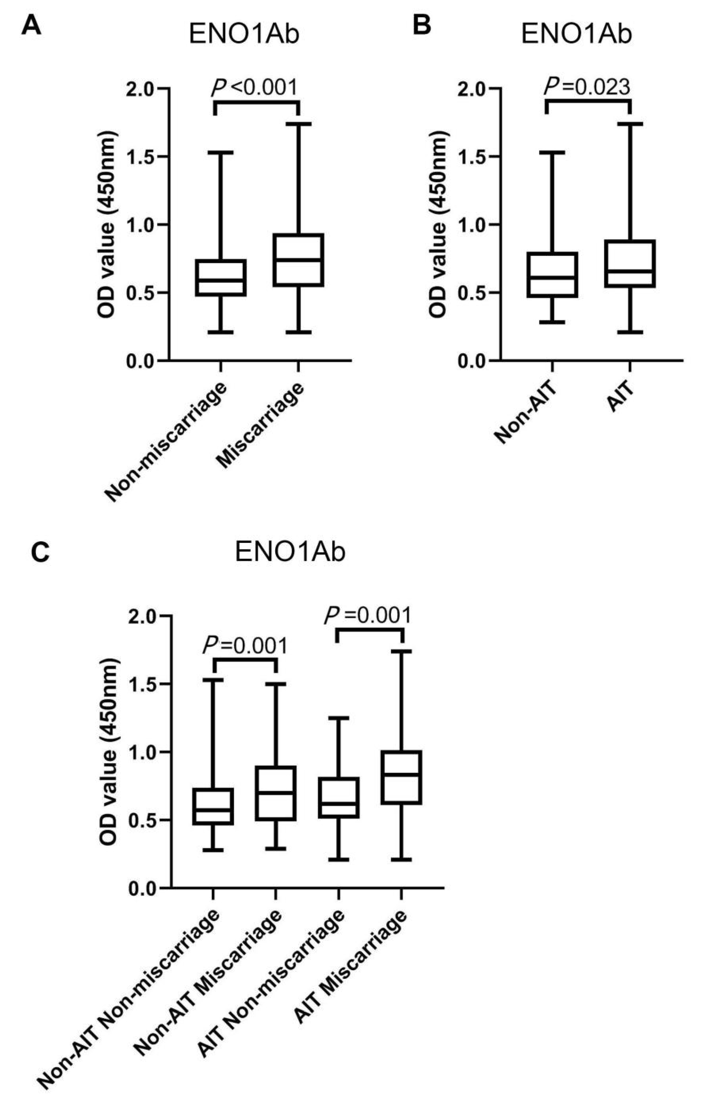
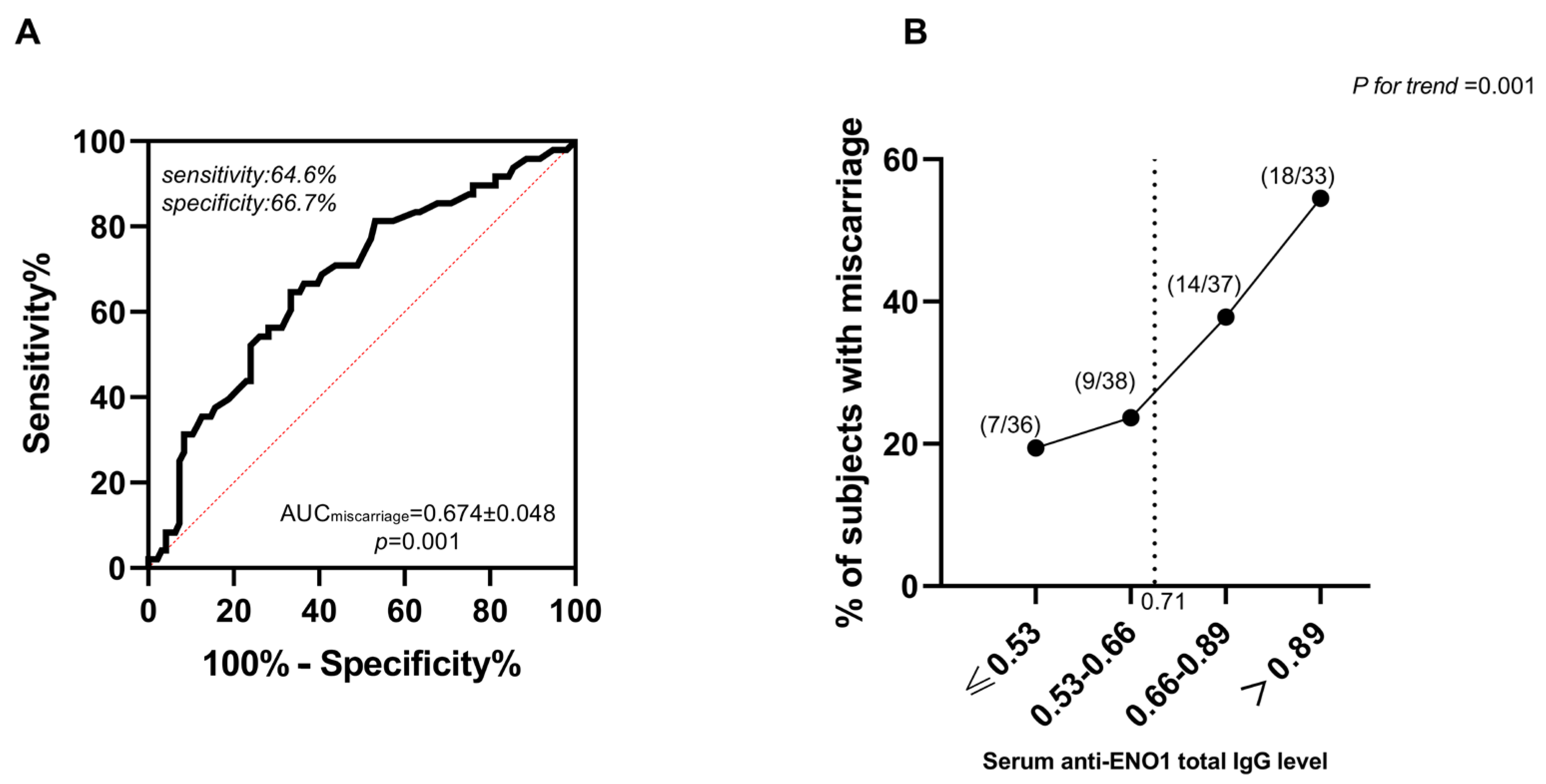
2.1. The Correlation between Serum Antibody Expressions against ENO1Ab and the Occurrence of Miscarriage in AIT Women

The characteristics of the 432 participants from the non-AIT non-miscarriage, non-AIT miscarriage, AIT non-miscarriage, and AIT miscarriage groups were described in our previous study [20]. There were no significant differences in age, BMI, serum levels of free thyroxine (FT4), and serum ferritin (SF) between the four groups.
The serum total IgG against ENO1 was significantly higher in the women with miscarriage than in those without miscarriage (p < 0.001) (Figure 1A). It was also statistically higher in women with AIT than in those without AIT (Figure 1B). Furthermore, the serum ENO1Ab level was significantly different between AIT miscarriage and AIT non-miscarriage groups, and between non-AIT miscarriage and non-AIT non-miscarriage groups (Figure 1C).
In order to study the role of ENO1Ab in miscarriage, logistic regression analysis (LRA) was performed for those potential risk factors of miscarriage in our participants [20]. As in our previous study, participants with either a smoking or drinking history were too few to be included in the LRA [20]. According to the results of the LRA, serum ENO1Ab levels were found as an independent risk factor in both AIT (OR = 15.295) and non-AIT women (OR = 4.795) (Table 1 and Table 2). Interestingly, the serum levels of TgAb and TPOAb were not significantly correlated with an increased risk of miscarriage in euthyroid women with AIT.
Figure 1. The serum levels of ENO1Ab in all euthyroid participants. The Mann–Whitney U test was performed to compare the serum levels of ENO1Ab between (A) participants with (n = 144) and those without miscarriage (n = 288), (B) participants with (n = 144) and and those without AIT (n = 288), (C) non-AIT participants with (n = 96) and those without miscarriage (n = 192), and AIT participants with (n = 48) and those without miscarriage (n = 96). The serum levels of total IgG against ENO1Ab were detected using enzyme-linked immunosorbent assay (ELISA) and expressed by absorbance at 450 nm. All data were exhibited as medians (P25 and P75), together with the minimum and maximum values in the box plot.
Ijms 24 01021 g001
The predictive value of serum ENO1Ab levels in the occurrence of miscarriage among AIT participants was further explored by receiver operating characteristic (ROC) curves and titer-dependence trend analysis. The area under the ROC curve (AUC) of ENO1Ab was 0.674 ± 0.048 (Figure 2A). Meanwhile, according to the largest Youden index (0.313), the corresponding cut-off absorbance at 450 nm of the ENO1Ab serum level was 0.71 (Figure 2B). Every 0.1 OD value higher ENO1Ab level above the corresponding cut-off value (0.71) was related to a 39% higher risk of miscarriage in women with AIT. These results indicated that the total IgG against ENO1 may be an important independent risk factor for miscarriage in women with AIT, and the antigenic epitopes of ENO1 protein specifically contributing to the miscarriage occurrence in euthyroid women with AIT need be further explored.
The correlations between circulating ENO1Ab expression and serum FT4, TSH, TPOAb, and TgAb levels in the participants with AIT were explored by spearman correlation analysis (SCA). The results showed a negative correlation between serum ENO1Ab and TSH levels in non-miscarriage women with AIT, but not in those with pregnancy loss. There was a negative correlation between serum total IgG against ENO1 protein and TPOAb levels in AIT participants with miscarriage, but not in those without pregnancy loss (Table 3).
Figure 2. Relationship between serum levels of total IgG against ENO1 and the development of miscarriage in AIT participants. The ROC curve of the serum ENO1Ab expression for miscarriage prediction (A), and the titer-dependent association between serum ENO1Ab levels after quartile stratifications and the prevalence of miscarriage (B).
Ijms 24 01021 g002

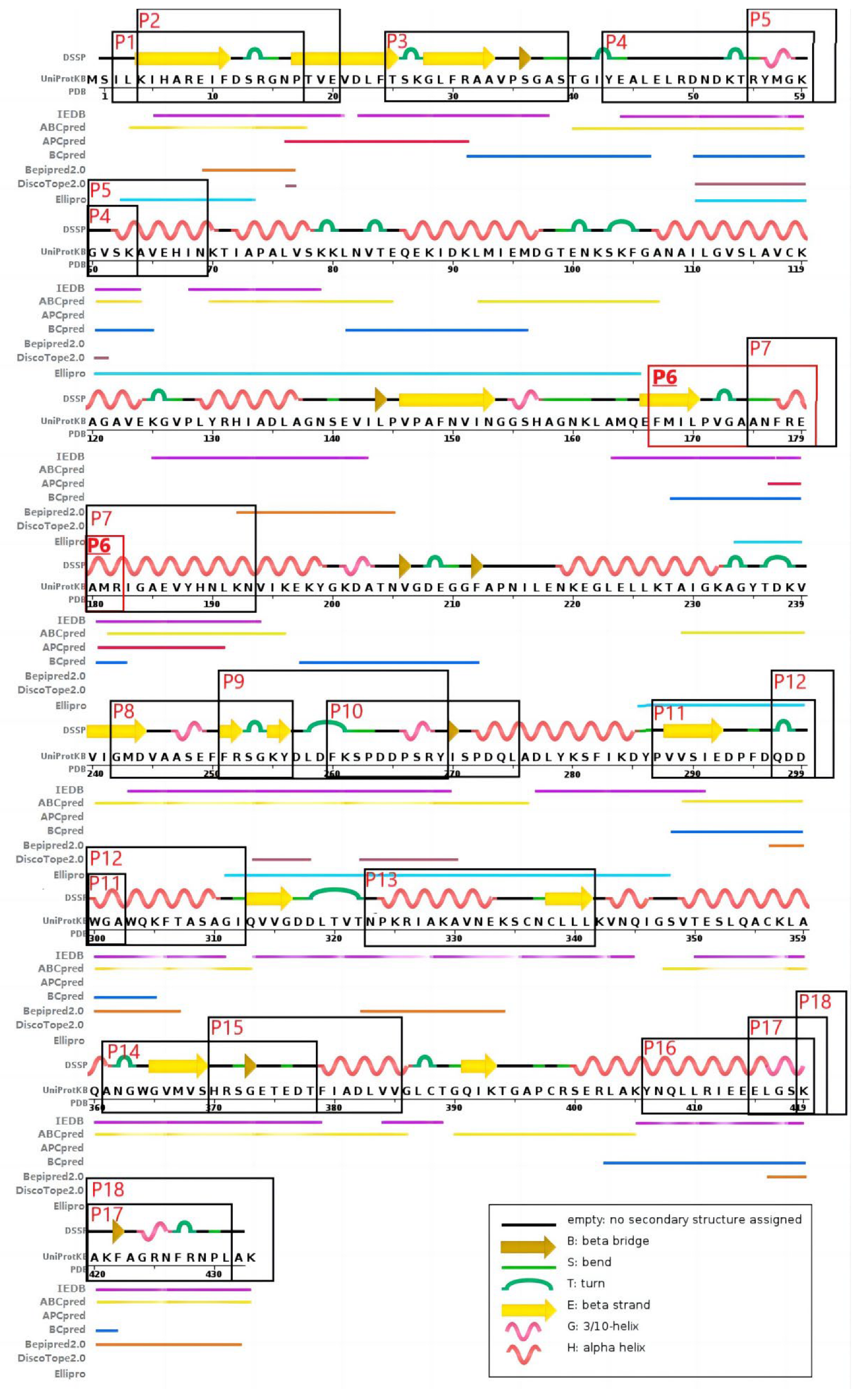
2.2. Serum Autoantibody Expressions against the 18 Predicted Antigenic Epitopes of ENO1

The serum autoantibody expressions against the predicted 18 antigenic epitopes (i.e., P1–P18) of human ENO1 protein in women randomly selected from each of the four groups (n = 8/group) were determined by enzyme-linked immunosorbent assay (ELISA) (Table 4). The characteristics of those 32 participants are listed in Supplementary Table S1. Interestingly, only the serum level of total IgG against P6 epitope of ENO1 (aa 168–183) was significantly increased in the AIT miscarriage group compared with that of the AIT non-miscarriage group. Autoantibodies specific to P4, P8, P9, P10, P11, P14, and P18 only showed a non-significant rise in the former group. Serum P9Ab levels were also markedly enhanced in non-AIT women with miscarriage when compared to those of the non-AIT non-miscarriage group. Autoantibodies against P1, P5, P8, P10, P15, and P18 also showed a non-significant rise in the non-AIT miscarriage group. Interestingly, the autoantibody levels against P1 and P13 were significantly decreased in women who had had a miscarriage compared with those of the non-miscarriage group among AIT subjects. In addition, the autoantibody levels against P6, P7, and P11 showed significant differences between AIT and non-AIT groups among those women who had had a miscarriage. The above findings indicated that P6 might be the specific antigenic epitope of ENO1 involved in the development of AIT-related pregnancy loss.

3. Discussion

Euthyroid women with AIT have been found to have a significantly higher risk for miscarriage than non-AIT ones, which cannot be decreased through L-thyroxine supplement [14]. The related pathogenic mechanisms are still unclear, and several factors potentially contribute to the occurrence of pregnancy loss [21,22]. There were some different non-organ specific autoantibodies (NOSA) found in the sera of more than 20% of women with AIT, such as autoantibodies against phospholipids, histones, and polynucleotides [23]. Those NOSA have been thought as important potential pathogenic factors for miscarriage in euthyroid women with AIT [22].
ENO1 is known as an important multifunctional protein and is distributed in many tissue cells such as thyrocytes, vascular, and trophoblast cells. It can be constructively expressed on the cell surface and in the cytoplasm and nucleus, and also maintain trafficking among the three sub-cellular locations [16,18,19,24]. ENO1Ab expression was exhibited in patients with some autoimmune and inflammatory disorders at different positive rates, such as rheumatoid arthritis (RA), systemic lupus erythematosus (SLE), antiphospholipid syndrome (APS), severe asthma, membranous nephropathy, and primary sclerosing cholangitis [25,26,27,28,29,30,31]. In fact, some of those diseases (e.g., RA, SLE, and APS) mentioned above have been reported as risk factors for spontaneous abortion [32,33,34]. Moreover, ENO1Ab has been found to be an important anti-trophoblast antibody; highly expressed in patients with unexplained recurrent miscarriages. Circulating ENO1Ab has been proposed to act as a potential biomarker for that disease [19]. It has been reported that ENO1Ab not only caused tissue cell injury through mediating antibody-dependent cytotoxicity (ADCC) and complement-dependent cytotoxicity (CDC), but also directly recognized the membrane-bound ENO1 protein and interfered with its functions [18,19,35,36]. All these findings indicate that ENO1Ab may play an important role in the pathogenesis of spontaneous abortion.
ENO1Ab expression was initially found in the sera of the patients with Hashimoto’s encephalopathy [37]. Later, we found that ENO1Ab was highly expressed in the serum of the experimental autoimmune thyroiditis (EAT) mouse model, a classical animal model of AIT [18]. Since the EAT mouse model has been established only through immunization by murine thyroglobulin and Freund adjuvant, the production of ENO1Ab may be secondary to the autoimmune responses against thyroid tissue other than the concurrent non-organ specific autoimmune disorders. Our previous study has shown that ENO1Ab caused damage to cerebral tissue in EAT mice due to a cross-autoimmune response. It has been shown that ENO1 protein is expressed in the placenta tissue as well as the thyroid gland and brain. ENO1Ab was able to directly suppress the hormone secretion function of trophoblast cells cultivated in vitro [19]. The above findings further suggest the expression of ENO1Ab may contribute to the development of spontaneous abortion in females with AIT. In the current pilot study, we systemically analyzed the relationship between serum ENO1Ab expression and the comorbidities of AIT and miscarriage. We found that the serum levels of ENO1Ab were completely different between females with and without AIT and between those with and without miscarriage. Its expression was not only significantly elevated in AIT women when compared to non-AIT women, but it was also pronouncedly higher in females suffering from miscarriage than those without a miscarriage history among either AIT or non-AIT patients. The LRA further showed that a high expression of serum ENO1Ab was an independent risk factor for miscarriage, especially in AIT females. The trend test has also directly demonstrated the titer-dependent association of pregnancy loss with ENO1Ab in AIT women and its potentially predictive value in the development of miscarriage. However, the AUC (0.674) from ROC analysis only showed a fair diagnostic value with moderate accuracy [38,39], which further reminds us of exploring the specific antigen epitope of ENO1 involved in AIT-related miscarriage.
Since serum ENO1Ab expression has been reported in a few autoimmune and inflammatory diseases, the identification of disease-specific antigen epitopes of ENO1 is gaining more and more attention to improve diagnostic and therapeutic strategies. Although multiple amino acid segments of ENO1 were found to be potentially specific epitopes to RA, cancer-associated retinopathy, lupus nephritis, and endometriosis [40,41,42,43], there is still a lack of systemic investigation into disease-specific ENO1 epitopes, and there are no reports on miscarriage. In the present study, we first predicted 18 antigenic epitopes of ENO1 from several databases by bioinformatic analysis. Then, the differential expression patterns of those epitopes were initially explored. There were significant differences in the serum levels of the autoantibodies against the predicted ENO1 epitopes between the women with and without miscarriage complicated with AIT or not. However, only serum P6Ab exhibited both significantly higher levels in AIT patients with miscarriage than those without pregnancy loss, and significantly higher levels in AIT patients when compared with those of non-AIT women who all suffered from miscarriage. Finally, the serum P6Ab levels were further detected in a total of 432 women with euthyroidism in our other study, which consisted of 48 AIT miscarriage women and matched controls in a 1:2 ratio (96 AIT patients without miscarriage, 96 non-AIT patients with miscarriage, and 192 without miscarriage) [20]. We found that serum titers of anti-ENO1-P6 total IgG and its four subtypes were not only pronouncedly increased in AIT females compared to non-AIT women, but also significantly higher in AIT females with spontaneous abortion than those without abortion. They were all independently associated with the occurrence of miscarriage in AIT patients. ROC showed that the largest AUC was 0.827 ± 0.043 for anti-ENO1-P6 IgG2 [20]. All these findings suggest that ENO1-P6Ab may be involved in the development of AIT-related miscarriage, and it can be potentially used as a good biomarker for that disorder, especially anti-ENO1-P6 IgG2. We acknowledge that we have not completed the investigation of the mechanisms of ENO1-P6Abs in the development of AIT-related miscarriage, so their cause–effect relationship awaits further study in the future.
Since ENO1 is a multiple-functioning protein, the precise activities of most of the 18 epitopes are still unclear. One previous study has suggested that the P6 epitope of ENO1 potentially contributes to the maintenance of its glycolytic enzymatic activity due to the involvement of Glu168 in the hydrogen bond formation with the PGA hydroxyl group [44]. Since placental development and energy supply in early pregnancy mainly depend on glycolysis [45], it is inferred that the direct binding of ENO1-P6 epitope and its autoantibodies may affect the glycolytic capacity of the enzyme and induce spontaneous abortion. Several studies have suggested that the IgG specific to ENO1 can directly inhibit the activities of ENO1 expressed in the cell membrane [19,36]. One in vitro study has found that membrane expression of ENO1 in MCF-7 cells, a human breast cancer cell line, was significantly increased after exposure to lipopolysaccharide (LPS), but both monoclonal and polyclonal antibodies against ENO1 were able to potentially downregulate the LPS-stimulated invasion of those cells, which was mediated by membrane ENO1 [36]. Another recent in vitro study has shown that ENO1-specific IgG could directly reduce β-hCG and progesterone production of cultured trophoblast cells, indicating a potential mechanism of causing miscarriage [19]. In addition, in vivo studies have found that ENO1Ab could damage cells through ADCC and CDC [18,35]. The constructive expression of ENO1 in the placenta [16] and increased production of P6Abs in AIT women suffering from miscarriage suggest that ENO1-P6Ab might injure the placenta and embryo through ADCC and/or CDC. We admit that these speculated pathogeneses need to be demonstrated in further experimental studies. We have established a pregnant mouse model with highly expressed ENO1-P6Ab in the serum by immunization with synthesized ENO1-P6 peptide. The fetal absorption rate, placental immune-complex deposition, energy metabolism, and other related mechanisms are being investigated. In addition, passive transfer of the IgG extracted from the above pregnant mouse model to female EAT mice without P6 peptide immunization just after mating may help decipher the role of ENO1-P6Ab in the pathogenesis of AIT-related miscarriage. At the same time, the control mice will be immunized with the synthesized mutant peptide, whose amino-acid sequence is partially changed and different from the intact ENO1-P6 epitope. In order to further investigate the exact role of the P6 epitope in the functional activities of ENO1, trophoblast cells will be cultured in vitro and their ENO1 gene will be knocked out, then they would be transfected with ENO1 gene containing either wild-type or mutated P6 coding DNA. The above experiments have been designed and will be completed in the near future.
It is still unknown why the serum levels of ENO1Abs were markedly increased in AIT patients, especially the production of ENO1-P6Ab. It has been found that more ENO1 may be expressed in the membrane of early apoptotic cells as a kind of super protein and in those under exposure to proinflammatory factors, such as tumor necrosis factor α and LPS. Previous studies have indicated that thyrocytes can express ENO1 protein [18] and many of them may develop apoptosis or be exposed to the pro-inflammatory cytokines [46,47]. We infer that the increased surface expression of ENO1 might result in the external exposure of some cryptic epitopes, and finally induce a cross-autoimmune reaction between thyroid and placenta tissues. We will also design a series of experiments to study whether the above mechanisms are existent and how the immune tolerance targeted to the ENO1-P6 epitope can be established in future work.
There are several limitations to our study. Firstly, our findings were obtained from a relatively small retrospective investigation, which awaits further confirmation in a larger-sample prospective study in the future. Secondly, the experimental studies of the mechanisms related to the production of ENO1Abs and the contribution ENO1Abs to AIT-related miscarriage have not been completed. The serum expression patterns of 18 predicted ENO1 epitopes still need to be examined in a larger-sample study using both intact and mutant epitope peptides. Thirdly, serum ENO1Ab expression has been found in some other disorders (e.g., APS, RA, and SLE), which are also suggested as risk factors for miscarriage [31,32,33,34]. Although we had excluded patients with a known personal history of other autoimmune and inflammatory disorders, we admit that we did not test those other non-organ specific autoantibodies associated with spontaneous abortion in this study, and we did not analyze the synergistic or additive actions between ENO1Ab and other NOSA, which will be performed in the future. Lastly, serum autoantibody levels show a decreasing trend from first trimester to the final trimester during normal pregnancy. According to a previous study, serum levels of antithyroid Abs were highest in the first trimester, although they were reduced by about 60% during the whole pregnancy with a half-life of 21 days [48,49]. In this study, there was a statistical difference in gestational ages between the women with and without miscarriage among the non-AIT group, the medians of which were 6 and 7 weeks of pregnancy, respectively [20]. This difference did not seem to cause a significant change in serum ENO1Ab levels between them or be the main responsible factor for the development of miscarriage in these non-AIT participants. In the future large-size investigation, gestational ages will be balanced between the different groups of patients.
In conclusion, our findings in this study have shown that ENO1Ab may be independently associated with the occurrence of miscarriage in AIT patients. A previous study has indicated that ENO1Ab expression is a novel autoimmune biomarker for unexplained recurrent pregnancy loss. This study suggests serum ENO1Ab might have a fair predictive value for AIT-related miscarriage, and especially ENO1-P6Ab may be more specifically related to this disorder. This pilot study has provided some clinical epidemiological clues for the utilization of the ENO1-P6 epitope to develop a clinical prediction biomarker of AIT-related miscarriage and establish the specific therapeutic strategy of immune tolerance. However, a large-sample clinical investigation and a series of experimental studies need to be performed to further verify our findings and to establish the cause–effect relationship between ENO1-P6Ab expression and AIT-related miscarriage.

4. Materials and Methods
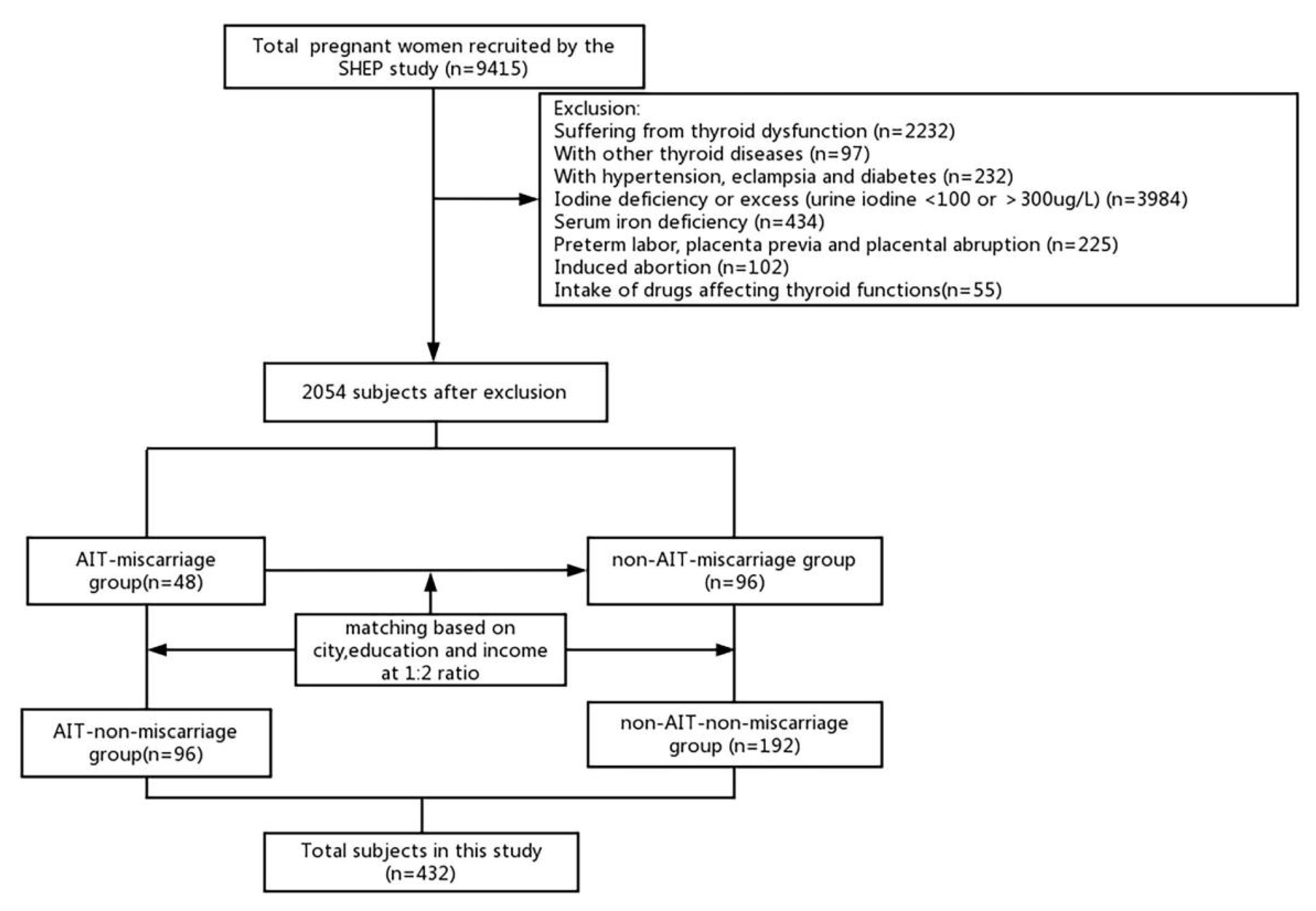
4.1. Patients

All participants enrolled in this study were pregnant females in the first 4–12 weeks of pregnancy from the Subclinical Hypothyroid during Early Pregnancy (SHEP) project [20]. SHEP was a prospective cohort study performed from 2011 to 2014. The project was approved by the Ethics Committee of China Medical University ((2012)2011-32-4). Each participant was required to provide personal information, including education level, family income level, residential city, history of smoking, history of drinking, personal history of chronic diseases, personal and family history of thyroid diseases, and multiple-micronutrient supplementation. Informed consent was obtained from all patients. Fasting serum and urine samples were collected from the participants upon their first visit. Electrochemiluminescence immunoassays were used to examine TSH, FT4, TPOAb, TgAb, and SF concentrations in all participants with Cobas Elesys 601 (Roche Diagnostics, Basel, Switzerland). The ammonium persulfate method was used to determine the concentration of urinary iodine based on the Sandell–Kolthoff reaction.
A total of 9415 pregnant women from the SHEP study were recruited for our study. Participants were excluded if they had other thyroid diseases or autoimmune diseases other than AIT, if they were taking drugs affecting thyroid function, if they had hypertension, eclampsia, diabetes, a urinary iodine concentration <100 or >300 μg/L, SF < 20 μg/L, preterm labor, placenta previa, placental abruption during this pregnancy, or an induced abortion. The remaining 2054 participants were further selected based on their miscarriage history and serum TgAb and/or TPOAb levels.
Miscarriages that had occurred in the preceding 5 years or in the current pregnancy were investigated and counted in this study. Levels of serum TgAb > 115 IU/mL and/or TPOAb > 34 IU/L according to the manufacturer (Roche Diagnostics, Switzerland) reference range and the Guidelines on Diagnosis and Management of Thyroid Diseases during Pregnancy and Postpartum were characterized as AIT [3]. A total of 48 euthyroid miscarriage participants with AIT (AIT miscarriage group) were collected from SHEP. SPSS 23.0 was used to conduct random sampling with a ratio of 1:2 for miscarriage women without AIT (n = 96, non-AIT miscarriage group), AIT women without miscarriage (n = 96, AIT non-miscarriage group), and those without either AIT or miscarriage (n = 192, non-AIT non-miscarriage group). The matching factors were city of residence, education level, and family income level. Finally, a total of 432 participants were included in this study (Figure 3).

4.2. Prediction of the Potential Epitopes in ENO1 Protein

The amino acid sequence (PubMed protein database, GI: 1167843), secondary structure, and 3D structure (PDB database, PDB ID: 2PSN) of the human ENO1 protein were used to predict its potential antigenic epitopes by bioinformatics [50]. Antigenic epitopes recognized by B cells were predicted by the following network servers according to the amino acid sequence: ABCpred, APCpred, Bcpred, Bepipred 2.0, and IEDB. Antigenic epitopes based on crystal structure were predicted by the DissoTope2.0 and Ellipro web servers. The epitopes recognized by B cells which could be predicted by at least two web servers were selected for further study. Antigenic epitopes recognized by T cells were predicted by the IEDB database. A total of 18 antigenic epitopes in the human ENO1 protein were further explored in this study, named P1 to P18 according to their location on the protein, and synthesized and purified by Sangon Biotech (Shanghai, China) (Table 5, Figure 4).

4.3. Enzyme-Linked Immunosorbent Assay

The serum levels of autoantibodies against the ENO1 protein and its 18 related epitopes of ENO1 were determined by ELISA as previously described [52]. Microtiter plates coated with recombinant ENO1 (1 μg/well) or the 18 recombinant peptides (10 ng/well) were incubated at 4 °C overnight and blocked with 1% bovine serum albumin (Sigma-Aldrich, St. Louis, MO, USA). Serum levels of ENO1Ab were measured at a dilution of 1:50, and serum levels of autoantibodies against the 18 epitopes were measured at a dilution of 1:250 in participants from our study. HRP-labeled rabbit anti-human total IgG antibodies (Bioss, Beijing, China) were used as secondary antibodies at a dilution of 1:1000. The enzymatic reaction was measured by TMB, and color development was stopped using hydrochloric acid at room temperature. Absorbance was measured at 450 nm using Infinite 2000 PRO (TECAN, Port Melbourne, VIC, Australia).

4.4. Statistical Analysis

The data were analyzed using SPSS 23.0 statistical software. The skewed distributed variables were reported as the medians with 25–75th percentiles for continuous variables. The normally distributed variables were reported as the means ± SD. The Mann–Whitney U test with a skewed distribution and a two-sample t-test were used to analyze variables with a normal distribution. Categorical variable frequencies were compared by the chi-squared test. SCA was performed to examine the relationship between ENO1Ab and serum levels of FT4, TSH, TPOAb, and TgAb. The independent risk variables for miscarriage were evaluated by LRA. ROC curve analysis was used to determine the optimal cut-off values of ENO1Ab in participants with AIT and miscarriage. The titer-dependent association between serum ENO1Ab levels and miscarriage prevalence in participants with AIT was examined by the trend test. Differences were considered statistically significant if p < 0.05 in two groups or p < 0.017 (0.05/3) or 0.0125 (0.05/4) for adjusted p values in multiple comparisons.

Supplementary Materials

The following supporting information can be downloaded at: https://www.mdpi.com/article/10.3390/ijms24021021/s1.

Author Contributions

Conceptualization, J.G., Y.L. and J.L. (Jing Li); methodology, J.G., Y.L. and X.H.; software, J.G., Y.L. and X.H.; validation, J.G. and X.H.; resources, J.L. (Jiashu Li), C.F., H.Z., W.T., Z.S. and J.L. (Jing Li); data curation, writing—original draft preparation, J.G.; writing—review and editing, X.H. and J.L. (Jing Li); project administration, J.L. (Jing Li); funding acquisition, J.L. (Jing Li). All authors have read and agreed to the published version of the manuscript.

Funding

J.L. (Jing Li) was supported by the National Nature Science Foundation of China (grant numbers 81771741 and 81570709), the Distinguished Professor at Educational Department of Liaoning Province (grant number (2014)187), and Z.S. was supported by the National Science and Technology Support Program (grant number 2014BA106B02).

Institutional Review Board Statement

SHEP was approved by the Ethics Committee of China Medical University ((2012)2011-32-4).

Informed Consent Statement

All of the participants who took part in the research provided their written consent.

Data Availability Statement

The article contains all of the findings obtained from the study.

Acknowledgments

The authors thank all staff for their excellent laboratory work.

Conflicts of Interest

The authors have no conflict of interest in this manuscript.

References

  1. Yang, W.; Xiang, Y.; Zhang, H.; Shan, Z.; Li, J.; Teng, W. The role of protein disulphide-isomerase A3 as autoantigen in the pathogenesis of autoimmune thyroiditis and related brain damage in adult mice. Clin. Immunol. 2020, 212, 108350. [Google Scholar] [CrossRef] [PubMed]
  2. De Leo, S.; Pearce, E.N. Autoimmune thyroid disease during pregnancy. Lancet Diabetes Endocrinol. 2018, 6, 575–586. [Google Scholar] [CrossRef] [PubMed]
  3. Liu, H.; Shan, Z.; Li, C.; Mao, J.; Xie, X.; Wang, W.; Fan, C.; Wang, H.; Zhang, H.; Han, C.; et al. Maternal subclinical hypothyroidism, thyroid autoimmunity, and the risk of miscarriage: A prospective cohort study. Thyroid 2014, 24, 1642–1649. [Google Scholar] [CrossRef] [PubMed] [Green Version]
  4. Karakosta, P.; Alegakis, D.; Georgiou, V.; Roumeliotaki, T.; Fthenou, E.; Vassilaki, M.; Boumpas, D.; Castanas, E.; Kogevinas, M.; Chatzi, L. Thyroid dysfunction and autoantibodies in early pregnancy are associated with increased risk of gestational diabetes and adverse birth outcomes. J. Clin. Endocrinol. Metab. 2012, 97, 4464–4472. [Google Scholar] [CrossRef] [PubMed]
  5. Cleary-Goldman, J.; Malone, F.D.; Lambert-Messerlian, G.; Sullivan, L.; Canick, J.; Porter, T.F.; Luthy, D.; Gross, S.; Bianchi, D.W.; D’Alton, M.E. Maternal thyroid hypofunction and pregnancy outcome. Obstet. Gynecol. 2008, 112, 85–92. [Google Scholar] [CrossRef]
  6. Thangaratinam, S.; Tan, A.; Knox, E.; Kilby, M.D.; Franklyn, J.; Coomarasamy, A. Association between thyroid autoantibodies and miscarriage and preterm birth: Meta-analysis of evidence. BMJ 2011, 342, d2616. [Google Scholar] [CrossRef] [Green Version]
  7. Poppe, K.; Velkeniers, B.; Glinoer, D. The role of thyroid autoimmunity in fertility and pregnancy. Nat. Rev. Endocrinol. 2008, 4, 394–405. [Google Scholar] [CrossRef]
  8. Stagnaro-Green, A.; Roman, S.H.; Cobin, R.H.; el-Harazy, E.; Alvarez-Marfany, M.; Davies, T.F. Detection of at-risk pregnancy by means of highly sensitive assays for thyroid autoantibodies. JAMA 1990, 264, 1422–1425. [Google Scholar] [CrossRef]
  9. Colicchia, M.; Campagnolo, L.; Baldini, E.; Ulisse, S.; Valensise, H.; Moretti, C. Molecular basis of thyrotropin and thyroid hormone action during implantation and early development. Hum. Reprod. Update 2014, 20, 884–904. [Google Scholar] [CrossRef]
  10. Matalon, S.T.; Blank, M.; Levy, Y.; Carp, H.J.; Arad, A.; Burek, L.; Grunebaum, E.; Sherer, Y.; Ornoy, A.; Refetoff, S.; et al. The pathogenic role of anti-thyroglobulin antibody on pregnancy, evidence from an active immunization model in mice. Hum. Reprod. 2003, 18, 1094–1099. [Google Scholar] [CrossRef]
  11. Twig, G.; Shina, A.; Amital, H.; Shoenfeld, Y. Pathogenesis of infertility and recurrent pregnancy loss in thyroid autoimmunity. J. Autoimmun. 2012, 38, J275–J281. [Google Scholar] [CrossRef]
  12. Vissenberg, R.; Manders, V.D.; Mastenbroek, S.; Fliers, E.; Afink, G.B.; Ris-Stalpers, C.; Goddijn, M.; Bisschop, P. Pathophysiological aspects of thyroid hormone disorders/thyroid peroxidase autoantibodies and reproduction. Hum. Reprod. Update 2015, 21, 378–387. [Google Scholar] [CrossRef] [Green Version]
  13. Monteleone, P.; Faviana, P.; Artini, P.G. Thyroid peroxidase identified in human granulosa cells, another piece to the thyroid-ovary puzzle? Gynecol. Endocrinol. 2017, 33, 574–576. [Google Scholar] [CrossRef]
  14. Wang, H.; Gao, H.; Chi, H.; Zeng, L.; Xiao, W.; Wang, Y.; Li, R.; Liu, P.; Wang, C.; Tian, Q.; et al. Effect of Levothyroxine on Miscarriage Among Women With Normal Thyroid Function and Thyroid Autoimmunity Undergoing In Vitro Fertilization and Embryo Transfer: A Randomized Clinical Trial. JAMA 2017, 318, 2190–2198. [Google Scholar] [CrossRef]
  15. Dhillon-Smith, R.K.; Middleton, L.J.; Sunner, K.K.; Cheed, V.; Baker, K.; Farrell-Carver, S.; Bender-Atik, R.; Agrawal, R.; Bhatia, K.; Edi-Osagie, E.; et al. Levothyroxine in Women with Thyroid Peroxidase Antibodies before Conception. N. Engl. J. Med. 2019, 380, 1316–1325. [Google Scholar] [CrossRef]
  16. Gharesi-Fard, B.; Zolghadri, J.; Kamali-Sarvestani, E. Alteration in the expression of proteins in unexplained recurrent pregnancy loss compared with in the normal placenta. J. Reprod. Dev. 2014, 60, 261–267. [Google Scholar] [CrossRef] [Green Version]
  17. Pietsch, J.; Sickmann, A.; Weber, G.; Bauer, J.; Egli, M.; Wildgruber, R.; Infanger, M.; Grimm, D. Metabolic enzyme diversity in different human thyroid cell lines and their sensitivity to gravitational forces. Proteomics 2012, 12, 2539–2546. [Google Scholar] [CrossRef]
  18. Lu, Y.; Qin, J.; Xiang, Y.; Sun, R.; Feng, Y.; Zhang, H.; Ding, S.; Li, J.; Shan, Z.; Teng, W. Experimental evidence for alpha enolase as one potential autoantigen in the pathogenesis of both autoimmune thyroiditis and its related encephalopathy. Int. Immunopharmacol. 2020, 85, 106563. [Google Scholar] [CrossRef]
  19. Ye, Y.; Kuhn, C.; Kösters, M.; Arnold, G.J.; Ishikawa-Ankerhold, H.; Schulz, C.; Rogenhofer, N.; Thaler, C.J.; Mahner, S.; Fröhlich, T.; et al. Anti α-enolase antibody is a novel autoimmune biomarker for unexplained recurrent miscarriages. EBioMedicine 2019, 41, 610–622. [Google Scholar] [CrossRef] [Green Version]
  20. He, X.; Liu, Y.; Wang, H.; Sun, W.; Lu, Y.; Shan, Z.; Teng, W.; Li, J. A Predictive Role of Autoantibodies Against the Epitope aa168-183 of ENO1 in the Occurrence of Miscarriage Related to Thyroid Autoimmunity. Front. Immunol. 2022, 13, 890502. [Google Scholar] [CrossRef]
  21. Lin, C.; Xiang, Y.; Li, J. The contribution of thyroid autoimmunity to miscarriage in euthyroid women. Minerva. Med. 2016, 107, 173–181. [Google Scholar] [PubMed]
  22. Zhu, Q.; Xu, Q.-H.; Xie, T.; Wang, L.-L.; Liu, H.; Muyayalo, K.P.; Huang, X.-B.; Zhao, S.-J.; Liao, A.-H. Recent insights into the impact of immune dysfunction on reproduction in autoimmune thyroiditis. Clin. Immunol. 2021, 224, 108663. [Google Scholar] [CrossRef] [PubMed]
  23. Pratt, D.; Novotny, M.; Kaberlein, G.; Dudkiewicz, A.; Gleicher, N. Antithyroid antibodies and the association with non-organ-specific antibodies in recurrent pregnancy loss. Am. J. Obstet. Gynecol. 1993, 168, 837–841. [Google Scholar] [CrossRef] [PubMed]
  24. Didiasova, M.; Schaefer, L.; Wygrecka, M. When Place Matters: Shuttling of Enolase-1 Across Cellular Compartments. Front. Cell Dev. Biol. 2019, 7, 61. [Google Scholar] [CrossRef] [PubMed] [Green Version]
  25. Díaz-Ramos, A.; Roig-Borrellas, A.; García-Melero, A.; López-Alemany, R. α-Enolase, a multifunctional protein: Its role on pathophysiological situations. J. Biomed. Biotechnol. 2012, 2012, 156795. [Google Scholar] [CrossRef] [PubMed] [Green Version]
  26. Terrier, B.; Degand, N.; Guilpain, P.; Servettaz, A.; Guillevin, L.; Mouthon, L. Alpha-enolase: A target of antibodies in infectious and autoimmune diseases. Autoimmun. Rev. 2007, 6, 176–182. [Google Scholar] [CrossRef]
  27. Thébault, S.; Gilbert, D.; Hubert, M.; Drouot, L.; Machour, N.; Lange, C.; Charlionet, R.; Tron, F. Orderly pattern of development of the autoantibody response in (New Zealand White × BXSB)F1 lupus mice: Characterization of target antigens and antigen spreading by two-dimensional gel electrophoresis and mass spectrometry. J. Immunol. 2002, 169, 4046–4053. [Google Scholar] [CrossRef] [Green Version]
  28. Lee, H.; Kwon, B.; Hur, G.; Choi, S.; Nahm, D.; Park, H. Isotype and IgG subclass distribution of autoantibody response to alpha-enolase protein in adult patients with severe asthma. Yonsei. Med. J. 2008, 49, 923–930. [Google Scholar] [CrossRef] [Green Version]
  29. Kimura, Y.; Miura, N.; Debiec, H.; Morita, H.; Yamada, H.; Banno, S.; Ronco, P.; Imai, H. Circulating antibodies to α-enolase and phospholipase A receptor and composition of glomerular deposits in Japanese patients with primary or secondary membranous nephropathy. Clin. Exp. Nephrol. 2017, 21, 117–126. [Google Scholar] [CrossRef] [Green Version]
  30. Orth, T.; Kellner, R.; Diekmann, O.; Faust, J.; Meyer, B.; Mayet, W. Identification and characterization of autoantibodies against catalase and alpha-enolase in patients with primary sclerosing cholangitis. Clin. Exp. Immunol. 1998, 112, 507–515. [Google Scholar] [CrossRef]
  31. Pratesi, F.; Moscato, S.; Sabbatini, A.; Chimenti, D.; Bombardieri, S.; Migliorini, P. Autoantibodies specific for alpha-enolase in systemic autoimmune disorders. J. Rheumatol. 2000, 27, 109–115. [Google Scholar]
  32. Versini, M. Thyroid Autoimmunity and Antiphospholipid Syndrome: Not Such a Trivial Association. Front. Endocrinol. (Lausanne) 2017, 8, 175. [Google Scholar] [CrossRef] [Green Version]
  33. Hee, J.Y.; Huang, S.; Leong, K.P.; Chun, L.; Zhang, Y.O.; Gongye, R.; Tang, K. Pregnancy loss and the risk of rheumatoid arthritis in Chinese women: Findings from the China Kadoorie biobank. BMC Public Health 2022, 22, 1768. [Google Scholar] [CrossRef]
  34. Bălănescu, A.; Donisan, T.; Bălănescu, D. An ever-challenging relationship: Lupus and pregnancy. Reumatologia 2017, 55, 29–37. [Google Scholar] [CrossRef]
  35. Sun, W.; Feng, Y.; Li, H.; He, X.; Lu, Y.; Shan, Z.; Teng, W.; Li, J. The effects of maternal anti-alpha-enolase antibody expression on the brain development in offspring. Clin. Exp. Immunol. 2022, 210, 187–198. [Google Scholar] [CrossRef]
  36. Perconti, G.; Maranto, C.; Romancino, D.P.; Rubino, P.; Feo, S.; Bongiovanni, A.; Giallongo, A. Pro-invasive stimuli and the interacting protein Hsp70 favour the route of alpha-enolase to the cell surface. Sci. Rep. 2017, 7, 3841. [Google Scholar] [CrossRef] [Green Version]
  37. Fujii, A.; Yoneda, M.; Ito, T.; Yamamura, O.; Satomi, S.; Higa, H.; Kimura, A.; Suzuki, M.; Yamashita, M.; Yuasa, T.; et al. Autoantibodies against the amino terminal of alpha-enolase are a useful diagnostic marker of Hashimoto’s encephalopathy. J. Neuroimmunol. 2005, 162, 130–136. [Google Scholar] [CrossRef]
  38. Swets, J. Measuring the accuracy of diagnostic systems. Source Sci. New Ser. 1988, 240, 285–293. [Google Scholar] [CrossRef] [Green Version]
  39. Fischer, J.; Bachmann, L.; Jaeschke, R. A readers’ guide to the interpretation of diagnostic test properties: Clinical example of sepsis. Intensive Care Med. 2003, 29, 1043–1051. [Google Scholar] [CrossRef] [Green Version]
  40. Walter, M.; Berg, H.; Leidenberger, F.; Schweppe, K.; Northemann, W. Autoreactive epitopes within the human alpha-enolase and their recognition by sera from patients with endometriosis. J Autoimmun. 1995, 8, 931–945. [Google Scholar] [CrossRef]
  41. Adamus, G.; Amundson, D.; Seigel, G.; Machnicki, M. Anti-enolase-alpha autoantibodies in cancer-associated retinopathy: Epitope mapping and cytotoxicity on retinal cells. J. Autoimmun. 1998, 11, 671–677. [Google Scholar] [CrossRef] [PubMed]
  42. Bruschi, M.; Sinico, R.A.; Moroni, G.; Pratesi, F.; Migliorini, P.; Galetti, M.; Murtas, C.; Tincani, A.; Madaio, M.; Radice, A.; et al. Glomerular autoimmune multicomponents of human lupus nephritis in vivo: α-enolase and annexin AI. J. Am. Soc. Nephrol. 2014, 25, 2483–2498. [Google Scholar] [CrossRef] [PubMed] [Green Version]
  43. Gerstner, C.; Dubnovitsky, A.; Sandin, C.; Kozhukh, G.; Malmstrm, V. Functional and structural characterization of a novel hla-drb1*04:01-restricted α-enolase t cell epitope in rheumatoid arthritis. Front. Immunol. 2016, 7, 494. [Google Scholar] [CrossRef] [PubMed] [Green Version]
  44. Pancholi, V. Multifunctional alpha-enolase: Its role in diseases. Cell Mol. Life Sci. 2001, 58, 902–920. [Google Scholar] [CrossRef] [PubMed]
  45. Pringle, K.; Kind, K.; Sferruzzi-Perri, A.; Thompson, J.; Roberts, C. Beyond oxygen: Complex regulation and activity of hypoxia inducible factors in pregnancy. Hum. Reprod. Update 2010, 16, 415–431. [Google Scholar] [CrossRef] [Green Version]
  46. Hammond, L.; Lowdell, M.; Cerrano, P.; Goode, A.; Bottazzo, G.; Mirakian, R. Analysis of apoptosis in relation to tissue destruction associated with Hashimoto’s autoimmune thyroiditis. J. Pathol. 1997, 182, 138–144. [Google Scholar] [CrossRef]
  47. Giordano, C.; Stassi, G.; De Maria, R.; Todaro, M.; Richiusa, P.; Papoff, G.; Ruberti, G.; Bagnasco, M.; Testi, R.; Galluzzo, A. Potential involvement of Fas and its ligand in the pathogenesis of Hashimoto’s thyroiditis. Science 1997, 275, 960–963. [Google Scholar] [CrossRef]
  48. Vidarsson, G.; Dekkers, G.; Rispens, T. IgG subclasses and allotypes: From structure to effector functions. Front. Immunol. 2014, 5, 520. [Google Scholar] [CrossRef] [Green Version]
  49. Glinoer, D.; Riahi, M.; Grün, J.P.; Kinthaert, J. Risk of subclinical hypothyroidism in pregnant women with asymptomatic autoimmune thyroid disorders. J. Clin. Endocrinol. Metab. 1994, 79, 197–204. [Google Scholar]
  50. Berman, H.M.; Westbrook, J.; Feng, Z.; Gilliland, G.; Bhat, T.N.; Weissig, H.; Shindyalov, I.N.; Bourne, P.E. The Protein Data Bank. Nucleic Acids Res. 2000, 28, 235–242. [Google Scholar] [CrossRef] [Green Version]
  51. Kang, H.; Jung, S.; Kim, S.; Chung, S. Biological crystallography, Structure of human alpha-enolase (hENO1), a multifunctional glycolytic enzyme. Acta Crystallogr. D Biol. Crystallogr. 2008, 64, 651–657. [Google Scholar] [CrossRef] [Green Version]
  52. Xiang, Y.; Jin, Q.; Li, L.; Yang, Y.; Zhang, H.; Liu, M.; Fan, C.; Li, J.; Shan, Z.; Teng, W. Physiological low-dose oestrogen promotes the development of experimental autoimmune thyroiditis through the up-regulation of Th1/Th17 responses. J. Reprod. Immunol. 2018, 126, 23–31. [Google Scholar] [CrossRef]
Figure 3. The enrollment flowchart of all the euthyroid participants in the current study.
Ijms 24 01021 g003
Figure 4. The 18 selected antigenic epitopes of human ENO1 protein predicted by databases.
Ijms 24 01021 g004
Table 1. Logistic regression of potential risk factors of miscarriage in euthyroid pregnant women with AIT.
VariantsBSEWaldpOR (95%CI)
Age (years)0.0850.0631.7990.1801.088 (0.962–1.231)
Gestational age (weeks)−0.1660.1611.0620.3030.847 (0.618–1.161)
BMI (kg/m2)−0.0270.0780.1150.7350.974 (0.835–1.136)
Urine iodine/creatinine (μg/g)0.0070.0051.9850.1591.007 (0.997–1.017)
Serum ferritin (μg/L)0.0040.0040.960.3271.004 (0.996–1.012)
TSH (mIU/L)0.4030.2542.5140.1131.497 (0.909–2.464)
FT4 (pmol/L)−0.1330.1510.7730.3790.875 (0.651–1.178)
TPOAb (IU/mL)0.0010.0020.3010.5831.001 (0.998–1.004)
TgAb (IU/mL)−0.0010.0011.1060.2930.999 (0.997–1.001)
ENO1Ab a2.7280.9218.7730.00315.295 (2.516–92.982)
a Serum levels of ENO1Ab were detected using ELISA and reflected by absorbance at 450 nm.
Table 2. Logistic regression of potential risk factors of miscarriage in euthyroid pregnant women without AIT.
VariantsBSEWaldpOR(95%CI)
Age (years)0.0950.045.6420.0181.099 (1.017–1.189)
Gestational age (weeks)−0.6950.13825.519<0.0010.499 (0.381–0.654)
BMI (kg/m2)−0.040.0381.1130.2910.96 (0.891–1.035)
Urine iodine/creatinine (μg/g)−0.0020.0020.9910.320.998 (0.995–1.002)
Serum ferritin (μg/L)0.0020.0030.4790.4891.002 (0.997–1.007)
TSH (mIU/L)0.1730.1651.1030.2941.189 (0.86–1.644)
FT4 (pmol/L)−0.0060.0870.0060.9410.994 (0.838–1.178)
ENO1Ab a1.5680.567.8340.0054.795 (1.6–14.37)
a Serum levels of ENO1Ab were detected using ELISA and reflected by absorbance at 450 nm.
Table 3. Spearman correlation analysis of ENO1Ab expression with serum levels of FT4, TSH, TPOAb, and TgAb in AIT women.
ENO1AbFT4TSHTPOAbTgAb
AIT non-miscarriage grouprs = 0.078rs = −0.201rs = 0.131rs = −0.039
(n = 96)NS0.049NSNS
AIT miscarriage grouprs = −0.009rs = −0.055rs = −0.311rs = −0.171
(n = 48)NSNS0.031NS
NS, non-significant.
Table 4. The serum levels of autoantibodies against P1–P18 antigen epitopes of ENO1.
Groupp Value
Non-AITAITNon-AITAITMiscarriage
Non-Miscarriage
(n = 8)
Miscarriage
(n = 8)
Non-Miscarriage
(n = 8)
Miscarriage
(n = 8)
Miscarriage vs.
Non-Miscarriage
Miscarriage vs. Non-MiscarriageAIT vs.
non-AIT
P1Ab a0.37 (0.16–0.93)0.94 (0.37–1.20)0.71 (0.38–2.00)0.16 (0.13–0.47)0.1890.016 b0.018
P2Ab a0.67 (0.57–0.88)0.40 (0.32–0.80)0.48 (0.27–0.60)0.31 (0.23–0.50)0.0930.2480.248
P3Ab a0.48 (0.42–0.72)0.29 (0.20–0.57)0.69 (0.45–0.97)0.60 (0.34–0.87)0.1410.5630.115
P4Ab a1.07 (0.77–1.45)0.22 (0.17–0.68)0.55 (0.25–1.00)0.84 (0.47–1.03)0.0460.4010.036
P5Ab a0.17 (0.13–0.36)0.45 (0.24–1.00)0.34 (0.28–0.69)0.31 (0.12–1.09)0.0400.7520.344
P6Ab a0.29 (0.15–0.46)0.23 (0.16–0.71)0.46 (0.41–0.59)1.05 (0.86–1.14)0.7520.005 b0.013 b
P7Ab a1.19 (0.65–1.38)0.71 (0.59–1.10)0.37 (0.28–0.48)0.24 (0.17–0.40)0.2070.1030.005 b
P8Ab a0.62 (0.26–0.90)0.70 (0.39–1.10)0.76 (0.43–1.14)0.83 (0.70–0.89)0.4620.9160.529
P9Ab a0.71 (0.46–0.87)1.04 (0.88–1.21)0.73 (0.30–1.10)1.13 (0.49–1.18)0.013 b0.2471.000
P10Ab a0.52 (0.15–1.06)1.02 (0.63–1.28)0.23 (0.13–0.88)0.59 (0.18–0.79)0.1560.4310.052
P11Ab a0.23 (0.17–0.26)0.15 (0.12–0.20)0.27 (0.23–0.45)0.30 (0.23–0.42)0.030.9580.002 b
P12Ab a0.79 (0.53–1.67)0.43 (0.23–0.64)0.55 (0.48–1.29)0.35 (0.12–0.58)0.0360.0520.400
P13Ab a1.00 (0.40–1.22)0.26 (0.23–0.78)0.74 (0.44–1.10)0.20 (0.15–0.39)0.0460.003 b0.083
P14Ab a0.65 (0.58–0.78)0.56 (0.23–0.90)0.51 (0.44–0.66)0.68 (0.47–0.85)0.4310.2930.462
P15Ab a0.83 (0.34–1.27)0.93 (0.45–1.51)0.90 (0.34–1.16)0.76 (0.61–1.09)0.4610.9580.401
P16Ab a1.52 (1.29–1.85)1.14 (0.98–1.46)1.01 (0.84–1.16)0.80 (0.36–1.07)0.1150.1150.021
P17Ab a0.80 (0.56–0.81)0.53 (0.32–0.76)0.51 (0.42–0.60)0.37 (0.23–0.62)0.0450.2080.270
P18Ab a0.20 (0.17–0.24)0.29 (0.23–0.32)0.20 (0.18–0.40)0.22 (0.17–0.30)0.1140.5260.343
a Serum levels of total IgG against P1–P18 antigen epitopes were reflected by absorbance at 450 nm. b p value for multiple comparisons was 0.017 as the cutoff value indicating statistical significance.
Table 5. The 18 selected antigenic epitopes of human ENO1 protein predicted by databases.
No.Amino Acid SequenceLocation of EpitopeLength of PeptideDatabaseReported Potential Function
P1ILKIHAREIFDSRGNP3–1816IEDB, ABCpred, ElliproNA
P2KIHAREIFDSRGNPTVE5–2117IEDB, Bepipred2.0, DiscoTope2.0NA
P3TSKGLFRAAVPSGAS26–4015IEDBRelated to rheumatoid arthritis [43]
P4YEALELRDNDKTRYMGKGVSK44–6421IEDB, ABCpred,
DiscoTope2.0, Ellipro
Related to cancer-associated retinopathy syndrome and endometriosis [40,41]
P5RYMGKGVSKAVEHIN56–7015IEDBRelated to cancer-associated retinopathy syndrome and endometriosis [40,41]
P6FMILPVGAANFREAMR168–18316IEDB, BCpred, ElliproA functional glycolytic segment [44]
P7ANFREAMRIGAEVYHNLKN176–19419IEDB, ABCpred, APCpredNA
P8GMDVAASEFFRSGKY243–26321IEDB, ABCpredA functional glycolytic segment [44]
P9FRSGKYDLDFKSPDDPSRY252–27019IEDB, ABCpred,
DiscoTope2.0, Ellipro
A functional glycolytic segment anda plasminogen-binding site [44,51]
P10FKSPDDPSRYISPDQL261–27616ABCpred,
DiscoTope2.0
A functional glycolytic segment [44]
P11PVVSIEDPFDQDDWGA288–30716ABCpred, BCpred, Bepipred2.0
P12QDDWGAWQKFTASAGI298–31316IEDB, ABCpredNA
P13NPKRIAKAVNEKSCNCLL324–34118IEDB, Bepipred2.0NA
P14ANGWGVMVSHRSGETEDT362–37918IEDB, ABCpredNA
P15HRSGETEDTFIADLVV371–38919IEDB, ABCpred
P16YNQLLRIEEELGSKAK405–42216IEDB, BCpredBinding with c-myc promoter [44]
P17ELGSKAKFAGRNFRNPL415–43218IEDB, Bepipred2.0Binding with c-myc promoter [44]
P18KAKFAGRNFRNPLAK420–43415IEDBBinding with c-myc promoter [44]
Disclaimer/Publisher’s Note: The statements, opinions and data contained in all publications are solely those of the individual author(s) and contributor(s) and not of MDPI and/or the editor(s). MDPI and/or the editor(s) disclaim responsibility for any injury to people or property resulting from any ideas, methods, instructions or products referred to in the content.

