
NMR Studies of Two Lysine Based Dendrimers with Insertion of Similar Histidine-Arginine and Arginine-Histidine Spacers Having Different Properties for Application in Drug Delivery
 , , , and
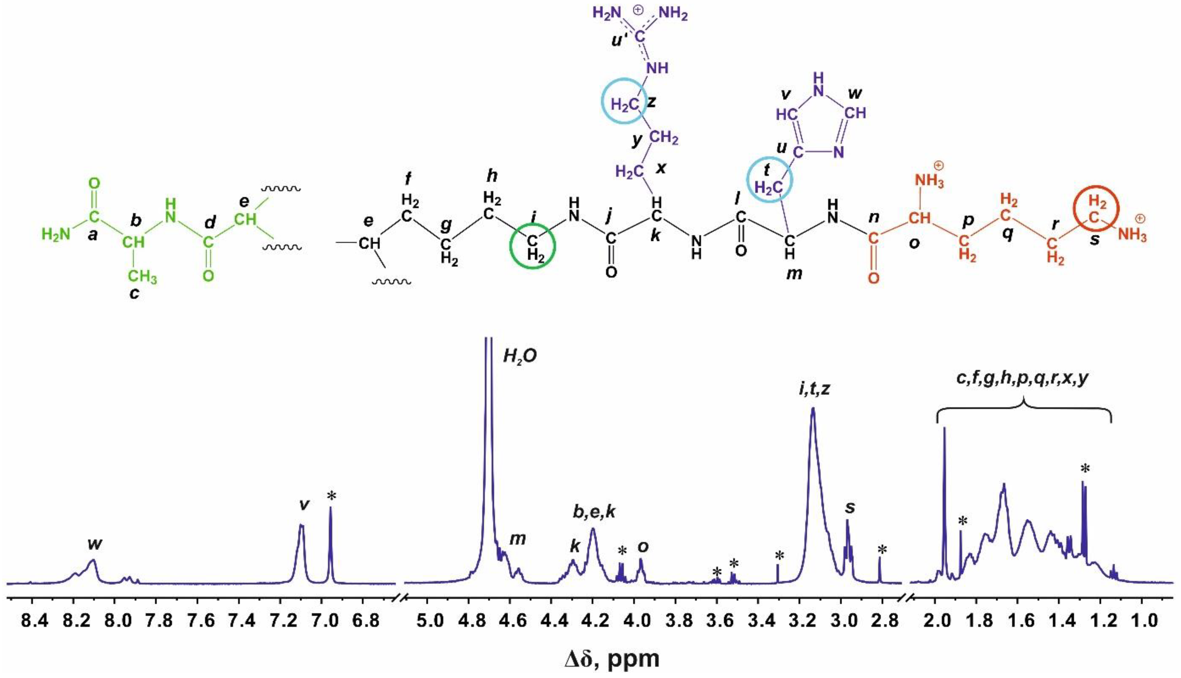
Abstract
:1. Introduction
2. Results
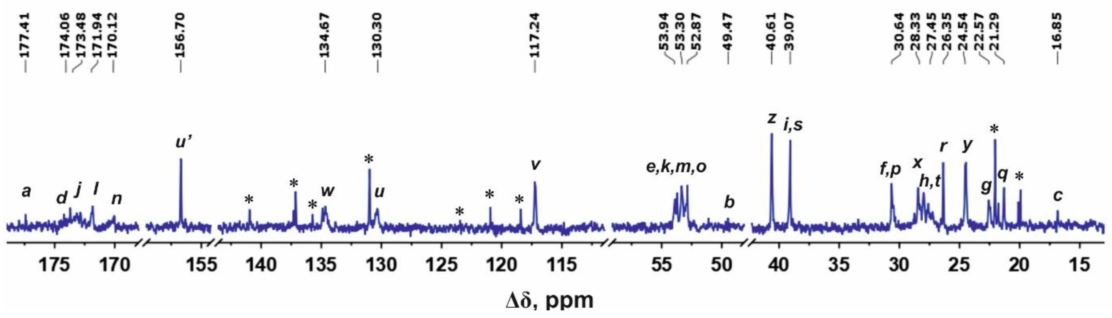
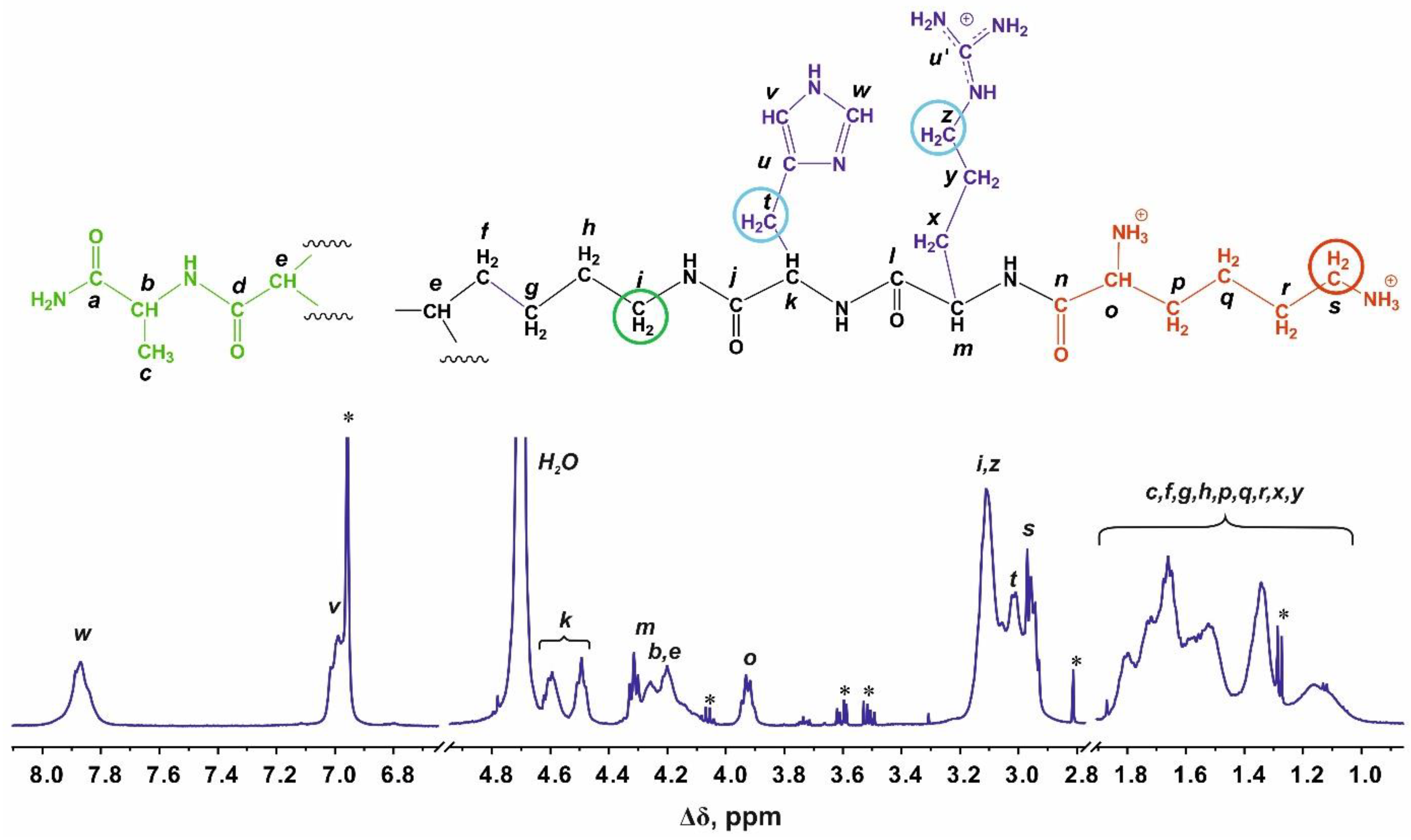
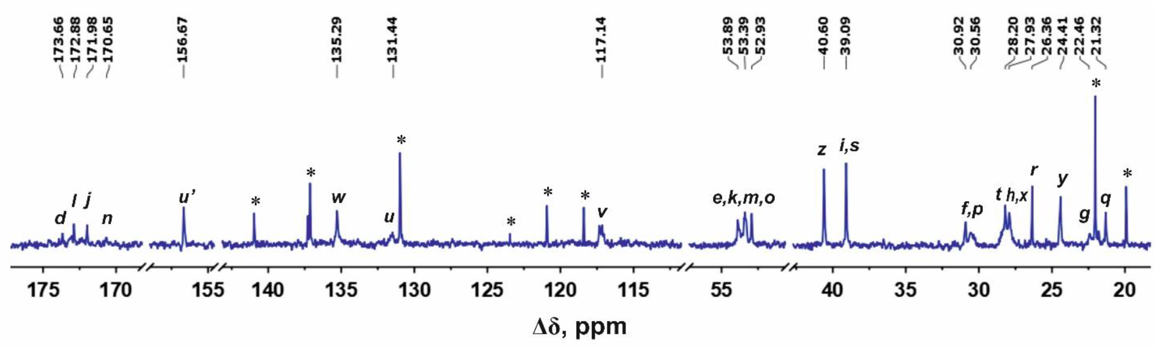
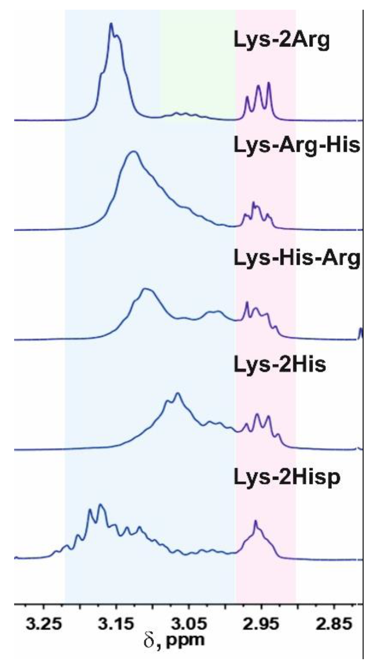
2.1. Analysis of 1H and 13C NMR Spectra
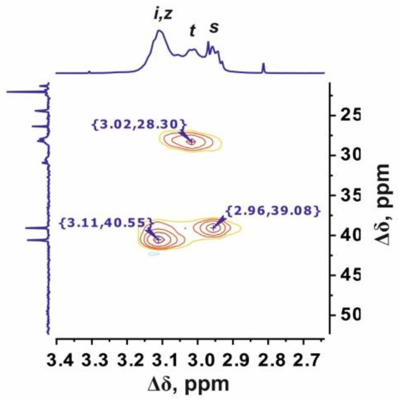
2.2. Lys-Arg-His
2.3. Lys-His-Arg
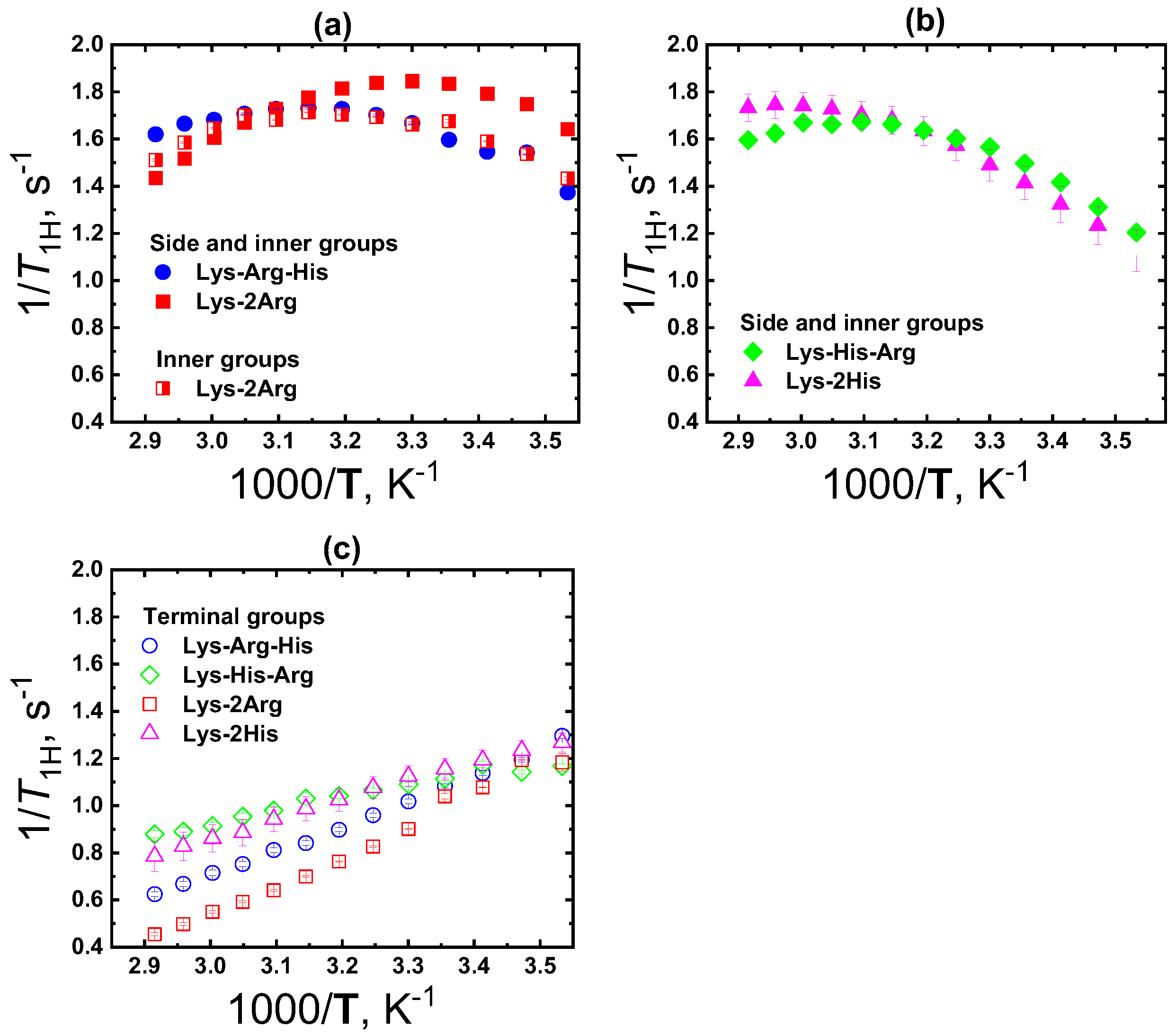
2.4. Local Orientational Mobility
3. Conclusions
4. Materials and Methods
4.1. Synthesis of Lys-His-Arg and Lys-Arg-His
4.2. NMR Experiments
Supplementary Materials
Author Contributions
Funding
Institutional Review Board Statement
Informed Consent Statement
Data Availability Statement
Acknowledgments
Conflicts of Interest
References
- Chis, A.A.; Dobrea, C.; Morgovan, C.; Arseniu, A.M.; Rus, L.L.; Butuca, A.; Juncan, A.M.; Totan, M.; Vonica-Tincu, A.L.; Cormos, G.; et al. Applications and Limitations of Dendrimers in Biomedicine. Molecules 2020, 25, 3982. [Google Scholar] [CrossRef] [PubMed]
- Dias, A.P.; da Silva Santos, S.; da Silva, J.V.; Parise-Filho, R.; Igne Ferreira, E.; El Seoud, O.; Giarolla, J. Dendrimers in the context of nanomedicine. Int. J. Pharm. 2020, 573, 118814. [Google Scholar] [CrossRef] [PubMed]
- Lee, C.C.; MacKay, J.A.; Frechet, J.M.J.; Szoka, F.C. Designing dendrimers for biological applications. Nat. Biotech. 2005, 23, 1517–1526. [Google Scholar] [CrossRef] [PubMed]
- Majoros, I.; Baker, J.J.R. Dendrimer-Based Nanomedicine; Majoros, I.J., Baker, J.R., Eds.; Pan Stanford Publishing: Singapore, 2008; ISBN 9789814241045. [Google Scholar]
- Svenson, S.; Tomalia, D.A. Dendrimers in biomedical applications—Reflections on the field. Adv. Drug Deliv. Rev. 2012, 64, 102–115. [Google Scholar] [CrossRef]
- Jang, W.-D.; Kamruzzaman Selim, K.M.; Lee, C.-H.; Kang, I.-K. Bioinspired application of dendrimers: From bio-mimicry to biomedical applications. Prog. Polym. Sci. 2009, 34, 1–23. [Google Scholar] [CrossRef]
- Liu, M.; Fréchet, J.M.J. Designing dendrimers for drug delivery. Pharm. Sci. Technolo. Today 1999, 2, 393–401. [Google Scholar]
- Esfand, R.; Tomalia, D.A. Poly(amidoamine) (PAMAM) dendrimers: From biomimicry to drug delivery and biomedical applications. Drug Discov. Today 2001, 6, 427–436. [Google Scholar] [CrossRef]
- Choi, Y.; Thomas, T.; Kotlyar, A.; Islam, M.T.; Baker, J.R. Synthesis and Functional Evaluation of DNA-Assembled Polyamidoamine Dendrimer Clusters for Cancer Cell-Specific Targeting. Chem. Biol. 2005, 12, 35–43. [Google Scholar] [CrossRef] [Green Version]
- Tomalia, D.A.; Reyna, L.A.; Svenson, S. Dendrimers as multi-purpose nanodevices for oncology drug delivery and diagnostic imaging. Biochem. Soc. Trans. 2007, 35, 61–67. [Google Scholar] [CrossRef] [Green Version]
- Gillies, E.R.; Fréchet, J.M.J. pH-Responsive Copolymer Assemblies for Controlled Release of Doxorubicin. Bioconjug. Chem. 2005, 16, 361–368. [Google Scholar] [CrossRef]
- Boas, U.; Christensen, J.B.; Heegaard, P.M.H.; Peng, L. Dendrimers in Medicine and Biotechnology: New Molecular Tools; The Royal Society of Chemistry: London, UK, 2006; ISBN 978-0-85404-852-6. [Google Scholar]
- Tekade, R.K.; Kumar, P.V.; Jain, N.K. Dendrimers in Oncology: An Expanding Horizon. Chem. Rev. 2009, 109, 49–87. [Google Scholar] [CrossRef] [PubMed]
- Singh, S.K.; Sharma, V.K. Dendrimers: A Class of Polymer in the Nanotechnology for Drug Delivery. In Nanomedicine for Drug Delivery and Therapeutics; John Wiley & Sons, Inc.: Hoboken, NJ, USA, 2013; pp. 373–409. ISBN 9781118414095. [Google Scholar]
- Svenson, S.; Chauhan, A.S. Dendrimers for enhanced drug solubilization. Nanomedicine 2008, 3, 679–702. [Google Scholar] [CrossRef] [PubMed]
- Duncan, R.; Izzo, L. Dendrimer biocompatibility and toxicity. Adv. Drug Deliv. Rev. 2005, 57, 2215–2237. [Google Scholar] [CrossRef] [PubMed]
- Khan, M.K.; Nigavekar, S.S.; Minc, L.D.; Kariapper, M.S.T.; Nair, B.M.; Lesniak, W.G.; Balogh, L.P. In Vivo Biodistribution of Dendrimers and Dendrimer Nanocomposites—Implications for Cancer Imaging and Therapy. Technol. Cancer Res. Treat. 2005, 4, 603–613. [Google Scholar] [CrossRef]
- Sadler, K.; Tam, J.P. Peptide dendrimers: Applications and synthesis. Rev. Mol. Biotechnol. 2002, 90, 195–229. [Google Scholar] [CrossRef]
- Martinho, N.; Silva, L.C.; Florindo, H.F.; Brocchini, S.; Zloh, M.; Barata, T.S. Rational design of novel, fluorescent, tagged glutamic acid dendrimers with different terminal groups and in silico analysis of their properties. Int. J. Nanomedicine 2017, 12, 7053–7073. [Google Scholar] [CrossRef] [Green Version]
- Hsu, H.; Bugno, J.; Lee, S.; Hong, S. Dendrimer-based nanocarriers: A versatile platform for drug delivery. WIREs Nanomed. Nanobiotechnol. 2017, 9, e1409. [Google Scholar] [CrossRef]
- Palmerston Mendes, L.; Pan, J.; Torchilin, V. Dendrimers as Nanocarriers for Nucleic Acid and Drug Delivery in Cancer Therapy. Molecules 2017, 22, 1401. [Google Scholar] [CrossRef] [Green Version]
- Gorzkiewicz, M.; Konopka, M.; Janaszewska, A.; Tarasenko, I.I.; Sheveleva, N.N.; Gajek, A.; Neelov, I.M.; Klajnert-Maculewicz, B. Application of new lysine-based peptide dendrimers D3K2 and D3G2 for gene delivery: Specific cytotoxicity to cancer cells and transfection in vitro. Bioorg. Chem. 2020, 95, 103504. [Google Scholar] [CrossRef]
- Kuang, T.; Fu, D.; Chang, L.; Yang, Z.; Chen, Z.; Jin, L.; Chen, F.; Peng, X. Recent Progress in Dendrimer-based Gene Delivery Systems. Curr. Org. Chem. 2016, 20, 1820–1826. [Google Scholar] [CrossRef] [Green Version]
- Pompilio, A.; Geminiani, C.; Mantini, P.; Siriwardena, T.N.; Di Bonaventura, I.; Reymond, J.L.; Di Bonaventura, G. Peptide dendrimers as “lead compounds” for the treatment of chronic lung infections by Pseudomonas aeruginosa in cystic fibrosis patients: In vitro and in vivo studies. Infect. Drug Resist. 2018, 11, 1767–1782. [Google Scholar] [CrossRef] [PubMed]
- Ben Jeddou, F.; Falconnet, L.; Luscher, A.; Siriwardena, T.; Reymond, J.-L.; van Delden, C.; Köhler, T. Adaptive and Mutational Responses to Peptide Dendrimer Antimicrobials in Pseudomonas aeruginosa. Antimicrob. Agents Chemother. 2020, 64, e02040-19. [Google Scholar] [CrossRef] [PubMed] [Green Version]
- Kawano, Y.; Jordan, O.; Hanawa, T.; Borchard, G.; Patrulea, V. Are Antimicrobial Peptide Dendrimers an Escape from ESKAPE? Adv. Wound Care 2020, 9, 378–395. [Google Scholar] [CrossRef] [PubMed]
- Cañas-Arranz, R.; de León, P.; Forner, M.; Defaus, S.; Bustos, M.J.; Torres, E.; Andreu, D.; Blanco, E.; Sobrino, F. Immunogenicity of a Dendrimer B2T Peptide Harboring a T-Cell Epitope from FMDV Non-structural Protein 3D. Front. Vet. Sci. 2020, 7, 498. [Google Scholar] [CrossRef]
- Shao, N.; Wang, F.; Chen, Y.; Wang, H.; Cheng, Y.; Wang, Y. Synergistic effect of amino acids modified on dendrimer surface in gene delivery. Biomaterials 2014, 35, 9187–9198. [Google Scholar]
- Crespo, L.; Sanclimens, G.; Pons, M.; Giralt, E.; Royo, M.; Albericio, F. Peptide and amide bond-containing dendrimers. Chem. Rev. 2005, 105, 1663–1681. [Google Scholar] [CrossRef]
- Johansson, E.M.V.; Crusz, S.A.; Kolomiets, E.; Buts, L.; Kadam, R.U.; Cacciarini, M.; Bartels, K.-M.; Diggle, S.P.; Cámara, M.; Williams, P.; et al. Inhibition and Dispersion of Pseudomonas aeruginosa Biofilms by Glycopeptide Dendrimers Targeting the Fucose-Specific Lectin LecB. Chem. Biol. 2008, 15, 1249–1257. [Google Scholar] [CrossRef]
- Gorain, B.; Choudhury, H.; Pandey, M.; Mohd Amin, M.C.I.; Singh, B.; Gupta, U.; Kesharwani, P. Dendrimers as Effective Carriers for the Treatment of Brain Tumor. In Nanotechnology-Based Targeted Drug Delivery Systems for Brain Tumors; Elsevier: Amsterdam, The Netherlands, 2018; pp. 267–305. [Google Scholar]
- Cooper, B.M.; Iegre, J.; O’ Donovan, D.H.; Ölwegård Halvarsson, M.; Spring, D.R. Peptides as a platform for targeted therapeutics for cancer: Peptide–drug conjugates (PDCs). Chem. Soc. Rev. 2021, 50, 1480–1494. [Google Scholar] [CrossRef]
- Maysinger, D.; Zhang, Q.; Kakkar, A. Dendrimers as Modulators of Brain Cells. Molecules 2020, 25, 4489. [Google Scholar] [CrossRef]
- Filipczak, N.; Yalamarty, S.S.K.; Li, X.; Parveen, F.; Torchilin, V. Developments in Treatment Methodologies Using Dendrimers for Infectious Diseases. Molecules 2021, 26, 3304. [Google Scholar] [CrossRef]
- Gorzkiewicz, M.; Kopeć, O.; Janaszewska, A.; Konopka, M.; Pędziwiatr-Werbicka, E.; Tarasenko, I.I.; Bezrodnyi, V.V.; Neelov, I.M.; Klajnert-Maculewicz, B. Poly(lysine) Dendrimers Form Complexes with siRNA and Provide Its Efficient Uptake by Myeloid Cells: Model Studies for Therapeutic Nucleic Acid Delivery. Int. J. Mol. Sci. 2020, 21, 3138. [Google Scholar] [CrossRef] [PubMed]
- Okuda, T.; Sugiyama, A.; Niidome, T.; Aoyagi, H. Characters of dendritic poly(L-lysine) analogues with the terminal lysines replaced with arginines and histidines as gene carriers in vitro. Biomaterials 2004, 25, 537–544. [Google Scholar] [CrossRef] [PubMed]
- Boas, U.; Heegaard, P.M.H. Dendrimers in drug research. Chem. Soc. Rev. 2004, 33, 43–63. [Google Scholar] [CrossRef] [PubMed]
- Stiriba, S.-E.; Frey, H.; Haag, R. Dendritic Polymers in Biomedical Applications: From Potential to Clinical Use in Diagnostics and Therapy. Angew. Chemie Int. Ed. 2002, 41, 1329–1334. [Google Scholar] [CrossRef]
- Agarwal, A.; Saraf, S.; Asthana, A.; Gupta, U.; Gajbhiye, V.; Jain, N.K. Ligand based dendritic systems for tumor targeting. Int. J. Pharm. 2008, 350, 3–13. [Google Scholar] [CrossRef]
- Yellepeddi, V.K.; Kumar, A.; Palakurthi, S. Surface modified poly(amido)amine dendrimers as diverse nanomolecules for biomedical applications. Expert Opin. Drug Deliv. 2009, 6, 835–850. [Google Scholar] [CrossRef]
- Kharwade, R.; More, S.; Warokar, A.; Agrawal, P.; Mahajan, N. Starburst pamam dendrimers: Synthetic approaches, surface modifications, and biomedical applications. Arab. J. Chem. 2020, 13, 6009–6039. [Google Scholar] [CrossRef]
- Wolinsky, J.; Grinstaff, M. Therapeutic and diagnostic applications of dendrimers for cancer treatment. Adv. Drug Deliv. Rev. 2008, 60, 1037–1055. [Google Scholar] [CrossRef]
- Chen, Y.; Tao, K.; Ji, W.; Kumar, V.B.; Rencus-Lazar, S.; Gazit, E. Histidine as a key modulator of molecular self-assembly: Peptide-based supramolecular materials inspired by biological systems. Mater. Today 2022, 60, 106–127. [Google Scholar] [CrossRef]
- Bradbury, J.H.; Chapman, B.E.; Crompton, M.W.; Norton, R.S.; Teh, J.S. Hydrogen–deuterium exchange of the C-2 protons of histidine and histidine peptides and proteins. J. Chem. Soc. Perkin Trans. 2 1980, 693–700. [Google Scholar] [CrossRef]
- Cebo, M.; Kielmas, M.; Adamczyk, J.; Cebrat, M.; Szewczuk, Z.; Stefanowicz, P. Hydrogen-deuterium exchange in imidazole as a tool for studying histidine phosphorylation. Anal. Bioanal. Chem. 2014, 406, 8013–8020. [Google Scholar] [CrossRef] [PubMed] [Green Version]
- Matthews, H.R.; Matthews, K.S.; Opella, S.J. Selectively deuterated amino acid analogues synthesis, Incorporation into proteins and NMR properties. BBA-Gen. Subj. 1977, 497, 1–13. [Google Scholar] [CrossRef]
- Sheveleva, N.N.; Markelov, D.A.; Vovk, M.A.; Tarasenko, I.I.; Mikhailova, M.E.; Ilyash, M.Y.; Neelov, I.M.; Lahderanta, E. Stable Deuterium Labeling of Histidine-Rich Lysine-Based Dendrimers. Molecules 2019, 24, 2481. [Google Scholar] [CrossRef] [PubMed] [Green Version]
- Sheveleva, N.N.; Bezrodnyi, V.V.; Mikhtaniuk, S.E.; Shavykin, O.V.; Neelov, I.M.; Tarasenko, I.I.; Vovk, M.A.; Mikhailova, M.E.; Penkova, A.V.; Markelov, D.A. Local Orientational Mobility of Collapsed Dendrimers. Macromolecules 2021, 54, 11083–11092. [Google Scholar] [CrossRef]
- Shavykin, O.V.; Mikhailov, I.V.; Darinskii, A.A.; Neelov, I.M.; Leermakers, F.A.M. Effect of an asymmetry of branching on structural characteristics of dendrimers revealed by Brownian dynamics simulations. Polymer 2018, 146, 256–266. [Google Scholar] [CrossRef]
- Shavykin, O.V.; Leermakers, F.A.M.; Neelov, I.M.; Darinskii, A.A. Self-Assembly of Lysine-Based Dendritic Surfactants Modeled by the Self-Consistent Field Approach. Langmuir 2018, 34, 1613–1626. [Google Scholar] [CrossRef]
- Sheveleva, N.N.; Markelov, D.A.; Vovk, M.A.; Mikhailova, M.E.; Tarasenko, I.I.; Neelov, I.M.; Lähderanta, E. NMR studies of excluded volume interactions in peptide dendrimers. Sci. Rep. 2018, 8, 8916. [Google Scholar] [CrossRef] [Green Version]
- Sheveleva, N.N.; Markelov, D.A.; Vovk, M.A.; Mikhailova, M.E.; Tarasenko, I.I.; Tolstoy, P.M.; Neelov, I.M.; Lähderanta, E. Lysine-based dendrimer with double arginine residues. RSC Adv. 2019, 9, 18018–18026. [Google Scholar] [CrossRef] [Green Version]
- Neelov, I.M.; Falkovich, S.; Markelov, D.; Paci, E.; Darinskii, A.; Tenhu, H. Molecular Dynamics of Lysine Dendrimers. Computer Simulation and NMR. In Dendrimers in Biomedical Applications; Royal Society of Chemistry: Cambridge, UK, 2013; pp. 99–114. ISBN 978-1-84973-611-4. [Google Scholar]
- Neelov, I.M.; Markelov, D.A.; Falkovich, S.G.; Ilyash, M.Y.; Okrugin, B.M.; Darinskii, A.A. Mathematical simulation of lysine dendrimers: Temperature dependences. Polym. Sci. Ser. C 2013, 55, 154–161. [Google Scholar] [CrossRef]
- Okrugin, B.; Ilyash, M.; Markelov, D.; Neelov, I. Lysine Dendrigraft Nanocontainers. Influence of Topology on Their Size and Internal Structure. Pharmaceutics 2018, 10, 129. [Google Scholar] [CrossRef] [Green Version]
- Mikhtaniuk, S.; Bezrodnyi, V.; Shavykin, O.; Neelov, I.; Sheveleva, N.; Penkova, A.; Markelov, D. Comparison of Structure and Local Dynamics of Two Peptide Dendrimers with the Same Backbone but with Different Side Groups in Their Spacers. Polymers 2020, 12, 1657. [Google Scholar] [CrossRef] [PubMed]
- Shavykin, O.V.; Neelov, I.M.; Darinskii, A.A. Is the manifestation of the local dynamics in the spin–lattice NMR relaxation in dendrimers sensitive to excluded volume interactions? Phys. Chem. Chem. Phys. 2016, 18, 24307–24317. [Google Scholar] [CrossRef] [PubMed]
- Okrugin, B.M.; Neelov, I.M.; Leermakers, F.A.M.; Borisov, O.V. Structure of asymmetrical peptide dendrimers: Insights given by self-consistent field theory. Polymer 2017, 125, 292–302. [Google Scholar] [CrossRef]
- Heyda, J.; Mason, P.E.; Jungwirth, P. Attractive Interactions between Side Chains of Histidine-Histidine and Histidine-Arginine-Based Cationic Dipeptides in Water. J. Phys. Chem. B 2010, 114, 8744–8749. [Google Scholar] [CrossRef] [PubMed]
- Chen, Y.; Yang, Y.; Orr, A.A.; Makam, P.; Redko, B.; Haimov, E.; Wang, Y.; Shimon, L.J.W.; Rencus-Lazar, S.; Ju, M.; et al. Self-Assembled Peptide Nano-Superstructure towards Enzyme Mimicking Hydrolysis. Angew. Chemie Int. Ed. 2021, 60, 17164–17170. [Google Scholar] [CrossRef]
- Bezrodnyi, V.V.; Mikhtaniuk, S.E.; Shavykin, O.V.; Neelov, I.M.; Sheveleva, N.N.; Markelov, D.A. Size and Structure of Empty and Filled Nanocontainer Based on Peptide Dendrimer with Histidine Spacers at Different pH. Molecules 2021, 26, 6552. [Google Scholar] [CrossRef]
- Bezrodnyi, V.V.; Shavykin, O.V.; Mikhtaniuk, S.E.; Neelov, I.M.; Sheveleva, N.N.; Markelov, D.A. Why the Orientational Mobility in Arginine and Lysine Spacers of Peptide Dendrimers Designed for Gene Delivery Is Different? Int. J. Mol. Sci. 2020, 21, 9749. [Google Scholar] [CrossRef]
- Abragam, A. The Principles of Nuclear Magnetism; Oxford University Press: Oxford, UK, 1983; ISBN 9780198520146. [Google Scholar]
- Bloembergen, N.; Purcell, E.M.; Pound, R.V. Relaxation effects in nuclear magnetic resonance absorption. Phys. Rev. 1948, 73, 679–712. [Google Scholar] [CrossRef] [Green Version]
- Solomon, I. Relaxation Processes in a System of Two Spins. Phys. Rev. 1955, 99, 559–565. [Google Scholar] [CrossRef]
- Chizhik, V.I.; Chernyshev, Y.S.; Donets, A.V.; Frolov, V.V.; Komolkin, A.V.; Shelyapina, M.G. Magnetic Resonance and Its Applications; Springer International Publishing: Berlin/Heidelberg, Germany, 2014; ISBN 978-3-319-05298-4. [Google Scholar]
- Levitt, M.H. Spin Dynamics: Basics of Nuclear Magnetic Resonance, 2nd ed.; Wiley: Chichester, UK; Hoboken, NJ, USA, 2008; ISBN 978-0-470-51118-3. [Google Scholar]
- Gotlib, Y.Y.; Markelov, D.A. Permittivity of a dendrimer containing polar groups. Polym. Sci. Ser. A 2004, 46, 815–832. [Google Scholar]
- Gotlib, Y.Y.; Markelov, D.A. Theory of orientational relaxation of individual specified units in a dendrimer. Polym. Sci. Ser. A 2007, 49, 1137–1154. [Google Scholar] [CrossRef]
- Markelov, D.A.; Dolgushev, M.; Gotlib, Y.Y.; Blumen, A. NMR relaxation of the orientation of single segments in semiflexible dendrimers. J. Chem. Phys. 2014, 140, 244904. [Google Scholar] [CrossRef] [PubMed] [Green Version]
- Novoa-Carballal, R.; Säwén, E.; Fernandez-Megia, E.; Correa, J.; Riguera, R.; Widmalm, G. The dynamics of GATG glycodendrimers by NMR diffusion and quantitative 13C relaxation. Phys. Chem. Chem. Phys. 2010, 12, 6587. [Google Scholar] [CrossRef] [PubMed]
- Markelov, D.A.; Matveev, V.V.; Ingman, P.; Nikolaeva, M.N.; Lähderanta, E.; Shevelev, V.A.; Boiko, N.I. NMR Studies of Carbosilane Dendrimer with Terminal Mesogenic Groups. J. Phys. Chem. B 2010, 114, 4159–4165. [Google Scholar] [CrossRef]
- Pinto, L.F.; Correa, J.; Martin-Pastor, M.; Riguera, R.; Fernandez-Megia, E. The dynamics of dendrimers by NMR relaxation: Interpretation pitfalls. J. Am. Chem. Soc. 2013, 135, 1972–1977. [Google Scholar] [CrossRef]
- Pinto, L.F.; Riguera, R.; Fernandez-Megia, E. Stepwise filtering of the internal layers of dendrimers by transverse-relaxation-edited NMR. J. Am. Chem. Soc. 2013, 135, 11513–11516. [Google Scholar] [CrossRef]
- Markelov, D.A.; Dolgushev, M.; Lähderanta, E. Chapter One—NMR Relaxation in Dendrimers. In Annual Reports on NMR Spectroscopy; G.A. Webb, Ed.; Academic Press: Cambridge, MA, USA, 2017; Volume 91, pp. 1–66. ISBN 0066-4103. [Google Scholar]
- Markelov, D.A.; Shishkin, A.N.; Matveev, V.V.; Penkova, A.V.; Lähderanta, E.; Chizhik, V.I. Orientational Mobility in Dendrimer Melts: Molecular Dynamics Simulations. Macromolecules 2016, 49, 9247–9257. [Google Scholar] [CrossRef]
- Gupta, S.; Biswas, P. Orientational Relaxation of Poly(propylene imine) Dendrimers at Different pH. J. Phys. Chem. B 2020, 124, 4193–4202. [Google Scholar] [CrossRef]
- Sørland, G.H. The Spoiler Recovery Approach (SR). In Dynamic Pulsed-Field-Gradient NMR; Springer Series in Chemical Physics; Springer: Berlin/Heidelberg, Germany, 2014; Volume 110, pp. 169–190. ISBN1 978-3-662-44500-6. ISBN2 978-3-662-44499-3. [Google Scholar]
- Sadovnichy, V.; Tikhonravov, A.; Voevodin, V.l.; Opanasenko, V. “Lomonosov”: Supercomputing at Moscow State University. In Contemporary High Performance Computing: From Petascale toward Exascale; Vetter, J.S., Ed.; Chapman and Hall/CRC Press: London, UK, 2013; pp. 283–307. ISBN 9781466568341. [Google Scholar]


Disclaimer/Publisher’s Note: The statements, opinions and data contained in all publications are solely those of the individual author(s) and contributor(s) and not of MDPI and/or the editor(s). MDPI and/or the editor(s) disclaim responsibility for any injury to people or property resulting from any ideas, methods, instructions or products referred to in the content. |
© 2023 by the authors. Licensee MDPI, Basel, Switzerland. This article is an open access article distributed under the terms and conditions of the Creative Commons Attribution (CC BY) license (https://creativecommons.org/licenses/by/4.0/).
Share and Cite
Sheveleva, N.N.; Tarasenko, I.I.; Vovk, M.A.; Mikhailova, M.E.; Neelov, I.M.; Markelov, D.A. NMR Studies of Two Lysine Based Dendrimers with Insertion of Similar Histidine-Arginine and Arginine-Histidine Spacers Having Different Properties for Application in Drug Delivery. Int. J. Mol. Sci. 2023, 24, 949. https://doi.org/10.3390/ijms24020949
Sheveleva NN, Tarasenko II, Vovk MA, Mikhailova ME, Neelov IM, Markelov DA. NMR Studies of Two Lysine Based Dendrimers with Insertion of Similar Histidine-Arginine and Arginine-Histidine Spacers Having Different Properties for Application in Drug Delivery. International Journal of Molecular Sciences. 2023; 24(2):949. https://doi.org/10.3390/ijms24020949
Chicago/Turabian StyleSheveleva, Nadezhda N., Irina I. Tarasenko, Mikhail A. Vovk, Mariya E. Mikhailova, Igor M. Neelov, and Denis A. Markelov. 2023. "NMR Studies of Two Lysine Based Dendrimers with Insertion of Similar Histidine-Arginine and Arginine-Histidine Spacers Having Different Properties for Application in Drug Delivery" International Journal of Molecular Sciences 24, no. 2: 949. https://doi.org/10.3390/ijms24020949