
Natural Killer Cell Receptors and Endometriosis: A Systematic Review
by
 , , ,
, , ,
José Lourenço Reis
1,
Natacha Nurdine Rosa
2,
Miguel Ângelo-Dias
3,4,
Catarina Martins
3,4,
Luís Miguel Borrego
3,4,5 and
Jorge Lima
1,3,4,* 1
Department of Obstetrics and Gynecology, LUZ SAÚDE, Hospital da Luz, 1500-650 Lisboa, Portugal
2
UCD School of Medicine, University College Dublin, D04 V1W8 Dublin, Ireland
3
Comprehensive Health Research Centre (CHRC), NOVA Medical School|Faculdade de Ciências Médicas (NMS|FCM), Universidade Nova de Lisboa, 1169-056 Lisboa, Portugal
4
Immunology Department, NOVA Medical School|Faculdade de Ciências Médicas (NMS|FCM), Universidade Nova de Lisboa, 1169-056 Lisboa, Portugal
5
Department of Imunoallergy, LUZ SAÚDE, Hospital da Luz, 1500-650 Lisboa, Portugal
*
Author to whom correspondence should be addressed.
Int. J. Mol. Sci. 2023, 24(1), 331; https://doi.org/10.3390/ijms24010331
Submission received: 9 November 2022
/
Revised: 12 December 2022
/
Accepted: 21 December 2022
/
Published: 25 December 2022
(This article belongs to the Special Issue Reproductive Immunology and Pregnancy 3.0)
Abstract
:Endometriosis is a chronic inflammatory disorder, characterized by the presence of endometrial cells outside the uterine cavity. An increasing number of studies correlate the immune system with endometriosis, particularly NK receptors (NKR), which have been suggested to play an essential role in the pathogenesis of the disease. This systematic review aims to enlighten the role of NKR in endometriosis. A literature search was performed independently by two reviewers, to identify studies assessing the role of NKR in endometriosis. In total, 18 studies were included. Endometriosis pathogenesis seems to be marked by the overexpression of NK inhibitor receptors (KIRS), namely, CD158a+, KIR2DL1, CD94/NKG2A, PD-1, NKB1, and EB6, and inhibiting ligands such as PD-L1, HLA-E, HLA-G, and HLA-I. Concurrently, there is a decrease in NK-activating receptors and natural cytotoxicity receptors (NCRs), such as NKp46, NKp30, and NKG2D. The immune shift from NK surveillance to NK suppression is also apparent in the greater relative number of ITIM domains compared with ITAM domains in NKRs. In conclusion, NK receptor activity seems to dictate the immunocompetency of women to clear endometriotic cells from the peritoneal cavity. Future research could explore NKRs as therapeutic targets, such as that which is now well established in cancer therapy through immunotherapy.
1. Introduction
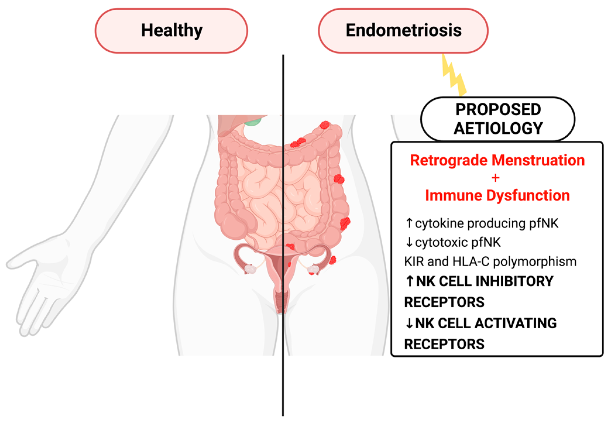
Endometriosis is a chronic gynecological disorder characterized by the presence of endometriotic tissue outside the uterine cavity. It is a very incapacitating disease affecting 1 in 10 women during reproductive age [1], having a great impact on their quality of life. Although endometriosis was first noted by Carl von Rokitansky more than one hundred years ago, the pathogenesis of this condition is still not clear. Regarding pathogenesis, the theory of retrograde menstruation and implantation of ectopic endometrium is the most accepted [2]. Retrograde menstruation occurs in almost all women, but only 10% of them develop endometriosis [1]. Thus, the question arises as to what facilitates ectopic endometriotic cell implantation and growth. The hypothesis of immunological dysfunction has gained greater evidence in recent years. In the peritoneal cavity microenvironment, immune dysfunction enhances the capacity of ectopic endometrial cells to adhere and invade, leading to poor clearance of the ectopic endometrium and increased proliferation and angiogenesis [3]. NK cells have been suggested to play an important role in the pathogenesis of the disease by either tolerating or inhibiting the survival, implantation, and proliferation of endometrial cells [4]. An increasing number of studies [5,6,7,8,9,10,11,12,13,14,15,16,17,18,19,20] address the role of NK receptors (activating and inhibiting) in endometriosis pathogenesis (Figure 1). To the best of our knowledge, there are no systematic reviews concerning the involvement of NK cell receptors in endometriosis; therefore, this assessment is important for a better understanding of the topic.
2. Methods
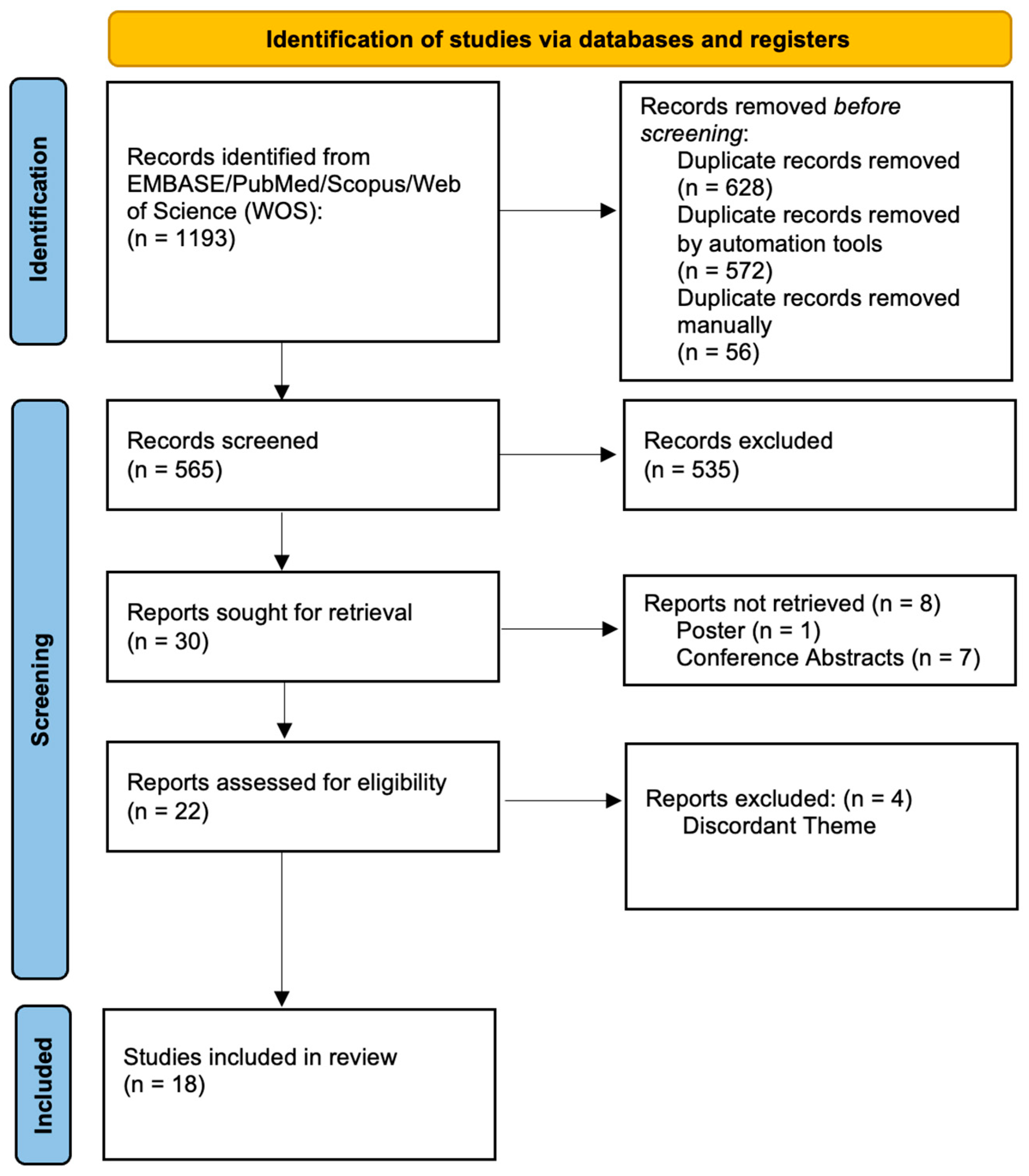
This systematic review identified and analyzed human studies which assess the role of NK cell receptors in women suffering from endometriosis compared with healthy women, and was conducted according to the PRISMA guidelines (Preferred Reporting Items for Systematic Reviews and Meta-analyses) [21,22] (Figure 2) and registered in PROSPERO (CRD42020205783).
2.1. Search Strategy
An extensive computerized literature search strategy was performed to retrieve the studies included in this review. The searches were performed in PubMed/Medline, EMBASE, Scopus, and Web of Science databases, using database-specific subject heading terms and all its variants in free-text words, according to the specificities of each database (Table S1). To ensure literature saturation, we performed a supplementary search of grey literature and of the reference sections of the selected studies and reviews, to identify any additional relevant missing publications as well as conference papers not retrieved in the electronic search. No date or language limits were imposed on the search.
2.2. Eligibility Criteria and Study Selection
The study selection was performed independently by two individuals (J.L.R. and N.N.R.), who screened the titles and abstracts of all suitable articles from the queries according to the eligibility criteria (Table S2). No restrictions to geographical location, language of publication, or year of publication were applied, and all non-primary studies were excluded.
Studies that did not meet eligibility criteria were excluded and the remaining articles were thoroughly analyzed and read in full and included or discarded according to the eligibility criteria. Any disagreement was resolved after discussion with a third reviewer (J.L.). All decisions, including reasons for exclusion and the number of selected articles in each step, were recorded and depicted in a flow chart, following the PRISMA 2020 guidelines.
EndNote version 20 was used to store, organize, and manage all the references arising from the literature search, including the management and removal of duplicates, and scanning of the titles/abstracts of all the records.
2.3. Data Extraction and Quality Assessment
All relevant data were extracted from each selected study by two independent reviewers and any disagreement was referred to a third reviewer (J.L.) to reach consensus. The following data were extracted:
- Identification of the study—title, authors, year of publication, journal title, country of origin, study design, number of participants, and recruitment procedure and duration.
- Participant characteristics—sample size, age, BMI, parity, rAFS classification of endometriosis [23], previous treatments, and preoperative painful symptoms scores—of subjects in endometriosis and control groups.
- Methodological features—sample characteristics (endometriotic tissue, peritoneal fluid, peripheral venous blood, and eutopic endometrium), methodology used for NK cell marker characterization (51Cr cytotoxicity assay, ELISA, flow cytometry, immunohistochemistry, in situ hybridization, RNA extraction, and quantitative real-time Western blotting) and NK cell markers.
- Outcomes—comparison of NK cell markers between the different samples studied.
The methodological quality of each individual study was assessed independently by two reviewers on 20 October 2022 (J.L.R. and N.N.R.), using the NHLBI quality assessment tool of case–control studies (https://www.nhlbi.nih.gov/health-topics/study-quality-assessment-tools). This scale evaluates 12 components of a study to determine its methodological quality. Each study evaluation criterion was assigned “Yes”, “No”, “NR” (not reported), “NA” (not applicable), or “Unc” (Unclear). Subsequently, studies were graded as “Poor”, “Fair”, or “Good”. The level of bias influences the evidence and results obtained in this systematic review. Conflicts regarding study quality were resolved with a third reviewer (J.L.)
2.4. Data Synthesis
Due to the high heterogenicity of the selected studies evaluating several NK cell receptors in different types of samples (peripheral blood, peritoneal fluid, and eutopic and ectopic endometrium), a quantitative meta-analysis was not feasible to perform. Data from the selected studies were narratively and qualitatively summarized.
3. Results
3.1. Study Selection
In total, 1193 studies were obtained. Upon the exclusion of 628 duplicates, titles and abstracts were screened, and 535 studies that did not meet eligibility criteria were excluded. There were 30 reports sought for retrieval, where an additional 8 studies were excluded because they were classified as conference abstracts/posters. The remaining 22 articles were thoroughly analyzed and read in full, leading to the exclusion of 4 more studies due to research theme incongruency. In total, 18 studies [5,6,7,8,9,10,11,12,13,14,15,16,17,18,19,20,24,25] were included in the final qualitative analysis, as per Figure 2.
3.2. Study Quality Assessment and Risk of Bias
Quality assessment was conducted on the 18 selected studies as presented in Table S3 (following NHLBI Assessment Tool guidelines). Among these studies, 15 were deemed “good quality” [5,6,8,9,10,14,15,16,17,18,19,20,24,25]. This assessment was based on the sample size, consideration/adjustment to key confounding variables (demographic factors, menstrual cycle stage, hormonal therapy, and disease staging as per revised American Society for Reproductive Medicine (rASRM) classification) [23], and appropriateness of the methodology according to the research hypothesis. None of the studies included sample size justification or blinded evaluators of outcomes.
3.3. Heterogenicity of the Studies Assessed
NK cell receptors (and their ligands) were evaluated in 18 case–control (observational) studies in the context of endometriosis, all using a human model. One of these was also classified as quasi-experimental [24], analyzing the impact of 17β-estradiol treatment on results. Heterogenicity was noted in the samples evaluated in each study, as presented in Table 1. Studies used a wide array of samples, including peritoneal fluid [6,7,8,9,10,11,13,14,16,17,18,19,24,25], endometriotic lesions/eutopic endometrium samples [12,15,18,19,24], and peripheral blood [7,8,9,10,18,20,24].
Additionally, each study employed different techniques to evaluate the NK cell receptor activity and expression in endometriosis. Flow cytometry [7,8,9,10,11,14,16,19,24,25] was most commonly used, followed by Western blotting [6,7,8,9,10,19,24], immunohistochemistry [12,15,18,24], polymerase chain reaction (PCR) [14,19,20], enzyme-linked immunosorbent assay (ELISA) [13,17,18], RNA in situ hybridization [12], and 51Cr cytotoxicity assays [5].
The study results are summarized in Table 1.

Table 1.
Summary of findings and methods of selected studies regarding NK cell-activating/inhibiting receptors in endometriosis.
Table 1.
Summary of findings and methods of selected studies regarding NK cell-activating/inhibiting receptors in endometriosis.
| Samples | Design | n | Comparison | Methods | Markers | Results | Reference |
|---|---|---|---|---|---|---|---|
| PF | Case–control | 11 I/II EDT; 22 III/IV EDT; 11 controls | NK cytotoxicity and NK inhibition receptors in early and late EDT | FC cytotoxicity assay 51Cr | FITC anti-CD45/PE-anti-CD14 γ1 FITC/γ2a PE, FITC-anti-CD3/PE-anti-CD19, FITC-anti-CD3/PE-anti-CD56, FITC-anti-CD3/PE-anti-NKB1/PerCP-anti-CD56, FITC-anti-CD3/PE-anti-GL183/PerCP-anti-CD56, FITC-anti-CD3/PE-anti-EB6/PerCP-anti-CD56. | peritoneal cytotoxicity against K562 EDT (I/II/III/IV) vs. controls; KIR expression (NKB1, EB6) in III/IV EDT vs. controls; KIR expression (NKB1, EB6) in III/IV EDT vs. I/II EDT | Wu et al., 2000 [5] |
| PF | Case–control | 10 EDT; 10 controls | HLA-G inhibitory ligand expression in women with and without EDT | Western blotting | mAb aa 61-83 1 domain of HLA-G | No statistical significance between groups | Hornung et al., 2001 [6] |
| PB, PF | Case–control | 11 I/II EDT; 17 III/IV EDT; 6 controls | ICAM-1 and KIR expression in women with and without EDT | FC Western blotting | FITC-labeled anti-CD3 mAb, anti-CD4 mAb, PE-labeled anti-CD8 mAb, PE-labeled anti-CD19 mAb, FITC-labeled anti-CD16 mAb, FITC-labeled anti-CD14 mAb, PE-labeled anti-CD54 (ICAM-1) mAb, PE-labeled anti-CD158a, anti-CD158b, and CD94 | ICAM in PF macrophages EDT vs. controls; KIR2DL1+ NK among CD16+ NK in PB and PF of EDT vs. controls (more pronounced in III/IV EDT) | Maeda, Izumiya, Oguri et al., 2002 [7] |
| PB, PF | Case–control | 12 I/II EDT; 30 III/IV; 40 controls | KIR2DL1+ NK cell expression in women with and without EDT | FC Western blotting | FITC-anti-CD16 mAb PE-labeled anti-CD158a mAb, anti-CD158b mAb, PE anti-CD94 mAb | KIR2DL1+ NK cells in PB and PF of EDT vs. controls (more pronounced in III/IV EDT) | Maeda, Izumiya, Yamamoto et al., 2002 [8] |
| PB, PF | Case–control | 18 I/II EDT; 70 III/IV EDT; 104 controls | CD158a+ cells (KIR subtype) expression in women with and without EDT | FC Western blotting | FITC-labeled anti-CD16 mAB, PE-labeled anti-CD158a and anti-CD158b mAbs, PE-labeled anti-CD94 mAbs | CD158a+ cells in PF of EDT (I/II/III/IV and III/IV) vs. controls; CD158a+ cells in PB of EDT (I/II and III/IV) | Maeda et al., 2004 [9] |
| PB, PF | Case–control | 6 I/II EDT; 18 III/IV; 25 controls | ITIM and ITAM KIR expression in women with and without EDT | FC Western blotting | FITC-labeled anti-CD56 mAb PE-labeled anti-CD158a mAb, anti-CD158b mAb, Anti-CD158a mAb, anti-CD158b mAb | ITIM-KIR > ITAM-KIR in PB EDT; CD158a+CD56+ NK cells in PB and PF EDT vs. controls | Matsuoka et al., 2005 [10] |
| Endometrium Peritoneal EDT | Case–control | 15 EDT and 12 controls (IHC); 24 endometrium EDT and 14 peritoneal fluid EDT and 17 controls (RNA ISH) | HLA-G expression in eutopic and ectopic endometrium in women with and without EDT | IHC RNA ISH | mAb4H84 cDNA probe | HLA-G protein and gene transcripts found in >90% glandular peritoneal fluid EDT but not in stromal endometrial epithelium (controls and EDT) | Barrier et al., 2006 [12] |
| PF | Case–control | 26 I/II EDT; 20 III/IV EDT; 24 controls | CD56+ cell expression, Fas antigen CD95 and early activation molecule CD69 in PF of women with and without EDT | FC | Anti-CD45FITC/CD14PE, IgG1FITC/IgG2aPE, anti-CD69FITC, anti-95FITC and anti-CD56PE | CD56+ dim expression in I/II EDT vs. controls; CD95 in I/II EDT vs. controls; CD69+CD56+ in I/II EDT vs. controls; CD69+CD56+ in III/IV EDT vs. controls | Eidukaite et al., 2006 [11] |
| PF | Case–control | 17 I/II EDT; 14 III/IV EDT; 27 controls | HLA-G expression in PF of women with and without EDT | ELISA | Anti-sHLA-G | No statistical significance between groups | Eidukaite and Tamosiunas, 2008 [13] |
| PB, PF, Endometrium Peritoneal EDT | Case–control | 20 III/IV EDT; 13 controls | (HLA)-E receptor CD94/NKG2A expression in women with and without EDT | FC, RT-PCR | Anti-CD56 (clone C218), anti-NKG2A (clone Z199), PE-conjugated anti-NKG2A, PE-conjugated anti-NKG2C, fluorescein-conjugated anti-CD56, peridin chlorophyll protein-conjugated anti-CD3, anti-CD45, cytokeratin 20 | CD94/NKG2A cells in PF in EDT vs. controls; HLA-E mRNA in peritoneal EDT | Galandrini et al., 2008 [14] |
| Endometrium | Case–control | 15 EDT; 15 controls | HLA-I and HLA-II expression in women with and without EDT | IHC | IgG2a (HLA-I), IgG1 (HLA-II) | HLA-I and HLA-II expression in EDT endometrial stroma and glands vs. controls | Baka et al., 2011 [15] |
| PF | Case–control | 3 I/II EDT; 18 III/IV EDT; 28 controls | Expression of NK Cell surface antigens (CD16 and CD56+ cells), NCRs (NKp46/40/30) and cytokine production (TNF-, IFN- etc.) of PF NK cells in women with and without EDT | FC | Anti-CD45 PerCP-Cy5.5/anti-CD56 PE/anti-CD16 fluorescein isothiocyanate, anti-CD45 PerCP-Cy5.5/anti-CD56 FITC/anti-CD335 (NKp46) PE, anti-CD45 PerCP-Cy5.5/anti-CD56 FITC/anti-CD336 (NKp44) PE, and anti-CD45 PerCP-Cy5.5/anti-CD56 FITC/anti-CD337 (NKp30) PE | NKp46+ NK cells in III/IV EDT vs. controls; CD56dim/NKp46+ cells in III/IV EDT vs. controls; TNF- producing NK cells in III/IV EDT vs. controls; IFN- producing NK cells in III/IV EDT vs. controls | Funamizu et al., 2014 [16] |
| PF | Case–control | 121 EDT; 81 controls | Levels of soluble NKG2D ligands (MICA, MICB and ULBP-2) in women with and without EDT | ELISA | ELISA (R&D Systems, Inc., Minneapolis, MN, USA) | MICA in EDT vs. controls; MICB in EDT vs. controls; MICA, MICB, ULBP-2 in deep infiltrating EDT | González-Foruria et al., 2015 [17] |
| Serum, PF, Endometrium EDT | Cross-sectional observational/Case–control | 60 I/II EDT; 83 III/IV EDT; 77 controls (ELISA) 26 EDT; 22 controls (IHC) | Soluble HLA-G expression in endometrium, EDT, PF, and serum in women with and without EDT | ELISA IHC | mAb 4H84 MEM-G/9 mouse mAb | sHLA-G in serum but not PF of III/IV EDT vs. controls; HLA-G protein expression in EDT but not eutopic endometrium | Rached et al., 2019 [18] |
| Endometrium EDT PB | Case–control Quasi-experimental | 15 EDT; 15 controls | PD-1/PDL-1 expression in women with and without EDT, post and prior estrogen and cytokine treatment | IHC FC Western blotting | Rabbit anti-human PD-1, CD4, CD8, PD-L1. anti-GAPDH CD279 (PD-1)-PE, CD274 (B7-H1)-PE, CD8-FITC, and CD4-PerCP-Cyanine5.5 | PD-1/PDL-1 and PD-1 in eutopic and ectopic endometrium EDT vs. controls; PD-1/PDL-1 and PD-1 in PB EDT vs. controls; PD-1/PDL-1 and PD-1 in eutopic endometrium EDT post 17-estradiol treatment vs. controls | Wu et al., 2019 [24] |
| Endometrium EDT PF | Case–control | 20 EDT; 13 controls | NKG2D expression and its ligands in women with and without EDT | FC RT-PCR Western blotting | FITC-conjugated mouse, anti-human CD16 mAb, PE-Cy5-conjugated mouse, anti-human CD56 mAb PE-mouse, anti-human NKp30 mAb, PE-mouse, anti-human NKp44 mAb, PE-mouse, anti-human NKp46 mAb, PE-mouse, and anti-human NKG2D mAb | NKp30 CD56+ in PF in EDT; NKp46 CD16+ in PF in EDT; NKG2D CD56+ in PF in EDT; ULBP-2 in eutopic endometrium in EDT vs. controls; ULBP-3 in ectopic endometrium in EDT vs. controls and eutopic EDT | Xu, 2019 [19] |
| PB | Case–control | 147 EDT; 117 controls | HLA-C and KIR polymorphism combinations in women with and without EDT | PCR | Commercial kits (HLAssure SE kit, Accutype Software, SSO typing kit) | HLA-C*03:03:01 occurrence in EDT vs. controls; KIR centromeric A/A haplotypes in EDT vs. controls; KIR2DS2-positive individuals in EDT vs. controls | Chou et al., 2020 [20] |
| PF | Case-control | 32 EDT; 30 controls | NKp46 co-expression patterns in women with and without EDT | FC | anti-TNF-α-BV421, anti-IFN-γ-PE- Cy7, anti-IL-4-PerCP-Cy5, anti-IL-10- APC, anti-TGF-β-PE | CD56+/NKp46+ in PF in EDT; NKG2C+/NKp46+ in PF in EDT; CD16+/NKp46+ in PF in EDT; NKG2D+/NKp46+ in PF in EDT No significant co-expression of inhibitory receptors (CD158a and NKG2A) and NKp46 IFN-γ-producing NK cells in PF in EDT | Saeki et al., 2022 [25] |
aa, amino acids; EDT, endometriosis; FC, flow cytometry; IHC, immunohistochemistry; ISH, in situ hybridization; ITAM, tyrosine-based activation motifs; ITIM, tyrosine-based inhibitory motifs; KIR, natural killer inhibitor receptor; mAb, monoclonal antibody; PB, peripheral venous blood; PF, peritoneal fluid. Scoring and staging of EDT (I/II/III/IV) as per American Fertility Society Guidelines [23].
3.4. NK Cell Receptors in Peritoneal Fluid
Fourteen studies evaluated the activity and expression of NK cell receptors in the peritoneal fluid of endometriosis subjects [5,6,7,8,9,10,11,13,14,16,17,18,19,25]. The role of immune dysfunction in the progression of endometriosis was highlighted by Maeda and colleagues [7,8,9], who found, by using flow cytometry, that the levels of NK cell inhibitory receptors such as CD158a+ and KIR2DL1+ were significantly higher in the peritoneal fluid of endometriosis subjects than controls. This increase was even more pronounced in stage III/IV endometriosis (KIR2DL1+ 24.2% ± 16.1% vs. 12.3% ± 6.9%, p = 0.007 [8], 17.5% ± 8.7% vs. 11.7% ± 5.5%, p = 0.008 [7], CD158a+ 17.0% ± 12.3% vs. 12.3% ± 7.1%, p = 0.017 [9]) Matsuoka et al. [10] corroborated these findings, also reporting an increased percentage of CD158a+ in CD56+ NK cells in the peritoneal fluid of the endometriosis group (13.9% ± 7.5% vs. 7.6% ± 3.0%, p = 0.028). Galandrini et al. [14] conducted a similar study using cytofluorometric analysis and reported an increased frequency of human leukocyte antigen–E inhibitory receptor CD94/NKG2A-expressing peritoneal NK cells in subjects with endometriosis (75.9% ± 8.2% vs. 52.1% ± 16.3%, p < 0.0001). Wu et al. [5] also explored NK inhibitor receptor (KIR) expression in endometriosis with flow cytometry, noting a significant increase in the mean fluorescence intensity of receptors NKB1 (183 ± 56 vs. 97 ± 45, p < 0.001) and EB6 (82 ± 45 vs. 53 ± 14, p = 0.021) in the peritoneal fluid of subjects with severe (III/IV) endometriosis compared with the controls. The increased inhibition of NK cell activity at the peritoneal level in endometriosis was also explored through NK cell inhibitory ligands in three studies [6,13,18], which analyzed HLA-G expression in peritoneal fluid using different methods (ELISA, Western blotting, and immunohistochemistry). All the studies reached the same result: there were no significant statistical differences in HLA-G in the peritoneal fluid of endometriosis subjects compared with the controls. These results were different in other samples ((serum and endometrium) [12,18]), as expressed in the following sections of this report.
NK cell-activating receptors were also investigated in the studies selected. Using flow cytometry, Xu [19] reported a decrease in NKp30 (p = 0.006) and NKG2D (p = 0.01) levels in CD56+ NK cells in the peritoneal fluid of endometriosis subjects. In this same study, however, Xu noted a significant increase in activating receptor NKp46 in CD16+ NK cells (p = 0.04) 19. This finding is in contrast to that of Funamizu et al. [16], who reported a decrease in peritoneal activating receptor NKp46 in advanced (III/IV) endometriosis: 22.84% (11.58–46.34) vs. 30.20% (18.72–51.20), p < 0.05. Furthermore, these authors noted a decrease in peritoneal CD56dim/NKp46 cells, 16.67% (8.95–33.09) vs. 26.12% (14.60–41.14, p < 0.05), as well as an increase in TNF-α (33.87% (25.21–60.73) vs. 25.42% (17.56–30.43), p < 0.05) and IFN-γ (50.04% (26.98–71.33) vs. 19.41% (6.01–38.37), p < 0.05) producing NK cells [16]. This decrease in peritoneal cytotoxic cells (CD56dim/NKp46) in endometriosis is complemented by the findings of Wu et al. [5], whose 51Cr release assay showed a decrease in cytotoxicity against K562 in the peritoneal fluid of subjects with severe (III/IV) (LU20 4.6 ± 3.6 vs. 8.0 ± 4.0 p = 0.011) and early (I/II) (LU20 4.7 ± 2.7 vs. 8.0 ± 4.0 p = 0.028) endometriosis. The decreased activation of NK cell activity at the peritoneal level in endometriosis was also explored through ligands. González-Foruria et al. [17] noted an increase in soluble forms or the shedding of NK-activating receptor ligands (namely, MICA (p = 0.015), MICB (p = 0.003), and ULBP-2 (0.045)) in the peritoneal fluid of subjects with deep infiltrating endometriosis relative to controls. Further elucidating the role of NK cell-activating receptors, Saeki et al. [25] noted a decrease in co-expression of CD56+/NKp46+ and CD56dim/NKp46+ cells (p < 0.05) with a negatively correlated increase in IFN--producing cells (p < 0.01) in subjects with endometriosis. Contrastingly, they also observed an increase in the co-expression of activating receptor NKG2D+/NKp46+ cells in endometriosis subjects (p < 0.05).
NK cell activity may also be suppressed through the loss of NK cells (apoptosis). Eidukaite et al. [11] implicated the Fas–FasL mechanism in the pathophysiology of early endometriosis by reporting a cytometric decrease in CD56dim NK cells (24.0% ± 4.9% vs. 36.3% ± 15.9%, p < 0.02), and an increase in CD95 (Fas antigen that promotes NK apoptosis) (66.6% ± 10.6% vs. 42.4% ± 10.1%, p < 0.01) in the peritoneal fluid of subjects with early endometriosis. They also reported an increase in NK cells containing the Fas early activation molecule (i.e., CD69+ CD56+ NK cells) in the peritoneal fluid of subjects with early (66.8 ± 16.5% vs. 43.3 ± 13.7%, p < 0.01) and advanced (74.2% ± 7.9% vs. 43.3% ± 13.7%, p < 0.01) endometriosis [11].
3.5. NK Cell Receptors in Endometrium and Endometriotic Lesions
Endometrium samples and endometriotic lesions were assessed for the expression of a variety of NK receptors/ligands in the studies selected, to ascertain which features promoted immune evasion and ectopic proliferation/implantation. Two studies [12,18] explored HLA-G (inhibitory NK cell ligand) protein expression in the endometrium and endometriotic lesion samples via immunohistochemistry. As per Barrier et al. [12], HLA-G protein was found in the glandular epithelia of 14 out of 15 peritoneal endometriotic lesion samples (93.3%) (none in stroma), whereas none was found in eutopic endometrium of both control and endometriosis samples. Similarly, Rached et al. [18] found a significant increase in the HLA-G protein expression in endometriotic lesions of advanced-stage endometriosis compared with the eutopic endometrium (both control and endometriosis) (p = 0.018). Barrier et al. [12] went further to analyze HLA-G gene transcripts in endometrial samples using RNA in situ hybridization. With this technique, they found that the glandular epithelia of 13 out of 14 peritoneal lesions (92.8%) contained the HLA-G gene transcript, but this was not present in the eutopic endometrium (control and endometriosis samples). The phenomenon of NK cell inhibition was also explored through the expression of programmed-death 1 (PD-1) and its ligand (PD-L1) in endometriosis using immunohistochemistry and Western blotting [24]. Immunohistochemistry results showed an increase in PD-1 and PD-L1 expression in the eutopic and ectopic endometria of endometriosis samples compared with the controls (p < 0.05 and p < 0.01, respectively) [24]. PD-L1 of the eutopic endometrium of endometriosis samples was upregulated by 17β-estradiol treatment (p < 0.01). Regarding Western blot results, ectopic endometriotic lesions showed a significant increase in PD-1 and PD-L1 expression compared with the controls and the eutopic endometrium from endometriosis samples (p < 0.01); eutopic endometrium from endometriosis also differed significantly from the controls, exhibiting greater PD-L1 expression (p < 0.01) [24]. Additionally, a significant increase in CD4+/CD8+ T cell infiltrate occurrence and size was reported in endometriosis samples (eutopic and ectopic) compared with controls (p < 0.01) [24]. In contrast, in the context of activation, NK cell-activating receptor NKG2D ligands were assessed via real-time PCR and Western blotting. Although Xu [19] did not find any statistically significant differences in ligand mRNA expression, changes at a translational level were observed, namely, a significant decrease in ULBP-2 protein expression in the eutopic endometrium of endometriosis samples (relative to the eutopic endometrium of control samples and ectopic lesions (p < 0.05)), and a decrease in ULBP-3 protein expression in the ectopic endometrium samples compared with the eutopic endometrium (control and endometriosis) (p < 0.05). Factors of immune evasion were also measured [15]. Using immunohistochemistry on endometrial samples obtained via curettage from endometriosis samples and controls, Baka et al. [15] noted a significant increase in HLA-I and HLA-II expression in both the glandular (HLA-I 87.5% vs. 56.3%, p < 0.02, HLA-II 46.7% vs. 20.0%, p < 0.04) and stromal (HLAI 100% vs. 87.5%, p < 0.02, HLAII 66.7% vs. 40.0%, p < 0.007) epithelia of endometriosis samples compared with the controls.
3.6. NK Cell Receptors in Peripheral Blood and Serum
Several studies [7,8,9,10,18,20,24] have explored NK cell receptor expression in the peripheral venous blood of endometriosis patients. Chou et al. [20] investigated the impact of HLA-C (KIR ligand) and KIR genotype/polymorphisms on the risk of endometriosis in a Han Chinese cohort using a DNA-based method of PCR amplification. They showed that a genotype combination HLA-C*03:03:01 was associated with an increased risk of endometriosis (OR = 2.811, p = 0.0473), while the presence of KIR2DS2 acted as a protective factor against endometriosis with an odds ratio of 0.5577 (p = 0.0394). Moreover, the number of KIR centromeric A/A haplotypes was significantly increased in the endometriosis group compared with the control group (OR = 1.793, p = 0.0394) [20]. Maeda et al. [9] studied the expression of NK inhibitory receptor CD158a+ in the peripheral blood of endometriosis subjects using flow cytometry and observed a statistically significant increase of 3.8% (17.5% ± 9.9% vs. 13.8% ± 7.7%, p = 0.009) in the endometriosis group compared with the control group. Matsuoka et al. [10] observed similar flow cytometry results, reporting a significant increase in CD158+ CD56+NK cells in the endometriosis group compared with the control group (18.8% ± 8.2% vs. 13.4% ± 5.8%, p = 0.021). Additionally, the authors also noted a trend whereby KIRs possessing ITIM domains were predominant over KIRs possessing ITAM domains (65.6% ± 32.1% vs. 3.9% ± 2.7%) in the endometriosis group’s peripheral blood samples [10]. Other NK cell inhibitory receptors such as KIR2DL1 + NK cells were also explored in the studies selected [8]. Similarly to CD158a+, endometriosis KIR2DL1 + NK cell levels were significantly higher than those of the control groups (17.4% ± 8.4% vs. 11.7% ± 5.5%, p = 0.011) [8]. Another study by Maeda et al. [7] corroborated this, demonstrating higher levels of KIR2DL1 + NK cells among CD16 + NK cells in the endometriosis group compared with the control group; however, the results were only statistically significant in advanced endometriosis (24.3% ± 15.6% vs. 11.6% ± 6.8%, p = 0.008). Soluble levels of the NK cell inhibitory ligand HLA-G were also investigated by Rached et al. [18] in serum, using ELISA, reporting significantly higher levels of sHLA-G in sera in the endometriosis group compared with the controls (9.3 U/mL (1.2–192.6) vs. 6.1 U/mL (1.0–36.9), p = 0.013). Lastly, natural cytotoxicity receptors (NCRs) and NKG2D (NK cell-activating receptors) were studied in the peripheral blood of endometriosis patients by Wu et al. [24]. Unlike the significant differences observed in the peritoneal fluid mentioned above, peripheral blood yielded no statistically significant differences regarding NCRs or NKG2D expression between endometriosis and control groups [24].
4. Discussion
Endometriosis is a multi-factorial disease, characterized by genetic propensity, environmental insult, and, as highlighted in this review, immune dysfunction [26]. Following Sampson’s theory of retrograde menstruation, tissue fragments and cells must not only escape the uterine cavity, but also evade apoptosis, adhere to ectopic tissue, generate a local vascular supply, and evade immune surveillance systems to develop into endometriotic lesions [2,26]. NK cell suppression via receptor-mediated dysfunction plays a crucial part in evading immunosurveillance and apoptosis, enabling the proliferation and adhesion of ectopic endometrial cells [27]. Secondary to inflammation associated with endometriosis, the NK cell-mediated release of cytokines and pro-angiogenic factors such as TNF-α and IFN-γ creates an optimal microenvironment for the proliferation of ectopic endometriotic lesions [16]. The studies explored in this review [5,6,7,8,9,10,11,12,13,14,15,16,17,18,19,20,24] corroborate this notion, detailing a pathological local environment of endometriosis whereby the aberrant expression/activity of NK cell receptors results in less cytotoxicity (via the increased inhibition, decreased activation, and apoptosis of NK cells themselves) and contribute to a pro-inflammatory state which promotes proliferation and angiogenesis. NK cell suppression has been implicated in the pathogenesis of endometriosis, given its role in immunocompetency and the clearance of atypic debris.
The increased apoptosis of NK cell populations (through aberrant expression of NK cell death receptors) results in increased immune evasion, leaving dysregulated cellular mechanisms and proliferation unconstrained. This is observed in endometriosis, as evidenced by Eidukaite et al. [11] through their flow cytometry results: endometriosis samples do not only contain decreased levels of CD56+ cytotoxic NK cells in peritoneal fluid, but also exhibit an increased presence of Fas antigen (CD95) and increased levels of early-activation molecules (i.e., CD69) in the initial stages of endometriosis (I/II). This suggests that, in the early stages of endometriosis, the Fas–FasL mechanism of induced cell death is overexpressed, leading to a decrease in NK cell populations, and thus, the increased immune evasion of ectopic endometriotic cells [11].
NK cell inhibitor receptors have also been implicated in immune dysfunction associated with endometriosis. Inhibitor receptor overexpression appears to be a feature of endometriosis, evidenced by several of the studies explored. Matsuoka et al. [10] observed that ITIM (inhibition) receptor domains were more prevalent than ITAM (activation) receptor domains in NK cells found in the peripheral blood of endometriosis subjects. This suggests that the immune balance of activation/inhibition is shifted in endometriosis, favoring NK cell suppression, and thus promoting ectopic endometrial cell survival. This study, however, had the limitation of only having analyzed ITIM/ITAM domains in the peripheral blood of subjects. Most ectopic implantation sites are in the peritoneal cavity; therefore, the peritoneal fluid analysis of ITAM and ITIM domain levels of local NK cells may be more accurate in depicting endometriotic disease profiles than the peripheral blood. Matsuoka et al. [10] also identified a significantly greater expression of NK inhibitor receptor CD158a in CD16+ NK cells in the peripheral blood and peritoneal fluid of endometriosis subjects compared with controls. This finding is supported by Maeda et al. [7,8], who also reported an increase in inhibitor receptor CD158a+ NK cells in the peritoneal fluid and peripheral blood of endometriosis subjects [9]. Unlike Matsuoka et al. [10], however, Maeda et al. [7,8] also found a significant overexpression of NK inhibitor receptor KIR2DL1; the results yielded by Matsuoka et al. [10] did not show significant results for KIR2DL1 levels. This could be due to variability in study populations—it may be that subjects studied by Maeda et al. [7,8] had a greater relative disease severity than those studied by Matsuoka et al. [10], because it is recognized that KIRD2DL1 overexpression appears to be progressive. Stage III/IV endometriosis exhibits significantly greater KIR2DL1+ NK levels than stage I/II samples. As for other inhibitor receptors, Galandrini et al. [14] reported a significant increase in CD94/NKG2A receptors in the peritoneal fluid of endometriosis subjects compared with controls. All these results support the hypothesis of NK cell inhibition being one of the underlying factors promoting the immune evasion of ectopic endometrial cells. Maeda et al. [8] also propose that the level of inhibition is potentially correlated with disease severity. A limitation of these studies, however, is that although they report greater KIR expression, the methodologies did not explore whether this indeed reduces NK cell activity. A complementary cytotoxicity assay would be appropriate to confirm the hypothesis. Wu et al. [5] bridged this knowledge gap by conducting a 51Cr cytotoxicity assay against K562 cells. They found that in all stages of endometriosis, there was significantly less peritoneal cytotoxicity against K562 cells than in controls [5]. Additionally, they observed a significant increase in KIR (NKB1, EB6) expression, which appeared to be correlated with disease severity (III/IV was significantly greater than I/II endometriosis) [5]. These findings together support the notion that NK cell activity is suppressed via KIR overexpression in endometriosis. Notably, in these studies, it is unclear whether KIR overexpression and reduced cytotoxicity were the cause or the result of endometriosis. To elucidate this, the influence of the genetic background on KIR expression/activity could be considered. As reported by Chou et al. [20], specific gene polymorphisms which affect inhibitor receptors such as HLA-C*03:03:01 and KIR centromeric A/A haplotypes occurred more often in subjects with endometriosis than controls. This finding suggests that altered KIR expression and immune dysfunction in endometriosis is associated with individual genetic backgrounds; specific genetic profiles seemed to be risk factors, associated with the higher occurrence of endometriosis. This defends the notion that KIR overexpression has a causal relationship with endometriosis, rather than occurring because of the disease. It is pertinent to consider, however, that this study was conducted in a Han Chinese cohort [20]. Whether these genetic findings would also apply to the general population is uncertain. Studies with more diverse samples are necessary to ascertain whether the extrapolation of findings to the general population would be accurate.
Ligands that bind to NK inhibitor receptors are also upregulated in endometriosis. This is evidenced, for instance, by the finding of significantly higher levels of HLA-E mRNA (which binds to the C-type lectin protein HLAI-specific inhibitory NK receptor CD94/NKG2A) in the peritoneal fluid of endometriosis subjects compared with controls [14]. Similar results have been reported on ligands which bind to the other family of HLA-I-specific inhibitory NK receptors or killer immunoglobulin-like receptors. HLA-I is an established inhibitory ligand that binds to KIRs on NK cells to prevent autoimmune responses; cells missing an HLA-I complex are targeted by NK cells for cell death following the “missing-self hypothesis” [28]. As per Baka et al. [15], another mechanism of endometrial immune evasion is the overexpression of HLA-I and HLA-II in both the glandular and stromal epithelia of endometrial tissue. The higher levels of HLA-I and HLA-II favor the suppression of cellular immunity in the peritoneal cavity, providing optimal conditions for the establishment of ectopic lesions. Nevertheless, this experiment is not without fault; Baka et al. only investigated samples of eutopic endometrium (found within the uterine cavity); therefore, it is unclear whether these expression trends are maintained in ectopic sites [15].
HLA-G, another NK receptor inhibitory ligand, has a stress-inducible gene; therefore, it is commonly found in tumors or in states of cellular stress, facilitating immune evasion and atypic cellular survival [29]. The selected studies explored this molecule extensively, mainly noting the absence of significant expression in the peritoneal fluid of endometriosis subjects [6,13]. Rached et al. [18] contradicted these findings by noting an increase in soluble HLA-G levels in the peritoneal fluid of endometriosis subjects in the menstrual phase compared with the secretory phase, although this increase was not statistically significant. These results align with Sampson’s theory [2] of retrograde menstruation, whereby endometrial cells escape to the peritoneal cavity during menstruation via patent fallopian tubes. Rached et al. [18] also observed significantly increased levels of soluble HLA-G in the serum of III/IV endometriosis subjects. Additionally, a significant increase in HLA-G protein and gene expression in ectopic peritoneal endometrial lesions’ glandular epithelium, but not the eutopic endometrium, was observed [12,18]. According to these results, it is suggested that ectopic endometrial lesions, upon encountering a new local immune environment, express HLA-G to promote cell survival and immune evasion via NK cell inhibition: this expression appears to be localized to the tissue fragments only. Furthermore, the absence of HLA-G expression in peritoneal fluid highlights that immune dysfunction associated with endometriosis derives from both NK inhibitor receptor and ligand overexpression.
NK immune checkpoint/inhibitory receptor programmed death-1 (PD-1) and its ligand, PD-L1, have also been investigated [30]. As per Wu et al. [24], increased proportions of PD-1- and PD-L1-positive cells were found in the endometriosis endometrium and blood samples compared with controls. The PD-1/PD-L1 axis has been thoroughly investigated in the oncological setting, and these molecules seem to be upregulated in a variety of cancer cells (ovarian/gastric carcinoma/leukemia) [31,32,33]. Thus, it has been postulated that PD-1 and PDL-1 serve as immunoinhibitory mechanisms contributing to tumor evasion from host defensive responses. These results suggest that this type of immune dysfunction also occurs in the pathogenesis of endometriosis. Further supporting this notion is the fact that PD-1/PDL-1 and PD-1 levels are upregulated in the eutopic endometrium of endometriosis samples following 17β-estradiol treatment. These results align with the established knowledge of endometriosis being an estrogen-dominant condition, whereby estrogen exposure promotes established ectopic endometrial lesion growth and exacerbates associated pain and inflammation [34]. Additionally, Wu et al. [24] noted a significant increase in the occurrence and size of CD4+/CD8+ T cell infiltrates in endometriosis samples. This observation is congruent with the inflammatory landscape of endometriosis, where the increased T cell-mediated release of mediators results in higher levels of circulating cytokines. Funamizu et al. [16] also explored the endometriotic cytokine environment, observing an increase in TNF-α- and IFN-γ-producing NK cells in III/IV endometriosis compared with controls. Saeki et al. [25] corroborates these findings, having also observed an increase in IFN-γ-producing NK cells in endometriosis. This aligns with the pathogenic setting of endometriosis as cytokines promoting an inflammatory state, optimal for angiogenesis and tissue invasion required for ectopic lesion establishment and survival.
The suppression of NK cell-activating receptors has also been implicated in immune dysfunction associated with endometriosis. Funamizu et al. [16] investigated this phenomenon, observing differences in the expression of several NCRs such as NKp46, NKp40, and NKp30. Of these, only NKp46 receptors exhibited a significant decrease in III/IV endometriosis compared with the controls [16]. Nevertheless, this decrease in receptor expression suggests that NK cell cytotoxicity suppression in endometriosis is also achieved by decreased activation. Xu [19] examined NKG2D receptor levels and respective ligands (MICA, MICB, ULBP-2, and ULBP-3), finding a significant decrease in NKG2D in CD56+ NK cells in the peritoneal fluid of endometriosis subjects, decreased expression of ULBP-2 in the eutopic endometrium of endometriosis subjects, and decreased ULBP-3 expression in ectopic endometrial lesions. Furthermore, González-Foruria et al. [17] explored NKG2D ligands. Unlike Xu [19], these authors found a significant increase in MICA, MICB, and ULBP-2 in the peritoneal fluid of subjects with deep infiltrating endometriosis compared with controls, and proposed that this increase was due to the shedding of ligands from endometrial cells onto the peritoneal space, resulting in the lower expression of these ligands on ectopic endometrial cell surfaces [17]. In turn, this promotes immune evasion and decreased NK cell cytotoxicity (less activation/triggering of NK cells by the ectopic lesions). It is possible that the results may not be fully representative of the ligand environment in endometriosis, whereby some allelic variants of MICA are not shed into the peritoneal space, and thus compensate for the absence of other allelic forms. This, although unlikely, cannot be confirmed due to the lack of literature on NKG2D ligands in endometriosis. Chou et al.’s [20] methodology may also be used to ascertain whether this apparent decrease in NK-activating receptors is the cause or the result of endometriosis. As per the study’s observations, there were fewer KIR2DS2-positive individuals in the endometriosis cohort compared with the controls [20]. KIR2DS2 (a gene encoding for an NK-activating receptor) appears to be a protective factor against endometriosis, suggesting that the inherent presence of fewer activating receptors creates an optimal environment for the development of endometriosis. However, as mentioned before, given the specificity of the cohort chosen in this study [20], it is unclear whether these results can be extrapolated to the general population. Saeki et al. [25] observed an increase in co-expression of NKp46+ and activating receptor NKG2D+. This prompts the hypothesis that aberrant or excess expression of NK cell-activating receptors may also be involved in the immune dysfunction of endometriosis. When chronically stimulated, NKG2D may exert inhibitory effects, decreasing missing-self signaling, as observed in murine models [25,35].
5. Conclusions
The overall objective of this systematic review was to assess the role of NK receptors (activating and inhibiting) in endometriosis pathogenesis, as depicted in the current literature. This was achieved by analyzing a total of 18 studies [5,6,7,8,9,10,11,12,13,14,15,16,17,18,19,20,24,25]. As per their results, endometriosis pathogenesis is marked by the overexpression of NK inhibitor receptors (KIRS), namely, CD158a+, KIR2DL1, CD94/NKG2A, PD-1, NKB1, and EB6, and inhibiting ligands such as PD-L1, HLA-E, HLA-G, and HLA-I in ectopic endometrial lesions. Concurrently, there is also a decrease in NK-activating receptors and NCRs such as NKp46, NKp30, and NKG2D, and a decreased expression (or increased shedding into the peritoneal space) of NK-activating ligands such as MICA, MICB, ULBP-2, and ULBP-3. Additionally, aberrant/excess expression of NKG2D+ has also been suggested to decrease immune response to ectopic endometrial cells. The notion of an immune shift from NK surveillance to NK suppression is also apparent in the greater relative number of ITIM domains compared with ITAM domains in NK receptors found in endometriosis subjects. Further exacerbating conditions for immune evasion and ectopic proliferation are NK cell death via the Fas–FasL mechanism in early endometriosis, decreased NK cell cytotoxicity, and increased NK cell-mediated cytokine release of TNF-α and IFN-γ, a state which promotes inflammation and angiogenesis required for ectopic endometrial cell lesion survival. Immune dysfunction associated with endometriosis also appears to have a genetic component—certain genotypes, namely, those which cause NK receptor dysfunction, have been associated with a greater risk of endometriosis. Overall, NK receptor activity dictates the immunocompetency of women to clear ectopic endometrial lesions from the peritoneal cavity. The suppression of NK cell activity (either by over-inhibition or decreased activation) creates optimal conditions for the development of endometriosis because it promotes lesion immune evasion, proliferation, and survival. If it is proven that the immune escape of endometriosis cells, allowing their implantation and growth, is related to the function of NK cell receptors, this may open the door to the application of immunotherapy in the treatment of severe endometriosis, as is already the case today with NK inhibitor receptor blockers in cancer treatment [36]. Further research and understanding on NK cell receptor involvement in endometriosis potentiates the development of novel therapeutic strategies, bringing about improvements in patients’ quality of life (clinical significance) and a novel approach towards endometriosis-associated infertility. Future research could explore NK inhibitor receptors as therapeutic targets, counteracting their overexpression in endometriosis with a local antagonist.
Supplementary Materials
The following supporting information can be downloaded at: https://www.mdpi.com/article/10.3390/ijms24010331/s1.
Author Contributions
Conceptualization: J.L.; data curation: J.L.R.; investigation: N.N.R. and J.L.R.; methodology: J.L. and M.Â.-D.; project administration: J.L.R. and J.L.; resources: J.L.; supervision: J.L., C.M. and L.M.B.; validation: M.Â.-D.; visualization: M.Â.-D.; writing—original draft preparation: J.L.R. and N.N.R.; writing—review and editing: M.Â.-D., C.M., L.M.B. and J.L. All authors have read and agreed to the published version of the manuscript.
Funding
This work in the context of the “Women’s Health and Maternal & Fetal Research Group” was partially co-financed by Hospital da Luz Lisboa under the initiative “Luz Investigação”.
Institutional Review Board Statement
Not applicable.
Informed Consent Statement
Not applicable.
Data Availability Statement
Not applicable.
Conflicts of Interest
The authors declare no conflict of interest.
References
- Shafrir, A.; Farland, L.; Shah, D.; Harris, H.; Kvaskoff, M.; Zondervan, K.; Missmer, S. Risk for and consequences of endometriosis: A critical epidemiologic review. Best Pract. Res. Clin. Obstet. Gynaecol. 2018, 51, 1–15. [Google Scholar] [CrossRef] [PubMed]
- Sampson, J.A. Metastatic or Embolic Endometriosis, due to the Menstrual Dissemination of Endometrial Tissue into the Venous Circulation. Am. J. Pathol. 1927, 3, 93–110. [Google Scholar] [PubMed]
- Riccio, L.D.G.C.; Santulli, P.; Marcellin, L.; Abrão, M.S.; Batteux, F.; Chapron, C. Immunology of endometriosis. Best Pract. Res. Clin. Obstet. Gynaecol. 2018, 50, 39–49. [Google Scholar] [CrossRef]
- Thiruchelvam, U.; Wingfield, M.; O’Farrelly, C. Natural Killer Cells: Key Players in Endometriosis. Am. J. Reprod. Immunol. 2015, 74, 291–301. [Google Scholar] [CrossRef] [PubMed]
- Wu, M.-Y.; Yang, J.-H.; Chao, K.-H.; Hwang, J.-L.; Yang, Y.-S.; Ho, H.-N. Increase in the expression of killer cell inhibitory receptors on peritoneal natural killer cells in women with endometriosis. Fertil. Steril. 2000, 74, 1187–1191. [Google Scholar] [CrossRef] [PubMed]
- Hornung, D.; Fujii, E.; Lim, K.-H.; Vigne, J.-L.; McMaster, M.T.; Taylor, R.N. Histocompatibility leukocyte antigen-G is not expressed by endometriosis or endometrial tissue. Fertil. Steril. 2001, 75, 814–817. [Google Scholar] [CrossRef] [PubMed]
- Maeda, N.; Izumiya, C.; Oguri, H.; Kusume, T.; Yamamoto, Y.; Fukaya, T. Aberrant expression of intercellular adhesion molecule-1 and killer inhibitory receptors induces immune tolerance in women with pelvic endometriosis. Fertil. Steril. 2002, 77, 679–683. [Google Scholar] [CrossRef] [PubMed] [Green Version]
- Maeda, N.; Izumiya, C.; Yamamoto, Y.; Oguri, H.; Kusume, T.; Fukaya, T. Increased killer inhibitory receptor KIR2DL1 expression among natural killer cells in women with pelvic endometriosis. Fertil. Steril. 2002, 77, 297–302. [Google Scholar] [CrossRef] [Green Version]
- Maeda, N.; Izumiya, C.; Kusum, T.; Masumoto, T.; Yamashita, C.; Yamamoto, Y.; Oguri, H.; Fukaya, T. Killer Inhibitory Receptor CD158a Overexpression Among Natural Killer Cells in Women With Endometriosis is Undiminished by Laparoscopic Surgery and Gonadotropin Releasing Hormone Agonist Treatment*. Am. J. Reprod. Immunol. 2004, 51, 364–372. [Google Scholar] [CrossRef]
- Matsuoka, S.; Maeda, N.; Izumiya, C.; Yamashita, C.; Nishimori, Y.; Fukaya, T. Expression of Inhibitory-Motif Killer Immunoglobulin-Like Receptor, KIR2DL1, is Increased in Natural Killer Cells from Women with Pelvic Endometriosis. Am. J. Reprod. Immunol. 2005, 53, 249–254. [Google Scholar] [CrossRef]
- Eidukaite, A.; Siaurys, A.; Tamosiunas, V. Aberrant Expression of CD95 and CD69 Molecules among CD56+ Cells in Women with Endometriosis. Am. J. Reprod. Immunol. 2006, 55, 276–281. [Google Scholar] [CrossRef]
- Barrier, B.; Kendall, B.; Ryan, C.; Sharpe-Timms, K. HLA-G is expressed by the glandular epithelium of peritoneal endometriosis but not in eutopic endometrium. Hum. Reprod. 2005, 21, 864–869. [Google Scholar] [CrossRef] [Green Version]
- Eidukaite, A.; Tamosiunas, V. Soluble HLA-G in the peritoneal fluid of women with endometriosis. Fertil. Steril. 2008, 89, 465–467. [Google Scholar] [CrossRef] [PubMed]
- Galandrini, R.; Porpora, M.G.; Stoppacciaro, A.; Micucci, F.; Capuano, C.; Tassi, I.; Di Felice, A.; Benedetti-Panici, P.; Santoni, A. Increased frequency of human leukocyte antigen-E inhibitory receptor CD94/NKG2A-expressing peritoneal natural killer cells in patients with endometriosis. Fertil. Steril. 2008, 89, 1490–1496. [Google Scholar] [CrossRef] [PubMed] [Green Version]
- Baka, S.; Frangou-Plemenou, M.; Panagiotopoulou, E.; Makrakis, E.; Kaltsakas, G.; Hassiakos, D.; Kondi-Pafiti, A. The expression of human leukocyte antigens class I and II in women with endometriosis or adenomyosis. Gynecol. Endocrinol. 2010, 27, 419–424. [Google Scholar] [CrossRef] [PubMed]
- Funamizu, A.; Fukui, A.; Kamoi, M.; Fuchinoue, K.; Yokota, M.; Fukuhara, R.; Mizunuma, H. Expression of Natural Cytotoxicity Receptors on Peritoneal Fluid Natural Killer Cell and Cytokine Production by Peritoneal Fluid Natural Killer Cell in Women with Endometriosis. Am. J. Reprod. Immunol. 2014, 71, 359–367. [Google Scholar] [CrossRef] [PubMed] [Green Version]
- González-Foruria, I.; Santulli, P.; Chouzenoux, S.; Carmona, F.; Batteux, F.; Chapron, C. Soluble Ligands for the NKG2D Receptor Are Released during Endometriosis and Correlate with Disease Severity. PLoS ONE 2015, 10, e0119961. [Google Scholar] [CrossRef] [Green Version]
- Rached, M.R.; Coelho, V.; Marin, M.L.C.; Pincerato, K.; Fujita, A.; Kalil, J.E.; Abrão, M.S. HLA-G is upregulated in advanced endometriosis. Eur. J. Obstet. Gynecol. Reprod. Biol. 2019, 235, 36–41. [Google Scholar] [CrossRef]
- Xu, H. Expressions of natural cytotoxicity receptor, NKG2D and NKG2D ligands in endometriosis. J. Reprod. Immunol. 2019, 136, 102615. [Google Scholar] [CrossRef]
- Chou, Y.-C.; Chen, C.-H.; Chen, M.-J.; Chang, C.-W.; Chen, P.-H.; Yu, M.-H.; Chen, Y.-J.; Tsai, E.-M.; Yang, P.-S.; Lin, S.-Y.; et al. Killer cell immunoglobulin-like receptors (KIR) and human leukocyte antigen-C (HLA-C) allorecognition patterns in women with endometriosis. Sci. Rep. 2020, 10, 1–9. [Google Scholar] [CrossRef] [PubMed]
- Page, M.J.; McKenzie, J.E.; Bossuyt, P.M.; Boutron, I.; Hoffmann, T.C.; Mulrow, C.D.; Shamseer, L.; Tetzlaff, J.M.; Akl, E.A.; Brennan, S.E.; et al. The PRISMA 2020 statement: An updated guideline for reporting systematic reviews. Int. J. Surg. 2021, 88, 105906. [Google Scholar] [CrossRef] [PubMed]
- Page, M.J.; Moher, D.; Bossuyt, P.M.; Boutron, I.; Hoffmann, T.C.; Mulrow, C.D.; Shamseer, L.; Tetzlaff, J.M.; Akl, E.A.; Brennan, S.E.; et al. PRISMA 2020 explanation and elaboration: Updated guidance and exemplars for reporting systematic reviews. BMJ 2021, 372, n160. [Google Scholar] [CrossRef] [PubMed]
- Andrews, W.C.; Buttram, V.C., Jr.; Weed, J.C.; Hammond, C.B.; Thomas, H.H.; Behrman, S.J. Revised American Fertility Society classification of endometriosis. Fertil. Steril. 1985, 43, 351–352. [Google Scholar]
- Wu, L.; Lv, C.; Su, Y.; Li, C.; Zhang, H.; Zhao, X.; Li, M. Expression of programmed death-1 (PD-1) and its ligand PD-L1 is upregulated in endometriosis and promoted by 17beta-estradiol. Gynecol. Endocrinol. 2018, 35, 251–256. [Google Scholar] [CrossRef] [PubMed]
- Saeki, S.; Fukui, A.; Mai, C.; Takeyama, R.; Yamaya, A.; Shibahara, H. Co-expression of activating and inhibitory receptors on peritoneal fluid NK cells in women with endometriosis. J. Reprod. Immunol. 2022, 155, 103765. [Google Scholar] [CrossRef] [PubMed]
- Ahn, S.H.; Monsanto, S.P.; Miller, C.; Singh, S.S.; Thomas, R.; Tayade, C. Pathophysiology and Immune Dysfunction in Endometriosis. BioMed Res. Int. 2015, 2015, 795976. [Google Scholar] [CrossRef] [Green Version]
- Fukui, A.; Mai, C.; Saeki, S.; Yamamoto, M.; Takeyama, R.; Kato, T.; Ukita, Y.; Wakimoto, Y.; Yamaya, A.; Shibahara, H. Pelvic endometriosis and natural killer cell immunity. Am. J. Reprod. Immunol. 2021, 85, e13342. [Google Scholar] [CrossRef]
- Brooks, A.G.; Boyington, J.C.; Sun, P.D. Natural killer cell recognition of HLA class I molecules. Rev. Immunogenet. 2000, 2, 433–448. [Google Scholar]
- Ibrahim, E.C.; Morange, M.; Dausset, J.; Carosella, E.D.; Paul, P. Heat shock and arsenite induce expression of the nonclassical class I histocompatibility HLA-G gene in tumor cell lines. Cell Stress Chaperon. 2000, 5, 207–218. [Google Scholar] [CrossRef]
- Quatrini, L.; Mariotti, F.; Munari, E.; Tumino, N.; Vacca, P.; Moretta, L. The Immune Checkpoint PD-1 in Natural Killer Cells: Expression, Function and Targeting in Tumour Immunotherapy. Cancers 2020, 12, 3285. [Google Scholar] [CrossRef]
- Rusak, M.; Eljaszewicz, A.; Bołkun, Ł.; Łuksza, E.; Łapuć, I.; Piszcz, J.; Singh, P.; Dąbrowska, M.; Bodzenta-Łukaszyk, A.; Kłoczko, J.; et al. Prognostic significance of PD-1 expression on peripheral blood CD4+ T cells in patients with newly diagnosed chronic lymphocytic leukemia. Pol. Arch. Intern. Med. 2015, 125, 553–559. [Google Scholar] [CrossRef] [PubMed]
- Dong, M.; Wang, H.Y.; Zhao, X.X.; Chen, J.N.; Zhang, Y.W.; Huang, Y.; Xue, L.; Li, H.G.; Du, H.; Wu, X.Y.; et al. Expression and prognostic roles of PIK3CA, JAK2, PD-L1, and PD-L2 in Epstein-Barr virus-associated gastric carcinoma. Hum. Pathol. 2016, 53, 25–34. [Google Scholar] [CrossRef] [PubMed]
- Webb, J.R.; Milne, K.; Kroeger, D.R.; Nelson, B.H. PD-L1 expression is associated with tumor-infiltrating T cells and favorable prognosis in high-grade serous ovarian cancer. Gynecol. Oncol. 2016, 141, 293–302. [Google Scholar] [CrossRef] [PubMed] [Green Version]
- Chantalat, E.; Valera, M.-C.; Vaysse, C.; Noirrit, E.; Rusidze, M.; Weyl, A.; Vergriete, K.; Buscail, E.; Lluel, P.; Fontaine, C.; et al. Estrogen Receptors and Endometriosis. Int. J. Mol. Sci. 2020, 21, 2815. [Google Scholar] [CrossRef] [Green Version]
- Coudert, J.D.; Scarpellino, L.; Gros, F.; Vivier, E.; Held, W. Sustained NKG2D engagement induces cross-tolerance of multiple distinct NK cell activation pathways. Blood 2008, 111, 3571–3578. [Google Scholar] [CrossRef] [Green Version]
- Evert, J.H.-V.; Paap, R.; Nap, A.; van der Molen, R. The Promises of Natural Killer Cell Therapy in Endometriosis. Int. J. Mol. Sci. 2022, 23, 5539. [Google Scholar] [CrossRef]
Figure 1.
Diagrammatic representation of endometriosis and proposed etiology.

Figure 2.
PRISMA flowchart [22].
Figure 2.
PRISMA flowchart [22].

Disclaimer/Publisher’s Note: The statements, opinions and data contained in all publications are solely those of the individual author(s) and contributor(s) and not of MDPI and/or the editor(s). MDPI and/or the editor(s) disclaim responsibility for any injury to people or property resulting from any ideas, methods, instructions or products referred to in the content. |
© 2022 by the authors. Licensee MDPI, Basel, Switzerland. This article is an open access article distributed under the terms and conditions of the Creative Commons Attribution (CC BY) license (https://creativecommons.org/licenses/by/4.0/).
Share and Cite
MDPI and ACS Style
Reis, J.L.; Rosa, N.N.; Ângelo-Dias, M.; Martins, C.; Borrego, L.M.; Lima, J. Natural Killer Cell Receptors and Endometriosis: A Systematic Review. Int. J. Mol. Sci. 2023, 24, 331. https://doi.org/10.3390/ijms24010331
AMA Style
Reis JL, Rosa NN, Ângelo-Dias M, Martins C, Borrego LM, Lima J. Natural Killer Cell Receptors and Endometriosis: A Systematic Review. International Journal of Molecular Sciences. 2023; 24(1):331. https://doi.org/10.3390/ijms24010331
Chicago/Turabian StyleReis, José Lourenço, Natacha Nurdine Rosa, Miguel Ângelo-Dias, Catarina Martins, Luís Miguel Borrego, and Jorge Lima. 2023. "Natural Killer Cell Receptors and Endometriosis: A Systematic Review" International Journal of Molecular Sciences 24, no. 1: 331. https://doi.org/10.3390/ijms24010331
Note that from the first issue of 2016, this journal uses article numbers instead of page numbers. See further details here.