
The Influence of Novel, Biocompatible, and Bioresorbable Poly(3-hydroxyoctanoate) Dressings on Wound Healing in Mice

 ,
,  , , ,
, , ,
Abstract
:1. Introduction
2. Results and Discussion
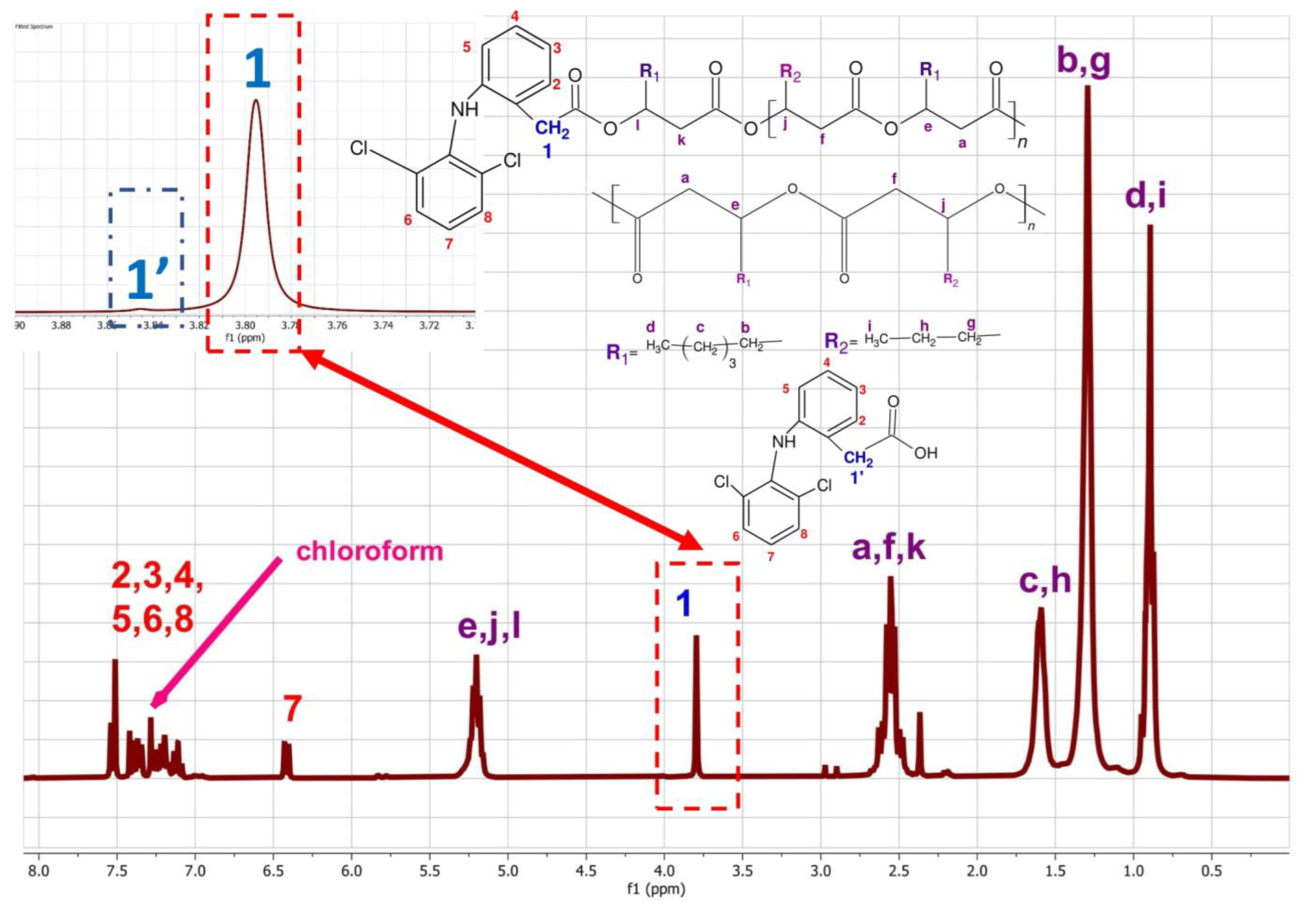
2.1. The Physicochemical Characterisation of Diclofenac Conjugates
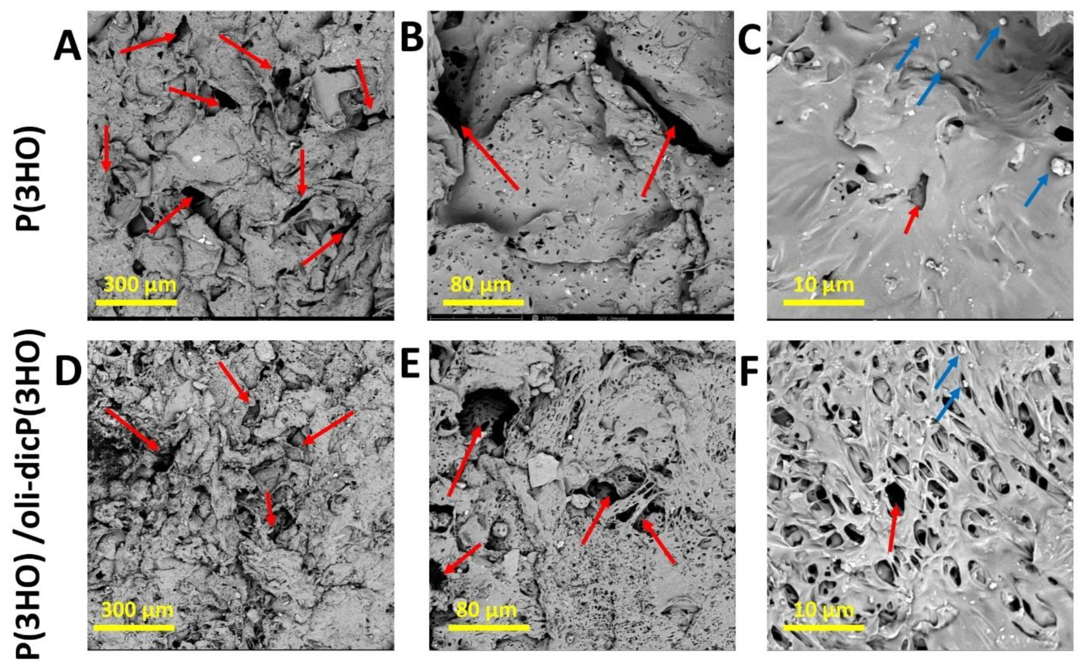
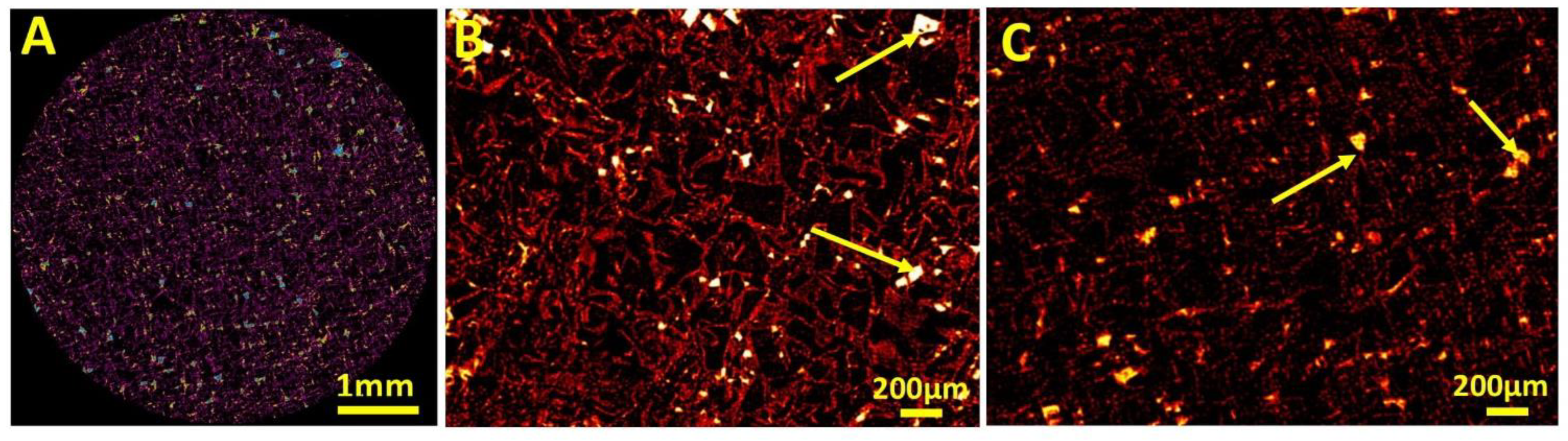
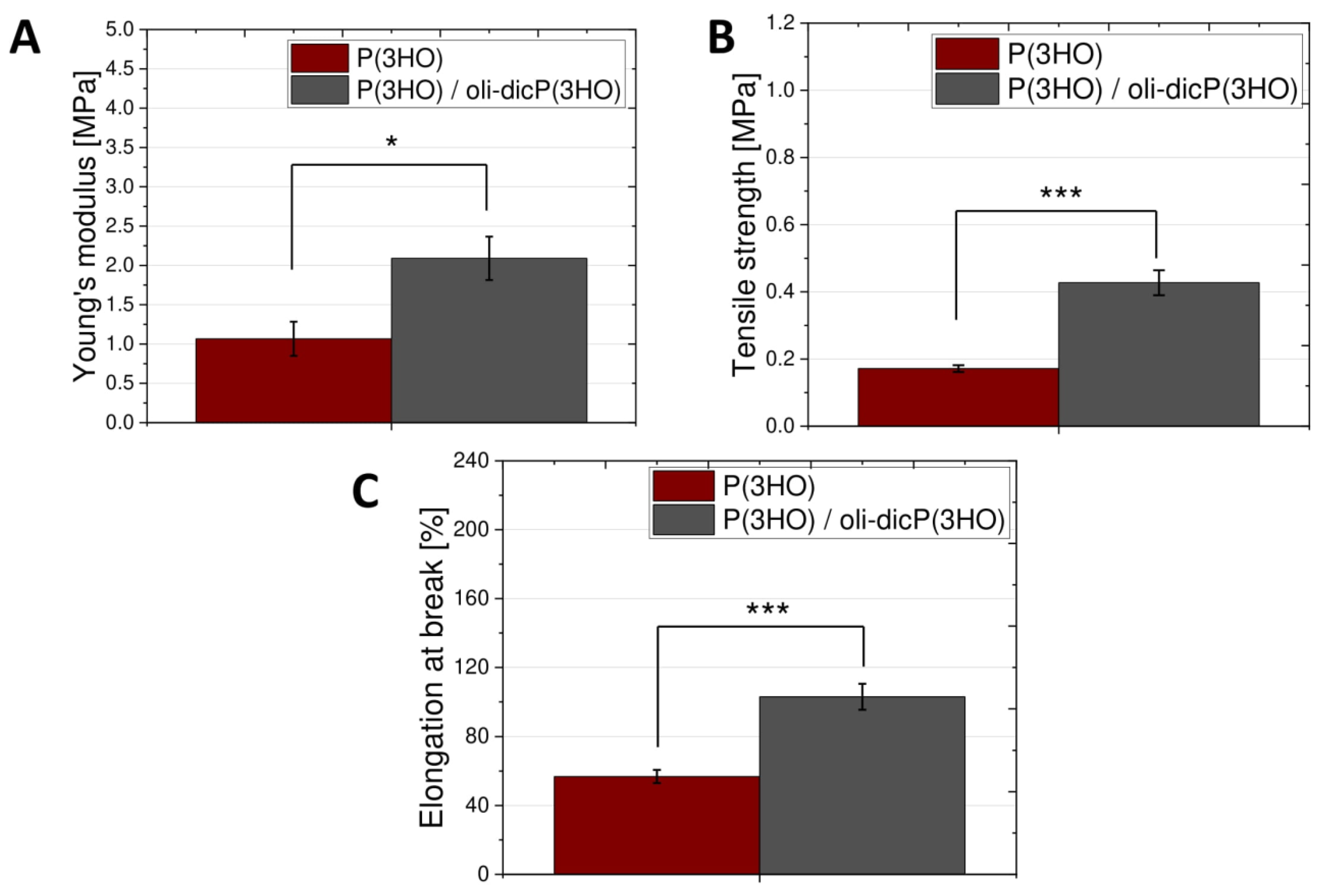
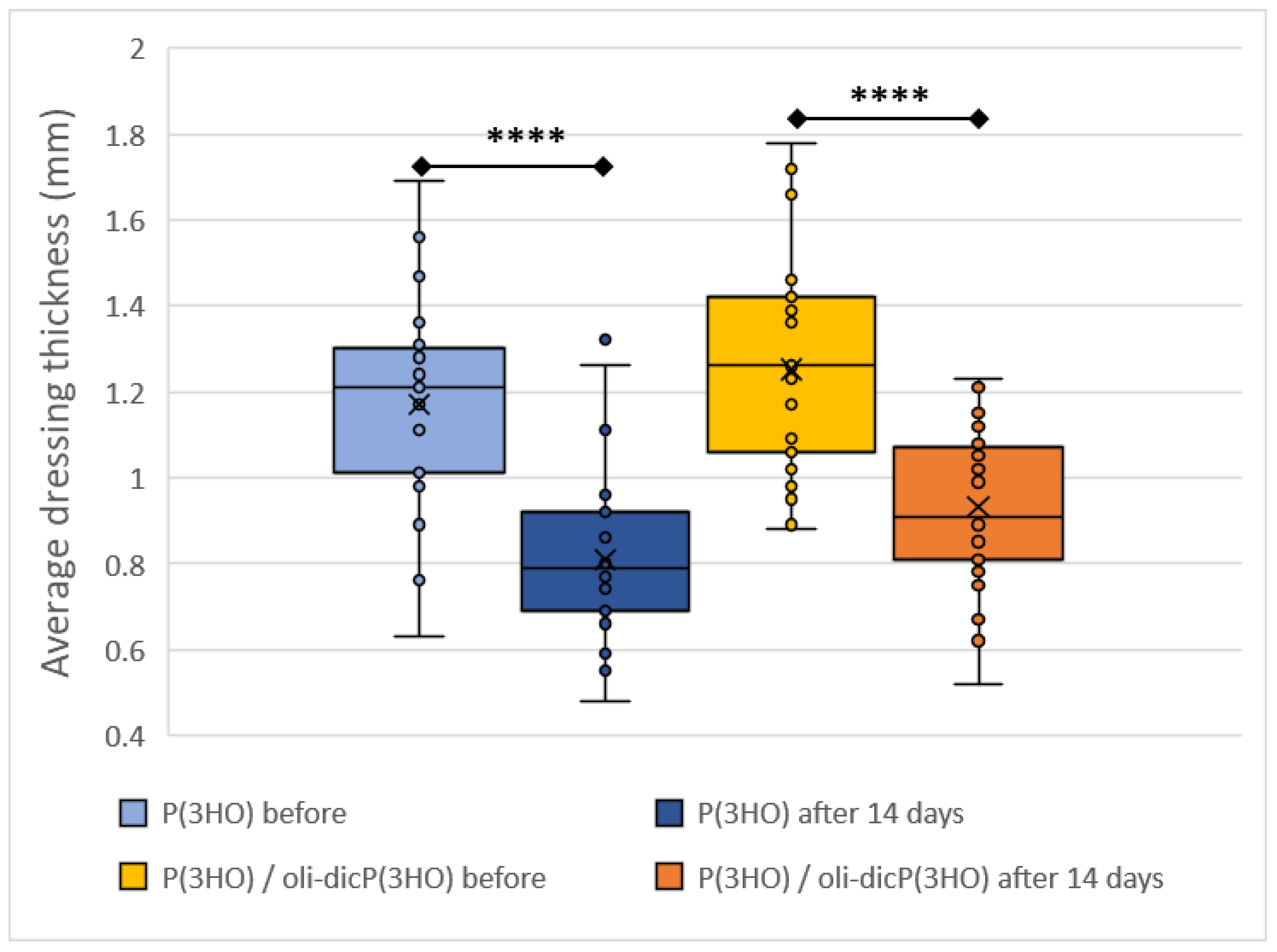
2.2. Description of Surface and Mechanical Properties of Materials
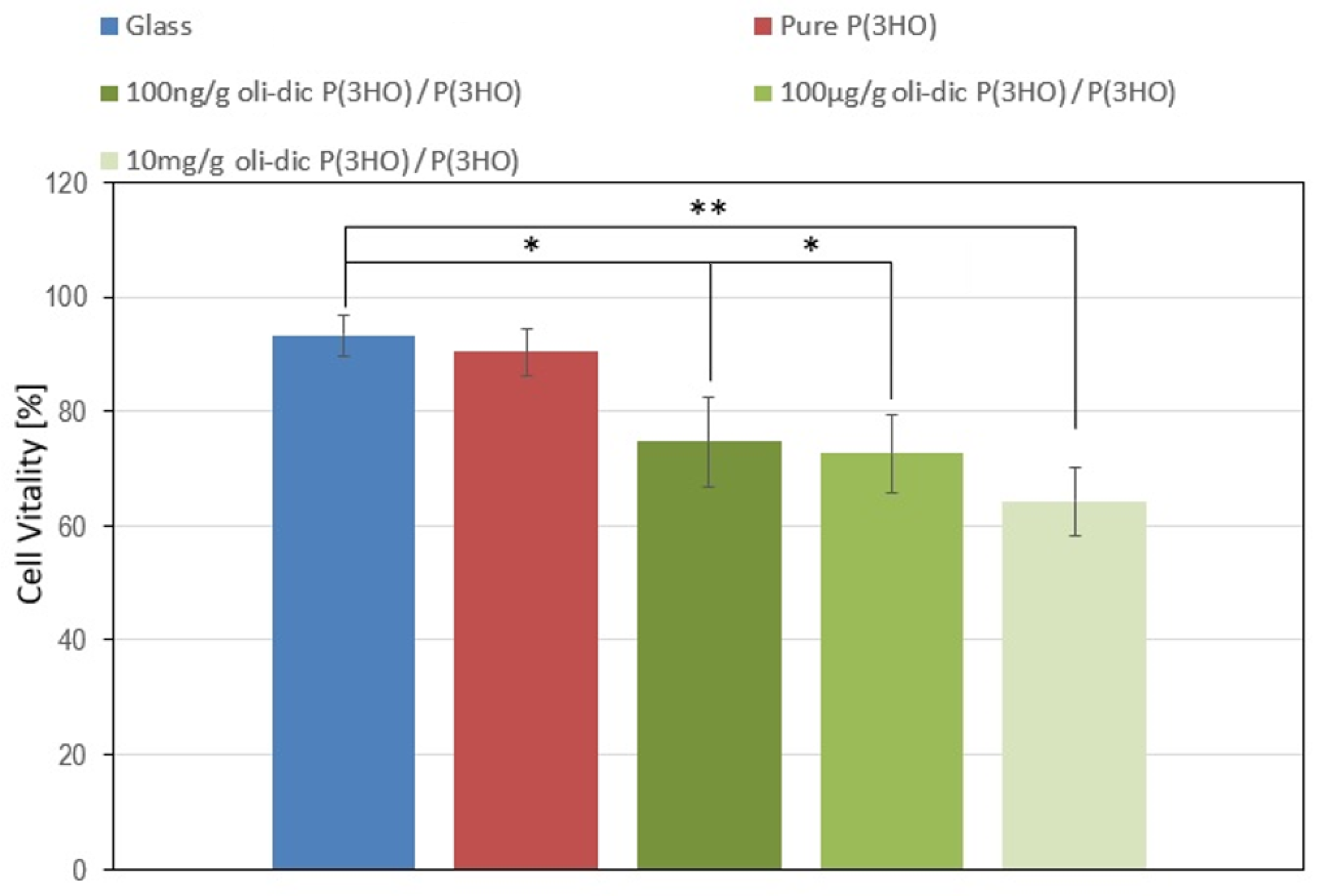
2.3. Cytotoxicity Assessment of Diclofenac-Modified Conjugates (Oli-dicP(3HO))
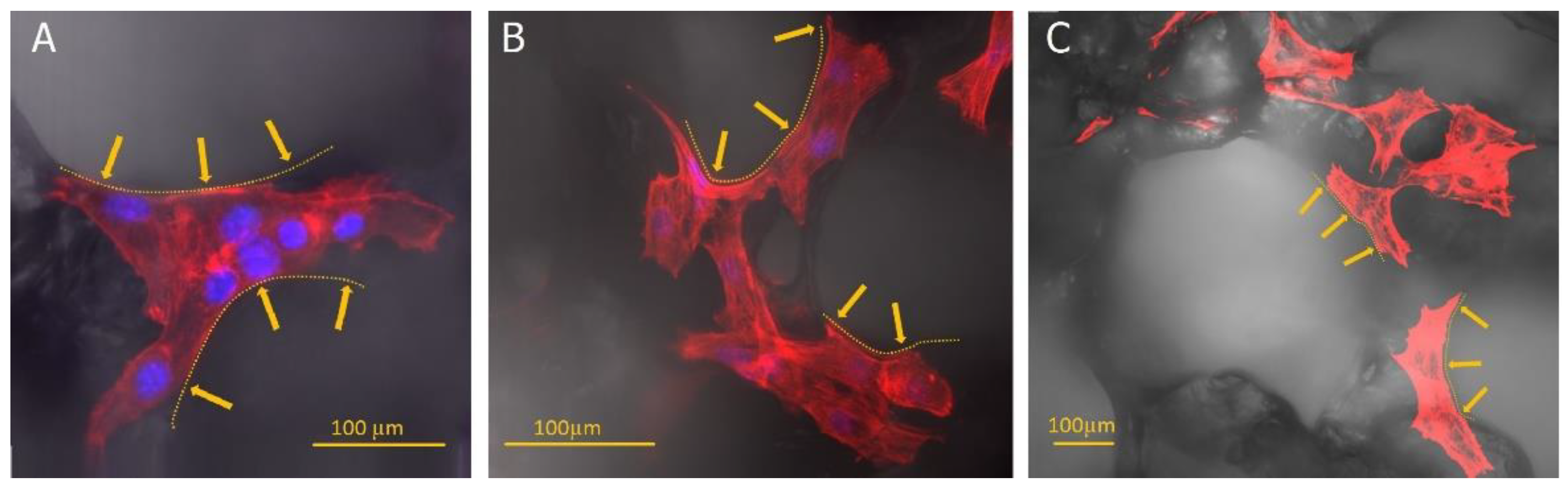
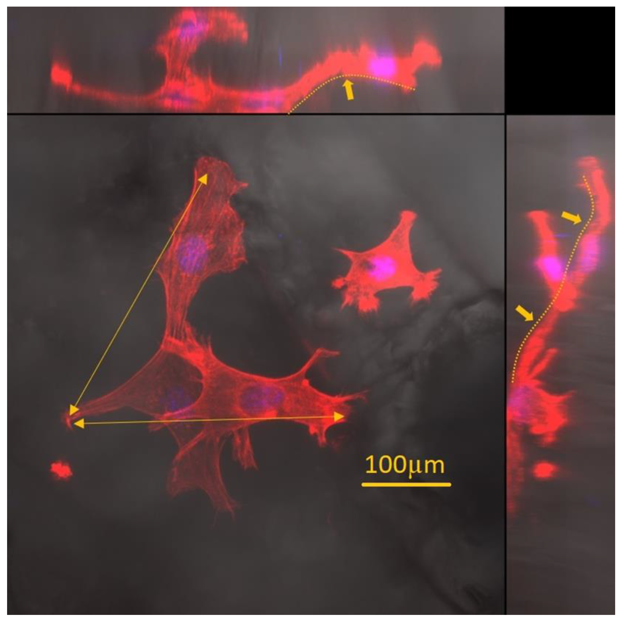
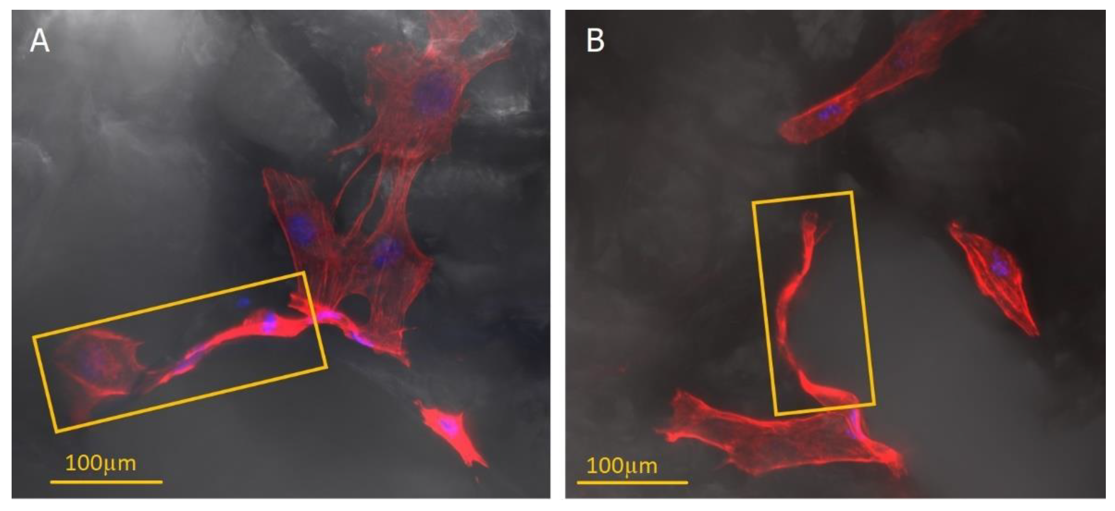
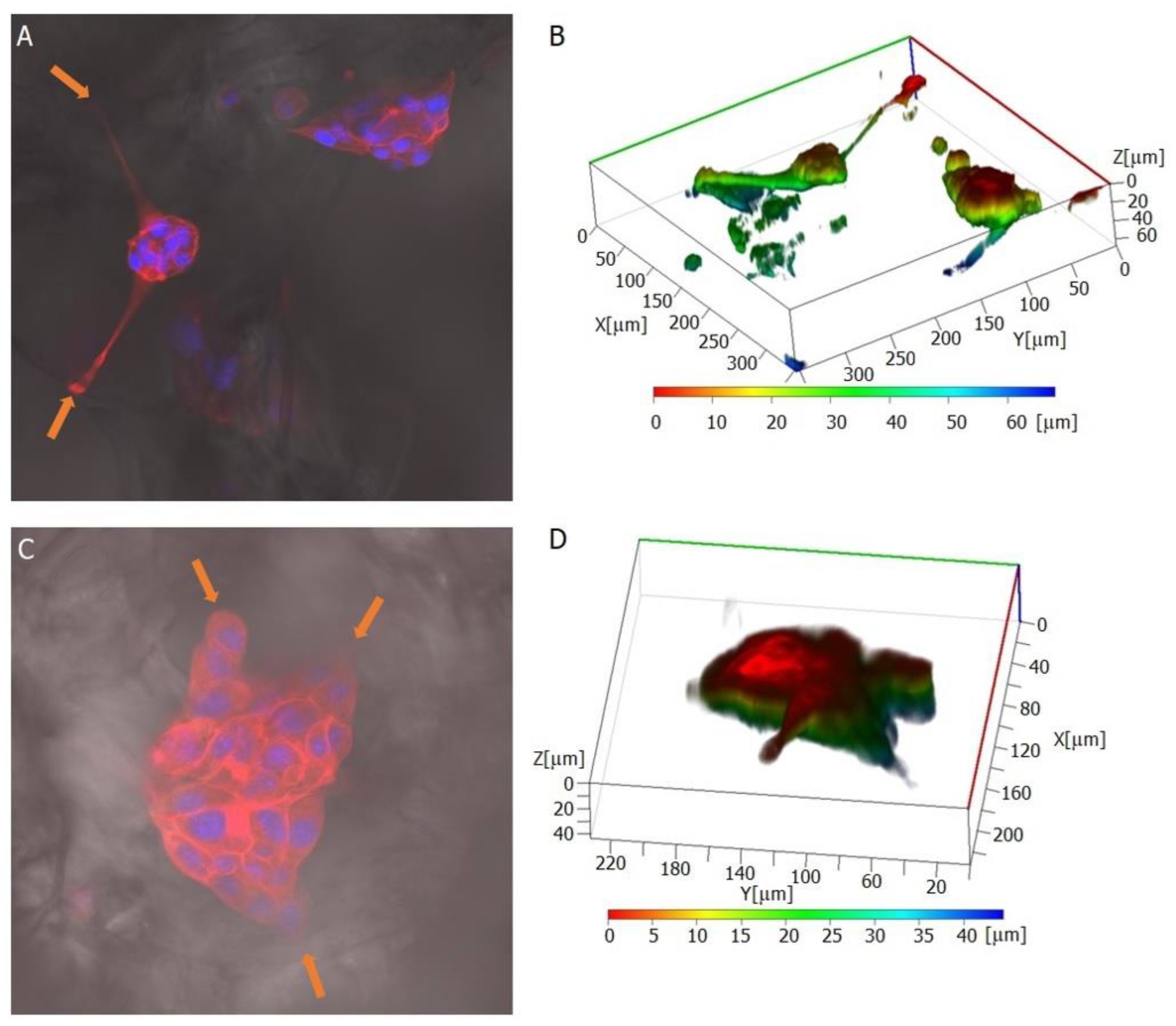
2.4. MEF3T3 Cells’ Behaviour on the Surface of Prepared Materials
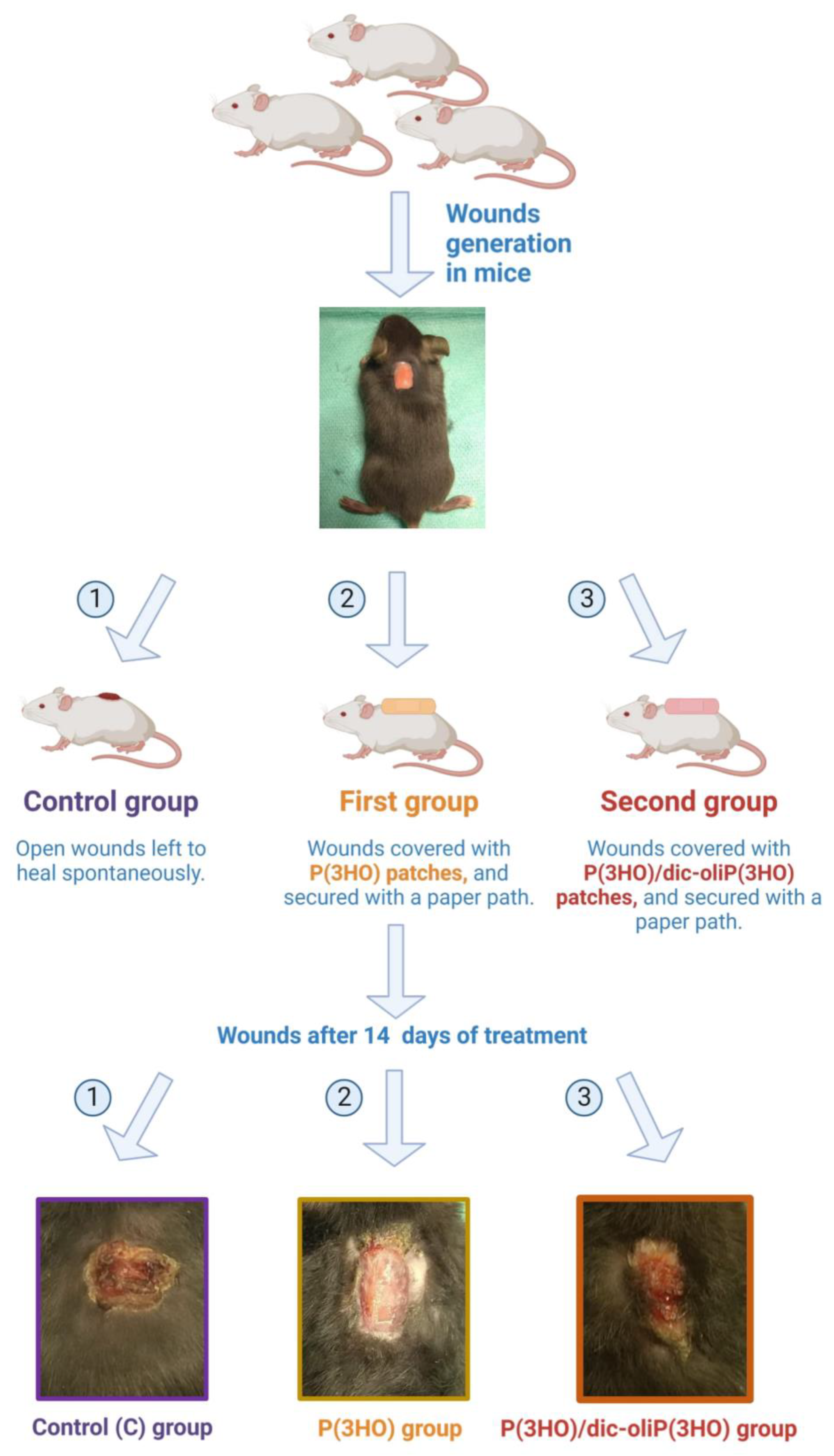
2.5. In Vivo Study
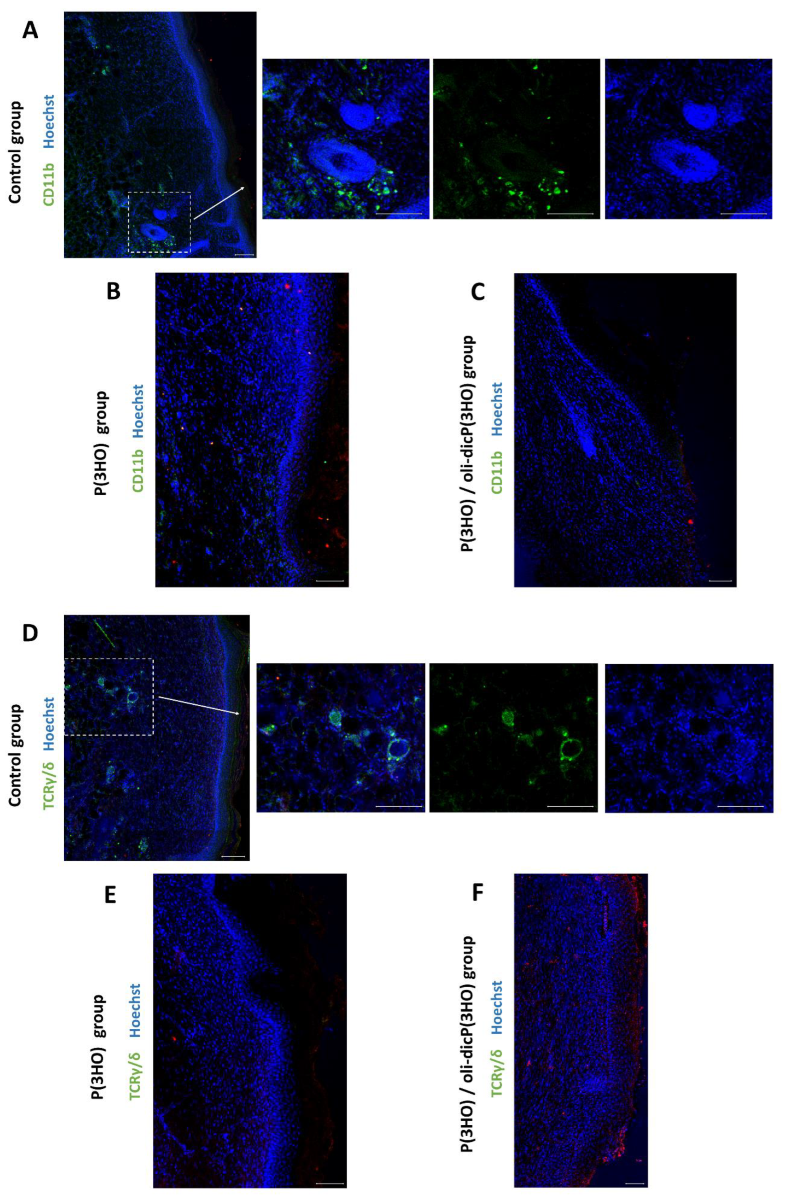
2.5.1. Inflammation
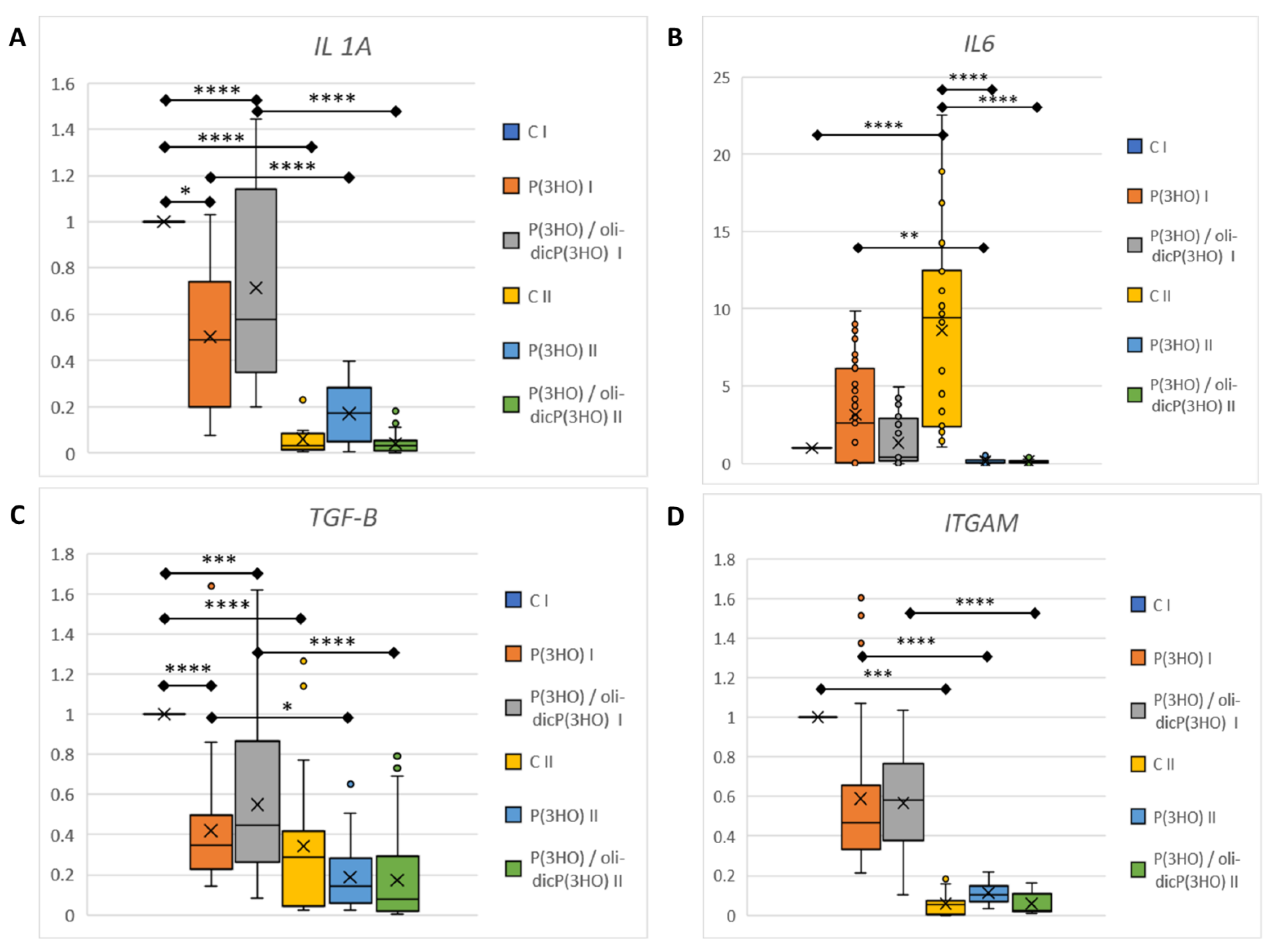
- IL1a is one of the essential proinflammatory cytokines activating innate inflammation after injury. IL18 is also a proinflammatory cytokine that stimulates NK cells to produce IFN-γ. It is known that skin keratinocytes, as a first defence line, contain, inter alia, preformed IL1α and IL18, which are released immediately after injury from dying cells [57]. In the case of an uninfected, properly healing wound, proinflammatory cytokine levels should drop over time. In our study, at the mRNA level, there was a significant decrease in proinflammatory IL1a levels in all study groups between day 7 and day 14 of wound healing (Figure 13A). In addition, a significantly lower expression of IL1a was observed in the animals from the groups treated with P(3HO) and P(3HO)/oli-dicP(3HO) dressings on day 7. Furthermore, it was shown that IL18 expression was at a very low level (limit of detection), which was comparable for all groups. In peripheral blood, a slight (not statistically significant) decrease in IL1a levels between days 7 and 14 was observed in all tested groups (Figure 14A).
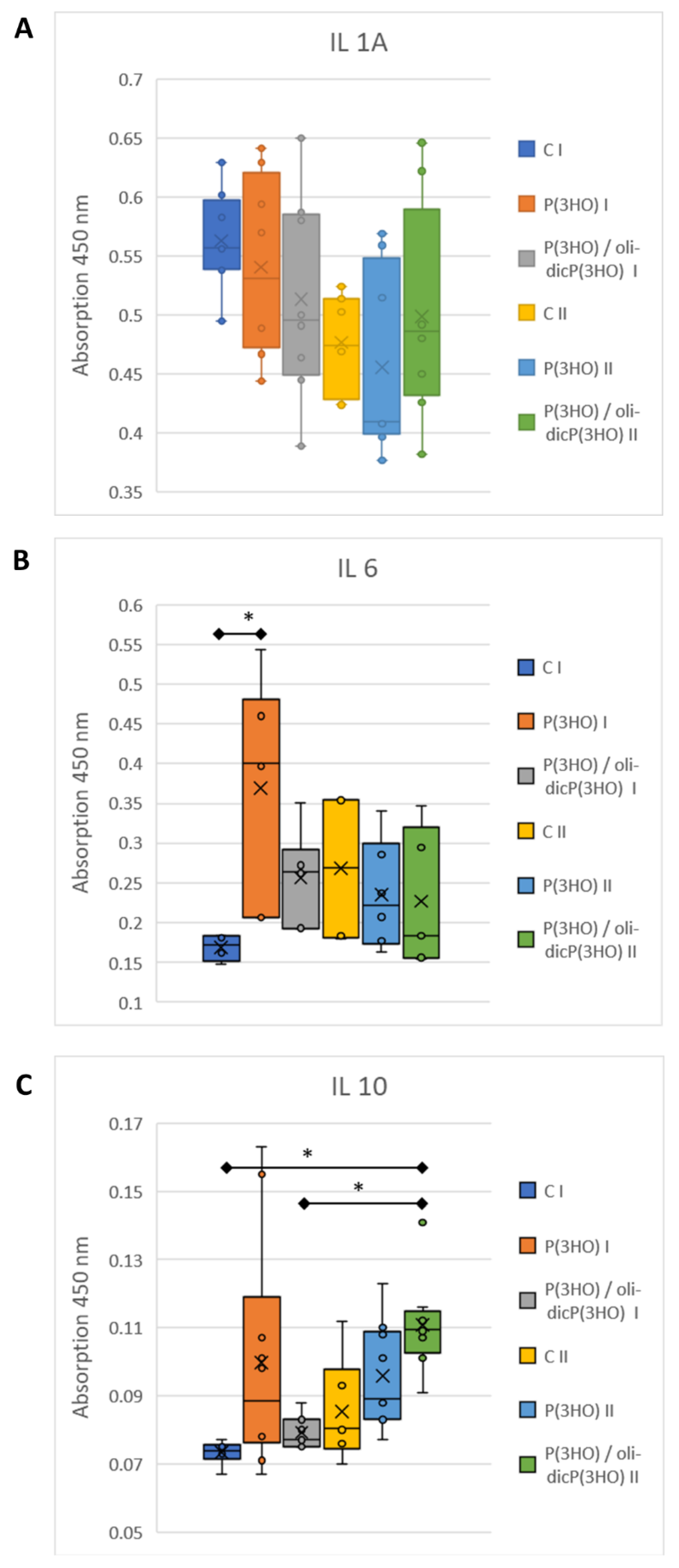
- IL6 is a pleiotropic cytokine responsible for activating the acute inflammatory phase, haematopoiesis, and immune reactions in response to an infection or tissue injuries. Prolonged expression of IL6 is associated with chronic inflammation, and its increased level is detected in chronic inflammatory diseases. Moreover, it was found that IL6 stimulates fibroblasts to exhibit collagen and glycosaminoglycans (GAG) production, and the IL6/JAK2/STAT3 signalling pathway is related to keloid formation [58,59]. In the presented experiment, it was observed that on day 7, the level of IL6 mRNA was comparable in all groups, but on day 14 increased significantly in the control group, at the same time decreasing and being at a very low level in the experimental groups (Figure 13B). Interestingly, on day 7, an increase in the IL6 level in peripheral blood was observed in the animals treated with P(3HO) compared to the control group. Still, on day fourteen, it decreased (Figure 14B).
- CCL20 is a strong chemokine produced by keratinocytes, which plays a role in immune cells (e.g., murine skin γδ T cells) recruitment after skin injury. It was found that dermis-resident Vγ4 T cells, activated by the CCR6-CCL20 pathway, infiltrate into the epidermis and are a significant source of IL17A. IL17A enhances the production of IL1β and IL23, leading to an increase in local inflammation at the early stages of wound healing, and excessive activation of CCL20—IL17A-IL1/23 loop activation delays skin wound healing [60]. In the presented work, on both days of observation, days 7 and 14, the mRNA level of CCL20 was below the detection level, suggesting that initial inflammation had been suppressed.
- TSLP (thymic stromal lymphopoietin) is one of the cytokines expressed mainly by epithelial cells and keratinocytes in the skin, lung, and intestine and plays a vital role in the initiation and maintenance of the allergic immune response. Its expression may be induced by mechanical injury, microbes, and the inflammatory cytokines IL4, IL13, and TNFα [61]. TSLP promotes the maturation of Langerhans and myeloid cells associated with the skin’s immune system. Activation of Langerhans cells by TSLP induces their synthesis of pro-inflammatory cytokines (e.g., TNFα) and is a sign of developing inflammation. In the tested animals in the control and experimental groups, TSLP mRNA levels in the wound and adjacent tissue were under the detection level on days 7 and 14.
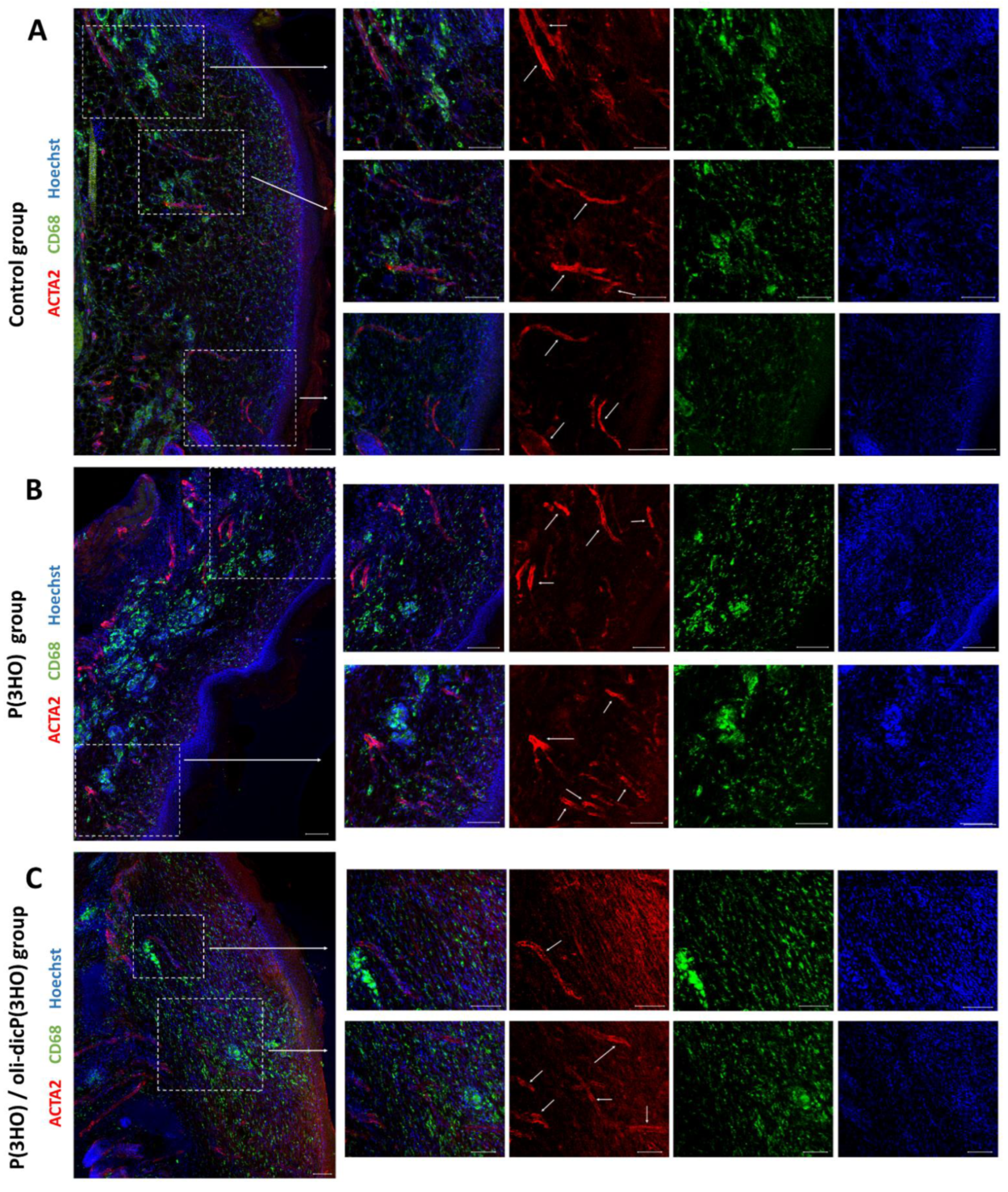
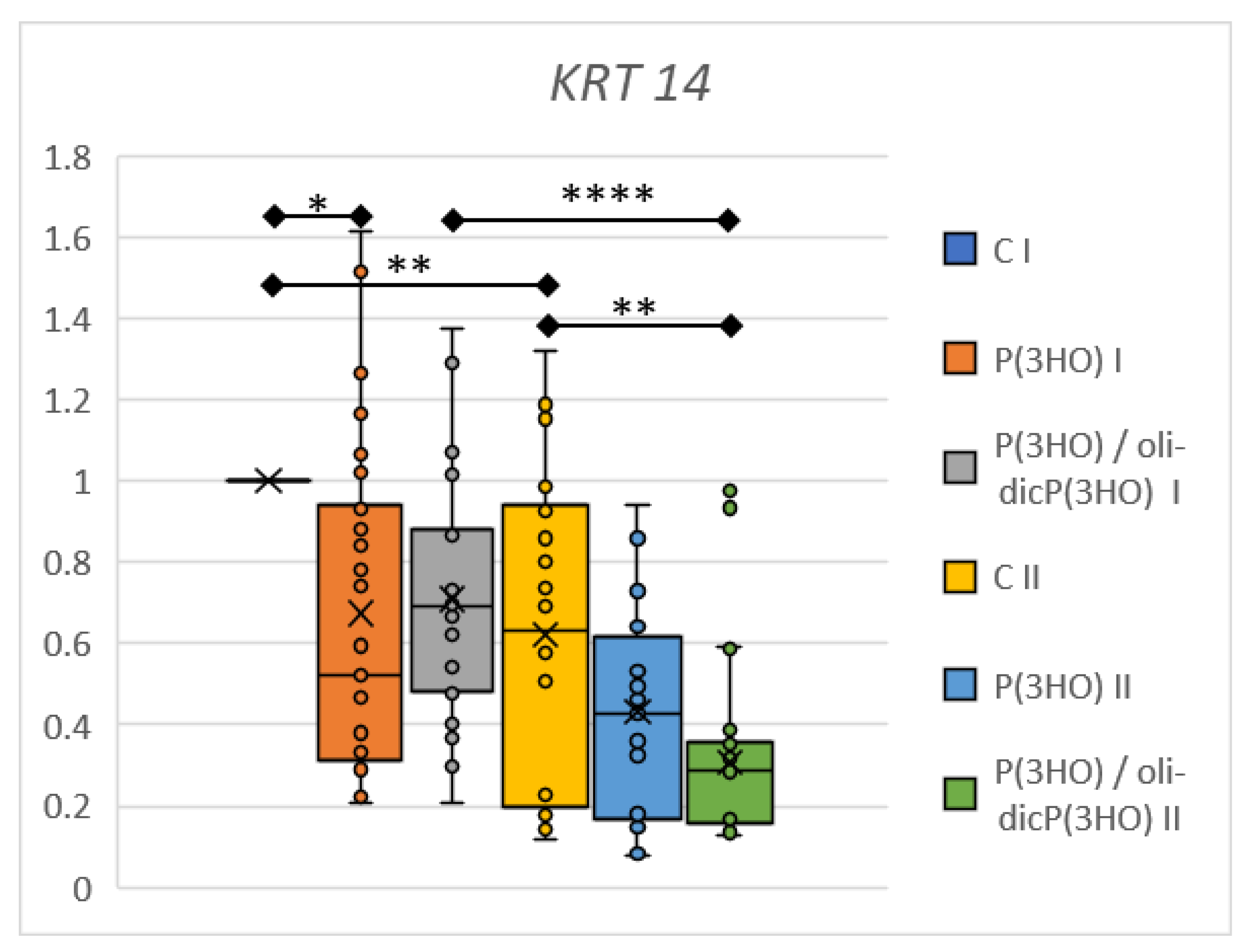
- ITGAM (integrin alpha M) gene code protein CD11b. CD11b is one of the fibrocytes (peripheral blood fibroblast-like cells) and monocytes marker. Fibrocytes are unique cells that exhibit properties of macrophages as well as fibroblasts. Fibrocytes play a pivotal role in wound healing during inflammatory and proliferation phases using numerous mechanisms: wound debridement (by, among other things, acting as antigen-presenting cells or phagocytic activity), tissue regeneration (by producing cytokines, chemokines, and growth factors), ECM synthesis, and wound closure via α-SMA-mediated contraction or angiogenesis (FGF2, VEGF, and PDGF synthesis). During the sub-healing phase, fibrocytes differentiate into myofibroblasts, depositing high levels of ECM components and MMPs [62]. In the experiments conducted in all study groups on day 7, the comparable expression of the ITGAM gene was demonstrated (Figure 13D). It was also shown that this expression significantly decreased on day 14 of observation, which is probably related to the transition of fibrocytes from the pro-inflammatory variant into the myofibroblast. In addition, this result was confirmed by immunocytochemical analysis (Figure 12A–C), in which, on day 14, single CD11b-positive cells were present only in the control group.
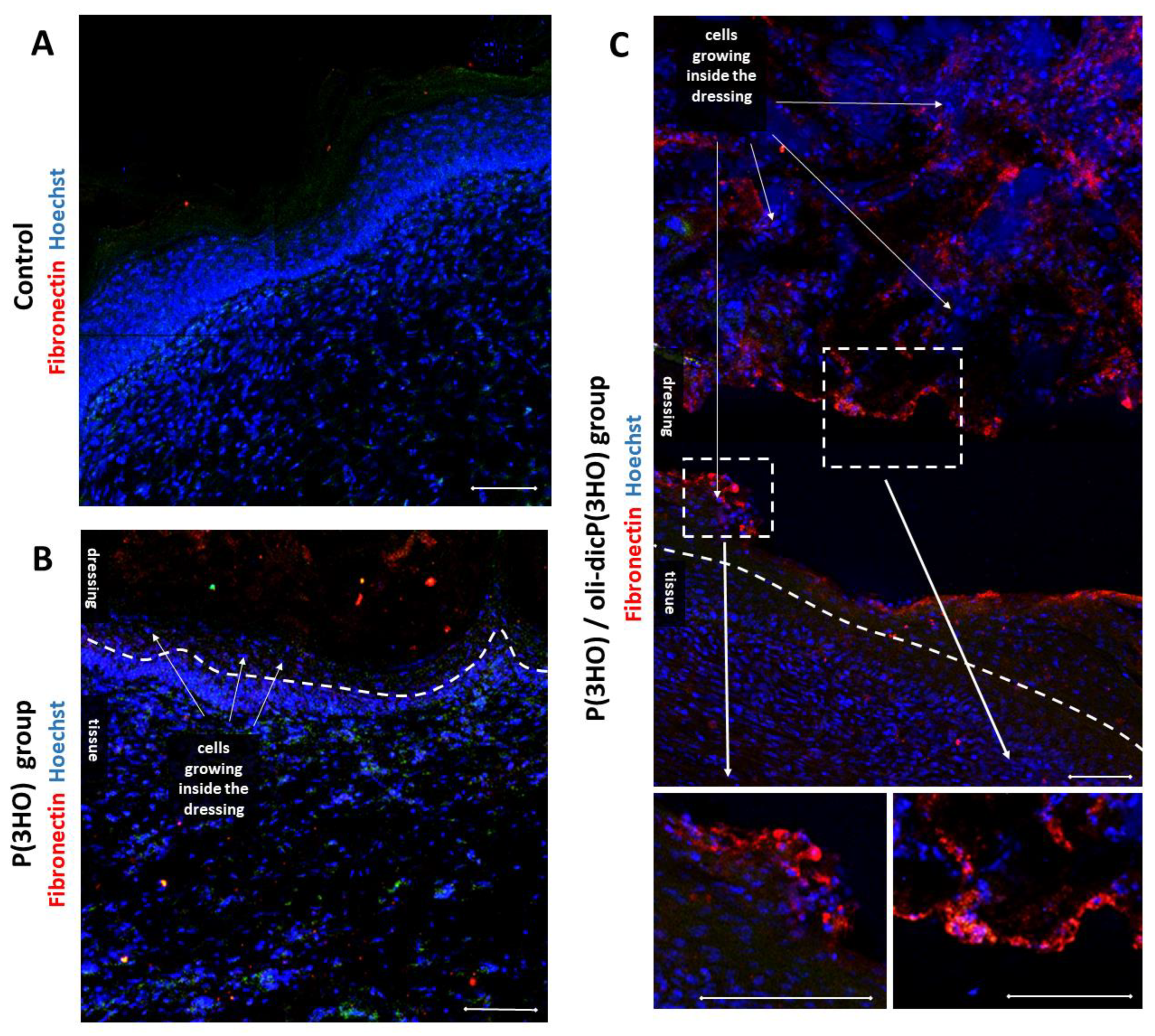
- IL10 is a crucial anti-inflammatory cytokine that suppresses an excessive host immune response to injury or bacterial infection. After the injury, IL10 is produced by keratinocytes. IL10 triggers a robust suppressive response in macrophages and neutrophils, mainly via the transcriptional inhibition of cytokines and chemokines. It plays an important role in sterile wound healing. More importantly, it was shown that IL10 reduced the gene expression of type I collagen, fibronectin, and upregulated decorin expression in human skin fibroblasts by the TGF-beta downregulation [63]. In our study, IL10 mRNA levels were deficient, almost undetectable. Still, an increase was observed in protein levels in peripheral blood, especially on day 14 in the case of animals treated with P(3HO)/oli-dicP(3HO) dressings (Figure 14C). Moreover, there is an apparent suppression of TGF-beta expression at the mRNA level (Figure 13C), which aligns with the authors’ observations. It was also found that high IL10 and reduced IL6 levels play an essential role in scarless, regenerative healing typical for the foetus [64]. In the presented experiment, a slight increase in IL10 levels in the blood was observed, with a concomitant decrease in IL6 and TGF-beta expression at the mRNA level. This is consistent with the observed smaller scab in animals treated with P(3HO) and P(3HO)/oli-dicP(3HO) dressings.
2.5.2. Angiogenesis and Remodelling
3. Materials and Methods
3.1. Materials Preparation with Physicochemical and Mechanical Characterisation Procedures
3.2. In Vitro Characterisation of Prepared Materials
3.3. In Vivo Study
3.3.1. Animals
3.3.2. mRNA Isolation and Gene Expression Analysis
3.3.3. Immunohistochemistry
3.3.4. The Enzyme-Linked Immunosorbent Assay (ELISA)
3.4. Statistical Analysis
4. Conclusions
Author Contributions
Funding
Institutional Review Board Statement
Informed Consent Statement
Data Availability Statement
Acknowledgments
Conflicts of Interest
Appendix A


References
- Kabashima, K.; Honda, T.; Ginhoux, F.; Egawa, G. The immunological anatomy of the skin. Nat. Rev. Immunol. 2019, 19, 19–30. [Google Scholar] [CrossRef] [PubMed]
- Raju, N.R.; Silina, E.; Stupin, V.; Manturova, N.; Chidambaram, S.B.; Achar, R.R. Multifunctional and Smart Wound Dressings—A Review on Recent Research Advancements in Skin Regenerative Medicine. Pharmaceutics 2022, 14, 1574. [Google Scholar] [CrossRef] [PubMed]
- Boateng, J.S.; Matthews, K.H.; Stevens, H.N.E.; Eccleston, G.M. Wound Healing Dressings and Drug Delivery Systems: A Review. J. Pharm. Sci. 2008, 97, 2892–2923. [Google Scholar] [CrossRef] [PubMed]
- Ghomi, E.R.; Khalili, S.; Khorasani, S.N.; Neisiany, R.E. Wound dressings : Current advances and future directions. J. Appl. Polym. Sci. 2019, 136, 47738. [Google Scholar] [CrossRef] [Green Version]
- Alven, S.; Nqoro, X.; Aderibigbe, A. Polymer-Based Materials Loaded with Curcumin for Wound Healing Applications. Polymers 2020, 12, 2286. [Google Scholar] [CrossRef]
- Portela, R.; Leal, C.R.; Pedro, L.; Sobral, R. Bacterial cellulose : A versatile biopolymer for wound dressing applications. Microb. Biotechnol. 2020, 12, 586–610. [Google Scholar] [CrossRef]
- Ruppert, D.S.; Mohammed, M.M.; Ibrahim, M.M.; Bachtiar, E.O.; Erning, K.; Ansari, K.; Everitt, J.I.; Brown, D.; Klitzman, B.; Koshut, W.; et al. Poly(lactide-co-ε-caprolactone) scaffold promotes equivalent tissue integration and supports skin grafts compared to a predicate collagen scaffold. Wound Repair Regen. 2021, 29, 1035–1050. [Google Scholar] [CrossRef]
- Negut, I.; Dorcioman, G.; Grumezescu, V. Scaffolds for Wound Healing Applications. Polymers 2020, 12, 2010. [Google Scholar] [CrossRef]
- Bolivar-Monsalve, E.J.; Alvarez, M.M.; Hosseini, S.; Espinoza-Hernandez, M.A.; Ceballos-Gonzalez, C.F.; Sanchez-Dominguez, M.; Shin, S.R.; Cecen, B.; Hassan, S.; Di Maio, E.; et al. Engineering bioactive synthetic polymers for biomedical applications: A review with emphasis on tissue engineering and controlled release. Mater. Adv. 2021, 2, 4447–4478. [Google Scholar] [CrossRef]
- Poonguzhali, R.; Khaleel Basha, S.; Sugantha Kumari, V. Novel asymmetric chitosan/PVP/nanocellulose wound dressing: In vitro and in vivo evaluation. Int. J. Biol. Macromol. 2018, 112, 1300–1309. [Google Scholar] [CrossRef]
- Khodabakhshi, D.; Eskandarinia, A.; Kefayat, A.; Rafienia, M.; Navid, S.; Karbasi, S.; Moshtaghian, J. In vitro and in vivo performance of a propolis-coated polyurethane wound dressing with high porosity and antibacterial efficacy. Colloids Surf. B Biointerfaces 2019, 178, 177–184. [Google Scholar] [CrossRef] [PubMed]
- Cao, Y.; Mitchell, G.; Messina, A.; Price, L.; Thompson, E.; Penington, A.; Morrison, W.; O’Connor, A.; Stevens, G.; Cooper-White, J. The influence of architecture on degradation and tissue ingrowth into three-dimensional poly(lactic-co-glycolic acid) scaffolds in vitro and in vivo. Biomaterials 2006, 27, 2854–2864. [Google Scholar] [CrossRef] [PubMed]
- Lee, S.Y.; Oh, J.H.; Kim, J.C.; Kim, Y.H.; Kim, S.H.; Choi, J.W. In vivo conjunctival reconstruction using modified PLGA grafts for decreased scar formation and contraction. Biomaterials 2003, 24, 5049–5059. [Google Scholar] [CrossRef] [PubMed]
- Garg, T.; Singh, O.; Arora, S.; Murthy, R.S.R. Scaffold: A Novel Carrier for Cell and Drug Delivery. Crit. Rev. Ther. Drug Carr. Syst. 2012, 29, 1–63. [Google Scholar] [CrossRef] [Green Version]
- Aramwit, P.; Ratanavaraporn, J.; Ekgasit, S.; Tongsakul, D.; Bang, N. A green salt-leaching technique to produce sericin/PVA/glycerin scaffolds with distinguished characteristics for wound-dressing applications. J. Biomed. Mater. Res.-Part B Appl. Biomater. 2015, 103, 915–924. [Google Scholar] [CrossRef]
- Kucińska-Lipka, J.; Gubańska, I.; Lewandowska, A.; Terebieniec, A.; Przybytek, A.; Cieśliński, H. Antibacterial polyurethanes, modified with cinnamaldehyde, as potential materials for fabrication of wound dressings. Polym. Bull. 2019, 76, 2725–2742. [Google Scholar] [CrossRef]
- Oustadi, F.; Nazarpak, M.H.; Mansouri, M. Preparation, characterization, and drug release study of ibuprofen-loaded poly (vinyl alcohol)/poly (vinyl pyrrolidone) bilayer antibacterial membrane. Int. J. Polym. Mater. Polym. Biomater. 2020, 71, 14–23. [Google Scholar] [CrossRef]
- Rancan, F.; Contardi, M.; Jurisch, J.; Blume-peytavi, U.; Vogt, A.; Bayer, I.S.; Schaudinn, C. Evaluation of Drug Delivery and E ffi cacy of Ciprofloxacin-Loaded Povidone Foils and Nanofiber Mats in a Wound-Infection Model Based on Ex Vivo Human Skin. Pharmaceutics 2019, 11, 527. [Google Scholar] [CrossRef] [Green Version]
- Laurano, R.; Boffito, M.; Ciardelli, G.; Chiono, V. Wound dressing products: A translational investigation from the bench to the market. Eng. Regen. 2022, 3, 182–200. [Google Scholar] [CrossRef]
- Postolovic, K.S.; Antonijevic, M.D.; Ljujic, B.; Radenkovic, S.; Kovacevic, M.M.; Hiezl, Z.; Pavlovic, S.; Radojevic, I.; Stanic, Z. Curcumin and Diclofenac Therapeutic Efficacy Enhancement Applying Transdermal Hydrogel Polymer Films, Based on carrageenan, alginate and poloxamer. Polymers 2022, 14, 4091. [Google Scholar] [CrossRef]
- Costa FL, D.S.; Tiussi, L.D.; Nascimento, M.S.; Corrêa, A.C.D.S.; Yasojima, E.Y.; Pires, C.A.A. Diclofenac topical gel in excisional wounds maintain heal quality and reduce phlogistic signals 1. Acta Chir. Bras. 2014, 29, 328–333. [Google Scholar] [CrossRef] [PubMed] [Green Version]
- Draczynski, Z.; Kolesinska, B.; Latanska, I.; Sujka, W. Preparation Method of Porous Dressing Materials Based on Butyric-Acetic Chitin Co-Polyesters. Materials 2018, 11, 2359. [Google Scholar] [CrossRef] [PubMed] [Green Version]
- Gustavo, J.; Ossa, D.; Fusco, A.; Azimi, B.; Salsano, J.E.; Digiacomo, M.; Coltelli, M.; De Clerck, K.; Roy, I.; Macchia, M.; et al. Immunomodulatory Activity of Electrospun Polyhydroxyalkanoate Fiber Scaffolds Incorporating Olive Leaf Extract. Appl. Sci. 2021, 11, 4006. [Google Scholar] [CrossRef]
- Sofińska, K.; Barbasz, J.; Witko, T.; Dryzek, J.; Haraźna, K.; Witko, M.; Kryściak-Czerwenka, J.; Guzik, M. Structural, topographical, and mechanical characteristics of purified polyhydroxyoctanoate polymer. J. Appl. Polym. Sci. 2019, 136, 47192. [Google Scholar] [CrossRef]
- Cichoń, E.; Haraźna, K.; Skibiński, S.; Witko, T.; Zima, A.; Ślósarczyk, A.; Zimowska, M.; Witko, M.; Leszczyński, B.; Wróbel, A.; et al. Novel bioresorbable tricalcium phosphate/polyhydroxyoctanoate (TCP/PHO) composites as scaffolds for bone tissue engineering applications. J. Mech. Behav. Biomed. Mater. 2019, 98, 235–245. [Google Scholar] [CrossRef]
- Haraźna, K.; Szyk-Warszyńska, L.; Witko, M.; Guzik, M. Preparation, characterisation and modification of bacterial polyhydroxyoctanoate for construction of wound patches. In Proceedings of the 7th International Seminar including Special session “Recent Advances in Polymer Nanocomposites and Hybrid Materials”, Kraków, Poland, 15–17 May 2019; pp. 139–152, ISBN 978-83-937270-6-3. [Google Scholar]
- Witko, T.; Solarz, D.; Feliksiak, K.; Haraźna, K.; Rajfur, Z.; Guzik, M. Insights into in vitro wound closure on two biopolyesters –polylactide and polyhydroxyoctanoate. Materials 2020, 13, 2793. [Google Scholar] [CrossRef]
- Skibiński, S.; Cichoń, E.; Haraźna, K.; Marcello, E.; Roy, I.; Witko, M.; Ślósarczyk, A.; Czechowska, J.; Guzik, M.; Zima, A. Functionalized tricalcium phosphate and poly(3-hydroxyoctanoate) derived composite scaffolds as platforms for the controlled release of diclofenac. Ceram. Int. 2021, 47, 3876–3883. [Google Scholar] [CrossRef]
- Witko, T.; Solarz, D.; Feliksiak, K.; Rajfur, Z.; Guzik, M. Cellular architecture and migration behavior of fibroblast cells on polyhydroxyoctanoate (PHO): A natural polymer of bacterial origin. Biopolymers 2019, 110, e23324. [Google Scholar] [CrossRef]
- Guzik, M.W. Polyhydroxyalkanoates, bacterially synthesized polymers, as a source of chemical compounds for the synthesis of advanced materials and bioactive molecules. Appl. Microbiol. Biotechnol. 2021, 105, 7555–7566. [Google Scholar] [CrossRef]
- Haraźna, K.; Cichoń, E.; Skibiński, S.; Witko, T.; Solarz, D.; Kwiecień, I.; Marcello, E.; Zimowska, M.; Socha, R.; Szefer, E.; et al. Physicochemical and biological characterisation of diclofenac oligomeric poly(3-hydroxyoctanoate) hybrids as β-TCP ceramics modifiers for bone tissue regeneration. Int. J. Mol. Sci. 2020, 21, 9452. [Google Scholar] [CrossRef]
- Ward, A.C.; Dubey, P.; Basnett, P.; Lika, G.; Newman, G.; Corrigan, D.K.; Russell, C.; Kim, J.; Chakrabarty, S.; Connolly, P.; et al. Toward a Closed Loop, Integrated Biocompatible Biopolymer Wound Dressing Patch for Detection and Prevention of Chronic Wound Infections. Front. Bioeng. Biotechnol. 2020, 8, 1039. [Google Scholar] [CrossRef] [PubMed]
- Skibiński, S.; Czechowska, J.; Cichoń, E.; Seta, M.; Gondek, A.; Cudnoch-Jędrzejewska, A.; Ślósarczyk, A.; Guzik, M.; Zima, A. Study on β TCP/P (3HB) Scaffolds—Physicochemical Properties and Biological Performance in Low Oxygen Concentration. Int. J. Mol. Sci. 2022, 23, 11587. [Google Scholar] [CrossRef] [PubMed]
- Achterberg, V.F.; Buscemi, L.; Diekmann, H.; Smith-clerc, J.; Schwengler, H.; Meister, J.; Wenck, H.; Gallinat, S.; Hinz, B. The Nano-Scale Mechanical Properties of the Extracellular Matrix Regulate Dermal Fibroblast Function. J. Investig. Dermatol. 2014, 134, 1862–1872. [Google Scholar] [CrossRef] [PubMed] [Green Version]
- Chen, X.; Wang, X.; Wang, S.; Zhang, X.; Yu, J.; Wang, C. Mussel-inspired polydopamine-assisted bromelain immobilization onto electrospun fibrous membrane for potential application as wound dressing. Mater. Sci. Eng. C 2020, 110, 110624. [Google Scholar] [CrossRef]
- Dziemidowicz, K.; Brocchini, S.; Williams, G.R. A simple route to functionalising electrospun polymer scaffolds with surface biomolecules. Int. J. Pharm. 2021, 597, 120231. [Google Scholar] [CrossRef]
- Yang, J.; Van Lith, R.; Baler, K.; Hoshi, R.A.; Ameer, G.A. A thermoresponsive biodegradable polymer with intrinsic antioxidant properties. Biomacromolecules 2014, 15, 3942–3952. [Google Scholar] [CrossRef]
- Yannas, I.V.; Lee, E.; Orgill, D.P.; Skrabut, E.M.; Murphyt, G.F. Synthesis and characterization of a model extracellular matrix that induces partial regeneration of adult mammalian skin. Proc. Natl. Acad. Sci. USA 1989, 86, 933–937. [Google Scholar] [CrossRef] [Green Version]
- Choi, D.J.; Parka, S.J.; Gua, B.K.; Kimc, Y.-J.; Chungb, S.; Kima, C.-H. Effect of the pore size in a 3D bioprinted gelatin scaffold on fi broblast proliferation. J. Ind. Eng. Chem. 2018, 67, 388–395. [Google Scholar] [CrossRef]
- Zeltinger, J.; Sherwood, J.K.; Graham, D.A.; Müeller, R.; Griffith, L.G. Effect of Pore Size and Void Fraction on Cellular Adhesion, Proliferation, and Matrix Deposition. Tissue Eng. 2001, 7, 557–572. [Google Scholar] [CrossRef]
- Plast, A.; Author, S.; July, P.M.C.; Cavallo, J.A.; Gangopadhyay, N.; Dudas, J.; Andres, A.; Jasielec, M.S.; Baty, J.; Baalman, S.; et al. Remodeling Characteristics And Collagen Distributions Of Biologic Scaffold Materials Biopsied From Postmastectomy Breast Reconstruction Sites. Ann. Plast. Surg. 2015, 75, 74–83. [Google Scholar] [CrossRef]
- Oh, S.; Kim, S.; Jeong, H.; Lee, J.; Cho, J.W.; Park, J. The Mechanical Properties of Polyurethane Foam Wound Dressing Hybridized with Alginate Hydrogel and Jute Fiber. Fibers Polym. 2013, 14, 173–181. [Google Scholar] [CrossRef]
- Lukasiewicz, B.; Basnett, P.; Nigmatullin, R.; Matharu, R.; Knowles, J.C.; Roy, I. Binary Polyhydroxyalkanoate Systems for Soft Tissue Engineering. Acta Biomater. 2018, 71, 225–234. [Google Scholar] [CrossRef] [PubMed] [Green Version]
- Al-nimer, M.S.M.; Hameed, H.G.; Mahmood, M.M. Antiproliferative effects of aspirin and diclofenac against the growth of cancer and fibroblast cells : In vitro comparative study. Saudi Pharm. J. 2015, 23, 483–486. [Google Scholar] [CrossRef] [PubMed] [Green Version]
- Pan, H.; Jiang, H.; Chen, W. Interaction of dermal fibroblasts with electrospun composite polymer scaffolds prepared from dextran and poly lactide-co-glycolide. Biomaterials 2006, 27, 3209–3220. [Google Scholar] [CrossRef] [PubMed]
- Ghibaudo, M.; Trichet, L.; Le Digabel, J.; Richert, A.; Hersen, P.; Ladoux, B. Substrate topography induces a crossover from 2D to 3D behavior in fibroblast migration. Biophys. J. 2009, 97, 357–368. [Google Scholar] [CrossRef] [Green Version]
- Dziob, D.; Kołodziej, T.; Nowak, J.; Cyzio, P.; Raczkowska, J.; Laska, J.; Rajfur, Z. Effect of substrate elasticity on macroscopic parameters of fish keratocyte migration. Phys. Biol. 2016, 13, 054001. [Google Scholar] [CrossRef]
- Chang, S.S.; Guo, W.H.; Kim, Y.; Wang, Y.L. Guidance of cell migration by substrate dimension. Biophys. J. 2013, 104, 313–321. [Google Scholar] [CrossRef] [Green Version]
- Kraning-Rush, C.M.; Carey, S.P.; Califano, J.P.; Smith, B.N.; Reinhart-King, C.A. The role of the cytoskeleton in cellular force generation in 2D and 3D environments. Phys. Biol. 2011, 8, 015009. [Google Scholar] [CrossRef] [Green Version]
- Trautmann, A.; Rüth, M.; Lemke, H.D.; Walther, T.; Hellmann, R. Two-photon polymerization based large scaffolds for adhesion and proliferation studies of human primary fibroblasts. Opt. Laser Technol. 2018, 106, 474–480. [Google Scholar] [CrossRef]
- Do, A.-V.; Khorsand, B.; Geary, S.M.; Salem, A.K. 3D Printing of Scaffolds for Tissue Regeneration Applications. Adv. Healthc. Mater. 2015, 4, 1742–1762. [Google Scholar] [CrossRef]
- Hou, Q.; De Bank, P.A.; Shakesheff, K.M. Injectable scaffolds for tissue regeneration. J. Mater. Chem. 2004, 14, 1915–1923. [Google Scholar] [CrossRef]
- Khan, F.; Tare, R.S.; Oreffo, R.O.C.; Bradley, M. Versatile biocompatible polymer hydrogels: Scaffolds for cell growth. Angew. Chemie-Int. Ed. 2009, 48, 978–982. [Google Scholar] [CrossRef] [PubMed] [Green Version]
- Kim, M.; Doh, H.; Choi, M.; Chung, S.; Kim, D.; Kim, J.S.; Yong, C.; Choi, H.; Doh, H.; Choi, M.; et al. Skin Permeation Enhancement of Diclofenac by Fatty Acids Skin Permeation Enhancement of Diclofenac by Fatty Acids. Drug Deliv. 2008, 7544, 373–379. [Google Scholar] [CrossRef] [PubMed]
- Przekora, A. A Concise Review on Tissue Engineered Artificial Skin Grafts for Chronic Wound Treatment : Can We Reconstruct Functional Skin Tissue In Vitro ? Cells 2020, 9, 1622. [Google Scholar] [CrossRef]
- Volova, T.G.; Shishatskaya, E.I.; Nikolaeva, E.D.; Sinskey, A.J. In vivo study of 2D PHA matrices of different chemical compositions: Tissue reactions and biodegradations. Mater. Sci. Technol. 2014, 30, 549–557. [Google Scholar] [CrossRef]
- Dinarello, C.A.; Netea, M.G. The Interleukin-1 Family. In Cytokine Frontiers; Yoshimoto, T., Yoshimoto, T., Eds.; Springer: Tokyo, Japan, 2014; pp. 3–51. [Google Scholar]
- Duncan, M.R.; Berman, B. Stimulation of collagen and glycosaminoglycan production in cultured human adult dermal fibroblasts by recombinant human interleukin 6. J. Investig. Dermatol. 1991, 97, 687–692. [Google Scholar] [CrossRef] [Green Version]
- Feng, Q.L.; Gu, J.J.; Chen, J.Y.; Zheng, W.Y.; Pan, H.H.; Xu, X.Y.; Deng, C.C.; Yang, B. TSP1 promotes fibroblast proliferation and extracellular matrix deposition via the IL6/JAK2/STAT3 signalling pathway in keloids. Exp. Dermatol. 2022, 31, 1533–1542. [Google Scholar] [CrossRef]
- Li, Y.; Luo, G.; He, W. Functions of Vγ4T Cells and Dendritic epidermal T Cells on Skin wound Healing. Front. Immunol. 2018, 9, 1–9. [Google Scholar] [CrossRef] [Green Version]
- Allakhverdi, Z.; Comeau, M.R.; Jessup, H.K.; Yoon, B.P.; Brewer, A.; Chartier, S.; Paquette, N.; Ziegler, S.F.; Sarfati, M.; Delespesse, G. Thymic stromal lymphopoietin is released by human epithelial cells in response to microbes, trauma, or infl ammation and potently activates mast cells. J. Exp. Med. 2007, 204, 253–258. [Google Scholar] [CrossRef]
- Oliveira-filho, E.R.; Silva, J.G.P.; Macedo, M.A. De Investigating Nutrient Limitation Role on Improvement of Growth and Accumulation by Burkholderia sacchari LMG 19450 From Xylose as the Sole Carbon Source. Front. Bioeng. Biotechnol. 2020, 7, 416. [Google Scholar] [CrossRef]
- Yamamoto, T.; Eckes, B.; Krieg, T. Effect of Interleukin-10 on the Gene Expression of Type I Collagen, Fibronectin, and Decorin in Human Skin Fibroblasts : Differential Regulation by Transforming Growth Factor-B and Monocyte Chemoattractant Protein-1. Biochem. Biophys. Res. Commun. 2001, 205, 200–205. [Google Scholar] [CrossRef]
- King, A.; Balaji, S.; Le, L.D.; Crombleholme, T.M. Regenerative Wound Healing: The Role of Interleukin-10. Adv. Wound Care 2014, 3, 315–323. [Google Scholar] [CrossRef] [Green Version]
- Gredes, T.; Gedrange, T.; Hinuber, C.; Gelinsky, M.; Kunert-Keil, C. Histological and molecular-biological analyses of poly(3-hydroxybutyrate) (PHB) patches for enhancement of bone regeneration. Ann. Anat. 2014, 199, 36–42. [Google Scholar] [CrossRef]
- Gumel, A.M.; Razaif-Mazinah, M.R.M.; Syairah Anis, S.N.; Annuar, M.S.M. Poly (3-hydroxyalkanoates)-co-(6-hydroxyhexanoate) hydrogel promotes angiogenesis and collagen deposition during cutaneous wound healing in rats. Biomed. Mater. 2015, 10, 045001. [Google Scholar] [CrossRef]
- Chistiakov, D.A.; Killingsworth, M.C.; Myasoedova, V.A.; Orekhov, A.N.; Bobryshev, Y. V CD68/macrosialin: Not just a histochemical marker. Lab. Investig. 2017, 97, 4–13. [Google Scholar] [CrossRef] [Green Version]
- Ashley, J.W.; Shi, Z.; Zhao, H.; Li, X.; Kesterson, R.A.; Feng, X. Genetic Ablation of CD68 Results in Mice with Increased Bone and Dysfunctional Osteoclasts. PLoS ONE 2011, 6, e25838. [Google Scholar] [CrossRef]
- Leek, R.D.; Lewis, C.E.; Whitehouse, R.; Greenall, M.; Clarke, J.; Harris, A.L. Association of macrophage infiltration with angiogenesis and Prognosis in Invasive Breast Carcinoma. Cancer Res. 1996, 56, 4625–4629. [Google Scholar]
- Lenselink, E.A. Role of fibronectin in normal wound healing. Int. Wound J. 2015, 12, 313–316. [Google Scholar] [CrossRef]
- Sumer, C.; Bozer, A.B.; Dincer, T. Keratin 14 is a novel interaction partner of keratinocyte differentiation regulator: Receptor-interacting protein kinase 4. Turkish J. Biol. 2019, 43, 225–234. [Google Scholar] [CrossRef]
- Guzik, M.W.; Duane, F.; Kenny, S.T.; Casey, E.; Wojnarowska, M.; Mielcarek, P.; O’Connor, K.E. A polyhydroxyalkanoates bioprocess improvement case study based on four fed-batch feeding strategies. Microb. Biotechnol. 2021, 15, 996–1006. [Google Scholar] [CrossRef]


| Mn | Mw | Dispersity Index | |
|---|---|---|---|
| P(3HO) [24] | 73.0 | 137.0 | 1.88 |
| Oli-dicP(3HO) | 6.19 ± 0.00 | 12.36 ± 0.16 | 2.00 ± 0.02 |
| Gene Name | Assay ID: | Catalogue Number | Dye Label |
|---|---|---|---|
| Krt14 | Mm00516876_m1 | 4331182 | FAM-MGB |
| Itgam | Mm00434455_m1 | 4331182 | FAM-MGB |
| Pecam1 | Mm01242576_m1 | 4331182 | FAM-MGB |
| Ccl20 | Mm01268754_m1 | 4331182 | FAM-MGB |
| Tslp | Mm01157588_m1 | 4331182 | FAM-MGB |
| Il1a | Mm00439620_m1 | 4331182 | FAM-MGB |
| Il6 | Mm00446190_m1 | 4331182 | FAM-MGB |
| Il10 | Mm01288386_m1 | 4331182 | FAM-MGB |
| Il18 | Mm00434225_m1 | 4331182 | FAM-MGB |
| Tgfb1 | Mm01178820_m1 | 4331182 | FAM-MGB |
| Gapdh | Mm99999915_g1 | 4448490 | VIC-MGB |
| Protein Name | Producent | Catalogue Number |
|---|---|---|
| Primary Antibodies | ||
| Krt14 | Invitrogen/ThermoFisher | MA5-11599 |
| CD11B | Invitrogen/ThermoFisher | 14-0112-82 |
| CD68 | Invitrogen/ThermoFisher | 14-0681-82 |
| ACTA2 | Proteintech | 14395-1-AP |
| Fibronectin | Chemicon | AB2033 |
| TCR gamma/delta | Invitrogen/ThermoFisher | 14-5711-85 |
| Secondary antibodies | ||
| Goat anti-Rabbit IgG (H+L) 488 | Invitrogen/ThermoFisher | A32731 |
| Goat anti-Rat IgG (H+L) 488 | Invitrogen/ThermoFisher | A48262 |
| Goat anti-Hamster IgG (H+L) 488 | Invitrogen/ThermoFisher | A-21110 |
| Goat anti-Rabbit IgG (H+L) 555 | Invitrogen/ThermoFisher | A32732 |
| Goat anti-Mouse IgG (H+L) 555 | Invitrogen/ThermoFisher | A32723 |
Publisher’s Note: MDPI stays neutral with regard to jurisdictional claims in published maps and institutional affiliations. |
© 2022 by the authors. Licensee MDPI, Basel, Switzerland. This article is an open access article distributed under the terms and conditions of the Creative Commons Attribution (CC BY) license (https://creativecommons.org/licenses/by/4.0/).
Share and Cite
Seta, M.; Haraźna, K.; Kasarełło, K.; Solarz-Keller, D.; Cudnoch-Jędrzejewska, A.; Witko, T.; Rajfur, Z.; Guzik, M. The Influence of Novel, Biocompatible, and Bioresorbable Poly(3-hydroxyoctanoate) Dressings on Wound Healing in Mice. Int. J. Mol. Sci. 2022, 23, 16159. https://doi.org/10.3390/ijms232416159
Seta M, Haraźna K, Kasarełło K, Solarz-Keller D, Cudnoch-Jędrzejewska A, Witko T, Rajfur Z, Guzik M. The Influence of Novel, Biocompatible, and Bioresorbable Poly(3-hydroxyoctanoate) Dressings on Wound Healing in Mice. International Journal of Molecular Sciences. 2022; 23(24):16159. https://doi.org/10.3390/ijms232416159
Chicago/Turabian StyleSeta, Martyna, Katarzyna Haraźna, Kaja Kasarełło, Daria Solarz-Keller, Agnieszka Cudnoch-Jędrzejewska, Tomasz Witko, Zenon Rajfur, and Maciej Guzik. 2022. "The Influence of Novel, Biocompatible, and Bioresorbable Poly(3-hydroxyoctanoate) Dressings on Wound Healing in Mice" International Journal of Molecular Sciences 23, no. 24: 16159. https://doi.org/10.3390/ijms232416159