Review

The Mystery of EVP4593: Perspectives of the Quinazoline-Derived Compound in the Treatment of Huntington’s Disease and Other Human Pathologies

by
Dmitriy A. Grekhnev
,
Anna A. Kruchinina
,
Vladimir A. Vigont
and
Elena V. Kaznacheyeva
*,†
Laboratory of Ionic Channels of Cell Membranes, Department of Molecular Physiology of the Cell, Institute of Cytology, Russian Academy of Sciences, 4 Tikhoretsky Ave., St. Petersburg 194064, Russia
*
Author to whom correspondence should be addressed.
These authors contributed equally to this work.
Int. J. Mol. Sci. 2022, 23(24), 15724; https://doi.org/10.3390/ijms232415724
Submission received: 31 October 2022 / Revised: 6 December 2022 / Accepted: 9 December 2022 / Published: 11 December 2022

Abstract

:
Quinazoline derivatives have various pharmacological activities and are widely used in clinical practice. Here, we reviewed the proposed mechanisms of the physiological activity of the quinazoline derivative EVP4593 and perspectives for its clinical implication. We summarized the accumulated data about EVP4593 and focused on its activities in different models of Huntington’s disease (HD), including patient-specific iPSCs-based neurons. To make a deeper insight into its neuroprotective role in HD treatment, we discussed the ability of EVP4593 to modulate calcium signaling and reduce the level of the huntingtin protein. Moreover, we described possible protective effects of EVP4593 in other pathologies, such as oncology, cardiovascular diseases and parasite invasion. We hope that comprehensive analyses of the molecular mechanisms of EVP4593 activity will allow for the expansion of the scope of the EVP4593 application.

1. Introduction

Quinazoline derivatives are a large pool of natural and synthetic compounds. The first derivatives of quinazoline were synthesized at the end of the 19th century [1]. To date, more than 200 derivatives of quinazoline with various biological activities, including anticancer, antihypertensive, antiviral, antimicrobial, antifungal, antiparasitic, anti-inflammatory and anticonvulsant, are known [2]. Many quinazoline derivatives have low toxicity and are widely used in clinical treatment [3]. Nevertheless, the mechanisms of pharmacological activities of many quinazoline derivatives remain poorly understood. Among the key targets of quinazoline derivatives are kinases, transcription factors, receptors and ion channels [3,4,5,6,7]. Here, we focused on one quinazoline derivative (4-N-[2-(4-phenoxyphenyl)ethyl]quinazoline-4,6-diamine)—EVP4593 (also marked as QNZ in papers). This compound was originally synthesized in 2003 as a modulator of the nuclear factor kappa B (NF-κB) signal transduction pathway [8,9]. Since that time, EVP4593 has been widely used as a blocker of NF-κB signaling (Sigma-Aldrich, cat #481417). Then, it has been postulated that EVP4593 affects calcium entry through store-operated calcium (SOC) channels [10], which are well-known to be required for the initiation of NF-κB signaling [11,12,13].
Firstly, the ability of EVP4593 to reduce store-operated calcium entry (SOCE) and protect neurons was shown in human neuroblastoma SK-N-SH cells, modeling Huntington’s disease (HD) [10]. In this review, we analyzed possible molecular mechanisms of EVP4593 activity and summarized the experience of using this compound in the treatment of various human pathologies.

2. The Molecular Mechanisms of EVP4593 Activity

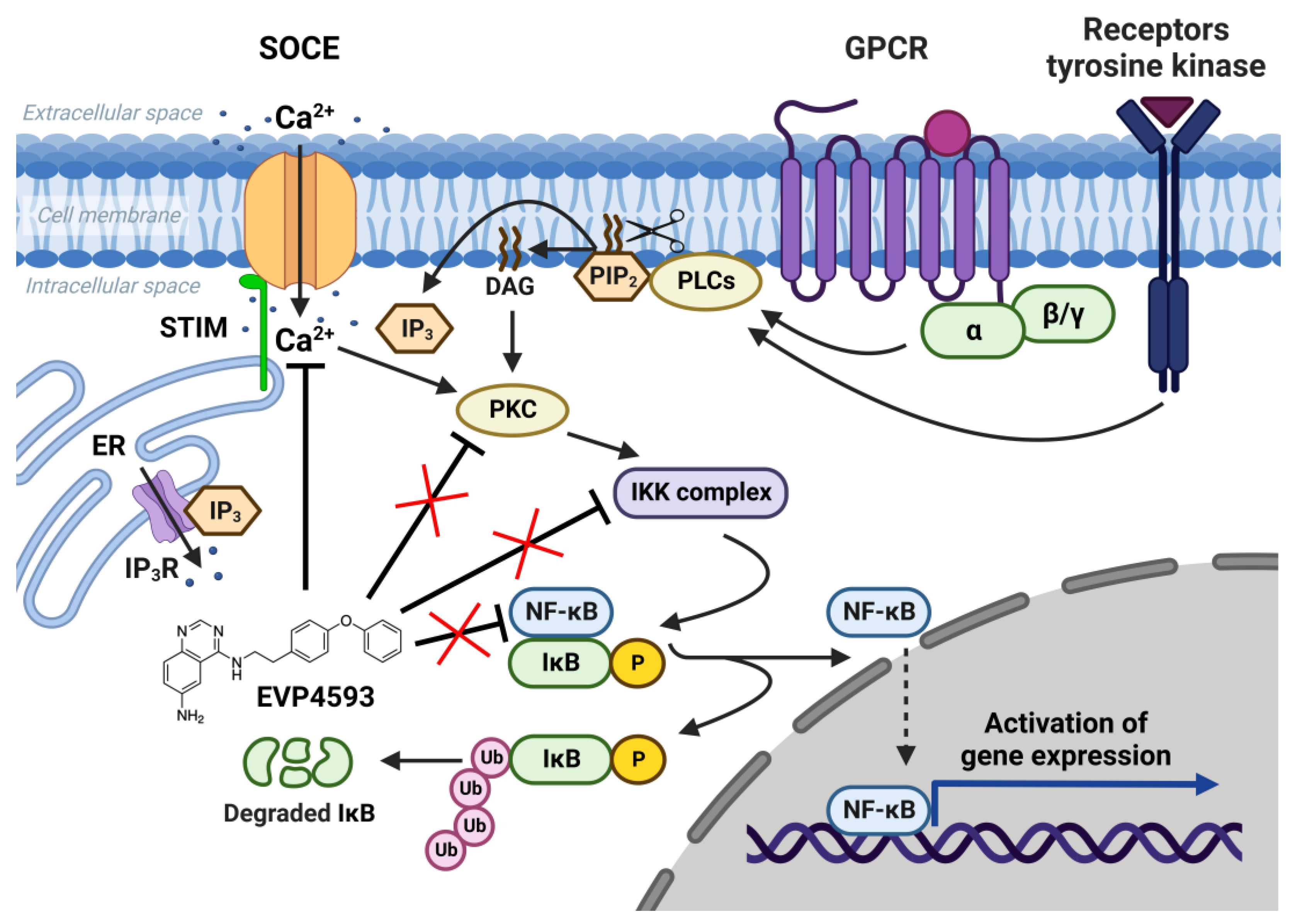
2.1. EVP4593 and NF-κB Signaling

The NF-κB signaling pathway is one of the general signal transduction pathways that mediate gene expression of proinflammatory and antiapoptotic factors, cell proliferation, differentiation, adhesion, migration and angiogenesis [14]. The hyperactivation of NF-κB signaling is a hallmark of many types of cancer [14] and inflammatory-associated pathologies [14,15]. Additionally, the chronic inflammation associated with upregulation of NF-κB signaling is a common feature of multiple neurodegenerative diseases [16]. Thus, the search for selective blockers of NF-κB signaling is an attractive task for both anticancer and anti-neurodegenerative disorders’ drug discovery. In 2003, Tobe et al. synthesized a novel pool of quinazoline derivatives that could act as antagonists of the NF-κB signaling pathway [8,9]. Analyzing various quinazoline core derivatives, Tobe et al. revealed the most effective compound, EVP4593 (4-N-[2-(4-phenoxyphenyl)ethyl]quinazoline-4,6-diamine), blocking NF-κB signaling at nanomolar concentrations (IC50 11 nM) [9]. However, even almost 20 years later, the specific molecular target for EVP4593 remains unclear. It has been established that the EVP4593 does not directly influence any key protein involved in NF-κB signaling, including protein kinase C (PKC), IκB kinase (IKK) complex and transcriptional factor NF-κB (Figure 1) [10]. Therefore, it was assumed that the molecular target of the EVP4593 should be located upstream of NF-κB signaling. The key upstream activators of NF-κB signaling are receptors of interleukins, tumor necrosis factor, lipopolysaccharides and calcium ions [14]. It is well-known that calcium influx through SOC channels triggers the activation of the NF-κB cascade [9,10,11] (Figure 1). Moreover, established SOCE blockers SKF96365 [17] and BTP2 [18] were reported to suppress NF-κB activity. The hypothesis that EVP4593 affects SOC channels, thus modulating NF-κB signaling, was proven for the first time in 2011 by Wu et al. [10].

2.2. EVP4593 and SOCE

In 2011, it was shown for the first time that EVP4593 at a concentration of 300 nM can reversibly block SOC channels and reduce pathologically elevated SOCE in an HD neuroblastoma cell model, expressing full-length mutant huntingtin (138Q), and in striatal neurons isolated from HD-specific YAC128 mice [10]. The current–voltage relationships of the registered SOC currents corresponded to the SOC channels formed by the transient receptor potential canonical (TRPC) proteins [10]. Moreover, suppression of TRPC1 significantly reduced SOC currents in SK-N-SH cells, modeling HD; at that point, the remaining currents were almost insensitive to further inhibition by EVP4593. Nevertheless, EVP4593 failed to inhibit SOC currents in SK-N-SH cells with overexpressed TRPC1 [10]. Therefore, it was concluded that EVP4593 affects heteromeric channels containing TRPC1 as one of the subunits but not homomeric TRPC1 channels. Besides SOC channels’ inhibitory activity, EVP4593 was shown to delay a progression of a motor phenotype in the fly model of HD [10]. Thus, SOC channels (especially TRPC1-containing) have become a potential molecular target for EVP4593, and EVP4593 was established as a promising pharmacological substance for HD treatment.
At the same time, the future studies of HD-specific induced pluripotent stem cells (iPSCs)-based GABAergic medium spiny neurons (MSNs) obtained from adult onset HD patients (40–47 glutamine residues in the polyglutamine tract of mutant huntingtin) demonstrated that both Orai- and TRPC-contained SOC channels were sensitive to treatment by 100 nM EVP4593 [19]. Thus, it has been supposed that the molecular target for EVP4593 may represent a common regulator of SOC channels, such as STIM1 or STIM2 proteins (Figure 2).
Additional experimental data obtained by using fluorescent calcium imaging (the method’s description is available in [20]) indicated that EVP4593 could not discriminate between STIM1 and STIM2 in HEK293 cells, affecting SOC in both STIM1 and STIM2 knockout cell lines (Figure 3).
To summarize the results obtained and published data, we cannot unequivocally explain the inhibitory effect of EVP4593 on SOCE. So, despite the low effective concentration of EVP4593, which supposes an existing direct protein target, we cannot exclude affecting SOC channels through the modulation of plasma membrane features.

2.3. EVP4593 and Mitochondrial Complex I

In addition to the well-studied effects of EVP4593 on SOCE and NF-κB signaling, Robin Krishnathas and colleagues identified mitochondrial complex I as a novel target for EVP4593 [21]. EVP4593 has been shown to exhibit strong inhibitory activity against mitochondrial complex I at nanomolar concentration. Molecular docking predictions illustrated that EVP4593 may incorporate into the quinone binding site, thus inhibiting mitochondrial complex I [22]. However, EVP4593 targeting mitochondrial complex I did not explain the inhibitory effect on SOCE and NF-κB signaling. Perhaps there are some independent targets for EVP4593. It should be noted that the velocity of the EVP4593 effects detected in experiments does not unequivocally conclude which target is primary from between mitochondrial complex I or SOC channels.
Mitochondrial complex I dysfunctions are also associated with oncogenesis and some neurodegenerative disorders [23,24,25]. Thus, EVP4593 can be successfully used in the treatment of cancer and neurodegenerative diseases, with a suitable overlap in effects on SOCE and mitochondrial complex I.

2.4. EVP4593 and mTOR Signaling

mTOR (Mechanistic/Mammalian Target of Rapamycin) signaling controls a wide range of processes involved in cellular metabolism and plays a crucial role in autophagy regulation [26,27]. Aberrant mTOR signaling is associated with a number of pathologies. Hyperactivated mTOR signaling marks some types of cancer and promotes proliferation and tumor progression [26]. mTOR signaling inhibitors are considered promising anticancer drugs [28]. Indeed, the promising therapeutic strategy is the suppression of the mTOR signaling pathway and, consequently, the enhancement of autophagy [27]. Ran Marciano, Manu Prasad and colleagues firstly found that EVP4593 inhibited the mTOR pathway in tumor cells growing under glucose starvation but not under normal conditions [29]. This fact indicates the absence of direct interaction of EVP4593 with the components of mTOR signaling. At the same time, low levels of phosphorylated-4EBP1, phosphorylated-P70S6K and phosphorylated-S6RP under EVP4593 treatment indicates inhibition of the mTORC1 pathway [29]. Since EVP4593 may inhibit mitochondrial complex I, the tumor cells pretreated by EVP4593 may have a significantly lower level of ATP under glucose deprivation, which leads to an inhibition of the AMPK/mTORC1 pathway and suppression of tumor progression. Additionally, EVP4593 inhibits the NF-ĸB pathway that promotes survival under glucose starvation (as well as other NF-ĸB inhibitors) [30]. Thus, EVP4593 promotes the selective death of the most glucose-dependent tumor cells.
The upregulation and involvement of the mTOR signaling pathway in neurodegenerative pathologies, including Huntington’s disease, is excellently discussed in the review by Professor Henry Querfurth [27]. The autophagy induction by mTOR signaling antagonists is accompanied by the removal of mutant protein aggregates and may protect neurons from degeneration [27]. Previously, we have reported larger numbers of lysosomes and autophagosomes in patient-specific models of HD compared to control healthy donors [31], supposing a compensatory enhancement of autophagy in HD-specific neurons aimed at cell survival. Since EVP4593 was shown to inhibit mTOR signaling in cancer [29], we anticipated the induction of autophagy upon EVP4593 treatment. However, HD-specific MSNs (but not WT MSNs) demonstrated a decreased number of lysosomes/autophagosomes after the EVP4593 application [31]. Moreover, it was also reported that other NF-κB inhibition may reduce the number of autophagosomes [32]. Possible controversies can be potentially solved because the reduced number of autophagosomes may indicate the high efficacy of autophagy, whereas a large number of autophagosomes may be a result of autophagy block and the accumulation of “pre-autophagosomes”.
The connection between mTORC1 signaling and SOCE is interesting. The data suggest that mTORC1 signaling is a positive regulator of SOCE [33,34,35]. Additionally, SOCE inhibition led to enhanced autophagy probably through the Akt/mTOR signaling pathway [36,37]. Furthermore, mTORC1 is a positive regulator of NF-κB signaling [38]. mTORC1 may phosphorylate IKKα/β and induce the transcriptional activity of NF-κB [38]. Resveratrol, which is known to inhibit NF-κB and mTOR signaling, reduce SOCE in preincubation experiments and also enhance autophagy [39]. However, the role of calcium signaling in the regulation of autophagy remains complicated [40]. Therefore, for the long-term effect of EVP4593, the following axis can be formed: inhibition of mitochondrial complex I—inhibition of the AMPK/mTORC1 pathway (or Akt/mTOR pathway)—inhibition NF-κB signaling—inhibition of SOCE (Figure 4). Nevertheless, EVP4593 also demonstrates an acute inhibitory SOCE effect confirmed by patch-clamp experiments and fluorescent calcium imaging [10,19,31,41] (Figure 3 and Figure 4).

2.5. EVP4593 and Gene Expression

In addition to an acute inhibitory effect of EVP4593 on SOCE, the long-term effects of incubation cells with EVP4593 have also been observed. The modulation of gene expression by EVP4593 is obvious since the expression of many genes depends on NF-κB. Indeed, it was found that incubation of HD-specific MSNs with 300 nM EVP4593 for 24–48 h reduced excessive levels of the huntingtin protein [42]. These data were not surprising because the expression of the huntingtin gene depends on NF-κB [43] (Figure 5). Notably, modulation of huntingtin expression may have therapeutic applications. It has been shown that even a 10% decrease in mutant huntingtin level has a neuroprotective effect, while even a 90% decrease in normal huntingtin level has no pathological effect [44].
What is interesting is that proteins, encoded by NF-κB-independent genes, may also change their levels upon treatment by EVP4593. SOC channels’ activator STIM2, which has a high level associated with excessive SOCE in HD-specific neurons [42], can also be downregulated by the application of EVP4593, thus protecting neurons from toxic calcium influx. Another research group reported that excessive STIM2-dependent SOC channels’ activity appears to lead to spine loss in YAC128 (HD mice model) MSN [45], confirming the key role of STIM2 at a high level in HD pathogenesis and establishing STIM2 as a promising target for anti-HD drugs. On the other hand, it is extremely important that EVP4593 reduces excessive STIM2 levels to control values since it has been reported that downregulation of STIM2 can be dangerous because STIM2-dependent stability of mushroom spines was shown to be a mechanism of hippocampal synaptic loss in a mice model of Alzheimer’s disease [46]. Curiously, despite the expression of the STIM2 encoding gene, it does not depend on NF-κB; another blocker of NF-κB signaling, wogonin, can also reduce STIM2 levels [47]. Wogonin also inhibits the mTOR pathway and can have a similar effect as rapamycin on the STIM protein level [48].

3. Perspectives of EVP4593 in Clinical Trials

3.1. Neurodegeneration

Impairment of calcium signaling and neuroinflammation are the key features accompanying various neurodegenerative pathologies [49,50,51,52]. Correction of pathological calcium signaling, including SOCE and suppression of the NF-κB signaling pathway, has a neuroprotective effect and can prevent the development of pathological processes [10,31,52,53,54]. Here, we summarized the reported effects of EVP4593 on various models of human diseases (Table 1).
It emphasized the low toxicity of EVP4593 and its pronounced neuroprotective effect in different HD models, including mice [46] and fly models [10]. Moreover, the majority of studies indicate that EVP4593 is effective at high nanomolar concentrations, which makes it suitable for further clinical implications. The main question is, what are the reasons for the neuroprotective effect of EVP4593? Is the observed neuroprotection associated with the inhibition of the NF-κB pathway or mitochondrial complex I or it is a consequence of the SOCE reduction? It was shown that EVP4593-mediated inhibition of the NF-κB pathway is probably not the main cause of the neuroprotective effect observed in experiments with transgenic HD flies [10]. Additionally, the structurally unrelated inhibitor of IKK and NF-κB pathway BMS-345541 were not effective in the fly HD model [10]. Thus, the suppression of NF-κB signaling is probably a necessary but not sufficient condition for neuroprotective effect in HD.
In the case of HD, we can speculate about the key effect of EVP4593 to attenuate excessive huntingtin level. Since HD is a monogenic disorder, all alterations in cellular processes are obviously caused by the presence of the toxic mutant huntingtin. However, EVP4593 cannot completely eliminate mutant huntingtin from the cells. Moreover, it should be noted that despite huntingtin expression being ubiquitous, many neurodegenerative pathologies, including HD, are characterized by selective death of specific types of neurons. Indeed, the most affected neurons in HD are MSNs [55], whereas Parkinson’s disease is characterized by predominant degeneration of dopaminergic neurons in the substantia nigra [56], and significant loss of hippocampal neurons is observed in Alzheimer’s disease [57]. Trying to develop the previously established calcium hypothesis of neurodegeneration [58], we suggest formulating “The calcium hypothesis of selective neuronal death”, where the selective vulnerability of neurons can be determined by different patterns of calcium signaling alterations and different potentials of the neurons to compensate them (for example, by the different levels of calcium-binding proteins). The accumulated data indicated that improper functioning of calcium channels, including SOC channels, plays a crucial role in a number of neurodegenerative pathologies [50,54,59]. Thus, the ability of EVP4593 to reduce excessive SOCE opens up perspectives to develop novel neuroprotective drugs based on EVP4593 for the selective treatment of neurodegenerative disorders. It also requires future studying of potentially selective effects of EVP4593 on different neuronal types.

3.2. Other Pathologies

EVP4593 has been also established as an antineoplastic compound that can suppress carcinogenesis [60,61]. In various carcinoma cell lines, EVP4593 exhibits strong antitumor effects (Table 1). To sum up, EVP4593 suppresses uncontrolled cell proliferation and migration, inhibits expression of the antiapoptotic proteins and increases the level of the proapoptotic proteins [60,61,62,63,64]. Noted antitumor effects of EVP4593 make it to become a prospective anticancer drug.
Another area of potential EVP4593 implication is cardiovascular pathologies. These aspects of EVP4593 actioning are studied poorly. Nevertheless, the ability of EVP4593 to enhance morphine-induced cardio protection in a myocardial ischemia/reperfusion rat model was shown [64]. Moreover, SOCE was registered in adult ventricular cardiomyocytes isolated from mice [65], suggesting its physiological role in the functioning of the cardiac cells. So, the ability of SOCE modulators to correct aberrant calcium signaling in cardiomyocytes may be in demand soon.
Finally, NF-kB is a key inflammatory mediator that regulates the production of many proinflammatory factors, including NO, which plays a central role in the elimination of parasitic infestations. Notably, the published data demonstrated the anthelmintic [66] and antileishmanial [67] activities of EVP4593 further expand the range of its possible implication.
Table 1. The physiological effects of EVP4593 in various models of human diseases.
ModelMechanism of EVP4593 ActivityEVP4593
Concentration
Potential TreatmentReferences
Neurodegeneration
Juvenile and adult-onset HD iPSCs-derived GABA neuronsEVP4593 attenuates pathologically enhanced huntingtin and STIM2 level. Pretreated-by-EVP4593 HD-specific neurons demonstrate reduced SOCE. 300 nMHD[42]
Adult-onset HD iPSCs-derived GABA neuronsEVP4593 inhibits both ICRAC and ISOC channels.100 nMHD[19]
Adult-onset HD iPSCs-derived GABA neuronsEVP4593 attenuates SOCE both in wild-type and HD-specific neurons. EVP4593 reduces the number of lysosomes/
autophagosomes. EVP4593 reduces MG132-induced HD-specific neuronal death.
100 nMHD[31]
The primary culture of mice MSNs with expressed
Htt138Q-1exon
EVP4593 reduces SOCE to the normal level.300 nMHD[41]
YAC128 transgenic HD mice. Mixed cortical/striatal (MSNs) culturesEVP4593 reduces synaptic neuronal SOCE and rescues spine loss. Intraventricular delivery of EVP4593 in YAC128 mice rescues age-dependent striatal spine loss in vivo.30 nM and
0.25 mg/mL in vivo
HD[45]
YAC128 transgenic HD mice. The primary culture of mice MSNs.
Fly HD model (Drosophila melanogaster) with expressed first 4 exons of human huntingtin (128Q)
EVP4593 attenuates enhanced SOCE. EVP4593 affects heteromeric channels containing the TRPC1 subunit but has no effect on homooligomer channels composed of TRPC1. EVP4593 delays a progression of a motor dysfunction phenotype in a transgenic fly HD model and protects YAC128 MSNs in a glutamate toxicity assay.300 nM and
100–400 µM
in vivo
HD[10]
R6/2 transgenic HD mice. The primary culture of cortical pyramidal neuronsEVP4593 reduces somatic calcium transient oscillations.3 µMHD[68]
The primary hippocampal cultures obtained from mice with PSEN1ΔE9 expressionEVP4593 decreases PSEN1ΔE9-mediated SOCE and rescues mushroom spines in PSEN1ΔE9-expressing neurons.30 nM AD[46]
Oncology
Hepatoblastoma cell line (HepG2) and hepatocellular carcinoma cell lines (Huh7 and SNU-387)EVP4593 affects NF-κB signaling and has an antitumor effect. Huh7 and SNU-387—5 µM and
HepG2—10 µM
Liver cancer[60]
Head and neck squamous cell carcinoma (HNSCC) cell lines PCI1, PCI9, PCI13 and PCI52EVP4593 inhibits the NF-κB signaling. Antitumor effects. 1–10 μMCancer[61]
Breast cancer cell lineMCF7 (Michigan Cancer Foundation-7)EVP4593 inhibits the NF-κB signaling and the mTOR pathway. Antitumor effects.1 mg/kgCancer[29]
Nonsmall cell lung cancer (NSCLC) CL1-5-F4 cellsEVP4593 suppresses the NF-ĸB activation. EVP4593 inhibited the expression of metastasis-associated proteins. EVP4593 reduced cell migration and invasion.0.25 μMLung cancer[62]
SK-HEP-1 cellsEVP4593 significantly inhibits the expression of antiapoptotic proteins and triggers extrinsic and intrinsic apoptosis pathways. EVP4593 reduces cell viability. EVP4593 inhibits the expression of NF-κB p65 and antiapoptotic proteins (XIAP, MCL-1 and c-FLIP) and increases levels of proapoptotic proteins (caspase-3 and -8 and cytochrome c). 0.4 µMHuman hepatocellular carcinoma[63]
Parasites invasion
L.amazonensis-infected macrophages EVP4593 inhibits amastigote growth and induces the production of high levels of NO and IL-1β.10 μMAntileishmanial activity[67]
L3 larvae of C. oncophoraAnthelmintic activity 1.9–3.4
μM
Anthelmintic activity[66]
Cardiovascular diseases
Myocardial ischemia/reperfusion rat modelEVP4593 inhibits the NF-κB pathway, decreases the expression of NF-κB and has an anti-inflammatory effect. EVP4593 enhances morphine-induced cardio protection.1 mg/kgMyocardial infarction[64]
Diabetic retinopathy
Streptozotocin-induced diabetes model rats EVP4593 could alleviate the aggravation of retinopathy. EVP4593 decrease the endothelial cell proliferation and significantly reduces p65 expression.
EVP4593 reduces blood glucose level.
80 mg/kgDiabetic retinopathy [69]

4. Conclusions

To date, EVP4593 is a widely used inhibitor of NF-κB signaling. It is also established as an effective blocker of SOCE, which is probably associated with the suppression of NF-κB activity. However, it is still confusing to unequivocally explain the molecular mechanism of EVP4593 acute effects on SOC channels. Nevertheless, the repeatedly demonstrated protective effects of EVP4593 in various models of oncological and neurodegenerative disorders make it attractive for further clinical implications. Moreover, in the last decade, EVP4593 have become an important instrument for studies of the NF-κB signaling and SOC channels.
The multiple actions described for EVP4593 at first glance complicate its application for the development of selective therapy. However, today, the polypharmacological approach has become more and more popular, in particular, for the treatment of multifactorial pathologies such as neurodegenerative diseases [70]. Thus, the ability of EVP4593 to modulate NF-κB signaling, calcium signaling, mitochondrial activity, mTOR pathway and, consequently, autophagy makes it attractive for the development of a polyfunctional drug, generally affecting intracellular signaling and targeting overall cell survival. We hope that future investigations determine the direct molecular target for EVP4593 and expand the area of its clinical implication.

Author Contributions

D.A.G.: writing—original draft preparation, writing—review and editing, visualization and conceptualization; A.A.K.: writing—original draft preparation and visualization; V.A.V.: writing—review and editing, conceptualization, supervision and project administration; E.V.K.: writing—review and editing, conceptualization, visualization and supervision. All authors have read and agreed to the published version of the manuscript.

Funding

This research was funded by a grant from the Ministry of Science and Higher Education of the Russian Federation (Agreement № 075-15-2021-1075, signed 28 September 2021, local identifier 415/21–23) (EVK) and a grant from the Russian Science Foundation N 22-14-00218 (DAG, AAK, VAV).

Institutional Review Board Statement

Not applicable.

Informed Consent Statement

Not applicable.

Data Availability Statement

Not applicable.

Acknowledgments

Figure 1, Figure 2, Figure 4 and Figure 5 were created with BioRender.com (accessed on 6 December 2022). We also thank Kharchenko M.V. (Institute of Cytology RAS) for the valuable comments and discussion of the intracellular signaling machinery.

Conflicts of Interest

The authors declare no conflict of interest.

References

  1. Asif, M. Chemical characteristics, synthetic methods, and biological potential of quinazoline and quinazolinone derivatives. Int. J. Med. Chem. 2014, 2014, 395637. [Google Scholar] [CrossRef] [PubMed]
  2. Dhuguru, J.; Ghoneim, O.A. Quinazoline Based HDAC Dual Inhibitors as Potential Anti-Cancer Agents. Molecules 2022, 27, 2294. [Google Scholar] [CrossRef] [PubMed]
  3. Das, D.; Hong, J. Recent advancements of 4-aminoquinazoline derivatives as kinase inhibitors and their applications in medicinal chemistry. Eur. J. Med. Chem. 2019, 170, 55–72. [Google Scholar] [CrossRef] [PubMed]
  4. Bolteau, R.; Duroux, R.; Laversin, A.; Vreulz, B.; Shiriaeva, A.; Stauch, B.; Han, G.W.; Cherezov, V.; Renault, N.; Barczyk, A.; et al. High ligand efficiency quinazoline compounds as novel A2A adenosine receptor antagonists. Eur. J. Med. Chem. 2022, 241, 114620. [Google Scholar] [CrossRef] [PubMed]
  5. Bolteau, R.; Descamps, F.; Ettaoussi, M.; Caignard, D.H.; Delagrange, P.; Melnyk, P.; Yous, S. Quinazoline and phthalazine derivatives as novel melatonin receptor ligands analogues of agomelatine. Eur. J. Med. Chem. 2020, 189, 112078. [Google Scholar] [CrossRef] [PubMed]
  6. Ran, D.; Gomez, K.; Moutal, A.; Patek, M.; Perez-Miller, S.; Khanna, R. Comparison of quinazoline and benzoylpyrazoline chemotypes targeting the CaVα-β interaction as antagonists of the N-type CaV2.2 channel. Channels 2021, 15, 128–135. [Google Scholar] [CrossRef]
  7. Darwish, S.S.; Chen, P.J.; Hamed, M.M.; Wagdy, R.A.; Chen, S.H.; Abadi, A.H.; Abdel-Halim, M.; Hwang, T.L.; Engel, M. Development of (4-Phenylamino)quinazolineAlkylthiourea Derivatives as Novel NF-κB Inhibitors. Pharmaceuticals 2022, 15, 778. [Google Scholar] [CrossRef]
  8. Tobe, M.; Isobe, Y.; Tomizawa, H.; Nagasaki, T.; Takahashi, H.; Hayashi, H. A novel structural class of potent inhibitors of NF-kappa B activation: Structure-activity relationships and biological effects of 6-aminoquinazoline derivatives. Bioorg. Med. Chem. 2003, 11, 3869–3878. [Google Scholar] [CrossRef]
  9. Tobe, M.; Isobe, Y.; Tomizawa, H.; Nagasaki, T.; Takahashi, H.; Fukazawa, T.; Hayashi, H. Discovery of quinazolines as a novel structural class of potent inhibitors of NF-kappa B activation. Bioorg. Med. Chem. 2003, 11, 383–391. [Google Scholar] [CrossRef]
  10. Wu, J.; Shih, H.P.; Vigont, V.; Hrdlicka, L.; Diggins, L.; Singh, C.; Mahoney, M.; Chesworth, R.; Shapiro, G.; Zimina, O.; et al. Neuronal store-operated calcium entry pathway as a novel therapeutic target for Huntington’s disease treatment. Chem. Biol. 2011, 18, 777–793. [Google Scholar] [CrossRef]
  11. Choi, S.; Kim, J.H.; Roh, E.J.; Ko, M.J.; Jung, J.E.; Kim, H.J. Nuclear factor-kappaB activated by capacitative Ca2+ entry enhances muscarinic receptor-mediated soluble amyloid precursor protein (sAPPalpha) release in SH-SY5Y cells. J. Biol. Chem. 2006, 281, 12722–12728. [Google Scholar] [CrossRef] [PubMed] [Green Version]
  12. Stankunas, K.; Graef, I.A.; Neilson, J.R.; Park, S.H.; Crabtree, G.R. Signaling through calcium, calcineurin, and NF-AT in lymphocyte activation and development. Cold Spring Harb. Symp. Quant. Biol. 1999, 64, 505–516. [Google Scholar] [CrossRef] [PubMed]
  13. Okamura, H.; Rao, A. Transcriptional regulation in lymphocytes. Curr. Opin. Cell. Biol. 2001, 13, 239–243. [Google Scholar] [CrossRef] [PubMed]
  14. Zhang, Q.; Lenardo, M.J.; Baltimore, D. 30 Years of NF-κB: A Blossoming of Relevance to Human Pathobiology. Cell 2017, 168, 37–57. [Google Scholar] [CrossRef] [PubMed] [Green Version]
  15. Jamornwan, S.; Chokpanuwat, T.; Uppakara, K.; Soodvilai, S.; Saengsawang, W. Anti-Inflammatory Activity of Panduratin A against LPS-Induced Microglial Activation. Biomedicines 2022, 10, 2587. [Google Scholar] [CrossRef]
  16. Srinivasan, M.; Lahiri, D.K. Significance of NF-κB as a pivotal therapeutic target in the neurodegenerative pathologies of Alzheimer’s disease and multiple sclerosis. Expert. Opin. Ther. Targets 2015, 19, 471–487. [Google Scholar] [CrossRef] [Green Version]
  17. Zhu, M.; Lv, B.; Ge, W.; Cui, Z.; Zhao, K.; Feng, Y.; Yang, X. Suppression of store-operated Ca2+ entry regulated by silencing Orai1 inhibits C6 glioma cell motility via decreasing Pyk2 activity and promoting focal adhesion. Cell Cycle 2020, 19, 3468–3479. [Google Scholar] [CrossRef]
  18. Dragoni, S.; Laforenza, U.; Bonetti, E.; Lodola, F.; Bottino, C.; Berra-Romani, R.; Carlo Bongio, G.; Cinelli, M.P.; Guerra, G.; Pedrazzoli, P.; et al. Vascular endothelial growth factor stimulates endothelial colony forming cells proliferation and tubulogenesis by inducing oscillations in intracellular Ca2+ concentration. Stem Cells 2011, 29, 1898–1907. [Google Scholar] [CrossRef]
  19. Vigont, V.; Nekrasov, E.; Shalygin, A.; Gusev, K.; Klushnikov, S.; Illarioshkin, S.; Lagarkova, M.; Kiselev, S.L.; Kaznacheyeva, E. Patient-Specific iPSC-Based Models of Huntington’s Disease as a Tool to Study Store-Operated Calcium Entry Drug Targeting. Front. Pharmacol. 2018, 9, 696. [Google Scholar] [CrossRef]
  20. Grekhnev, D.A.; Novikova, I.V.; Krisanova, A.V.; Yuskovets, V.N.; Chernov, N.M.; Yakovlev, I.P.; Kaznacheyeva, E.V.; Vigont, V.A. Dithiadiazole derivative 3-(4-nitrophenyl)-5-phenyl-3H-1,2,3,4-dithiadiazole-2-oxide—Novel modulator of store-operated calcium entry. Biochem. Biophys. Res. Commun. 2022, 626, 38–43. [Google Scholar] [CrossRef]
  21. Krishnathas, R.; Bonke, E.; Dröse, S.; Zickermann, V.; Nasiri, H.R. Identification of 4-N-[2-(4-phenoxyphenyl)ethyl]quinazoline-4,6-diamine as a novel, highly potent and specific inhibitor of mitochondrial complex I. Medchemcomm 2017, 8, 657–661. [Google Scholar] [CrossRef] [PubMed]
  22. Kurelac, I.; Cavina, B.; Sollazzo, M.; Miglietta, S.; Fornasa, A.; De Luise, M.; Iorio, M.; Lama, E.; Traversa, D.; Nasiri, H.R.; et al. NDUFS3 knockout cancer cells and molecular docking reveal specificity and mode of action of anti-cancer respiratory complex I inhibitors. Open Biol. 2022, 12, 220198. [Google Scholar] [CrossRef] [PubMed]
  23. Bassal, M.A.; Samaraweera, S.E.; Lim, K.; Benard, B.A.; Bailey, S.; Kaur, S.; Leo, P.; Toubia, J.; Thompson-Peach, C.; Nguyen, T.; et al. Germline mutations in mitochondrial complex I reveal genetic and targetable vulnerability in IDH1-mutant acute myeloid leukaemia. Nat. Commun. 2022, 13, 2614. [Google Scholar] [CrossRef] [PubMed]
  24. Jain, S.; Hu, C.; Kluza, J.; Ke, W.; Tian, G.; Giurgiu, M.; Bleilevens, A.; Campos, A.R.; Charbono, A.; Stickeler, E.; et al. Metabolic targeting of cancer by a ubiquinone uncompetitive inhibitor of mitochondrial complex I. Cell Chem. Biol. 2022, 29, 436–450. [Google Scholar] [CrossRef]
  25. Hu, Q.; Wang, G. Mitochondrial dysfunction in Parkinson’s disease. Transl. Neurodegener. 2016, 5, 14. [Google Scholar] [CrossRef] [Green Version]
  26. Saxton, R.A.; Sabatini, D.M. mTOR Signaling in Growth, Metabolism, and Disease. Cell 2017, 168, 960–976. [Google Scholar] [CrossRef] [Green Version]
  27. Querfurth, H.; Lee, H.K. Mammalian/mechanistic target of rapamycin (mTOR) complexes in neurodegeneration. Mol. Neurodegener. 2021, 16, 44. [Google Scholar] [CrossRef]
  28. Occhiuzzi, M.A.; Lico, G.; Ioele, G.; De Luca, M.; Garofalo, A.; Grande, F. Recent advances in PI3K/PKB/mTOR inhibitors as new anticancer agents. Eur. J. Med. Chem. 2022, 246, 114971. [Google Scholar] [CrossRef]
  29. Marciano, R.; Prasad, M.; Ievy, T.; Tzadok, S.; Leprivier, G.; Elkabets, M.; Rotblat, B. High-Throughput Screening Identified Compounds Sensitizing Tumor Cells to Glucose Starvation in Culture and VEGF Inhibitors In Vivo. Cancers 2019, 11, 156. [Google Scholar] [CrossRef] [Green Version]
  30. Mauro, C.; Leow, S.C.; Anso, E.; Rocha, S.; Thotakura, A.K.; Tornatore, L.; Moretti, M.; De Smaele, E.; Beg, A.A.; Tergaonkar, V.; et al. NF-KB controls energy homeostasis and metabolic adaptation by upregulating mitochondrial respiration. Nat. Cell Biol. 2011, 13, 1272–1279. [Google Scholar] [CrossRef] [Green Version]
  31. Nekrasov, E.D.; Vigont, V.A.; Klyushnikov, S.A.; Lebedeva, O.S.; Vassina, E.M.; Bogomazova, A.N.; Chestkov, I.V.; Semashko, T.A.; Kiseleva, E.; Suldina, L.A.; et al. Manifestation of Huntington’s disease pathology in human induced pluripotent stem cell-derived neurons. Mol. Neurodegener. 2016, 11, 27. [Google Scholar] [CrossRef] [PubMed] [Green Version]
  32. Fu, J.; Lu, L.; Wang, H.; Hou, Y.; Dou, H. Hirsutella sinensis mycelium regulates autophagy of alveolar macrophages via TLR4/NF-κB signaling pathway. Int. J. Med. Sci. 2021, 18, 1810–1823. [Google Scholar] [CrossRef] [PubMed]
  33. Ogawa, A.; Firth, A.L.; Smith, K.A.; Maliakal, M.V.; Yuan, J.X. PDGF enhances store-operated Ca2+ entry by upregulating STIM1/Orai1 via activation of Akt/mTOR in human pulmonary arterial smooth muscle cells. Am. J. Physiol. Cell. Physiol. 2012, 302, 405–411. [Google Scholar] [CrossRef] [PubMed] [Green Version]
  34. Peng, H.; Liu, J.; Sun, Q.; Chen, R.; Wang, Y.; Duan, J.; Li, C.; Li, B.; Jing, Y.; Chen, X.; et al. mTORC1 enhancement of STIM1-mediated store-operated Ca2+ entry constrains tuberous sclerosis complex-related tumor development. Oncogene 2013, 32, 4702–4711. [Google Scholar] [CrossRef] [Green Version]
  35. Vaeth, M.; Maus, M.; Klein-Hessling, S.; Freinkman, E.; Yang, J.; Eckstein, M.; Cameron, S.; Turvey, S.E.; Serfling, E.; Berberich-Siebelt, F.; et al. Store-Operated Ca2+ Entry Controls Clonal Expansion of T Cells through Metabolic Reprogramming. Immunity 2017, 47, 664–679. [Google Scholar] [CrossRef] [Green Version]
  36. Tang, B.D.; Xia, X.; Lv, X.F.; Yu, B.X.; Yuan, J.N.; Mai, X.Y.; Shang, J.Y.; Zhou, J.G.; Liang, S.J.; Pang, R.P. Inhibition of Orai1-mediated Ca2+ entry enhances chemosensitivity of HepG2 hepatocarcinoma cells to 5-fluorouracil. J. Cell. Mol. Med. 2017, 21, 904–915. [Google Scholar] [CrossRef]
  37. Chen, Y.W.; Chen, Y.F.; Chen, Y.T.; Chiu, W.T.; Shen, M.R. The STIM1-Orai1 pathway of store-operated Ca2+ entry controls the checkpoint in cell cycle G1/S transition. Sci. Rep. 2016, 6, 22142. [Google Scholar] [CrossRef] [Green Version]
  38. Li, Y.; Yang, L.; Dong, L.; Yang, Z.W.; Zhang, J.; Zhang, S.L.; Niu, M.J.; Xia, J.W.; Gong, Y.; Zhu, N. Crosstalk between the Akt/mTORC1 and NF-κB signaling pathways promotes hypoxia-induced pulmonary hypertension by increasing DPP4 expression in PASMCs. Acta Pharmacol. Sin. 2019, 40, 1322–1333. [Google Scholar] [CrossRef]
  39. Selvaraj, S.; Sun, Y.; Sukumaran, P.; Singh, B.B. Resveratrol activates autophagic cell death in prostate cancer cells via downregulation of STIM1 and the mTOR pathway. Mol. Carcinog. 2016, 55, 818–831. [Google Scholar] [CrossRef]
  40. Decuypere, J.P.; Bultynck, G.; Parys, J.B. A dual role for Ca2+ in autophagy regulation. Cell Calcium. 2011, 50, 242–250. [Google Scholar] [CrossRef]
  41. Vigont, V.; Kolobkova, Y.; Skopin, A.; Zimina, O.; Zenin, V.; Glushankova, L.; Kaznacheyeva, E. Both Orai1 and TRPC1 are Involved in Excessive Store-Operated Calcium Entry in Striatal Neurons Expressing Mutant Huntingtin Exon 1. Front. Physiol. 2015, 6, 337. [Google Scholar] [CrossRef] [PubMed] [Green Version]
  42. Vigont, V.A.; Grekhnev, D.A.; Lebedeva, O.S.; Gusev, K.O.; Volovikov, E.A.; Skopin, A.Y.; Bogomazova, A.N.; Shuvalova, L.D.; Zubkova, O.A.; Khomyakova, E.A.; et al. STIM2 Mediates Excessive Store-Operated Calcium Entry in Patient-Specific iPSC-Derived Neurons Modeling a Juvenile Form of Huntington’s Disease. Front. Cell Dev. Biol. 2021, 9, 625231. [Google Scholar] [CrossRef] [PubMed]
  43. Bečanović, K.; Nørremølle, A.; Neal, S.J.; Kay, C.; Collins, J.A.; Arenillas, D.; Lilja, T.; Gaudenzi, G.; Manoharan, S.; Doty, C.N.; et al. A SNP in the HTT promoter alters NF-κB binding and is a bidirectional genetic modifier of Huntington disease. Nat. Neurosci. 2015, 18, 807–816. [Google Scholar] [CrossRef] [PubMed]
  44. Lu, B.; Palacino, J. A novel human embryonic stem cell-derived Huntington’s disease neuronal model exhibits mutant huntingtin (mHTT) aggregates and soluble mHTT-dependent neurodegeneration. FASEB J. 2013, 27, 1820–1829. [Google Scholar] [CrossRef] [PubMed] [Green Version]
  45. Wu, J.; Ryskamp, D.A.; Liang, X.; Egorova, P.; Zakharova, O.; Hung, G.; Bezprozvanny, I. Enhanced Store-Operated Calcium Entry Leads to Striatal Synaptic Loss in a Huntington’s Disease Mouse Model. J. Neurosci. 2016, 36, 125–141. [Google Scholar] [CrossRef] [Green Version]
  46. Chernyuk, D.; Zernov, N.; Kabirova, M.; Bezprozvanny, I.; Popugaeva, E. Antagonist of neuronal store-operated calcium entry exerts beneficial effects in neurons expressing PSEN1ΔE9 mutant linked to familial Alzheimer disease. Neuroscience 2019, 410, 118–127. [Google Scholar] [CrossRef]
  47. Sukkar, B.; Hauser, S.; Pelzl, L.; Hosseinzadeh, Z.; Sahu, I.; Al-Maghout, T.; Bhuyan, A.A.M.; Zacharopoulou, N.; Stournaras, C.; Schöls, L.; et al. Inhibition of Lithium Sensitive Orai1/ STIM1 Expression and Store Operated Ca2+ Entry in Chorea-Acanthocytosis Neurons by NF-κB Inhibitor Wogonin. Cell. Physiol. Biochem. 2018, 51, 278–289. [Google Scholar] [CrossRef]
  48. Zhu, Y.; Wang, J. Wogonin increases β-amyloid clearance and inhibits tau phosphorylation via inhibition of mammalian target of rapamycin: Potential drug to treat Alzheimer’s disease. Neurol. Sci. 2015, 36, 1181–1188. [Google Scholar] [CrossRef]
  49. Skobeleva, K.; Shalygin, A.; Mikhaylova, E.; Guzhova, I.; Ryazantseva, M.; Kaznacheyeva, E. The STIM1/2-Regulated Calcium Homeostasis Is Impaired in Hippocampal Neurons of the 5xFAD Mouse Model of Alzheimer’s Disease. Int. J. Mol. Sci. 2022, 23, 14810. [Google Scholar] [CrossRef]
  50. Grekhnev, D.A.; Kaznacheyeva, E.V.; Vigont, V.A. Patient-Specific iPSCs-Based Models of Neurodegenerative Diseases: Focus on Aberrant Calcium Signaling. Int. J. Mol. Sci. 2022, 23, 624. [Google Scholar] [CrossRef]
  51. Kolobkova, Y.A.; Vigont, V.A.; Shalygin, A.V.; Kaznacheyeva, E.V. Huntington’s Disease: Calcium Dyshomeostasis and Pathology Models. Acta Naturae 2017, 9, 34–46. [Google Scholar] [CrossRef] [PubMed]
  52. Ureshino, R.P.; Erustes, A.G.; Bassani, T.B.; Wachilewski, P.; Guarache, G.C.; Nascimento, A.C.; Costa, A.J.; Smaili, S.S.; Pereira, G.J.D.S. The Interplay between Ca2+ Signaling Pathways and Neurodegeneration. Int. J. Mol. Sci. 2019, 20, 6004. [Google Scholar] [CrossRef] [PubMed] [Green Version]
  53. Guan, P.P.; Cao, L.L.; Yang, Y.; Wang, P. Calcium Ions Aggravate Alzheimer’s Disease Through the Aberrant Activation of Neuronal Networks, Leading to Synaptic and Cognitive Deficits. Front. Mol. Neurosci. 2021, 14, 757515. [Google Scholar] [CrossRef] [PubMed]
  54. Bezprozvanny, I. Calcium signaling and neurodegenerative diseases. Trends Mol. Med. 2009, 15, 89–100. [Google Scholar] [CrossRef] [PubMed] [Green Version]
  55. Vonsattel, J.P.; DiFiglia, M. Huntington disease. J. Neuropathol. Exp. Neurol. 1998, 57, 369–384. [Google Scholar] [CrossRef] [Green Version]
  56. Langston, J.W. The Parkinson’s complex: Parkinsonism is just the tip of the iceberg. Ann. Neurol. 2006, 59, 591–596. [Google Scholar] [CrossRef]
  57. Braak, H.; Braak, E. Neuropathologicalstageing of Alzheimer-related changes. Acta Neuropathol. 1991, 82, 239–259. [Google Scholar] [CrossRef]
  58. Khachaturian, Z.S. Towards theories of brain aging. In Handbook of Studies on Psychiatry and Old Age; Kay, D.S., Burrows, G.W., Eds.; Elsevier: Amsterdam, The Netherlands, 1984; pp. 7–30. [Google Scholar]
  59. Secondo, A.; Bagetta, G.; Amantea, D. On the Role of Store-Operated Calcium Entry in Acute and Chronic Neurodegenerative Diseases. Front. Mol. Neurosci. 2018, 11, 87. [Google Scholar] [CrossRef] [Green Version]
  60. Gu, Z.L.; Huang, J.; Zhen, L.L. Knockdown of otubain 2 inhibits liver cancer cell growth by suppressing NF-κB signaling. Kaohsiung J. Med. Sci. 2020, 36, 399–404. [Google Scholar] [CrossRef]
  61. Scheurer, M.J.J.; Brands, R.C.; El-Mesery, M.; Hartmann, S.; Müller-Richter, U.D.A.; Kübler, A.C.; Seher, A. The Selection of NFκB Inhibitors to Block Inflammation and Induce Sensitisation to FasL-Induced Apoptosis in HNSCC Cell Lines Is Critical for Their Use as a Prospective Cancer Therapy. Int. J. Mol. Sci. 2019, 20, 1306. [Google Scholar] [CrossRef]
  62. Wu, J.Y.; Lin, S.S.; Hsu, F.T.; Chung, J.G. Fluoxetine Inhibits DNA Repair and NF-ĸB-modulated Metastatic Potential in Non-small Cell Lung Cancer. Anticancer Res. 2018, 38, 5201–5210. [Google Scholar] [CrossRef] [PubMed]
  63. Tsai, J.J.; Pan, P.J.; Hsu, F.T. Regorafenib induces extrinsic and intrinsic apoptosis through inhibition of ERK/NF-κB activation in hepatocellular carcinoma cells. Oncol. Rep. 2017, 37, 1036–1044. [Google Scholar] [CrossRef] [PubMed] [Green Version]
  64. Tu, R.H.; Wang, D.X.; Zhong, G.Q.; Meng, J.J.; Wen, H.; Feng, J.; Bi, Q.; He, Y. New targets of morphine postconditioning protection of the myocardium in ischemia/reperfusion injury: Involvement of HSP90/Akt and C5a/NF-κB. Open Med. 2021, 6, 1552–1563. [Google Scholar] [CrossRef] [PubMed]
  65. Gusev, K.O.; Vigont, V.V.; Grekhnev, D.A.; Shalygin, A.V.; Glushankova, L.N.; Kaznacheeva, E.V. Store-Operated Calcium Entry in Mouse Cardiomyocytes. Bull. Exp. Biol. Med. 2019, 167, 311–314. [Google Scholar] [CrossRef] [PubMed]
  66. Liu, M.; Landuyt, B.; Klaassen, H.; Geldhof, P.; Luyten, W. Screening of a drug repurposing library with a nematode motility assay identifies promising anthelmintic hits against Cooperiaoncophora and other ruminant parasites. Vet. Parasitol. 2019, 265, 15–18. [Google Scholar] [CrossRef] [PubMed]
  67. Macedo, S.R.; de FigueiredoNicolete, L.D.; Ferreira Ados, S.; de Barros, N.B.; Nicolete, R. The pentavalent antimonial therapy against experimental Leishmania amazonensis infection is more effective under the inhibition of the NF-κB pathway. Int. Immunopharmacol. 2015, 28, 554–559. [Google Scholar] [CrossRef] [PubMed]
  68. Li, R.; Yuan, H.; Zhao, T.; Yan, Y.; Liu, Z.; Cai, J.; Qiu, C.; Li, C. miR-874 ameliorates retinopathy in diabetic rats by NF-κB signaling pathway. Adv. Clin. Exp. Med. 2021, 30, 421–430. [Google Scholar] [CrossRef]
  69. Oikonomou, K.D.; Donzis, E.J.; Bui, M.T.N.; Cepeda, C.; Levine, M.S. Calcium dysregulation and compensation in cortical pyramidal neurons of the R6/2 mouse model of Huntington’s disease. J. Neurophysiol. 2021, 126, 1159–1171. [Google Scholar] [CrossRef]
  70. Garcia-Romero, E.M.; López-López, E.; Soriano-Correa, C.; Medina-Franco, J.L.; Barrientos-Salcedo, C. Polypharmacological drug design opportunities against Parkinson’s disease. F1000Research 2022, 11, 1176. [Google Scholar] [CrossRef]
Figure 1. NF-κB signaling pathway and SOCE. SOC channels are activated as a result of intracellular calcium store depletion caused by the activation of inositol-1,4,5-trisphosphate receptor (IP3R). Calcium influx through SOC channels activates protein kinase C (PKC), which activates the IκB kinase (IKK) complex containing the kinases IKKα and IKKβ and the regulatory protein IKKγ. Activated IKK complex phosphorylates inhibitory protein IκB and recruits NF-κB dimers (p50, p52, p65/RelA, RelB and c-Rel). NF-κB free from inhibitory protein IκB migrate to the nucleus and initiate gene expression. EVP4593 does not directly affect any key protein of the NF-κB signal pathway but inhibits SOCE, which is required for NF-κB activation.
Ijms 23 15724 g001
Figure 2. EVP4593 inhibits both Orai- and TRPC-contained store-operated calcium channels. EVP4593 blocks currents through both types of SOC channels that are Orai-contained (ICRAC) and TRPC-contained (ISOC).
Ijms 23 15724 g002
Figure 3. EVP4593 suppresses SOCE in both STIM1 (STIM1KO) and STIM2 (STIM2KO) knockout HEK293 cell lines. Normalized relative fluorescence of Fura-2AM associated with cytosolic calcium level in HEK293 (A), HEK293 STIM1KO (B) and HEK STIM2KO (C) cells during thapsigargin-induced calcium response. The arrow indicates the supply of 1 µM EVP4593 or 0.1% DMSO, respectively. The used solutions are indicated above the curves. The curves are plotted as the mean ± SEM.
Ijms 23 15724 g003
Figure 4. Effects of EVP4593 on cell signaling. EVP4593 has both acute and long-term effects. The acute effect is to directly inhibit SOCE. Long-term effects may be associated with the inhibition of the mitochondrial complex I. Energy deficit induced by EVP4593 inhibits the AMPK/mTORC1 pathway or activates Unc-51 such as autophagy activating kinase 1 (ULK1), which leads to enhanced autophagy and suppressed protein synthesis. Additionally, EVP4593 can negatively affect NF-κB signaling by inhibiting SOCE or mTORC1 signaling. Consequently, EVP4593 also suppresses the expression of NF-κB -dependent genes. The lower levels of STIM proteins due to EVP4593 action may also result in reduced SOCE.
Ijms 23 15724 g004
Figure 5. EVP4593 reduces the level of the huntingtin protein. Huntingtin gene expression is regulated by NF-κB-dependent promoter/enhancer. (A) Elevated SOCE in HD induces hyperactivation of NF-κB signaling, resulting in a high level of the huntingtin protein. Mutant huntingtin enhances calcium influx through SOC channels and potentiates the IKK complex. Thus, a pathological vicious circle is formed. (B) EVP4593 attenuates pathologically enhanced SOCE and decreases NF-κB-dependent huntingtin production.
Ijms 23 15724 g005
Publisher’s Note: MDPI stays neutral with regard to jurisdictional claims in published maps and institutional affiliations.

