Article

Molecular Characterization of Vitellogenin and Its Receptor in Spodoptera frugiperda (J. E. Smith, 1797), and Their Function in Reproduction of Female

College of Plant Protection, Hebei Agricultural University, Baoding 071000, China
*
Author to whom correspondence should be addressed.
These authors contributed equally to this work.
Int. J. Mol. Sci. 2022, 23(19), 11972; https://doi.org/10.3390/ijms231911972
Submission received: 5 September 2022 / Revised: 24 September 2022 / Accepted: 26 September 2022 / Published: 9 October 2022
(This article belongs to the Section Molecular Biology)

Abstract

:
The fall armyworm Spodoptera frugiperda is a highly polyphagous invasive pest. The strong reproductive capacity is an important factor in the rapid colonization and expansion of S. frugiperda. Vitellogenin (Vg) and vitellogenin receptor (VgR) play important roles in insect reproduction. As the precursor of vitellin (Vn), Vg provides essential nutrition for embryonic development, and VgR mediates the uptake of Vg by oocytes. In this context, we cloned and characterized these two genes of S. frugiperda (SfVg and SfVgR) and evaluated their expression profiles in different developmental stages and tissues. The RNA interference experiment was used to investigate their function in vitellogenesis. The ORF values of SfVg and SfVgR were 5250 and 5445 bp, encoding 1749 and 1815 amino acid residues, respectively. The qRT-PCR results revealed that both SfVg and SfVgR were highly expressed in female adults; SfVg was specifically expressed in the fat body, whereas SfVgR was highly expressed in the ovary. In addition, the depletion of either SfVg or SfVgR hindered oocyte maturation and ovarian development, leading to a significant decrease in fecundity. The present study reveals the importance of SfVg and SfVgR in the vitellogenesis of S. frugiperda, laying a theoretical foundation for the development of pollution-free pest control strategies with SfVg and SfVgR as new targets.

1. Introduction

For oviparous insects, vitellogenesis is an essential process for population multiplication and involves the synthesis and absorption of vitellogenin (Vg) [1,2]. As the precursor of the yolk protein vitellin (Vn), Vg provides the nutrition necessary for embryonic development, such as amino acids, fats, vitamins, phosphates, and other trace elements [3]. In most insects, such as Aedes aegypti (Linnaeus) (Diptera: Culicidae), Locusta migratoria (Linnaeus) (Orthoptera: Acrididae), and Periplaneta americana (Linnaeus) (Blattodea: Blattidae), Vg is mainly synthesized in the fat body and secreted into the hemolymph, then absorbed by developing oocytes via vitellogenin receptor (VgR)-mediated endocytosis [1,2,4,5,6]. Thus, Vg and VgR play important roles in insect reproduction.
At present, Vg and VgR have been studied extensively in many insects. Vgs are multipart oligomeric glycolipophospho proteins and encode a polypeptide with a molecular weight of about 200 kDa [7,8]. Structural analysis indicates that Vgs belong to the large lipid transfer protein (LLTP) superfamily. In general, the LLTP family members have three amino acid domains: an N-terminal lipid-binding domain (LPD_N), an unknown functional region (DUF1943), and a von Willebrand factor type D similar domain (VWD) [3,4,5,9]. There is only one single VgR gene in most insects, encoding a large protein of 180–214 kDa [3,5]. VgRs are members of the low-density lipoprotein receptor (LDLR) family, which contains five typical domains: a ligand-binding domain (LBD), an epidermal growth factor precursor domain (EGF) with low-density lipoprotein-receptor Tyr-Trp-Thr-Asp (YWTD) repeats, an O-linked sugar domain, a transmembrane domain (TMD), and a cytoplasmic tail (CD) [10,11,12].
Since the first Vg and VgR genes were identified in Hyalophora cecropia (Linnaeus) (Lepidoptera: Saturniidae) and L. migratoria [13,14], respectively, these homologous genes have been cloned in many insect species in recent years, including Sogatella furcifera (Horváth) (Hemiptera: Delphacidae) [3], Neoseiulus barkeri (Hughes) (Mesostigmata: Phytoseiidae) [15], Thitarodes pui (Zhang) (Lepidoptera: Hepialidae) [16], and Conopomorpha sinensis (Bradley) (Lepidoptera: Gracillariidae) [17]. Knockdown of either gene can arrest ovarian development and reduce oviposition. Studies have proved that Vg and VgR are essential for the successful reproduction of insects. In addition, Vg has been found to have several non-reproductive functions in other physiological responses, such as the extended life span of eusocial insects, immunity reaction, gustatory responsiveness, and the spread of viruses [18,19,20,21]. Undoubtedly, the primary role of Vg is to form yolk protein and provide the nutrients required for ovarian development.
The fall armyworm Spodoptera frugiperda (J. E. Smith) (Lepidoptera: Noctuidae) is a highly polyphagous invasive pest native to tropical and subtropical America. It has a wide suitable region, wide host range, high reproductive capacity, and strong migration ability, and the strong fecundity of S. frugiperda represents a primary cause of serious economic losses [22,23,24,25]. The female moths can mate and spawn multiple times, and each female adult can produce more than 1500 offspring throughout their lifetime under appropriate conditions [23]. Because vitellogenesis is essential for the reproduction of oviparous pests, disrupting this specific process could be a new idea for pest management.
In this study, we first cloned vitellogenin (SfVg) and the vitellogenin receptor (SfVgR) of S. frugiperda and analyzed their molecular characteristics and phylogenetic relationships. Furthermore, we evaluated the expression profiles of SfVg and SfVgR in different developmental stages and tissues. Finally, we used dsRNA-mediated gene silencing to determine the function of Vg and VgR in the vitellogenesis of S. frugiperda. These findings contribute to clarifying the reproductive regulation mechanism and provide new ideas and targets for the sustainable management of S. frugiperda.

2. Results

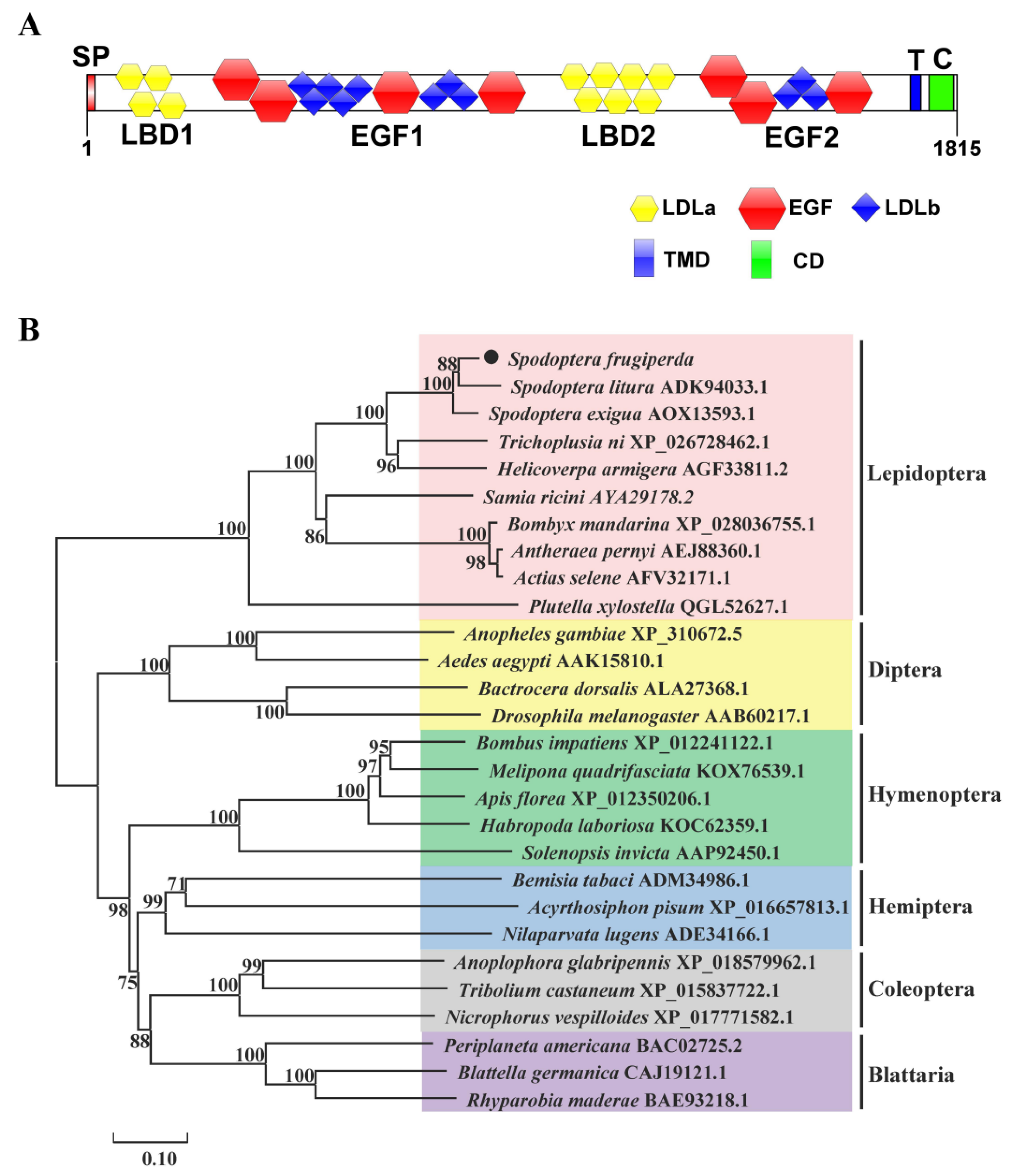
2.1. Cloning and Sequence Analysis of SfVg and SfVgR

The sequences of SfVg and SfVgR were identified from transcriptome data of S. frugiperda and deposited in GenBank with accession no. MT955597 and MT955598. The open reading frame (ORF) of SfVg consists of 5250 base pairs encoding a protein of 1749 amino acids with a 15 aa signal protein located at the N-terminus (Figure 1). The predicted molecular weight (MW) is 198.92 kDa and the isoelectric point (pI) is 8.92. The amino acid sequence of SfVg contains five putative RXXR cleavage sites, a DGQR conserved motif, and a GLCG conserved motif (Figure 1). Moreover, SfVg has 185 phosphorylation sites and five N-glycosylation sites (Supplementary Files S1 and S2). The results of BLASTP and SMART indicated that SfVg protein belongs to the large lipid transfer protein (LLTP) superfamily with three conservative domains of LPD_N (29-714 aa), DUF1943 (746-1024 aa), and VWD (1410-1583 aa) (Figure 1 and Figure 2A). The BLASTP analysis of NCBI database revealed that SfVg shares similarities of 91.94% and 88.47% with the Vg of Spodoptera litura (Fabricius) (Lepidoptera: Noctuidae) and Spodoptera exigua (Hübner) (Lepidoptera: Noctuidae), respectively. The phylogenetic analysis indicated that SfVg clusters together with the branch of Lepidoptera, and forms a clade similar to cotton leafworm, S. litura (Figure 2B).
The ORF of SfVgR is 5445 bases and encodes a protein of 1815 amino acids with MW of 204.059 kDa and pI of 4.83. The signal peptide has 17 amino acids at the N-terminus. SfVgR has 14 N-linked glycosylation sites and 164 phosphorylation sites (Supplementary Files S3 and S4). BLASTP and SMART analysis revealed that SfVgR belongs to the low-density lipoprotein receptor (LDLR) family, containing two ligand-binding repeats (LBD1, 29–217 aa; LBD2, 942–1284 aa), two epidermal growth factor precursor domains (EGF1, 218–938 aa; EGF2, 1285–1663 aa), a transmembrane domain (TMD, 1691–1713 aa), and a cytoplasmic tail (CD, 1714–1815 aa) (Figure 3). There are four class A (LDLa) repeats in LBD1 and seven repeats in LBD2. Each LBD is followed by an EGF. The EGF1 domain possesses four EGF-like repeats and seven LDLb domains, and the EGF2 domain possesses three EGF-like repeats and three LDLb domains (Figure 4A). The BLASTP analysis indicated that SfVgR shares 94.33% and 92.89% similarity with the VgR of S. litura and S. exigua, respectively. Phylogenetic analysis showed that SfVgR is clustered together with VgRs of other Lepidopteran species, and also forms a clade similar to cotton leafworm, S. litura (Figure 4B).

2.2. Expression Profiling of SfVg and SfVgR

The qRT-PCR analysis indicated that SfVg and SfVgR were both highly expressed in female adults (Figure 5A, F = 137.912, df = 10, 22, p < 0.001; Figure 5D, F = 352.644, df = 10, 22, p < 0.001). In different ages of females, SfVg was first detected in 7 d old pupae and then the expression level gradually increased. The expression peaked in the 3-day-old adults and then continued to decrease (Figure 5B, F = 107.865, df = 5, 12, p < 0.001). The SfVgR had similar expression patterns with SfVg in different ages of females, while SfVgR expression peaked in the 4-day-old adults (Figure 5E, F = 94.871, df = 5, 12, p < 0.001). The female tissue expression analysis showed that SfVg was specifically and highly expressed in the fat body (Figure 5C, F = 112.082, df = 5, 12, p < 0.001) and SfVgR was significantly highly expressed in the ovaries (Figure 5F, F = 149.254, df = 5, 12, p < 0.001).

2.3. Functional Analysis of SfVg and SfVgR by RNA Interference Experiment

For functional analysis, 10 μg dsRNA of SfVg, SfVgR, and GFP was injected into 2 d old female pupae, and then the expression levels of target genes were determined by qPCR. The results showed that the expression level of SfVg in the dsSfVg injection treatment was downregulated by 48.22% after 24 h of female adult eclosion (F = 0.173, df = 4, p = 0.003), 76.62% after 48 h (F = 14.111, df = 4, p = 0.026), and 73.01% after 72 h (F = 0.991, df = 4, p < 0.001) compared with the dsGFP injection control group (Figure 6A). The expression levels of SfVgR in the dsSfVgR injection treatment after 24, 48, and 72 h of eclosion were downregulated by 46.07%, 67.94%, and 58.47%, respectively, compared with the dsGFP injection control treatment (Figure 6A; F24h = 3.212, df24h = 4, P24h = 0.002; F48h = 0.269, df48h = 4, P48h = 0.001; F72h = 1.302, df72h = 4, P72h = 0.008). Meanwhile, the SfVgR expression level was significantly decreased at each time point in the dsSfVg injection treatment (Figure 6A; F24h = 0, df24h = 4, P24h < 0.001; F48h = 2.262, df48h = 4, P48h < 0.001; F72h = 6.371, df72h = 4, P72h = 0.001). However, knockdown of SfVgR had no significant effect on the expression of SfVg at 24 h and 48 h after emergence, but downregulated the SfVg transcription 72 h after emergence (Figure 6A; F24h = 0.574, df24h = 4, P24h = 0.085; F48h = 12.911, df48h = 4, P48h = 0.524; F72h = 0.980, df72h = 4, P72h = 0.018). Western blot analysis indicated that knockdown of SfVg decreased the content of Vg protein in the hemolymph and ovaries of females; downregulation of SfVgR did not affect the synthesis of Vg, but hindered the uptake of Vg protein by oocytes and increased the content of Vg in the hemolymph (Figure 6B).
In addition, the developmental status of ovaries from each treatment was dissected and observed at 72 h after emergence. Photographs of ovarian morphology showed that knockdown of SfVg or SfVgR obviously hindered ovarian development compared with the dsGFP injection treatment (Figure 6C). The ovary lengths in the dsSfVg injection treatment and the dsSfVgR injection treatment were significantly shorter than in the dsGFP injection treatment (Figure 6D; p < 0.001).
Furthermore, the effects of SfVg and SfVgR knockdown on the reproduction of S. frugiperda were further explored. The results showed that the oviposition of the SfVg and SfVgR knockdown moths was significantly less than that in the control group (Figure 7A,B). Specifically, the number of eggs per female of dsSfVg injection moths was reduced by 68.10% compared with dsGFP injection treatment, and dsSfVg injection treatment exhibited an 80.15% decline (Figure 7B; p < 0.001). Moreover, the pre-oviposition period in dsSfVg/dsSfVgR-treated females (dsSfVg, 4.17 d; dsSfVgR, 4.00 d) was significantly prolonged compared with the control groups (3.53 d) (Figure 7C; p < 0.01). The oviposition duration and egg hatchability after SfVg or SfVgR silencing were significantly lower than in the control group (Figure 7D,E; p < 0.001).

3. Discussion

Vitellogenesis, an essential process for insect reproduction, entails the synthesis of Vg in the fat body and its adsorption by developing oocytes via VgR-mediated endocytosis [1,2,3]. The research on insects’ reproductive mechanisms can facilitate the search for new targets for pest control. S. frugiperda is a highly polyphagous invasive pest, and its strong fecundity is a key factor in its explosive damage [23,26]. However, little is known about the role of Vg and VgR in S. frugiperda. In this study, we cloned and identified the Vg and VgR genes of S. frugiperda and analyzed their expression patterns, then explored their function in the reproductive process of female adults by RNA interference.
Similar to previous reports, SfVg had conserved domains of the LLTP superfamily, including the LPD_N, VWD, and DUF1943 domains [3,17,27]. The motifs GL/ICG and DGXR in the amino acid sequence of SfVg were also highly conserved domains in insect Vg proteins, which was regarded as a necessary condition for oligomerization [28,29]. The Vg protein can bind inactive ecdysone-containing lipids via oligomerization and release ecdysone during embryogenesis [30]. The SfVg contains five putative RXXR cleavage sites, which exist in most insect Vg sequences and play an important role in the maturation of primary Vg protein [3,5]. However, the number of these cleavage sites was different in various insects. For example, the HmVg of Harmonia axyridis (Pallas) (Coleoptera: Coccinellidae) contains three putative cleavage recognition sites [31], while the amino acid sequence of CsVg in Chrysopa septempunctata (Wesmael) (Neuroptera: Chrysopidae) contains only one RXXR cleavage site [32].
SfVgR belongs to the low-density lipoprotein receptor (LDLR) superfamily and has several typical domains. Insect VgRs generally have two LBD domains, with five LDLRa repeats in the first domain and eight LDLRa repeats in the second domain [6,33,34]. In some Lepidoptera insects, LBD1 and LBD2 have four and seven LDLRa repeats, respectively, such as S. litura [35], Helicoverpa armigera (Hübner) (Lepidoptera: Noctuidae) [36], S. exigua [37], and T. pui [16]. A previous report indicated that LBD domains of VgR could mediate the interaction between Vg and VgR [6]. In addition, similar to previous studies on insect Vgs and VgRs, SfVg and SfVgR are also highly phosphorylated proteins with multiple putative phosphorylation sites [4,6]. Phosphorylation can promote the combination of Vg and VgR. For example, Havukainen et al. found that phosphorylation could prevent Vg from being decomposed by protease [38]. In addition, Jing et al. reported that juvenile hormone-stimulated VgR phosphorylation promotes Vg uptake by oocytes [39].
Gene expression patterns are usually associated with their physiological functions. In this study, the qPCR results indicated that SfVg and SfVgR were female-specific genes. High expression levels of Vg and VgR in female adults were also confirmed in other insects [3,16]. However, Vg and VgR are no longer female-specific proteins, according to recent studies. In sub-social honeybees, Vg and VgR are expressed in both male and female adults; this may be related to the differentiation of gender and behavior [40,41]. The expression of Vg and VgR in different insects has developmental stage specificity. In our study, the expressions of SfVg and SfVgR in larvae were not detected, but in certain insects, the expression of these two genes can be detected in larvae or nymphs [3,35,42]. In different ages of female pupae and adults, both SfVg and SfVgR were first detected in 7-day-old pupae, and the expression peaks of SfVg and SfVgR were 3 d and 4 d after emergence, respectively. This result is similar to that of S. litura and Chilo suppressalis (Walker) (Lepidoptera: Crambidae); that is, the expression of Vg and VgR can be first detected at the end of the pupa stage and increases rapidly in the adult stage [11,35,43]. The expression of Vg and VgR usually has a similar trend, and this expression pattern is closely related to the process of vitellogenesis and the state of ovarian development [3,44]. Thus, the expression of Vg and VgR could become new targets for forecasting the occurrence of pests in the field. In addition, Zhao et al. found a significant linear regression correlation between Vg and VgR expression levels [37]. The expression of Vg and VgR has obvious tissue specificity in most insects. Similar to previous studies, SfVg and SfVgR were highly expressed in the fat body and ovaries, respectively. However, the expression of Vg and VgR has also been detected in other tissues of Apis mellifera (Linnaeus) (Hymenoptera: Apidae), suggesting that these two genes have functional diversity [40,41].
RNA interference technology has been widely used in various fields of life science research, including gene function research, clinical disease treatment, animal model establishment, and anti-virus research. In recent years, the expression of dsRNAs in transgenic plants has emerged as a potential approach to control agricultural pests [45,46]. Studies show that transgenic cotton, which can produce dsRNA that targets the key gene (FAR) in insect reproduction, can be used to control plant bugs [46]. At present, the RNAi-mediated silencing of target genes has been implemented in a variety of Lepidopteran insects, such as S. litura, H. armigera, Cadra cautella (Walker) (Lepidoptera: Pyralidae), and C. suppressalis [10,11,35,36,47]. In our study, the expressions of SfVg and SfVgR in female adults were apparently decreased by injecting dsRNA into the 2-day-old female pupae. Depletion of SfVg or SfVgR hindered oocyte maturation and ovarian development, leading to a significant decrease in fecundity. Similarly, Hu et al. reported that silencing of either Vg or VgR genes of S. furcifera could reduce vitellin deposition in oocytes and arrest oocyte maturation [3]. Yang et al. found that knockdown of the BmVg gene could interfere with egg formation and embryonic development of Bombyx mori (Linnaeus) (Lepidoptera: Bombycidae) [48]. In almond moth C. cautella, downregulation of VgR can significantly reduce fecundity and hatchability [10].
As previously found, there is a feedback loop between the transcription levels of Vg and VgR in insects. However, the feedback loop between Vg and VgR transcription varies in different insects. In Aphis citricidus (Kirkaldy) (Hemiptera: Aphididae), downregulation of AcVg can decrease the mRNA transcript of AcVgR [42], whereas knockdown of Vg did not affect the expression levels of VgR in S. furcifera and Nilaparvata lugens (Stål) (Hemiptera: Delphacidae) [3,49]. Moreover, some researchers indicated that a lack of VgR impedes the Vg uptake by ovaries and increases the accumulation of Vg proteins in the hemolymph, whereas VgR knockdown does not influence Vg synthesis [35,36]. In cabbage beetle Colaphellus bowringi (Baly) (Coleoptera: Chrysomelidac), knockdown of VgR also increases the accumulation of Vg proteins in the hemolymph, but downregulates the Vg transcription [50]. In our study, silencing of SfVg decreased the expression of SfVgR, whereas knockdown of SfVgR had no significant effect on the expression of SfVg at 24 h and 48 h after emergence, but downregulated the SfVg transcription 72 h after emergence. Western blot analysis indicated that knockdown of SfVgR hindered the uptake of Vg proteins by oocytes and increased the accumulation of Vg in the hemolymph. Therefore, there is indeed a feedback loop between Vg and VgR transcription in S. frugiperda, but the specific feedback mode needs further verification.
In summary, we identified and cloned the full ORF sequences of SfVg and SfVgR and analyzed their expression profiles in different developmental stages and tissues. The results of RNA interference experiments confirm that SfVg and SfVgR play crucial roles in ovarian development and oviposition in S. frugiperda. Our findings enrich the basic theoretical knowledge of the reproductive biology of S. frugiperda and confirm that SfVg and SfVgR can be potential targets for controlling this pest. For example, transgenic plant-mediated RNAi or RNAi preparations synthesized in vitro to target these two genes may provide a new strategy to control this pest.

4. Materials and Methods

4.1. Insect Collection and Rearing

S. frugiperda was obtained from Henan Academy of Agricultural Sciences, Zhengzhou, China. The insect samples were reared on fresh corn seedlings in an incubator at 26 ± 1 °C, with relative humidity of 65 ± 5% and under a 16:8 h (L:D) photoperiod. The samples of different developmental stages were collected from eggs (20 mg), first-instar larvae (50 individuals), second-instar larvae (20 individuals), third-instar larvae (5 individuals), fourth-instar larvae (1 individual), fifth-instar larvae (1 individual), sixth-instar larvae (1 individual), third-day pupae (1 male and female individual), and third-day adults (1 male and female individual).
The female samples with different emergence ages included 0, 1, 2, 3,…, and 10 d old female adults (1 individual). For tissue sample collection, the heads (10 individuals), thoraxes (5 individuals), ovaries (5 individuals), fat body (20 mg), legs (20 individuals), and wings (20 individuals) were obtained from 3-day-old females. The samples were collected in 1.5 mL centrifuge tubes and rapidly frozen in liquid nitrogen and stored at −80 °C. Each sample had three biological replicates.

4.2. RNA Isolation and cDNA Synthesis

The total RNA was extracted using the RNA prep Pure Tissue Kit (Tiangen, Beijing, China) according to the manufacturer’s instructions. The concentration and purity of total RNA were detected using a NanoDrop spectrophotometer (MD2000C; Biofuture, London, UK). Subsequently, a PrimeScript RT Master Mix (Takara, Dalian, China) was used to synthesize first-strand complementary DNA (cDNA) with 1 µg of total RNA in a 20 µL reaction mixture.

4.3. Molecular Cloning of SfVg and SfVgR

The open-reading frame (ORF) sequences of SfVg and SfVgR were obtained from the transcriptome of S. frugiperda, and gene-specific primers were designed by DNAMAN V6 (Table 1). The PCR reactions were performed with Super Pfx MasterMix (Cwbio, Jiangsu, China) and were followed as initial denaturation at 94 °C for 3 min, then 30 cycles of 94 °C for 45 s and 56 °C for 50 s, with a final extension at 72 °C for 10 min. The PCR products were then gel-purified, cloned into the pEASY-Blunt Zero Cloning vector (Transgen, Beijing, China), and transformed into Trans1-T1 cells for amplification. The positive clones were selected and sequenced.

4.4. Bioinformatics Analysis of SfVg and SfVgR

The gene sequences used in the bioinformatics analysis were obtained from the National Center of Biotechnology Information (NCBI) (https://www.ncbi.nlm.nih.gov/, accessed on 4 September 2020). The online programs of BLASTP and SMART were used to identify structural domains (https://blast.ncbi.nlm.nih.gov/, accessed on 4 September 2020; http://smart.emblheidelberg.de/, accessed on 4 September 2020), and the SIGNALP 5.0 server was used to predict signal peptides (http:// www.cbs.dtu.dk/services/SignalP/, accessed on 4 September 2020). The NetNGlyc Server (NXS/T) was used to predict potential phosphorylation and glycosylation sites (https://services.healthtech.dtu.dk/service/NetNGlyc, accessed on 4 September 2020). A phylogenetic tree was constructed using MEGA 7.0 software by the neighbor joining method with a bootstrap test of 1000 replicates [51].

4.5. Expression Profiling Analysis of SfVg and SfVgR

Firstly, the total RNA isolation and cDNA synthesis of all samples were performed as mentioned above. Then, qRT-PCR was used to determine the expression levels of SfVg and SfVgR in different developmental stages and tissues of S. frugiperda. The gene-specific primers were designed by DNAMAN V6, and RPL10 was used as a reference gene (Table 1) [52]. The qRT-PCR amplification was performed in 20 μL reaction volume containing 10 μL Fast Super EvaGreen® Master Mix (US Everbright Inc., Suzhou, China), 1 μL cDNA of a sample, 0.5 μL of each primer (10 μM), and 8 μL ddH2O. The PCR reaction was conducted by C1000 Touch Thermal Cycler (Bio-Rad, Hercules, CA, USA) under the following conditions: 95 °C for 2 min, and 40 cycles of 95 °C for 5 s, 56 °C for 30 s, and 72 °C for 30 s. The melting curves of amplicons were determined by taking continuous fluorescence readings with increasing temperatures from 65 °C to 95 °C. Three biological repeats were set for each reaction, and three technical repeats were set for each biological repeat. The 2−ΔΔCT method was used to calculate and analyze the relative expression levels of the SfVg and SfVgR [53].

4.6. Functional Verification of SfVg and SfVgR by RNA Interference Experiment

The functions of SfVg and SfVgR were verified by RNAi experiment with dsGFP injection as the control. The dsRNA templates of SfVg (600 bp), SfVgR (574 bp), and GFP (417 bp) were amplified using corresponding primers with the T7 promoter sequence (Table 1). Then, the T7 RiboMAXTM Express RNAi System (Promega, Madison, WI, USA) was used to synthesize dsRNA according to the manufacturer’s instructions. The dsRNA was diluted to final concentrations of 2.5 g/L with RNase-free water.
Two-day-old female pupae of uniform size were selected and injected with 4 μL (10 μg) of dsRNA from the septal membrane of their 7th and 8th abdominal segments using a microsyringe (Hamilton, Bonaduz, Switzerland). After the injection, the needle should stay for about 30 s and then be slowly pulled out to prevent excessive body fluid outflow. Each treatment was injected into 50 female pupae with three repetitions, 150 in total. To detect the interference efficiency of the target gene, the female adults of each treatment group were collected after eclosion for 24, 48, and 72 h. Three biological replicates were set for each treatment at each time point; each biological repeat contained three individuals.
Furthermore, the functions of SfVg and SfVgR were evaluated by observing ovarian development and statistical reproductive parameters. For each treatment, the ovaries of twenty 72-hour-old females were dissected and observed using a stereoscope (Optec SZ810, Optec Instrument Co., Chongqing, China), and the length of the ovaries was measured. Then, the ovarian phenotypes were photographed using an E-M5Mark Ⅲ SLR camera (Olympus Corporation, Tokyo, Japan). The pre-oviposition periods, oviposition period, and oviposition per female of 30 paired female adults were recorded for each treatment under the mentioned conditions. In addition, the egg masses were collected at the peak of spawning and the hatching rate was recorded; approximately 300 eggs were collected from each treatment and replicated three times.

4.7. Western Blot Analysis

At 72 h after eclosion, the tissues of hemolymph and ovaries were dissected and used for Western blot analysis (5 individuals). All samples were homogenized in RIPA lysis buffer containing a mixture of protease inhibitors (Cowin, Taizhou, China). After incubation at 4 °C for 20 min, samples were centrifuged at 14,000 rpm for 10 min and the supernatant was extracted. The total protein concentration was determined by a BCA Protein Assay Kit using bovine serum albumin (BSA) as the standard (Cowin, TaiZhou, China). Then, 20 μg of total protein mixed with loading buffer was fractionated on 6% SDS-PAGE and transferred to PVDF membrane. Western blots were performed using S. frugiperda SfVg antibody (1:2000 dilution) and the corresponding HRP-conjugated secondary antibodies (1:2000 dilution; Biodragon, Suzhou, China). Anti-β-actin antibody (1:2000 dilution) was used for the loading control (Biodragon, Suzhou, China). The result was detected using an chemiluminescence imaging system kit (TransGen, Beijing, China) and photographed by Image Quant LAS4000 mini (GE-Healthcare, Boston, MA, USA).

4.8. Statistical Analysis

Statistical analyses were performed using IBM SPSS Statistics version 22 (IBM, Armonk, NY, USA). The significant differences in expression levels of SfVg and SfVgR in different developmental stages and tissues were analyzed with a one-way ANOVA analysis followed by Tukey’s test (α = 0.05). The significant differences in interference efficiency and reproductive parameters in the RNAi experiment were assessed by Student’s t-test (* p < 0.05, ** p < 0.01, *** p < 0.001). All data were shown as means ± standard errors (SE).

Supplementary Materials

The supporting information can be downloaded at: https://www.mdpi.com/article/10.3390/ijms231911972/s1.

Author Contributions

Conceptualization, Y.H., S.H. and D.W.; methodology, S.H. and D.W.; software, S.Z.; data curation, Y.H.; writing—original draft preparation, S.H. and P.S.; writing—review and editing, D.W. and S.Z.; visualization, D.W.; supervision, Y.H.; project administration, Y.H. All authors have read and agreed to the published version of the manuscript.

Funding

This research was funded by the Hebei Modern Agriculture Industry Technology System China (HBCT2018060204) and the Hebei Natural Science Foundation (C2022204009).

Institutional Review Board Statement

Not applicable.

Informed Consent Statement

Not applicable.

Data Availability Statement

The data presented in this study are available in the Supplementary Materials.

Conflicts of Interest

The authors declare no conflict of interest.

References

  1. Roy, S.; Saha, T.T.; Zou, Z.; Raikhel, A.S. Regulatory pathways controlling female insect reproduction. Annu. Rev. Entomol. 2018, 63, 489–511. [Google Scholar] [CrossRef] [PubMed]
  2. Wu, Z.; Yang, L.; He, Q.; Zhou, S. Regulatory mechanisms of vitellogenesis in insects. Front. Cell Dev. Biol. 2020, 8, 593613. [Google Scholar] [CrossRef] [PubMed]
  3. Hu, K.; Tian, P.; Tang, Y.; Yang, L.; Qiu, L.; He, H.; Ding, W.; Li, Z.; Li, Y. Molecular characterization of vitellogenin and its receptor in Sogatella furcifera, and their function in oocyte maturation. Front. Physiol. 2019, 10, 1532. [Google Scholar] [CrossRef] [PubMed] [Green Version]
  4. Tufail, M.; Takeda, M. Molecular characteristics of insect vitellogenins. J. Insect Physiol. 2008, 54, 1447–1458. [Google Scholar] [CrossRef] [PubMed]
  5. Sappington, T.W.; Raikhel, A.S. Molecular characteristics of insect vitellogenins and vitellogenin receptors. Insect Biochem. Mol. Biol. 1998, 28, 277–300. [Google Scholar] [CrossRef]
  6. Tufail, M.; Takeda, M. Insect vitellogenin/lipophorin receptors: Molecular structures, role in oogenesis, and regulatory mechanisms. J. Insect Physiol. 2009, 55, 87–103. [Google Scholar] [CrossRef]
  7. Zhang, H.; Wang, Y.; Liu, Y.; Zhao, M.; Jin, J.; Zhou, Z.; Guo, J. Identification and expression patterns of three vitellogenin genes and their roles in reproduction of the alligatorweed flea beetle Agasicles hygrophila (Coleoptera: Chrysomelidae). Front. Physiol. 2019, 10, 368. [Google Scholar] [CrossRef] [PubMed] [Green Version]
  8. Kim, H.R.; Ko, Y.G.; Mayer, R.T. Purification, characterization, and synthesis of vitellin from the cabbage butterfly, Pieris rapae L. Arch. Insect Biochem. Physiol. 1988, 9, 67–79. [Google Scholar] [CrossRef]
  9. Guo, J.; Wang, L.; Wu, H.; Cao, Y.; Xiao, R.; Lai, X.; Liu, J.; Yi, J.; Zhang, G. Molecular characterization and expression of vitellogenin genes from the wolf spider Pardosa pseudoannulata (Araneae: Lycosidae). Physiol. Entomol. 2018, 43, 295–305. [Google Scholar] [CrossRef]
  10. Husain, M.; Rasool, K.G.; Tufail, M.; Aldawood, A.S. Molecular characterization, expression pattern, and RNAi ediated silencing of vitellogenin receptor gene in almond moth, Cadra cautella. Insect Mol. Biol. 2020, 29, 417–430. [Google Scholar] [CrossRef]
  11. Miao, L.U.; Zhang, N.; Jiang, H.; Dong, F.; Wang, J.U. Molecular characterization and functional analysis of the vitellogenin receptor in the rice stem borer, Chilo suppressalis. Arch. Insect Biochem. Physiol. 2019, 103, e21636. [Google Scholar] [CrossRef]
  12. Tufail, M.; Elmogy, M.; Fouda, M.M.A.; Elgendy, A.M.; Bembenek, J.; Trang, L.T.D.; Shao, Q.; Takeda, M. Molecular cloning, characterization, expression pattern and cellular distribution of an ovarian lipophorin receptor in the cockroach, Leucophaea maderae. Insect Mol. Biol. 2010, 18, 281–294. [Google Scholar] [CrossRef]
  13. Pan, M.L.; Bell, W.J.; Telfer, W.H. Vitellogenic blood protein synthesis by insect fat body. Science 1969, 165, 393–394. [Google Scholar] [CrossRef]
  14. Ferenz, H. Yolk protein accumulation in Locusta migratoria (R. & F.) (Orthoptera: Acrididae) oocytes. Int. J. Insect Morphol. 1993, 22, 295–314. [Google Scholar]
  15. Jiang, J.; Zhang, Y.; Ma, L.; Niu, T.; Dong, T.; Sheng, R.; Li, L.; Xu, Y.; Xi, L.; Li, G. Molecular characterization of Neoseiulus barkeri vitellogenin genes and vitellogenin receptor during reproductive diapause. Insects 2020, 11, 203. [Google Scholar] [CrossRef] [Green Version]
  16. Wu, H.; Jiang, F.; Guo, J.; Yi, J.; Liu, J.; Cao, Y.; Lai, X.; Zhang, G. Molecular characterization and expression of vitellogenin and vitellogenin receptor of Thitarodes pui (Lepidoptera: Hepialidae), an insect on the tibetan plateau. J. Insect Sci. 2018, 18, 23. [Google Scholar] [CrossRef]
  17. Yao, Q.; Xu, S.; Dong, Y.; Que, Y.; Chen, B. Characterization of vitellogenin and vitellogenin receptor of Conopomorpha sinensis Bradley and their responses to sublethal concentrations of insecticide. Front. Physiol. 2018, 9, 1250. [Google Scholar] [CrossRef]
  18. Garcia, J.; Munro, E.S.; Monte, M.M.; Fourrier, M.C.S.; Whitelaw, J.; Smail, D.A.; Ellis, A.E. Atlantic salmon (Salmo salar L.) serum vitellogenin neutralises infectivity of infectious pancreatic necrosis virus (IPNV). Fish Shellfish Immunol. 2010, 29, 293–297. [Google Scholar] [CrossRef]
  19. Salmela, H.; Amdam, G.V.; Freitak, D. Transfer of immunity from mother to offspring is mediated via egg-yolk protein vitellogenin. PLoS Pathog. 2015, 11, e1005015. [Google Scholar] [CrossRef] [Green Version]
  20. Amdam, G.V.; Norberg, K.; Hagen, A.; Omholt, S.W. Social exploitation of vitellogenin. Proc. Natl. Acad. Sci. USA 2003, 100, 1799–1802. [Google Scholar] [CrossRef] [Green Version]
  21. Huo, Y.; Yu, Y.; Chen, L.; Li, Q.; Zhang, M.; Song, Z.; Chen, X.; Fang, R.; Zhang, L. Insect tissue-specific vitellogenin facilitates transmission of plant virus. PLoS Pathog. 2018, 14, e1006909. [Google Scholar] [CrossRef] [PubMed]
  22. Todd, E.L.; Poole, R.W. Keys and illustrations for the armyworm moths of the noctuid genus Spodoptera Guenée from the western hemisphere. Ann. Entomol. Soc. Am. 1980, 6, 722–738. [Google Scholar] [CrossRef]
  23. Wan, J.; Huang, C.; LI, C.Y.; Zhou, H.X.; Wan, F.H. Biology, invasion and management of the agricultural invader: Fall armyworm, Spodoptera frugiperda (Lepidoptera: Noctuidae). J. Integr. Agr. 2021, 20, 646–663. [Google Scholar] [CrossRef]
  24. Montezano, D.G.; Specht, A.; Sosa-Gómez, D.R.; Roque-Specht, V.F.; Hunt, T. Host plants of Spodoptera frugiperda (Lepidoptera: Noctuidae) in the Americas. Afr. Entomol. 2018, 26, 286–300. [Google Scholar] [CrossRef] [Green Version]
  25. Jiang, Y.Y.; Zhang, Y.Y.; Zhou, X.Y.; Hong, X.Y.; Chen, L. Population genetics reveal multiple independent invasions of Spodoptera frugiperda (Lepidoptera: Noctuidae) in China. Bull. Entomol. Res. 2022, 1–11. [Google Scholar] [CrossRef]
  26. Tepa-Yotto, G.T.; Chinwada, P.; Rwomushana, I.; Goergen, G.; Subramanian, S. Integrated management of Spodoptera frugiperda six years post-detection in Africa: A review. Curr. Opin. Insect Sci. 2022, 52, 100928. [Google Scholar] [CrossRef]
  27. Tufail, M.; Naeemullah, M.; Elmogy, M.; Sharma, P.N.; Takeda, M.; Nakamura, C. Molecular cloning, transcriptional regulation, and differential expression profiling of vitellogenin in two wing-morphs of the brown planthopper, Nilaparvata lugens Stl (Hemiptera: Delphacidae). Insect Mol. Biol. 2010, 19, 787–798. [Google Scholar] [CrossRef]
  28. Lee, K.Y.; Yoon, H.J.; Jin, B.R. Osmia cornifrons vitellogenin: cDNA cloning, structural analysis and developmental expression. Entomol. Res. 2015, 45, 94–101. [Google Scholar] [CrossRef]
  29. Giorgi, F.; Bradley, J.T.; Nordin, J.H. Differential vitellin polypeptide processing in insect embryos. Micron 1999, 30, 579–596. [Google Scholar] [CrossRef]
  30. Tufail, M.; Lee, J.M.; Hatakeyama, M.; Oishi, K.; Takeda, M. Cloning of vitellogenin cDNA of the American cockroach, Periplaneta americana (Dictyoptera), and its structural and expression analyses. Arch. Insect Biochem. Physiol. 2000, 45, 37–46. [Google Scholar] [CrossRef]
  31. Liang, C.; Liu, T.H.; Han, S.P.; He, Y.Z. Molecular cloning, expression profiling and RNA interference of a vitellogenin gene from Harmonia axyridis (Coleoptera: Coccinellidae). J. Integr. Agric. 2019, 18, 133–142. [Google Scholar] [CrossRef]
  32. Liu, C.; Mao, J.; Zeng, F. Chrysopa septempunctata (Neuroptera: Chrysopidae) vitellogenin functions through effects on egg production and hatching. J. Econ. Entomol. 2015, 108, 2779–2788. [Google Scholar] [CrossRef] [PubMed]
  33. Schonbaum, C.P.; Lee, S.; Mahowald, A.P. The Drosophila yolkless gene encodes a vitellogenin receptor belonging to the low density lipoprotein receptor superfamily. Proc. Natl. Acad. Sci. USA 1995, 92, 1485–1489. [Google Scholar] [CrossRef] [PubMed] [Green Version]
  34. Yang, Z.M.; Lu, T.Y.; Wu, Y.; Yu, N.; Xu, G.M.; Han, Q.Q.; Liu, Z.W. The importance of vitellogenin receptors in the oviposition of the pond wolf spider, Pardosa pseudoannulata. Insect Sci. 2022, 29, 443–452. [Google Scholar] [CrossRef]
  35. Shu, Y.H.; Wang, J.W.; Lu, K.; Zhou, J.L.; Zhou, Q.; Zhang, G.R. The first vitellogenin receptor from a Lepidopteran insect: Molecular characterization, expression patterns and RNA interference analysis. Insect Mol. Biol. 2010, 20, 61–73. [Google Scholar] [CrossRef]
  36. Zhang, W.; Ma, L.; Xiao, H.; Xie, B.; Smagghe, G.; Guo, Y.; Liang, G. Molecular Characterization and function analysis of the vitellogenin receptor from the cotton bollworm, Helicoverpa armigera (Hübner) (Lepidoptera, Noctuidae). PLoS ONE 2016, 11, e155785. [Google Scholar] [CrossRef] [PubMed] [Green Version]
  37. Zhao, J.; Sun, Y.; Xiao, L.; Tan, Y.; Jiang, Y.; Bai, L. Vitellogenin and vitellogenin receptor gene expression profiles in Spodoptera exigua are related to host plant suitability. Pest Manag. Sci. 2018, 74, 950–958. [Google Scholar] [CrossRef]
  38. Havukainen, H.; Underhaug, J.; Wolschin, F.; Amdam, G.; Halskau, O. A vitellogenin polyserine cleavage site: Highly disordered conformation protected from proteolysis by phosphorylation. J. Exp. Biol. 2012, 215, 1837–1846. [Google Scholar] [CrossRef] [Green Version]
  39. Jing, Y.; Wen, X.; Li, L.; Zhang, S.; Zhang, C.; Zhou, S. The vitellogenin receptor functionality of the migratory locust depends on its phosphorylation by juvenile hormone. Proc. Natl. Acad. Sci. USA 2021, 118, e2106908118. [Google Scholar] [CrossRef] [PubMed]
  40. Roy-Zokan, E.M.; Cunningham, C.B.; Hebb, L.E.; Mckinney, E.C.; Moor, A.J. Vitellogenin and vitellogenin receptor gene expression is associated with male and female parenting in a subsocial insect. Proc. Biol. Sci. 2015, 282, 20150787. [Google Scholar] [CrossRef] [PubMed] [Green Version]
  41. Piulachs, M.D.; Guidugli, K.R.; Barchuk, A.R.; Cruz, J.; Simões, Z.L.P.; Bellés, X. The vitellogenin of the honey bee, Apis mellifera: Structural analysis of the cDNA and expression studies. Insect Biochem. Molec. Biol. 2003, 33, 459–465. [Google Scholar] [CrossRef]
  42. Shang, F.; Niu, J.; Ding, B.; Zhang, Q.; Ye, C.; Zhang, W.; Smagghe, G.; Wang, J. Vitellogenin and its receptor play essential roles in the development and reproduction of the brown citrus aphid, Aphis (Toxoptera) citricidus. Insect Mol. Biol. 2017, 27, 221–233. [Google Scholar] [CrossRef]
  43. Shu, Y.; Zhou, J.; Tang, W.; Lu, K.; Qiang, Z.; Zhang, G. Molecular characterization and expression pattern of Spodoptera litura (Lepidoptera: Noctuidae) vitellogenin, and its response to lead stress. J. Insect Physiol. 2009, 55, 608–616. [Google Scholar] [CrossRef] [PubMed]
  44. Han, H.; Han, S.; Qin, Q.; Chen, J.; Wang, D.; He, Y. Molecular identification and functional characterization of vitellogenin receptor from Harmonia axyridis (Coleoptera: Coccinellidae). J. Econ. Entomol. 2022, 115, 325–333. [Google Scholar] [CrossRef] [PubMed]
  45. Baum, J.A.; Bogaert, T.; Clinton, W.; Heck, G.R.; Feldmann, P.; Ilagan, O.; Johnson, S.; Plaetinck, G.; Munyikwa, T.; Pleau, M.; et al. Control of coleopteran insect pests through RNA interference. Nat. Biotechnol. 2007, 25, 1322–1326. [Google Scholar] [CrossRef] [PubMed]
  46. Sharleeza, M.; Yasmin, O.R.; Ann, H.J. Global trends in research and commercialization of exogenous and endogenous RNAi technologies for crops. Crit. Rev. Biotechnol. 2018, 39, 67–78. [Google Scholar]
  47. Xu, J.; Wang, X.; Chen, P.; Liu, F.; Zheng, S.; Ye, H.; Mo, M. RNA interference in moths: Mechanisms, applications, and progress. Genes 2016, 7, 88. [Google Scholar] [CrossRef] [Green Version]
  48. Yang, C.; Lin, Y.; Shen, G.; Chen, E.; Wang, Y.; Luo, J.; Zhang, H.; Xing, R.; Xia, Q. Female qualities in males: Vitellogenin synthesis induced by ovary transplants into the male silkworm, Bombyx mori. Biochem. Biophys. Res. Commun. 2014, 453, 31–36. [Google Scholar] [CrossRef] [PubMed]
  49. Lu, K.; Shu, Y.; Zhou, J.; Zhang, X.; Zhang, X.; Chen, M.; Yao, Q.; Zhou, Q.; Zhang, W. Molecular characterization and RNA interference analysis of vitellogenin receptor from Nilaparvata lugens (Stål). J. Insect Physiol. 2015, 73, 20–29. [Google Scholar] [CrossRef]
  50. Liu, W.; Guo, S.; Sun, D.; Zhu, L.; Zhu, F.; Lei, C.; Sheng, L.; Phelps, B.; Wang, X. Molecular characterization and juvenile hormone-regulated transcription of the vitellogenin receptor in the cabbage beetle Colaphellus bowringi. Comp. Biochem. Phys. A 2019, 229, 69–75. [Google Scholar] [CrossRef]
  51. Tamura, K.; Peterson, D.; Peterson, N.; Stecher, G.; Nei, M.; Kumar, S. MEGA5: Molecular evolutionary genetics analysis using maximum likelihood, evolutionary distance, and maximum parsimony methods. Mol. Biol. Evol. 2011, 28, 2731–2739. [Google Scholar] [CrossRef] [PubMed] [Green Version]
  52. Han, S.; Qin, Q.; Wang, D.; Zhou, Y.; He, Y. Selection and evaluation of reference genes for qRT-PCR in Spodoptera frugiperda (Lepidoptera: Noctuidae). Insects 2021, 12, 902. [Google Scholar] [CrossRef]
  53. Livak, K.J.; Schmittgen, T.D. Analysis of relative gene expression data using real-time quantitative PCR and the 2ΔΔCT method. Methods 2001, 25, 402–408. [Google Scholar] [CrossRef]
Figure 1. Amino acid sequence alignments of SfVg. Signal peptides are underlined. The domains of LPD_N, DUF1943, and VWD are shown in red, yellow, and blue boxes, respectively. The putative RXXR cleavage sites are shown in bold. The DGQR and GLCG motifs are shown with red letters.
Ijms 23 11972 g001
Figure 2. Schematic of primary protein structures (A) and phylogenetic tree (B) of SfVg.
Ijms 23 11972 g002
Figure 3. Amino acid sequence alignments of SfVgR. Signal peptides are underlined. The domains of LBD, EGF, TMD, and CD are shown in red, yellow, blue, and green boxes, respectively.
Ijms 23 11972 g003
Figure 4. Schematic of primary protein structures (A) and phylogenetic tree (B) of SfVgR.
Ijms 23 11972 g004
Figure 5. Developmental and tissue-specific expression patterns of SfVg and SfVgR. Relative expression levels of SfVg (A) and SfVgR (D) in the egg, first to sixth instar larvae (1 L, 2 L, 3 L, 4 L, 5 L, 6 L), pupae and adult stages (males and females). Relative expression levels of SfVg (B) and SfVgR (E) in the female adults at different ages. Relative expression levels of SfVg (C) and SfVgR (F) in various tissues of females. The bar indicates the average ± SE of three biological repetitions. Different letters above the bars represent significant differences (ANOVA followed by Tukey’s test, p < 0.05).
Ijms 23 11972 g005
Figure 6. The effect of RNAi-mediated knockdown of SfVg and SfVgR on ovary development of S. frugiperda female adults. (A) Relative expression levels of SfVg and SfVgR in female adults at 24, 48, and 72 h after eclosion. The bar represents the mean ± SE of three biological repetitions. The asterisks indicate statistically significant differences and ns represents no significant difference between each treatment (t-test: * p < 0.05, ** p < 0.01, *** p < 0.001). (B) The protein contents of Vg in hemolymph (1) and ovary (2) after dsRNA injection were detected by Western blot, with actin as an internal parameter. (C,D) Effect of SfVg and SfVgR silencing on ovarian development and length with GFP as a control. Ovaries were dissected 72 h after eclosion. The scale bar in the figures is 5 mm. The horizontal line in (D) represents the median.
Ijms 23 11972 g006
Figure 7. The effect of RNAi-mediated knockdown of SfVg and SfVgR on the reproduction of S. frugiperda female adults. (A) The average number of eggs per day. (B) The number of eggs per female. The horizontal line represents the median. (C) The preoviposition period. (D) The oviposition duration. (E) The hatching rate. The asterisks indicate statistically significant differences and ns represents no significant difference between each treatment (t-test: ** p < 0.01, *** p < 0.001).
Ijms 23 11972 g007
Table 1. Primers used in this study.
PurposeNamePrimer Sequences (5′–3′)
gene cloningVgFATGAAGTTGTTGGTATTGGC
VgRTTGCTATGTAGTGAGGCTCTTAC
VgRFATAATGAAGTATCAAAGCTTGGTATT
VgRRTTAATTATATTTTAAAATTCTCTGACTCTC
qRT-PCRqVgFCAATGAAACTGCTCACAACTACTA
qVgRAGAATCTCACGGTGTCCTAAG
qVgRFGTGGTTCGGATGAGATACTTT
qVgRRCCTCGTCTTCACTCTTAGGAC
RNAiRNAiVgFTCCAGGGTGCTTTCTTCTAC
RNAiVgRTTCCTTGGTCGCTCTACAA
T7RNAiVgFtaatacgactcactataggTCCAGGGTGCTTTCTTCTAC
T7RNAiVgRtaatacgactcactataggTTCCTTGGTCGCTCTACAA
RNAiVgRFGCAGTGGATTACTTAGGCG
RNAiVgRRAGATGCGAGCAGTTGTTGT
T7RNAiVgRFtaatacgactcactataggGCAGTGGATTACTTAGGCG
T7RNAiVgRRtaatacgactcactataggAGATGCGAGCAGTTGTTGT
GFPFCCACAAGTTCAGCGTGTCCG
GFPRtaatacgactcactataggAAGTTCACCTTGATGCCGTTCT
T7GFPFCCACAAGTTCAGCGTGTCCG
T7GFPRtaatacgactcactataggAAGTTCACCTTGATGCCGTTCT
Publisher’s Note: MDPI stays neutral with regard to jurisdictional claims in published maps and institutional affiliations.

