
Progress and Current Status in Hajdu-Cheney Syndrome with Focus on Novel Genetic Research


1
Department of Biochemistry, Tokyo Dental College, 2-9-18 Kandamisaki-cho Chiyoda-ku, Tokyo 101-0061, Japan
2
Oral Health Science Center, Tokyo Dental College, 2-9-18 Kanadamisaki-cho Chiyoda-ku, Tokyo 101-0061, Japan
*
Author to whom correspondence should be addressed.
Int. J. Mol. Sci. 2022, 23(19), 11374; https://doi.org/10.3390/ijms231911374
Submission received: 29 August 2022
/
Revised: 16 September 2022
/
Accepted: 19 September 2022
/
Published: 27 September 2022
(This article belongs to the Special Issue Genetics in Bone Diseases)
Abstract
:Hajdu-Cheney syndrome (HCS) is a rare autosomal dominant manifestation of a congenital genetic disorder caused by a mutation in the NOTCH2 gene. NOTCH signaling has variations from NOTCH 1 to 4 and maintains homeostasis by determining and regulating the proliferation and differentiation of various cells. In HCS, the over-accumulated NOTCH2 causes abnormal bone resorption due to its continuous excessive signaling. HCS is characterized by progressive bone destruction, has complex wide-range clinical manifestations, and significantly impacts the patient’s quality of life. However, no effective treatment has been established for HCS to date. There are genetic variants of NOTCH2 that have been reported in the ClinVar database of the U.S. National Institutes of Health. In total, 26 mutant variants were detected based on the American College of Medical Genetics and Genomics (ACMC). To date, there has been no comprehensive compilation of HCS mutations. In this review, we provide the most comprehensive list possible of HCS variants, nucleotide changes, amino acid definitions, and molecular consequences reported to date, following the ACMC guidelines.
1. Background and Clinical Manifestations
Hajdu-Cheney syndrome (HCS) is a rare autosomal dominant manifestation of a congenital genetic disorder. It was first described by Hajdu and Kauntze in 1948 [1]. In 1965, Cheney reported a familial form of the disease [2,3]. HCS is registered in the OMIM (Online Mendelian Inheritance in Man) project database with reference #102500 and in ORPHANET under the reference ORPHA955. The prevalence of HCS is less than 1 in 1,000,000 making it an extremely rare genetic syndrome. The gender or racial differences in HCS prevalence remain unclear. HCS is characterized by resorption of the distal phalanges of the feet and in the fingers, often with inflammatory epiphyseal lysis, which causes pain and swelling. Progressive bone destruction, especially severe osteoporosis, is also found, including spinal abnormalities such as compression fractures and deformities, as well as craniofacial deformities. Moreover, cardiac diseases such as cardiovascular abnormalities and valvular insufficiency as well as dental issues, including abnormal redness of the gingiva, caries, severe periodontal disease, premature tooth loss, cleft palates, and abnormal tooth eruption, are also observed in HCS [1,4]. Neurological disorders and polycystic kidney disease may also occur [5]. In most patients with HCS, mental development is reported to progress at a normal rate [2]. Overall, Hajdu-Cheney Syndrome has complex clinical manifestations. The significant features in patients with HCS are listed below (Table 1).
Thus, HCS is a severe genetic disorder with wide-ranging clinical manifestations and a significant impact on the patient’s quality of life. Furthermore, patients diagnosed with HCS have a variable clinical presentation that varies from early infancy to late adulthood, worsening over time because of age-dependent progression [6,7]. The complex clinical symptoms, which vary from patient to patient, hinder the diagnosis of HCS based on patient clinical features alone. Further, as this disease is extremely rare, no clinical trials with statistical analysis have been conducted, and the accumulation of clinical reports is extremely insufficient. Therefore, no effective treatment has been established for HCS to date. This review summarizes the mutations associated with HCS, and its clinical management along with recent work in animal models to provide potential insights into the treatment of this rare disease.
2. NOTCH Signaling
In 2011, a mutation in the NOTCH2 gene on chromosome 1 (locus 1p13-p11) was identified as a causative gene in patients with HCS based on whole exome analysis [8,9].
NOTCH signaling is an evolutionarily conserved pathway in multicellular organisms, which maintains homeostasis in living tissues by determining and regulating the proliferation and differentiation of various cells during development. Loss of function in NOTCH is known to cause Adams-Oliver syndrome, Alagille syndrome, spondylocostal dysostosis, and congenital cardiac disease. In contrast, gain of function in NOTCH causes HCS, serpentine fibula polycystic kidney syndrome, infantile myofibromatosis, and lateral meningocele syndrome [8,9,10,11,12,13].
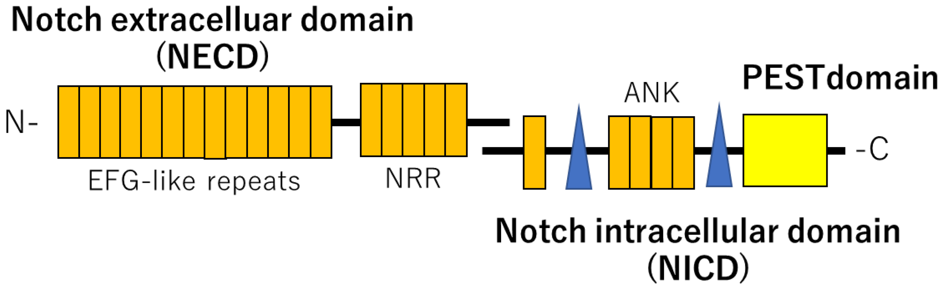
The NOTCH pathway is described as a cell-contact signaling pathway that requires physical contact between adjacent cells in a short-range. NOTCH proteins are heterodimeric, single transmembrane proteins functioning as receptors for DSL (Delta, Serrate, Lag2) family ligands. They are divided into three major parts: the extracellular domain (NECD), the transmembrane domain, and the intracellular domain (NICD). The extracellular domain, which is the receptor part, is transported to the cell surface and exits the cell by exocytosis, allowing it to bind ligands. There are four variants of NOTCH (NOTCH1-4) and their ligands include five known proteins (JAG1, JAG2, DLL1, DLL3, DLL4) on the adjacent cell surface [14,15]. NOTCH protein is activated by binding to one of these ligands. The intracellular domain of NOTCH (NICD) is then detached and transported into the nucleus, where it forms a complex with a DNA-binding protein called CSL to induce the expression of downstream target genes (Figure 1).
The NOTCH receptor consists of the NOTCH extracellular domain (NECD) and NOTCH intracellular domain (NICD). Mutations of NOTCH2 cause the deletion of the PEST domain in the NICD, which results in NOTCH2 overexpression, leading to the Hajdu-Cheney syndrome.
3. Gene Mutation in NOTCH2
Isidor B. et al. reported that a genetic mutation in NOTCH2 causes HCS [9]. Further reports indicated that a frameshift in exon 34, which is located upstream of the PEST (proline–glutamic acid–serine–threonine-rich) domain in the final exon on the C-terminal side of NOTCH2, results in deletion of the sequence the PEST domain. The PEST domain regulates NICD stability by its ubiquitination. The ubiquitination of PEST promotes proteolysis of the NICD domain. Thus, deletion of the PEST domain prolongs the survival of NICD and results in excessive NOTCH signaling. The NOTCH2 signaling pathway plays an important role in osteoclast differentiation and in the promotion of osteoclastogenesis [16]. Deviating from proteasome-dependent proteolysis via ubiquitination by the SCFFBW7 ubiquitin ligase complex, non-degraded stable NOTCH2 mutants accumulate in excess. The over-accumulated NOTCH2 involved in osteoclast differentiation continues to signal excessively, which enhances osteoclast differentiation and causes abnormal bone resorption.
Another mechanism for excessive bone resorption caused by the overactivation of NOTCH signaling is through increased tumor necrosis factor-alpha (TNFα), which may promote osteoclast differentiation [17].
In HCS, the proto-oncogene c-Fos (c-FOS) and nuclear factor of activated T cells 1 (NFATc1) show increased expression, which promotes osteoclast differentiation. HCS is a unique disorder that causes inflammation and bone destruction simultaneously, along with systemic osteolysis and osteoporosis. Therefore, it is necessary to elucidate why osteolysis is accompanied by inflammation and the pathogenesis of HCS.
4. Variants of the NOTCH2 Gene
High-throughput next-generation sequencing has led to remarkable advances in genetic analysis over the past decade. Simultaneously, the analysis of genetic disorders has become more complex, and base sequence differences (variants) between various individuals have been demonstrated to be more abundant than previously expected [18]. In 2015, the American College of Medical Genetics and Genomics (ACMC), in collaboration with the Association of Molecular Pathology (AMP) and the College of American Pathologists (CAP), developed standards and guidelines for sequence interpretation of variants in this genetic analysis [19]. These guidelines established specific criteria to define the variants identified in genes responsible for Mendelian diseases, including “Pathogenic”, “Likely pathogenic”, “Uncertain significance”, “Likely benign”, and “Benign”. Further, by evaluating multiple criteria in relation to pathogenicity, and by synthesizing each evaluation to ultimately determine whether the disease is pathogenic, these evaluation methods facilitate objective and accurate judgment.
We examined the genetic variants of NOTCH2 that have been reported to date and aggregated in the ClinVar database of the U.S. National Institutes of Health. In total, 377 mutant variants were detected; however, our analysis was conducted with the “Condition” parameter set as “Hajdu-Cheney Syndrome” and “Clinical significance” set as “Pathogenic” based on the ACMC guidelines. To our knowledge, this is the first report to classify Hajdu-Cheney Syndrome variants, nucleotide changes, amino acid definitions, and molecular consequences according to the ACMC guidelines. The variants of the 26 analyzed cases are summarized in Table 2. The variant types included 10 single nucleotide variants, 10 deletions, 2 microsatellites, 2 duplications, and 2 insertions/indels. The molecular consequence indicating a functional effect was 12 cases for nonsense and 13 for frameshift mutations, with one unknown. In summary, the NOTCH2 mutation that causes Hajdu-Cheney syndrome is a nonsense mutation that replaces a stop codon through a base swap, leading to translation of the transcribed messenger RNA beyond the original gene sequence after the mutation. These results also indicate that the disease onset is attributed to frameshifts in gene translation caused by deletion, duplication, indel, insertion, and microsatellite formation. Taken together, based on the current results, missense mutations by single nucleotide substitutions, which have been thought to be causative, may not cause Hajdu-Cheney syndrome under the pathogenic condition of the guidelines.
5. Clinical Treatment
The clinical treatment of patients with HCS is challenging, as no treatment strategies have been established and conventional conservative treatments with drugs and surgical treatment are being used at present. Although HCS is a genetic disease and therefore difficult to treat effectively, the goal of current treatment is to improve the quality of life of patients by reducing complications and alleviating their symptoms [8]. Bisphosphonates (BP) and denosumab are often used to treat osteoporosis, which is characterized by progressive bone resorption [20]. Bisphosphonates are anti-bone resorptive and anti-catabolic agents that have a P–C–P structure. They have an affinity for hydroxyapatite and affect bone turnover [21,22]. Denosumab is a monoclonal IgG2 antibody against RANKL, which inhibits osteoclast generation, and binds to RANKL. RANK, RANKL, and osteoprotegerin, which typically regulate bone remodeling; however, when this pathway is uncontrolled, as in patients with HCS, denosumab has not been applicable [20,23,24]. The off-label use of BP and denosumab is because they are both drugs that may induce drug-related osteonecrosis of the jaw (MRONJ) [25,26].
In the genetic disorder HCS, which causes progressive bone resorption with inflammation, impaired bone growth and remodeling, imbalance between the numbers of osteoblasts and osteoclasts, as well as excessive osteoclast activation are believed to form the underlying mechanism. In patients with HCS, anti-resorptive and anti-angiogenic agents have been used to primarily retard osteoporosis development and distal phalanx bone resorption. BPs have been reported to exert a positive effect on the bone in patients with HCS [27,28], but other studies have failed to confirm this effect. Thus, at present, there is no clear evidence that BPs are beneficial; in fact, it may prevent the initiation of bone formation in humans and may not allow activation of bone remodeling [29]. Denosumab has been shown to increase bone mineral density, but has no effect on osteolysis of the endosteum [20]. However, BPs and denosumab are not indicated for surgical treatment or surgical procedures related to the alveolar bone (extraction, periosteoplasty, etc.), nor for periodontal disease, peri-implantitis, and prosthetic procedures, because of the risk of MRONJ.
Abnormal tooth eruption and premature tooth loss are usual features that are particularly prominent in patients with HCS. Structural hard tissue changes in the cementum and dentin, along with hypoplastic dental roots, result in the premature exfoliation of the teeth [30]. Thus, any interference, stimulation or injury can lead to abnormal healing or osteonecrosis. Further, taking anti-resorptive or anti-angiogenic agents may also be a risk factor [4]. Therefore, the treatment of patients with HCS, in which the inflammatory destruction of bone is more advanced than in patients with osteoporosis, requires further management, prophylactic antibacterial agents, and appropriate diagnostic and radiographic studies in addition to the conventional osteoporosis treatment [4].
6. Recent Reports and Genetics of Mouse Models
Osteoporosis is a specific clinical manifestation of HCS, and the NOTCH pathway is assumed to be important in bone tissue homeostasis; however, the role of NOTCH2 in osteoblast differentiation has been rarely reported [31,32,33]. In recent years, NOTCH-related studies on bone have gradually gained increasing attention [9,34,35]. Engin et al. found that loss of NOTCH signaling is correlated with bone health [36]. However, some papers have reported no relationship between osteoclasts and bone resorption caused by NOTCH2 hyperaccumulation. In any case, there is currently no consensus on osteoclast activation via the NOTCH pathway [37,38].
In 2016, Canalis et al. created an HCS mouse model by introducing the 6955C>T mutation into exon 34 upstream of the PEST domain of Notch2 using homologous recombination. This single nucleotide substitution mutation is a nonsense mutation that creates a STOP codon upstream of Notch2 exon 34, resulting in the expression of a truncated Notch2 protein with loss of PEST domain in the model mouse (Notch2Q2319X) [39]. Notch2Q2319X heterozygous mutant mice were smaller and had shorter femurs and reduced trabecular and cortical bone mass in both males and females compared to those in the controls. They showed increased osteoclast numbers and bone resorption in the trabecular bone but no decrease in osteoblast numbers or bone formation. This indicated that Notch2Q2319X cells did not affect osteoblast differentiation or function [34]. Antibodies generated against the negative regulatory region (NRR) of NOTCH suppressed osteopenia and reduced the osteoclast numbers in mouse trabecular bone. Furthermore, subcutaneous administration of Notch2 antisense oligonucleotide (ASO) reduced osteoclast numbers and induction of TNF superfamily member 11 (Tnfsf11) mRNA levels in mice, suggesting that systemic administration of Notch2 ASO can improve the reduction of bone mass in NOTCH2 model mice [40,41]. Moreover, the study also examined whether the skeleton can be sensitized to the osteolytic effects of tumor necrosis factor α (TNFα). TNFα was injected into the cranium, resulting in an increase in osteoclast numbers and osteoclast surface. However, the inactivation of Hes1 reduced the effect of TNFα on osteoclastogenesis. This indicates that TNFα promotes osteoclastogenesis and inflammatory bone resorption, leading to epiphyseal resorption [17]. Another group has used this mouse model to suggest that HCS mutations increase the susceptibility to osteoarthritis and further reported that NOTCH2 activation is involved in HCS pathogenesis [42]. These studies demonstrate that HCS-like NOTCH2 mutant model mice are severely osteopenic with increased bone resorption. They concluded that this is caused by the sensitization of osteoclasts to the receptor activator of nuclear factor-B ligand and the increased expression of tumor necrosis factor superfamily member 11 (Tnfsf11) in osteoblasts. A NOTCH2 conditioned-by-inversion (Notch2COIN) model has also been created in which Cre recombination generates a Notch2ΔPEST allele that expresses a NOTCH2 mutant lacking the PEST domain. Interestingly, osteopenia was observed when these mice were crossed with mice expressing Cre from the BGLAP or Lyz2 promoter to induce HCS mutation only in osteoblasts and not in osteoclasts. The authors thus noted that the HCS mutation in osteoblasts instead of osteoclasts causes osteopenia [37].
Vollersen et al. introduced another HCS-related mutation (6272delT, Notch2/F2091SsX4) in the mouse NOTCH2 gene to create an HCS model mouse in 2018 [43]. Although no osteocyte abnormalities were observed in these mice, excessive skeletal remodeling was identified. In addition, trabecular bone loss was observed at all ages; however, acro-osteolysis, a hallmark of HCS, did not occur in these mice. Other phenotypic manifestations, such as renal cysts and craniofacial abnormalities, which vary in clinical manifestation from patient to patient, may not appear as a phenotype. Therefore, these mouse models have several limitations. However, treatment with alendronate prevented the osteopenia phenotype in these mice. Further, the observed high rates of bone formation were also normalized. The increased expression of Tnfs11 and IL6 in osteoblasts of model mice indicates that NOTCH2 does not regulate osteoblast activity directly but instead exhibits an osteoclast-promoting gene expression pattern, which results in high bone metabolic turnover. Yorgan et al. also observed NOTCH2 expression in osteoblasts and osteoclasts, and then inactivated Notch2 in Runx2-Cre and Lyz2-Cre mice. Notch2fl/fl/Lyz2-Cre mice showed no significant differences in skeletal growth, bone mass, and remodeling whereas Notch2fl/fl/Runx2-Cre mouse showed skeletal abnormalities in long bones. Therefore, NOTCH2 was considered to suppress trabecular bone formation in the skeleton and to regulate bone remodeling [44]. Furthermore, in a mouse model of HCS, Fukushima et al. reported that NOTCH2 overexpression in osteoclasts enhances osteoclast function by inhibiting NOTCH2 degradation [45].
7. Conclusions
Although NOTCH signaling is complex and is essential for human survival, studying the effects of its mutations in human genes is very challenging in some respects. Despite its importance, the relationship between NOTCH signaling and bone formation and development is beginning to be addressed only recently. Although it has recently become possible to explore the effects of NOTCH function in humans, for example by creating knockout mice, and to obtain information on genetic diseases, studies in mice have several practical limitations. The generation of disease-specific induced pluripotent stem cells using samples from patients and further research on genetic diseases can provide information that is more relevant to the human body. This information may play a significant role in establishing the pathogenic mechanism of genetic diseases and their treatment. Elucidating the detailed mechanism of this disease and establishing effective treatment methods can thus be expected to improve the quality of life for patients with HCS in the future.
Author Contributions
Conceptualization, N.A. and T.A.; Data curation, Investigation and Writing-original draft preparing, N.A.; Project administration and Visualization, T.O.; Supervision and Writing-review and editing, T.A. All authors have read and agreed to the published version of the manuscript.
Funding
This work was supported by JSPS KAKENHI (grant numbers 21H03146 and 20K10170). This study was funded by the Private University Research Branding Project from MEXT of Japan (Multidisciplinary Research Center for Jaw Disease (MRCJD): Achieving Longevity and Sustainability by Comprehensive Reconstruction of Oral and Maxillofacial functions), and the Science Research Promotion Fund from the Promotion and Mutual Aid Corporation for Private School of Japan. The funders were not involved in the study design, collection, analysis, interpretation of data, writing of this article, or decision to submit it for publication.
Institutional Review Board Statement
Not applicable.
Informed Consent Statement
Not applicable.
Data Availability Statement
Not applicable.
Acknowledgments
The authors would like to thank T. Nakamura, A. Saito, S. Onodera and Y. Nakamura for useful discussions and suggestions on data collection and analysis.
Conflicts of Interest
The authors declare no conflict of interest.
References
- Hajdu, N.; Kauntze, R. Cranio-skeletal dysplasia. Br. J. Radiol. 1948, 21, 42–48. [Google Scholar] [CrossRef] [PubMed]
- Canalis, E.; Zanotti, S. Hajdu-Cheney syndrome: A review. Orphanet J. Rare Dis. 2014, 9, 200. [Google Scholar] [CrossRef] [PubMed]
- Vingerhoedt, E.; Bailleul-Forestier, I.; Fellus, P.; Schoenaers, J.; Frijns, J.P.; Carels, C. Syndrome of Hajdu-Cheney: Three cases. Cleft Palate-Craniofacial J. 2010, 47, 645–653. [Google Scholar] [CrossRef]
- Antoniades, K.; Kaklamanos, E.; Kavadia, S.; Hatzistilianou, M.; Antoniades, V. Hajdu-Cheney syndrome (acro-osteolysis): A case report of dental interest. Oral Surg. Oral Med. Oral Pathol. Oral Radiol. Endodontol. 2003, 95, 725–731. [Google Scholar] [CrossRef] [PubMed]
- Kaczoruk-Wieremczuk, M.; Adamska, P.; Adamski, L.J.; Wychowanski, P.; Jereczek-Fossa, B.A.; Starzynska, A. Oral surgery procedures in a patient with Hajdu-Cheney syndrome treated with denosumab—A rare case report. Int. J. Environ. Res. Public Health 2021, 18, 9099. [Google Scholar] [CrossRef]
- Brennan, A.M.; Pauli, R.M. Hajdu-Cheney syndrome: Evolution of phenotype and clinical problems. Am. J. Med. Genet. 2001, 100, 292–310. [Google Scholar] [CrossRef]
- Cortés-Martín, J.; Díaz-Rodríguez, L.; Piqueras-Sola, B.; Rodríguez-Blanque, R.; Bermejo-Fernández, A.; Sánchez-García, J.C. Hajdu–Cheney syndrome: A systematic review of the literature. Int. J. Environ. Res. Public Health 2020, 17, 6174. [Google Scholar] [CrossRef]
- Simpson, M.A.; Irving, M.D.; Asilmaz, E.; Gray, M.J.; Dafou, D.; Elmslie, F.V.; Mansour, S.; Holder, S.E.; Brain, C.E.; Burton, B.K.; et al. Mutations in NOTCH2 cause Hajdu-Cheney syndrome, a disorder of severe and progressive bone loss. Nat. Genet. 2011, 43, 303–305. [Google Scholar] [CrossRef]
- Isidor, B.; Lindenbaum, P.; Pichon, O.; Bézieau, S.; Dina, C.; Jacquemont, S.; Martin-Coignard, D.; Thauvin-Robinet, C.; Le Merrer, M.; Mandel, J.L.; et al. Truncating mutations in the last exon of NOTCH2 cause a rare skeletal disorder with osteoporosis. Nat. Genet. 2011, 43, 306–308. [Google Scholar] [CrossRef]
- Gray, M.J.; Kim, C.A.; Bertola, D.R.; Arantes, P.R.; Stewart, H.; Simpson, M.A.; Irving, M.D.; Robertson, S.P. Serpentine fibula polycystic kidney syndrome is part of the phenotypic spectrum of Hajdu-Cheney syndrome. Eur. J. Hum. Genet. 2012, 20, 122–124. [Google Scholar] [CrossRef] [Green Version]
- Han, M.S.; Ko, J.M.; Cho, T.J.; Park, W.Y.; Cheong, H.I. A novel NOTCH2 mutation identified in a Korean family with Hajdu-Cheney syndrome showing phenotypic diversity. Ann. Clin. Lab. Sci. 2015, 45, 110–114. [Google Scholar] [PubMed]
- Isidor, B.; Le Merrer, M.; Exner, G.U.; Pichon, O.; Thierry, G.; Guiochon-Mantel, A.; David, A.; Cormier-Daire, V.; Le Caignec, C. Serpentine fibula-polycystic kidney syndrome caused by truncating mutations in NOTCH2. Hum. Mutat. 2011, 32, 1239–1242. [Google Scholar] [CrossRef] [PubMed]
- Majewski, J.; Schwartzentruber, J.A.; Caqueret, A.; Patry, L.; Marcadier, J.; Fryns, J.P.; Boycott, K.M.; Ste-Marie, L.G.; McKiernan, F.E.; Marik, I.; et al. Mutations in NOTCH2 in families with Hajdu-Cheney syndrome. Hum. Mutat. 2011, 32, 1114–1117. [Google Scholar] [CrossRef] [PubMed]
- Gazave, E.; Lapébie, P.; Richards, G.S.; Brunet, F.; Ereskovsky, A.V.; Degnan, B.M.; Borchiellini, C.; Vervoort, M.; Renard, E. Origin and evolution of the Notch signalling pathway: An overview from eukaryotic genomes. BMC Evol. Biol. 2009, 9, 249. [Google Scholar] [CrossRef] [PubMed]
- Helmi, S.A.; Rohani, L.; Zaher, A.R.; El Hawary, Y.M.; Rancourt, D.E. Enhanced osteogenic differentiation of pluripotent stem cells via γ-secretase inhibition. Int. J. Mol. Sci. 2021, 22, 5215. [Google Scholar] [CrossRef]
- Fukushima, H.; Nakao, A.; Okamoto, F.; Shin, M.; Kajiya, H.; Sakano, S.; Bigas, A.; Jimi, E.; Okabe, K. The association of Notch2 and NF-kappaB accelerates RANKL-induced osteoclastogenesis. Mol. Cell Biol. 2008, 28, 6402–6412. [Google Scholar] [CrossRef]
- Yu, J.; Canalis, E. The Hajdu Cheney mutation sensitizes mice to the osteolytic actions of tumor necrosis factor α. J. Biol. Chem. 2019, 294, 14203–14214. [Google Scholar] [CrossRef]
- Onodera, S.; Nakamura, Y.; Azuma, T. Gorlin Syndrome: Recent advances in genetic testing and molecular and cellular biol ogical research. Int. J. Mol. Sci. 2020, 21, 7559. [Google Scholar] [CrossRef]
- Richards, S.; Aziz, N.; Bale, S.; Bick, D.; Das, S.; Gastier-Foster, J.; Grody, W.W.; Hegde, M.; Lyon, E.; Spector, E.; et al. Standards and guidelines for the interpretation of sequence variants: A joint consensus recommendation of the American College of Medical Genetics and Genomics and the Association for Molecular Pathology. Genet. Med. 2015, 17, 405–423. [Google Scholar] [CrossRef]
- Adami, G.; Rossini, M.; Gatti, D.; Orsolini, G.; Idolazzi, L.; Viapiana, O.; Scarpa, A.; Canalis, E. Hajdu Cheney Syndrome; report of a novel NOTCH2 mutation and treatment with denosumab. Bone 2016, 92, 150–156. [Google Scholar] [CrossRef] [Green Version]
- Nancollas, G.; Tang, R.; Phipps, R.; Henneman, Z.; Gulde, S.; Wu, W.; Mangood, A.; Russell, R.G.G.; Ebetino, F. Novel insights into actions of bisphosphonates on bone: Differences in interactions with hydroxyapatite. Bone 2006, 38, 617–627. [Google Scholar] [CrossRef]
- Abed, H.H.; Al-Sahafi, E.N. The role of dental care providers in the management of patients prescribed bisphosphonates: Brief clinical guidance. Gen. Dent. 2018, 66, 18–24. [Google Scholar]
- Polyzos, S.A.; Makras, P.; Tournis, S.; Anastasilakis, A.D. Off-label uses of denosumab in metabolic bone diseases. Bone 2019, 129, 115048. [Google Scholar] [CrossRef]
- Kumaki, D.; Nakamura, Y.; Suzuki, T.; Kato, H. Efficacy of denosumab for osteoporosis in two patients with adult-onset Still’s disease—Denosumab efficacy in osteoporotic Still’s disease patients. J. Clin. Med. 2018, 7, 63. [Google Scholar] [CrossRef]
- Assili, Z.; Dolivet, G.; Salleron, J.; Griffaton-Tallandier, C.; Egloff-Juras, C.; Phulpin, B. A Comparison of the clinical and radiological extent of denosumab (Xgeva®) related osteonecrosis of the jaw: A retrospective study. J. Clin. Med. 2021, 10, 2390. [Google Scholar] [CrossRef]
- Sivolella, S.; Lumachi, F.; Stellini, E.; Favero, L. Denosumab and anti-angiogenetic drug-related osteonecrosis of the jaw: An uncommon but potentially severe disease. Anticancer Res. 2013, 33, 1793–1797. [Google Scholar]
- Drake, M.T.; Clarke, B.L.; Khosla, S. Bisphosphonates: Mechanism of action and role in clinical practice. Mayo Clin. Proc. 2008, 83, 1032–1045. [Google Scholar] [CrossRef]
- Khosla, S.; Bilezikian, J.P.; Dempster, D.W.; Lewiecki, E.M.; Miller, P.D.; Neer, R.M.; Recker, R.R.; Shane, E.; Shoback, D.; Potts, J.T. Benefits and risks of bisphosphonate Ttherapy for osteoporosis. J. Clin. Endocrinol. Metab. 2012, 97, 2272–2282. [Google Scholar] [CrossRef]
- Jensen, P.R.; Andersen, T.L.; Chavassieux, P.; Roux, J.P.; Delaisse, J.M. Bisphosphonates impair the onset of bone formation at remodeling sites. Bone 2021, 145, 115850. [Google Scholar] [CrossRef]
- Bazopoulou-Kyrkanidou, E.; Vrahopoulos, T.P.; Eliades, G.; Vastardis, H.; Tosios, K.; Vrotsos, I.A. Periodontitis associated with Hajdu-Cheney syndrome. J. Periodontol. 2007, 78, 1831–1838. [Google Scholar] [CrossRef]
- Shen, J.; Bronson, R.T.; Chen, D.F.; Xia, W.; Selkoe, D.J.; Tonegawa, S. Skeletal and CNS defects in Presenilin-1-deficient mice. Cell 1997, 89, 629–639. [Google Scholar] [CrossRef]
- Wong, P.C.; Zheng, H.; Chen, H.; Becher, M.W.; Sirinathsinghji, D.J.; Trumbauer, M.E.; Chen, H.Y.; Price, D.L.; Van der Ploeg, L.H.; Sisodia, S.S. Presenilin 1 is required for Notch1 and DII1 expression in the paraxial mesoderm. Nature 1997, 387, 288–292. [Google Scholar] [CrossRef]
- Dunwoodie, S.L.; Clements, M.; Sparrow, D.B.; Sa, X.; Conlon, R.A.; Beddington, R.S. Axial skeletal defects caused by mutation in the spondylocostal dysplasia/pudgy gene Dll3 are associated with disruption of the segmentation clock within the presomitic mesoderm. Development 2002, 129, 1795–1806. [Google Scholar] [CrossRef]
- Canalis, E. Clinical and experimental aspects of notch receptor signaling: Hajdu-Cheney syndrome and related disorders. Metabolism 2018, 80, 48–56. [Google Scholar] [CrossRef]
- Pittaway, J.F.; Harrison, C.; Rhee, Y.; Holder-Espinasse, M.; Fryer, A.E.; Cundy, T.; Drake, W.M.; Irving, M.D. Bisphosphonate therapy for spinal osteoporosis in Hajdu-Cheney syndrome—New data and literature review. Orphanet J. Rare Dis. 2018, 13, 47. [Google Scholar] [CrossRef]
- Engin, F.; Yao, Z.; Yang, T.; Zhou, G.; Bertin, T.; Jiang, M.M.; Chen, Y.; Wang, L.; Zheng, H.; Sutton, R.E.; et al. Dimorphic effects of Notch signaling in bone homeostasis. Nat. Med. 2008, 14, 299–305. [Google Scholar] [CrossRef]
- Zanotti, S.; Yu, J.; Sanjay, A.; Schilling, L.; Schoenherr, C.; Economides, A.N.; Canalis, E. Sustained Notch2 signaling in osteoblasts, but not in osteoclasts, is linked to osteopenia in a mouse model of Hajdu-Cheney syndrome. J. Biol. Chem. 2017, 292, 12232–12244. [Google Scholar] [CrossRef]
- Drake, W.M.; Hiorns, M.P.; Kendler, D.L. Hajdu-Cheney syndrome: Response to therapy with bisphosphonates in two patients. J. Bone Miner. Res. 2003, 18, 131–133. [Google Scholar] [CrossRef]
- Canalis, E.; Schilling, L.; Yee, S.P.; Lee, S.K.; Zanotti, S. Hajdu Cheney mouse mutants exhibit osteopenia, increased osteoclastogenesis, and bone resorption. J. Biol. Chem. 2016, 291, 1538–1551. [Google Scholar] [CrossRef]
- Canalis, E.; Grossman, T.M.; Carrer, M.; Schilling, L.; Yu, J. Antisense oligonucleotides targeting Notch2 ameliorate the osteopenic phenotype in a mouse model of Hajdu-Cheney syndrome. J. Biol. Chem. 2020, 295, 3952–3964. [Google Scholar] [CrossRef]
- Canalis, E.; Zanotti, S. Hajdu-Cheney syndrome, a disease associated with NOTCH2 mutations. Curr. Osteoporos. Rep. 2016, 14, 126–131. [Google Scholar] [CrossRef] [PubMed]
- Zanotti, S.; Yu, J.; Bridgewater, D.; Wolf, J.M.; Canalis, E. Mice harboring a Hajdu Cheney syndrome mutation are sensitized to osteoarthritis. Bone 2018, 114, 198205. [Google Scholar] [CrossRef] [PubMed]
- Vollersen, N.; Hermans-Borgmeyer, I.; Cornils, K.; Fehse, B.; Rolvien, T.; Triviai, I.; Jeschke, A.; Oheim, R.; Amling, M.; Schinke, T.; et al. High bone turnover in mice carrying a pathogenic NOTCH2 mutation causing Hajdu-Cheney syndrome. J. Bone Miner. Res. 2018, 33, 70–83. [Google Scholar] [CrossRef] [PubMed]
- Yorgan, T.; Vollersen, N.; Riedel, C.; Jeschke, A.; Peters, S.; Busse, B.; Amling, M.; Schinke, T. Osteoblast-specific Notch2 inactivation causes increased trabecular bone mass at specific sites of the appendicular skeleton. Bone 2016, 87, 136–146. [Google Scholar] [CrossRef] [PubMed]
- Fukushima, H.; Shimizu, K.; Watahiki, A.; Hoshikawa, S.; Kosho, T.; Oba, D.; Sakano, S.; Arakaki, M.; Yamada, A.; Nagashima, K.; et al. NOTCH2 Hajdu-Cheney mutations escape SCF FBW7-dependent proteolysis to promote osteoporosis. Mol. Cell. 2017, 68, 645–658.e5. [Google Scholar] [CrossRef] [PubMed]
Figure 1.
An overview of NOTCH signaling, and its involvement in Hajdu-Cheney Syndrome.


Table 1.
Clinical features of patients with Hajdu-Cheney Syndrome.
| Craniofacial Abnormality | Dental Abnormality | Skeletal Abnormality | Cardiac Diseases | Others |
|---|---|---|---|---|
| Micrognathism | Highly arched palates | Acroosteolysis | Cardiovascular abnormalities | Polycystic kidneys |
| Facial dysmorphism | Caries | Fibular deformities, severe osteoporosis | Valvular insufficiency | Neurological disorders |
| Open sutures, wormian bones | Severe periodontal disease | Severe osteoporosis | ||
| Platybasia and basilar invagination | Premature tooth loss | Fractures | ||
| Abnormal redness of gingiva | Joint hyperlaxity | |||
| Abnormal of tooth eruption | Compression fractures and deformities | |||
| Short stature, developmental delay |
Table 2.
Classification of Hajdu-Cheney Syndrome variants according to the standards and Guidelines for sequence interpretation of variants of the American College of Medical Genetics and Genomics (ACMC). This classification shows 26 cases of “Pathogenic” variants including nucleotide changes, amino acid definitions, and molecular consequences.
| Human Genome Variation Society (HGVS) | Variant Type | Nucleotide Change | Protein (Amino Acid Definition) | Molecular Consequence (Functional Effect) | dbSNP |
|---|---|---|---|---|---|
| M_024408.4(NOTCH2):c.1668C>A(p.Cys556Ter) | SNV | c.1668C>A | p.Cys556Ter | nonsense | - |
| NM_024408.4(NOTCH2):c.2235_2236del (p.Cys745_Asp746delinsTer) | Microsatelite | c.2235_2236del | p.Cys745_Asp746delinsTer | nonsense | - |
| NM_024408.4(NOTCH2):c.3415del(p.Leu1139fs) | Deletion | c.3415del | p.Leu1139fs | frameshift | - |
| NM_024408.4(NOTCH2):c.4174C>T(p.Gln1392Ter) | SNV | c.4174C>T | p.Gln1392Ter | nonsense | rs1649449471 |
| NM_024408.4(NOTCH2):c.5123_5132delinsAGA(p.Gln1392Ter) | Indel | c.5123_5132delinsAGA | p.Gln1392Ter | nonsense | rs1649314295 |
| NM_024408.4(NOTCH2):c.5345del(p.Asp1782fs) | Deletion | c.5345del | p.Asp1782fs | frameshift | rs1553193977 |
| NM_024408.4(NOTCH2):c.6272del(p.Phe2091fs) | Deletion | c.6272del | p.Phe2091fs | frameshift | rs1557802353 |
| NM_024408.4(NOTCH2):c.6386del(p.Ser2129fs) | Deletion | c.6386del | p.Ser2129fs | frameshift | - |
| NM_024408.4(NOTCH2):c.6403_6404del(p.Leu2135fs) | Microsatelite | c.6403_6404del | p.Leu2135fs | frameshift | rs1649067817 |
| NM_024408.4(NOTCH2):c.6424_6427del(p.Ser2142fs) | Deletion | c.6424_6427del | p.Ser2142fs | frameshift | rs1064793515 |
| NM_024408.4(NOTCH2):c.6426_6427insTT(p.Leu2135fs) | Insertion | c.6426_6427insTT | p.Leu2135fs | frameshift | rs1649066485 |
| NM_024408.4(NOTCH2):c.6449_6450del(p.Pro2150fs) | Deletion | c.6449_6450del | p.Pro2150fs | frameshift | rs1553193574 |
| NM_024408.4(NOTCH2):c.6503del(p.Pro2168fs) | Deletion | c.6503del | p.Pro2168fs | frameshift | rs1557802165 |
| NM_024408.4(NOTCH2):c.6622C>T(p.Gln2208Ter) | SNV | c.6622C>T | p.Gln2208Ter | nonsense | rs387906746 |
| NM_024408.4(NOTCH2):c.6832dup(p.Thr2278fs) | Duplication | c.6832dup | p.Thr2278fs | frameshift | - |
| NM_024408.4(NOTCH2):c.6853C>T(p.Gln2285Ter) | SNV | c.6853C>T | p.Gln2285Ter | nonsense | rs1553193507 |
| NM_024408.4(NOTCH2):c.6877del(p.His2293fs) | Deletion | c.6877del | p.His2293fs | frameshift | rs1649047546 |
| NM_024408.4(NOTCH2):c.6895G>T(p.Glu2299Ter) | SNV | c.6895G>T | p.Glu2299Ter | nonsense | rs387906748 |
| NM_024408.4(NOTCH2):c.6909del(p.Ile2304fs) | Deletion | c.6909del | p.Ile2304fs | frameshift | rs771237928 |
| NM_024408.4(NOTCH2):c.6909dup(p.Ile2304fs) | Duplication | c.6909dup | p.Ile2304fs | frameshift | rs771237928 |
| NM_024408.4(NOTCH2):c.6949C>T(p.Gln2317Ter) | SNV | c.6949C>T | p.Gln2317Ter | nonsense | rs387906747 |
| NM_024408.4(NOTCH2):c.7078C>T(p.Gln2360Ter) | SNV | c.7078C>T | p.Gln2360Ter | nonsense | rs1553193485 |
| NM_024408.4(NOTCH2):c.7090del(p.Gln2364fs) | Deletion | c.7090del | p.Gln2364fs | frameshift | rs1649037695 |
| NM_024408.4(NOTCH2):c.7119T>G(p.Tyr2373Ter) | SNV | c.7119T>G | p.Tyr2373Ter | nonsense | rs1557801639 |
| NM_024408.4(NOTCH2):c.7165C>T(p.Gln2389Ter) | SNV | c.7165C>T | p.Gln2389Ter | nonsense | rs387906749 |
| NOTCH2, 1-BP DEL, 6460T | Deletion | 6460T | - | - | - |
Publisher’s Note: MDPI stays neutral with regard to jurisdictional claims in published maps and institutional affiliations. |
© 2022 by the authors. Licensee MDPI, Basel, Switzerland. This article is an open access article distributed under the terms and conditions of the Creative Commons Attribution (CC BY) license (https://creativecommons.org/licenses/by/4.0/).
Share and Cite
MDPI and ACS Style
Aida, N.; Ohno, T.; Azuma, T. Progress and Current Status in Hajdu-Cheney Syndrome with Focus on Novel Genetic Research. Int. J. Mol. Sci. 2022, 23, 11374. https://doi.org/10.3390/ijms231911374
AMA Style
Aida N, Ohno T, Azuma T. Progress and Current Status in Hajdu-Cheney Syndrome with Focus on Novel Genetic Research. International Journal of Molecular Sciences. 2022; 23(19):11374. https://doi.org/10.3390/ijms231911374
Chicago/Turabian StyleAida, Natsuko, Tatsukuni Ohno, and Toshifumi Azuma. 2022. "Progress and Current Status in Hajdu-Cheney Syndrome with Focus on Novel Genetic Research" International Journal of Molecular Sciences 23, no. 19: 11374. https://doi.org/10.3390/ijms231911374
Note that from the first issue of 2016, this journal uses article numbers instead of page numbers. See further details here.