Article

Inhibition of GABAAR or Application of Lactobacillus casei Zhang Alleviates Ulcerative Colitis in Mice: GABAAR as a Potential Target for Intestinal Epithelial Renewal and Repair

College of Veterinary Medicine, China Agricultural University, Beijing 100193, China
*
Authors to whom correspondence should be addressed.
Int. J. Mol. Sci. 2022, 23(19), 11210; https://doi.org/10.3390/ijms231911210
Submission received: 24 July 2022 / Revised: 2 September 2022 / Accepted: 13 September 2022 / Published: 23 September 2022
(This article belongs to the Special Issue Molecular Advances in Colorectal Cancer)

Abstract

:
Emerging evidence indicates that the gamma−aminobutyric acid type A receptor (GABAAR) and Lactobacillus casei Zhang regulate colitis in a variety of ways, such as by participating in host immune and inflammatory responses, altering the gut microbiota, and influencing intestinal barrier function. However, not much is known about the mechanisms by which GABAAR and L. casei affect colon epithelial cell renewal and the interaction between GABAAR and L. casei during this process. To elucidate this, we established a dextran sulfate sodium (DSS)−induced model and measured the mouse body weights, colon length, the disease activity index (DAI), and histological scores. Our results indicated that inhibition of GABAAR alleviated the DSS−induced colitis symptoms, resulting in less weight loss and more intact colon tissue. Moreover, treatment with bicuculline (Bic, a GABAAR inhibitor) increased the levels of PCNA, β−catenin, and TCF4 in mice with colitis. Interestingly, open field test performances showed that inhibition of GABAAR also attenuated colitis−related anxiety−like behavior. By 16S RNA gene sequencing analysis, we showed that inhibition of GABAAR partially reversed the gut dysbacteriosis of DSS−induced mice and increased the abundance of beneficial bacteria. Additionally, L. casei Zhang supplementation inhibited the expression of GABAAR in mice with colitis, promoted the proliferation and renewal of colon epithelial cells, and alleviated anxiety−like behavior and intestinal microflora disorder in mice. Thus, GABAAR plays a key role in the beneficial effects of L. casei on DSS−induced colitis in mice.

1. Introduction

Ulcerative colitis (UC) is an inflammatory bowel disease (IBD) that results in abdominal pain, diarrhea, and blood and mucus in the stool, as well as an increased propensity for malignancy [1]. It is characterized by the upregulation of proinflammatory cytokines, apoptotic signals, and an imbalance between microbiota and mucosal immunity [2]. The etiology and pathogenesis of UC are complicated, including genetic factors, microbiota, dysregulation of immune responses, and dysfunction of the mucosal barrier [3]. Even though it is widely accepted that dysfunction of the intestinal barrier plays a key pathophysiological role in UC, the specific causes of UC are not completely identified [4]. As a protective interface in the gastrointestinal (GI) tract, the colonic epithelium is not only a physical barrier separating the microbes from the host but also a key physiological feature in maintaining immune homeostasis [5]. Colonic epithelial injury represents a major physiological challenge that requires the rapid renewal and continuous replenishment of stem cells present in the niche at the base of the crypt [6]. Multiple pathways including the Notch, Hippo/Yes−associated protein, and Wnt/β−catenin pathways are involved in the epithelial regeneration process [7]. The Wnt/β−catenin signaling pathway is essential in stem cell proliferation and differentiation capacity. β−catenin is a key component of the Wnt/β−catenin signaling pathway. Upon activation, β−catenin is separated from the β−catenin destruction complex comprising APC, Axin, and GSK−3β, accumulated, and transferred to the nucleus, where it binds to TCF/LEF to activate target genes and promote the proliferation of intestinal stems cells [8]. In the present study, we mainly focused on the effect of the Wnt/β−catenin pathway on colonic epithelial proliferation and renewal in DSS−induced colitis in mice.
Gamma−aminobutyric acid (GABA) is mainly known as a major inhibitory neurotransmitter in the central nervous system. GABA is produced from glutamate through decarboxylation by glutamic acid decarboxylase (GAD) and is widely found in relatively large amounts, regulating various physiological and psychological processes such as anxiety and depression [9,10]. The GABA type A receptor (GABAAR) is one of the main GABA receptors. Previous studies have shown that GAD, GABA, and GABAAR are expressed in the small intestine and colon and that the GABA−GABAAR system directly alters the excitability of enteric nervous system neurons, spontaneous colonic contractility, and GI motility [11,12,13]. Although it is well known that colonic epithelial cells are enriched with a functional GABAergic signaling system [12], relatively little is known about the function of GABAAR in epithelial tissue renewal.
Patients with UC more often experience mental symptoms such as depression and anxiety compared to the general population [14,15,16]. The underlying mechanism of UC−induced behavioral disorders is not completely understood. Data from recent years indicate that the gut microbiome is closely related to neuroinflammation and further emotional behavior disorders [17,18]. Probiotics can be utilized to modify the intestinal microbiota of the host and may have a therapeutic potential in IBD patients [19,20]. As an important probiotic genus, Lactobacillus have been widely used in clinical trials and showed a wide range of positive effects, including reducing fat mass accumulation and central obesity, maintaining the diversity of intestinal microbiota, and preventing stress−related abdominal symptoms. [21,22,23]. Although a growing body of evidence has shown a close connection between Lactobacillus and UC, very few studies have pinpointed the role of the GABA system in the regulatory effects of Lactobacillus in emotional behavior disorders. The present research aimed to investigate the protective effect of Lactobacillus in DSS−induced colitis in mice by evaluating its impact on colonic epithelial proliferation and associating this with its well−known microbiota regulatory properties, paying special attention to the role of GABAAR.

2. Results

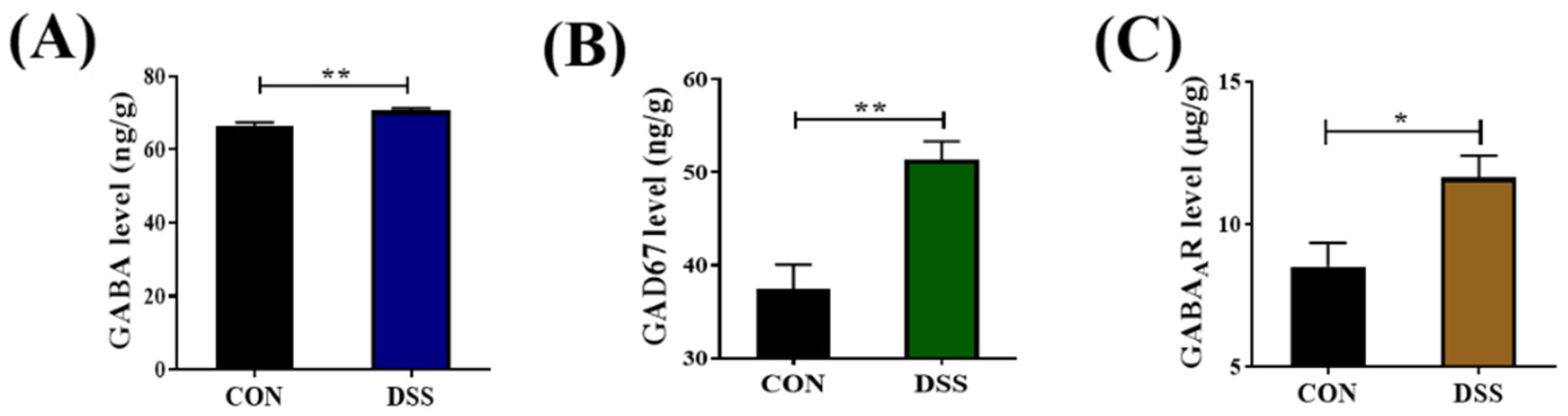
2.1. GABAergic Signals Were Upregulated in the Colon of DSS−nduced Mice

To investigate the role of GABAergic signals in DSS−induced colitis, we first determined the expression levels of GABA, glutamic acid decarboxylase 67 (GAD67), and GABAAR in the colon by ELISA. GAD67 is the major enzyme converting glutamate into GABA. In comparison with the control group, the expression levels of GABA, GAD67, and GABAAR in the DSS group were significantly increased (p < 0.05), as shown in Figure 1.

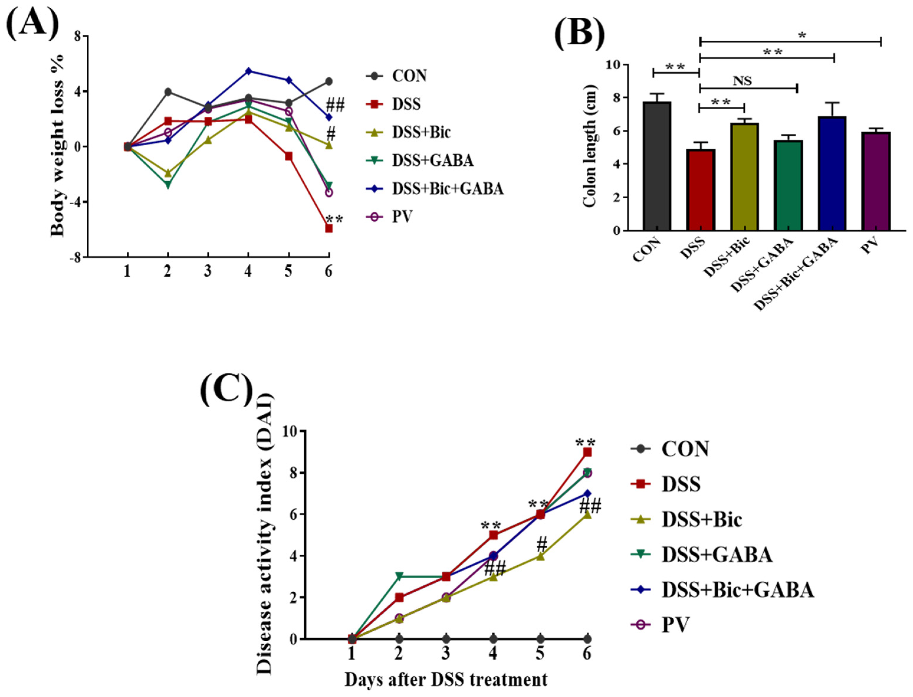
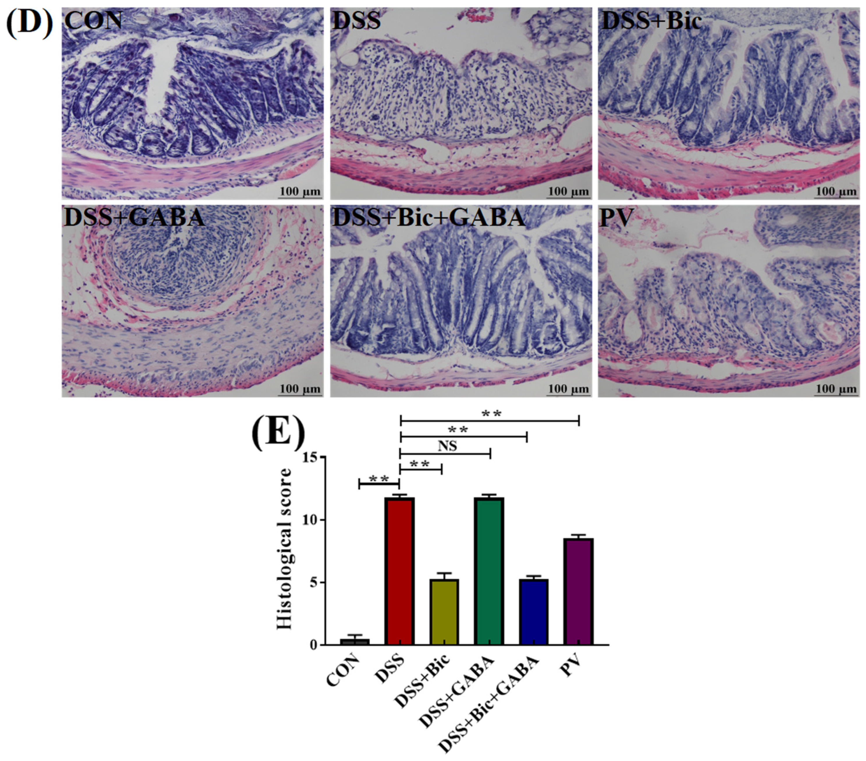
2.2. Clinical Symptoms

Based on the high expression levels of GABAAR, GAD67, and GABA in DSS−induced colitis mice and our previous studies on Lactobacillus casei Zhang [24], we sought to investigate the effects of the GABAergic system and L. casei Zhang on DSS−induced acute colitis in mice. As shown in Figure 2A,B, the DSS−induced model mice showed obvious clinical characteristics, such as weight loss, diarrhea, and blood stool, as indicated by a significantly increased disease activity index (DAI) score during 6 days of DSS treatment (p < 0.05). Mice treated with bicuculline (Bic, a GABAAR inhibitor) and L. casei Zhang showed significant improvements in weight loss and a reduction in DAI scores, indicating remission of colitis in mice. Further, we found that Bic, Bic + GABA, and L. casei Zhang treatment significantly increased colon length in DSS−induced mice (p < 0.05). Histological analysis was used to assess the severity of colonic inflammation in each group. Compared to control group, DSS−induced mice displayed severe pathological changes in their colons, including epithelial erosion, loss of crypts, destroyed muscularis mucosae, and immune cell infiltration. On the contrary, these symptoms were remarkably ameliorated by Bic, Bic + GABA, and L. casei Zhang treatments (p < 0.05, Figure 2D). The histopathology scores in the DSS + Bic, DSS + Bic + GABA, and L. casei Zhang prevention (PV) groups were significantly lower than those of the DSS group (p < 0.05). These results suggested that GABAAR inhibition could alleviate colitis in mice, and the therapeutic effect of L. casei Zhang on acute colitis was similar to that of GABAAR inhibition.

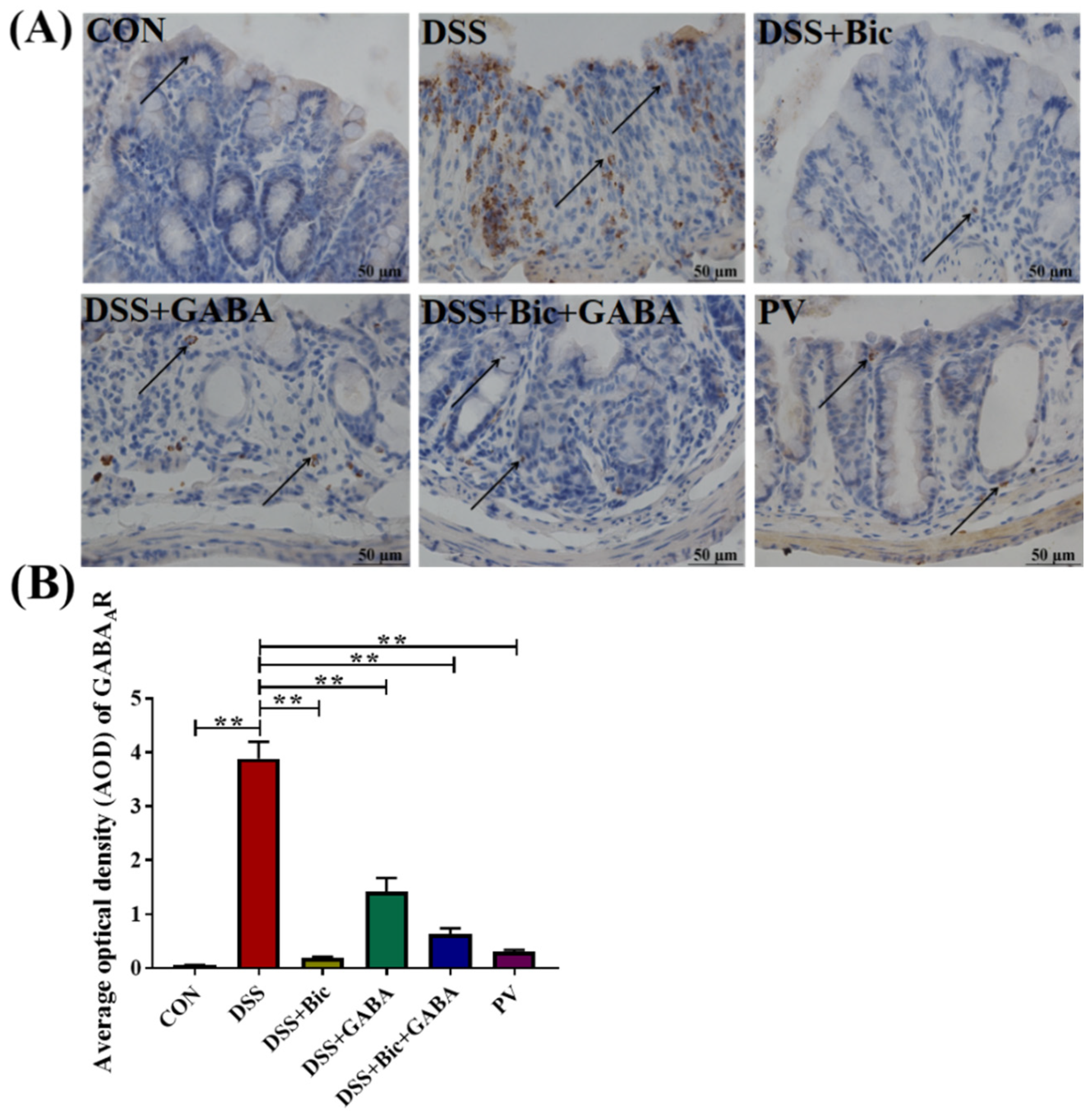
2.3. L. casei Zhang Reduced the Expression of GABAAR in Colitis

In order to study the possible mechanism of the protective effect of L. casei Zhang, we analyzed GABAAR expression in the colonic segments in six groups. The expression of GABAAR was significantly increased in the DSS group compared with the control group (p < 0.05, Figure 3A,B). In addition, GABAAR was widely expressed in glandular epithelium and submucosa. Notably, the expression levels of GABAAR in the Bic, GABA, Bic + GABA, and L. casei Zhang treatment groups were much lower than that in the DSS group, demonstrating that L. casei Zhang could alleviate DSS−induced acute colitis by inhibiting the expression of GABAAR.

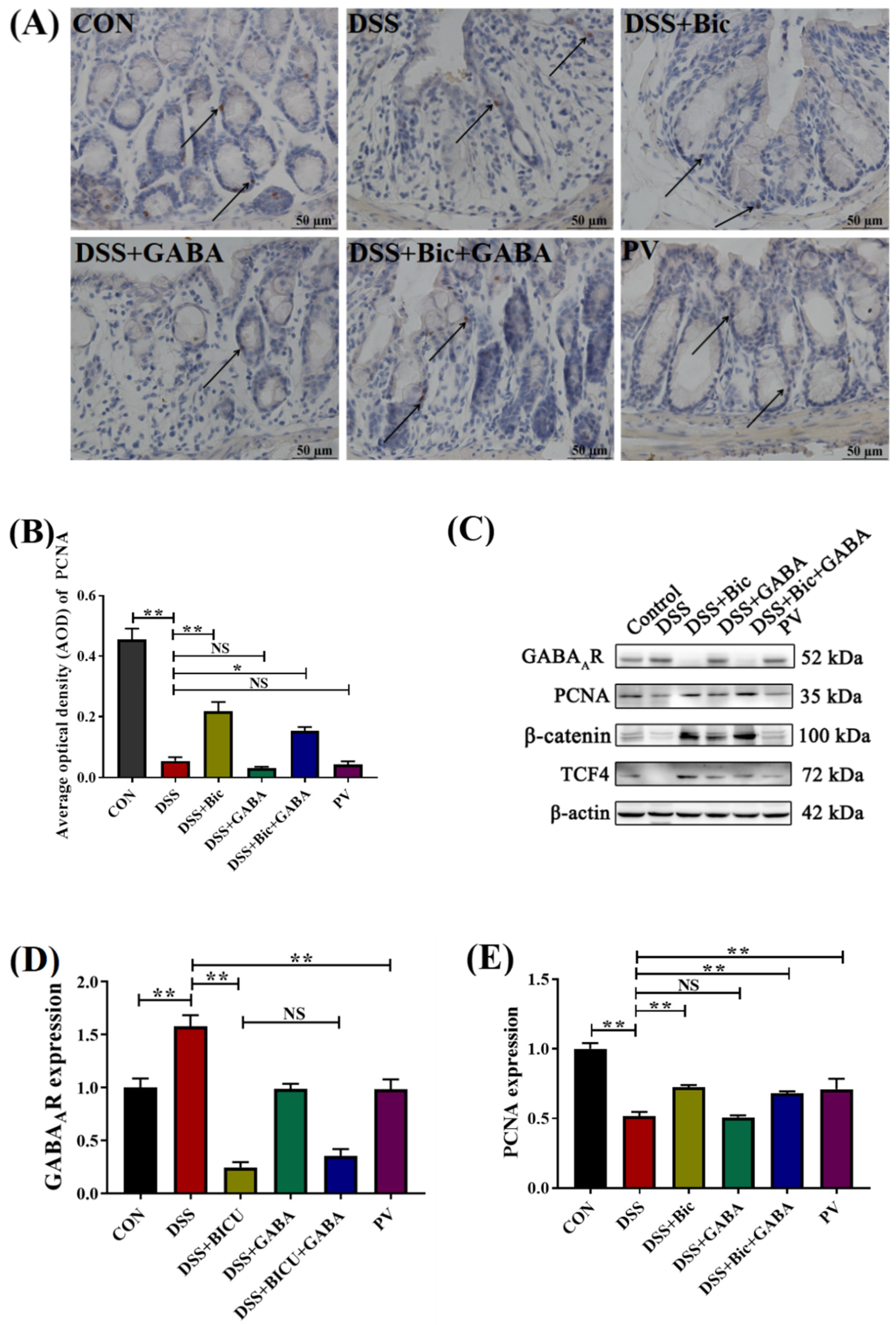
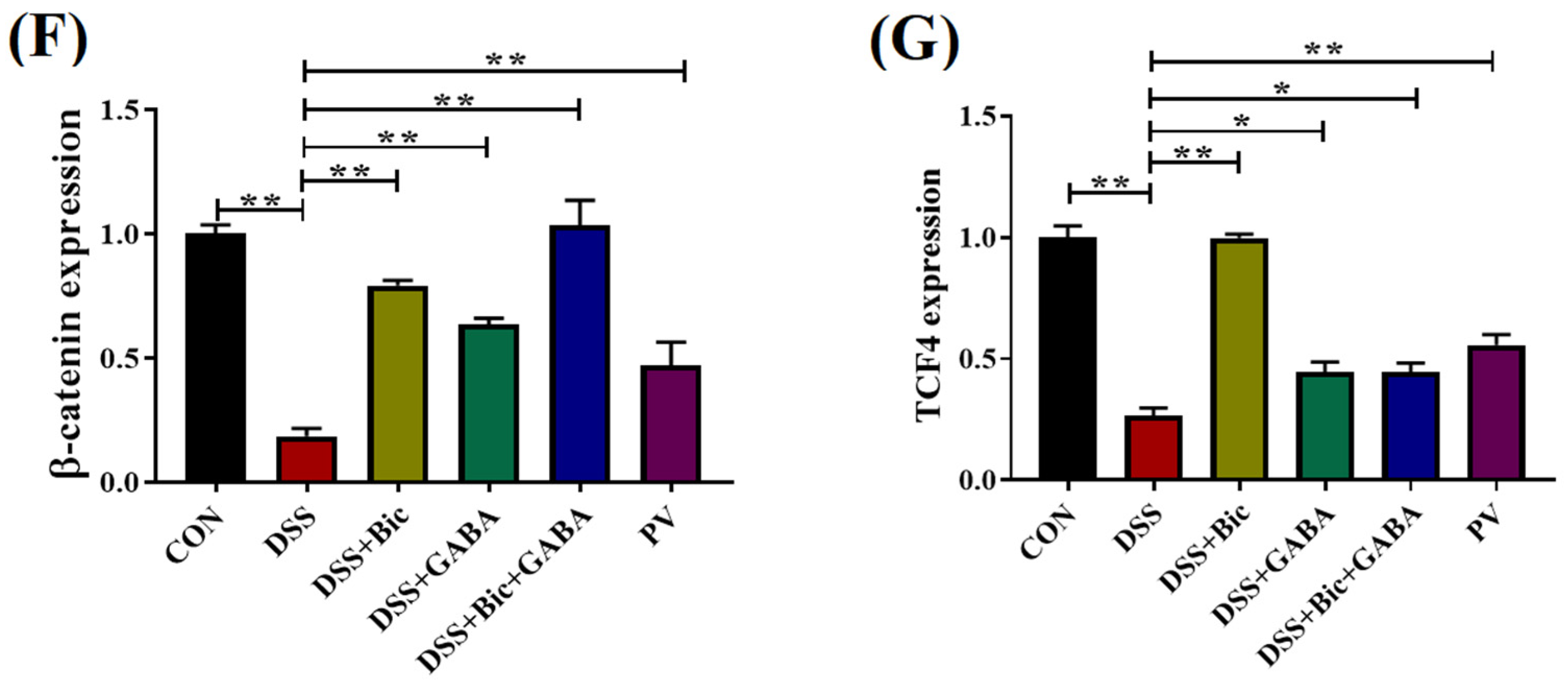
2.4. Inhibition of GABAAR Promotes Colonic Epithelial Renewal in Mice with Colitis

To examine the mechanism whereby inhibition of GABAAR is able to promote the proliferation of intestinal cells, the expression levels of Wnt signaling pathway−associated proteins and proliferating cell nuclear antigen (PCNA) in the colon of mice were assessed. PCNA expression could be detected in the nucleus of the crypts and intestinal gland epithelial cells (Figure 4A). The average optical density (AOD) values of the sections showed a significantly decreased cellular proliferation in the DSS and DSS + GABA groups (p < 0.05). In comparison to the DSS group, the expression of PCNA was significantly increased in the DSS + Bic and DSS + Bic + GABA groups (p < 0.05). The results in Figure 4C–G, suggest that β−catenin and TCF4 were significantly downregulated in the DSS group compared with the control group (p < 0.05). It is noteworthy that the protein levels of PCNA, β−catenin, and TCF4 were significantly increased after Bic, Bic + GABA, and L. casei Zhang treatment (p < 0.05). The protein level of GABAAR significantly decreased after treatment with L. casei Zhang, which further confirmed the results in Figure 3. Taken together, these results demonstrate that inhibition of GABAAR could contribute to maintenance of intestinal integrity in acute colitis by increasing the expression levels of PCNA and Wnt signaling pathway proteins. In addition, L. casei Zhang also promoted the renewal and repair of colonic epithelium in mice with colitis.

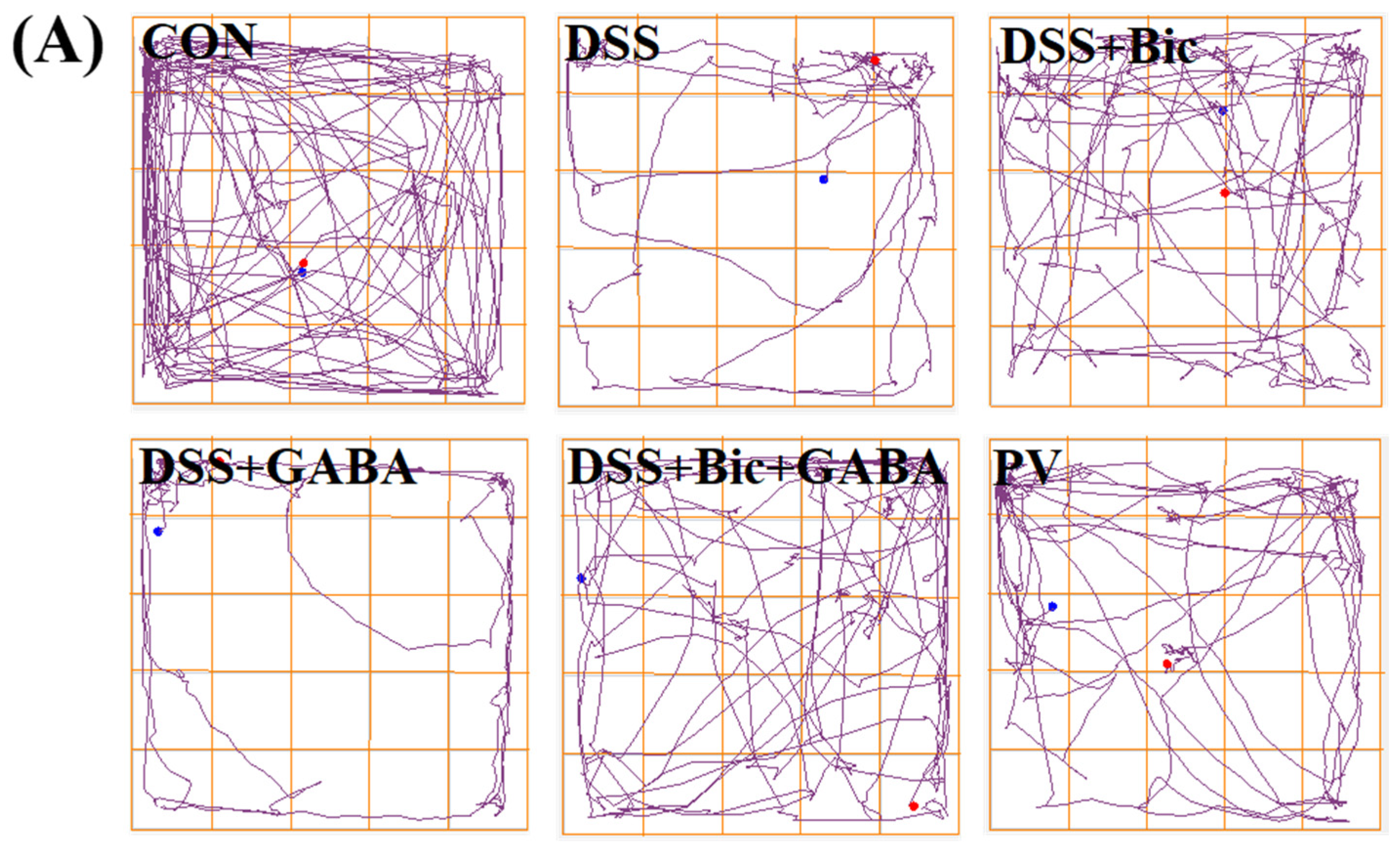
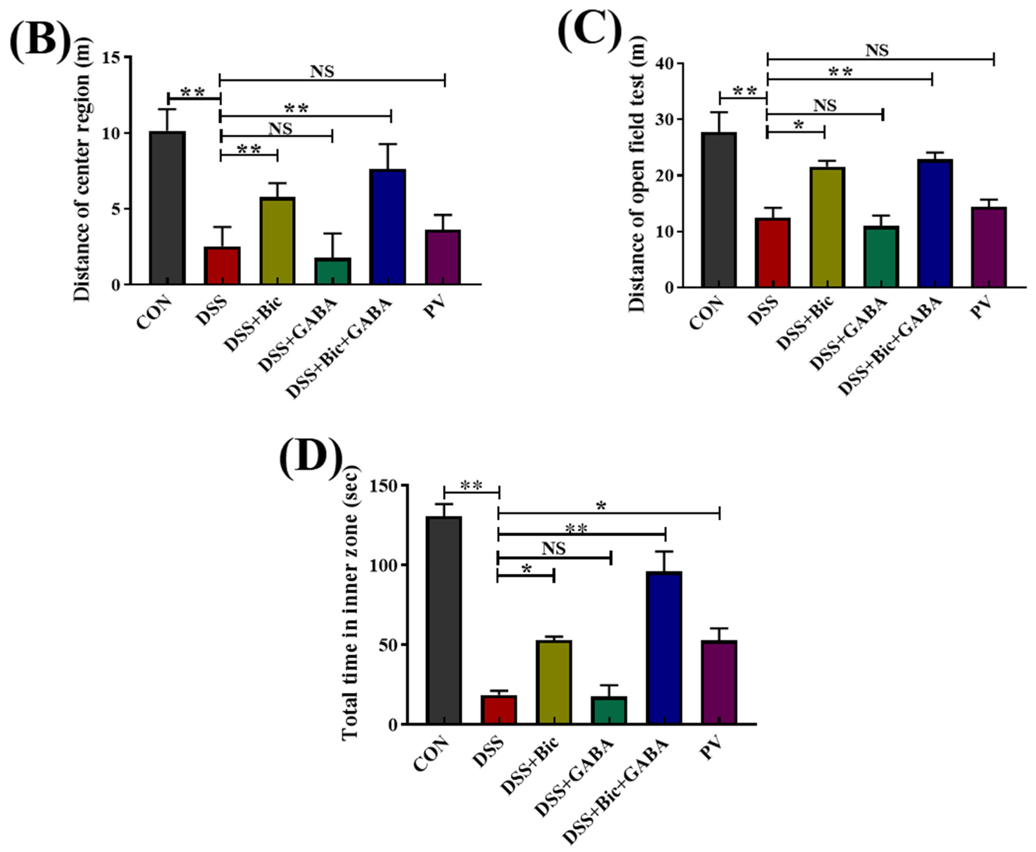
2.5. DSS−Induced Colitis Is Associated with Increased Anxiety−like Behavior

In order to determine whether DSS can alter behavior in mice, the open field test (OFT) was used to assess behavior. Our results showed that DSS−induced mice exhibited an increase in anxiety−like behavior, as demonstrated by the shorter distance in the total and center regions (Figure 5). Meanwhile, the results revealed that control mice spent more time exploring than DSS−induced animals in the center of the open field. However, colitis mice treated with Bic exhibited a significantly increased distance in the total and center regions (p < 0.05), with increased time spent in the center of the open field. In addition, the time spent in the center region of the PV group was significantly increased compared to the DSS group (p < 0.05), and no difference between the DSS group and the DSS + GABA group was found. Taken together, these results suggest that Bic treatment attenuated anxiety−like behavior in mice with colitis.

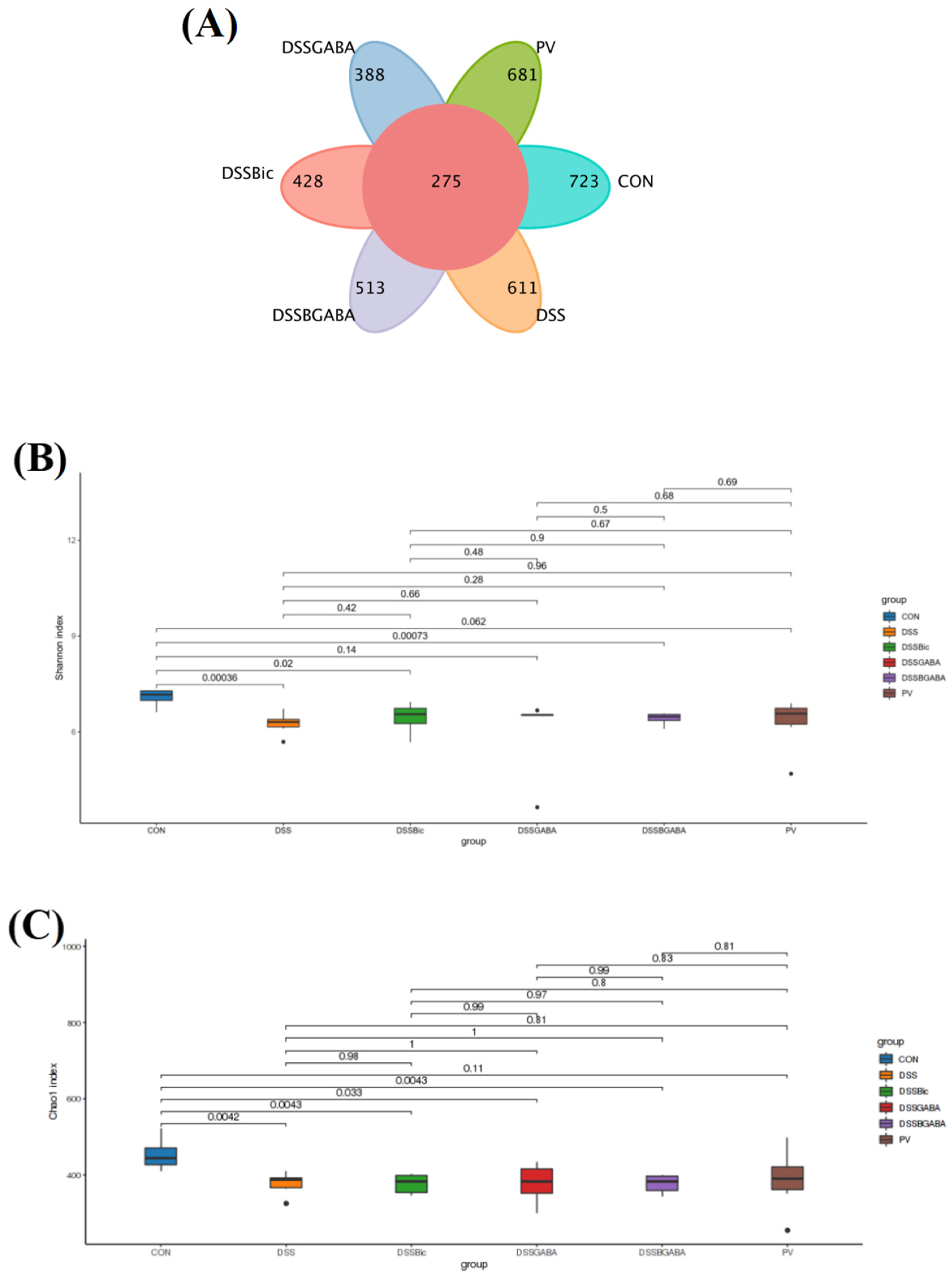
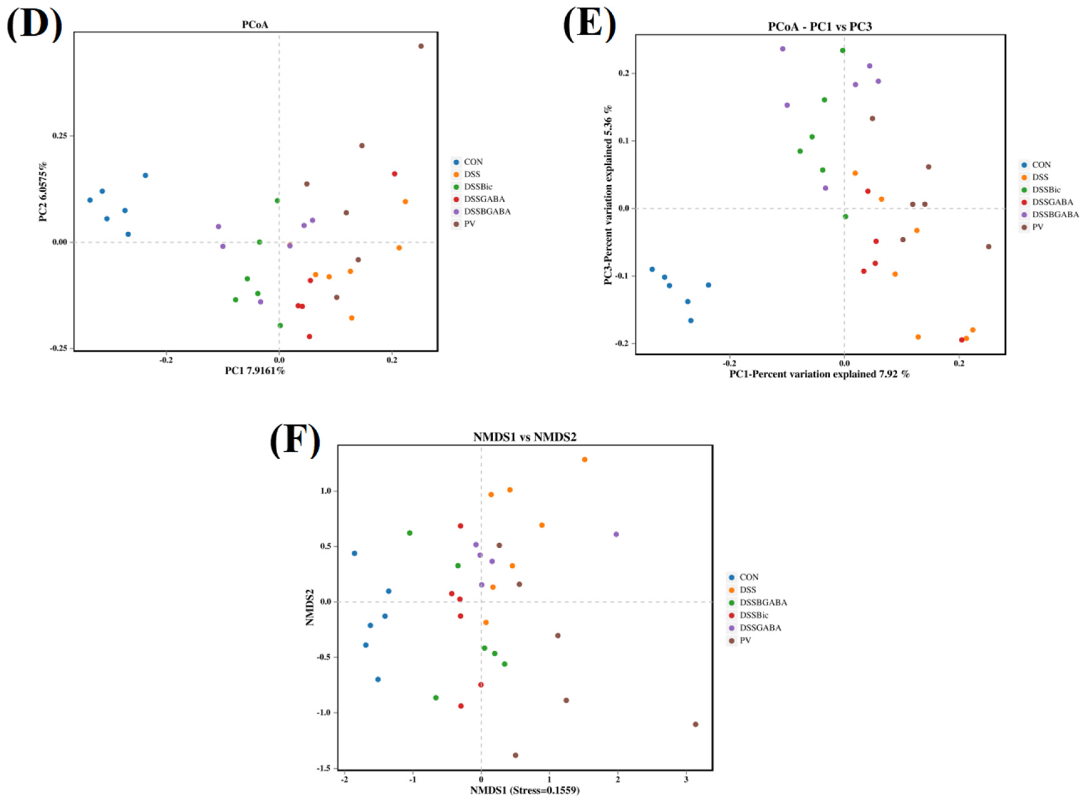
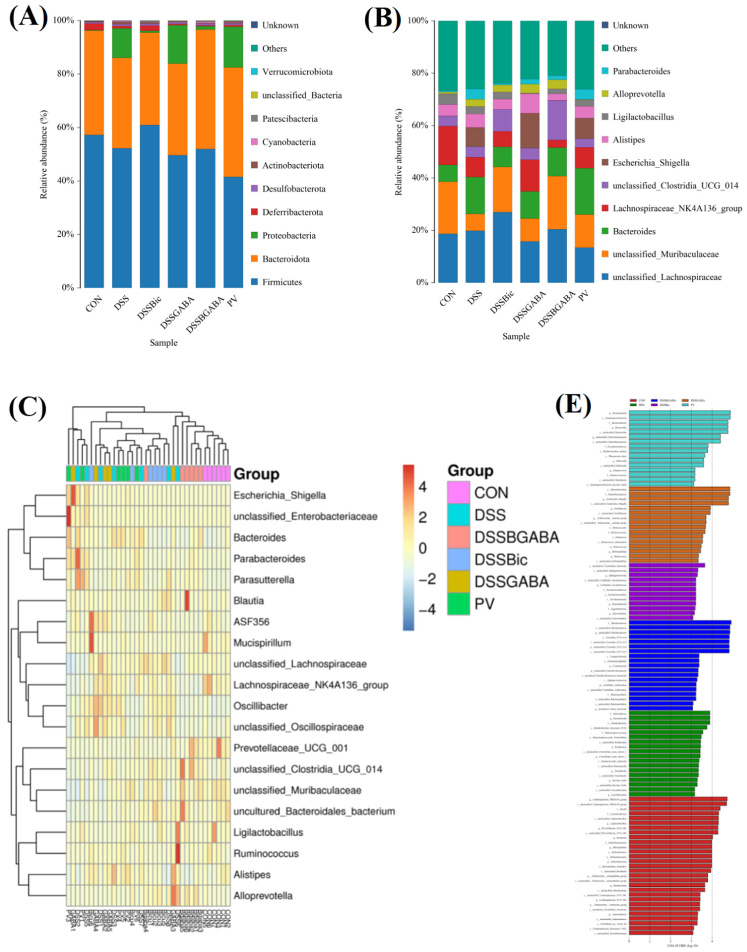
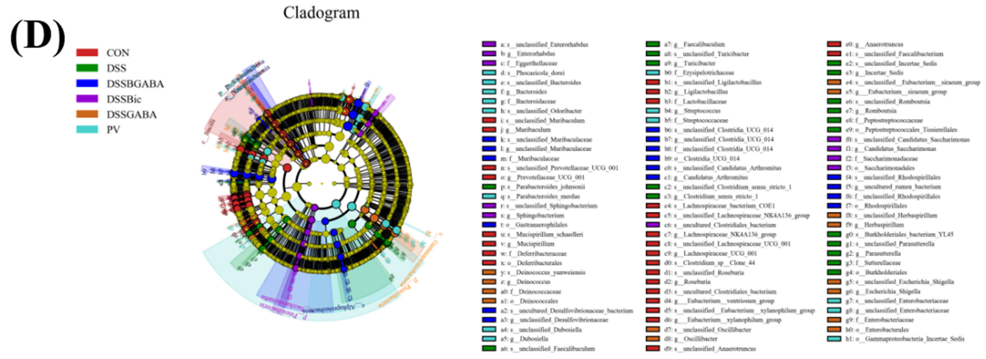
2.6. GABAAR Modulates the Gut Microbiota in Mice with Colitis

Next, we assessed whether GABAAR regulates intestinal microflora. As shown in Figure 6A, the numbers of unique operational taxonomic units (OTUs) in the control, DSS, DSS + Bic, DSS + GABA, DSS + Bic + GABA, and PV groups were 723, 611, 428, 513, 388, and 681, respectively. These six groups shared 275 OTUs among intestinal microbiota. Next, α−diversity was analyzed using the observed species diversity. As shown in Figure 6B,C, the Chao1 and Shannon indices indicated that DSS treatment significantly decreased species richness of bacteria in the gut, and no changes in α−diversity were found between the DSS group and other groups. The overall structure of gut microbiota that was investigated by principal coordinates analysis (PCOA) and non−metric multidimensional scaling (NMDS) analysis showed that the DSS group displayed a shifted clustering of bacterial composition, which was distinct from the control group, suggesting that gut microbiota dysbiosis was induced by DSS treatment (Figure 6D–F). However, Bic treatment remarkably changed the structure of microbiota. Collectively, these results suggest that inhibition of GABAAR in the colon could modulate the structure of gut microbiota, partly alleviating the reduction in species richness of the bacterial population upon DSS treatment.
In order to further study specific differences in the microbiota composition between different groups, we characterized the microbiota community structure profiles of the mice in the six groups. At the phylum level, the bacterial population was primarily composed of Firmicutes, Bacteroidota, Proteobacteria, Deferribacterota, Desulfobacterota, Actinobacteriota, Cyanobacteria, and Patescibacteria (Figure 7A). At the genus level, the DSS group showed an increase in Bacteroides, Escherichia Shigella, and Alistipes and a decrease in Muribaculaceae and Lachnospiraceae_NK4A136_group compared with the control group (Figure 7B). Meanwhile, compared to the DSS group, the increased content of beneficial Muribaculaceae in the intestinal microbiota of DSS−treated mice was further elevated by Bic or L. casei Zhang. A community heatmap was used to determine the microbial populations at the genus level (Figure 7C). The populations of Prevotellaceae_UCG_001, Muribaculaceae, Bacteroidales, and Ligilactobacillus were found to be significantly decreased, and the abundance of Escherichia Shigella, Parabacteroides, and Parasutterella was obviously enhanced after DSS or DSS + GABA treatment. Interestingly, Bic or L. casei Zhang treatment partly reversed the DSS−induced upregulation of the relative abundance of Escherichia Shigella, Parabacteroides, and Parasutterella. Moreover, we performed a linear discrimination analysis (LDA) using the LDA effect size (LEfSe) algorithm to identify operational microbial taxa that were differentially abundant in each group (Figure 7D,E). After L. casei Zhang treatment, relative abundances of Proteobacteria, Bacteroides, and Enterobacteriaceae were significantly increased.

3. Discussion

GABAAR proteins, which are the major mediators of rapid inhibitory synaptic transmission in the mammalian nervous system, mediate the effects of the neurotransmitter GABA. Recent reports suggested that GABAAR could regulate various processes such as learning, memory, and seizure and stress responses [25,26,27]. However, emerging evidence now points to the expression of GABAAR in the enteric nervous system, and GABAAR plays a key role in the spontaneous contractility of the mouse colon and colonic inflammation caused by psychological stress [11,28]. However, the effect of the GABAergic system on colon epithelial proliferation and renewal is not clear. In this study, we used a DSS−induced mouse model to explore the effect of GABAergic signaling on colonic epithelial proliferation and renewal in mice with colitis. Consistent with previously reported results [29], we also observed increased expression of GABAergic−related molecules in DSS−induced mouse colon tissue, while treatment with GABA aggravated colitis in mice, and application of a GABAAR inhibitor attenuated the colitis. However, further research is needed to describe the exact molecular mechanisms by which the GABAergic system affects colitis.
UC can induce a variety of extra−intestinal inflammatory manifestations and complications, which poses a great threat to human health. At present, the main therapeutic strategy for UC is drug therapy, including aminosalicylates, corticosteroids, immunosuppressants, antibiotics, and biologic agents, which have many side effects that limit their therapeutic use [20]. Probiotics are living, non−pathogenic bacteria that are increasingly being used to treat colitis. Probiotics such as Lactobacillus and Bifidobacterium could maintain remission in adults with UC by building a healthy ecosystem and preventing pathogenic infections [30,31]. In this study, we investigated the effects of L. casei Zhang on (i) the expression of GABAergic signal proteins in the colon and (ii) DSS−induced colitis in mice. To detect the potential roles of GABAAR and L. casei Zhang, colon length, body weight loss, and DAI scores were determined, and a histological analysis was performed to compare DSS, DSS + Bic, DSS + GABA, DSS + Bic + GABA, and PV groups. Our findings showed that Bic partially reversed DSS−induced body weight loss and colon length shortening, and the DAI and histological scores of the colon were increased in DSS−induced mice. These improved clinical parameters were likely due to the maintenance of the intestinal barrier by inducing epithelial cell proliferation. It is worth noting that compared with the DSS group, these clinical parameters did not change significantly after GABA addition, which was probably due to the relatively high expression levels of GABAergic signaling proteins in the colon of DSS−induced mice. Therefore, it was difficult to evaluate the effect of exogenous GABA addition on mice with colitis. To further explore the effect of L. casei Zhang on the GABAergic system in the DSS−induced mouse colon, we used immunohistochemistry to detect the expression of GABAAR in the colon in each group of mice. We found that oral administration of L. casei Zhang significantly decreased the expression of GABAAR in colonic tissue of colitis mice, suggesting that the protective effect of L. casei Zhang against colitis in mice was probably achieved by inhibiting GABAAR.
Compared with the study by Ma X [29], our study further revealed that inhibition of GABAAR promoted colonic epithelial cell proliferation, regulated gut microbiota and alleviated anxiety−like behavior in mice with colitis. HE and immunohistochemical staining showed that inhibition of GABAAR contributed to maintenance of the integrity of colon epithelium after DSS treatment. Considering this positive impact of GABAAR inhibition on colonic epithelium, we hypothesized that the GABAergic signals may play a role in the development of colitis by influencing colonic epithelial proliferation. Wnt/β−catenin signaling is a critical driver of stem cell maintenance and cell specialization, which is essential for maintaining the epithelial proliferative compartment and controlling differentiation [32,33,34]. We showed that inhibition of GABAAR significantly increased the expression levels of β−catenin, TCF4, and PCNA, suggesting that inhibition of GABAAR may exert protective effects against colitis through the Wnt signaling pathway. Meanwhile, similar to GABAAR inhibition, oral administration of L. casei Zhang also promoted colon epithelial cell proliferation. The above results demonstrate that L. casei Zhang decreased GABAAR expression in colonic colitis, so we concluded that L. casei Zhang upregulated the Wnt signaling pathway by inhibiting GABAAR, thus maintaining the integrity of the colon epithelium. GABA and L. casei both have long been shown to be associated with anxiety spectrum disorders. L. casei could reduce anxiety symptoms in elderly patients by improving mood disorders [35]. In the control of emotionality, GABAAR plays a key role in regulating vigilance, anxiety, and mood [36,37]. Thus, we detected the behavioral changes of mice in each group by the OFT. The OFT is a widely used test to assess anxiety−like behavior in rodents [38,39]. The results showed that inhibition of GABAAR or L. casei Zhang treatment could alleviate anxiety−like behaviors in DSS−treated mice, suggesting a new treatment method for colitis. Studies have demonstrated that ingestion of Lactobacillus regulates emotional behavior and central GABA receptor expression in mice via the vagus nerve [40]. However, L. casei could also alleviate stress and anxiety in humans and mice through the regulation of gut immune responses and microbiota composition [41,42]. Our results demonstrated that oral L. casei Zhang alleviated anxiety−like behaviors in DSS−treated mice by modulating the gut microbiota. Moreover, inhibition of GABAAR also alleviated anxiety−like behaviors in colitis, and our above results have shown that L. casei Zhang decreased the expression of GABAAR in colitis mice. Therefore, the alleviation of anxiety−like behavior in colitis mice by L. casei Zhang is probably the result of both inhibition of GABAAR and regulation of intestinal microbiota.
There is increasing evidence that probiotics exert positive effects, not only by reinforcing the intestinal barrier of the host, but also by preventing colonization by pathogenic bacteria and modulating the activity of the intestinal immune system at both the cellular and the molecular level [43,44,45]. Lactobacillus species have been reported to exert beneficial effects in the context of intestinal inflammation in experimental mouse models [46]. In addition, recent studies have shown that members of the gut microbiome, such as lactic acid bacteria, are able to produce GABA to regulate mental disorders such as depression and anxiety [47,48]. Our results showed that the diversity of the colonic microbial community decreased and dysbiosis of gut microbiota was induced after DSS treatment. PCOA and NMDS analysis indicated that Bic or L. casei Zhang treatment could improve the structure of microbiota after DSS treatment. We found a lower relative abundance of Pharabacteroides, Alistipes, and Muribaculaceae and a higher relative abundance of Escherichia Shigella in the DSS and DSS + GABA groups than in other groups. It has been found that treating mice with Parabacteroides and Alistipes could induce depressive−like behavior [49,50]. Alistipes is a genus of indole−positive bacteria that bind to serotonin receptors, reducing serotonin availability and causing a decrease in pleasant feelings. Meanwhile, Alistipes has been reported to express GAD, which metabolizes glutamate into GABA. This may be an important reason for the depressive−like behavior induced by DSS in mice [51]. Furthermore, we discovered that the relative abundance of the genus Muribaculaceae was significantly increased in the L. casei Zhang and Bic treatment groups. Heatmap and LEfSe analysis showed that L. casei Zhang or Bic treatment increased the numbers of Bacteroides and Ligilactobacillus, which are beneficial bacteria that maintain intestinal health. A growing number of studies have shown that Lactobacillus mitigation of DSS−induced colitis is associated with reduced proinflammatory cytokine levels, increased goblet cells, and secretion of antimicrobial peptides, regulating the ratio of Firmicutes/Bacteroidetes and increasing acetate levels [45,52]. Combined with our results, the protective effects of L. casei Zhang on colitis were closely related to the inhibition of GABAAR, providing new ideas for the clinical treatment of UC with L. casei Zhang.

4. Materials and Methods

4.1. Bacterial Strain

L. casei Zhang is a probiotic strain with good tolerance to acid and bile, which was isolated from the natural fermentation of koumiss (mare’s milk) in the Zhenglanqi herder family in Xilin Gol League, Inner Mongolia [53]. In this experiment, L. casei Zhang was fermented and freeze−dried to make a powder, which was dissolved to a concentration of 5 × 10⁸ CFU/mL and gavaged daily (300 μL per day).

4.2. Animals

Six− to eight−week−old male C57/BL mice weighing 18−22 g were purchased from Beijing Vital River Laboratory Animal Technology Co., Ltd., Beijing, China. They were acclimatized for 1 week before the experiments and maintained in a room with a controlled temperature (22 ± 1 °C) and a 12/12-h light/dark cycle, with free access to food and water. Acute colitis was induced by administering 4% (w/v) DSS (molecular weight, 36,000−50,000, Yeasen Biotechnology Co., Ltd. (Shanghai, China)) in drinking water for six days. A total of 60 mice were randomly divided into six groups (n = 10 mice per group). Mice in the control group received water only during the six−day experiment. Mice in the DSS group received 4% DSS in drinking water. Mice in the DSS + Bic group received a daily i.p. injection of Bic (HY−N0219, purchased from MedChemExpress, MedChemExpress, NJ, USA) at 0.1 mg/kg once per day for seven days with 4% DSS in drinking water. Mice in the DSS + GABA group received a daily i.p. injection of GABA (03835, purchased from Sigma−Aldrich, St. Louis, MO, USA) at 5 mg/kg once per day for seven days with 4% DSS in drinking water. Mice in the DSS + Bic + GABA group received a daily i.p. injection of Bic at 0.1 mg/kg and GABA at 5 mg/kg once per day for seven days with 4% DSS in drinking water. Mice in the PV group received L. casei Zhang by oral gavage for seven days before DSS administration. Mice were monitored daily for weight loss, stool consistency, and fecal bleeding. The changes of DAI were measured using the three criteria mentioned above (Table 1) [54].

4.3. Behavioral Test

The OFT is widely used to assess the level of anxiety in mice by providing activity tracks, activity time and exploration habits. The mice were randomly selected and placed in a white OFT chamber (45 cm × 45 cm × 45 cm). The bottom surface of the apparatus was divided into 25 squares (9 cm × 9 cm), which were divided into a surrounding area (16 squares near the walls of the chamber) and a central area (9 squares in the middle). The activities of the mice were recorded by a digital camera set above the apparatus. For each test, each mouse was placed in a corner of the apparatus to acclimatize for 1 min, after which all its activities were recorded for 5 min. The apparatus was cleaned with 75% ethanol before the testing of each animal. The methodology used for the OFT was described previously [55]. The total distance of the mice moving in the open box and the time of staying in the central area within the 5 min was recorded; the sum of these two parameters was used to evaluate the general activity of the mice.

4.4. Sample Collection

On day six, the mice were anesthetized with 1% pentobarbital sodium (5 mg/100 g body weight) by i.p. injection and euthanized by neck dislocation. The colon samples and intestinal contents were collected, and the colon lengths were measured. Next, approximately 2 cm long distal colonic intestinal segments were harvested, after phosphate−buffered saline (PBS) cleaning, segments were placed in 4% paraformaldehyde for 24 h for hematoxylin and eosin (H&E) staining, and immunohistochemistry. Furthermore, the remaining colons were rinsed with PBS and frozen in liquid nitrogen and stored at −80 °C until protein expression analysis.

4.5. Histological Analysis

For histological examination, colons fixed in 4% paraformaldehyde were removed and embedded in paraffin, sectioned (10 μm thick) and stained with H&E. Histological scores were based on four criteria: number of ulcers, depletion of goblet cells, crypt damage, and inflammatory cell infiltration (Table 2) [56,57].

4.6. Immunohistochemistry

Paraffin embedded colon tissue was dewaxed with dimethyl−benzene and rehydrated in gradient ethanol. After antigen heat recovery in citrate buffer, the sections were treated with 1% H2O2 for 20 min to remove endogenous peroxidase activity and blocked with 5% donkey serum for 30 min. The sections were incubated overnight at 4 °C with polyclonal rabbit anti−GABAARα3 antibody (12708-1-AP, 1:200, Proteintech Group, Chicago, IL, USA) and monoclonal mouse anti−PCNA antibody (1:300, BM-0104, Boster Biological Technology Co., Ltd., Wuhan, China). Then, these sections were washed three times with PBS for 5 min each time. Next, sections were incubated with a 1:100 dilution of the biotinylated donkey anti−mouse/anti−rabbit IgG (H&L) (715-065-151/711-065-152, 1:100, Jackson Immunoresearch, PA, USA) secondary antibody for 1 h at room temperature. After washing three times with PBS, ABC−peroxidase solution (PK−6102; Vector Laboratories, Burlingame, CA, USA) was added, and allowed to stand for 30 min at room temperature. The sections were incubated in 0.01 M Tris−HCl containing 0.02% 3−3′−diaminobenzidine (D5637; Sigma−Aldrich, Saint Louis, MO, USA) and 0.003% H2O2 for 5−10 min in the dark to observe immunoreactivity. Finally, the sections were stained with hematoxylin to counterstain the nuclei for 5 s. Three slices were randomly chosen from each group to further select five different regions of each slice. Image J 6.0 software (National Institutes of Health, USA) was used to calculate the average OD of GABAARα3 and PCNA immunopositive substances in colon.

4.7. Western Blotting Analysis

The colon tissue was lysed with RIPA lysis buffer (CW2334S; CWBIO, Beijing, China) containing a protease inhibitor cocktail (CW2200S; CWBIO, Beijing, China), and a bicinchoninic acid (BCA) protein assay kit (CW0014; CWBIO, Beijing, China) was used to quantify the total protein. Subsequently, equal amounts of protein (30 μg per lane) were separated utilizing 10% SDS−PAGE gel (Solarbio, Beijing, China), and then transferred onto PVDF membranes (IPVH00010; Millipore, Danvers, MA, USA). After 1 h of protein blocking with 5% skimmed milk, the membranes were incubated with specific primary antibodies, including rabbit anti−GABAARα3 (12708-1-AP, 1:1000, Proteintech), mouse anti−PCNA antibody (BM−0104, 1:1000, Boster Biological Technology Co., Ltd.), polyclonal rabbit anti−β−catenin (51067-2-AP, 1:1000, Proteintech), polyclonal rabbit anti−TCF4 (22337-1-AP, 1:1000, Proteintech), and monoclonal mouse anti−β−actin (50201, 1:1000, Kemei Borui Science and Technology, Beijing, China). Later, the membranes were incubated with HRP−conjugated goat anti−rabbit IgG (H&L) (A0208, 1:1000, Beyotime Biotechnology, Shanghai, China) or HRP−conjugated goat anti−mouse IgG (H&L) (A0216, 1:1000, Beyotime Biotechnology) for 1 h at 37 °C. The protein bands were detected by ECL developer and chemiluminescence imager. The protein band intensities were quantified by Image J 6.0 software (National Institutes of Health, USA).

4.8. Microbial Sequencing

Fecal genomic DNA was extracted by using a QIAamp DNA stool mini kit (Qiagen). PCR amplification was performed using total DNA and primers targeting the V3−V4 region of the 16S rRNA gene. The amplifications were performed using the following conditions: 95 °C for 5 min, followed by 25 cycles of 95 °C for 30 s, 50 °C for 30 s, and 72 °C for 40 s and a final extension step at 72 °C for 7 min. Next, we constructed the genomic library and conducted high−throughput pyrosequencing of the PCR products using an Illumina MiSeq platform. Finally, the obtained reads of data were processed by cutting and removing the chimeric sequences. According to UCLUST, OTUs were clustered at 97% sequence similarity and classified using the NCBI and RDP reference databases. With QIIME 2.0 software (Knight and Caporaso labs, USA), α−diversity analysis including the Shannon and Chao1 indices and β−diversity analysis including PCOA and NMDS analysis were conducted. The biomarkers of different groups were identified by LEfSe analysis.

4.9. Statistical Analysis

GraphPad Prism7 software (GraphPad software, USA) was used for statistical analysis. All data are shown as mean ± SEM. Differences between individual groups were analyzed via one−way ANOVA followed by Tukey’s multiple comparison test. A value of p < 0.05 was considered statistically significant.

5. Conclusions

In conclusion, oral administration of L. casei Zhang could alleviate DSS−induced colitis in mice by inhibiting GABAAR. GABAergic signaling plays a vital role in colonic epithelial proliferation and is probably involved in modulating the Wnt/β−catenin signaling pathway and gut dysbacteriosis in the colon. GABAergic signaling may serve as a new drug target to treat IBD.

Author Contributions

The study was conceived and designed by Y.M. and Q.W.; all data were collected by Q.W.; statistical analyses were conducted by Q.W., Z.D., J.L. and D.L.; the manuscript was drafted by Q.W. and Y.M.; administrative, technical, and material supports were provided by Y.M., J.C. and K.F. All authors have read and agreed to the published version of the manuscript.

Funding

This work was funded by the National Natural Science Foundation of China (No. 31772686).

Institutional Review Board Statement

The present research complied with the Guide for the Care and Use of Laboratory Animals published by the Animal Welfare Committee of the Agricultural Research Organization, China Agricultural University (Beijing, China), and was approved by the University Ethical Committee. An effort was made to minimize the suffering and the number of mice used.

Informed Consent Statement

No applicable.

Data Availability Statement

All figures and data used to support to this study are induced within this article.

Acknowledgments

Thanks to the L. casei Zhang provided by State Key Laboratory of DirectFed Microbial Engineering, Beijing DaBeiNong Science and Technology Group Co., Ltd.

Conflicts of Interest

The authors have no financial conflict of interest to declare.

References

  1. Kobayashi, K.; Tomita, H.; Shimizu, M.; Tanaka, T.; Suzui, N.; Miyazaki, T.; Hara, A. p53 Expression as a Diagnostic Biomarker in Ulcerative Colitis-Associated Cancer. Int. J. Mol. Sci. 2017, 18, 1284. [Google Scholar] [CrossRef]
  2. Saber, S.; Khalil, R.M.; Abdo, W.S.; Nassif, D.; El-Ahwany, E. Olmesartan ameliorates chemically-induced ulcerative colitis in rats via modulating NFκB and Nrf-2/HO-1 signaling crosstalk. Toxicol. Appl. Pharmacol. 2019, 364, 120–132. [Google Scholar] [CrossRef]
  3. Nunes, S.; Danesi, F.; Del Rio, D.; Silva, P. Resveratrol and inflammatory bowel disease: The evidence so far. Nutr. Res. Rev. 2018, 31, 85–97. [Google Scholar] [CrossRef] [PubMed]
  4. Liang, L.; Liu, L.; Zhou, W.; Yang, C.; Mai, G.; Li, H.; Chen, Y. Gut microbiota-derived butyrate regulates gut mucus barrier repair by activating the macro-phage/WNT/ERK signaling pathway. Clin. Sci. 2022, 136, 291–307. [Google Scholar] [CrossRef]
  5. Jamwal, D.R.; Laubitz, D.; Harrison, C.A.; da Paz, V.F.; Cox, C.M.; Wong, R.; Midura-Kiela, M.; Gurney, M.A.; Besselsen, D.G.; Setty, P.; et al. Intestinal Epithelial Expression of MHCII Determines Severity of Chemical, T-Cell–Induced, and Infectious Colitis in Mice. Gastroenterology 2020, 159, 1342–1356.e6. [Google Scholar] [CrossRef]
  6. Kinchen, J.; Chen, H.H.; Parikh, K.; Antanaviciute, A.; Jagielowicz, M.; Fawkner-Corbett, D.; Ashley, N.; Cubitt, L.; Mellado-Gomez, E.; Attar, M.; et al. Structural Remodeling of the Human Colonic Mesenchyme in Inflammatory Bowel Disease. Cell 2018, 175, 372–386.e17. [Google Scholar] [CrossRef]
  7. Deng, F.; Peng, L.; Li, Z.; Tan, G.; Liang, E.; Chen, S.; Zhao, X.; Zhi, F. YAP triggers the Wnt/β-catenin signalling pathway and promotes enterocyte self-renewal, regen-eration and tumorigenesis after DSS-induced injury. Cell Death Dis. 2018, 9, 153. [Google Scholar] [CrossRef]
  8. Zhang, M.; Yang, D.; Yu, H.; Li, Q. MicroRNA-497 inhibits inflammation in DSS-induced IBD model mice and lipopolysaccha-ride-induced RAW264.7 cells via Wnt/β-catenin pathway. Int. Immunopharmacol. 2021, 101, 108318. [Google Scholar] [CrossRef]
  9. Cui, Y.; Miao, K.; Niyaphorn, S.; Qu, X. Production of Gamma-Aminobutyric Acid from Lactic Acid Bacteria: A Systematic Review. Int. J. Mol. Sci. 2020, 21, 995. [Google Scholar] [CrossRef]
  10. Barrett, E.; Ross, R.P.; O’Toole, P.W.; Fitzgerald, G.F.; Stanton, C. γ-aminobutyric acid production by culturable bacteria from the human intestine. J. Appl. Microbiol. 2012, 113, 411–417. [Google Scholar] [CrossRef]
  11. Seifi, M.; Brown, J.F.; Mills, J.J.; Bhandari, P.; Belelli, D.; Lambert, J.J.; Rudolph, U.; Swinny, J.D. Molecular and functional diversity of GABA-A receptors in the enteric nervous system of the mouse colon. J. Neurosci. 2014, 34, 10361–10378. [Google Scholar] [CrossRef]
  12. Aggarwal, S.; Ahuja, V.; Paul, J. Attenuated GABAergic Signaling in Intestinal Epithelium Contributes to Pathogenesis of Ulcerative Colitis. Am. J. Dig. Dis. 2017, 62, 2768–2779. [Google Scholar] [CrossRef]
  13. Seifi, M.; Swinny, J.D. Developmental and age-dependent plasticity of GABAA receptors in the mouse colon: Implications in colonic motility and inflammation. Auton. Neurosci. 2019, 221, 102579. [Google Scholar] [CrossRef] [PubMed]
  14. Yuan, X.; Chen, B.; Duan, Z.; Xia, Z.; Ding, Y.; Chen, T.; Liu, H.; Wang, B.; Yang, B.; Wang, X.; et al. Depression and anxiety in patients with active ulcerative colitis: Crosstalk of gut microbiota, metabolomics and proteomics. Gut Microbes 2021, 13, 1987779. [Google Scholar] [CrossRef]
  15. Trindade, I.A.; Ferreira, C.; Pinto-Gouveia, J. Ulcerative colitis symptomatology and depression: The exacerbator role of maladap-tive psychological processes. Dig. Dis. Sci. 2015, 60, 3756–3763. [Google Scholar] [CrossRef]
  16. Kurina, L.M.; Goldacre, M.J.; Yeates, D.; E Gill, L. Depression and anxiety in people with inflammatory bowel disease. J. Epidemiol. Community Health 2001, 55, 716–720. [Google Scholar] [CrossRef]
  17. Zhang, X.; Zou, Q.; Zhao, B.; Zhang, J.; Zhao, W.; Li, Y.; Liu, R.; Liu, X.; Liu, Z. Effects of alternate-day fasting, time-restricted fasting and intermittent energy restriction DSS-induced on colitis and behavioral disorders. Redox Biol. 2020, 32, 101535. [Google Scholar] [CrossRef]
  18. Zhao, B.; Wu, J.; Li, J.; Bai, Y.; Luo, Y.; Ji, B.; Xia, B.; Liu, Z.; Tan, X.; Lv, J.; et al. Lycopene Alleviates DSS-Induced Colitis and Behavioral Disorders via Mediating Microbes-Gut–Brain Axis Balance. J. Agric. Food Chem. 2020, 68, 3963–3975. [Google Scholar] [CrossRef]
  19. Li, S.C.; Lin, H.P.; Chang, J.S.; Shih, C.K. Lactobacillus acidophilus-fermented germinated brown rice sSuppresses preneoplastic lesions of the colon in rats. Nutrients 2019, 11, 2718. [Google Scholar] [CrossRef]
  20. Rodríguez-Nogales, A.; Algieri, F.; Garrido-Mesa, J.; Vezza, T.; Utrilla, M.P.; Chueca, N.; García, F.; Rodríguez-Cabezas, M.E.; Gálvez, J. Intestinal anti-inflammatory effect of the probiotic Saccharomyces boulardii in DSS-induced colitis in mice: Impact on microRNAs expression and gut microbiota composition. J. Nutr. Biochem. 2018, 61, 129–139. [Google Scholar] [CrossRef]
  21. He, Q.; Zhang, Y.; Ma, D.; Zhang, W.; Zhang, H. Lactobacillus casei Zhang exerts anti-obesity effect to obese glut1 and gut-specific-glut1 knockout mice via gut microbiota modulation mediated different metagenomic pathways. Eur. J. Nutr. 2022, 61, 2003–2014. [Google Scholar] [CrossRef] [PubMed]
  22. Liu, M.; Ding, J.; Zhang, H.; Shen, J.; Hao, Y.; Zhang, X.; Qi, W.; Luo, X.; Zhang, T.; Wang, N. Lactobacillus casei LH23 modulates the immune response and ameliorates DSS-induced colitis via suppressing JNK/p-38 signal pathways and enhancing histone H3K9 acetylation. Food Funct. 2020, 11, 5473–5485. [Google Scholar] [CrossRef] [PubMed]
  23. Zhang, J.; Wang, L.; Guo, Z.; Sun, Z.; Gesudu, Q.; Kwok, L.; Menghebilige; Zhang, H. 454 pyrosequencing reveals changes in the faecal microbiota of adults consuming Lactobacillus casei Zhang. FEMS Microbiol. Ecol. 2014, 88, 612–622. [Google Scholar] [CrossRef]
  24. Deng, Z.; Han, D.; Wang, Y.; Wang, Q.; Yan, X.; Wang, S.; Liu, X.; Song, W.; Ma, Y. Lactobacillus casei protects intestinal mucosa from damage in chicks caused by Salmonella pul-lorum via regulating immunity and the Wnt signaling pathway and maintaining the abundance of gut microbiota. Poult. Sci. 2021, 100, 101283. [Google Scholar] [CrossRef]
  25. Deidda, G.; Parrini, M.; Naskar, S.; Bozarth, I.F.; Contestabile, A.; Cancedda, L. Reversing excitatory GABAAR signaling restores synaptic plasticity and memory in a mouse model of Down syndrome. Nat. Med. 2015, 21, 318–326. [Google Scholar] [CrossRef]
  26. Zhang, Y.; Li, Z.; Gu, J.; Zhang, Y.; Wang, W.; Shen, H.; Chen, G.; Wang, X. Plic-1, a new target in repressing epileptic seizure by reg-ulation of GABAAR function in patients and a rat model of epilepsy. Clin. Sci. 2015, 129, 1207–1223. [Google Scholar] [CrossRef] [PubMed]
  27. Gunn, B.G.; Cunningham, L.; Mitchell, S.G.; Swinny, J.D.; Lambert, J.J.; Belelli, D. GABAA receptor-acting neurosteroids: A role in the development and regulation of the stress response. Front. Neuroendocr. 2015, 36, 28–48. [Google Scholar] [CrossRef] [PubMed]
  28. Seifi, M.; Rodaway, S.; Rudolph, U.; Swinny, J.D. GABAA Receptor Subtypes Regulate Stress-Induced Colon Inflammation in Mice. Gastroenterology 2018, 155, 852–864. [Google Scholar] [CrossRef]
  29. Ma, X.; Sun, Q.; Sun, X.; Chen, D.; Wei, C.; Yu, X.; Liu, C.; Li, Y.; Li, J. Activation of GABAA receptors in colon epithelium exacerbates acute colitis. Front. Immunol. 2018, 9, 987. [Google Scholar] [CrossRef] [PubMed]
  30. Chen, P.; Xu, H.; Tang, H.; Zhao, F.; Yang, C.; Kwok, L.; Cong, C.; Wu, Y.; Zhang, W.; Zhou, X.; et al. Modulation of gut mucosal microbiota as a mechanism of probiotics-based adjunctive therapy for ulcerative colitis. Microb. Biotechnol. 2020, 13, 2032–2043. [Google Scholar] [CrossRef]
  31. Qu, S.; Fan, L.; Qi, Y.; Xu, C.; Hu, Y.; Chen, S.; Liu, W.; Liu, W.; Si, J. Akkermansia muciniphila alleviates dextran sulfate sodium (DSS)-induced acute colitis by NLRP3 activation. Microbiol. Spectr. 2021, 9, e0073021. [Google Scholar] [CrossRef] [PubMed]
  32. Nusse, R.; Clevers, H. Wnt/β-Catenin Signaling, Disease, and Emerging Therapeutic Modalities. Cell 2017, 169, 985–999. [Google Scholar] [CrossRef] [PubMed]
  33. Li, C.; Zhou, Y.; Rychahou, P.; Weiss, H.L.; Lee, E.Y.; Perry, C.L.; Barrett, T.A.; Wang, Q.; Evers, B.M. SIRT2 Contributes to the Regulation of Intestinal Cell Proliferation and Differentiation. Cell. Mol. Gastroenterol. Hepatol. 2020, 10, 43–57. [Google Scholar] [CrossRef] [PubMed]
  34. Li, V.S.; Ng, S.S.; Boersema, P.J.; Low, T.Y.; Karthaus, W.R.; Gerlach, J.P.; Mohammed, S.; Heck, A.J.; Maurice, M.M.; Mahmoudi, T.; et al. Wnt Signaling through Inhibition of β-Catenin Degradation in an Intact Axin1 Complex. Cell 2012, 149, 1245–1256. [Google Scholar] [CrossRef] [PubMed]
  35. Takada, M.; Nishida, K.; Kataoka-Kato, A.; Gondo, Y.; Ishikawa, H.; Suda, K.; Kawai, M.; Hoshi, R.; Watanabe, O.; Igarashi, T.; et al. Probiotic Lactobacillus casei strain Shirota relieves stress-associated symptoms by modulating the gut-brain interaction in human and animal models. Neurogastroenterol. Motil. 2016, 28, 1027–1036. [Google Scholar] [CrossRef]
  36. Möhler, H. The GABA system in anxiety and depression and its therapeutic potential. Neuropharmacology 2012, 62, 42–53. [Google Scholar] [CrossRef]
  37. Kalueff, A.V.; Nutt, D.J. Role of GABA in anxiety and depression. Depress. Anxiety 2007, 24, 495–517. [Google Scholar] [CrossRef]
  38. Kuniishi, H.; Ichisaka, S.; Yamamoto, M.; Ikubo, N.; Matsuda, S.; Futora, E.; Harada, R.; Ishihara, K.; Hata, Y. Early deprivation increases high-leaning behavior, a novel anxiety-like behavior, in the open field test in rats. Neurosci. Res. 2017, 123, 27–35. [Google Scholar] [CrossRef]
  39. Sturman, O.; Germain, P.-L.; Bohacek, J. Exploratory rearing: A context- and stress-sensitive behavior recorded in the open-field test. Stress 2018, 21, 443–452. [Google Scholar] [CrossRef]
  40. Bravo, J.A.; Forsythe, P.; Chew, M.V.; Escaravage, E.; Savignac, H.M.; Dinan, T.G.; Bienenstock, J.; Cryan, J.F. Ingestion of Lactobacillus strain regulates emotional behavior and central GABA receptor expression in a mouse via the vagus nerve. Proc. Natl. Acad. Sci. USA 2011, 108, 16050–16055. [Google Scholar] [CrossRef] [Green Version]
  41. Jang, H.-M.; Lee, K.-E.; Kim, D.-H. The Preventive and Curative Effects of Lactobacillus reuteri NK33 and Bifidobacterium adolescentis NK98 on Immobilization Stress-Induced Anxiety/Depression and Colitis in Mice. Nutrients 2019, 11, 819. [Google Scholar] [CrossRef]
  42. Nishida, K.; Sawada, D.; Kuwano, Y.; Tanaka, H.; Rokutan, K. Health Benefits of Lactobacillus gasseri CP2305 Tablets in Young Adults Exposed to Chronic Stress: A Randomized, Double-Blind, Placebo-Controlled Study. Nutrients 2019, 11, 1859. [Google Scholar] [CrossRef] [PubMed]
  43. Najera, C.A.I.; Del Rosario Huizar-López, M.; Casas-Solís, J.; Castro-Félix, P.; Santerre, A. Protective Effect of Lactobacillus casei on DMH-Induced Colon Carcinogenesis in Mice. Probiotics Antimicrob. Proteins 2017, 9, 163–171. [Google Scholar] [CrossRef]
  44. Chu, B.; Zhu, Y.; Su, J.; Xia, B.; Zou, Y.; Nie, J.; Zhang, W.; Wang, J. Butyrate-mediated autophagy inhibition limits cytosolic Salmonella Infantis replication in the colon of pigs treated with a mixture of Lactobacillus and Bacillus. Vet. Res. 2020, 51, 99. [Google Scholar] [CrossRef] [PubMed]
  45. Wang, M.-X.; Lin, L.; Chen, Y.-D.; Zhong, Y.-P.; Lin, Y.-X.; Li, P.; Tian, X.; Han, B.; Xie, Z.-Y.; Liao, Q.-F. Evodiamine has therapeutic efficacy in ulcerative colitis by increasing Lactobacillus acidophilus levels and acetate production. Pharmacol. Res. 2020, 159, 104978. [Google Scholar] [CrossRef]
  46. Dias, A.M.M.; Douhard, R.; Hermetet, F.; Regimbeau, M.; Lopez, T.E.; Gonzalez, D.; Masson, S.; Marcion, G.; Chaumonnot, K.; Uyanik, B.; et al. Lactobacillus stress protein GroEL prevents colonic inflammation. J. Gastroenterol. 2021, 56, 442–455. [Google Scholar] [CrossRef]
  47. Duranti, S.; Ruiz, L.; Lugli, G.A.; Tames, H.; Milani, C.; Mancabelli, L.; Mancino, W.; Longhi, G.; Carnevali, L.; Sgoifo, A.; et al. Bifidobacterium adolescentis as a key member of the human gut microbiota in the production of GABA. Sci. Rep. 2020, 10, 14112. [Google Scholar] [CrossRef] [PubMed]
  48. Zheng, P.; Zeng, B.; Liu, M.; Chen, J.; Pan, J.; Han, Y.; Liu, Y.; Cheng, K.; Zhou, C.; Wang, H.; et al. The gut microbiome from patients with schizophrenia modulates the glutamate-glutamine-GABA cycle and schizophrenia-relevant behaviors in mice. Sci. Adv. 2019, 5, eaau8317. [Google Scholar] [CrossRef]
  49. Gomez-Nguyen, A.; Basson, A.R.; Dark-Fleury, L.; Hsu, K.; Osme, A.; Menghini, P.; Pizarro, T.T.; Cominelli, F. Parabacteroides distasonis induces depressive-like behavior in a mouse model of Crohn’s disease. Brain Behav. Immun. 2021, 98, 245–250. [Google Scholar] [CrossRef]
  50. Parker, B.J.; Wearsch, P.A.; Veloo, A.C.M.; Rodriguez-Palacios, A. The Genus Alistipes: Gut Bacteria with Emerging Implications to Inflammation, Cancer, and Mental Health. Front. Immunol. 2020, 11, 906. [Google Scholar] [CrossRef]
  51. Polansky, O.; Sekelova, Z.; Faldynova, M.; Sebkova, A.; Sisak, F.; Rychlik, I. Important Metabolic Pathways and Biological Processes Expressed by Chicken Cecal Microbiota. Appl. Environ. Microbiol. 2015, 82, 1569–1576. [Google Scholar] [CrossRef] [PubMed]
  52. Hu, T.; Fan, Y.; Long, X.; Pan, Y.; Mu, J.; Tan, F.; Zhao, X. Protective effect of Lactobacillus plantarum YS3 on dextran sulfate sodium-induced colitis in C57BL/6J mice. J. Food Biochem. 2021, 45, e13632. [Google Scholar] [CrossRef] [PubMed]
  53. Wu, R.; Wang, L.; Wang, J.; Li, H.; Menghe, B.; Wu, J.; Guo, M.; Zhang, H. Isolation and preliminary probiotic selection of lactobacilli from koumiss in Inner Mongolia. J. Basic Microbiol. 2009, 49, 318–326. [Google Scholar] [CrossRef] [PubMed]
  54. Nunes, N.S.; Chandran, P.; Sundby, M.; Visioli, F.; Gonçalves, F.d.; Burks, S.R.; Paz, A.H.; Frank, J.A. Therapeutic ultrasound attenuates DSS-induced colitis through the cholinergic an-ti-inflammatory pathway. EBioMedicine 2019, 45, 495–510. [Google Scholar] [CrossRef]
  55. Zheng, S.; Zhu, J.; Li, J.; Zhang, S.; Ma, Y. Leonurine protects ischemia-induced brain injury via modulating SOD, MDA and GABA levels. Front. Agric. Sci. Eng. 2019, 6, 197–205. [Google Scholar] [CrossRef]
  56. Jang, Y.J.; Kim, W.-K.; Han, D.H.; Lee, K.; Ko, G. Lactobacillus fermentum species ameliorate dextran sulfate sodium-induced colitis by regulating the immune response and altering gut microbiota. Gut Microbes 2019, 10, 696–711. [Google Scholar] [CrossRef]
  57. Santucci, L.; Fiorucci, S.; Rubinstein, N.; Mencarelli, A.; Palazzetti, B.; Federici, B.; Rabinovich, G.A.; Morelli, A. Galectin-1 suppresses experimental colitis in mice. Gastroenterology 2003, 124, 1381–1394. [Google Scholar] [CrossRef]
Figure 1. The expression levels of GABAergic signaling proteins were upregulated in the colon of ulcerative colitis (UC) mice. The levels of (A) GABA, (B) GAD67, and (C) GABAAR in the colon of DSS−induced mice were detected by ELISA kits. Data are presented as mean ± SEM (n = 5 per group). * p < 0.05, ** p < 0.01 vs. the control group.
Ijms 23 11210 g001
Figure 2. Effect of GABAergic signal and L. casei Zhang on DSS−induced colitis mice. Mice receiving 4% DSS in drinking water were treated with or without Bic and GABA during the six−day experiment. (A) Body weight loss. (B) Colon length. (C) DAI scores. (D) Representative H&E staining images of a colon. Scale bar = 100 μm. (E) Histological score. Data are presented as mean ± SEM (n = 10 per group). * p < 0.05, ** p < 0.01 vs. the control group; # p < 0.05, ## p < 0.01 vs. the DSS group. NS indicates no significant change.
Ijms 23 11210 g002aIjms 23 11210 g002b
Figure 3. Evaluation of GABAAR expression in the mouse colon. (A) Representative immunohistochemistry data for GABAAR staining (brown) in the colon. Arrows point towards the GABAAR signal. (B) Statistical analysis of GABAAR expression. Data are presented as mean ± SEM (n = 5 per group). ** p < 0.01 vs. the control group.
Ijms 23 11210 g003
Figure 4. Expression levels of Wnt signaling pathway−associated proteins and PCNA in colon tissues of DSS−induced mice. (A) Representative pictures of colon tissue stained by immunohistochemistry with anti−PCNA (brown). Arrows point towards the PCNA signal. (B) Statistical analysis of PCNA expression. (C) GABAAR, PCNA, β−catenin, and TCF4 expression in the mouse colon was determined by Western blot. β−Actin was used as loading control. (DG), Quantitative analysis of GABAAR, PCNA, β−catenin, and TCF4 protein levels by ImageJ (n = 6 per group). Data are presented as mean ± SEM. * p < 0.05, ** p < 0.01. NS indicates no significant change.
Ijms 23 11210 g004aIjms 23 11210 g004b
Figure 5. Behavior test of mice in each group. (A) Activity tracks from representative mice in an open field during a 5 min recording time. The blue dot represents the start of the track, and the red dot represents the end of the track. (B) Distance traveled in the center region. (C) Total distance traveled. (D) Time spent in the center region. Data are presented as mean ± SEM (n = 10 per group). * p < 0.05, ** p < 0.01. NS indicates no significant change.
Ijms 23 11210 g005aIjms 23 11210 g005b
Figure 6. Diversity indexes of microbiota in the mice colon. (A) Venn diagram of identified OTUs. (B,C) α−diversity analyzed by Chao1 and Shannon indices. (DF) β−diversity analysis of gut microbiota of each group.
Ijms 23 11210 g006aIjms 23 11210 g006b
Figure 7. Analysis of the microbial composition. (A,B) Relative abundances of bacterial taxa at the phylum and genus levels. (C) Heatmap of taxa in six groups at the genus level. (D,E) LEfSe analysis of the dominant biomarker taxa among the six groups.
Ijms 23 11210 g007aIjms 23 11210 g007b
Table 1. Disease activity index.
Score% Weight LossStool ConsistencyBleeding
00normal stoolno bleeding
11~5%slightly loose stoolfew blood−tinged stools
25~10%loose stoolsslight blooding
310~20%watery stoolgross blooding
4>20%blood filling the whole colon
Table 2. Histological score.
ScoreUlcersDepletion of Goblet Cells and Crypt DamageInflammatory Cell Infiltration
00normalno signs of inflammation
11moderate goblet cell losslow leukocyte infiltration
22high goblet cell lossmoderate leukocyte infiltration
33focal loss of cryptshigh leukocyte infiltration
4>3extensive fibrosis and diffuse loss of cryptstransmural infiltrations
Publisher’s Note: MDPI stays neutral with regard to jurisdictional claims in published maps and institutional affiliations.

