
Ultrasonic-Assisted Synthesis of Benzofuran Appended Oxadiazole Molecules as Tyrosinase Inhibitors: Mechanistic Approach through Enzyme Inhibition, Molecular Docking, Chemoinformatics, ADMET and Drug-Likeness Studies

 , ,
, ,
Abstract
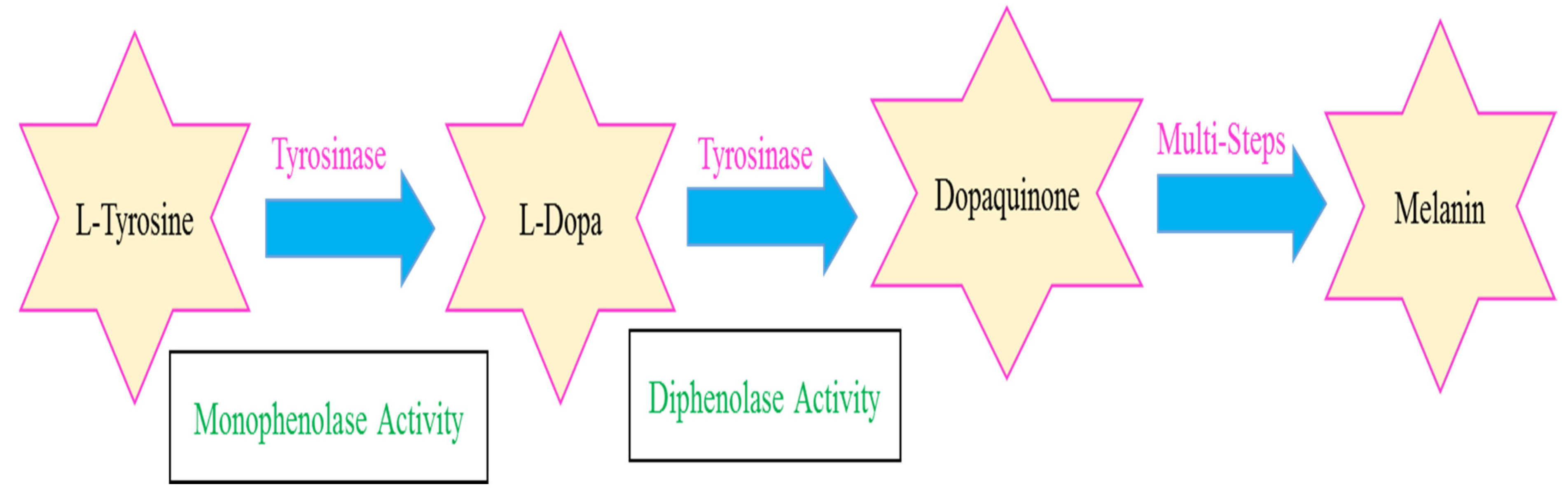
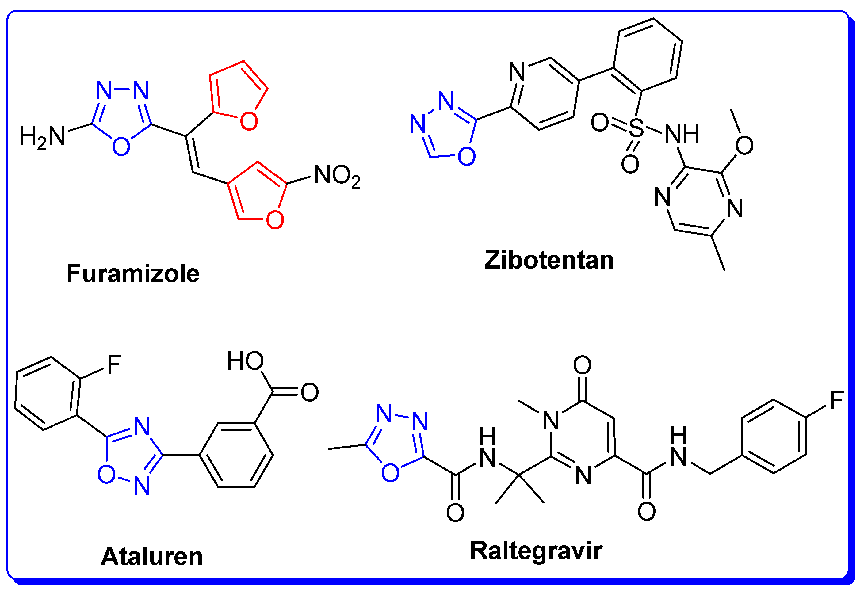
:1. Introduction
2. Results and Discussion
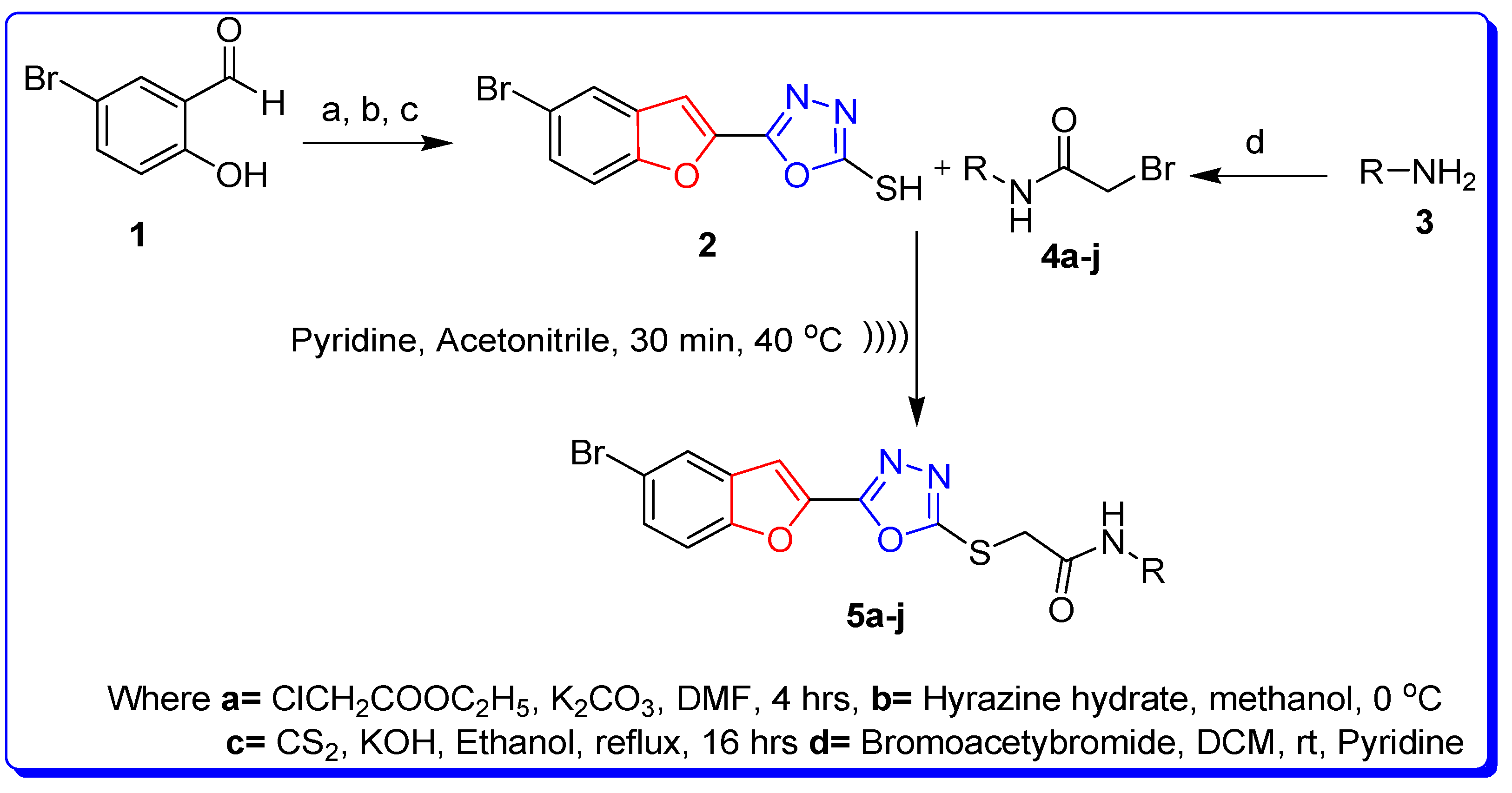
2.1. Synthesis of Furan-Oxadiazole Derivatives 5a–j
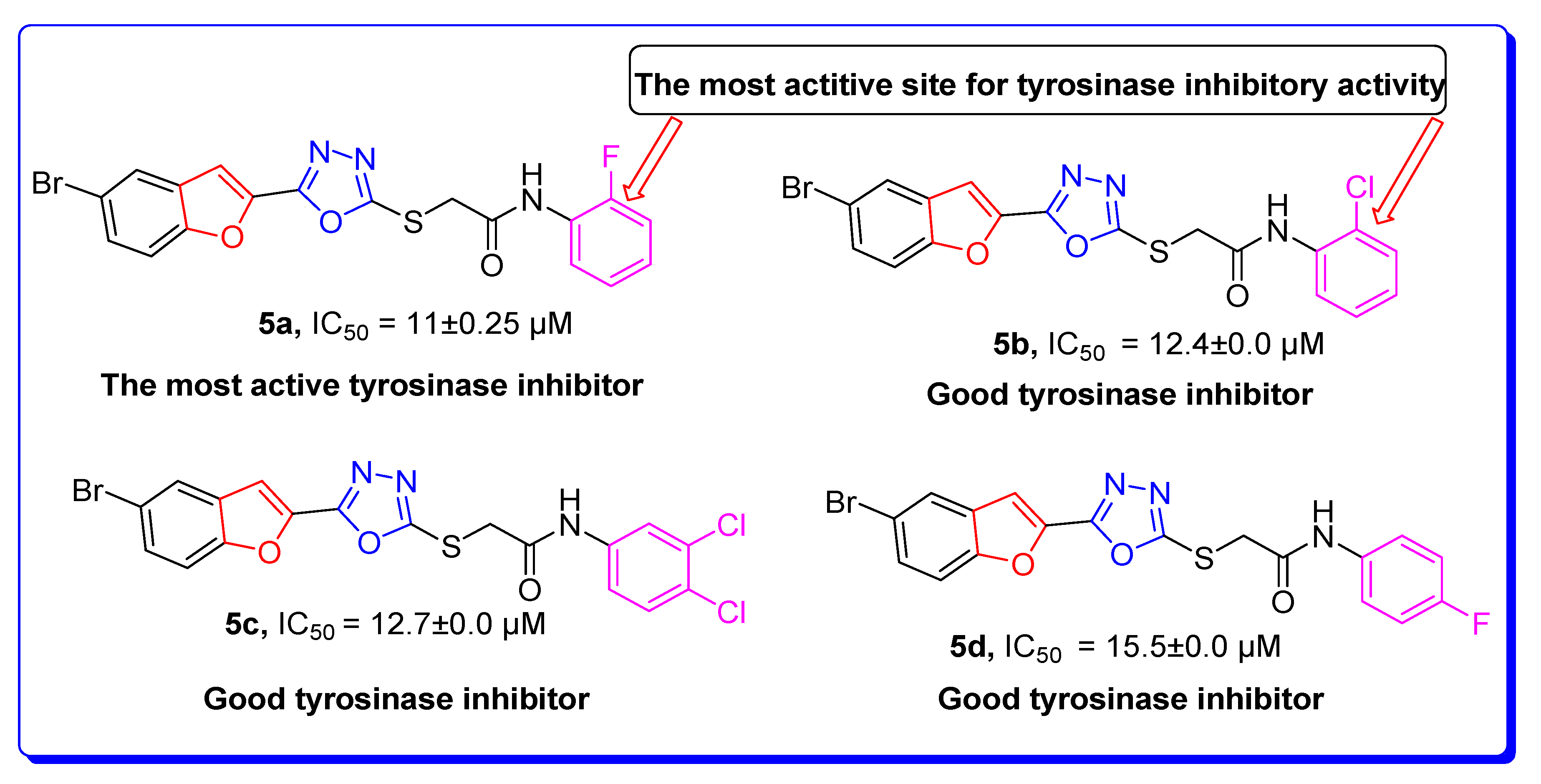
2.2. Bacterial Tyrosinase Inhibition and SAR
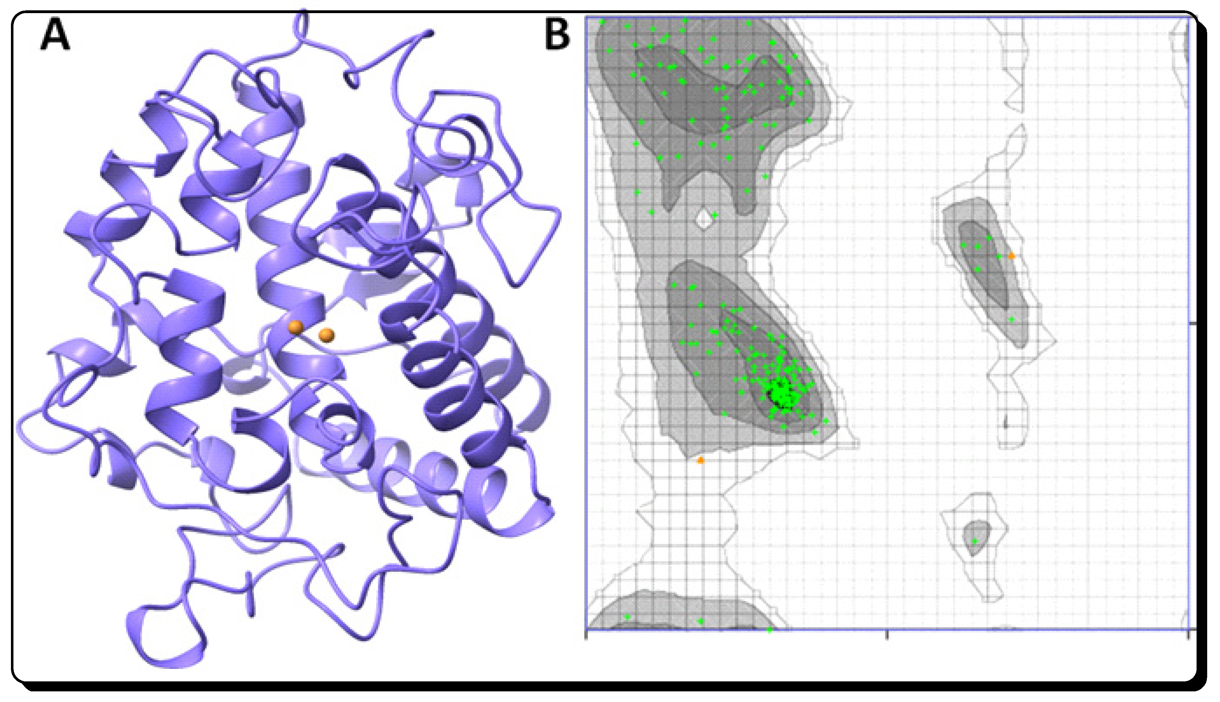
2.3. Structural Assessment of Bacterial Tyrosinase
2.4. RO5 Validation of Newly Designed Furan-Oxadiazole Ligands
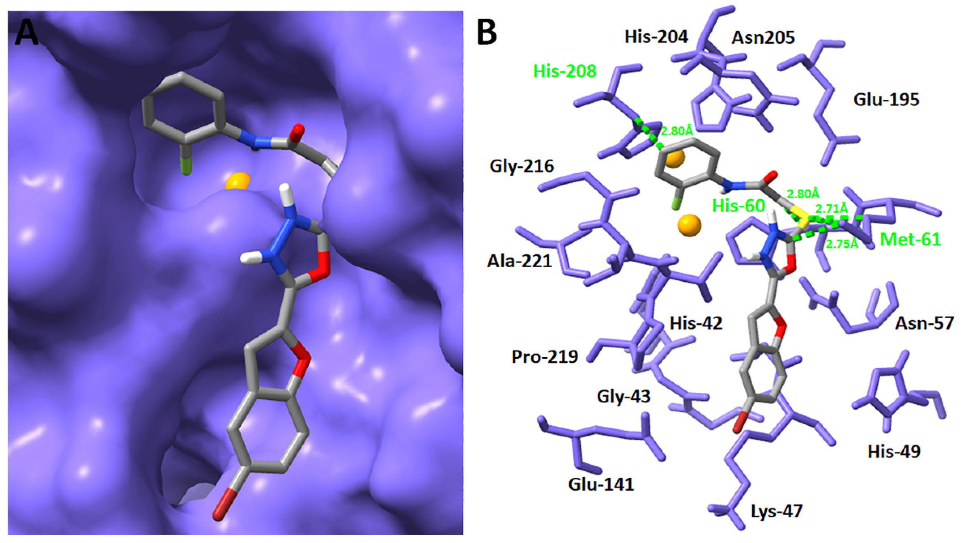
2.5. Molecular Docking Studies of Furan-Oxadiazoles 5a–j
Binding Pocket and Hydrogen (H) Binding of Furan-Oxadiazole 5a
2.6. ADMET and Drug-Likeness Studies of Furan-Oxadiazoles 5a–j
3. Materials and Methods
3.1. Synthesis and Characterization Techniques
3.2. Synthesis of Furan-Oxadiazole Scaffolds 5a–j by Ultrasonic Irradiated Synthetic Approach
3.2.1. 2-((5-(5-Bromobenzofuran-2-yl)-1,3,4-oxadiazol-2-yl)thio)-N-(2-fluorophenyl)acetamide (5a)
 .
.3.2.2. 2-((5-(5-Bromobenzofuran-2-yl)-1,3,4-oxadiazol-2-yl)thio)-N-(2-chlorophenyl)acetamide (5b)

3.2.3. 2-((5-(5-Bromobenzofuran-2-yl)-1,3,4-oxadiazol-2-yl)thio)-N-(3,4-dichlorophenyl)acetamide (5c)

3.2.4. 2-((5-(5-Bromobenzofuran-2-yl)-1,3,4-oxadiazol-2-yl)thio)-N-(4-fluorophenyl)acetamide (5d)

3.2.5. 2-((5-(5-Bromobenzofuran-2-yl)-1,3,4-oxadiazol-2-yl)thio)-N-(3,4-dimethylphenyl)acetamide (5e)

3.2.6. 2-((5-(5-Bromobenzofuran-2-yl)-1,3,4-oxadiazol-2-yl)thio)-N-(2-methoxyphenyl)acetamide (5f)

3.2.7. 2-((5-(5-Bromobenzofuran-2-yl)-1,3,4-oxadiazol-2-yl)thio)-N-phenylacetamide (5g)

3.2.8. 2-((5-(5-Bromobenzofuran-2-yl)-1,3,4-oxadiazol-2-yl)thio)-N-(2,4-dimethylphenyl)acetamide (5h)

3.2.9. 2-((5-(5-Bromobenzofuran-2-yl)-1,3,4-oxadiazol-2-yl)thio)-N-(2,5-dimethoxyphenyl)acetamide (5i)

3.2.10. 2-((5-Benzofuran-2-yl)-1,3,4-oxadiazol-2-yl)thio)-N,Ndiethylacetamide (5j)

3.3. Tyrosinase Inhibition Assay
3.4. Computational Methodology
3.4.1. Retrieval of Bacterial Tyrosinase Structure from Protein Data Bank (PDB)
3.4.2. Designing of Ligands 5a–j and Chemoinformatic Analysis
3.4.3. Molecular Docking of Furan-Oxadiazoles 5a–j
3.4.4. ADMET and Drug-Likeness Studies
3.5. Statistical Data
4. Conclusions
Supplementary Materials
Author Contributions
Funding
Institutional Review Board Statement
Informed Consent Statement
Data Availability Statement
Acknowledgments
Conflicts of Interest
References
- Zolghadri, S.; Bahrami, A.; Hassan Khan Mahmud, T.; Munoz-Munoz, J.; Garcia-Molina, F.; Garcia-Canovas, F.; Saboury, A.A. A comprehensive review on tyrosinaseinhibitors. J. Enzym. Inhib. Med. Chem. 2019, 34, 279–309. [Google Scholar] [CrossRef]
- Nazir, Y.; Saeed, A.; Rafiq, M.; Afzal, S.; Ali, A.; Latif, M.; Zuegg, J.; Hussein, W.M.; Fercher, C.; Barnard, R.T.; et al. Hydroxyl substituted benzoic acid/cinnamic acid derivatives: Tyrosinase inhibitory kinetics, anti-melanogenic activity and molecular docking studies. Bioorg. Med. Chem. Lett. 2019, 30, 126722. [Google Scholar] [CrossRef] [PubMed]
- Li, J.; Feng, L.; Liu, L.; Wang, F.; Ouyang, L.; Zhang, L.; Hu, X.; Wang, G. Recent advances in the design and discovery of synthetic tyrosinase inhibitors. Eur. J. Med. Chem. 2021, 224, 113744. [Google Scholar] [CrossRef] [PubMed]
- Ashraf, Z.; Rafiq, M.; Seo, S.Y.; Kwon, K.S.; Babar, M.M. Kinetic and in silico studies of novel hydroxy-based thymol analogues as inhibitors of mushroom tyrosinase. Eur. J. Med. Chem. 2015, 98, 203–211. [Google Scholar] [CrossRef] [PubMed]
- Collins, R.P.; Warner, L.B.; Paige, L. The Tyrosinase Activity of StrobilomycesStrobilaceus. Mycologia 1963, 55, 764–774. [Google Scholar] [CrossRef]
- Parvez, S.; Kang, M.; Hwan-Suck, C.; Cho, C.; Moo-Chang, H.; Min-Kyu, S.; Bae, H. Survey and mechanism of skin depigmenting and lightening agents. Phytother. Res. 2006, 20, 921–934. [Google Scholar] [CrossRef]
- Asanuma, M.; Miyazaki, I.; Ogawa, N. Dopamine- or L-DOPA-induced neurotoxicity: The role of dopamine quinone formation and tyrosinase in a model of Parkinson’s disease. Neurotox. Res. 2003, 5, 165–176. [Google Scholar] [CrossRef]
- Zhu, Y.J.; Zhou, H.T.; Hu, Y.H. Antityrosinase and antimicrobial activities of 2-phenylethanol, 2-phenylacetaldehyde and 2-phenylacetic acid. Food Chem. 2011, 124, 298–302. [Google Scholar] [CrossRef]
- Xu, Y.; Stokes, A.H.; Roskoski, R., Jr.; Vrana, K.J. Dopamine, in the presence of tyrosinase, covalently modifies and inactivates tyrosine hydroxylase. J. Neurosci. Res. 1998, 54, 691–697. [Google Scholar] [CrossRef]
- Bang, E.; Noh, S.-G.; Ha, S.; Jung, H.J.; Kim, D.H.; Lee, A.K.; Hyun, M.K.; Kang, D.; Lee, S.; Park, C.; et al. Evaluation of the novel synthetic tyrosinase inhibitor(z)-3-(3-bromo-4-hydroxybenzylidene) thiochroman-4-one (MHY1498) In vitro and in silico. Molecules 2018, 23, 3307. [Google Scholar] [CrossRef] [Green Version]
- Dige, N.C.; Mahajan, P.G.; Raza, H.; Hassan, M.; Vanjare, B.D.; Hong, H.; Hwan, L.K.; Latip, J.; Seo, S.Y. Ultrasound mediated efficient synthesis of new4-oxoquinazolin-3(4H)-yl) furan-2-carboxamides as potent tyrosinaseinhibitors: Mechanistic approach through chemoinformatics and molecular docking studies. Bioorg. Chem. 2019, 92, 103201. [Google Scholar] [CrossRef]
- Ben-Yosef, V.S.; Sendovski, M.; Fishman, A. Directed evolution of tyrosinase for enhanced monophenolase/diphenolase activity ratio. Enzym. Microb. Technol. 2010, 47, 372–376. [Google Scholar] [CrossRef]
- El-Shora, H.M.; Metwally, M. Use of tyrosinase from Bacillusthuringiensis for the decontamination of water polluted with phenols. Biotechnology 2008, 7, 305–310. [Google Scholar] [CrossRef]
- Abdullah, J.; Ahmad, M.; Karuppiah, N.; Heng, L.Y.; Sidek, H. Immobilization oftyrosinase in chitosan film for an optical detection of phenol. Sens. Actuators B Chem. 2006, 114, 604–609. [Google Scholar] [CrossRef]
- Tatsuma, T.; Komori, K.; Yeoh, H.; Oyama, N. Disposable test plates with tyrosinaseand-glucosidases for cyanide and cyanogenic glycosides. Anal. Chim. Acta 2000, 408, 233–240. [Google Scholar] [CrossRef]
- Fairhead, M.; Thöny-Meyer, L. Bacterial tyrosinases: Old enzymes with new relevance to biotechnology. New Biotechnol. 2012, 29, 183–191. [Google Scholar] [CrossRef]
- Faccio, G.; Kruus, K.; Saloheimo, M.; Thöny-Meyera, L. Bacterialtyrosinases and their applications. Process Biochem. 2012, 47, 1749–1760. [Google Scholar] [CrossRef]
- Yi, W.; Cao, R.; Peng, W.; Wen, H.; Yan, Q.; Zhou, B.; Ma, L.; Song, H. Synthesis and biological evaluation ofnovel 4-hydroxybenzaldehyde derivatives as tyrosinaseinhibitors. Eur. J. Med. Chem. 2010, 45, 639–646. [Google Scholar] [CrossRef]
- Seo, S.Y.; Sharma, V.K.; Sharma, N. Mushroom Tyrosinase: Recent Prospects. J. Agric. Food Chem. 2003, 51, 2837–2853. [Google Scholar] [CrossRef]
- Maertens, J.A. History of the development of azole derivatives. Clin. Microbiol. Infect. 2004, 10, 1–10. [Google Scholar] [CrossRef] [Green Version]
- Ghani, U. Azole inhibitors of mushroom and human tyrosinases: Current advances and prospects of drug development for melanogenic dermatological disorders. Eur. J. Med. Chem. 2022, 239, 114525. [Google Scholar] [CrossRef]
- Rehman, S.B.A.; Athar, A.M.; Rehman, U.A.; Zahra, S.S.; Raza, H.; Hassan, M.; Adnan, A.S.S.; Shahid, M.; Seo, S.Y. Synthesis and Structure-activity relationship of Tyrosinase Inhibiting novel biheterocyclicacetamides: Mechanistic Insights through Enzyme Inhibition, Kinetics and Computational Studies. Bioorg. Chem. 2019, 86, 459–472. [Google Scholar] [CrossRef]
- Jha, K.K.; Samad, A.; Kumar, Y.; Shaharyar, M.; Khosa, L.M.; Kumar, V.J.J.; Singh, P. Design, synthesis and biological evaluation of 1,3,4-oxadiazole derivatives. Eur. J. Med. Chem. 2010, 45, 4963–4967. [Google Scholar] [CrossRef]
- Mohd, A.M.F.F.; Rullah, K.; Tan, H.H.; Chan, K.M.; Tan, J.S.; Leong, S.W.; Mansor, A.H.; Yamin, B.M.; Wai, L.K. Synthesis and effects of oxadiazole derivatives on tyrosinase activity and human SK-MEL-28 malignant melanoma cells. RSC Adv. 2016, 6, 72177–72184. [Google Scholar] [CrossRef]
- Bala, S.; Kamboj, S.; Kajal, A.; Saini, V.; Prasad, D.N. 1,3,4-Oxadiazole derivatives: Synthesis, characterization, antimicrobial potential, and computational studies. Biomed. Res. Int. 2014, 2014, 172791. [Google Scholar] [CrossRef]
- Bondock, S.; Adel, S.; Etman, H.A.; Badria, F.A. Synthesis and antitumor evaluation of some new 1,3,4-oxadiazole-based heterocycles. Eur. J. Med. Chem. 2012, 48, 192–199. [Google Scholar] [CrossRef]
- Napiórkowska, M.; Cieślak, M.; Barańska, K.J.; Golińska, K.K.; Nawrot, B. Synthesis of new derivatives of benzofuran as potential anticancer agents. Molecules 2019, 24, 1529. [Google Scholar] [CrossRef]
- Aslam, N.A.; Stevenson, C.P.; Phythian, J.S.; Veitch, C.N.; Hall, R.D. Synthesis of cicerfuran, an antifungal benzofuran, and some related analogues. Tetrahedron 2006, 62, 4214–4226. [Google Scholar] [CrossRef]
- Clive, D.L.J.; Stoffman, E.J.L. Total synthesis of (-)-conocarpan and assignment of the absolute configuration by chemical methods. Chem. Commun. 2007, 21, 2151–2153. [Google Scholar] [CrossRef]
- Faiz, S.; Zahoor, A.F.; Ajmal, M.; Kamal, S.; Ahmad, S.; Abdelgawad, A.M.; Elnaggar, E.M. Design, synthesis, antimicrobial evaluation, and laccase catalysis effect of novel benzofuran–oxadiazole and benzofuran–triazolehybrids. J. Heterocycl. Chem. 2019, 56, 2839–2852. [Google Scholar] [CrossRef]
- Shi, Z.; Zhao, Z.; Huang, M.; Fu, X. Ultrasound-assisted, one-pot, three-component synthesis and antibacterial activities of novel indole derivatives containing 1,3,4-oxadiazole and 1,2,4-triazole moieties. C. R. Chim. 2015, 18, 1320–1327. [Google Scholar] [CrossRef]
- Irfan, A.; Faiz, S.; Rasul, A.; Zafar, R.; Zahoor, A.F.; Kotwica-Mojzych, K.; Mojzych, M. Exploring the Synergistic Anticancer Potential of Benzofuran–Oxadiazoles and Triazoles: Improved Ultrasound- and Microwave-Assisted Synthesis, Molecular Docking, Hemolytic, Thrombolytic and Anticancer Evaluation of Furan-Based Molecules. Molecules 2022, 27, 1023. [Google Scholar] [CrossRef] [PubMed]
- Zeni, G.; Ludtke, S.D.; Nogueira, W.C.; Panatieri, B.R.; Antonio, L.; Braga, L.A.; Silveira, C.C.; Stefanib, A.H.; Rochaa, B.T.J. New acetylenic furan derivatives: Synthesis and anti-inflammatory activity. Tetrahedron. Lett. 2001, 42, 8927–8930. [Google Scholar] [CrossRef]
- Farag, A.A.; El Shehry, F.M.; Abbas, Y.S.; Abd-Alrahman, N.S.; Atrees, A.A.; Al-basheer, Z.H.; Ammar, A.Y. Synthesis of pyrazoles containing benzofuran and trifluoromethyl moieties as possible anti-inflammatory and analgesic agents. Z. Nat. B 2015, 70, 519–526. [Google Scholar] [CrossRef]
- Lu, D.; Zhou, Y.; Li, Q.; Luo, J.; Jiang, Q.; He, B.; Tang, Q. Synthesis, in vitro antitumor activity and molecular mechanism of novel furan derivatives and their precursors, anticancer agents. Med. Chem. 2020, 20, 1475–1486. [Google Scholar] [CrossRef]
- Matsuya, Y.; Sasaki, K.; Nagaoka, M.; Kakuda, H.; Toyooka, N.; Imanishi, N.; Ochiai, H.; Nemoto, H. Synthesis of a new class of furan-fused tetracyclic compounds using o-quinodimethane chemistry and investigation of their antiviral activity. J. Org. Chem. 2004, 69, 7989–7993. [Google Scholar] [CrossRef]
- Tighadouini, S.; Radi, S.; Benabbes, R.; Youssoufi, H.M.; Shityakov, S.; El Massaoudi, M.; Garcia, Y. Synthesis, biochemical characterization, and theoretical studies of novel β-keto-enol pyridine and furan derivatives as potent antifungal agents. ACS Omega 2020, 5, 7743–17752. [Google Scholar] [CrossRef]
- Singh, V.F.; Chaurasia, S.; Joshi, D.M.; Srivastava, K.A.; Goel, A. Synthesis and in vivo antihyperglycemic activity of nature-mimicking furanyl-2-pyranones in STZ-S model. Bioorganic Med. Chem. Lett. 2007, 17, 2425–2429. [Google Scholar] [CrossRef]
- Rangaswamy, J.; Kumar, V.H.; Harini, T.S.; Naik, N. Functionalized 3-(benzofuran-2-yl)-5-(4-methoxyphenyl)-4,5-dihydro-1H-pyrazole scaffolds: A new class of antimicrobials and antioxidants. Arab. J. Chem. 2017, 10, S2685–S2696. [Google Scholar] [CrossRef]
- Barros, M.R.; Menezes, T.M.; Da Silva, L.P.; Pires, D.S.; Princival, J.L.; Seabra, G.; Neves, J.L. Furan inhibitory activity against tyrosinase and impact on B16F10 cell toxicity. Int. J. Biol. Macromol. 2019, 136, 1034–1041. [Google Scholar] [CrossRef]
- Jung, H.J.; Noh, S.G.; Ryu, I.Y.; Park, C.; Lee, J.Y.; Chun, P.; Moon, H.R.; Chung, H.Y. (E)-1-(Furan-2-yl)-(substituted phenyl) prop-2-en-1-one Derivatives as Tyrosinase Inhibitors and Melanogenesis Inhibition: An In Vitro and In Silico Study. Molecules 2020, 25, 5460. [Google Scholar] [CrossRef]
- Kowalewska, M.; Kwiecień, H.; Śmist, M.; Wrześniewska, A. Synthesis of new benzofuran-2-carboxylic acid derivatives. J. Chem. 2013, 2013, 183717. [Google Scholar] [CrossRef]
- Parekh, S.; Bhavsar, D.; Savant, M.; Thakrar, S.; Bavishi, A.; Parmar, M.; Vala, H.; Radadiya, A.; Pandya, N.; Serly, J.; et al. Synthesis of some novel benzofuran-2-yl(4,5-dihyro-3,5-substituted diphenylpyrazol-1-yl) methanones and studies on the antiproliferative effects and reversal of multidrug resistance of human MDR1-gene transfected mouse lymphoma cells in vitro. Eur. J. Med. Chem. 2011, 46, 1942–1948. [Google Scholar] [CrossRef]
- Almasirad, A.; Tabatabai, S.A.; Faizi, M.; Kebriaeezadeh, A.; Mehrabi, N.; Dalvandi, A.; Shafiee, A. Synthesis and anticonvulsant activity of new 2-substituted-5-[2-(2-fluorophenoxy)phenyl]-1,3,4-oxadiazoles and 1,2,4-triazoles. Bioorg. Med. Chem. Lett. 2004, 14, 6057–6059. [Google Scholar] [CrossRef]
- Cormier, R.; Burda, W.N.; Harrington, L.; Edlinger, J.; Kodigepalli, K.M.; Thomas, J.; Kapolka, R.; Roma, G.; Anderson, B.E.; Turos, E.; et al. Studies on the antimicrobial properties of N-acylatedciprofloxacins. Bioorg. Med. Chem. Lett. 2012, 22, 6513–6520. [Google Scholar] [CrossRef]
- Shiino, M.; Watanabe, Y.; Umezawa, K. Synthesis of N-substituted N-nitrosohydroxylamines as inhibitors of Mushroom Tyrosinase. Bioorg. Med. Chem. 2001, 9, 1233–1240. [Google Scholar] [CrossRef]
- Shiino, M.; Watanabe, Y.; Umezawa, K. Synthesis and tyrosinase inhibitory activity of novel N-hydroxybenzyl-N-nitrosohydroxylamines. Bioorg. Med. Chem. 2003, 31, 129–135. [Google Scholar] [CrossRef]
- Xinyang, C.; Aya, H.; Takehiro, K.; Hiroyuki, W.; Takashi, W.; Yoshino, O.; Masanobu, S.; Chul-Sa, K. The evaluation of the synergistic effect of3-(2,4-dihydroxyphenyl) propionic acid and l-ascorbic acid on tyrosinase inhibition. Z. Nat. B 2017, 72, 119–121. [Google Scholar] [CrossRef]
- Senol, F.S.; Khan, M.T.; Orhan, G.; Gurkas, E.; Orhan, I.E.; Oztekin, N.S.; Ak, F. In silico approach to inhibition of tyrosinase by ascorbic acid using molecular docking simulations. Curr. Top. Med. 2014, 14, 1469–1472. [Google Scholar] [CrossRef]
- Nokinsee, D.; Shank, L.; Lee, S.V.; Nimmanpipug, P. Estimation of Inhibitory Effect against Tyrosinase Activity through Homology Modeling and Molecular Docking. Enzym. Res. 2015, 2015, 262364. [Google Scholar] [CrossRef] [Green Version]
- Sendovski, M.; Kanteev, M.; Ben-Yosef, V.S.; Adir, N.; Fishman, A. First structures of an active bacterial tyrosinasereveal copper plasticity. J. Mol. Biol. 2011, 405, 227–237. [Google Scholar] [CrossRef]
- Hassan, M.; Vanjare, B.D.; Sim, K.-Y.; Raza, H.; Lee, K.H.; Shahzadi, S.; Kloczkowski, A. Biological and Cheminformatics Studies of Newly Designed Triazole Based Derivatives as Potent Inhibitors against Bacterial Tyrosinase. Molecules 2022, 27, 1731. [Google Scholar] [CrossRef]
- Hassan, M.; Abbasi, M.A.; Siddiqui, S.Z.; Shahzadi, S.; Raza, H.; Hussain, G.; Shah, S.A.A.; Ashraf, M.; Shahid, M.; Seo, S.Y.; et al. Designing of promising medicinal scaffolds for Alzheimer’s disease through enzyme inhibition, lead optimization, molecular docking and dynamic simulation approaches. Bioorg. Chem. 2019, 91, 103138. [Google Scholar] [CrossRef]
- Ghose, A.K.; Herbertz, T.; Hudkins, R.L.; Dorsey, B.D.; Mallamo, J.P. Knowledge-based, central nervous system (CNS) lead selection and lead optimization for CNS drug discovery. ACS Chem. Neurosci. 2012, 3, 50–68. [Google Scholar] [CrossRef]
- Hassan, M.; Ashraf, Z.; Abbas, Q.; Raza, H.; Seo, S.-Y. Exploration of novel human tyrosinase inhibitors by molecular modeling, docking and simulation studies. Interdiscip. Sci. Comput. Life Sci. 2018, 10, 68–80. [Google Scholar] [CrossRef]
- Hassan, M.; Abbas, Q.; Ashraf, Z.; Moustafa, A.A.; Seo, S.Y. Pharmacoinformatics exploration of polyphenol oxidases leading to novel inhibitors by virtual screening and molecular dynamic simulation study. Comput. Biol. Chem. 2017, 68, 131–142. [Google Scholar] [CrossRef]
- Gupta, A.; Zaheer, M.R.; Iqbal, S.; Ahmad, A.R.; Alshammari, B.M. Photodegradation and in silico molecular docking study of a diuretic D drug: Clopamide. ACS Omega 2022, 7, 13870–13877. [Google Scholar] [CrossRef]
- Elsayed, E.A.; Danial, E.N. Isolation, Identification and Medium Optimization for Tyrosinase Production by a Newly Isolated Bacillus subtilis NA2 Strain. J. Appl. Pharm. Sci. 2018, 8, 93–101. [Google Scholar]
- Hussain, F.; Kamal, S.; Rehman, S.; Azeem, M.; Bibi, I.; Ahmed, T.; Iqbal, H. Alkaline Protease Production Using Response Surface Methodology, Characterization and Industrial Exploitation of Alkaline Protease of Bacillus subtilissp. Catal. Lett. 2017, 147, 1204–1213. [Google Scholar] [CrossRef]
- Kim, J.H.; Yoon, J.-Y.; Yang, S.Y.; Choi, S.-K.; Kwon, S.J.; Cho, I.S.; Jeong, M.H.; Ho Kim, Y.; Choi, G.S. Tyrosinase inhibitory components from Aloevera and their antiviral activity. J. Enzym. Inhib. Med. Chem. 2017, 32, 78–83. [Google Scholar] [CrossRef] [PubMed]
- Pettersen, E.F.; Goddard, T.D.; Huang, C.C.; Couch, G.S.; Greenblatt, D.M.; Meng, E.C.; Ferrin, T.E. UCSF Chimera-A visualization system for exploratory research and analysis. J. Comput. Chem. 2004, 25, 1605–1612. [Google Scholar] [CrossRef] [PubMed] [Green Version]
- Lovell, S.C.; Davis, I.W.; Arendall III, W.B.; De Bakker, P.I.; Word, J.M.; Prisant, M.G.; Richardson, J.S.; Richardson, D.C. Structure validation by Cα geometry: ϕ, ψ and Cβ deviation. Proteins Struct. Funct. Bioinform. 2003, 50, 437–450. [Google Scholar] [CrossRef]
- Discovery Studio; Version 2.1; Accelrys: San Diego, CA, USA, 2008.
- Lipinski, C.A. Lead- and drug-like compounds: The rule-of-five revolution. Drug Discov. Today Technol. 2004, 1, 337–341. [Google Scholar] [CrossRef]
- Dallakyan, S.; Olson, A.J. Small-molecule library screening by docking with PyRx. Methods Mol. Biol. 2015, 1263, 243–350. [Google Scholar] [CrossRef]
- Daina, A.; Michielin, O.; Zoete, V. SwissADME: A Free Web Tool to Evaluate Pharmacokinetics, Drug-Likeness and Medicinal Chemistry Friendliness of Small Molecules. Sci. Rep. 2017, 7, 42717. [Google Scholar] [CrossRef]
- Yang, H.; Lou, C.; Sun, L.; Li, J.; Cai, Y.; Wang, Z.; Li, W.; Liu, G.; Tang, Y. AdmetSAR 2.0: Web-Service for Prediction and Optimization of Chemical ADMET Properties. Bioinformatics 2019, 35, 1067–1069. [Google Scholar] [CrossRef]
- Cheng, F.; Li, W.; Zhou, Y.; Shen, J.; Wu, Z.; Liu, G.; Lee, P.W.; Tang, Y. ADMET SAR: A Comprehensive Source and Free Tool for Assessment of Chemical ADMET Properties. J. Chem. Inf. Model. 2012, 52, 3099–3105. [Google Scholar] [CrossRef]


| Compound | R-NH | Product | Percentage Yield |
|---|---|---|---|
| 5a |  |  | 55 |
| 5b |  |  | 53 |
| 5c |  |  | 77 |
| 5d |  |  | 63 |
| 5e |  |  | 65 |
| 5f |  |  | 79 |
| 5g |  |  | 65 |
| 5h |  |  | 69 |
| 5i |  |  | 76 |
| 5j |  |  | 66 [30] |
| Compound | Tyrosinase Inhibition IC50 (µM) |
|---|---|
| 5a | 11 ± 0.25 |
| 5b | 12.4 ± 0.0 |
| 5c | 12.7 ± 0.0 |
| 5d | 15.5 ± 0.0 |
| 5e | 25 ± 0.75 |
| 5f | 27 ± 1.00 |
| 5g | 30 ± 1.50 |
| 5h | 36 ± 0.25 |
| 5i | 48 ± 0.96 |
| 5j | 49.5 ± 0.92 |
| Ascorbic acid Standard [43,44,45,47,48,49] | 11.5 ± 1.00 |
| Ligands | Molecular Weight (g/mol) | LogP | HBA | HBD | PSA (Å2) | RO5 |
|---|---|---|---|---|---|---|
| 5a | 446 | 4.18 | 6 | 1 | 61.81 | Yes |
| 5b | 462 | 4.48 | 6 | 1 | 60.81 | Yes |
| 5c | 496 | 5.88 | 6 | 1 | 61.51 | No |
| 5d | 446 | 4.46 | 6 | 1 | 61.51 | Yes |
| 5e | 457 | 5.29 | 6 | 1 | 75.98 | No |
| 5f | 458 | 4.27 | 7 | 1 | 68.44 | Yes |
| 5g | 428 | 4.21 | 6 | 1 | 61.51 | Yes |
| 5h | 457 | 4.90 | 6 | 1 | 60.81 | Yes |
| 5i | 489 | 4.19 | 8 | 1 | 75.98 | Yes |
| 5j | 331 | 2.92 | 6 | 0 | 54.03 | Yes |
| Ascorbic Acid | 176 | −1.59 | 6 | 4 | 85.73 | Yes |
| Synthesized Ligands | Docking Energy (kcal/mol) |
|---|---|
| 5a | −3.2 |
| 5b | −3.4 |
| 5c | −0.4 |
| 5d | −1.0 |
| 5e | −1.1 |
| 5f | −3.2 |
| 5g | −3.0 |
| 5h | 1.2 |
| 5i | −1.4 |
| 5j | −4.8 |
| Ascorbic Acid | −6.3 |
| Compound | GI-Absorption | iLogP | Log S (ESOL) H2O Solubility | Bio-Availability Score | TPSA | P-gp Substrate | Lipinski RO5 |
|---|---|---|---|---|---|---|---|
| 5a | High | 3.41 | −5.54 moderately soluble | 0.55 | 106.46 Å2 | No | No violations |
| 5b | High | 3.66 | −5.97 moderately soluble | 0.55 | 106.46 Å2 | No | No violations |
| 5c | Low | 3.76 | −6.56 poorly soluble | 0.55 | 106.46 Å2 | No | No violations |
| 5d | High | 3.21 | −5.54 moderately soluble | 0.55 | 106.46 Å2 | No | No violations |
| 5e | High | 3.78 | −5.98 moderately soluble | 0.55 | 106.46 Å2 | No | No violations |
| 5f | High | 3.75 | −5.44 moderately soluble | 0.55 | 115.69 Å2 | No | No violations |
| 5g | High | 3.38 | −5.38 moderately soluble | 0.55 | 106.46 Å2 | No | No violations |
| 5h | High | 3.70 | −5.98 moderately soluble | 0.55 | 106.46 Å2 | No | No violations |
| 5i | Low | 3.99 | −5.51 moderately soluble | 0.55 | 124.92 Å2 | No | No violations |
| 5j | High | 3.48 | −3.75 soluble | 0.55 | 97.67 Å2 | No | No violations |
Publisher’s Note: MDPI stays neutral with regard to jurisdictional claims in published maps and institutional affiliations. |
© 2022 by the authors. Licensee MDPI, Basel, Switzerland. This article is an open access article distributed under the terms and conditions of the Creative Commons Attribution (CC BY) license (https://creativecommons.org/licenses/by/4.0/).
Share and Cite
Irfan, A.; Zahoor, A.F.; Kamal, S.; Hassan, M.; Kloczkowski, A. Ultrasonic-Assisted Synthesis of Benzofuran Appended Oxadiazole Molecules as Tyrosinase Inhibitors: Mechanistic Approach through Enzyme Inhibition, Molecular Docking, Chemoinformatics, ADMET and Drug-Likeness Studies. Int. J. Mol. Sci. 2022, 23, 10979. https://doi.org/10.3390/ijms231810979
Irfan A, Zahoor AF, Kamal S, Hassan M, Kloczkowski A. Ultrasonic-Assisted Synthesis of Benzofuran Appended Oxadiazole Molecules as Tyrosinase Inhibitors: Mechanistic Approach through Enzyme Inhibition, Molecular Docking, Chemoinformatics, ADMET and Drug-Likeness Studies. International Journal of Molecular Sciences. 2022; 23(18):10979. https://doi.org/10.3390/ijms231810979
Chicago/Turabian StyleIrfan, Ali, Ameer Fawad Zahoor, Shagufta Kamal, Mubashir Hassan, and Andrzej Kloczkowski. 2022. "Ultrasonic-Assisted Synthesis of Benzofuran Appended Oxadiazole Molecules as Tyrosinase Inhibitors: Mechanistic Approach through Enzyme Inhibition, Molecular Docking, Chemoinformatics, ADMET and Drug-Likeness Studies" International Journal of Molecular Sciences 23, no. 18: 10979. https://doi.org/10.3390/ijms231810979