
Thiogenistein—Antioxidant Chemistry, Antitumor Activity, and Structure Elucidation of New Oxidation Products

 , , and
Abstract
:1. Introduction
2. Results and Discussion
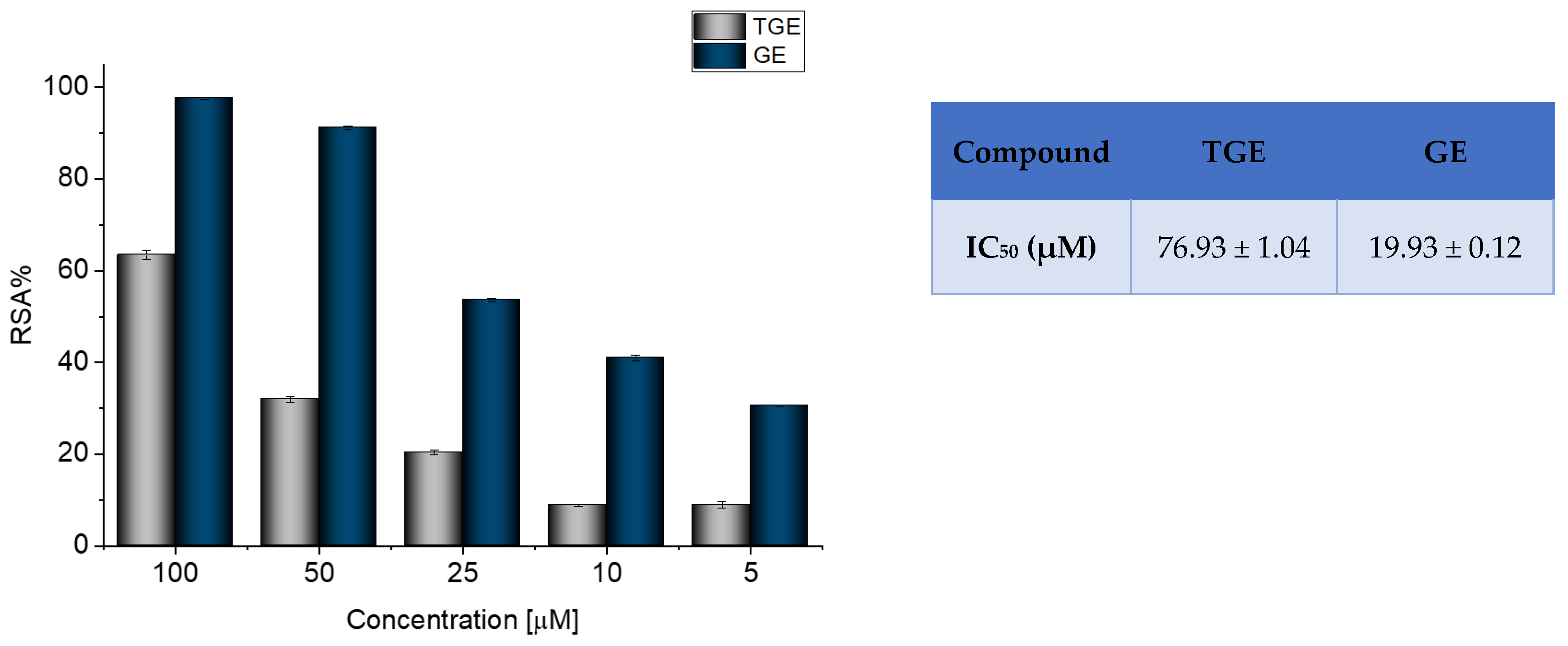
2.1. ABTS Radical ScavengingAssay
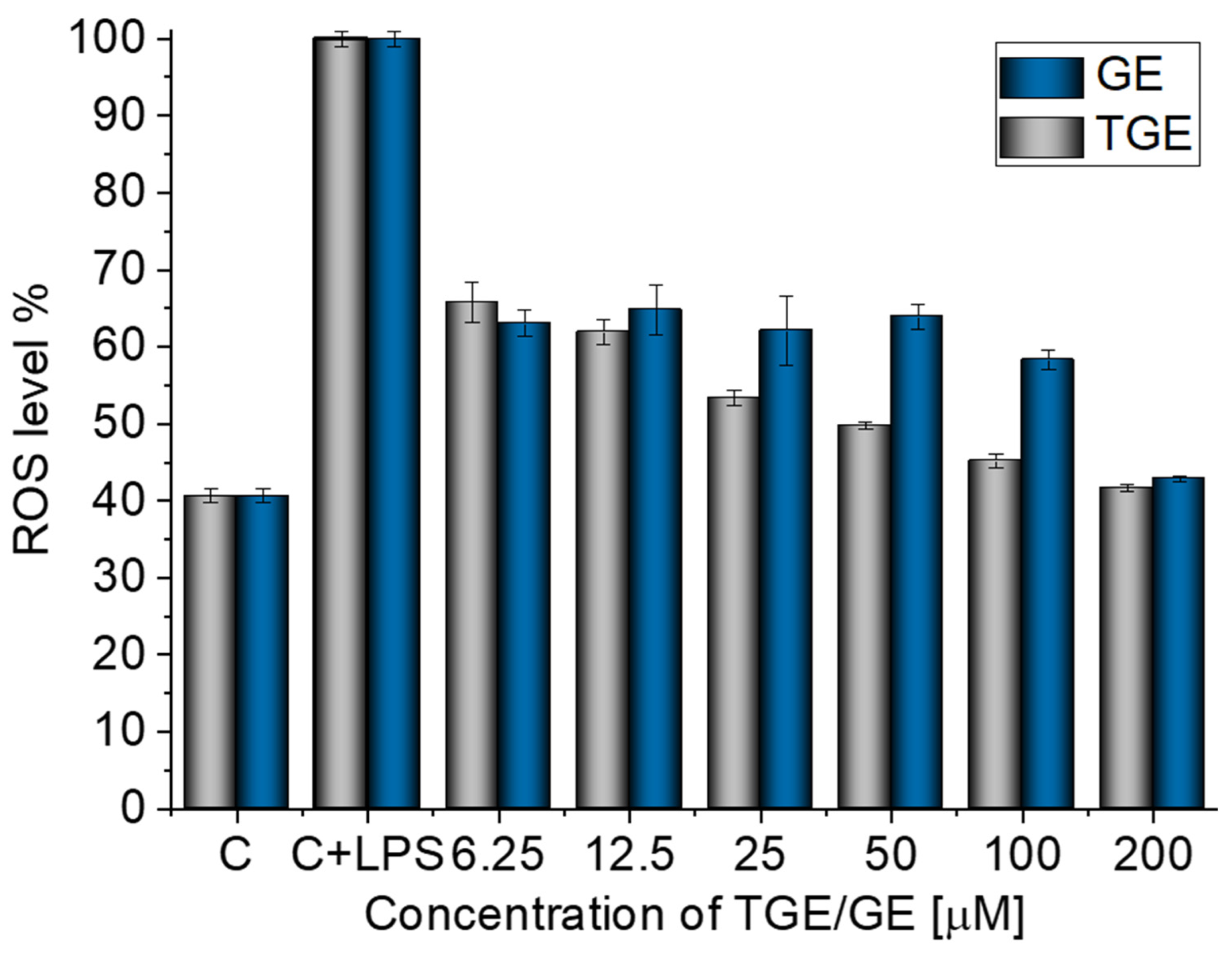
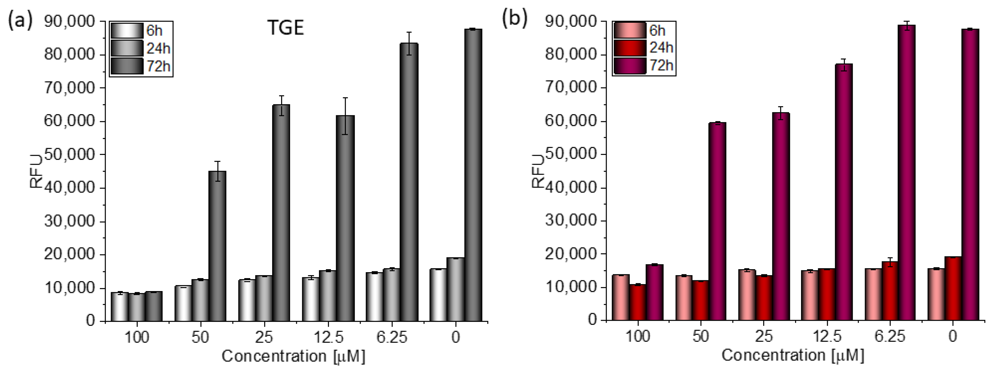
2.2. H2DCF-DA ROS Detection Assay
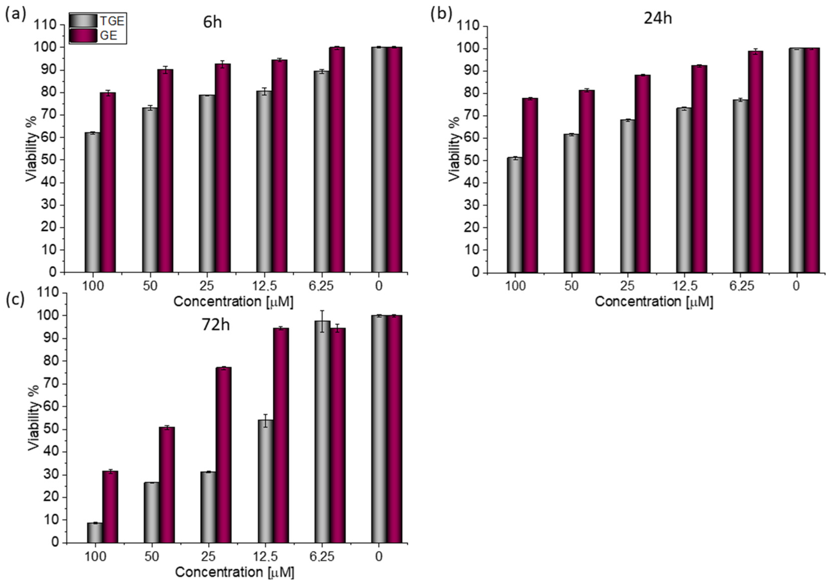
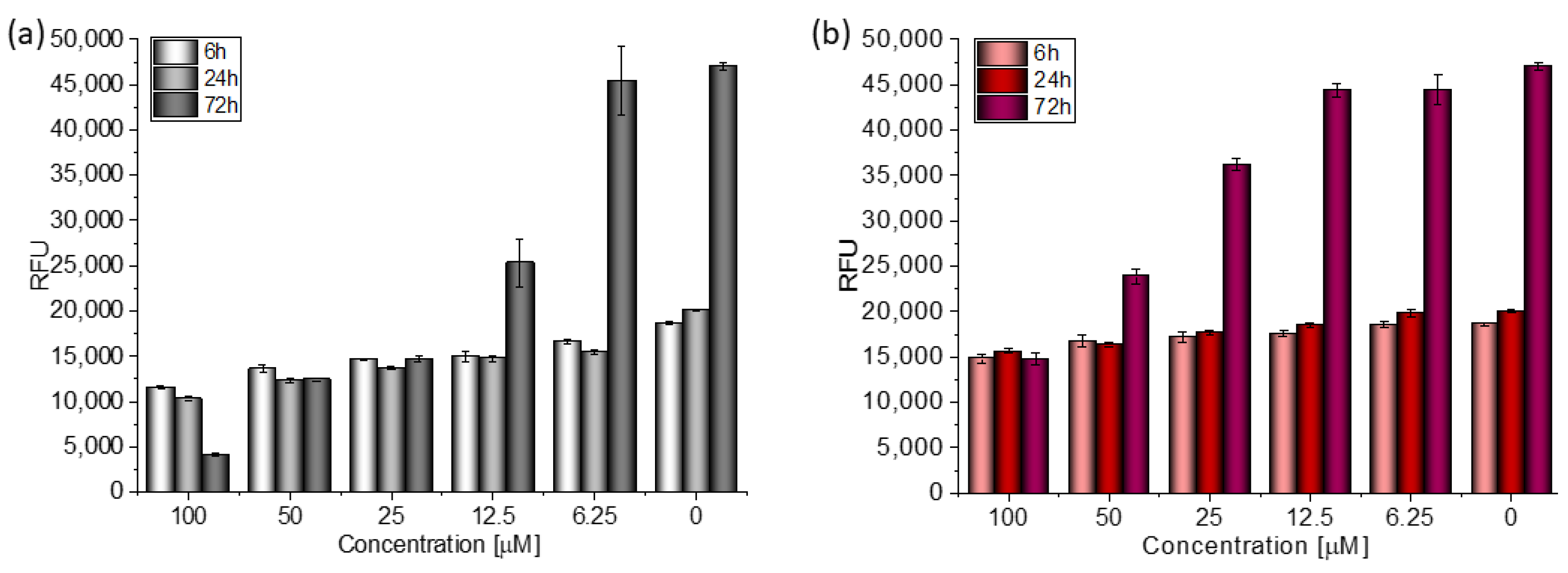
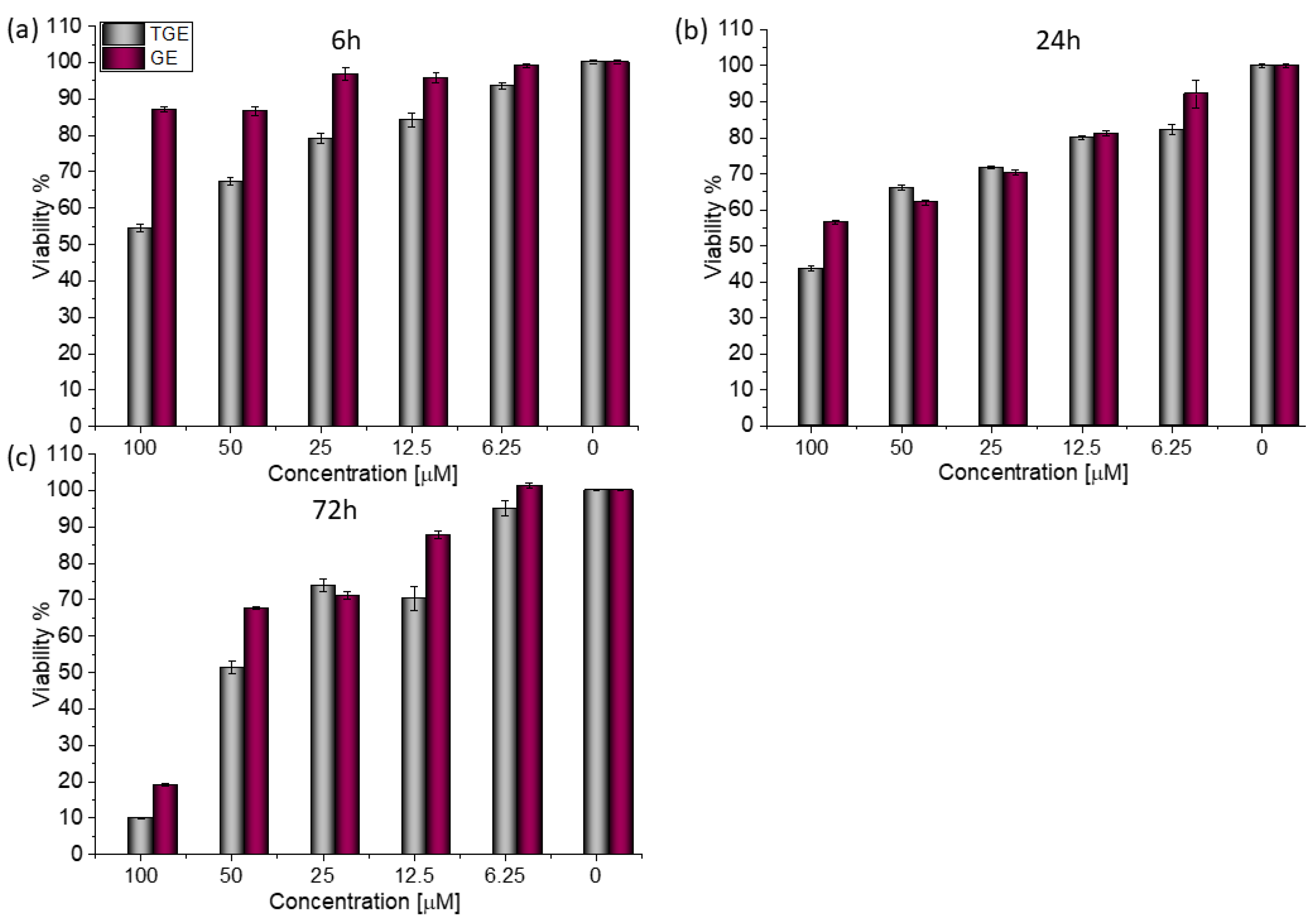
2.3. In Vitro Study–Cell Viability, Cytotoxicity
Antitumor Activity–Breast Cancer
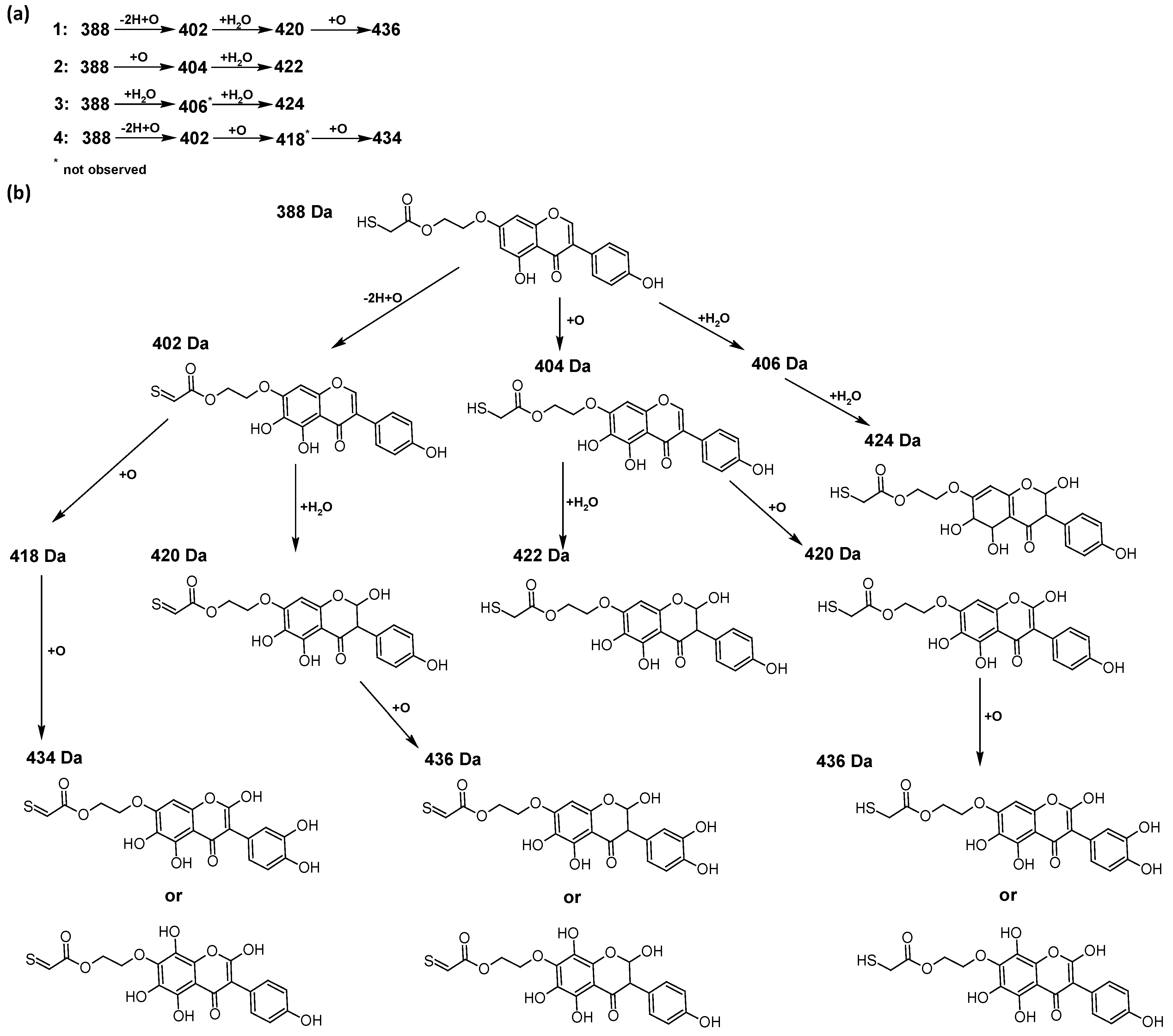
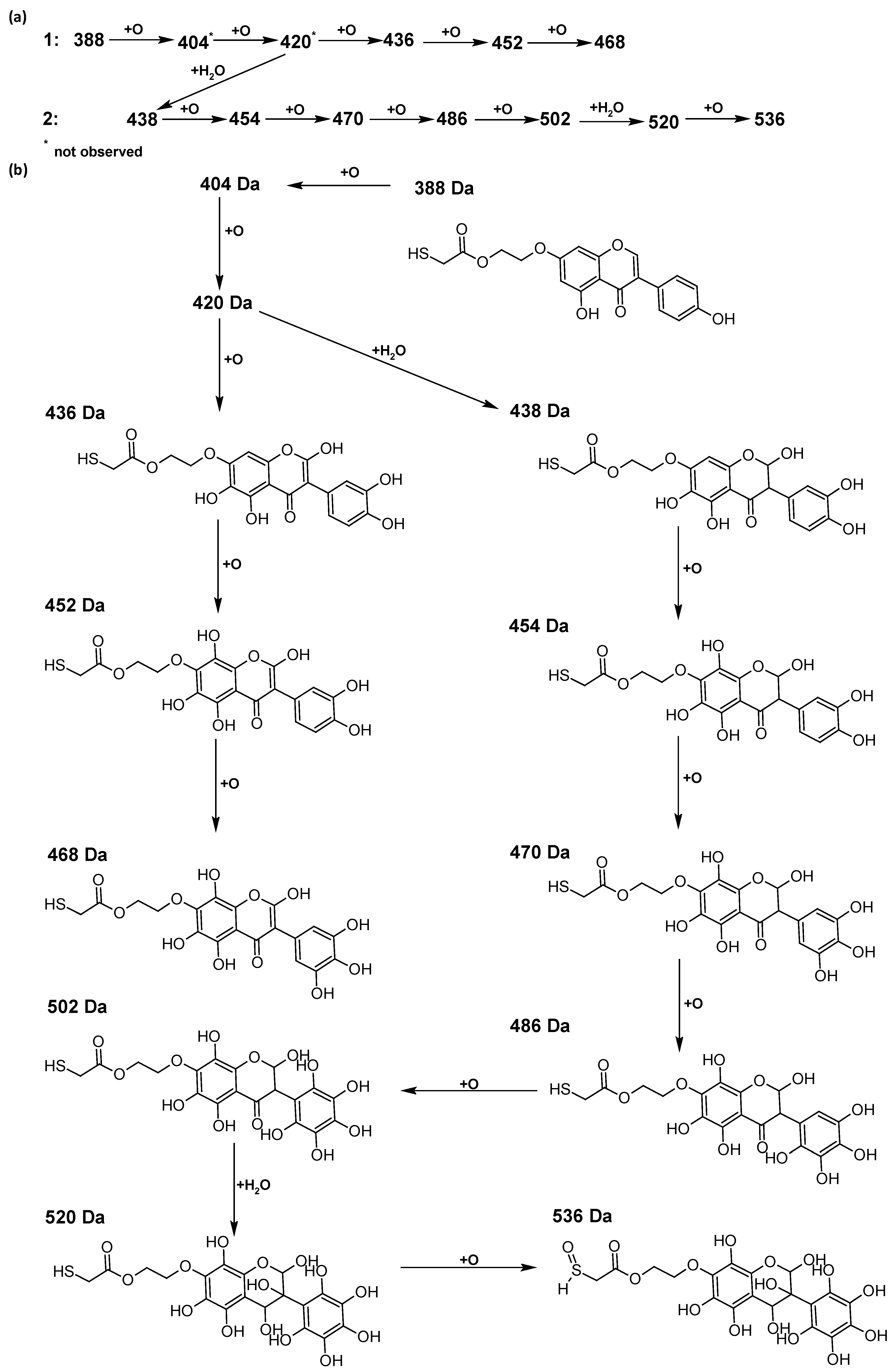
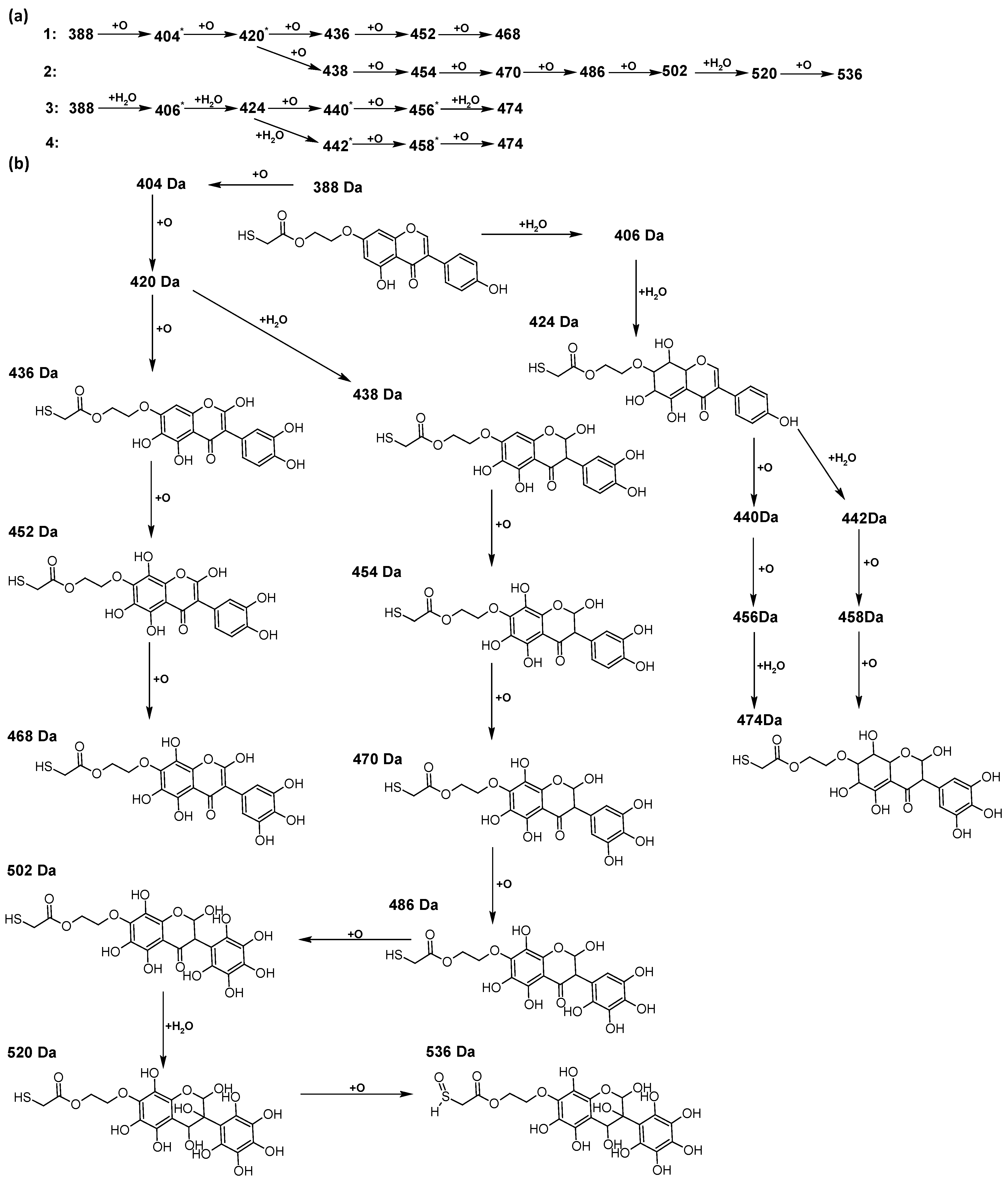
2.4. Antioxidant Chemistry–Oxidation of TGE by Potential and With Hydrogen Peroxide
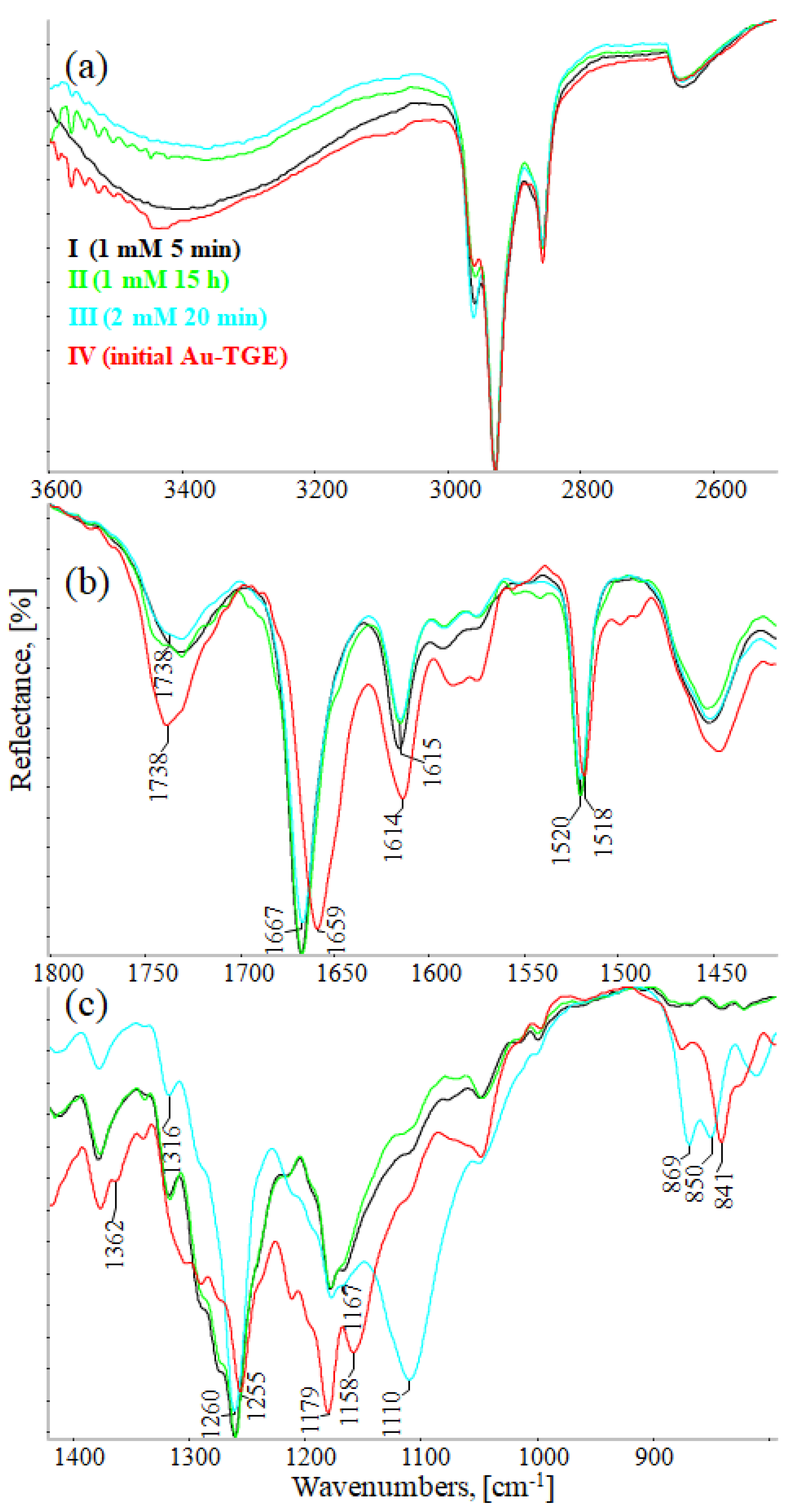
2.5. Description of Qualitative Changes Observed in the IR-ATR Spectra during the Oxidation of the TGE Monolayer on the Au Electrode
Vibrational Studies on Oxidative Monolayers–Simulation of Active Substance Immobilization on the Surface of Biological Membranes
2.6. Molecular Modeling and the Quantum Mechanical Density Functional Calculations
3. Materials and Methods
3.1. Materials
3.2. Antioxidant Study
3.2.1. ABTS Radical Scavenging Assay
3.2.2. H2DCF-DA ROS Detection Assay
3.3. In Vitro Study–Cell Viability, Cytotoxicity
3.4. Electrochemical Measurements
3.5. MS Spectrometry
3.6. IR Measurements
3.7. Quantum Mechanical Modeling
4. Conclusions
Supplementary Materials
Author Contributions
Funding
Institutional Review Board Statement
Informed Consent Statement
Data Availability Statement
Acknowledgments
Conflicts of Interest
References
- Plattner, S.; Erb, R.; Chervet, J.P.; Oberacher, H. Studying the Reducing Potencies of Antioxidants with the Electrochemistry Inherently Present in Electrospray Ionization-Mass Spectrometry. Anal. Bioanal. Chem. 2014, 406, 213–224. [Google Scholar] [CrossRef] [PubMed] [Green Version]
- Gutteridge, J.M. Free Radicals and Aging. Rev. Clin. Gerontol. 1994, 4, 279–288. [Google Scholar] [CrossRef] [Green Version]
- Wang, T.Y.; Li, Q.; Bi, K.S. Bioactive Flavonoids in Medicinal Plants: Structure, Activity and Biological Fate. Asian J. Pharm. Sci. 2018, 13, 12–23. [Google Scholar] [CrossRef]
- Harborne, J.B.; Williams, C.A. Advances in flavonoid Research since 1992. Phytochemistry 2000, 55, 481–504. [Google Scholar] [CrossRef]
- Heim, K.E.; Tagliaferro, A.R.; Bobilya, D.J. Flavonoid Antioxidants: Chemistry, Metabolism and Structure-Activity Relationships. J. Nutr. Biochem. 2002, 13, 572–584. [Google Scholar] [CrossRef]
- Waldmann, S.; Almukainzi, M.; Bou-Chacra, N.; Amidon, G.; Lee, B.; Feng, J.; Kanfer, I.; Zuo, J.; Wei, H.; Bolger, M.; et al. Provisional Biopharmaceutical Classification of Some Common Herbs Used in Western Medicine. Mol. Pharm. 2012, 9, 815–822. [Google Scholar] [CrossRef]
- Sepehr, E.; Cooke, G.; Robertson, P.; Gilani, G.S. Bioavailability of Soy Isoflavones in Rats Part I: Application of Accurate Methodology for Studying the Effects of Gender and Source of Isoflavones. Mol. Nutr. Food Res. 2007, 51, 799–812. [Google Scholar] [CrossRef]
- Sidoryk, K.; Michalak, O.; Kubiszewski, M.; Leś, A.; Cybulski, M.; Stolarczyk, E.U.; Doubsky, J. Synthesis of Thiol Derivatives of Biological Active Compounds for Nanotechnology Application. Molecules 2020, 25, 3470. [Google Scholar] [CrossRef]
- Vo, Q.V.; Nam, P.C.; Thong, N.M.; Trung, N.T.; Phan, C.T.D.; Mechler, A. Antioxidant Motifs in Flavonoids: O−H versus C−H Bond Dissociation. ACS Omega 2019, 4, 8935–8942. [Google Scholar] [CrossRef] [Green Version]
- Chen, Z.; Liu, Q.; Zhao, Z.; Bai, B.; Sun, Z.; Cai, L.; Fu, Y.; Ma, Y.; Wang, Q.; Xi, G. Effect of Hydroxyl on Antioxidant Properties of 2,3-Dihydro-3,5-Dihydroxy-6-Methyl-4: H -Pyran-4-One to Scavenge Free Radicals. RSC Adv. 2021, 11, 34456–34461. [Google Scholar] [CrossRef]
- Han, R.M.; Tian, Y.X.; Liu, Y.; Chen, C.H.; Ai, X.C.; Zhang, J.P.; Skibsted, L.H. Comparison of Flavonoids and Isoflavonoids as Antioxidantsz. J. Agric. Food Chem. 2009, 57, 3780–3785. [Google Scholar] [CrossRef]
- Castellano, G.; Torrens, F. Quantitative Structure-Antioxidant Activity Models of Isoflavonoids: A Theoretical Study. Int. J. Mol. Sci. 2015, 16, 12891–12906. [Google Scholar] [CrossRef] [PubMed] [Green Version]
- Wright, J.; Johnson, E.; DiLabio, G. Predicting the Activity of Phenolic Antioxidants: Theoretical Method, Analysis of Substituent Effects, and Application to Major Families of Antioxidants. J. Am. Chem. Soc. 2001, 123, 1173–1183. [Google Scholar] [CrossRef]
- Mahmoud, M.A.A.; Chedea, V.S.; Detsi, A.; Kefalas, P. Ascorbic Acid Modifies the Free Radical Scavenging Behaviour of Catechin: An Insight into the Mechanism. Food Res. Int. 2013, 51, 907–913. [Google Scholar] [CrossRef]
- Proença, C.; Freitas, M.; Ribeiro, D. Inhibition of Protein Tyrosine Phosphatase 1B by Flavonoids: A Structure–Activity Relationship Study. Food Chem. Toxicol. 2018, 111, 474–481. [Google Scholar] [CrossRef] [PubMed]
- Firuzia, O.; Lacanna, A.; Petrucci, R.; Marrosu, G.S.L. Evaluation of the Antioxidant Activity of Flavonoids by “Ferric Reducing Antioxidant Power” Assay and Cyclic Voltammetry. Biochim. Biophys. Acta-Gen. Subj. 2005, 1721, 174–184. [Google Scholar] [CrossRef] [PubMed]
- Marshall, G.M.; Bensebaa, F.; Dubowski, J.J. Observation of Surface Enhanced IR Absorption Coefficient in Alkanethiol Based Self-Assembled Monolayers on GaAs(001). J. Appl. Phys. 2009, 105, 094310. [Google Scholar] [CrossRef] [Green Version]
- Skoda, M.W.A.; Jacobs, R.M.J.; Willis, J.; Schreiber, F. Hydration of Oligo(Ethylene Glycol) Self-Assembled Monolayers Studied Using Polarization Modulation Infrared Spectroscopy. Langmuir 2007, 23, 970–974. [Google Scholar] [CrossRef]
- Galato, D.; Ckless, K.; Susin, M.F.; Giacomelli, C.; Ribeiro-do-Valle, R.M.; Spinelli, A. Antioxidant Capacity of Phenolic and Related Compounds: Correlation among Electrochemical, Visible Spectroscopy Methods and Structure-Antioxidant Activity. Redox Rep. 2001, 6, 243–250. [Google Scholar] [CrossRef]
- Stolarczyk, E.U.; Strzempek, W.; Łaszcz, M.; Leś, A.; Menaszek, E.; Sidoryk, K.; Stolarczyk, K. Anti-Cancer and Electrochemical Properties of Thiogenistein–New Biologically Active Compound. Int. J. Mol. Sci. 2021, 22, 8783. [Google Scholar] [CrossRef]
- Gupta, V.K.; Jain, R.; Radhapyari, K.; Jadon, N.; Agarwal, S. Voltammetric Techniques for the Assay of Pharmaceuticals-A Review. Anal. Biochem. 2011, 408, 179–196. [Google Scholar] [CrossRef]
- Stolarczyk, E.U.; Stolarczyk, K.; Łaszcz, M.; Kubiszewski, M.; Maruszak, W.; Olejarz, W.; Bryk, D. Synthesis and Characterization of Genistein Conjugated with Gold Nanoparticles and the Study of Their Cytotoxic Properties. Eur. J. Pharm. Sci. 2017, 96, 176–185. [Google Scholar] [CrossRef] [PubMed]
- Tuli, H.S.; Tuorkey, M.J.; Thakral, F.; Sak, K.; Kumar, M.; Sharma, A.K.; Sharma, U.; Jain, A.; Aggarwal, V.; Bishayee, A. Molecular Mechanisms of Action of Genistein in Cancer: Recent Advances. Front. Pharmacol. 2019, 10, 1336. [Google Scholar] [CrossRef] [PubMed] [Green Version]
- Grosso, R.; de-Paz, M.V. Thiolated-Polymer-Based Nanoparticles as an Avant-Garde Approach for Anticancer Therapies–Reviewing Thiomers from Chitosan and Hyaluronic Acid. Pharmaceutics 2021, 13, 854. [Google Scholar] [CrossRef] [PubMed]
- Mueller, S.O.; Simon, S.; Chae, K.; Metzler, M.; Korach, K.S. Phytoestrogens and Their Human Metabolites Show Distinct Agonistic and Antagonistic Properties on Estrogen Receptor α (ERα) and ERβ in Human Cells. Toxicol. Sci. 2004, 80, 14–25. [Google Scholar] [CrossRef] [PubMed] [Green Version]
- Bovee, T.F.H.; Schoonen, W.G.E.J.; Hamers, A.R.M.; Bento, M.J.; Peijnenburg, A.A.C.M. Screening of Synthetic and Plant-Derived Compounds for (Anti)Estrogenic and (Anti)Androgenic Activities. Anal. Bioanal. Chem. 2008, 390, 1111–1119. [Google Scholar] [CrossRef]
- Pan, H.; Zhou, W.; He, W.; Liu, X.; Ding, Q.; Ling, L.; Zha, X.; Wang, S. Genistein Inhibits MDA-MB-231 Triple-Negative Breast Cancer Cell Growth by Inhibiting NF-ΚB Activity via the Notch-1 Pathway. Int. J. Mol. Med. 2012, 30, 337–343. [Google Scholar] [CrossRef] [Green Version]
- Zhang, W.; Bao, L.; Zhang, X.; He, J.; Wei, G. Electropolymerization Treatment of Phenol Wastewater and the Reclamation of Phenol. Water Environ. Res. 2012, 84, 2028–2036. [Google Scholar] [CrossRef]
- Marik, R.; Allu, M.; Anchoori, R.; Stearns, V.; Umbricht, C.B.; Khan, S. Potent Genistein Derivatives as Inhibitors of Estrogen Receptor Alpha-Positive Breast Cancer. Cancer Biol. Ther. 2011, 11, 883–892. [Google Scholar] [CrossRef] [Green Version]
- Szultka-Młyńska, M.; Bajkacz, S.; Baranowska, I.; Buszewski, B. Structural Characterization of Electrochemically and in Vivo Generated Potential Metabolites of Selected Cardiovascular Drugs by EC-UHPLC/ESI-MS Using an Experimental Design Approach. Talanta 2018, 176, 262–276. [Google Scholar] [CrossRef]
- Szultka-Młyńska, M.; Bajkacz, S.; Kaca, M.; Baranowska, I.; Buszewski, B. Electrochemical Simulation of Three Novel Cardiovascular Drugs Phase I Metabolism and Development of a New Method for Determination of Them by Liquid Chromatography Coupled with Tandem Mass Spectrometry. J. Chromatogr. B Anal. Technol. Biomed. Life Sci. 2018, 1093–1094, 100–112. [Google Scholar] [CrossRef] [PubMed]
- Kallinich, C.; Schefer, S.; Rohn, S. Analysis of Protein-Phenolic Compound Modifications Using Electrochemistry Coupled to Mass Spectrometry. Molecules 2018, 23, 264. [Google Scholar] [CrossRef] [PubMed] [Green Version]
- Deng, H.; Van Berkel, G.J. A Thin-Layer Electrochemical Flow Cell Coupled on-Line with Electrospray-Mass Spectrometry for the Study of Biological Redox Reactions. Electroanalysis 1999, 11, 857–865. [Google Scholar] [CrossRef]
- Sagandykova, G.N.; Szultka-Młyńska, M.; Walczak-Skierska, J.; Pomastowski, P.P.; Buszewski, B. Combination of Electrochemical Unit and ESI-MS in Fragmentation of Flavonoids. Phytochem. Anal. 2021, 32, 601–620. [Google Scholar] [CrossRef]
- Konermann, L.; Ahadi, E.; Rodriguez, A.D.; Vahidi, S. Unraveling the Mechanism of Electrospray Ionization. Anal. Chem. 2013, 85, 2–9. [Google Scholar] [CrossRef]
- Potęga, A.; Żelaszczyk, D.; Mazerska, Z. Electrochemical Simulation of Metabolism for Antitumor-Active Imidazoacridinone C-1311 and in Silico Prediction of Drug Metabolic Reactions. J. Pharm. Biomed. Anal. 2019, 169, 269–278. [Google Scholar] [CrossRef]
- Kulling, S.E.; Honig, D.M.; Metzler, M. Oxidative Metabolism of the Soy Isoflavones Daidzein and Genistein in Humans in Vitro and in Vivo. J. Agric. Food Chem. 2001, 49, 3024–3033. [Google Scholar] [CrossRef]
- Roberts-Kirchhoff, E.S.; Crowley, J.R.; Hollenberg, P.F.; Kim, H. Metabolism of Genistein by Rat and Human Cytochrome P450s. Chem. Res. Toxicol. 1999, 12, 610–616. [Google Scholar] [CrossRef]
- Maessen, D.E.; Stehouwer, C.D.; Schalkwijk, C.G. The Role of Methylglyoxal and the Glyoxalase System in Diabetes and Other Age-Related Diseases. Clin. Sci. 2015, 128, 839–861. [Google Scholar] [CrossRef]
- Kwak, H.S.; Park, S.Y.; Kim, M.G.; Yim, C.H.; Yoon, H.K.; Han, K.O. Marked Individual Variation in Isoflavone Metabolism after a Soy Challenge Can Modulate the Skeletal Effect of Isoflavones in Premenopausal Women. J. Korean Med. Sci. 2009, 24, 867–873. [Google Scholar] [CrossRef] [Green Version]
- Barclay, L.R.C. 1992 Syntex Award Lecture. Model Biomembranes: Quantitative Studies of Peroxidation, Antioxidant Action, Partitioning, and Oxidative Stress. Can. J. Chem. 1993, 71, 1–16. [Google Scholar] [CrossRef]
- Arora, A.; Byrem, T.M.; Nair, M.G.; Strasburg, G.M. Modulation of Liposomal Membrane Fluidity by Flavonoids and Isoflavonoids. Arch. Biochem. Biophys. 2000, 373, 102–109. [Google Scholar] [CrossRef] [PubMed]
- Xu, P.; Uyama, H.; Whitten, J.E.; Kobayashi, S.; Kaplan, D.L. Peroxidase-Catalyzed in Situ Polymerization of Surface Orientated Caffeic Acid. J. Am. Chem. Soc. 2005, 127, 11745–11753. [Google Scholar] [CrossRef]
- Zhang, T.; Andrukhov, O.; Haririan, H.; Müller-Kern, M.; Liu, S.; Liu, Z.; Rausch-Fan, X. Total Antioxidant Capacity and Total Oxidant Status in Saliva of Periodontitis Patients in Relation to Bacterial Load. Front. Cell. Infect. Microbiol. 2016, 5, 97. [Google Scholar] [CrossRef] [PubMed]
- Zhang, J.; Du, F.; Peng, B.; Lu, R.; Gao, H.; Zhou, Z. Structure, Electronic Properties, and Radical Scavenging Mechanisms of Daidzein, Genistein, Formononetin, and Biochanin A: A Density Functional Study. J. Mol. Struct. THEOCHEM 2010, 955, 1–6. [Google Scholar] [CrossRef]
- Eskandari, M.; Rembiesa, J.; Startaitė, L.; Holefors, A.; Valančiūtė, A.; Faridbod, F.; Ganjali, M.R.; Engblom, J.; Ruzgas, T. Polyphenol-Hydrogen Peroxide Reactions in Skin: In Vitro Model Relevant to Study ROS Reactions at Inflammation. Anal. Chim. Acta 2019, 1075, 91–97. [Google Scholar] [CrossRef]
- Di Meo, F.; Lemaur, V.; Cornil, J.; Lazzaroni, R.; Duroux, J.L.; Olivier, Y.; Trouillas, P. Free Radical Scavenging by Natural Polyphenols: Atom versus Electron Transfer. J. Phys. Chem. A 2013, 117, 2082–2092. [Google Scholar] [CrossRef]
- Vásquez-Espinal, A.; Yañez, O.; Osorio, E.; Areche, C.; García-Beltrán, O.; Ruiz, L.M.; Cassels, B.K.; Tiznado, W. Structure-Antioxidant Activity Relationships in Boldine and Glaucine: A DFT Study. New J. Chem. 2021, 45, 590–596. [Google Scholar] [CrossRef]
- Chiorcea-Paquim, A.M.; Enache, T.A.; De Souza Gil, E.; Oliveira-Brett, A.M. Natural Phenolic Antioxidants Electrochemistry: Towards a New Food Science Methodology. Compr. Rev. Food Sci. Food Saf. 2020, 19, 1680–1726. [Google Scholar] [CrossRef]
- Rice-Evans, C.A.; Miller, N.J.; Paganga, G. Structure-Antioxidant Activity Relationships of Flavonoids and Phenolic Acids. Free Radic. Biol. Med. 1996, 20, 933–956. [Google Scholar] [CrossRef]
- Majewska, M.; Czeczot, H. Flawonoidy w Profilaktyce i Terapii. Farm. Pol. 2009, 65, 369–377. [Google Scholar]
- Kalinowski, D.S.; Yu, Y.; Sharpe, P.C.; Islam, Y.; Liao, T.; Lovejoy, D.B.; Kumar, N.; Bernhardt, P.V.; Richardson, D.R. Design, Synthesis, and Characterization of Novel Iron Chelators: Structure-Activity Relationships of the 2-Benzoylpyridine Thiosemicarbazone Series and Their 3-Nitrobenzoyl Analogues as Potent Antitumor Agents. J. Med. Chem. 2007, 50, 3716–3729. [Google Scholar] [CrossRef] [PubMed]
- Richardson, D.R.; Sharpe, P.C.; Lovejoy, D.B.; Senaratne, D.; Kalinowski, D.S.; Islam, M.; Bernhardt, P.V. Dipyridyl Thiosemicarbazone Chelators with Potent and Selective Antitumor Activity Form Iron Complexes with Redox Activity. J. Med. Chem. 2006, 49, 651–6521. [Google Scholar] [CrossRef] [PubMed]
- Re, R.; Pellegrini, N.; Proteggente, A.; Pannala, A.; Yang, M.; Rice-Evans, C. Antioxidant activity applying an improved abts radical cation decolorization assay roberta. Free Radic. Biol. Med. 1999, 26, 1231–1237. [Google Scholar] [CrossRef]
- Frisch, M.J.; Truck, G.W.; Schlegel, H.B.; Scuseria, G.E.; Robb, M.A.; Cheeseman, J.R.; Scalmani, G.; Barone, V.; Petersson, G.A.; Nakatsuji, H.; et al. Gaussian G16 Gaussian 16, Revis. A.03; Gaussian, Inc.: Wallingford, CT, USA, 2016; Available online: http://www.gaussian.com (accessed on 1 December 2021).
- Wang, P.; Chen, H.; Sang, S. Trapping Methylglyoxal by Genistein and Its Metabolites in Mice. Chem. Res. Toxicol. 2016, 29, 406–414. [Google Scholar] [CrossRef]
- Santos-Sánchez, N.F.; Salas-Coronado, R.; Villanueva-Cañongo, C.; Hernández-Carlos, B. Antioxidant Compounds and Their Antioxidant Mechanism. In Antioxidants; Shalaby, E., Ed.; IntechOpen: London, UK, 2019. [Google Scholar] [CrossRef] [Green Version]


| Product Mass, Characteristic Ions m/z, (rel. int., % for EPI) | No. Oxidation Product/Intensity | Proposed Metabolic Reaction-Suggested Reactions |
|---|---|---|
| TGE [M-H]− = 387 Da; DP (−80), CE (−40) 0V, m/z (EPI−) 387: 369(1); 343(1); 327(1); 313(1); 299(1); 295(1); 286(1); 269(100); 268(13); 255(1); 241(1); 225(1); 224(1); 211(1); 201(1); 199(1); 196(1); 181(1); 157(1)  Electrode: BDD; pulse 5: pulse of 1 s/+2.5 V + pulse of 0.5 s/−0.3 V m/z: EPI− | ||
| 402: 384(4); 374(8); 368(3); 358(11); 342(3); 339(5); 329(9); 326(11); 317(7); 312(9); 303(2); 294(5); 284(20); 283(7); 268(22); 255(7); 241(4); 195(9); 177(10) M = 402 Da:  15-thioformyl-6-hydroxy-TGE (1) | 1/Major | O gain (+16) + 2H loss (−2) |
M = 404 Da: 6-hydroxy-TGE (2) | 2/Minor | O gain (+16) |
| 419: 401(1); 391(57); 385(29); 375(57); 371(14); 357(14); 351 (43); 347(1); 329(43); 327(29); 315(29); 301(29); 299(29); 295 (29); 285(28); 283(42); 271(14); 269(86); 257(14); 255(28); 227(14); 195(43); 177(14); 165(57); 151(14) M = 420 Da:  15-thioformyl-2,3-dihydro-2,6-dihydroxy-TGE (3) | 3/Medium | O gain (+16) + H2O gain (+18) + 2H loss (−2) |
M = 422 Da: 2,3-dihydro-2,6-dihydroxy-TGE (4) | 4/Minor | O gain (+16) + H2O gain (+18) |
| 423: 405(6); 395(25); 359(1); 339(19); 331(6); 307(1); 283 (6); 269(6); 257(100); 255(1); 229(13); 212(6); 185(13); 176(12); 120(13) M = 424 Da:  2,3,6,7-tetrahydro-2,6-dihydroxy-TGE (5) | 5/Minor | 2H2O gain (+36) |
| 433: 415(9); 405(13); 399(4); 387(9); 369(9); 315(9); 287(9); 269(100); 243(4); 213(9); 193(17); 177(4); 139(4) M = 434 Da:  15-thioformyl-2,6,3′-trihydroxy-TGE (6) or  | 6/Medium | 3O gain (+48) + 2H loss (−2) |
| 435: 417(7); 389(7); 372(1); 363(2); 345(5); 301(10); 286(6); 280(1); 271(17); 269(100); 256 (3), 255(4); 241(2); 227(3); 212(7); 194(7); 176(1); 150(48) M = 436 Da:  15-thioformyl-2,3-dihydro-2,6,3′-trihydroxy-TGE (7) or  or  or  | 7/Medium | 2H loss (−2) + 2O gain (+32) + H2O gain (+18) or 3O gain (+48) |
| Product Mass, Characteristic Ions, m/z, (rel. int., % for EPI) | No. Oxidation Product/Intensity | Proposed Metabolic Reaction–Suggested Reactions |
|---|---|---|
| TGE [M-H]− = 387 Da m/z: EPI− | ||
| 435: 349(1); 286(1); 269(100); 241(1); 224(1); 213(1); 201(1); 181(1); 165(1); 157 (1); 121(2) M = 436 Da:  2,6,3′-trihydroxy-TGE (8) | 8/Major | 3O gain (+48) |
M = 438 Da: 2,3-dihydro-2,6,3′-trihydroxy-TGE (9) | 9/Minor | 2O gain (+32) + H2O gain (+18) |
| 451: 423(2); 285(100); 283(2); 269(3); 257(5); 229(2); 217(3); 213(1); 121(2) M = 452 Da:  2,6,8,3′-tetrahydroxy-TGE (10) | 10/Minor | 4O gain (+64) |
| 453: 438(67); 435(17); 425(50); 409(17); 407(33); 395(17); 364(100); 338(17); 309(33); 295(17); 287(67); 285(100); 269(17); 241(17); 229(17); 123(33) M = 454 Da:  2,3-dihydro-2,6,8,3′-tetrahydroxy-TGE (11) | 11/Medium | 3O gain (+48) + H2O gain (+18) |
| 467: 450(2); 449 (1); 434(61); 433(1); 421(2); 405(1); 370(4); 369(1); 328(13); 317(2); 301(10); 284(20); 283(17); 268(100); 267(7); 255(6); 239(4); 211(2); 193(6); 151(3); 121(3) M = 468 Da:  2, 6, 8,3′,5′-pentahydroxy-TGE (12) | 12/Minor | 5O gain (+80) |
| 469: 454(5); 441(7); 437(30); 425(6); 423(20); 411(5); 409(100); 397(4); 383(8); 380(4); 355(2); 353(20); 339(5); 325(13); 317(3); 311(7); 307(11); 303(7); 295(2); 285(28); 283(4); 269(8); 257(3); 241(4); 225(1); 199(2); 185(2); 171(6); 151(4); 147(2); 121(2) M = 470 Da:  2,3-dihydro-2,6,8,3′,5′-pentahydroxy-TGE (13) | 13/Medium | 4O gain (+64) + H2O gain (+18) |
| 485: 467(1); 451(3); 439(2); 437(30); 435(11); 409(100); 407(3); 393(6); 375(3); 350(3); 319(2); 301(5); 285(5); 269(5); 257(2); 243(1); 227(2); 199(2); 183(1); 151(2); 143(1); 121(2) M = 486 Da:  2,3-dihydro-2,6,8,3′,5′,6′-hexahydroxy-TGE (14) | 14/Medium | 5O gain (+80) + H2O gain (+18) |
| 501: 483(1); 473(1); 453(11); 437(7); 425(10); 409(100); 407(5); 393(5); 379(5); 364(8); 352(3); 336(3); 309(3); 291(2); 282(5); 268(2); 155(1); 139(1); 123(1) M = 502 Da:  2,3-dihydro-2,6,8,2′,3′,5′,6′-heptahydroxy-TGE (15) | 15/Minor | 6O gain (+96) + H2O gain (+18) |
| 520: 502(3); 476(6); 474(98); 457(58); 448(10); 435(83); 430(29); 429(6); 406(5); 404(8); 390(5); 372(3); 358(6); 353(6); 338(5); 326(6); 306(3); 283(4); 269(100); 197(5); 177(8); 121(10) M = 520 Da:  2-hydro-2,3,4,6,8,2′,3′,5′,6′-nonahydroxy-TGE (16) | 16/Minor | 6O gain (+96) + 2H2O gain (+36) |
| 536: 518(1); 502(1); 490(3); 473(11); 455(1); 454(1); 435(86); 429(1); 402(1); 384(1); 382(1); 370(3); 353(3); 338(1); 308(1); 285(2); 269(100); 214(2); 165(1); 121(2) M = 536 Da:  16-sulfenic-2-hydro-2,3,4,6,8,2′,3′,5′,6′-nonahydroxy-TGE (17) | 17/Minor | 7O gain (+112) + 2H2O gain (+36) |
| Product Mass, Characteristic Ions m/z, (rel. int., % for EPI) | No. Oxidation Product/Intensity | Proposed Metabolic Reaction-Suggested Reactions |
|---|---|---|
| TGE [M-H]− = 387 Da m/z: EPI− | ||
| 423: 395(7); 339(7); 307(27); 287(3), 269(3); 257(100); 229(10); 213(1); 185(10); 177(3); 159(1); 143(1); 135(1); 121(10) M = 424 Da:  6,7,8,9-tetrahydro-6,8-dihydroxy-TGE (18) | 18/Medium | 2H2O gain (+36) |
| 435: 349(1); 286(1); 269(100); 241(1); 224(1); 213(1); 201(1); 181(1); 165(1); 157 (1); 121(2) M = 436 Da:  2,6,3′-trihydroxy-TGE (8) | 8/Major | 3O gain (+48) |
M = 438 Da: 2,3-dihydro-2,6,3′-trihydroxy-TGE (9) | 9/Minor | 2O gain (+32) + H2O gain (+18) |
| 451: 423(2); 285(100); 283(2); 269(3); 257(5); 229(2); 217(3); 213(1); 121(2) M = 452 Da:  2,6,8,3′-tetrahydroxy-TGE (10) | 10/Medium | 4O gain (+64) |
| 453: 438(67); 435(17); 425(50); 409(17); 407(33); 395(17); 364(100); 338(17); 309(33); 295(17); 287(67); 285(100); 269(17); 241(17); 229(17); 123(33) M = 454 Da:  2,3-dihydro-2,6,8,3′-tetrahydroxy-TGE (11) | 11/Medium | 3O gain (+48) + H2O gain (+18) |
| 467: 450(2); 434(61); 421(2); 370(4); 328(13); 317(2); 301(10); 284(20); 268(100); 255(6); 239(4); 211(2); 193(6); 151(3); 121(3) M = 468 Da:  2,6,8,3′,5′-pentahydroxy-TGE (12) | 12/Medium | 5O gain (+80) |
| 469: 454(5); 441(7); 437(30); 425(6); 423(20); 411(5); 409(100); 397(4); 383(8); 380(4); 355(2); 353(20); 339(5); 325(13); 317(3); 311(7); 307(11); 303(7); 295(2); 285(28); 283(4); 269(8); 257(3); 241(4); 225(1); 199(2); 185(2); 171(6); 151(4); 147(2); 121(2) M = 470 Da:  2,3-dihydro-2,6,8,3′,5′-pentahydroxy-TGE (13) | 13/Minor | 4O gain (+64) + H2O gain (+18) |
| 473: 457(1); 409(1); 399(13); 351(13); 323(1); 313(25); 307(100); 306(12); 305(1); 295(25); 284(1); 281(13); 269(63); 239(1); 223(13); 181(1); 177(88); 165(1); 139(1); 121(50) M = 474 Da:  2,3,6,7,8,9-hexahydro-2,6,8,3′,5′-pentahydroxy-TGE (19) | 19/Medium | 2O gain (+32) + 3 H2O gain (+54) |
| 485: 467(1); 451(3); 439(2); 437(30); 435(11); 409(100); 407(3); 393(6); 375(3); 350(3); 319(2); 301(5); 285(5); 269(5); 257(2); 243(1); 227(2); 199(2); 183(1); 151(2); 143(1); 121(2) M = 486 Da:  2,3-dihydro-2,6,8,3′,5′,6′-hexahydroxy-TGE (14) | 14/Minor | 5O gain (+80)+ H2O gain (+18) |
| 501: 483(2); 473(1); 453(3); 437(9); 425(8); 409(100); 407(9); 393(5); 379(5); 364(11); 352(3); 336(1); 309(3); 291(7); 282(5); 268(1); 155(1); 139(1); 123(1) M = 502 Da:  2-hydro-2,6,8,2′,3′,5′,6′-heptahydroxy-TGE (15) | 15/Minor | 6O gain (+96) + H2O gain (+18) |
| 520: 502(6); 476(2); 474(30); 457(16); 448(7); 435(13); 430(12); 429(7); 404(5); 390(1); 358(3); 353(7); 338(4); 326(9); 306(4); 283(3); 269(100); 197(6); 177(5); 121(12) M = 520 Da:  2,3-dihydro-2,3,4,6,8,2′,3′,5′,6′-nonahydroxy-TGE (16) | 16/Minor | 6O gain (+96) + 2H2O gain (+36) |
| 536: 518(2); 502(2); 490(2); 473(4); 455(1); 435(12); 429(1); 402(1); 370(3); 353(4); 338(1); 308(1); 285(2); 269(100); 214(1); 165(1); 121(3) M = 536 Da:  16-sulfenic-2-hydro-2,3,4,6,8,2′,3′,5′,6′-nonahydroxy-TGE (17) | 17/Minor | 7O gain (+112) + 2H2O gain (+36) |
| Compound | BDE |
|---|---|
| Phenol | 82.90 |
| Genistein | 81.56 |
| Thiogenistein | 81.82 |
| Trolox | 73.91 |
| Curcumin (*) | 75.56 |
| Antioxidant | ΔG, kcal/mol |
|---|---|
| Genistein (GE) | 16.02 |
| Genistein thiolated at O-7 (TGE) | 15.37 |
| Genistein thiolated at O-4′ | 14.88 |
| Genistein thiolated at O-5 | 14.99 |
| Genistein thiolated at O-7 by a modified thiol residue with the extra -CH2- group (M26P) | 14.49 |
| Trolox | 11.44 |
Publisher’s Note: MDPI stays neutral with regard to jurisdictional claims in published maps and institutional affiliations. |
© 2022 by the authors. Licensee MDPI, Basel, Switzerland. This article is an open access article distributed under the terms and conditions of the Creative Commons Attribution (CC BY) license (https://creativecommons.org/licenses/by/4.0/).
Share and Cite
Stolarczyk, E.U.; Strzempek, W.; Łaszcz, M.; Leś, A.; Menaszek, E.; Stolarczyk, K. Thiogenistein—Antioxidant Chemistry, Antitumor Activity, and Structure Elucidation of New Oxidation Products. Int. J. Mol. Sci. 2022, 23, 7816. https://doi.org/10.3390/ijms23147816
Stolarczyk EU, Strzempek W, Łaszcz M, Leś A, Menaszek E, Stolarczyk K. Thiogenistein—Antioxidant Chemistry, Antitumor Activity, and Structure Elucidation of New Oxidation Products. International Journal of Molecular Sciences. 2022; 23(14):7816. https://doi.org/10.3390/ijms23147816
Chicago/Turabian StyleStolarczyk, Elżbieta U., Weronika Strzempek, Marta Łaszcz, Andrzej Leś, Elżbieta Menaszek, and Krzysztof Stolarczyk. 2022. "Thiogenistein—Antioxidant Chemistry, Antitumor Activity, and Structure Elucidation of New Oxidation Products" International Journal of Molecular Sciences 23, no. 14: 7816. https://doi.org/10.3390/ijms23147816