Article

Detection of Human Papillomavirus in Squamous Lesions of the Conjunctiva Using RNA and DNA In-Situ Hybridization

1
Department of Molecular and Comparative Pathobiology, The Johns Hopkins University School of Medicine, Baltimore, MD 21205, USA
2
Department of Ophthalmology, The Johns Hopkins University School of Medicine, Baltimore, MD 21287, USA
3
Department of Pathology, The Johns Hopkins University School of Medicine, Baltimore, MD 21231, USA
*
Author to whom correspondence should be addressed.
Int. J. Mol. Sci. 2022, 23(13), 7249; https://doi.org/10.3390/ijms23137249
Submission received: 14 May 2022 / Revised: 24 June 2022 / Accepted: 28 June 2022 / Published: 29 June 2022

Abstract

:
In-situ hybridization provides a convenient and reliable method to detect human papillomavirus (HPV) infection in formalin-fixed paraffin-embedded tissue. Cases of conjunctival papillomas, conjunctival intraepithelial neoplasia (CIN), conjunctival carcinoma in situ (cCIS), and invasive squamous cell carcinoma (SCC), in which low-risk (LR) and/or high-risk (HR) HPV types were evaluated by RNA or DNA in-situ hybridization, were retrospectively identified. LR HPV types were frequently detected in conjunctival papillomas (25/30, 83%), including 17/18 (94%) with RNA probes, compared to 8/12 (75%) with DNA probes. None of the CIN/cCIS or SCC cases were positive for LR HPV by either method. HR HPV was detected by RNA in-situ hybridization in 1/16 (6%) of CIN/cCIS cases and 2/4 (50%) of SCC cases, while DNA in-situ hybridization failed to detect HPV infection in any of the CIN/cCIS lesions. Reactive atypia and dysplasia observed in papillomas was generally associated with the detection of LR HPV types. Collectively, our findings indicate RNA in-situ hybridization may provide a high-sensitivity approach for identifying HPV infection in squamous lesions of the conjunctiva and facilitate the distinction between reactive atypia and true dysplasia. There was no clear association between HPV infection and atopy in papillomas or dysplastic lesions.

1. Introduction

The ocular surface barrier, predominately comprising the conjunctival epithelium, is uniquely vulnerable to chronic environmental exposure and microbial infection [1,2]. As a result, numerous etiologies have been proposed for the spectrum of squamous conjunctival proliferations, which range from benign papillomas to dysplastic lesions. The latter are also referred to as ocular surface squamous neoplasms (OSSNs) and include pre-invasive conjunctival intraepithelial neoplasia (CIN) and conjunctival carcinoma in situ (cCIS), in addition to invasive squamous cell carcinomas (SCC) [3,4,5,6,7,8,9]. While complete surgical excision with a no-touch technique is generally curative, when margins are positive, recurrence can occur, and medical therapies are frequently now incorporated into treatment [10]. Chronic exposure to ultraviolet radiation (UVR) is commonly implicated in both premalignant and malignant OSSNs due to deleterious effects on the DNA damage repair system, while the epitheliotropic and oncogenic human papillomavirus (HPV) is frequently detected in conjunctival papillomas [9,11,12,13,14,15,16,17,18,19,20,21].
Atopy has also been reported as a risk factor in OSSN development. Several proposed mechanisms for atopy-associated tumorigenesis have been hypothesized, including pruritus-induced epithelial microtrauma with secondary HPV infection, type I hypersensitivity-mediated inflammation and tissue remodeling, and competitive binding of IgE receptors by allergen-specific IgE, limiting anti-tumor immune responses [22,23,24,25,26]. Indeed, HPV infection is the most well documented of these proposed etiologic contributors, and as discussed below, a range of studies has investigated the potential role of HPV in OSSN.
It has been estimated that as many as 400 types of HPV, a small, spherical, non-enveloped, double-stranded DNA virus, exist, and approximately 200 have been fully characterized, with mucosal types grouped into low- (LR HPV) and high-risk (HR HPV) subtypes based on potential for malignant transformation [13,22,27,28,29,30,31,32,33]. HPV types 6 and 11 are categorized as LR HPV and are most commonly associated with conjunctival papillomas, while HR HPV 16 and 18, among others, have been implicated as contributing factors in the development of some conjunctival papillomas and OSSNs [1,9,12,13,14,19,22,31,34,35,36,37,38,39,40]. HR HPV DNA integrates into the host genome to induce expression of viral genes E6 and E7 to disrupt p53 and pRB, respectively, effectively propagating and dysregulating the cell cycle [31,41,42].
A number of studies suggest HPV, alone, is sufficient as a causative agent for conjunctival lesions; however, others propose a potential two-hit etiology, requiring prior UVR damage, chemical mutagenesis, or immunosuppression in addition to HPV infection [1,5,12,13,16,18,20,22,31,35,43,44,45]. Considerable variability in the correlation of HPV infection with squamous lesions has been reported, and this may be due to the variety of methods for both sampling and viral detection [31,35,46,47]. Immunohistochemical approaches, including direct detection of HPV-encoded proteins or alterations in p16 expression, have been investigated, but more recent studies suggest that these methods are prone to false-positive results in conjunctival lesions [13,22,34,41,46,48,49,50,51,52,53,54,55,56,57,58,59]. Historically, HPV DNA was detected with Southern blotting or in-situ hybridization assays, which can have poor sensitivity, and polymerase chain reactions (PCR), with high false-positive rates [31,47]. More recently, RNA-based methodologies, such as real-time PCR (RT-PCR) and in-situ hybridization for HPV mRNA, have become more reliable alternatives, particularly for squamous lesions of the head and neck [31,47,60].
RNA in-situ hybridization permits direct visualization of intralesional transcriptionally active virus, with probes capable of detecting E6 and E7 mRNA, and provides morphologic evaluation of nuclear HPV localization within virally infected neoplastic cells [51,52,60,61,62,63,64,65,66,67]. In addition to the reported high sensitivity and specificity, viral mRNA in-situ hybridization continues to gain popularity due to the automated staining platform, which facilitates rapid turnaround time and standardization across diagnostic laboratories and incorporation into standard slide-based workflows [60,61,62,65,68,69,70,71]. Here, we present a case series of in-situ hybridization for detection of LR and HR HPV in conjunctival papillomas and OSSNs with the objective of describing the detection of HPV infection in these lesions, using either DNA or RNA approaches, and determining the degree to which HPV is associated with atopy. The study period included a transition in our pathology laboratory from DNA to RNA in-situ hybridization, and cases assessed using either method are included. Lesions were evaluated by DNA in-situ hybridization prior to 2015 and RNA in-situ hybridization from 2015 onward, based solely on the availability of testing offered by the clinical laboratory at the time of initial case evaluation.

2. Results

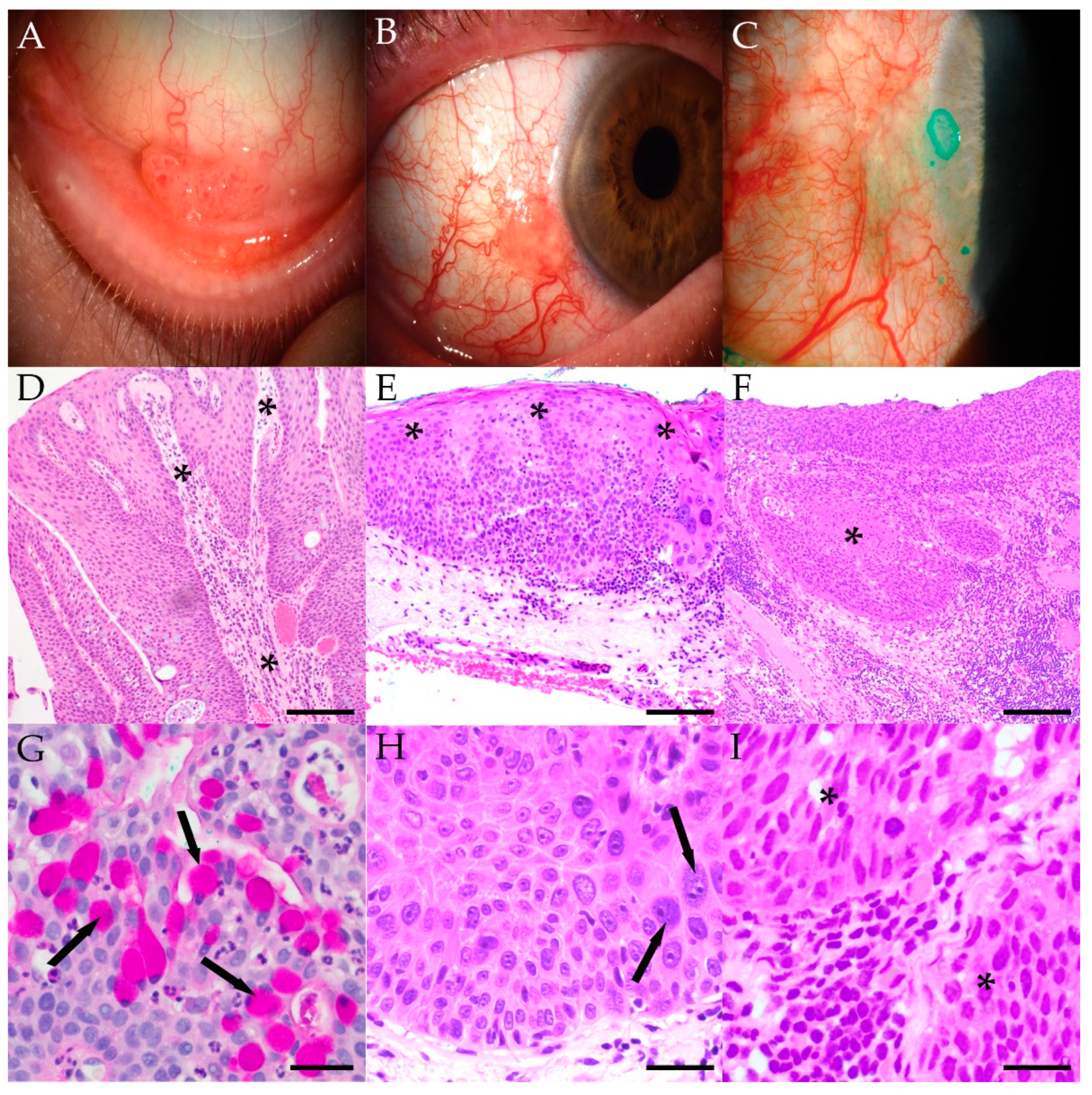
2.1. Clinicopathologic Characteristics

HPV in-situ hybridization was performed on 53 lesions, including 33 papillomas, 16 CIN/cCIS, and 4 invasive SCCs. Demographic and clinicopathologic information for these patients is provided in Table 1, with complete information for individual cases presented in Table S1.
Representative clinical photographs and photomicrographs of each type of lesion are presented in Figure 1. Conjunctival papillomas were generally rich in goblet cells, with epithelium covering fibrovascular cores. Cases of CIN and CIS featured dysplastic cells involving the conjunctival epithelium to varying extents, with sharply defined peripheral edges, but no invasion of underlying stroma. Superficial keratinization was variable, and in some cases, with more prominent thickening of the epithelium, sessile papillomatous changes were noted. Invasive SCC featured nests and strands of tumor cells in the substantia propria. No goblet cells, mucinous, or glandular components were identified in the CIN, CIS, or SCC cases reported here.

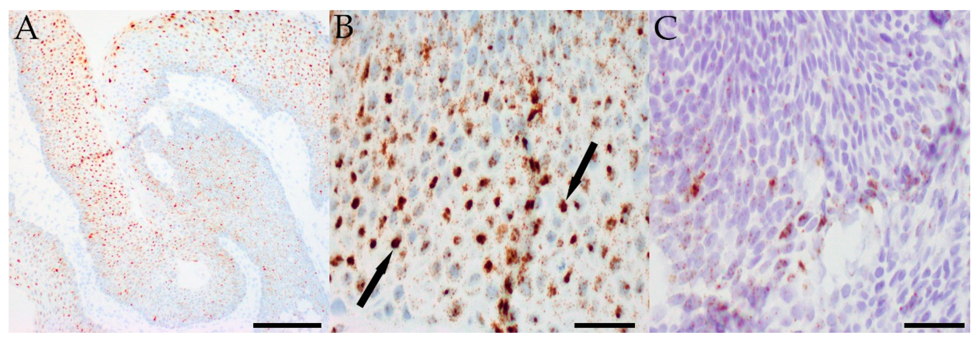
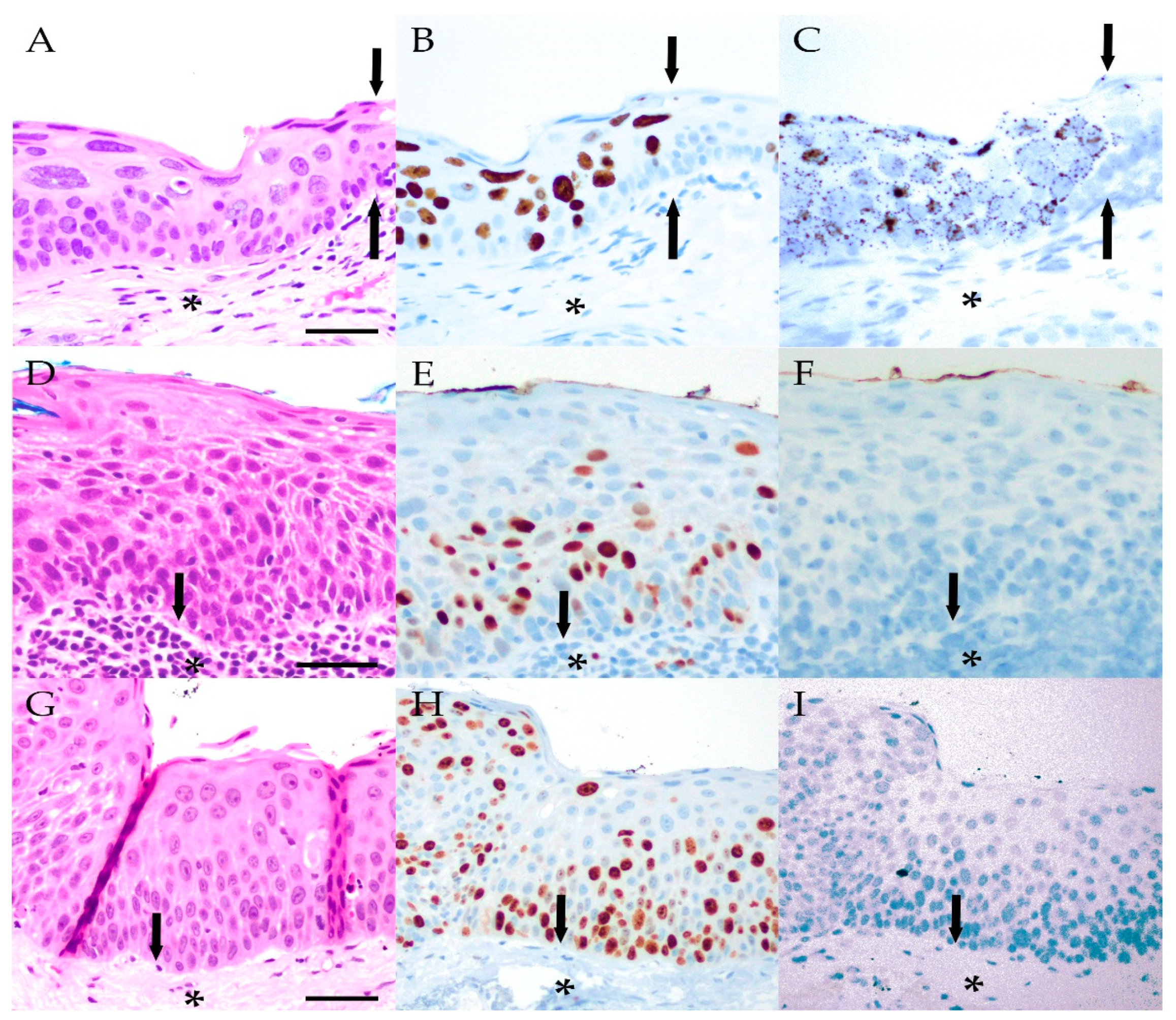
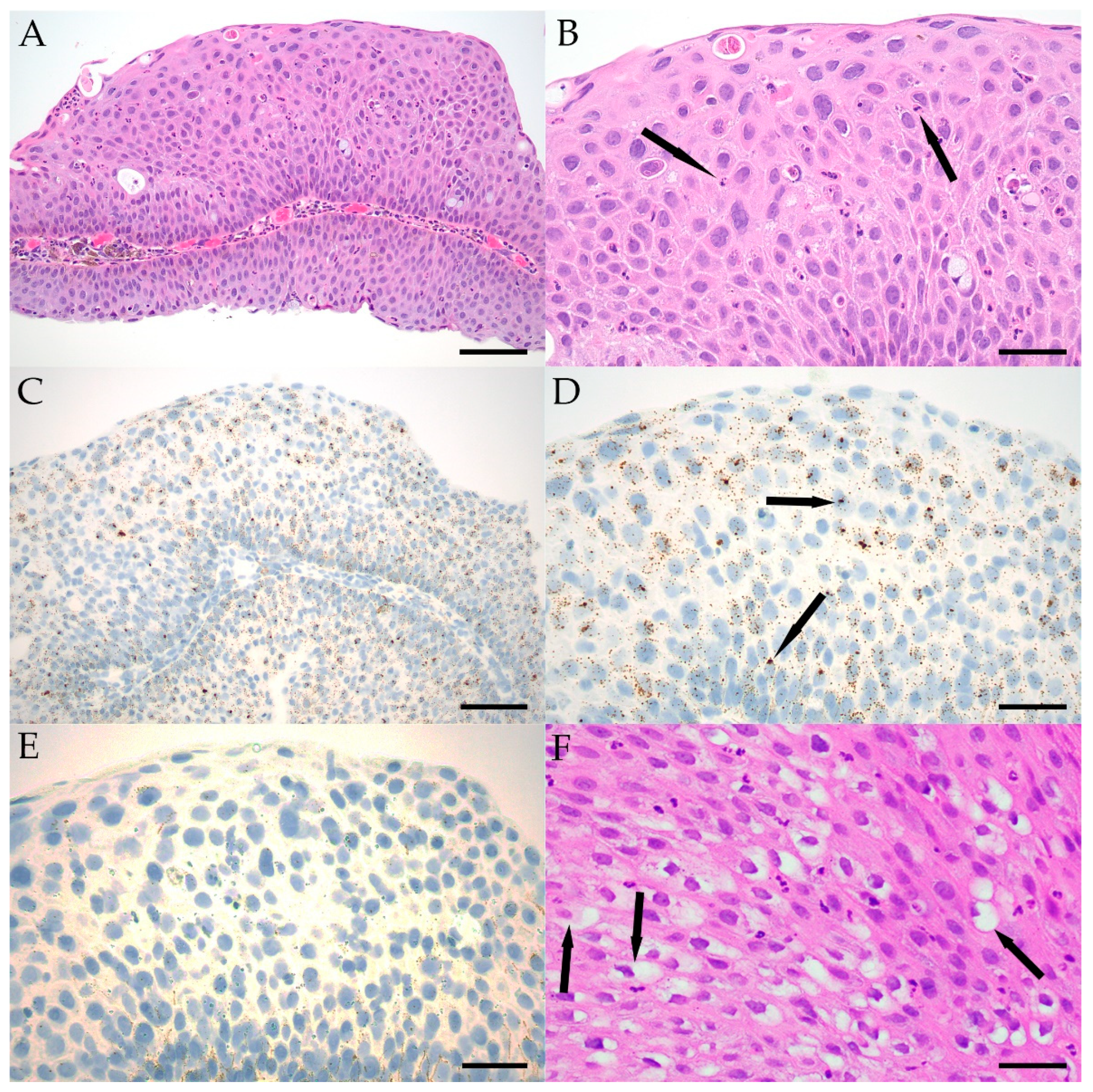
2.2. In-Situ Hybridization

In-situ hybridization using DNA probes was performed in 14/33 (42%) of papillomatous lesions and 4/16 (25%) CIN/cCIS lesions. In-situ hybridization using RNA probes was performed in the remaining lesions: (19/33 (58%) papillomas, 12/16 (75%) CIN/cCIS, and 3/3 invasive SCCs). Low-risk types were detected in 25/30 (83%) papillomas, 0/11 CIN/cCIS, and 0/1 invasive SCCs. High-risk types were detected in 2/28 (7%) papillomas, 1/16 (6%) CIN/cCIS, and 2/4 (50%) invasive SCCs. Detection of LR and HR HPV types was mutually exclusive in papillomatous lesions, and four lesions (12%) were negative for both LR and HR HPV.
HPV signal presented in epithelial cells as dot-like foci visible at 100× magnification in the nucleus. Multiple dots in at least a subset of cells were required to call a case positive. In some cases, the small intracellular signals were quite numerous, and could become almost confluent within a single nucleus. Staining was restricted to epithelial cells, and stromal elements, including blood vessels and mesenchymal cells, were negative in all cases examined. In cases containing normal conjunctiva adjacent to a lesion, the non-neoplastic epithelium was also always negative.
With respect to the extent of staining within individual lesions, HPV was present diffusely in most papillomas. Focal HPV positivity was observed in 8/25 (32%) LR HPV-positive papillomas and 1/2 (50%) HR HPV-positive papillomas, while diffuse staining was noted in 17/25 (68%) and 1/2 (50%) LR and HR HPV-positive papillomas, respectively. In contrast, HPV expression was more localized in the OSSN cases that contained virus. Focal HPV positivity was observed in the single HR HPV-positive CIN (Case 39) and both HR HPV-positive invasive SCC lesions. Further details of HPV in-situ hybridization are summarized in Table 2, and representative images for positive LR and HR HPV in-situ hybridization are presented in Figure 2 and Figure 3.

2.3. Correlation of HPV with Histologic Features

Conjunctival papillomas can become quite inflamed, leading to reactive epithelial atypia, in which epithelial cells show generally mild nuclear enlargement and pleomorphism, along with some chromatin changes and more prominent nucleoli. Reactive atypia can sometimes be difficult to distinguish from dysplasia, although the latter process is often less uniform and more severe. Detection of LR HPV could potentially be helpful in such cases and support a less aggressive biological potential if positive. In 18 of 33 (55%) papilloma cases, intraepithelial inflammation and epithelial atypia diagnosed as reactive were detected. Dysplasia was rarer, and noted in 4/25 (16%) LR HPV-positive papillomas, 1/2 (50%) HR-positive papillomas (Case 29), and none of the HPV-negative papillomas. In three LR HPV-positive papillomas diagnosed with dysplasia, the focal in-situ hybridization signal corresponded to the foci of dysplastic epithelium, while in Case 33 and Case 39, discrete foci of epithelial dysplasia were observed, but more diffuse LR and HR HPV signal was noted. An inflamed papilloma with reactive atypia and LR HPV-positive epithelium is demonstrated in Figure 4A–E.
Koilocytic cells were identified in all lesion types but were relatively rare, with focal change observed in 2/6 (33%) of HPV-negative papillomas, 10/25 (40%) LR HPV-positive papillomas, 1/15 (7%) HPV-negative CIN/cCIS, and 1/2 (50%) HR HPV-positive invasive SCCs. More diffuse koilocytic-appearing cells were observed in 1/25 (4%) LR HPV-positive papillomas, as shown in Figure 4F. Immunolabeling for p16 was performed on seven papillomas, five CIN/cCIS, and three invasive SCCs at the time of initial case evaluation. Generally, p16 labeling corresponded to HPV detection. However, p16 labeling was observed in two CIN/cCIS and one SCC in which HPV was not detected.

2.4. Patient Follow-Up and Atopic History

Patients returned for follow-up care in 17/33 (52%) papilloma cases, 8/16 (50%) CIN/cCIS cases, and none of the invasive SCCs. The length of follow-up (mean ± SD) was 605 ± 1219 days and 887 ± 1534 days for papilloma and CIN/cCIS cases, respectively. Recurrence was noted in only one case (Case 14) following an initial diagnosis of an HPV-negative papilloma. The recurrent lesion was originally treated with excision and subsequently observed.
History of atopic disease, including asthma, eczema, or allergic rhinitis or conjunctivitis, was also recorded for patients when possible. Electronic medical records were available for review for 18/33 (56%) of all papillomas, including 3 HPV-negative lesions, 14 LR HPV-positive lesions, and 1 HR HPV-positive lesion. Atopic disease, including asthma (1/3), rhinitis/conjunctivitis (1/3), and a combination of the two (1/3), was reported in patients with HPV-negative papillomas. A history of asthma was present in 9/14 (64%) patients with LR HPV-positive papillomas, 2 of whom also had additional atopic disease, including eczema and rhinitis/conjunctivitis, respectively. Two additional patients with LR HPV-positive papillomas had histories of eczema and rhinitis/conjunctivitis, alone. There was no reported history of atopic disease in the single HR HPV-positive papilloma. For cases of CIN/cCIS, there was no history of atopic disease in the HR HPV-positive lesion that had medical records available for review.

3. Discussion

The prevalence of HPV infection in conjunctival epithelial proliferations has been difficult to establish, due, at least in part, to the range of detection methods used [35,72]. To some degree, this has been dependent on the type of lesion, and findings in conjunctival papillomas have been more uniform over time [14,31,36]. Our results using DNA and RNA in-situ hybridization were similar to previous studies demonstrating LR HPV infection in the majority of conjunctival papillomas using PCR, as shown in Table 3 [12,13,14,18,31,36,37,44]. In the current study, LR HPV was detected in 94% of papillomas using RNA probes, while only 75% were positive for LR HPV when evaluated by DNA probes. Interestingly, prior studies using DNA probes had lower rates of HPV detected, perhaps reflecting the age of the studies [73]. While some single cases reports examined papillomas using RNA in-situ hybridization, this study represents the first series of cases, and the larger percentage of papillomas found to be positive using RNA in-situ hybridization could potentially reflect the increased sensitivity of this approach [74,75]. However, given the relatively small number of cases in each group, the differences between DNA and RNA in-situ approaches could also be due to normal variation. The cases that were negative for HPV by DNA in-situ hybridization were all several years old, and because of the known potential for RNA degradation, these were not retested using RNA in-situ hybridization.
Conjunctival papillomas can occasionally become inflamed and show prominent reactive atypia, which can be difficult to distinguish from clinically worrisome dysplasia. Among the 33 papillomas we examined, generally mild dysplasia was identified in 5 cases (15%). This number may be elevated due to selection bias, as papillomas with concerning microscopic features were more likely to have had HPV status analyzed during the study period. LR HPV was detected in four of five potentially dysplastic papillomas, while HR HPV was only detected in one. This suggests that the majority of conjunctival papillomas with atypia or dysplasia are associated with LR HPV, and that in-situ analysis could be useful in supporting limited malignant potential in many of these cases. Similar to these findings at the ocular surface, LR HPV was detected in the majority of papillomas, including those with atypical features of the nasopharynx, oral cavity, and larynx, while HR types are more commonly detected in invasive SCCs of these tissues [78,79,80,81,82,83,84].
The reported prevalence of HPV in CIN and other more aggressive OSSN is more variable, with positive cases almost always containing high-risk types [31,34,35,85]. In prior studies, 40–100% of CIN/cCIS were HPV-positive by DNA in-situ hybridization and 0–31% positive by RNA in-situ hybridization [22,34,46,86]. Prevalence was higher in SCC (30–36% positive by DNA in-situ hybridization and 16–26% positive by RNA in-situ hybridization) [34,46,85,86]. In our cohort, 1 CIN/cCIS out of 12 (8%) was positive for HR HPV using RNA in-situ hybridization, while none of the 4 cases evaluated by DNA in-situ hybridization were positive, suggesting that RNA approaches may provide higher sensitivity for detection. The overall positive rate for the 16 cases examined was, thus, 6%.
Immunohistochemical approaches, including direct detection of HPV-encoded proteins or alterations in p16 expression, have been investigated, but more recent studies suggest that these methods are prone to false-positive results [13,22,41,46,49,51,53,55,56,58,59,82]. In particular, it has been found that many lesions negative for HPV express p16 [49,58]. Next-generation sequencing represents another potential method by which HPV would be detected in routine clinical samples [87,88,89]. With respect to clinical advances in care, imaging, including optical coherence tomography, is increasingly being used to distinguish benign and malignant ocular surface lesions [10,87,88,89,90].
Many demographic and clinical associations in our study were similar to prior reports. Papillomas of the conjunctiva reportedly occur more commonly in younger patients, while more dysplastic OSSN lesions tend to present in older ones, and our cases had a mean age of 43, 68, and 74 years at the time of presentation for papillomas, CIN/cCIS, and invasive SCCs, respectively [3,40,77,87]. Prior studies suggested that conjunctival papillomas and OSSN are more common in male patients, and this was true overall in our group as well, except for the relatively small set of SCC [3,7,21,40,46]. Papillomas in our cohort presented more frequently on palpebral conjunctiva (78%), while CIN/cCIS cases were more common for bulbar conjunctiva (82%). However, while a recent report suggested correlation of HR HPV-positive OSSN with atopy, our study did not identify a history of atopic disease in patients with HR HPV lesions [22]. Another group reported increased keratinization in HPV-negative OSSN, but we did not identify a similar association in our cases [46].
Limitations of the current study include the relatively small number of invasive SCC cases, precluding the possibility of statistical comparisons between groups. In addition, detailed review of electronic medical records was not possible for all cases in this series, as many were older pathology consultations submitted by outside physicians. While RNA in archived formalin-fixed paraffin-embedded tissue is labile and sensitive to degradation, potentially affecting the sensitivity of in-situ hybridization, we do not believe this is of concern in our study, as all in-situ hybridization assays were performed around the time of initial histologic review [63,65].

4. Materials and Methods

4.1. Study Design

A retrospective review of the Ophthalmic Pathology Archives of The Johns Hopkins Hospital for cases matching keyword searches including “conjunctiva,” “in-situ hybridization,” and “human papillomavirus” between 2007–2021 was conducted. Electronic medical records were reviewed for the following clinical and pathologic information: age at presentation, gender, lesion type and location, clinical treatment modalities, length of clinical follow-up, history of recurrence, history of atopic disease, HPV in-situ hybridization assay details, and details regarding any additional immunohistochemical stains. Any case for which in-situ hybridization was performed to detect LR and/or HR HPV in a conjunctival papilloma, CIN/cCIS, or invasive squamous cell carcinoma was included. Cases were reviewed to confirm the initial diagnosis, and intralesional koilocytic or dysplastic features were noted.

4.2. HPV In-Situ Hybridization

HPV in-situ hybridization was performed by the Johns Hopkins Hospital clinical laboratories at the time of initial review of histology using standard protocols. Lesions assayed prior to 2015 were subject to DNA ISH (Ventana Medical Systems; Tucson, AZ, USA), and those evaluated from 2015 and onward were analyzed using RNA probes (Advanced Cell Diagnostics Inc; Hayward, CA, USA). Both DNA and RNA probes for LR HPV detect the same two types (6, 11), while DNA probes for HR HPV detect 12 types (16, 18, 31, 33, 35, 39, 45, 51, 52, 56, 58, and 68) and RNA probes detect 18 HR HPV types (16, 18, 26, 31, 33, 35, 39, 45, 51, 52, 53, 56, 58, 59, 66, 68, 73, and 82). While 37/53 lesions (70%) were evaluated for both LR and HR types, evaluation by either DNA or RNA approaches was mutually exclusive. The commercially available probes utilized in this study were designed to hybridize with E6/E7 in the various HPV types. All determinations of in-situ hybridization positivity were performed by an ocular pathologist (CGE).

5. Conclusions

We confirmed that LR HPV is frequently detected in conjunctival papillomas while HR HPV is rarely detected in these lesions. The fact that most papillomas with histopathological dysplasia were positive for LR HPV suggests that their malignant potential is limited, and that in-situ hybridization may help with prognostic assessment, although analysis of more cases with long-term follow-up will be needed to confirm this. HR HPV was detected by RNA in-situ hybridization in a relatively small number of CIN and SCC, but was not clearly linked to atopy or other clinical and histopathological features.

Supplementary Materials

The following supporting information can be downloaded at: https://www.mdpi.com/article/10.3390/ijms23137249/s1.

Author Contributions

Conceptualization, R.N.P., S.S., C.G.E.; data curation, C.P., R.N.P., C.G.E.; formal analysis, C.P., R.N.P.; methodology, M.T.A., A.A.C., Y.D., N.M.; project administration, C.G.E.; supervision, CGE; writing—original draft preparation, R.N.P., C.P.; writing—review and editing, C.P., M.T.A., A.A.C., Y.D., N.M., S.S., C.G.E. All authors have read and agreed to the published version of the manuscript.

Funding

The authors received no financial support for authorship and/or publication of this article. C.P. was supported by NIH T32 OD011089 (PI: Mankowski).

Institutional Review Board Statement

The study was conducted in accordance with the Declaration of Helsinki and approved by the Institutional Review Board of Johns Hopkins Hospital (NA_00011133, approved 10/9/2009) for studies involving humans.

Informed Consent Statement

Informed consent was obtained from all subjects involved in the study.

Data Availability Statement

Not applicable.

Conflicts of Interest

The authors declare no conflict of interest.

References

  1. Di Girolamo, N. Association of Human Papilloma Virus with Pterygia and Ocular-Surface Squamous Neoplasia. Eye 2012, 26, 202–211. [Google Scholar] [CrossRef] [PubMed] [Green Version]
  2. Yin, V.T.; Merritt, H.A.; Sniegowski, M.; Esmaeli, B. Eyelid and Ocular Surface Carcinoma: Diagnosis and Management. Clin. Dermatol. 2015, 33, 159–169. [Google Scholar] [CrossRef] [PubMed]
  3. Bellerive, C.; Berry, J.L.; Polski, A.; Singh, A.D. Conjunctival Squamous Neoplasia: Staging and Initial Treatment. Cornea 2018, 37, 1287–1291. [Google Scholar] [CrossRef] [PubMed]
  4. Boneva, S.; Schlecht, A.; Zhang, P.; Boehringer, D.; Lapp, T.; Mittelviefhaus, H.; Reinhard, T.; Auw-Haedrich, C.; Schlunck, G.; Wolf, J.; et al. MACE RNA Sequencing Analysis of Conjunctival Squamous Cell Carcinoma and Papilloma Using Formalin-Fixed Paraffin-Embedded Tumor Tissue. Sci. Rep. 2020, 10, 21292. [Google Scholar] [CrossRef]
  5. Cervantes, G.; Rodríguez, A.A.; Leal, A.G. Squamous Cell Carcinoma of the Conjunctiva: Clinicopathological Features in 287 Cases. Can. J. Ophthalmol. J. Can. Ophtalmol. 2002, 37, 14–19, discussion 19–20. [Google Scholar] [CrossRef]
  6. Crim, N.; Forniés-Paz, M.E.; Monti, R.; Espósito, E.; Maccio, J.P.; Urrets-Zavalía, J.A. In Situ Carcinoma of the Conjunctiva: Surgical Excision Associated with Cryotherapy. Clin. Ophthalmol. 2013, 7, 1889–1893. [Google Scholar] [CrossRef] [PubMed] [Green Version]
  7. Gichuhi, S.; Sagoo, M.S.; Weiss, H.A.; Burton, M.J. Epidemiology of Ocular Surface Squamous Neoplasia in Africa. Trop. Med. Int. Health TMIH 2013, 18, 1424–1443. [Google Scholar] [CrossRef] [PubMed] [Green Version]
  8. Basti, S.; Macsai, M.S. Ocular Surface Squamous Neoplasia: A Review. Cornea 2003, 22, 687–704. [Google Scholar] [CrossRef] [PubMed]
  9. Gichuhi, S.; Ohnuma, S.-I.; Sagoo, M.S.; Burton, M.J. Pathophysiology of Ocular Surface Squamous Neoplasia. Exp. Eye Res. 2014, 129, 172–182. [Google Scholar] [CrossRef] [PubMed] [Green Version]
  10. Alvarez, O.P.; Zein, M.; Galor, A.; Karp, C.L. Management of Ocular Surface Squamous Neoplasia: Bowman Club Lecture 2021. BMJ Open Ophthalmol. 2021, 6, e000842. [Google Scholar] [CrossRef] [PubMed]
  11. Mittal, R.; Rath, S.; Vemuganti, G.K. Ocular Surface Squamous Neoplasia—Review of Etio-Pathogenesis and an Update on Clinico-Pathological Diagnosis. Saudi J. Ophthalmol. 2013, 27, 177–186. [Google Scholar] [CrossRef] [PubMed]
  12. Hanbazazh, M.; Gyure, K.A. Ocular Human Papillomavirus Infections. Arch. Pathol. Lab. Med. 2018, 142, 706–710. [Google Scholar] [CrossRef] [PubMed] [Green Version]
  13. Moyer, A.B.; Roberts, J.; Olsen, R.J.; Chévez-Barrios, P. Human Papillomavirus—Driven Squamous Lesions: High-Risk Genotype Found in Conjunctival Papillomas, Dysplasia, and Carcinoma. Am. J. Dermatopathol. 2018, 40, 486–490. [Google Scholar] [CrossRef]
  14. Sjö, N.C.; von Buchwald, C.; Cassonnet, P.; Norrild, B.; Prause, J.U.; Vinding, T.; Heegaard, S. Human Papillomavirus in Normal Conjunctival Tissue and in Conjunctival Papilloma: Types and Frequencies in a Large Series. Br. J. Ophthalmol. 2007, 91, 1014–1015. [Google Scholar] [CrossRef] [PubMed] [Green Version]
  15. Ng, J.; Coroneo, M.T.; Wakefield, D.; Di Girolamo, N. Ultraviolet Radiation and the Role of Matrix Metalloproteinases in the Pathogenesis of Ocular Surface Squamous Neoplasia. Investig. Opthalmol. Vis. Sci. 2008, 49, 5295–5306. [Google Scholar] [CrossRef] [Green Version]
  16. Ateenyi-Agaba, C.; Dai, M.; Le Calvez, F.; Katongole-Mbidde, E.; Smet, A.; Tommasino, M.; Franceschi, S.; Hainaut, P.; Weiderpass, E. TP53 Mutations in Squamous-Cell Carcinomas of the Conjunctiva: Evidence for UV-Induced Mutagenesis. Mutagenesis 2004, 19, 399–401. [Google Scholar] [CrossRef]
  17. Yazu, H.; Dogru, M.; Miyauchi, J.; Tanaka, Y.; Yamaguchi, T.; Den, S.; Satake, Y.; Tsubota, K.; Shimazaki, J. Association of Epithelial Atypia with Recurrence after Surgical Excision in Conjunctival Papilloma. Eye Contact Lens 2018, 44 (Suppl. 1), S77–S81. [Google Scholar] [CrossRef]
  18. Karcioglu, Z.A.; Issa, T.M. Human Papilloma Virus in Neoplastic and Non-Neoplastic Conditions of the External Eye. Br. J. Ophthalmol. 1997, 81, 595–598. [Google Scholar] [CrossRef] [Green Version]
  19. Bredow, L.; Martin, G.; Reinhard, T.; Mittelviefhaus, H.; Auw-Haedrich, C. Recurrent Conjunctival Papilloma Progressing into Squamous Cell Carcinoma with Change of HPV-Finding During the Course. Br. J. Ophthalmol. 2009, 93, 1437. [Google Scholar] [CrossRef]
  20. de la Vega, L.L.; Bick, N.; Hu, K.; Rahrig, S.E.; Silva, C.D.; Matayoshi, S.; Picciarelli, P.; Wang, X.; Sugar, A.; Soong, H.K.; et al. Invasive Squamous Cell Carcinomas and Precursor Lesions on UV-Exposed Epithelia Demonstrate Concordant Genomic Complexity in Driver Genes. Mod. Pathol. 2020, 33, 2280–2294. [Google Scholar] [CrossRef]
  21. Hamam, R.; Bhat, P.; Foster, C.S. Conjunctival/Corneal Intraepithelial Neoplasia. Int. Ophthalmol. Clin. 2009, 49, 63–70. [Google Scholar] [CrossRef] [PubMed]
  22. Griffin, H.; Mudhar, H.S.; Rundle, P.; Shiraz, A.; Mahmood, R.; Egawa, N.; Quint, W.; Rennie, I.G.; Doorbar, J. Human Papillomavirus Type 16 Causes a Defined Subset of Conjunctival In Situ Squamous Cell Carcinomas. Mod. Pathol. 2020, 33, 74–90. [Google Scholar] [CrossRef]
  23. Cheng, J.; Zens, M.S.; Duell, E.; Perry, A.E.; Chapman, M.S.; Karagas, M.R. History of Allergy and Atopic Dermatitis in Relation to Squamous Cell and Basal Cell Carcinoma of the Skin. Cancer Epidemiol. Biomark. Prev. 2015, 24, 749–754. [Google Scholar] [CrossRef] [PubMed] [Green Version]
  24. Jensen, A.O.; Svaerke, C.; Körmendiné Farkas, D.; Olesen, A.B.; Kragballe, K.; Sørensen, H.T. Atopic Dermatitis and Risk of Skin Cancer: A Danish Nationwide Cohort Study (1977–2006). Am. J. Clin. Dermatol. 2012, 13, 29–36. [Google Scholar] [CrossRef] [PubMed]
  25. Arana, A.; Wentworth, C.E.; Fernández-Vidaurre, C.; Schlienger, R.G.; Conde, E.; Arellano, F.M. Incidence of Cancer in the General Population and in Patients with or without Atopic Dermatitis in the U.K. Br. J. Dermatol. 2010, 163, 1036–1043. [Google Scholar] [CrossRef]
  26. Wiemels, J.L.; Wiencke, J.K.; Li, Z.; Ramos, C.; Nelson, H.H.; Karagas, M.R. Risk of Squamous Cell Carcinoma of the Skin in Relation to IgE: A Nested Case—Control Study. Cancer Epidemiol. Biomark. Prev. 2011, 20, 2377–2383. [Google Scholar] [CrossRef] [Green Version]
  27. Sritippho, T.; Chotjumlong, P.; Iamaroon, A. Roles of Human Papillomaviruses and p16 in Oral Cancer. Asian Pac. J. Cancer Prev. APJCP 2015, 16, 6193–6200. [Google Scholar] [CrossRef] [Green Version]
  28. Bzhalava, D.; Eklund, C.; Dillner, J. International Standardization and Classification of Human Papillomavirus Types. Virology 2015, 476, 341–344. [Google Scholar] [CrossRef]
  29. Bernard, H.-U.; Burk, R.D.; Chen, Z.; van Doorslaer, K.; zur Hausen, H.; de Villiers, E.-M. Classification of Papillomaviruses (PVs) Based on 189 PV Types and Proposal of Taxonomic Amendments. Virology 2010, 401, 70–79. [Google Scholar] [CrossRef] [Green Version]
  30. Pim, D.; Banks, L. Interaction of Viral Oncoproteins with Cellular Target Molecules: Infection with High-Risk vs. Low-Risk Human Papillomaviruses. APMIS Acta Pathol. Microbiol. Immunol. Scand. 2010, 118, 471–493. [Google Scholar] [CrossRef]
  31. Chalkia, A.K.; Bontzos, G.; Spandidos, D.A.; Detorakis, E.T. Human Papillomavirus Infection and Ocular Surface Disease (Review). Int. J. Oncol. 2019, 54, 1503–1510. [Google Scholar] [CrossRef] [PubMed] [Green Version]
  32. Burk, R.D.; Harari, A.; Chen, Z. Human Papillomavirus Genome Variants. Virology 2013, 445, 232–243. [Google Scholar] [CrossRef] [PubMed] [Green Version]
  33. National Institute of Allergy and Infectious Diseases. PaVE: The Papillomavirus Episteme; National Institutes of Health: Bethesda, MD, USA, 2013. [Google Scholar]
  34. Afrogheh, A.H.; Jakobiec, F.A.; Hammon, R.; Grossniklaus, H.E.; Rocco, J.; Lindeman, N.I.; Sadow, P.M.; Faquin, W.C. Evaluation for High-Risk HPV in Squamous Cell Carcinomas and Precursor Lesions Arising in the Conjunctiva and Lacrimal Sac. Am. J. Surg. Pathol. 2016, 40, 519–528. [Google Scholar] [CrossRef] [PubMed]
  35. Ramberg, I.; Møller-Hansen, M.; Toft, P.B.; Funding, M.; Heegaard, S. Human Papillomavirus Infection Plays a Role in Conjunctival Squamous Cell Carcinoma: A Systematic Review and Meta-Analysis of Observational Studies. Acta Ophthalmol. 2021, 99, 478–488. [Google Scholar] [CrossRef]
  36. Mlakar, J.; Kocjan, B.J.; Hošnjak, L.; Pižem, J.; Beltram, M.; Gale, N.; Drnovšek-Olup, B.; Poljak, M. Morphological Characteristics of Conjunctival Squamous Papillomas in Relation to Human Papillomavirus Infection. Br. J. Ophthalmol. 2015, 99, 431–436. [Google Scholar] [CrossRef]
  37. Kalogeropoulos, C.; Koumpoulis, I.; Papadiotis, E.; Zioga, A.; Gkrepi, K.; Pappa, C.; Paschides, C.; Malamou-Mitsi, V.; Aspiotis, M. Squamous Cell Papilloma of the Conjunctiva Due to Human Papillomavirus (HPV): Presentation of Two Cases and Review of Literature. Clin. Ophthalmol. 2012, 6, 1553. [Google Scholar] [CrossRef] [Green Version]
  38. George, R.S.; Lewis, D.R.; Archibald, C.W.; Heathcote, G. Bilateral HPV Positive Squamous Cell Carcinoma In Situ of Conjunctiva. Ophthalmic Plast. Reconstr. Surg. 2018, 34, e1–e3. [Google Scholar] [CrossRef]
  39. Saegusa, M.; Takano, Y.; Hashimura, M.; Okayasu, I.; Shiga, J. HPV Type 16 in Conjunctival and Junctional Papilloma, Dysplasia, and Squamous Cell Carcinoma. J. Clin. Pathol. 1995, 48, 1106–1110. [Google Scholar] [CrossRef] [Green Version]
  40. Huang, Y.-M.; Huang, Y.-Y.; Yang, H.-Y.; Tsai, C.-C.; Yu, W.-K.; Kao, S.-C.; Kau, H.-C.; Liu, C.J.-L. Conjunctival Papilloma: Clinical Features, Outcome, and Factors Related to Recurrence. Taiwan J. Ophthalmol. 2018, 8, 15–18. [Google Scholar] [CrossRef]
  41. Yu, L.; Fei, L.; Liu, X.; Pi, X.; Wang, L.; Chen, S. Application of p16/Ki-67 Dual-Staining Cytology in Cervical Cancers. J. Cancer 2019, 10, 2654–2660. [Google Scholar] [CrossRef] [Green Version]
  42. Buitrago-Pérez, A.; Garaulet, G.; Vázquez-Carballo, A.; Paramio, J.M.; García-Escudero, R. Molecular Signature of HPV-Induced Carcinogenesis: pRb, p53 and Gene Expression Profiling. Curr. Genom. 2009, 10, 26–34. [Google Scholar] [CrossRef] [PubMed] [Green Version]
  43. Manzouri, B.; Luthert, P.J.; Larkin, D.F.P. Papilloma Development and Long-Term Ciclosporin Use in Chronic Ocular Allergy with Associated Keratoconus. Eye Contact Lens 2013, 39, 402–404. [Google Scholar] [CrossRef]
  44. Verma, V.; Shen, D.; Sieving, P.C.; Chan, C.-C. The Role of Infectious Agents in the Etiology of Ocular Adnexal Neoplasia. Surv. Ophthalmol. 2008, 53, 312–331. [Google Scholar] [CrossRef] [PubMed] [Green Version]
  45. Osahon, A.I.; Ukponmwan, C.U.; Uhunmwangho, O.M. Prevalence of HIV Seropositivity among Patients with Squamous Cell Carcinoma of the Conjunctiva. Asian Pac. J. Trop. Biomed. 2011, 1, 150–153. [Google Scholar] [CrossRef] [Green Version]
  46. Ramberg, I.; Toft, P.B.; Georgsen, J.B.; Siersma, V.D.; Funding, M.; Jensen, D.H.; von Buchwald, C.; Heegaard, S. Conjunctival Intraepithelial Neoplasia and Carcinoma: Distinct Clinical and Histological Features in Relation to Human Papilloma Virus Status. Br. J. Ophthalmol. 2021, 105, 878–883. [Google Scholar] [CrossRef]
  47. Abreu, A.L.P.; Souza, R.P.; Gimenes, F.; Consolaro, M.E.L. A Review of Methods for Detect Human Papillomavirusinfection. Virol. J. 2012, 9, 262. [Google Scholar] [CrossRef] [PubMed] [Green Version]
  48. Shrestha, T.; Choi, W.; Kim, G.E.; Yang, J.M.; Yoon, K.C. Human Papilloma Virus Identification in Ocular Surface Squamous Neoplasia by p16 Immunohistochemistry and DNA Chip Test: A Strobe-Compliant Article. Medicine 2019, 98, e13944. [Google Scholar] [CrossRef]
  49. Carrilho, C.; Miu, C.; Kim, Y.; Karki, S.; Balmaceda, A.; Challa, B.; Diamond, S.; Monteiro, E.; Marole, E.; Lorenzoni, C.; et al. p16 Expression Correlates with Invasive Ocular Surface Squamous Neoplasms in HIV-Infected Mozambicans. Ocul. Oncol. Pathol. 2020, 6, 123–128. [Google Scholar] [CrossRef]
  50. Mann, P.; Diamond, S.; Masanganise, R.; Gwanzura, L.; Liu, Y.-T.; Schooley, R.T.; Lin, J.H. HPV16/18 and p16 Gene Expression in Ocular Surface Squamous Neoplasia: A Retrospective Cross-Sectional Analysis. Investig. Ophthalmol. Vis. Sci. 2018, 59, 5594. [Google Scholar]
  51. Ukpo, O.C.; Flanagan, J.J.; Ma, X.-J.; Luo, Y.; Thorstad, W.L.; Lewis, J.S. High-Risk Human Papillomavirus E6/E7 mRNA Detection by a Novel In Situ Hybridization Assay Strongly Correlates with p16 Expression and Patient Outcomes in Oropharyngeal Squamous Cell Carcinoma. Am. J. Surg. Pathol. 2011, 35, 1343–1350. [Google Scholar] [CrossRef]
  52. Venuti, A.; Paolini, F. HPV Detection Methods in Head and Neck Cancer. Head Neck Pathol. 2012, 6 (Suppl. 1), 63–74. [Google Scholar] [CrossRef] [PubMed] [Green Version]
  53. Yu, L.; Wang, L.; Zhong, J.; Chen, S. Diagnostic value of p16INK4A, Ki-67, and Human Papillomavirus L1 Capsid Protein Immunochemical Staining on Cell Blocks from Residual Liquid-Based Gynecologic Cytology Specimens. Cancer Cytopathol. 2010, 118, 47–55. [Google Scholar] [CrossRef] [PubMed]
  54. Hoffmann, M.; Ihloff, A.S.; Görögh, T.; Weise, J.B.; Fazel, A.; Krams, M.; Rittgen, W.; Schwarz, E.; Kahn, T. p16INK4a Overexpression Predicts Translational Active Human Papillomavirus Infection in Tonsillar Cancer. Int. J. Cancer 2010, 127, 1595–1602. [Google Scholar] [CrossRef] [PubMed]
  55. Zhu, Y.; Ren, C.; Yang, L.; Zhang, X.; Liu, L.; Wang, Z. Performance of p16/Ki67 Immunostaining, HPV E6/E7 mRNA Testing, and HPV DNA Assay to Detect High-Grade Cervical Dysplasia in Women with ASCUS. BMC Cancer 2019, 19, 271. [Google Scholar] [CrossRef] [Green Version]
  56. Wang, X.; Zhao, Y.; Zou, X.; Wang, L. Relationship of p16 and Ki67 in Recurrence of HPV Infection and Cervical Intraepithelial Neoplasia. Int. J. Clin. Exp. Pathol. 2020, 13, 3174–3180. [Google Scholar]
  57. Bulten, J.; van der Avoort, I.A.M.; Melchers, W.J.G.; Massuger, L.F.A.G.; Grefte, J.M.M.; Hanselaar, A.G.J.M.; de Wilde, P.C.M. p14ARF and p16INK4A, Two Products of the Same Gene, Are Differently Expressed in Cervical Intraepithelial Neoplasia. Gynecol. Oncol. 2006, 101, 487–494. [Google Scholar] [CrossRef]
  58. Wong, K.S.; Krane, J.F.; Jo, V.Y. Heterogeneity of p16 Immunohistochemistry and Increased Sensitivity of RNA In Situ Hybridization in Cytology Specimens of HPV-Related Head and Neck Squamous Cell Carcinoma. Cancer Cytopathol. 2019, 127, 632–642. [Google Scholar] [CrossRef]
  59. Abi-Raad, R.; Prasad, M.L.; Gilani, S.; Garritano, J.; Barlow, D.; Cai, G.; Adeniran, A.J. Quantitative Assessment of p16 Expression in FNA Specimens from Head and Neck Squamous Cell Carcinoma and Correlation with HPV Status. Cancer Cytopathol. 2021, 129, 394–404. [Google Scholar] [CrossRef]
  60. Rooper, L.M.; Gandhi, M.; Bishop, J.A.; Westra, W.H. RNA In-Situ Hybridization is a Practical and Effective Method for Determining HPV Status of Oropharyngeal Squamous Cell Carcinoma Including Discordant Cases That Are p16 Positive by Immunohistochemistry but HPV Negative by DNA In-Situ Hybridization. Oral Oncol. 2016, 55, 11–16. [Google Scholar] [CrossRef]
  61. Bishop, J.A.; Ma, X.-J.; Wang, H.; Luo, Y.; Illei, P.B.; Begum, S.; Taube, J.M.; Koch, W.M.; Westra, W.H. Detection of Transcriptionally Active High-risk HPV in Patients with Head and Neck Squamous Cell Carcinoma as Visualized by a Novel E6/E7 mRNA In Situ Hybridization Method. Am. J. Surg. Pathol. 2012, 36, 1874–1882. [Google Scholar] [CrossRef] [Green Version]
  62. Lewis, J.S., Jr.; Ukpo, O.C.; Ma, X.-J.; Flanagan, J.J.; Luo, Y.; Thorstad, W.L.; Chernock, R.D. Transcriptionally-Active High-Risk Human Papillomavirus Is Rare in Oral Cavity and Laryngeal/Hypopharyngeal Squamous Cell Carcinomas—A Tissue Microarray Study Utilizing E6/E7 mRNA In Situ Hybridization. Histopathology 2012, 60, 982–991. [Google Scholar] [CrossRef] [PubMed]
  63. Wang, F.; Flanagan, J.; Su, N.; Wang, L.-C.; Bui, S.; Nielson, A.; Wu, X.; Vo, H.-T.; Ma, X.-J.; Luo, Y. RNAscope: A Novel In Situ RNA Analysis Platform for Formalin-Fixed, Paraffin-Embedded Tissues. J. Mol. Diagn. 2012, 14, 22–29. [Google Scholar] [CrossRef] [PubMed] [Green Version]
  64. Kelesidis, T.; Aish, L.; Steller, M.A.; Aish, I.S.; Shen, J.; Foukas, P.; Panayiotides, J.; Petrikkos, G.; Karakitsos, P.; Tsiodras, S. Human Papillomavirus (HPV) Detection Using In Situ Hybridization in Histologic Samples. Am. J. Clin. Pathol. 2011, 136, 119–127. [Google Scholar] [CrossRef] [PubMed] [Green Version]
  65. Garcia, J.A.; Best, S.R.; Rooper, L.M. HPV RNA In-Situ Hybridization as a Diagnostic aid in Papillary Laryngeal Lesions. Laryngoscope 2020, 130, 955–960. [Google Scholar] [CrossRef]
  66. Pagliusi, S.R.; Garland, S.M. International Standard Reagents for HPV Detection. Dis. Markers 2007, 23, 283–296. [Google Scholar] [CrossRef] [Green Version]
  67. Pandey, M.; Bhosale, P.G.; Mahimkar, M.B. Detection of HPV E6/E7 mRNA in Clinical Samples Using RNA In Situ Hybridization. Methods Mol. Biol. 2018, 1726, 167–175. [Google Scholar] [CrossRef]
  68. Coppock, J.D.; Willis, B.C.; Stoler, M.H.; Mills, A.M. HPV RNA In Situ Hybridization Can Inform Cervical Cytology-Histology Correlation. Cancer Cytopathol. 2018, 126, 533–540. [Google Scholar] [CrossRef] [Green Version]
  69. Suresh, K.; Shah, P.V.; Coates, S.; Alexiev, B.A.; Samant, S. In Situ Hybridization for High Risk HPV E6/E7 mRNA in Oropharyngeal Squamous Cell Carcinoma. Am. J. Otolaryngol. 2021, 42, 102782. [Google Scholar] [CrossRef]
  70. Outh-Gauer, S.; Augustin, J.; Mandavit, M.; Grard, O.; Denize, T.; Nervo, M.; Lépine, C.; Rassy, M.; Tartour, E.; Badoual, C. Chromogenic In Situ Hybridization as a Tool for HPV-Related Head and Neck Cancer Diagnosis. J. Vis. Exp. JoVE 2019, 148, e59422. [Google Scholar] [CrossRef] [Green Version]
  71. Henley-Smith, R.; Santambrogio, A.; Andoniadou, C.L.; Odell, E.; Thavaraj, S. RNA In Situ Hybridization for Human Papillomavirus Testing in Oropharyngeal Squamous Cell Carcinoma on a Routine Clinical Diagnostic Platform. J. Oral Pathol. Med. 2021, 50, 68–75. [Google Scholar] [CrossRef]
  72. Ramberg, I.; Heegaard, S. Human Papillomavirus Related Neoplasia of the Ocular Adnexa. Viruses 2021, 13, 1522. [Google Scholar] [CrossRef] [PubMed]
  73. Nakamura, Y.; Mashima, Y.; Kameyama, K.; Mukai, M.; Oguchi, Y. Detection of Human Papillomavirus Infection in Squamous Tumours of the Conjunctiva and Lacrimal Sac by Immunohistochemistry, In Situ Hybridisation, and Polymerase Chain Reaction. Br. J. Ophthalmol. 1997, 81, 308–313. [Google Scholar] [CrossRef] [PubMed] [Green Version]
  74. Annadanam, A.; Vizcaino, M.A.; Eberhart, C.G.; Khurshid, G.S.; Gupta, P. Long Standing Exophytic Conjunctival Papilloma Infected with Human Papillomavirus. J. Eye Ophthalmol. 2017, 4, 1. [Google Scholar] [CrossRef] [Green Version]
  75. Michel, J.; Guiguen, Y.; Léger, F.; Gain, P.; Valanconny, C.; Cambazard, F. Human papilloma virus 6/11 inconjunctival papilloma. Ann. Dermatol. Venereol. 1996, 123, 90–92. [Google Scholar] [PubMed]
  76. Minchiotti, S.; Masucci, L.; Santos, M.S.D.; Perrella, E.; Graffeo, R.; Lambiase, A.; Bonini, S. Conjunctival Papilloma and Human Papillomavirus: Identification of HPV Types by PCR. Eur. J. Ophthalmol. 2006, 16, 473–477. [Google Scholar] [CrossRef]
  77. Sjö, N.; Heegaard, S.; Prause, J.U. Conjunctival Papilloma. Acta Ophthalmol. Scand. 2000, 78, 663–666. [Google Scholar] [CrossRef]
  78. Ikegami, T.; Kise, N.; Kinjyo, H.; Kondo, S.; Suzuki, M.; Tsukahara, N.; Murakami, A.; Kiyuna, A.; Agena, S.; Tanaka, K.; et al. Development of Antibodies against HPV-6 and HPV-11 for the Study of Laryngeal Papilloma. Viruses 2021, 13, 2024. [Google Scholar] [CrossRef]
  79. Amiling, R.; Meites, E.; Querec, T.D.; Stone, L.; Singh, V.; Unger, E.R.; Derkay, C.S.; Markowitz, L.E. Juvenile-Onset Recurrent Respiratory Papillomatosis in the United States, Epidemiology and HPV Types—2015–2020. J. Pediatr. Infect. Dis. Soc. 2021, 10, 774–781. [Google Scholar] [CrossRef]
  80. Li, H.; Torabi, S.J.; Yarbrough, W.G.; Mehra, S.; Osborn, H.A.; Judson, B. Association of Human Papillomavirus Status at Head and Neck Carcinoma Subsites with Overall Survival. JAMA Otolaryngol. Head Neck Surg. 2018, 144, 519. [Google Scholar] [CrossRef] [Green Version]
  81. McCord, C.; Xu, J.; Xu, W.; Qiu, X.; Muhanna, N.; Irish, J.; Leong, I.; McComb, R.J.; Perez-Ordonez, B.; Bradley, G. Association of Human Papilloma Virus with Atypical and Malignant Oral Papillary Lesions. Oral Surg. Oral Med. Oral Pathol. Oral Radiol. 2014, 117, 722–732. [Google Scholar] [CrossRef] [Green Version]
  82. Mannarini, L.; Kratochvil, V.; Calabrese, L.; Gomes Silva, L.; Morbini, P.; Betka, J.; Benazzo, M. Human Papilloma Virus (HPV) in Head and Neck Region: Review of Literature. Acta Otorhinolaryngol. Ital. 2009, 29, 119–126. [Google Scholar] [PubMed]
  83. Paver, E.C.; Currie, A.M.; Gupta, R.; Dahlstrom, J.E. Human Papilloma Virus Related Squamous Cell Carcinomas of the Head and Neck: Diagnosis, Clinical Implications and Detection of HPV. Pathology 2020, 52, 179–191. [Google Scholar] [CrossRef] [PubMed] [Green Version]
  84. Sabatini, M.E.; Chiocca, S. Human Papillomavirus as a Driver of Head and Neck Cancers. Br. J. Cancer 2020, 122, 306–314. [Google Scholar] [CrossRef] [PubMed]
  85. Nagarajan, P.; El-Hadad, C.; Gruschkus, S.K.; Ning, J.; Hudgens, C.W.; Sagiv, O.; Gross, N.; Tetzlaff, M.T.; Esmaeli, B. PD-L1/PD1 Expression, Composition of Tumor-Associated Immune Infiltrate, and HPV Status in Conjunctival Squamous Cell Carcinoma. Investig. Opthalmol. Vis. Sci. 2019, 60, 2388–2398. [Google Scholar] [CrossRef] [Green Version]
  86. Galor, A.; Garg, N.; Nanji, A.; Joag, M.; Nuovo, G.; Palioura, S.; Wang, G.; Karp, C.L. Human Papilloma Virus Infection Does Not Predict Response to Interferon Therapy in Ocular Surface Squamous Neoplasia. Ophthalmology 2015, 122, 2210–2215. [Google Scholar] [CrossRef] [Green Version]
  87. Kaliki, S.; Arepalli, S.; Shields, C.L.; Klein, K.; Sun, H.; Hysenj, E.; Lally, S.E.; Shields, J.A. Conjunctival Papilloma. JAMA Ophthalmol. 2013, 131, 585. [Google Scholar] [CrossRef] [Green Version]
  88. Theotoka, D.; Morkin, M.I.; Galor, A.; Karp, C.L. Update on Diagnosis and Management of Conjunctival Papilloma. Eye Vis. 2019, 6, 18. [Google Scholar] [CrossRef]
  89. Galati, L.; Combes, J.D.; Gupta, P.; Sen, R.; Robitaille, A.; Brancaccio, R.N.; Atsou, K.; Cuenin, C.; McKay-Chopin, S.; Tornesello, M.L.; et al. Detection of a Large Spectrum of Viral Infections in Conjunctival Premalignant and Malignant Lesions. Int. J. Cancer 2020, 147, 2862–2870. [Google Scholar] [CrossRef]
  90. Füst, Á.; Tóth, J.; Imre, L.; Nagy, Z.Z. Non-Malignant Conjunctival Epithelial Masses with Ocular Surface Squamous Neoplasia-Like Optical Coherence Tomography Features. Int. Ophthalmol. 2021, 41, 1827–1834. [Google Scholar] [CrossRef]
Figure 1. Clinical appearance of squamous bulbar conjunctival lesions including a lobulated papilloma extending toward the inferior fornix from Case 3 (A), a more nodular CIN encroaching on the limbus from Case 39 (B), and a larger invasive perilimbal SCC with prominent conjunctival hyperemia from Case 51 (C). Representative low-magnification histology of conjunctival lesions including a papilloma characterized by hyperplastic epithelium overlying fibrovascular core denoted with asterisks (D) (scale bar: 100 μM), a CIN exhibiting a thickened and dysplastic epithelium (asterisks) (E) (scale bar: 50 μM), and an invasive SCC with neoplastic cells breaching the epithelial basement membrane and infiltrating the substantia propria (asterisk) (F) (scale bar: 100 μM). Higher magnification of a representative conjunctival papilloma with large numbers of PAS-positive goblet cells (arrows) and infiltrating neutrophils (G) (scale bar: 25 μM), CIN with abnormal epithelial stratification and hyperchromatic and pleomorphic nuclei (arrows) (H) (scale bar: 25 μM), and invasive SCC with epithelial dysplasia (asterisks) and intervening inflamed stroma (I) (scale bar: 25 μM).
Ijms 23 07249 g001
Figure 2. Representative images of in-situ hybridization showing diffuse low-risk HPV positivity in a papilloma from Case 6 using RNA probes (A) (scale bar: 100 μM). At higher magnification, many nuclei are almost completely filled with smaller dot-like foci of staining (arrows) (B) (scale bar: 25 μM). More focal high-risk HPV positivity in a papilloma from Case 16 using DNA probes (C) (scale bar: 25 μM).
Ijms 23 07249 g002
Figure 3. Representative images from the periphery of an H&E-stained CIN from Case 39 showing the transition to non-neoplastic conjunctiva (A) (scale bar: 25 μM). Increased nuclear immunolabeling for Ki67 within the neoplastic epithelium (B) and corresponding RNA in-situ hybridization demonstrating positivity for high-risk HPV within the same dysplastic cells (C). Arrows in A–C mark the border between CIN and adjacent conjunctiva with non-dysplastic epithelium on the right side of each panel negative for Ki67 and HPV. The underlying substantia propria, containing blood vessels and inflammatory cells, is marked by asterisks and is negative for HPV. Representative images of an H&E-stained CIN from Case 40 (D) (scale bar: 25 μM) and cCIS from Case 44 (G) (scale bar: 25 μM) with arrows pointing to the epithelial base and asterisks in the underlying substantia propria. Increased nuclear immunolabeling for Ki67 within the neoplastic epithelium (E,H) and the corresponding RNA in-situ hybridization, which were both negative for high-risk HPV (F,I).
Ijms 23 07249 g003
Figure 4. H&E-stained section of Case 1, a conjunctival papilloma demonstrating reactive epithelial atypia associated with inflammation (A) (scale bar: 50 μM). Reactive atypia and mixed inflammation characterized by neutrophils (arrows) and fewer lymphocytes and plasma cells within the epithelium from Case 1 at higher magnification (B) (scale bar: 25 μM). HPV in Case 1 was confirmed with RNA in-situ hybridization, with diffuse LR HPV in (C) (scale bar: 50 μM) and shown at higher magnification with dot-like foci of signal, some of which coalesced into larger masses (arrows) (D) (scale bar: 25 μM). HR HPV was not detected in these same epithelial cells (E) (scale bar: 25 μM). Diffuse koilocytosis (arrows) in the well-differentiated epithelium of a recurrent LR HPV-positive papilloma from Case 14 (F) (scale bar: 25 μM).
Ijms 23 07249 g004
Table 1. Clinicopathologic Characteristics of Cases of Conjunctival Papillomas and OSSNs.
Lesion TypeCasesAge Mean ± SD (Range)Sex N (%)Bulbar (%)Palpebral (%)Forniceal (%)
Papilloma3343 ± 17
(9–86)
Male 18 (54)
Female 15 (46)
4/27 (15)21/27 (78)2/27 (7)
CIN/cCIS1668 ± 10
(49–90)
Male 11 (69)
Female 5 (31)
9/11 (82)2/11 (18)0/11
SCC474 ± 12
(61–90)
Male 1 (25)0/11/10/1
Female 3 (75)
Total5353 ± 20
(9–90)
Male 30 (56)
Female 23 (44)
13/39 (33)24/39 (62)2/39 (5)
CIN: conjunctiva intraepithelial neoplasia, cCIS: conjunctival carcinoma in situ, SCC: invasive squamous cell carcinoma.
Table 2. HPV Detection in Conjunctival Papillomas and OSSNs.
CasesLow-Risk Types PositiveHigh-Risk Types Positive
RNA ISHPapilloma1917/18 (94%)1/15 (7%)
CIN/cCIS120/81/12 (8%)
SCC40/12/4 (50%)
Papilloma148/12 (75%)1/13 (8%)
DNA ISHCIN/cCIS40/30/4
SCC0----
OverallPapilloma3325/30 (83%)2/28 (7%)
CIN/cCIS160/111/16 (6%)
SCC40/12/4 (50%)
ISH: in-situ hybridization, CIN: conjunctiva intraepithelial neoplasia, cCIS: conjunctival carcinoma in situ, SCC: invasive squamous cell carcinoma.
Table 3. HPV Detection in Conjunctival Papillomas Reported in the Literature.
HPV PrevalenceMethod of DetectionLow-Risk Types PositiveHigh-Risk Types PositiveReference
0/14 PCR0/140/14[13]
86/106 (81%)PCR85/86 (99%)1/86 (1%)[14]
19/25 (76%)PCR19/19--[36]
ISH (DNA)9/19 (47%)--
4/4PCR4/40/4[76]
48/52 (92%)PCR41/47 (87%)1/47 (2%)[77]
4/7 (57%)PCR3/4 (75%)0/4[73]
ISH (DNA)1/4 (25%)0/4
PCR: polymerase chain reaction, ISH: in-situ hybridization.
Publisher’s Note: MDPI stays neutral with regard to jurisdictional claims in published maps and institutional affiliations.

