Review

The Interplay between Hydrogen Sulfide and Phytohormone Signaling Pathways under Challenging Environments

1
International Genome Center, Jiangsu University, Zhenjiang 212013, China
2
Key Laboratory of Forest Genetics and Biotechnology, Ministry of Education of China, Co-Innovation Center for the Sustainable Forestry in Southern China, Nanjing Forestry University, Nanjing 210037, China
3
Department of Biological Sciences, University of South Carolina, Columbia, SC 29208, USA
4
State Key Laboratory of Wheat and Maize Crop Science, College of Agronomy, Henan Agricultural University, Zhengzhou 450002, China
*
Authors to whom correspondence should be addressed.
These authors contributed equally to this work.
Int. J. Mol. Sci. 2022, 23(8), 4272; https://doi.org/10.3390/ijms23084272
Submission received: 20 February 2022 / Revised: 8 April 2022 / Accepted: 11 April 2022 / Published: 12 April 2022

Abstract

:
Hydrogen sulfide (H2S) serves as an important gaseous signaling molecule that is involved in intra- and intercellular signal transduction in plant–environment interactions. In plants, H2S is formed in sulfate/cysteine reduction pathways. The activation of endogenous H2S and its exogenous application has been found to be highly effective in ameliorating a wide variety of stress conditions in plants. The H2S interferes with the cellular redox regulatory network and prevents the degradation of proteins from oxidative stress via post-translational modifications (PTMs). H2S-mediated persulfidation allows the rapid response of proteins in signaling networks to environmental stimuli. In addition, regulatory crosstalk of H2S with other gaseous signals and plant growth regulators enable the activation of multiple signaling cascades that drive cellular adaptation. In this review, we summarize and discuss the current understanding of the molecular mechanisms of H2S-induced cellular adjustments and the interactions between H2S and various signaling pathways in plants, emphasizing the recent progress in our understanding of the effects of H2S on the PTMs of proteins. We also discuss future directions that would advance our understanding of H2S interactions to ultimately mitigate the impacts of environmental stresses in the plants.

1. Introduction

The in-depth understanding of mechanisms/processes involved in plant growth and development is critical for improving crop quality and productivity, as well as the development of more stable and climate-resilient crops. Due to their sessile nature, plants have evolved several adaptive mechanisms for survival. Among them, phytohormones are complex signaling factors that regulate a myriad of physio-biochemical processes to maintain optimum growth, development, and performance [1]. The synthesis and level of hormones could vary significantly in different plant tissues, during different developmental stages, and under different environmental conditions [2]. Furthermore, there is less knowledge about the coordination of the spatial and temporal distribution of plant hormones and how these dynamic processes trigger diverse responses in plants [3].
Recently, numerous investigations have revealed hydrogen sulfide (H2S) as one of the critical components in various acclimation processes in plants under normal and stressful conditions (Figure 1). H2S is a colorless, lipophilic, toxic, volatile, inflammable, and water-soluble gas with a pungent odor, similar to that of rotten eggs. Amidst the emergence of life on Earth approximately 3.8 billion years ago, H2S acted as a major energy source; however, H2S-dependent organisms disappeared after a burst of oxygen [4]. Nevertheless, the biogeochemical sulfur cycle was preserved in organisms and is presently limited to some vital metabolic and signaling events [5,6]. H2S receives extensive attention in the animal field due to its multiple physiological and pathophysiological functions in different organs due to clear and well-established experimental models/approaches [7]. However, it was not until recently that the roles of H2S in plants have gained the attention of scientists due to the involvement of H2S in adverse stress conditions via regulation of gene expression, post-translational modifications (PTMs), and crosstalk with other gaseous signals and phytohormones [8,9].
The fine-tuned interaction of H2S with other gaseous signaling biomolecules and hormones orchestrates molecular, metabolic, and physiological adaptive responses and permits the plants to respond properly to changing environmental conditions. In this review article, we will explain the central role of H2S in the regulation of various physiological and molecular processes. We will also discuss how hormonal homeostasis plays a crucial role in stress conditions and how H2S synergistically/antagonistically regulates the biosynthesis and degradation of the associated plant hormones and modulates their signaling to generate adaptive responses in plants.

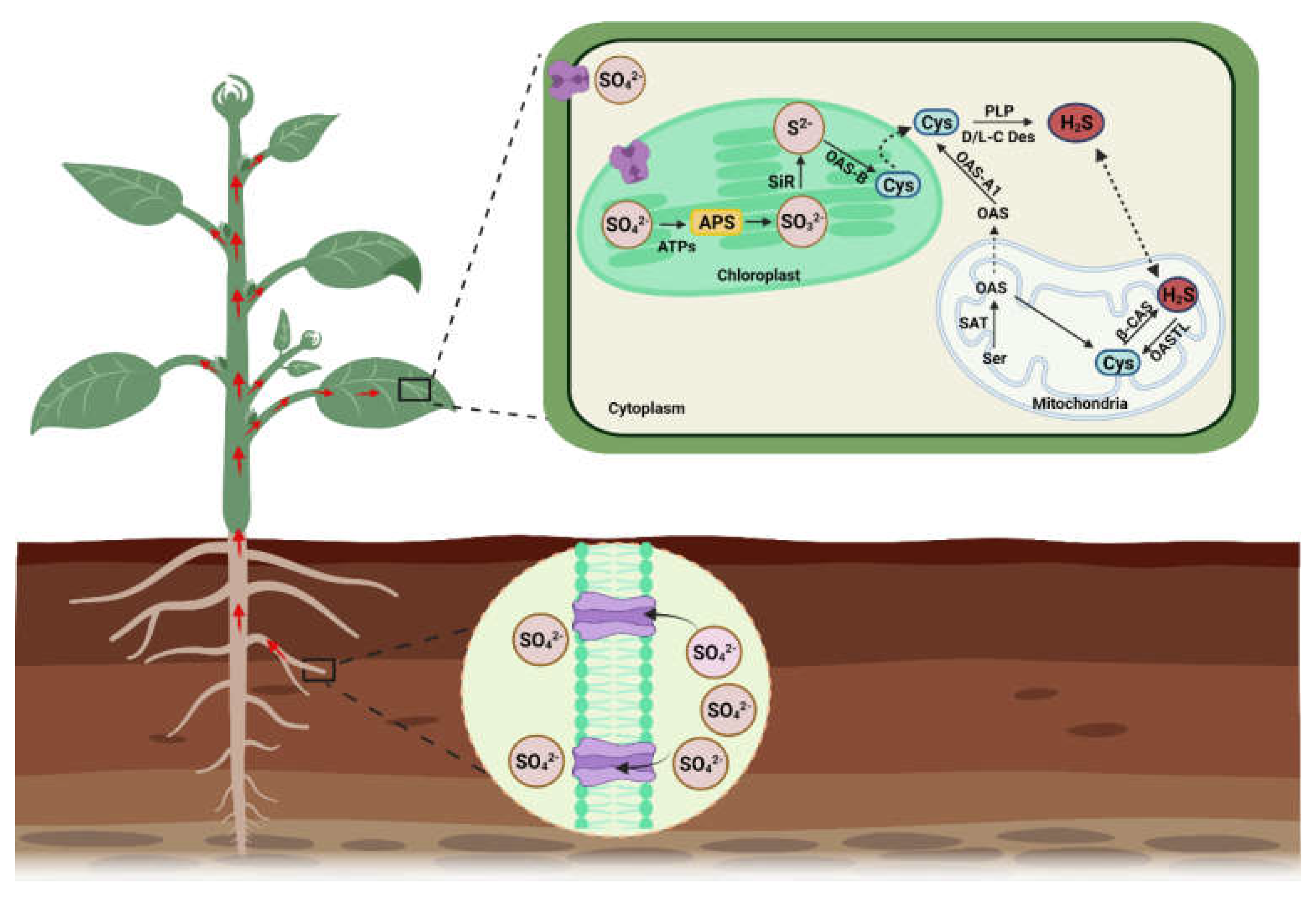
2. H2S Biosynthesis in Different Organelles and Associated Enzymes

Plant roots absorb sulfate (SO42−), which is reduced into H2S via the action of APS reductase (adenosine-5′-phosphoryl sulfate reductase) and SiR (sulfite reductase). H2S is later transformed into cysteine amino acid via catalysis of O-Acetylserine (thiol) lyase (OASTL), as a final step of sulfate assimilation in plants (Figure 2). In A. thaliana, cytosolic OAS-A1 (At4g14880), the plastid OAS-B (At2g43750), and the mitochondrial OAS-C (At3g59760) are considered true OASTL because they incorporate an O-acetylserine (OAS) and sulfide into cysteine synthesis [10,11,12]. The presence of functional OASTL was also identified in pollen [13]. Additionally, plant cells contain nutritional sulfur (SO42−) and SO2 (collected from the atmosphere) that is consequently converted into SO32− and is used to produce H2S in the presence of ferredoxin and APS reductase [14,15]. In salt-stressed tobacco plants, malfunction of SiR leads to decreased H2S production, correlating with less availability of SO2 on account of stomatal closure. This series represents the functional role of SiR in H2S metabolism under stress conditions [16].
H2S is also synthesized in the chloroplasts and mitochondria when cysteine is reduced by cysteine desulfhydrase (CDes) and β-cyanoalanine synthase (β-CAS), respectively (Figure 2). Genetic and molecular evidence indicated that mitochondrial isoforms of CAS are CYS-C1 (At3g61440) and OAS-C (At3g59760), and chloroplastic isoforms of CAS are OAS-B (At2g43750) and SCS (At3g03630) [17]. The cytosolic release of H2S is dependent upon the functioning of D/L cysteine desulfhydrases (L/D-CDes). Several L-CDes of the Arabidopsis plant are well characterized and are involved in the breakdown of L-cysteine to sulfide, NH3, and pyruvate [18,19,20]. However, D-CDes are completely different proteins and belong to the pyridoxal 5-phosphate (PLP)-dependent enzyme superfamily, and its activity is PLP dependent [21,22]. The model plant Arabidopsis contains two putative D-cysteine desulfhydrases (D-CDes) genes (At1g48420 and At3g26115) [21,22,23], while two D-CDes are also functionally characterized in rice (OsDCD1 and OsLCD2) and some other crops [24,25]. The D-cysteine desulfhydrases 2 carry out the decomposition of both L- and D-Cystine into H2S. Accumulating evidence signifies that NifS-like L-CDes are also involved in the generation of H2S. The presence of H2S in plant peroxisomes and its interaction with catalase is also observed; however, the synthesis mechanisms and involved enzymes are still unknown [26].
The mitochondria play a vital role in the catabolism of H2S and maintain its steady-state levels in cells. In mitochondria, H2S is generated during cyanide detoxification through the catalysis of β-CAS. The functional mitochondria isoform of CAS is CYS-C1 (At3g61440), which catalyzes the conversion of cysteine and cyanide into hydrogen sulfide and β-CAS and maintains optimum levels of cyanide to prevent phytotoxicity [27]. This yielded H2S is converted back into cysteine via mitochondrial OASTL (OAS-C, At3g59760), which will again be used in the detoxification of cyanide. This process is considered a cyclic pathway of cysteine generation via H2S consumption in mitochondria [28]. Under stress conditions, excess accumulation of H2S raises the pH of mitochondria, leading to the conversion of H2S into hydrosulfide ions (HS). Excess accumulation of H2S also prevents the loss of H2S from mitochondrial membranes and maintains H2S homeostasis (Figure 2). The environmental cues also modulate the endogenous H2S biosynthesis by stimulating desulfhydrase activities in plant cells [18].
In plastids, the reduction of sulfate to sulfide and its incorporation into the OAS is executed as an entry point of reduced sulfur to plant metabolism for growth and development via a photosynthetic sulfate assimilation pathway [18,29]. The OAS interaction with serine acetyltransferase (SAT) forms a cysteine synthase complex (CSC), which generates demand-driven synthesis of cystine in plant cells [30,31]. Subsequently, the breakdown of cysteine in the chloroplast generates H2S due to the catalysis of DES1 and L/D-cysteine desulfhydrase (Figure 2). The generation of H2S in chloroplasts acts as a signaling molecule because it substantially impacts cellular metabolism by limiting the rate of photosynthesis.
The peroxisome is an essential single membrane-bound organelle involved in the metabolism of reactive nitrogen species (RNS), including H2S [26,32,33]. Recent studies demonstrated the presence of H2S in plant peroxisomes [34]. Some studies speculated that peroxisomes have the capacity to transform sulfite to sulfate under the catalysis of Sulfite oxidase (At3g01910) in A. thaliana. Presently, no enzymatic source for H2S metabolism has been observed in the peroxisome of Arabidopsis, and tomato [34,35,36]; the mechanism of H2S production in peroxisome is still obscure. The H2S characterization study in Solanum lycopersicum showed the localization of OASTL9 in the peroxisome, which exhibited upregulation under different developmental stages and pathogenic bacterial treatments [36].
In the plant, several additional enzymes are also involved in H2S synthesis, and most of the H2S in the cell is produced during the necessary consumption of cysteine. For example, At5g28030 encodes a cysteine synthase (CS)-like protein that degrades L-cysteine and produces H2S [28]. This protein is also localized in the cytoplasm as AtDES1 (desulfhydrase). The homolog of this protein in Brassica napus (BnDES) is also involved in the breakdown of cysteine [37]. However, AtDES1 homolog in rice (OsLCD2) exhibits cysteine biosynthesis activity [38]. The Arabidopsis nitrogen fixation-like 1 and 2 (At5g65720; At1g08490) also use L-cysteine as a substrate and produce H2S during the synthesis of L-alanine in the cytosol [20,28,39]. This diversity in enzymatic functioning and discrepancies in their substrates’ catalyzation may allow the plants to calibrate endogenous H2S levels according to their requirements and external prompts.

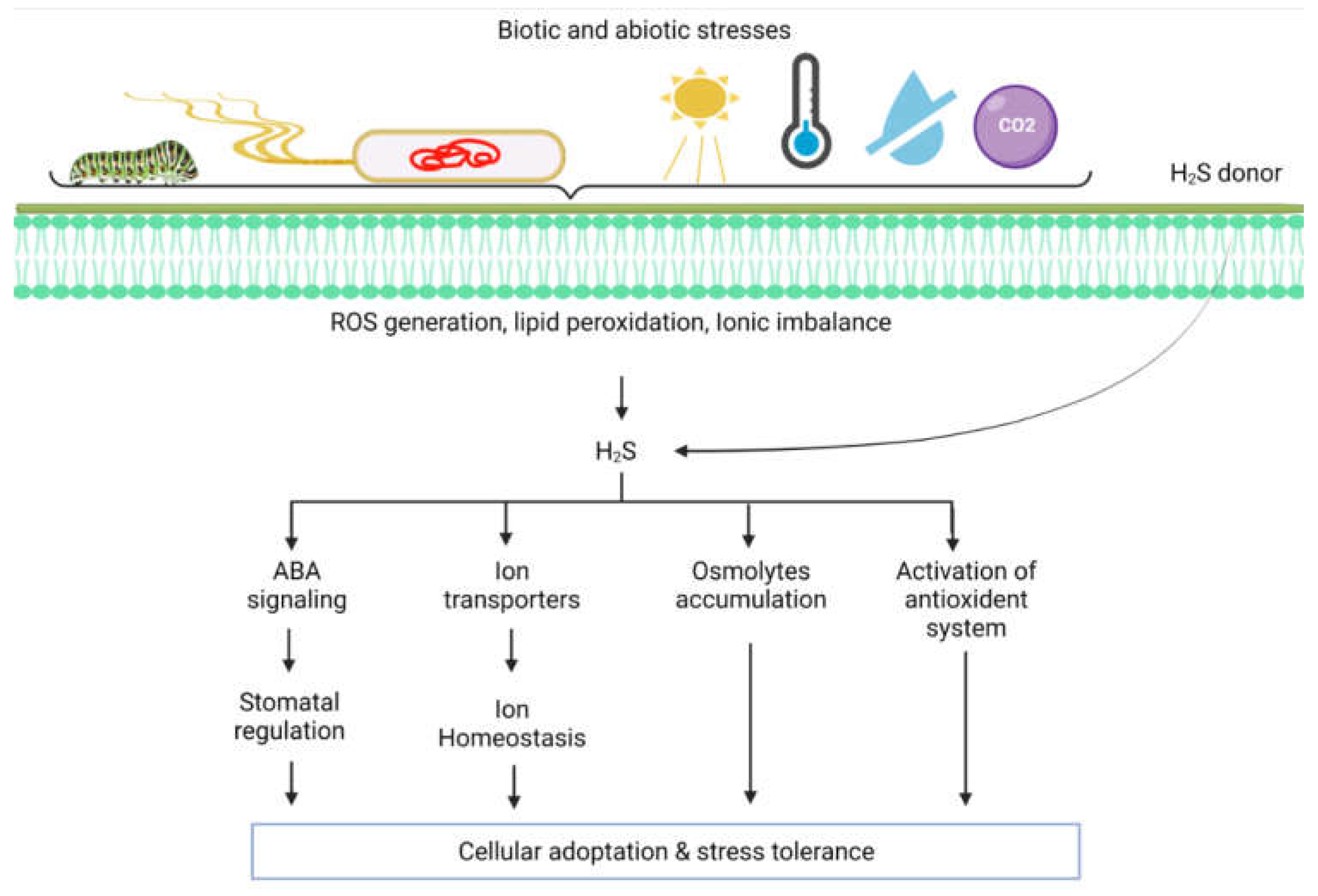
3. Role of H2S in the Modulation of Abiotic Stress Responses

H2S plays a vital role in protecting plants against several abiotic stressors. Environmental stress factors such as salinity, drought, waterlogging, high temperature, excessive light, heavy metals, and chilling could adversely affect plant growth and development (Figure 3) [40,41,42,43]. Generally, under most stress conditions, plants reduce uptake of CO2 due to the closure of stomata and limiting CO2 fixation. This condition causes alternation in cell metabolism due to restricted photosynthetic capacity that leads to the generation of reactive oxygen/nitrogen species (ROS/RNS) [44,45,46,47,48,49,50]. H2S directly regulates the cysteine (Cys) residues’ persulfidation via posttranslational modification (PTM), allowing the H2S to regulate protein functioning through persulfidation [51,52]. For example, APX protein was persulfidated in different compartments of cells (cytosol, chloroplasts, mitochondria, and peroxisomes) in Arabidopsis [26,53,54,55]. These findings indicate that the ROS-induced toxicity in stressed plants is regulated by H2S-mediated persulfidation post-translationally via triggering the ROS scavenging enzyme activities [56].

3.1. Application of H2S in Plant Drought Responses

During osmotic stress, improved water status of plants is a vital survival strategy that is achieved via accumulating osmolytes to maintain normal hydration levels. Exposure to drought stress or PEG-induced osmotic stress in plants enhances the accumulation of osmolytes such as proline and glycine betaine to maintain normal water status in stressed plants. However, sometimes the accumulation of these osmolytes fails to maintain adequate water status due to the severity of osmotic stress [50,52]. The endogenous stimulation of H2S regulates the proline synthesizing enzyme via stimulating the expression of 1-pyrroline-5-carboxylate synthetase, and by inhibiting the activity of the proline-degrading enzyme. On the other hand, H2S also triggers the activity of glycine betaine biosynthesis enzymes (aldehyde dehydrogenase), which reduce the osmotic stress and assist the plants in enhancing osmotic pressures to improve water uptake and relative water content in vital tissues [57,58]. The pre-exposure of SO2 to drought-stressed wheat plants showed a pronounced increase in endogenous H2S. This inflation may be caused by the conversion of SO2 into the SO32− and decomposition of L-/D-Cys, which generates enough H2S to initiate drought adaptive responses in the stressed seedling. However, when hypotaurine (HT; H2S scavenger) was applied on SO2-pretreated seedlings, reduced content of H2S and severe symptoms of drought toxicity appeared in seedlings. In addition, endogenous generation of H2S via pretreatment of SO2/NaHS, fully activated the antioxidant enzymes (SOD, CAT, and POD) and reduced the production of H2O2 and MDA content in drought-stressed plants [41,47,59,60]. The endogenous H2S modulation in plants also activated the expression of transcription factors (TFs) such as ERF1, NAC69, and MYB30 [41,61]. The findings of several studies indicated that TF NAC69 could confer resistance in drought-stressed plants via the H2S mediated ABA signaling pathway. Additionally, the upregulation of TFs such as ERF1 and MYB30 may activate signal transduction pathways and regulate stress-responsive gene expression profiling under drought stress conditions [62,63,64]. Since the application of H2S scavengers inhibited the transcript abundance of ERF1, NAC69, and MYB30 in wheat plants under drought stress conditions, there must be direct involvement of H2S in the regulation of stress-related TFs in response to drought stress [61,65,66]. Some studies also recognized that H2S signaling in response to drought stress influences the functioning of ABA biosynthesis genes such as NCED2, NCED3, and NCED5 and suppresses the ABA catabolic genes (ABA8ox1, ABA8ox2, and ABA8ox3), which is consistent with ABA accumulation in drought-stressed plants [47,50].

3.2. Role of H2S in the Alleviation of Metal Stress

Under metal toxicity, plants modulate several metal/metalloid ions from toxic to less toxic forms, such as reduction of arsenate (AsV) to arsenite (ASIII), and hexavalent chromium (Cr(VI)) to less toxic trivalent Cr(III), and sequester these metal ions via thiols (GSH) and phytochelatins (PCs) ligands [64]. These metabolites (GSH and PCs) actively participate in the intracellular redox balance and metal tolerance capacity of crop plants and prevent the cells from entering programmed cell death or necrosis phases [67,68]. Due to metal-induced oxidative stress, the intracellular redox becomes oxidized, decreasing levels of reduced molecules such as NADH/NADPH and allowing apoptosis or necrosis to be initiated. The endogenous production of H2S or exogenous application of H2S donors assists in maintaining the levels of GSH and phytochelatins in the plant to sustain optimum redox balance and the sequestration of toxic metal ions into the vacuoles [41,69]. The GSH and PCs are sulfur enriched compounds, whereas, in sulfur metabolism, metabolites such as sulfite, H2S, cysteine, and GSH are highly interconnected, and depletion of GSH during metal toxicity could potentially accelerate cysteine breakdown and ultimately enhance the GSH and H2S supply to the cell [16,67]. In several published studies, it is observed that the mitigation effects of H2S under different abiotic stresses and metal excess conditions are related to the upregulation or superior maintenance of redox-active compounds such as ASA-, GSH, and PCs [40,67,69,70]. This finding of these studies provides compelling evidence that modulation of endogenous H2S during stressful conditions could help the plant to maintain or reduce the loss of intracellular glutathione, which supports the overall redox positive state of the cell and verifies that H2S has an important influence on cell functions under stressful conditions [41,70,71].
H2S not only overcomes ROS-induced toxicity in metal exposed plants but also plays an effective role in the inhibition of metal transport and absorption. H2S has the ability to alter chemical forms of metal ions into insoluble phosphate compounds, which decreases metal toxicity and movements [72]. However, the metal reduction capacity of H2S is much lower than GSH, cysteine, phytochelatins, and metallothioneins [73]. H2S mediated reduction in metal transport/immobilization is usually associated with downregulation of metal transporters or secretion of chelating compounds to prevent the further translocation of metal ions to the sensitive tissues or uptake from the root zone. For example, in several crop plants, exogenous application of H2S intensifies the citrate secretion and expression of citrate transporters, so the non-toxic complexes of citrate with Al3+ could be formed in the rhizosphere [74,75,76]. Similarly, H2S also suppresses pectin methyl esterase activity, which suppresses Al3+ binding sites by reducing negative charge in root cells, which has direct implications for Al3+ tolerance [77,78]. In the case of Cd metal, H2S triggers the expression of phytochelatin synthase (PCS) and the Cd-ATPase gene to effectively chelate and transport metal ions into the vacuoles through the help of HMT transmembrane transporter channels [79]. The L-DC-mediated H2S accumulation modulates root pectin content with a lower degree of methylation to facilitate the binding of Cd2+ to the cell wall, which ultimately diminishes its further translocation from root to shoot and toxicity symptoms in exposed plants [80]. In Arabidopsis, exogenous application of H2S activated the generation of Cr6+ binding peptides, such as phytochelatins and metallothioneins, to carry toxic Cr6+ to insensitive regions mediated by compartmentalization [81,82]. Based on these studies, we infer that H2S plays a pivotal role in the chelation of heavy metals for inactivation and later sequesters them into the vacuole to increase the metal stress tolerance of plants.

3.3. Effect of H2S on Plant Salt Tolerance

Salinity is a major constraint limiting agriculture productivity due to poor irrigation practices and continuous climate fluctuations [83]. Saline stress imposes both osmotic stress and ionic toxicity, which retard plant growth and productivity. The unregulated accumulation of sodium (Na+) hinders water and nutrient uptake and induces water deficit conditions for plants. Furthermore, an excessive amount of Na+ and chloride (Cl) accumulation in plants disturbs ionic homeostasis. The depolarization of membranes leads to the loss of potential stress mitigating ions such as K+ and Ca2+ and induces changes in transpiration rate, photosynthesis, oxidative stress, etc. [84,85,86]. Saline stress in plants reinforces several physiological, molecular, and metabolic disorders that completely inhibit plant growth [87,88,89]. The maintenance of ionic homeostasis and a lower cytosolic Na+/K+ ratio is critical for salt adaptation and tolerance. It is observed that several Na+/K+ ion transporters and stress-responsive gene activation pathways are interconnected with plant hormones because stress and growth hormones are spatially involved in mediating salt-stress signaling and maintaining the balance between stress responses and growth in plants [83,87,88]. In this regard, H2S biosynthesis and signaling are implicated in saline stress tolerance in plants. [90,91,92,93]. Several studies demonstrated that exogenous application of H2S reduces the uptake of Na+ and increases the accumulation of K+ that untimely preserves an optimal Na+/K+ ratio for the plant’s vital functioning. [90,91,92,93]. It is proven via pharmacological studies that when H2S scavengers were applied to the salt-stressed plants, the depletion of endogenous H2S aggravated the saline stress symptoms and increased the Na+/K+ ratio and cytosolic concentration of Na+ in studied plants. These studies also highlighted that H2S application significantly maintains K+ homeostasis in plants by preventing K+ leakage by reducing oxidative stress-mediated lipid peroxidation and membrane depolarization. [90,91,92,93]. At the molecular level, it was observed that H2S regulated the activity of SKOR (outward rectifying K+ channel) by inhibiting its expression and preventing the loss of K+ into the xylem under saline stress conditions. However, when H2S scavengers (DL-propargylglycine or HT) were applied to the plants, SKOR expression was not compromised. [90,91,92,93]. Similarly, the K+ retention during saline stress conditions normalizes H+-ATPase, because H+ gradient-mediated H+-ATPase activity repolarizes the PM to accelerate potassium influx and sodium efflux [90,91,92,93]. This repolarization occurs because H2S is involved in the stimulation of gene expression and phosphorylation-mediated upregulation of H+-ATPase activity under salinity [94,95]. This observation suggests that H2S shows the implication of K+ uptake and its homeostasis via upregulating the K+/Na+ antiport system through modulating H+-ATPase activity [42,91,92,95]. Besides this, AKT1 (inward rectifying potassium channels) is located in root epidermal tissue [96], and HAK5 (potassium transporter) gene is located in the tonoplast and the PM [96]. These genes are also coupled with maintaining K+ and plant resistance to salt. The exogenous application of H2S donors improved the transcript expression of AKT1 and HAK5 and total K content in the salt-challenged Brassica napus plant [97]. Similarly, NaHS induced H2S promoted the expression of HvAKT1 and HvHAK5 in roots of barley seedlings under salinity [8]. All these findings advocate that the potential increase in H2S and its signaling is a positive regulator of K+ homeostasis and maintenance of the Na+/K+ ratio during saline stress in plants [8,95,98,99].
The (SOS) pathway is critical for the exclusion of Na+ under saline stress conditions. SOS1 is involved in the long-distance transport of Na+ from roots to shoots [95,100]. The increase in transcript abundance of SOS1 favors the accumulation of SOS1 proteins in the PM, which triggers the exclusion of Na+ from cells and minizines the Na+ load in the cytosol [60]. The H2S application under alkaline and normal salt stress conditions stabilizes the mRNA level of SOS1, which leads to the reduced Na+ content in the roots of cultivated apple plants [101]. SOS1 is regulated by the H+ gradient provided by PM H+-ATPase. Several studies identified that H2S positively influences the gene expression and phosphorylation of PM H+-ATPase under salinity [102]. In pharmacological experiments where endogenous H2S production was inhibited, the expression level of SOS1 and related Na+ antiporters were downregulated, and salinity tolerance of plants was compromised due to unregulated accumulation of Na+ in sensitive tissues [100]. The PM H+-ATPase on the membranes of vacuoles also regulates the expression and activation of the Na+/H+ antiporter, because the compartmentalization of Na+ ions into the vacuoles is an alternative solution to decrease the Na+ induced toxicity in cells [42,103]. The H2S application greatly induces the transcript accumulation of NHX2 and VHA-β genes (Na+/H+ antiporter) in salt-exposed plants. This finding also advocates that Na+ caging in vacuoles is influenced by H2S signaling [8,85]. Meanwhile, for the regulation of Na+/K+ homeostasis, H2S also controls the H2O2 mediated activity of PM-bound NADPH oxidases [104]. For instance, PM NADPH oxidase inhibitor (diphenyleneiodonium chloride) suppressed the H2S mediated increase in H2O2 in the root of Arabidopsis under salinity. The application of ROS scavenger (N,N’-Dimethylthiourea) abolished the H2S mediated H2O2 production in salt stress plants due to the Na+ uptake being high in salt-stressed plants from the absence of H2S mediated activation of NADPH oxidase [104]. This conclusion indicates that H2O2 might act as a downstream signal for H2S-mediated Na+/K+ homeostasis [85,104,105]. The findings of these studies demonstrate that H2S regulated signaling influences the activity of H+-ATPase and the expression of PM Na+/H+ antiporter that enhances the salt tolerance by maintaining Na+/K+ homeostasis in plants [85,106].

4. Crosstalk of H2S with Signaling/Phytohormones under Changing Environmental Conditions

Phytohormones, or plant growth regulators (PGRs), are the most significant signaling molecules, synthesized in specific locations within plants, and can be translocated to different parts to regulate stress responses [106]. PGR such as abscisic acid (ABA), auxins (IAA), brassinosteroids (BRs), cytokinins (CK), gibberellins (GA), jasmonic acid (JA), and salicylic acid (SA) help the plants to overcome numerous biotic/abiotic adversities by triggering physiological and molecular responses [107,108]. H2S, which acts as an endogenous gasotransmitter, is recognized in relevance with other signaling molecules such as NO [109], ROS [110], H2O2 [111], CO [112], and plant hormones such as ABA [113], JA [114], GA [115] and ethylene.
H2S in plants exhibits a dual role, either disseminated as pernicious cellular repercussion or as credible signaling molecules depending upon stress conditions. A study discovered that H2S operates downstream of NO and helps decrease oxidative stress during salt stress in tomatoes. H2S helps minimize postharvest ripening and senescence in bananas because it inhibits ethylene signaling as well as mitigating oxidative stress [115]. Additional studies revealed that H2S regulates NADPH oxidase (RBOH) activity, leading to ROS accumulation [116]. Simultaneously, the concentration of phosphatidic acid generated via phospholipase D [117,118] is also modulated by H2S, which helps further to inhibit the cellular signaling pathway [1]. In Arabidopsis, H2S operates upstream of the MAPKs pathway, and both of these work parallelly under cold stress conditions [119]. Various developmental processes such as organogenesis, seed germination, and the advent of senescence are spurred by H2S produced from sodium hydrosulfide (NaHS) and morpholin-4-ium 4-methoxyphenyl (morpholino) phosphinodithiolate (GYY4137) [119,120]. As a signaling molecule, H2S participates in several cross-talk networks amid H2O2, NO, CO, and phytohormone ABA during different stress conditions [121]. It is evident that signaling molecules such as H2S interplay an essential role in several stages of plant development because of the interaction between H2S and numerous phytohormones. In the future, genes involved in governing the new signaling molecules such as H2S could be targeted to develop a genetically improved crop.

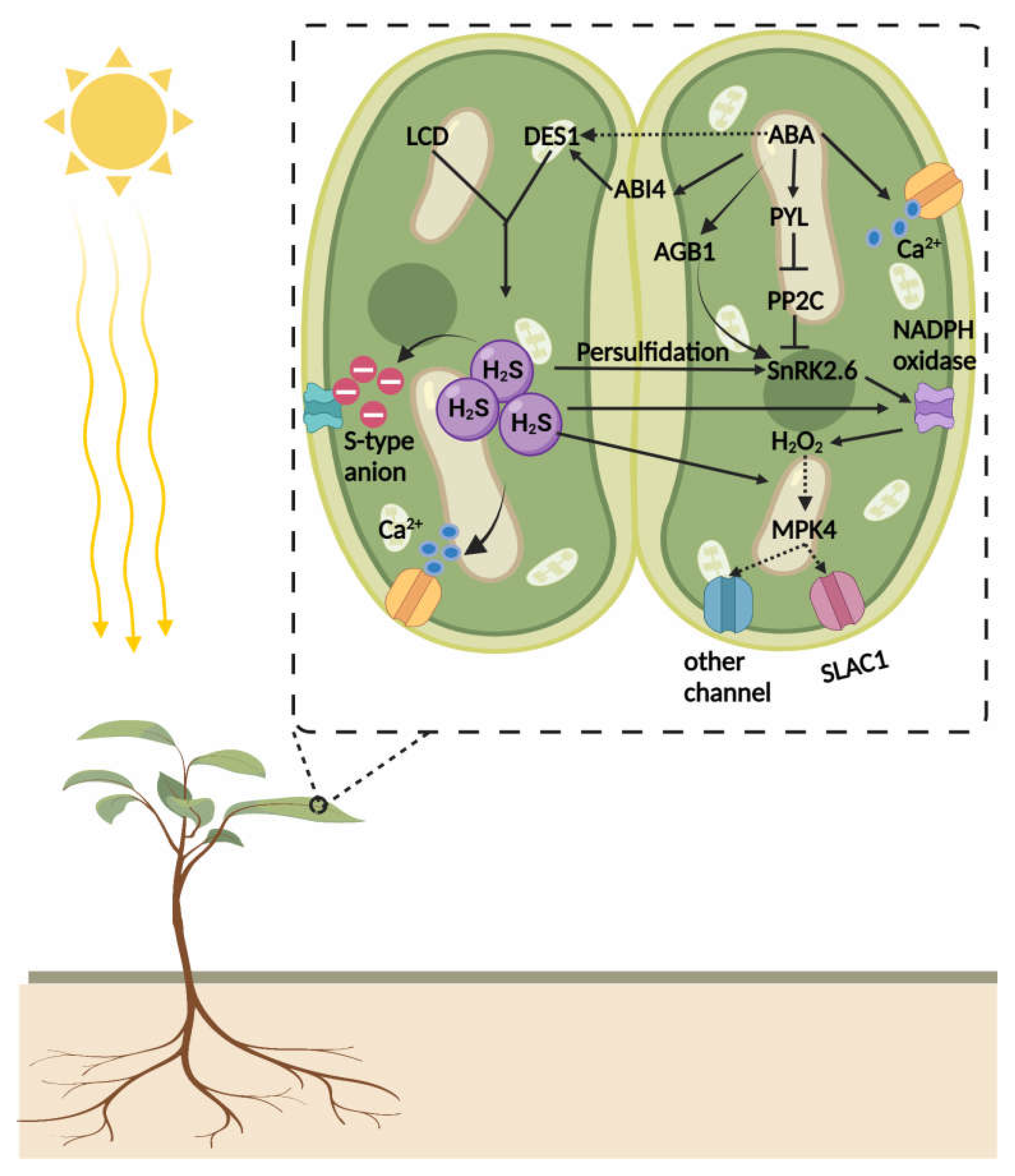
4.1. Crosstalk of H2S and Abscisic Acid (ABA)

Plants modify ABA levels continually in response to changing physiological and environmental conditions, while bioactive ABA levels are sustained through a fine balance between generation and catabolism [45,86,87]. Several ABA receptors are involved in signal perception and transduction [45]. Earlier studies revealed that the interaction of H2S with ABA receptor genes implied that H2S regulates ABA signaling via influencing ABA receptors [45,122,123]. H2S application in drought-stressed plants upregulated the expression of potential ABA receptors such as RCAR (The regulatory component of ABA), ABAR (abscisic acid receptor), PYR1 (pyrabactin resistant protein), GTG1 (GPCR-type G proteins), and CHLH (H subunit of the Mg-chelatase) [45,124]. Some studies point out that ABA regulates many physiological processes, and H2S sometimes regulates these responses in a similar way [45,113,124]. Exogenous application of ABA triggers the endogenous production of H2S, suggesting complex crosstalk between two signaling molecules exists under drought stress conditions [45]. Similarly, under heat stress, ABA could trigger the accumulation of endogenous H2S and act as a new downstream gaseous signaling molecule that regulates ABA-induced stress responses in heat-stressed plants [45].
In plants, stomatal closure or opening is regulated by guard cells. The plant hormone ABA regulates the function of several ion channels in an ABA-dependent manner to control stomatal closure and opening [124,125,126,127,128]. A wealth of literature provides ample evidence that H2S regulates stomatal aperture in various plant species, and it may have implications for ABA-dependent stomatal closures in plants under stressful conditions [124]. The earlier study of Wang et al. [129] illuminated this underlying mechanism and revealed that exogenous application of H2S activates the S-type anion currents in guard cells of Arabidopsis. Concurrently, the elevated level of free Ca2+ is a prerequisite for its activation [129]. H2S triggers Ca2+ waves in guard cells. In guard cells, Ca2+ sensing is perceived by a heterotrimeric G-protein β-subunit (AGB1) that collaborates in Ca2+ induced stomatal closure in Arabidopsis [130]. Ca2+ ions also activate SLAC1 by stimulating CPK (calcium-dependent protein kinase) activity. It was observed that lower concentrations of ABA partially impaired stomatal closure in CPK quadruple mutant plants; however, higher concentrations of ABA effectively close stomata. The application of Ca2+ chelator (1,2-bis(o-aminophenoxy) ethane-N,N,N,N-tetraacetic acid (BAPTA) completely inhibited the ABA-mediated activation of anion channel in guard cells and prevented the ABA-induced stomatal closure [131,132]. These studies showed that H2S and ABA are signaling components in stomatal closure in plants.
A recent study demonstrated that H2S mediated persulfidation of SnRK2.6/OST1 in response to ABA signaling initiated stomatal closure (Figure 4). In guard cells, SnRK2.6/OST1 acts as a core component of ABA signaling that controls stomatal movements, and its function is tightly regulated by H2S-mediated PTMs. Under certain physiological conditions, ABA induces the generation of H2S by activating DES1 in the guard cell. The accumulation of H2S persulfidates SnRK2.6 on Cyc131 and Cys137, which are close to the catalytic loop and near to Ser175 residues, which is vital for the phosphorylation of SnRK2.6 [133,134,135,136,137]. The Cys137 can also undergo S-nitrosylation and could inhibit the activity of SnRK2.6 [9,136]. However, persulfidation promotes SnRK2.6 activity, and it is believed that persulfidation occurs earlier than S-nitrosylation [9,137]. Due to Cyc131/137 persulfidation induced changes, Ser175 affinity for ATP-γ-phosphate proton acceptor site (Asp140) increases, which leads to the robust autophosphorylation of Ser175 and triggers efficient interaction of SnRK2.6 with its target. This observation confirms that H2S-mediated persulfidation positively impacts the function of SnRK2.6 in ABA-mediated stomatal closure in guard cells [9,135]. Likewise, Shen et al. [138] reported that during drought stress, ABA signaling in guard cells is promoted by H2S interaction with ABA. The drought stress mediates the accumulation of ABA, which stimulates persulfidation of DES1 in a redox-dependent manner. At the physiological level, enhanced accumulation of H2S in the guard cell leads to the persulfidation of H2O2 producing enzymes, such as NADPH oxidase, which triggers the generation of H2O2 in the guard cell that reinforces ABA signaling and the closure of stomata [138]. Another study revealed that abscisic acid insensitive 4 (ABI4) is involved in the facilitation of ABA and H2S crosstalk at the transcriptional level (Figure 4). ABI4 is a vital TF in the ABA signaling cascade, and little was known about the PTMs that regulate its activity in response to ABA/H2S interaction in plants. The ABA accumulation triggers a massive generation of H2S that leads to the persulfidation of ABI4, which allows the binding of ABI4 to the E1 motif of the MAPKKK18 (mitogen-activated protein kinase kinase kinase 18) promoter to activate DES1 transcription to close stomata under the ABA-dependent signaling cascade [43]. This study provides compelling evidence that the DES1/H2S-ABI4 module acts downstream of ABA signaling to regulate stomatal closure [43,139] (Figure 4).
In some of the recently published reports, it was also revealed that H2S might be involved in the biosynthesis of ABA in guard cells [140]. The H2S promotes the synthesis of cysteine, which is a substrate of ABA3 (molybdenum cofactor sulfurase) enzymes that regulate the activation of AAO3 (abscisic aldehyde oxidase 3) [141]. The higher accumulation of cysteine stimulates the activity of AAO (in vivo) and favors the synthesis of ABA [39] by stimulating the transcript abundance of NCED3 (9-cis-epoxycarotenoid dioxygenase 3). It was revealed that H2S could boost ABA synthesis, because in a cysteine-biosynthesis-depleted mutant with the disrupted ABA biosynthesis, the H2S was unable to induce stomatal closure [135,136]. All these studies point out the involvement/crosstalk of H2S with SnRK2.6, CPK6, MAPKKK18, ABI1, NADPH oxidase, Ca2+, and ROS in ABA-mediated signaling for stomatal movements in plants [135,136,137].

4.2. Nitric Oxide (NO) and H2S: Two Interacting Gaseous Molecules Essential for Plant Functioning

Nitric oxide (NO) is also a lipophilic gaseous hormone that could diffuse into inter- or intra cellular spaces without the need for any carrier or transport channel. NO is also involved in PTMs via tyrosine nitration, metal nitrosylation, and S-nitrosylation, whereas H2S mediated-PTM is associated with persulfidation. However, all these reactions led to the modification of structure, localization, and function of target proteins. Several studies have shown that H2S interacts with NO and other signaling molecules to modulate plant development and stress responses [7,26,32,34,142]. Earlier reports indicate that the interaction of H2S towards NO is complementary or inhibitory [55,143,144,145,146]. The positive or negative interaction of these two gaseous signaling molecules may be dependent upon the dosage of exogenous H2S or NO application. For instance, the level of NO was reduced in plant tissues that were treated with H2S modulator (NaSH) [126,147]. However, crosstalk of NO-H2S showed synergistic interaction during abiotic stresses and inhibition of ethylene-induced fruit ripening, whereas antagonistic interaction of H2S-NO-ethylene is also reported [16,148,149,150]. The discrepancy in H2S and NO interaction may depend upon the specific location of these gaseous molecules in the cell that deicide their signaling behavior [151]. There is also a possibility that both gaseous molecules may compete for the same targeting protein in the cell. For example, SnRK2.6 is a target of both NO and H2S biomolecules, and S-nitrosylation of SnRK2.6 via NO inhibits its activity while persulfidation enhances its activity and mediate stomatal movements [135,137]. Additionally, H2S and NO could react among themselves to produce nitrosothiol compounds that are also involved in signaling responses. The crosstalk of ROS with H2S–NO cascades also modulates their interactions in positive or negative ways [152]. Taken together, the nature of the interaction between NO and H2S may vary for different physiological functions based upon their location and concentration in the cell.
NO and H2S belong to the family of reactive nitrogen and sulfur species (RNS and RSS), and their positive combinations regulate various important physiological and molecular processes in plants. For example, the interaction of H2S with NO and Ca2+ regulate lateral root (LR) formation in tomato plants. The exogenous application of NO triggers the accumulation of H2S in tomato roots due to the upregulation of H2S biosynthesis enzymes, which induce later root formation [6]. However, when H2S inhibitor/scavengers were applied, LRs’ formation was partially arrested. These findings indicate that NO-induced H2S synthesis governs the later root formation [6,153].
Stomatal movements are regulated by many endogenous signaling molecules; among them, H2S and NO crosstalk are also responsible for stomatal closure. In a recent study, with the employment of pharmacological, spectrophotographic, and fluorescence microscope techniques, the coordinated action of H2S and NO in the presence of 2,4-epibrassinolide (EBR) was involved in stomatal regulation [154,155]. The authors demonstrated the application of EBR-induced stomatal closure in a dose and time-dependent manner via modifying the levels of NO, and H2S in Vicia faba. The application of EBR upregulated the activity of L-/D-cysteine desulfhydrase and enhanced the endogenous levels of H2S together with H2O2 and NO generation in guard cells. The application of the H2S inhibitor significantly reduced L-/D-cysteine desulfhydrase activity and H2S endogenous production, which in turn abolished the EBR mediated stomatal closure effect [154]. The H2S scavengers/inhibitors did not affect the NO and H2O2 levels in guard cells. However, the application of NO and H2O2 inhibitors/modulators significantly affected the endogenous production of H2S and its biosynthesis enzymes and compromised the EBR-induced stomatal closure [154]. Similarly, Jing et al. [156] found that H2S may function downstream of NO in ethylene-induced stomatal closure in V. faba. These results indicate that H2S and NO participate in EBR-mediated stomatal closure response and H2S signifies an essential constituent downstream of H2O2 and NO in EBR-induced stomatal closure in V. faba [154,157]. Previous studies demonstrated that H2S inhibits ABA-mediated NO generation in Arabidopsis and Capsicum annuum guard cells. Conversely, H2S increased NO levels in alfalfa seedlings [55,147], while H2S induces NO generation in Arabidopsis guard cells. Conversely, NO scavenger inhibited H2S-induced stomatal closure [145]. However, investigation of H2S-mediated guard cell signaling in Arabidopsis revealed that the H2S induced signaling cascade for stomatal closure is NO-dependent [128], and both H2S and NO equally contribute to the production of 8-mercapto-cGMP, which triggers stomatal closure. In the same way, H2S and NO collaborate in ethylene induce stomatal closure responses in Arabidopsis plants, and H2S generation is mediated by NO, which suggests that H2S acts as a downstream signaling agent in ethylene induce stomatal closure [158].
The crosstalk of H2S and NO in the alleviation of metal toxicity is also reported, but these studies focused more on stress physiology and lacked underlying molecular mechanisms of crosstalk [159]. The exogenous application of H2S donor alleviated Cd stress in alfalfa plants by triggering the synthesis of NO. The interaction mechanism between H2S and NO improved the Cd stress tolerance by reducing Cd accumulation and lowering the lipid peroxidation in stressed plants [136]. Another study, where H2S and NO scavenger and inhibitor were applied to Cd stressed bermudagrass plants, revealed that depletion of NO makes them more vulnerable to metal toxicity. Furthermore, through pharmacological experiments, it was demonstrated that NO-activated H2S was essential for cadmium stress responses in bermudagrass [160]. In Pisum sativum, positive interaction of NO and H2S was also explored under arsenate stress [109]. The application of H2S donor triggered endogenous H2S and NO accumulation in P. sativum, which led to the strengthening of the antioxidant defense system, reduced arsenate accumulation, and maintained the redox balance of P. sativum plant under metal toxicity [109]. Similarly, the crosstalk of NO and H2S reduced oxidative stress and increased salinity tolerance in alfalfa, while barley seedlings under H2S application regulate ion homeostasis under salinity via maintaining the NO signaling pathway [8,146]. Most of the published studies on the interaction of NO and H2S in the context of metal toxicity/salinity proposed that crosstalk of these gaseous molecules ameliorates stress-induced toxicity in exposed plants via (i) improving the antioxidant defense to prevent oxidative stress, (ii) reducing the metal uptake, and (iii) by modulating the expression of associated metal transporter genes [159].
In short, H2S and NO are both gaseous biomolecules with common signaling pathways, and it seems that one pathway controls the functions of the other [159]. The persulfidation promoted by H2S reacts with thiol groups in the same way as NO does in modification through S-nitrosation [159,161]. However, there is still a need to investigate the interaction of H2S and NO in different plant species, tissues, and diverse environmental conditions to unveil the regulatory mechanism of the NO–H2S signaling cascade in plants.

4.3. H2S-Mediated Manipulation of Auxin Signaling in Plants

The development of roots, including lateral and adventitious roots, is incredibly important for normal plant growth and the successful completion of the life cycle. Plant root architecture is mainly based on the LR that is generated from pericycle founder cells [155]. The plant hormone auxin and environmental factors (i.e., water and nutrient availability) are key influencers in lateral root formation [162,163]. Since auxin is a master regulator of root development in plants, there have always been complex crosstalks of auxin with other signaling agents in the root development [162,164,165].
Several studies have reported that H2S and auxin interact with each other to regulate root growth; however, mechanistic insight remains to be elucidated [120,154,166]. The earlier studies demonstrated that the application of exogenous H2S on the sweet potato seedling stimulated the numbers and length of adventitious roots by modulating the IAA levels in a dose-dependent manner [154]. It was also noted that pretreatment of H2S donor upregulated the transcript abundance of the auxin-dependent Cyclin-Dependent Kinases gene (CDKA1) and a cell cycle regulatory gene (CYCA2) [153,165]. The activity of both of these genes was inhibited either by auxin blocker or H2S inhibitor, which illustrated that H2S mediated LR development is dependent upon the IAA signaling via influencing the regulation of CDKA1 and CYCA2 [153,165]. Similarly, when higher doses of H2S donor (1 mM) were applied, the RBOH1 (respiration burst oxidase homologous) transcript was significantly upregulated and ROS accumulation triggered the later root formation [115] (Figure 5). The pharmacological studies revealed that H2S triggered the expression activity of RBOH1, which stimulated an H2O2-mediated increase in IAA signaling via regulation of CDKA1, CYCA2, and Kip-Related Protein 2 (KRP2), to activate LR formation [115]. A transcriptomic study revealed that exogenous application of H2S impacted the regulation of various auxin pathway-related genes. The accumulation of auxin biosynthesis genes (TAA1 and UGT74B1) was correlated with the increase in auxin levels in roots. The genes involved in auxin polar subcellular distribution, such as PIN2, ABCB1, ABCB19, PILS3, and PILS7, were differentially expressed, while PIN1c appeared as a hub gene on the basis of WGCNA analysis. This study provides sufficient evidence that H2S induced root development emanates from regulating the genes involved in transcriptional control and synthesis of auxin [166] (Figure 5).
In some studies, the application of higher dosages of H2S showed changes in root development and inhibition of auxin transport due to the alteration in the polar subcellular distribution of the PIN proteins [166]. The polar subcellular movement of auxin in root cells is an actin-dependent process, and H2S is involved in the regulation of actin dynamics due to the persulfidation and depolymerization of F actin [167]. Furthermore, during root hair development, the H2S fine-tuned polar auxin transport via persulfidation and actin filament growth [167,168]. In the root developmental process, actin-binding proteins work downstream of the H2S signal transduction pathway because actin-binding proteins are involved in the depolymerization of F-actin in root cells, which regulate the distribution and transport of auxin [168]. Auxin affects the patterning and organization of the actin cytoskeleton in root cells during cellular growth [169,170]. Conversely, the actin cytoskeleton modulates the directional transport of auxin by altering auxin efflux carriers [171,172]. This finding indicates that overproduction of H2S significantly increases the S-sulfhydration level of actin-2 and decreases the distribution of actin cytoskeleton in root cells, thereby reducing auxin’s polar transport, which restricts the LR and the root hair growth [44,167,168].
The exposure of plants to CH4 strongly induces H2S production and affects the root growth, adventitious root numbers, and root length in cucumber explants [106,173]. At the transcriptional level, it was observed that H2S modulated auxin-signaling genes (Aux22D-like and Aux22B-like) reinforce the CH4-induced cucumber adventitious rooting network [111,173,174,175]. Similarly, in tomato plants, LRs formation was also triggered by the CH4-mediated H2S signaling cascade. It was hypothesized that the possible involvement of auxin transport and auxin signaling in CH4-induced LR formation is involved [176]. However, more biochemical and genetic investigations are required to analyze the detailed targets and their functions in root organogenesis under CH4-H2S-Auxin crosstalks [173,176].
The signaling pathways of H2S and auxin interaction under the chilling stress were recently explored in cucumber plants [177,178,179] (Figure 6). The study demonstrated that chilling stress in cucumber arrested photosynthesis and induced oxidative stress; however, deleterious effects were alleviated due to exogenous application of H2S donor or IAA application [179]. The expression of YUCCA2 (auxin biosynthesis gene) and auxin contents were very high in chilling-exposed cucumber seedlings. This result may be due to the inhibition of polar transport of IAA in long-term chilling stress, which increases auxin concentration in leaves and inhibits plant growth. The complex interaction of H2S and IAA under chilling stress improved the activities and gene expression of key enzymes of the Calvin–Benson cycle (Ribulose-1,5-bisphosphatecarboxylase, fructose bisphosphatase, sedoheptulose-1,7-bisphosphatase, fructose-1,6-bisphosphate aldolase, and transketolase) and strengthened the photosynthetic carbon assimilation capacity [179] (Figure 6). The results also indicated that auxin is a downstream signal for the protective effects induced by H2S under chilling-induced tolerance in cucumber plants [179]. Furthermore, the overexpression of auxin response factor 5 (ARF5) in cucumber unveiled the molecular mechanism of cold tolerance. In transgenic plants overexpressing ARF5 under cold stress, ARF5 directly activates the expression of dehydration-responsive element-binding protein 3 (DREB3) for the reinforcement of auxin signaling to improve cold stress tolerance in cucumber in response to H2S application [180] (Figure 6). Previously, it was observed that auxin response factors (ARFs) and miR390 formed an auxin-responsive regulatory network (miR390-TAS3-ARF2/ARF3/ARF4) that strengthens auxin signaling in plants [181].

4.4. Interaction between H2S and Gibberellic Acid

Gibberellic acid (GA) is a phytohormone that substantially influences the seed germination and growth of seedlings. Imbibition of barley grains in 0.25 mM NaHS solution caused an upsurge in antioxidant enzymes such as CAT, POD, APX, and SOD in the aleurone layer [182]. In tomato plants, boron stress reduced dry weight, photosynthetic rate, water content, chlorophyll content, and increased H2O2, MDA, and endogenous H2S. GA foliar spray reduced the harmful effects of boron by raising endogenous H2S, Ca2+, and K+, as well as lowering the levels of H2O2, MDA, and boron, as well as membrane leakage. Surprisingly, NaHS further increased GA-induced boron tolerance, whereas H2S scavengers prevented it (HT). These findings indicate that H2S plays a signaling role downstream of GA in the development of boron stress tolerance in tomato plants. During cadmium stress, the NaHS treatment stimulated the activities of amylase and antioxidant enzymes in cucumber hypocotyls and radicles, which might be connected to H2S-induced Cd stress tolerance.
Moreover, GA can cause programmed cell death (PCD); however, NaHS application can prevent PCD by lowering L-cysteine desulfhydrase (LCD) activity and accumulating endogenous H2S in wheat aleurone layers [49]. GA-induced PCD is reduced in the aleurone layer in the NaHS-treated seeds by diminishing the endogenous GSH levels. H2S concentration regulates the GSH levels, which upsurges expression of the HEME OXYGENASE-1 (HO-1) gene, resulting in the alleviation of apoptosis in the aleurone layer and an overall decrease in PCD. Hence, in the aleurone layer, there are regulatory interactions between GA, H2S, GSH, and HO-1. Intriguingly, NaHS pretreatment slowed Arabidopsis seed germination, but Arabidopsis des1 mutant seedlings were more susceptible to ABA than the wild-type. These findings suggest that H2S interacts with GA in plants to control seed germination under normal and stressful circumstances.

4.5. Interaction between H2S and Melatonin

Melatonin (N-acetyl-5-methoxytryptamine) is a multifaceted phytohormone involved in germination, ripening, flowering, photosynthesis, and defense mechanisms [183]. In plants, melatonin alters the permeability of the cell layer governed by ion transporters, which control stomatal opening and closure. Studies have shown that melatonin can increase the photosynthetic capacity of plants, which leads to greater levels of nitrogen and chlorophyll. In tomato and wheat, increased transcription of stress-responsive genes was induced by melatonin, resulting in better tolerance to high temperature [184,185]. Furthermore, melatonin cross-talks with various plant hormones and signaling molecules. It was also discovered that H2S and melatonin conjointly helped alleviate salt stress-induced growth reduction in tomatoes, and exogenous melatonin treatment assisted in regulating early H2S signaling [186]. In wheat, the heat stress-induced oxidative damage was mitigated by exogenous melatonin and further increased the H2S production, suggesting that melatonin-mediated H2S was involved in alleviating the oxidative stress. However, the melatonin function was attenuated when H2S was inhibited by its inhibitor, indicating that the cross-talk between H2S and melatonin, and possibly melatonin, regulates heat stress signaling by acting upstream of H2S [187].

5. H2S-Plant Hormone Cross-Talk under Pathogen Attack

In plants, the dual roles of H2S in interactions with phytohormones determine the biological roles of H2S in plant growth, development, and responses to biotic stresses. In response to biotic stresses, the crosstalk between H2S and phytohormones, as well as several other signaling molecules, has been studied less; however, some critical molecular insights have been found in the recent past. In the following paragraph we discuss the H2S–phytohormone interplay under biotic stress.

5.1. Interaction between H2S and Salicylic Acid

Salicylic acid (SA) is a phytohormone that triggers a defense response in plants against biotrophic and hemibiotrophic phytopathogens. SA activates a large number of defense-related genes, especially those that encode pathogenesis-related (PR) proteins [188,189]. Susceptibility to virulent and avirulent pathogens develops as a result of mutations that impede SA production. In Nicotiana tabacum cv. Xanthi-nc, acetyl SA (aspirin) confers resistance to tobacco mosaic virus [190]. Previously, it was found that the expression of multiple WRKY transcription factors (TFs) is modulated by pathogen attack or SA treatment [191]. A subsequent study has shown that the mutation in WRKY18, WRKY40, and WRKY60 resulted in the up-regulation of LCD, DES, DCD1, and higher production of H2S in Arabidopsis [192]. In Arabidopsis, the expression level of a PR gene-regulating transcription factor WRKY54 was elevated in des1 mutants and decreased in oas-a1 mutants [193]. Furthermore, des1 mutants had lower levels of L-glutathione oxidation than oas-a1 mutants, and lesser intracellular redox potential was caused by higher L-Cys levels in des1 mutants, which may help boost plant resistance to pathogen invasion [193]. Later, Alvarez et al. [194] demonstrated that Arabidopsis des1 mutants have increased amounts of SA and developed more resilience against Pseudomonas syringae pv. tomato (Pst) DC3000 avrRpm1, while oas-a1 mutants were more vulnerable to this pathogen [194]. The des1 mutants exhibited all the constitutive systemic acquired resistance characteristics, including high resistance against biotrophic and necrotrophic pathogens, accumulation of salicylic acid, and induction of WRKY54 and PR1 [194]. In contrast to the oas-a1 mutants, Arabidopsis cad2-1 mutants showed lower levels of L-glutathione but a non-significant change in the L-Cys levels. In cad2-1 mutants, repression of WRKY54 was also not observed, which suggests that lower expression of PR genes in oas-a1 mutants might be due to reduced L-Cys level [192]. In order to determine if L-Cys is involved in plant immunity, researchers exposed oas-a1 mutants to the bacterial pathogen Pst DC3000, which releases effectors that suppress PAMP-triggered immunity (PTI). The Arabidopsis oas-a1 mutant plants were shown to be more susceptible to infection by this pathogen [195]. Thus, the results from the previously mentioned studies suggest that higher L-Cys decreases cytoplasmic redox potential, which may play a key role in pathogen defense in Arabidopsis and other plant species. Still, more research is needed in Arabidopsis and other plant species.
Among SA-biosynthesis genes in Arabidopsis, the phytoalexin deficient (PAD) genes (PAD1, PAD2, PAD3, and PAD4) encode regulatory proteins that function against the eukaryotic biotroph Peronospora parasitica and promote resistance to downy mildew [196]. Increased sensitivity to the bacterial pathogen Pst DC3000 has been observed in the pad1, pad2, and pad4 mutants [196]. Enhanced disease susceptibility1 (EDS1) gene codes for a lipases-like protein that acts in resistance (R) gene-dependent effector-triggered immunity and contributes to basal defense in plants. EDS1 is also required for pathogen-induced PAD4 mRNA accumulation [197]. The PAD4 and EDS1 genes involved in SA biosynthesis were found to be constitutively activated in Arabidopsis plants with high H2S concentrations but found to be reduced in plants with low H2S levels (Figure 7) [58]. NPR1 plays an essential function in SA signaling because it binds SA and initiates a SAR response [198,199,200]. Other similar molecules such as methyl salicylate (MeSA) or gentisic acid promote PR1 expression in addition to SA [201]. The deposition of SA is required for triggering the expression of SA-mediated genes, such as PRs [189]. Plants with greater H2S levels showed increased expression of SA-mediated PR genes, which improved pathogen resistance, and vice versa (Figure 7).

5.2. Interaction between H2S and Jasmonic Acid

Jasmonic acid (JA) is a lipid-derived signaling molecule that plays a significant role in many biological processes in plant cells. Herbivorous insects chewing on the leaves or necrotrophic diseases trigger the JA response pathway. Plants have evolved to remember these attacks and employ this pre-conditioned situation effectively and to their benefit in a mechanism termed induced systemic resistance (ISR). Interestingly, the biological pathways of JA and SA have been reported to function antagonistically [202]. JA and SA enhance plant defense against nematodes such as M. incognita [203]. This pathogen causes plants to trigger SA pathways and prevent JA in leaves to permit successful invasion of the pathogen. Furthermore, JA showed a higher concentration in roots following the nematodic infection that is subsequently transferred to leaves, helping plants to defend themselves against pathogens [204]. In another study, when Arabidopsis was deprived of the sulfur element, it led to activation of the JA and SA metabolism; but the plant showed susceptibility to necrotrophic Botrytis cinerea [205]. This discovery suggests that the presence of sulfur-containing compound H2S is essential for plant defense mechanisms through its interaction with SA and JA.
H2S interacts with JA to promote pathogen resistance in plants (Figure 7). The redox state of ascorbate is shown to be regulated in the leaves of A. thaliana by the interaction between H2S and mitogen-activated protein kinase (MEK1/2) (Figure 7) [206]. In Arabidopsis, the exogenous application of JA resulted in a significant increase in endogenous H2S generation, MEK1/2 phosphorylation, and a lower ascorbate to dehydroascorbate ratio (AsA/DHA) [195]. The increase in the phosphorylation level of MEK1/2, endogenous H2S generation, and the AsA/DHA ratio in wild-type hosts was shown to be caused by hypotaurine (HT), an H2S scavenger, resulting in a decrease in JA. The application of sodium hydrosulfide, which acts as an H2S donor in mutant A. thaliana plants, was observed to enhance these indicators. When these mutant plants were given an application of NaHS after being treated with HT and JA, the effects of hypotaurine on those JA-induced indicators were not reversed.

5.3. Interaction between H2S and Ethylene

Phytohormones play a critical role in the defense mechanism in plants against various pathogens. SA often controls biotrophic and hemibiotrophic pathogen defense responses, but ethylene and JA promote defense responses to necrotrophic pathogens. However, sometimes hormone signal transduction pathways that conferred resistance and vulnerability were found to be diametrically opposed. Plant resistance was shown to be associated with an increase in SA signaling, whereas susceptibility was found to be associated with an increase in the ethylene pathway and a decrease in SA and cytokinin signaling. According to Foucher et al. [207], two Phaseolus vulgaris L. genotypes (resistant and susceptible) were screened against common bacterial blight caused by Xanthomonas phaseoli pv. phaseoli. The transcriptomic study revealed that resistance was associated with an increase in the SA pathway and a decrease in photosynthetic activity as well as sugar metabolism. Susceptibility was associated with an increase in the ethylene pathway and genes that modify cell walls, as well as a decrease in the downregulation of resistance genes [207].
Pathogenic bacteria cannot form merism when exposed to exogenous NaHS, which helps plants recover from infection [208]. Fumigation with H2S has been shown to suppress spore germination, mycelial growth, and pathogenicity of Monilinia fructicola in peach fruit, as well as Aspergillus niger and Penicillium expansum in pear [209]. These findings show that H2S can promote a plant’s resistance to pathogen infection, and that immunological signals and exogenous sulfide can both trigger the production of endogenous H2S. Exogenous H2S reversed the impacts of ETH by reducing the activity of enzymes involved in cell wall modification (cellulase and polygalacturonase) via transcription suppression rather than direct post-translational modification (sulfhydration) by H2S [210]. H2S also controlled the expression of SlIAA3, SlIAA4, ILR-L3, and ILR-L4 (all of which are involved in auxin signaling), which suppressed petiole abscission by controlling the amount of free auxin in tomato abscission zone cells. In rose and lily plants, similar findings were observed in floral organ abscission and anther dehiscence [210]. These findings suggest that H2S interacts with ethylene and auxin during plant organ abscission.
Exogenous ethylene donor (ethephon) stimulated the activities of LCD and DCD in Arabidopsis and Vicia faba plants, resulting in H2S production in guard cells and stomatal closure, whereas H2S-synthesis inhibitors (PAG) reversed ethylene-induced stomatal closure, indicating H2S-mediated ethylene-induced stomatal closure [211]. Furthermore, early leaf senescence was seen in Arabidopsis des1 mutants (due to reduced endogenous H2S content), whereas NaHS treatment reversed the senescence and extended the vase life of cut flowers by elevating endogenous H2S levels. In addition, by reducing ethylene synthesis, H2S-delayed senescence was seen in green leafy crops [212]. These findings demonstrate that ethylene promotes stomatal closure and organ senescence in plants by independently increasing and suppressing endogenous H2S generation.

6. Conclusions and Future Prospects

For a long time, H2S was considered an undesirable by-product of sulfur metabolism, which could adversely affect plant cells. However, this perception was altered after it was discovered that H2S could have signaling properties. H2S is involved in many plant processes and can interact with other phytohormones to mitigate stress in plants. However, most research is focused on the H2S interaction with phytohormones under abiotic stress. In contrast, there is very limited research progress on the interaction of H2S with SA, JA, and especially, ethylene in plants under biotic stresses. The exogenous ethylene donor (ethephon) stimulated the activities of LCD and DCD in Arabidopsis and V. faba plants, resulting in H2S production in guard cells and stomatal closure, whereas H2S-synthesis inhibitors (PAG) reversed ethylene-induced stomatal closure, indicating H2S mediates ethylene-induced stomatal closure [211]. Since ethylene promotes stomatal closure, it might prevent the invasion of pathogens. Therefore, it is likely the crosstalk between H2S and ethylene plays a pivotal role in the regulation of stomatal closure during plant defense against pathogen invasion, which warrants further investigation.
In plants, the H2S-mediated persulfation can significantly impact protein function, altering protein conformation and regulating protein activity under stress response. According to Chen et al. [135], H2S positively regulates abscisic acid signaling by sulfidating SnRK2.6 in guard cells. H2S has also been reported to persulfidate MAPK in Arabidopsis to alleviate cold stress [213]. Numerous studies have been conducted to understand H2S-mediated persulfation of proteins in plants under abiotic stress; however, H2S-mediated persulfation is not studied sufficiently in plant–pathogen interaction. H2S can also be involved in protein functions through trans-persulfidation and regulating cellular redox state in other unexplored H2S-related molecules in the plant metabolism such as glutathione persulfide (GSSH) and cysteine persulfide (CysSSH).
In future studies, more fundamental research is required to investigate the fate and regulation of endogenous H2S production, and its subsequent interaction with and regulation of different plant processes under laboratory as well as in field conditions. However, the exogenous application of H2S on plants in controlled conditions has generated plenty of experimental results that have explained at least some of the underlying mechanisms of actions driven by H2S molecules in plants. In the animal field, several exogenous sources of H2S have been utilized that can slowly release H2S in media (mimicking the natural generation of H2S). However, for plants, NaHS and inorganic sodium polysulfides (Na2Sn) such as Na2S2, Na2S3, and Na2S4 are currently used in various research reports to study the H2S impacts in plants. The NaHS and related H2S generation compounds are usually short-lived donors and do not mimic the slow release of H2S in in-vivo conditions. Recently, dialkyldithiophosphate demonstrated the potential to release H2S slowly and enhance the maize plant biomass upon application [214]. In addition, more precise and advanced methods of H2S application to the plants under various growth stages and environmental stresses, and H2S suitable dosages for different crop species are also required.

Author Contributions

M.S.S.K. and F.I.: original draft preparation; M.S.S.K.: figure preparation; J.C. and Z.Q.F.: supervision and conceptualization; Y.Y., M.A., D.W., B.Z., Z.Q.F. and J.C.: reviewing and editing; B.Z. and J.C.: funding acquisition. All authors have read and agreed to the published version of the manuscript.

Funding

This work is supported by grants from Jiangsu University High-level Talent Funding (20JDG34), Natural Science Foundation of Jiangsu Province (BK20211319), and National Natural Science Foundation of China (32000201) to J.C., and National Natural Science Foundation of China (No. 31800386) and Chinese Postdoctoral Science Found (No. 2019M651721) to Z.B.

Institutional Review Board Statement

Not applicable.

Informed Consent Statement

Not applicable.

Data Availability Statement

Not applicable.

Acknowledgments

We would like to acknowledge BIORender.com, which was used to create all figures.

Conflicts of Interest

The authors declare no conflict of interest.

References

  1. Kour, J.; Khanna, K.; Sharma, P.; Singh, A.D.; Sharma, I.; Arora, P.; Kumar, P.; Devi, K.; Ibrahim, M.; Ohri, P. Hydrogen sulfide and phytohormones crosstalk in plant defense against abiotic stress. In Hydrogen Sulfide in Plant Biology; Elsevier: Amsterdam, The Netherlands, 2021; pp. 267–302. [Google Scholar] [CrossRef]
  2. Swarup, R.; Perry, P.; Hagenbeek, D.; Van Der Straeten, D.; Beemster, G.T.; Sandberg, G.r.; Bhalerao, R.; Ljung, K.; Bennett, M.J. Ethylene upregulates auxin biosynthesis in Arabidopsis seedlings to enhance inhibition of root cell elongation. Plant Cell 2007, 19, 2186–2196. [Google Scholar] [CrossRef] [PubMed] [Green Version]
  3. Isoda, R.; Yoshinari, A.; Ishikawa, Y.; Sadoine, M.; Simon, R.; Frommer, W.B.; Nakamura, M. Sensors for the quantification, localization and analysis of the dynamics of plant hormones. Plant J. 2021, 105, 542–557. [Google Scholar] [CrossRef] [PubMed]
  4. Olson, K.R.; Straub, K.D. The role of hydrogen sulfide in evolution and the evolution of hydrogen sulfide in metabolism and signaling. Physiology 2016, 31, 60–72. [Google Scholar] [CrossRef] [PubMed]
  5. Fike, D.A.; Bradley, A.S.; Rose, C.V. Rethinking the ancient sulfur cycle. Annu. Rev. Earth Planet. Sci. 2015, 43, 593–622. [Google Scholar] [CrossRef] [Green Version]
  6. Li, Y.J.; Chen, J.; Xian, M.; Zhou, L.G.; Han, F.X.; Gan, L.J.; Shi, Z.Q. In site bioimaging of hydrogen sulfide uncovers its pivotal role in regulating nitric oxide-induced lateral root formation. PLoS ONE 2014, 9, e90340. [Google Scholar] [CrossRef] [PubMed]
  7. Yamasaki, H.; Cohen, M.F. Biological consilience of hydrogen sulfide and nitric oxide in plants: Gases of primordial earth linking plant, microbial and animal physiologies. Nitric Oxide 2016, 55, 91–100. [Google Scholar] [CrossRef]
  8. Chen, J.; Wang, W.H.; Wu, F.H.; He, E.M.; Liu, X.; Shangguan, Z.P.; Zheng, H.L. Hydrogen sulfide enhances salt tolerance through nitric oxide-mediated maintenance of ion homeostasis in barley seedling roots. Sci. Rep. 2015, 5, 12516. [Google Scholar] [CrossRef] [Green Version]
  9. Chen, S.; Wang, X.; Jia, H.; Li, F.; Ma, Y.; Liesche, J.; Liao, M.; Ding, X.; Liu, C.; Chen, Y. Persulfidation-induced structural change in SnRK2. 6 establishes intramolecular interaction between phosphorylation and persulfidation. Mol. Plant 2021, 14, 1814–1830. [Google Scholar] [CrossRef]
  10. Bonner, E.R.; Cahoon, R.E.; Knapke, S.M.; Jez, J.M. Molecular basis of cysteine biosynthesis in plants: Structural and functional analysis of O-acetylserine sulfhydrylase from Arabidopsis thaliana. J. Biol. Chem. 2005, 280, 38803–38813. [Google Scholar] [CrossRef] [Green Version]
  11. Heeg, C.; Kruse, C.; Jost, R.; Gutensohn, M.; Ruppert, T.; Wirtz, M.; Hell, R.D. Analysis of the Arabidopsis O-acetylserine (thiol) lyase gene family demonstrates compartment-specific differences in the regulation of cysteine synthesis. Plant Cell 2008, 20, 168–185. [Google Scholar] [CrossRef] [Green Version]
  12. Jez, J.M.; Dey, S. The cysteine regulatory complex from plants and microbes: What was old is new again. Curr. Opin. Struct. Biol. 2013, 23, 302–310. [Google Scholar] [CrossRef] [PubMed]
  13. Birke, H.; Heeg, C.; Wirtz, M.; Hell, R. Successful fertilization requires the presence of at least one major O-acetylserine (thiol) lyase for cysteine synthesis in pollen of Arabidopsis. Plant Physiol. 2013, 163, 959–972. [Google Scholar] [CrossRef] [PubMed] [Green Version]
  14. Nakayama, M.; Akashi, T.; Hase, T. Plant sulfite reductase: Molecular structure, catalytic function and interaction with ferredoxin. J. Inorgan. Biochem. 2000, 82, 27–32. [Google Scholar] [CrossRef]
  15. Li, Z.G.; Xie, L.R.; Li, X.J. Hydrogen sulfide acts as a downstream signal molecule in salicylic acid-induced heat tolerance in maize (Zea mays L.) seedlings. J. Plant Physiol. 2015, 177, 121–127. [Google Scholar] [CrossRef] [PubMed]
  16. Da-Silva, C.J.; Modolo, L.V. Hydrogen sulfide: A new endogenous player in an old mechanism of plant tolerance to high salinity. Acta Bot. Bras. 2017, 32, 150–160. [Google Scholar] [CrossRef] [Green Version]
  17. Hatzfeld, Y.; Maruyama, A.; Schmidt, A.; Noji, M.; Ishizawa, K.; Saito, K. β-Cyanoalanine synthase is a mitochondrial cysteine synthase-like protein in spinach and Arabidopsis. Plant Physiol. 2000, 123, 1163–1172. [Google Scholar] [CrossRef] [Green Version]
  18. Gotor, C.; García, I.; Aroca, Á.; Laureano-Marín, A.M.; Arenas-Alfonseca, L.; Jurado-Flores, A.; Moreno, I.; Romero, L.C. Signaling by hydrogen sulfide and cyanide through post-translational modification. J. Exp. Bot. 2019, 70, 4251–4265. [Google Scholar] [CrossRef]
  19. Garcia-Arriaga, V.; Alvarez-Ramirez, J.; Amaya, M.; Sosa, E. H2S and O2 influence on the corrosion of carbon steel immersed in a solution containing 3 M diethanolamine. Corros. Sci. 2010, 52, 2268–2279. [Google Scholar] [CrossRef]
  20. Shen, J.; Xing, T.; Yuan, H.; Liu, Z.; Jin, Z.; Zhang, L.; Pei, Y. Hydrogen sulfide improves drought tolerance in Arabidopsis thaliana by microRNA expressions. PLoS ONE 2013, 8, e77047. [Google Scholar] [CrossRef]
  21. Riemenschneider, A.; Bonacina, E.; Schmidt, A.; Papenbrock, J. Isolation and characterization of a second D-cysteine desulfhydrase-like protein from Arabidopsis. In Sulfur Transport and Assimilation in Plants in the Post Genomic Era; Backhuys Publishers: Leiden, The Netherlands, 2005; pp. 103–106. [Google Scholar]
  22. Riemenschneider, A.; Wegele, R.; Schmidt, A.; Papenbrock, J. Isolation and characterization of ad-cysteine desulfhydrase protein from Arabidopsis thaliana. FEBS J. 2005, 272, 1291–1304. [Google Scholar] [CrossRef]
  23. Papenbrock, J.; Riemenschneider, A.; Kamp, A.; Schulz-Vogt, H.; Schmidt, A. Characterization of cysteine-degrading and H2S-releasing enzymes of higher plants-from the field to the test tube and back. Plant Biol. 2007, 9, 582–588. [Google Scholar] [CrossRef] [PubMed]
  24. Zhou, H.; Guan, W.; Zhou, M.; Shen, J.; Liu, X.; Wu, D.; Yin, X.; Xie, Y. Cloning and Characterization of a gene Encoding True D-cysteine Desulfhydrase from Oryza sativa. Plant Mol. Biol. Rep. 2020, 38, 95–113. [Google Scholar] [CrossRef]
  25. Khan, M.N.; AlZuaibr, F.M.; Al-Huqail, A.A.; Siddiqui, M.H.; Ali, H.M.; Al-Muwayhi, M.A.; Al-Haque, H.N. Hydrogen sulfide-mediated activation of O-Acetylserine (thiol) Lyase and L/D-Cysteine desulfhydrase enhance dehydration tolerance in Eruca sativa mill. Int. J. Mol. Sci. 2018, 19, 3981. [Google Scholar] [CrossRef] [PubMed] [Green Version]
  26. Corpas, F.J.; Del Río, L.A.; Palma, J.M. Plant peroxisomes at the crossroad of NO and H2O2 metabolism. J. Integr. Plant Biol. 2019, 61, 803–816. [Google Scholar] [CrossRef] [Green Version]
  27. García, I.; Castellano, J.M.; Vioque, B.; Solano, R.; Gotor, C.; Romero, L.C. Mitochondrial β-cyanoalanine synthase is essential for root hair formation in Arabidopsis thaliana. Plant Cell 2010, 22, 3268–3279. [Google Scholar] [CrossRef] [Green Version]
  28. Álvarez, C.; García, I.; Romero, L.C.; Gotor, C. Mitochondrial sulfide detoxification requires a functional isoform O-acetylserine (thiol) lyase C in Arabidopsis thaliana. Mol. Plant 2012, 5, 1217–1226. [Google Scholar] [CrossRef] [Green Version]
  29. García, I.; Romero, L.C.; Gotor, C. Cysteine Homeostasis; CABI Publishing: Sao Paulo, Brazil, 2015; Chapter 12; pp. 219–233. [Google Scholar]
  30. Feldman-Salit, A.; Wirtz, M.; Lenherr, E.D.; Throm, C.; Hothorn, M.; Scheffzek, K.; Hell, R.; Wade, R.C. Allosterically gated enzyme dynamics in the cysteine synthase complex regulate cysteine biosynthesis in Arabidopsis thaliana. Structure 2012, 20, 292–302. [Google Scholar] [CrossRef] [Green Version]
  31. Wirtz, M.; Birke, H.; Heeg, C.; Müller, C.; Hosp, F.; Throm, C.; König, S.; Feldman-Salit, A.; Rippe, K.; Petersen, G. Structure and function of the hetero-oligomeric cysteine synthase complex in plants. J. Biol. Chem. 2010, 285, 32810–32817. [Google Scholar] [CrossRef] [Green Version]
  32. Corpas, F.J.; González-Gordo, S.; Cañas, A.; Palma, J.M. Nitric oxide and hydrogen sulfide in plants: Which comes first? J. Exp. Bot. 2019, 70, 4391–4404. [Google Scholar] [CrossRef]
  33. Corpas, F.J.; González-Gordo, S.; Muñoz-Vargas, M.A.; Rodríguez-Ruiz, M.; Palma, J.M. The modus operandi of hydrogen sulfide (H2S)-dependent protein persulfidation in higher plants. Antioxidants 2021, 10, 1686. [Google Scholar] [CrossRef]
  34. Corpas, F.J.; Barroso, J.B.; González-Gordo, S.; Muñoz-Vargas, M.A.; Palma, J.M. Hydrogen sulfide: A novel component in Arabidopsis peroxisomes which triggers catalase inhibition. J. Integr. Plant Biol. 2019, 61, 871–883. [Google Scholar] [CrossRef] [Green Version]
  35. Corpas, F.J.; Palma, J.M. H2S signaling in plants and applications in agriculture. J. Adv. Res. 2020, 24, 131–137. [Google Scholar] [CrossRef] [PubMed]
  36. Choudhary, A.; Singh, S.; Khatri, N.; Gupta, R. Hydrogen sulphide: An emerging regulator of plant defence signalling. Plant Biol. 2021. [Google Scholar] [CrossRef]
  37. Xie, Y.; Lai, D.; Mao, Y.; Zhang, W.; Shen, W.; Guan, R. Molecular cloning, characterization, and expression analysis of a novel gene encoding L-cysteine desulfhydrase from Brassica napus. Mol. Biotechnol. 2013, 54, 737–746. [Google Scholar] [CrossRef] [PubMed]
  38. Shen, J.; Su, Y.; Zhou, C.; Zhang, F.; Zhou, H.; Liu, X.; Wu, D.; Yin, X.; Xie, Y.; Yuan, X.A. Putative rice L-cysteine desulfhydrase encodes a true L-cysteine synthase that regulates plant cadmium tolerance. Plant Growth Regul. 2019, 89, 217–226. [Google Scholar] [CrossRef]
  39. González-Gordo, S.; Palma, J.M.; Corpas, F.J. Appraisal of H2S metabolism in Arabidopsis thaliana: In silico analysis at the subcellular level. Plant Physiol. Biochem. 2020, 155, 579–588. [Google Scholar] [CrossRef]
  40. Cao, M.J.; Wang, Z.; Zhao, Q.; Mao, J.L.; Speiser, A.; Wirtz, M.; Hell, R.; Zhu, J.K.; Xiang, C.B. Sulfate availability affects ABA levels and germination response to ABA and salt stress in Arabidopsis thaliana. Plant J. 2014, 77, 604–615. [Google Scholar] [CrossRef]
  41. Shan, C.J.; Zhang, S.; Li, D.F.; Zhao, Y.Z.; Tian, X.; Zhao, X.; Wu, Y.X.; Wei, X.Y.; Liu, R.Q. Effects of exogenous hydrogen sulfide on the ascorbate and glutathione metabolism in wheat seedlings leaves under water stress. Acta Physiol. Plant. 2011, 33, 2533. [Google Scholar] [CrossRef]
  42. Zhao, N.; Zhu, H.; Zhang, H.; Sun, J.; Zhou, J.; Deng, C.; Zhang, Y.; Zhao, R.; Zhou, X.; Lu, C. Hydrogen sulfide mediates K+ and Na+ homeostasis in the roots of salt-resistant and salt-sensitive poplar species subjected to NaCl stress. Front. Plant Sci. 2018, 9, 1366. [Google Scholar] [CrossRef] [Green Version]
  43. Zhou, M.; Zhang, J.; Shen, J.; Zhou, H.; Zhao, D.; Gotor, C.; Romero, L.C.; Fu, L.; Li, Z.; Yang, J. Hydrogen sulfide-linked persulfidation of ABI4 controls ABA responses through the transactivation of MAPKKK18 in Arabidopsis. Mol. Plant 2021, 14, 921–936. [Google Scholar] [CrossRef]
  44. Zhang, J.; Zhou, H.; Zhou, M.; Ge, Z.; Zhang, F.; Foyer, C.H.; Yuan, X.; Xie, Y. The coordination of guard-cell autonomous ABA synthesis and DES1 function in situ regulates plant water deficit responses. J. Adv. Res. 2021, 27, 191–197. [Google Scholar] [CrossRef] [PubMed]
  45. Zhou, H.; Zhou, Y.; Zhang, F.; Guan, W.; Su, Y.; Yuan, X.; Xie, Y. Persulfidation of Nitrate Reductase 2 Is Involved in l-Cysteine Desulfhydrase-Regulated Rice Drought Tolerance. Int. J. Mol. Sci. 2021, 22, 12119. [Google Scholar] [CrossRef]
  46. Xuan, L.; Li, J.; Wang, X.; Wang, C. Crosstalk between hydrogen sulfide and other signal molecules regulates plant growth and development. Int. J. Mol. Sci. 2020, 21, 4593. [Google Scholar] [CrossRef] [PubMed]
  47. Ma, D.; Ding, H.; Wang, C.; Qin, H.; Han, Q.; Hou, J.; Lu, H.; Xie, Y.; Guo, T. Alleviation of drought stress by hydrogen sulfide is partially related to the abscisic acid signaling pathway in wheat. PLoS ONE 2016, 11, e0163082. [Google Scholar] [CrossRef] [PubMed]
  48. Xie, R.; Deng, L.; Jing, L.; He, S.; Ma, Y.; Yi, S.; Zheng, Y.; Zheng, L. Recent advances in molecular events of fruit abscission. Biol. Plant 2013, 57, 201–209. [Google Scholar] [CrossRef]
  49. Xie, Z.; Shi, M.; Xie, L.; Wu, Z.Y.; Li, G.; Hua, F.; Bian, J.S. Sulfhydration of p66Shc at cysteine59 mediates the antioxidant effect of hydrogen sulfide. Antioxid. Redox. Signal. 2014, 21, 2531–2542. [Google Scholar] [CrossRef]
  50. Zhou, H.; Chen, Y.; Zhai, F.; Zhang, J.; Zhang, F.; Yuan, X.; Xie, Y. Hydrogen sulfide promotes rice drought tolerance via reestablishing redox homeostasis and activation of ABA biosynthesis and signaling. Plant Physiol. Biochem. 2020, 155, 213–220. [Google Scholar] [CrossRef]
  51. Ziogas, V.; Tanou, G.; Filippou, P.; Diamantidis, G.; Vasilakakis, M.; Fotopoulos, V.; Molassiotis, A. Nitrosative responses in citrus plants exposed to six abiotic stress conditions. Plant Physiol. Biochem. 2013, 68, 118–126. [Google Scholar] [CrossRef]
  52. Ziogas, V.; Tanou, G.; Belghazi, M.; Filippou, P.; Fotopoulos, V.; Grigorios, D.; Molassiotis, A. Roles of sodium hydrosulfide and sodium nitroprusside as priming molecules during drought acclimation in citrus plants. Plant Mol. Biol. 2015, 89, 433–450. [Google Scholar] [CrossRef]
  53. Zhou, M.; Zhang, J.; Zhou, H.; Zhao, D.; Duan, T.; Wang, S.; Yuan, X.; Xie, Y. Hydrogen Sulfide-Linked Persulfidation Maintains Protein Stability of abscisic acid-insensitive 4 and Delays Seed Germination. Int. J. Mol. Sci. 2022, 23, 1389. [Google Scholar] [CrossRef]
  54. Begara-Morales, J.C.; López-Jaramillo, F.J.; Sánchez-Calvo, B.; Carreras, A.; Ortega-Muñoz, M.; Santoyo-González, F.; Corpas, F.J.; Barroso, J.B. Vinyl sulfone silica: Application of an open preactivated support to the study of transnitrosylation of plant proteins by S-nitrosoglutathione. BMC Plant Biol. 2013, 13, 61. [Google Scholar] [CrossRef] [PubMed] [Green Version]
  55. Begara-Morales, J.C.; Sánchez-Calvo, B.; Chaki, M.; Valderrama, R.; Mata-Pérez, C.; López-Jaramillo, J.; Padilla, M.N.; Carreras, A.; Corpas, F.J.; Barroso, J.B. Dual regulation of cytosolic ascorbate peroxidase (APX) by tyrosine nitration and S-nitrosylation. J. Exp. Bot. 2014, 65, 527–538. [Google Scholar] [CrossRef] [PubMed] [Green Version]
  56. Li, J.; Shi, C.; Wang, X.; Liu, C.; Ding, X.; Ma, P.; Wang, X.; Jia, H. Hydrogen sulfide regulates the activity of antioxidant enzymes through persulfidation and improves the resistance of tomato seedling to copper oxide nanoparticles (CuO NPs)-induced oxidative stress. Plant Physiol. Biochem. 2020, 156, 257–266. [Google Scholar] [CrossRef] [PubMed]
  57. Naz, R.; Batool, S.; Shahid, M.; Keyani, R.; Yasmin, H.; Nosheen, A.; Hassan, M.N.; Mumtaz, S.; Siddiqui, M.H. Exogenous silicon and hydrogen sulfide alleviates the simultaneously occurring drought stress and leaf rust infection in wheat. Plant Physiol. Biochem. 2021, 166, 558–571. [Google Scholar] [CrossRef]
  58. Shi, H.; Ye, T.; Han, N.; Bian, H.; Liu, X.; Chan, Z. Hydrogen sulfide regulates abiotic stress tolerance and biotic stress resistance in Arabidopsis. J. Integr. Plant Biol. 2015, 57, 628–640. [Google Scholar] [CrossRef]
  59. Li, L.; Wang, Y.; Shen, W. Roles of hydrogen sulfide and nitric oxide in the alleviation of cadmium-induced oxidative damage in alfalfa seedling roots. Biometals 2012, 25, 617–631. [Google Scholar] [CrossRef]
  60. Shi, H.; Ye, T.; Chan, Z. Exogenous application of hydrogen sulfide donor sodium hydrosulfide enhanced multiple abiotic stress tolerance in bermudagrass (Cynodon dactylon (L). Pers.). Plant Physiol. Biochem. 2013, 71, 226–234. [Google Scholar] [CrossRef]
  61. Christou, A.; Manganaris, G.A.; Papadopoulos, I.; Fotopoulos, V. Hydrogen sulfide induces systemic tolerance to salinity and non-ionic osmotic stress in strawberry plants through modification of reactive species biosynthesis and transcriptional regulation of multiple defence pathways. J. Exp. Bot. 2013, 64, 1953–1966. [Google Scholar] [CrossRef]
  62. Zhang, L.; Zhao, G.; Xia, C.; Jia, J.; Liu, X.; Kong, X. A wheat R2R3-MYB gene, TaMYB30-B, improves drought stress tolerance in transgenic Arabidopsis. J. Exp. Bot. 2012, 63, 5873–5885. [Google Scholar] [CrossRef] [Green Version]
  63. Xu, Z.S.; Xia, L.Q.; Chen, M.; Cheng, X.G.; Zhang, R.Y.; Li, L.C.; Zhao, Y.X.; Lu, Y.; Ni, Z.Y.; Liu, L. Isolation and molecular characterization of the Triticum aestivum L. ethylene-responsive factor 1 (TaERF1) that increases multiple stress tolerance. Plant Mol. Biol. 2007, 65, 719–732. [Google Scholar] [CrossRef]
  64. Joshi, R.; Wani, S.H.; Singh, B.; Bohra, A.; Dar, Z.A.; Lone, A.A.; Pareek, A.; Singla-Pareek, S.L. Transcription factors and plants response to drought stress: Current understanding and future directions. Front. Plant Sci. 2016, 7, 1029. [Google Scholar] [CrossRef] [PubMed] [Green Version]
  65. Baillo, E.H.; Kimotho, R.N.; Zhang, Z.; Xu, P. Transcription factors associated with abiotic and biotic stress tolerance and their potential for crops improvement. Genes 2019, 10, 771. [Google Scholar] [CrossRef] [PubMed] [Green Version]
  66. Xue, G.P.; Way, H.M.; Richardson, T.; Drenth, J.; Joyce, P.A.; McIntyre, C.L. Overexpression of TaNAC69 leads to enhanced transcript levels of stress up-regulated genes and dehydration tolerance in bread wheat. Mol. Plant 2011, 4, 697–712. [Google Scholar] [CrossRef] [PubMed]
  67. Li, L.H.; Yi, H.L.; Liu, X.P.; Qi, H.X. Sulfur dioxide enhance drought tolerance of wheat seedlings through H2S signaling. Ecotoxicol. Environ. Saf. 2021, 207, 111248. [Google Scholar] [CrossRef] [PubMed]
  68. Kaya, C.; Ashraf, M. Nitric oxide is required for aminolevulinic acid-induced salt tolerance by lowering oxidative stress in maize (Zea mays). J. Plant Growth Regul. 2021, 40, 617–627. [Google Scholar] [CrossRef]
  69. Schafer, F.Q.; Buettner, G.R. Redox environment of the cell as viewed through the redox state of the glutathione disulfide/glutathione couple. Free Radic. Biol. Med. 2001, 30, 1191–1212. [Google Scholar] [CrossRef]
  70. Hancock, J.T. Hydrogen sulfide and environmental stresses. Environ. Exp. Bot. 2019, 161, 50–56. [Google Scholar] [CrossRef]
  71. Foyer, C.H.; Theodoulou, F.L.; Delrot, S. The functions of inter-and intracellular glutathione transport systems in plants. Trend. Plant Sci. 2001, 6, 486–492. [Google Scholar] [CrossRef]
  72. Noctor, G.; Reichheld, J.P.; Foyer, C.H. ROS-related redox regulation and signaling in plants. In Proceedings of the Seminars in Cell & Developmental Biology; Academic Press: Cambridge, MA, USA, 2018; pp. 3–12. [Google Scholar]
  73. Fang, H.; Liu, Z.; Jin, Z.; Zhang, L.; Liu, D.; Pei, Y. An emphasis of hydrogen sulfide-cysteine cycle on enhancing the tolerance to chromium stress in Arabidopsis. Environ. Pollut. 2016, 213, 870–877. [Google Scholar] [CrossRef]
  74. Wang, H.R.; Che, Y.H.; Wang, Z.H.; Zhang, B.N.; Huang, D.; Feng, F.; Ao, H. The multiple effects of hydrogen sulfide on cadmium toxicity in tobacco may be interacted with CaM signal transduction. J. Hazard. Mater. 2021, 403, 123651. [Google Scholar] [CrossRef]
  75. Chen, J.; Wang, W.H.; Wu, F.H.; You, C.Y.; Liu, T.W.; Dong, X.J.; He, J.X.; Zheng, H.L. Hydrogen sulfide alleviates aluminum toxicity in barley seedlings. Plant Soil. 2013, 362, 301–318. [Google Scholar] [CrossRef]
  76. Shivaraj, S.M.; Vats, S.; Bhat, J.A.; Dhakte, P.; Goyal, V.; Khatri, P.; Kumawat, S.; Singh, A.; Prasad, M.; Sonah, H.; et al. Nitric oxide and hydrogen sulfide crosstalk during heavy metal stress in plants. Physiol. Plant. 2020, 168, 437–455. [Google Scholar] [CrossRef] [PubMed]
  77. Zhu, C.Q.; Zhang, J.H.; Sun, L.M.; Zhu, L.F.; Abliz, B.; Hu, W.J.; Zhong, C.; Bai, Z.G.; Sajid, H.; Cao, X.C. Hydrogen sulfide alleviates aluminum toxicity via decreasing apoplast and symplast Al contents in rice. Front. Plant Sci. 2018, 9, 294. [Google Scholar] [CrossRef] [PubMed] [Green Version]
  78. Fang, H.; Jing, T.; Liu, Z.; Zhang, L.; Jin, Z.; Pei, Y. Hydrogen sulfide interacts with calcium signaling to enhance the chromium tolerance in Setaria italica. Cell Calcium 2014, 56, 472–481. [Google Scholar] [CrossRef] [PubMed]
  79. Yu, Y.; Zhou, X.; Zhu, Z.; Zhou, K. Sodium hydrosulfide mitigates cadmium toxicity by promoting cadmium retention and inhibiting its translocation from roots to shoots in Brassica napus. J. Agric. Food Chem. 2018, 67, 433–440. [Google Scholar] [CrossRef] [PubMed]
  80. Kabil, O.; Banerjee, R. Redox biochemistry of hydrogen sulfide. J. Biol. Chem. 2010, 285, 21903–21907. [Google Scholar] [CrossRef] [Green Version]
  81. Jia, H.; Wang, X.; Shi, C.; Guo, J.; Ma, P.; Ren, X.; Wei, T.; Liu, H.; Li, J. Hydrogen sulfide decreases Cd translocation from root to shoot through increasing Cd accumulation in cell wall and decreasing Cd2+ influx in Isatis indigotica. Plant Physiol. Biochem. 2020, 155, 605–612. [Google Scholar] [CrossRef]
  82. He, H.; Li, Y.; He, L.F. The central role of hydrogen sulfide in plant responses to toxic metal stress. Ecotoxicol. Environ. Saf. 2018, 157, 403–408. [Google Scholar] [CrossRef]
  83. Islam, F.; Xie, Y.; Farooq, M.A.; Wang, J.; Yang, C.; Gill, R.A.; Zhu, J.; Zhou, W. Salinity reduces 2, 4-D efficacy in Echinochloa crusgalli by affecting redox balance, nutrient acquisition, and hormonal regulation. Protoplasma 2018, 255, 785–802. [Google Scholar] [CrossRef]
  84. Long, M.; Shou, J.; Wang, J.; Hu, W.; Hannan, F.; Mwamba, T.M.; Farooq, M.A.; Zhou, W.; Islam, F. Ursolic acid limits salt-induced oxidative damage by interfering with nitric oxide production and oxidative defense machinery in rice. Front. Plant Sci. 2020, 11, 697. [Google Scholar] [CrossRef]
  85. Huang, D.; Huo, J.; Liao, W. Hydrogen sulfide: Roles in plant abiotic stress response and crosstalk with other signals. Plant Sci. 2021, 302, 110733. [Google Scholar] [CrossRef] [PubMed]
  86. Huang, Q.; Farooq, M.A.; Hannan, F.; Chen, W.; Ayyaz, A.; Zhang, K.; Zhou, W.; Islam, F. Endogenous nitric oxide contributes to chloride and sulphate salinity tolerance by modulation of ion transporter expression and reestablishment of redox balance in Brassica napus cultivars. Environ. Exp. Bot. 2022, 194, 104734. [Google Scholar] [CrossRef]
  87. Cui, P.; Liu, H.; Islam, F.; Li, L.; Farooq, M.A.; Ruan, S.; Zhou, W. OsPEX11, a peroxisomal biogenesis factor 11, contributes to salt stress tolerance in Oryza sativa. Front. Plant Sci. 2016, 7, 1357. [Google Scholar] [CrossRef] [PubMed] [Green Version]
  88. Islam, F.; Ali, B.; Wang, J.; Farooq, M.A.; Gill, R.A.; Ali, S.; Wang, D.; Zhou, W. Combined herbicide and saline stress differentially modulates hormonal regulation and antioxidant defense system in Oryza sativa cultivars. Plant Physiol. Biochem. 2016, 107, 82–95. [Google Scholar] [CrossRef] [PubMed]
  89. Islam, F.; Wang, J.; Farooq, M.A.; Yang, C.; Jan, M.; Mwamba, T.M.; Hannan, F.; Xu, L.; Zhou, W. Rice responses and tolerance to salt stress: Deciphering the physiological and molecular mechanisms of salinity adaptation. In Advances in Rice Research for Abiotic Stress Tolerance; Elsevier: Amsterdam, The Netherlands, 2019; pp. 791–819. [Google Scholar] [CrossRef]
  90. Palmgren, M.G. Plant plasma membrane H+-ATPases: Powerhouses for nutrient uptake. Annu. Rev. Plant Biol. 2001, 52, 817–845. [Google Scholar] [CrossRef] [Green Version]
  91. Khan, M.N.; Mukherjee, S.; Al-Huqail, A.A.; Basahi, R.A.; Ali, H.M.; Al-Munqedhi, B.; Siddiqui, M.H.; Kalaji, H.M. Exogenous Potassium (K+) Positively regulates Na+/H+ antiport system, carbohydrate metabolism, and ascorbate-glutathione cycle in H2S-dependent manner in NaCl-stressed tomato seedling roots. Plants 2021, 10, 948. [Google Scholar] [CrossRef]
  92. Khan, M.N.; Siddiqui, M.H.; Mukherjee, S.; Alamri, S.; Al-Amri, A.A.; Alsubaie, Q.D.; Al-Munqedhi, B.M.; Ali, H.M. Calcium-hydrogen sulfide crosstalk during K+-deficient NaCl stress operates through regulation of Na+/H+ antiport and antioxidative defense system in mung bean roots. Plant Physiol. Biochem. 2021, 159, 211–225. [Google Scholar] [CrossRef]
  93. Li, J.; Yu, Z.; Choo, S.; Zhao, J.; Wang, Z.; Xie, R. Chemico-proteomics reveal the enhancement of salt tolerance in an invasive plant species via H2S signaling. ACS Omega 2020, 5, 14575–14585. [Google Scholar] [CrossRef]
  94. Li, L.; Jia, Y.; Li, P.; Yin, S.; Zhang, G.; Wang, X.; Wang, Y.; Zang, X.; Ding, Y. Expression and activity of V-H+-ATPase in gill and kidney of marbled eel Anguilla marmorata in response to salinity challenge. J. Fish Biol. 2015, 87, 28–42. [Google Scholar] [CrossRef]
  95. Deng, Y.Q.; Bao, J.; Yuan, F.; Liang, X.; Feng, Z.T.; Wang, B.S. Exogenous hydrogen sulfide alleviates salt stress in wheat seedlings by decreasing Na+ content. Plant Growth Regul. 2016, 79, 391–399. [Google Scholar] [CrossRef]
  96. Shabala, S.; Cuin, T.A. Potassium transport and plant salt tolerance. Physiol. Plant. 2008, 133, 651–669. [Google Scholar] [CrossRef] [PubMed]
  97. Cheng, P.; Zhang, Y.; Wang, J.; Guan, R.; Pu, H.; Shen, W. Importance of hydrogen sulfide as the molecular basis of heterosis in hybrid Brassica napus: A case study in salinity response. Environ. Exp. Bot. 2022, 193, 104693. [Google Scholar] [CrossRef]
  98. Mostofa, M.G.; Saegusa, D.; Fujita, M.; Tran, L.S.P. Hydrogen sulfide regulates salt tolerance in rice by maintaining Na+/K+ balance, mineral homeostasis and oxidative metabolism under excessive salt stress. Front. Plant Sci. 2015, 6, 1055. [Google Scholar] [CrossRef] [PubMed] [Green Version]
  99. Siddiqui, M.H.; Khan, M.N.; Mukherjee, S.; Alamri, S.; Basahi, R.A.; Al-Amri, A.A.; Alsubaie, Q.D.; Al-Munqedhi, B.M.; Ali, H.M.; Almohisen, I.A. Hydrogen sulfide (H2S) and potassium (K+) synergistically induce drought stress tolerance through regulation of H+-ATPase activity, sugar metabolism, and antioxidative defense in tomato seedlings. Plant Cell Rep. 2021, 40, 1543–1564. [Google Scholar] [CrossRef] [PubMed]
  100. Jiang, J.L.; Tian, Y.; Li, L.; Yu, M.; Hou, R.P.; Ren, X.M. H2S alleviates salinity stress in cucumber by maintaining the Na+/K+ balance and regulating H2S metabolism and oxidative stress response. Front. Plant Sci. 2019, 10, 678. [Google Scholar] [CrossRef] [PubMed] [Green Version]
  101. Li, H.; Shi, J.; Wang, Z.; Zhang, W.; Yang, H. H2S pretreatment mitigates the alkaline salt stress on Malus hupehensis roots by regulating Na+/K+ homeostasis and oxidative stress. Plant Physiol. Biochem. 2020, 156, 233–241. [Google Scholar] [CrossRef]
  102. Li, C.; Huang, D.; Wang, C.; Wang, N.; Yao, Y.; Li, W.; Liao, W. NO is involved in H 2-induced adventitious rooting in cucumber by regulating the expression and interaction of plasma membrane H+-ATPase and 14-3-3. Planta 2020, 252, 9. [Google Scholar] [CrossRef] [PubMed]
  103. Flowers, T.; Troke, P.; Yeo, A. The mechanism of salt tolerance in halophytes. Annu. Rev. Plant Physiol. 1977, 28, 89–121. [Google Scholar] [CrossRef]
  104. Amooaghaie, R.; Enteshari, S. Role of two-sided crosstalk between NO and H2S on improvement of mineral homeostasis and antioxidative defense in Sesamum indicum under lead stress. Ecotoxicol. Environ. Saf. 2017, 139, 210–218. [Google Scholar] [CrossRef]
  105. Levine, A.; Tenhaken, R.; Dixon, R.; Lamb, C. H2O2 from the oxidative burst orchestrates the plant hypersensitive disease resistance response. Cell 1994, 79, 583–593. [Google Scholar] [CrossRef]
  106. Janicka, M.; Reda, M.; Czyżewska, K.; Kabała, K. Involvement of signalling molecules NO, H2O2 and H2S in modification of plasma membrane proton pump in cucumber roots subjected to salt or low temperature stress. Funct. Plant Biol. 2017, 45, 428–439. [Google Scholar] [CrossRef]
  107. Asif, M.; Jamil, H.M.A.; Hayat, M.T.; Mahmood, Q.; Ali, S. Use of Phytohormones to Improve Abiotic Stress Tolerance in Wheat. In Wheat Production in Changing Environments; Springer: Berlin/Heidelberg, Germany, 2019; pp. 465–479. [Google Scholar] [CrossRef]
  108. Javid, M.G.; Sorooshzadeh, A.; Moradi, F.; Modarres Sanavy, S.A.M.; Allahdadi, I. The role of phytohormones in alleviating salt stress in crop plants. Aust. J. Crop Sci. 2011, 5, 726–734. [Google Scholar]
  109. Singh, V.P.; Singh, S.; Kumar, J.; Prasad, S.M. Hydrogen sulfide alleviates toxic effects of arsenate in pea seedlings through up-regulation of the ascorbate–glutathione cycle: Possible involvement of nitric oxide. J. Plant Physiol. 2015, 181, 20–29. [Google Scholar] [CrossRef] [PubMed]
  110. Hancock, J.T.; Whiteman, M. Hydrogen sulfide and cell signaling: Team player or referee? Plant Physiol. Biochem. 2014, 78, 37–42. [Google Scholar] [CrossRef] [PubMed]
  111. Aroca, A.; Gotor, C.; Bassham, D.C.; Romero, L.C. Hydrogen sulfide: From a toxic molecule to a key molecule of cell life. Antioxidants 2020, 9, 621. [Google Scholar] [CrossRef] [PubMed]
  112. Lin, Y.T.; Li, M.Y.; Cui, W.T.; Lu, W.; Shen, W.B. Haem oxygenase-1 is involved in hydrogen sulfide-induced cucumber adventitious root formation. J. Plant Growth Regul. 2012, 31, 519–528. [Google Scholar] [CrossRef]
  113. Scuffi, D.; Lamattina, L.; García-Mata, C. Gasotransmitters and stomatal closure: Is there redundancy, concerted action, or both? Front. Plant Sci. 2016, 7, 277. [Google Scholar] [CrossRef] [Green Version]
  114. Hou, Z.; Liu, J.; Hou, L.; Li, X.; Liu, X. H2S may function downstream of H2O2 in jasmonic acid-induced stomatal closure in Vicia faba. Chin. Bull. Bot. 2011, 46, 396. [Google Scholar] [CrossRef]
  115. Raya-González, J.; López-Bucio, J.S.; Prado-Rodríguez, J.C.; Ruiz-Herrera, L.F.; Guevara-García, Á.A.; López-Bucio, J. The MEDIATOR genes MED12 and MED13 control Arabidopsis root system configuration influencing sugar and auxin responses. Plant Mol. Biol. 2017, 95, 141–156. [Google Scholar] [CrossRef]
  116. Mei, Y.; Chen, H.; Shen, W.; Shen, W.; Huang, L. Hydrogen peroxide is involved in hydrogen sulfide-induced lateral root formation in tomato seedlings. BMC Plant Biol. 2017, 17, 162. [Google Scholar] [CrossRef]
  117. Khan, M.S.S.; Basnet, R.; Islam, S.A.; Shu, Q. Mutational analysis of OsPLDα1 reveals its involvement in phytic acid biosynthesis in rice grains. J. Agric. Food Chem. 2019, 67, 11436–11443. [Google Scholar] [CrossRef] [PubMed]
  118. Khan, M.S.S.; Basnet, R.; Ahmed, S.; Bao, J.; Shu, Q. Mutations of OsPLDa1 increase lysophospholipid content and enhance cooking and eating quality in rice. Plants 2020, 9, 390. [Google Scholar] [CrossRef] [PubMed] [Green Version]
  119. Du, J.; Jin, H.; Yang, L. Role of hydrogen sulfide in retinal diseases. Front. Pharmacol. 2017, 8, 588. [Google Scholar] [CrossRef] [PubMed] [Green Version]
  120. Wojtyla, Ł.; Lechowska, K.; Kubala, S.; Garnczarska, M. Different modes of hydrogen peroxide action during seed germination. Front. Plant Sci. 2016, 7, 66. [Google Scholar] [CrossRef] [Green Version]
  121. Guo, M.; Liu, J.H.; Ma, X.; Luo, D.X.; Gong, Z.H.; Lu, M.H. The plant heat stress transcription factors (HSFs): Structure, regulation, and function in response to abiotic stresses. Front. Plant Sci. 2016, 7, 114. [Google Scholar] [CrossRef] [Green Version]
  122. Jin, Z.; Wang, Z.; Ma, Q.; Sun, L.; Zhang, L.; Liu, Z.; Liu, D.; Hao, X.; Pei, Y. Hydrogen sulfide mediates ion fluxes inducing stomatal closure in response to drought stress in Arabidopsis thaliana. Plant Soil 2017, 419, 141–152. [Google Scholar] [CrossRef]
  123. Li, Z.G.; Jin, J.Z. Hydrogen sulfide partly mediates abscisic acid-induced heat tolerance in tobacco (Nicotiana tabacum L.) suspension cultured cells. Plant Cell Tissue Organ Cult. 2016, 125, 207–214. [Google Scholar] [CrossRef]
  124. Jin, Z.; Xue, S.; Luo, Y.; Tian, B.; Fang, H.; Li, H.; Pei, Y. Hydrogen sulfide interacting with abscisic acid in stomatal regulation responses to drought stress in Arabidopsis. Plant Physiol. Biochem. 2013, 62, 41–46. [Google Scholar] [CrossRef]
  125. García-Mata, C.; Lamattina, L. Hydrogen sulphide, a novel gasotransmitter involved in guard cell signalling. New Phytol. 2010, 188, 977–984. [Google Scholar] [CrossRef]
  126. Lisjak, M.; Srivastava, N.; Teklic, T.; Civale, L.; Lewandowski, K.; Wilson, I.; Wood, M.; Whiteman, M.; Hancock, J.T. A novel hydrogen sulfide donor causes stomatal opening and reduces nitric oxide accumulation. Plant Physiol. Biochem. 2010, 48, 931–935. [Google Scholar] [CrossRef]
  127. Honda, K.; Yamada, N.; Yoshida, R.; Ihara, H.; Sawa, T.; Akaike, T.; Iwai, S. 8-Mercapto-cyclic GMP mediates hydrogen sulfide-induced stomatal closure in Arabidopsis. Plant Cell Physiol. 2015, 56, 1481–1489. [Google Scholar] [CrossRef] [PubMed] [Green Version]
  128. Papanatsiou, M.; Scuffi, D.; Blatt, M.R.; García-Mata, C. Hydrogen sulfide regulates inward-rectifying K+ channels in conjunction with stomatal closure. Plant Physiol. 2015, 168, 29–35. [Google Scholar] [CrossRef] [PubMed] [Green Version]
  129. Wang, L.; Ma, X.; Che, Y.; Hou, L.; Liu, X.; Zhang, W. Extracellular ATP mediates H2S-regulated stomatal movements and guard cell K+ current in a H2O2-dependent manner in Arabidopsis. Sci. Bull. 2015, 60, 419–427. [Google Scholar] [CrossRef] [Green Version]
  130. Jeon, B.W.; Acharya, B.R.; Assmann, S.M. The Arabidopsis heterotrimeric G-protein β subunit, AGB 1, is required for guard cell calcium sensing and calcium-induced calcium release. Plant J. 2019, 99, 231–244. [Google Scholar] [CrossRef]
  131. Brault, M.; Amiar, Z.; Pennarun, A.-M.; Monestiez, M.; Zhang, Z.; Cornel, D.; Dellis, O.; Knight, H.; Bouteau, F.; Rona, J.P. Plasma membrane depolarization induced by abscisic acid in Arabidopsis suspension cells involves reduction of proton pumping in addition to anion channel activation, which are both Ca2+ dependent. Plant Physiol. 2004, 135, 231–243. [Google Scholar] [CrossRef] [Green Version]
  132. Siegel, R.S.; Xue, S.; Murata, Y.; Yang, Y.; Nishimura, N.; Wang, A.; Schroeder, J.I. Calcium elevation-dependent and attenuated resting calcium-dependent abscisic acid induction of stomatal closure and abscisic acid-induced enhancement of calcium sensitivities of S-type anion and inward-rectifying K+ channels in Arabidopsis guard cells. Plant J. 2009, 59, 207–220. [Google Scholar] [CrossRef] [Green Version]
  133. Belin, C.; de Franco, P.O.; Bourbousse, C.; Chaignepain, S.; Schmitter, J.M.; Vavasseur, A.; Giraudat, J.; Barbier-Brygoo, H.; Thomine, S. Identification of features regulating OST1 kinase activity and OST1 function in guard cells. Plant Physiol. 2006, 141, 1316–1327. [Google Scholar] [CrossRef] [Green Version]
  134. Chen, S.; Jia, H.; Wang, X.; Shi, C.; Wang, X.; Ma, P.; Wang, J.; Ren, M.; Li, J. Hydrogen sulfide positively regulates abscisic acid signaling through persulfidation of SnRK2.6 in guard cells. Mol. Plant 2020, 13, 732–744. [Google Scholar] [CrossRef]
  135. Chen, J.; Zhou, H.; Xie, Y. SnRK2. 6 phosphorylation/persulfidation: Where ABA and H2S signaling meet. Trends Plant Sci. 2021, 26, 1207–1209. [Google Scholar] [CrossRef]
  136. Cavallari, N.; Artner, C.; Benkova, E. Auxin-regulated lateral root organogenesis. Cold Spring Harb. Perspect. Biol. 2021, 13, a039941. [Google Scholar] [CrossRef]
  137. Zhang, J.; Zhou, M.; Zhou, H.; Zhao, D.; Gotor, C.; Romero, L.C.; Shen, J.; Ge, Z.; Zhang, Z.; Shen, W.; et al. Hydrogen sulfide, a signaling molecule in plant stress responses. J. Integr. Plant Biol. 2021, 63, 146–160. [Google Scholar] [CrossRef] [PubMed]
  138. Shen, J.; Zhang, J.; Zhou, M.; Zhou, H.; Cui, B.; Gotor, C.; Romero, L.C.; Fu, L.; Yang, J.; Foyer, C.H. Persulfidation-based modification of cysteine desulfhydrase and the NADPH oxidase RBOHD controls guard cell abscisic acid signaling. Plant Cell 2020, 32, 1000–1017. [Google Scholar] [CrossRef] [PubMed]
  139. Liu, H.; Xue, S. Interplay between hydrogen sulfide and other signaling molecules in the regulation of guard cell signaling and abiotic/biotic stress response. Plant Commun. 2021, 2, 100179. [Google Scholar] [CrossRef] [PubMed]
  140. Batool, S.; Uslu, V.V.; Rajab, H.; Ahmad, N.; Waadt, R.; Geiger, D.; Malagoli, M.; Xiang, C.-B.; Hedrich, R.; Rennenberg, H. Sulfate is incorporated into cysteine to trigger ABA production and stomatal closure. Plant Cell 2018, 30, 2973–2987. [Google Scholar] [CrossRef] [PubMed] [Green Version]
  141. Bittner, F.; Oreb, M.; Mendel, R.R. ABA3 is a molybdenum cofactor sulfurase required for activation of aldehyde oxidase and xanthine dehydrogenase in Arabidopsis thaliana. J. Biol. Chem. 2001, 276, 40381–40384. [Google Scholar] [CrossRef] [Green Version]
  142. Rajab, H.; Khan, M.S.; Malagoli, M.; Hell, R.; Wirtz, M. Sulfate-induced stomata closure requires the canonical ABA signal transduction machinery. Plants 2019, 8, 21. [Google Scholar] [CrossRef] [Green Version]
  143. Fancy, N.N.; Bahlmann, A.K.; Loake, G.J. Nitric oxide function in plant abiotic stress. Plant Cell Environ. 2017, 40, 462–472. [Google Scholar] [CrossRef]
  144. Mishra, V.; Singh, P.; Tripathi, D.K.; Corpas, F.J.; Singh, V.P. Nitric oxide and hydrogen sulfide: An indispensable combination for plant functioning. Trends Plant Sci. 2021, 26, 1270–1285. [Google Scholar] [CrossRef]
  145. Christou, A.; Fotopoulos, V.; Manganaris, G.A. Hydrogen sulfide confers systemic tolerance to salt and polyethylene glycol stress in strawberry plants. Mol. Approaches Plant Abiotic Stress 2011. Available online: http://ktisis.cut.ac.cy/handle/10488/5071 (accessed on 17 February 2022).
  146. Wang, Y.; Li, L.; Cui, W.; Xu, S.; Shen, W.; Wang, R. Hydrogen sulfide enhances alfalfa (Medicago sativa) tolerance against salinity during seed germination by nitric oxide pathway. Plant Soil 2012, 351, 107–119. [Google Scholar] [CrossRef]
  147. Lisjak, M.; Teklic, T.; Wilson, I.D.; Whiteman, M.; Hancock, J.T. Hydrogen sulfide: Environmental factor or signalling molecule? Plant Cell Environ. 2013, 36, 1607–1616. [Google Scholar] [CrossRef] [PubMed]
  148. Gong, T.; Li, C.; Bian, B.; Wu, Y.; Dawuda, M.M.; Liao, W. Advances in application of small molecule compounds for extending the shelf life of perishable horticultural products: A review. Sci. Hortic. 2018, 230, 25–34. [Google Scholar] [CrossRef]
  149. Li, D.; Limwachiranon, J.; Li, L.; Du, R.; Luo, Z. Involvement of energy metabolism to chilling tolerance induced by hydrogen sulfide in cold-stored banana fruit. Food Chem. 2016, 208, 272–278. [Google Scholar] [CrossRef] [PubMed]
  150. Peng, R.; Bian, Z.; Zhou, L.; Cheng, W.; Hai, N.; Yang, C.; Yang, T.; Wang, X.; Wang, C. Hydrogen sulfide enhances nitric oxide-induced tolerance of hypoxia in maize (Zea mays L.). Plant Cell Rep. 2016, 35, 2325–2340. [Google Scholar] [CrossRef] [PubMed]
  151. Mukherjee, S. Recent advancements in the mechanism of nitric oxide signaling associated with hydrogen sulfide and melatonin crosstalk during ethylene-induced fruit ripening in plants. Nitric Oxide 2019, 82, 25–34. [Google Scholar] [CrossRef] [PubMed]
  152. Whiteman, M.; Li, L.; Kostetski, I.; Chu, S.H.; Siau, J.L.; Bhatia, M.; Moore, P.K. Evidence for the formation of a novel nitrosothiol from the gaseous mediators nitric oxide and hydrogen sulphide. Biochem. Biophys. Res. Commun. 2006, 343, 303–310. [Google Scholar] [CrossRef] [PubMed]
  153. Zhang, H.; Tang, J.; Liu, X.P.; Wang, Y.; Yu, W.; Peng, W.Y.; Fang, F.; Ma, D.F.; Wei, Z.J.; Hu, L.Y. Hydrogen sulfide promotes root organogenesis in Ipomoea batatas, Salix matsudana and Glycine max. J. Integr. Plant Biol. 2009, 51, 1086–1094. [Google Scholar] [CrossRef]
  154. Ma, Y.; Wang, L.; Zhang, W. The role of hydrogen sulfide and its relationship with hydrogen peroxide and nitric oxide in brassinosteroid-induced stomatal closure of Vicia faba L. S. Afr. J. Bot. 2022, 146, 426–436. [Google Scholar] [CrossRef]
  155. Ma, Y.; Shao, L.; Zhang, W.; Zheng, F. Hydrogen sulfide induced by hydrogen peroxide mediates brassinosteroid-induced stomatal closure of Arabidopsis thaliana. Funct. Plant Biol. 2020, 48, 195–205. [Google Scholar] [CrossRef]
  156. Jing, L.; Hou, Z.; Liu, G.H.; Hou, L.X.; Xin, L. Hydrogen sulfide may function downstream of nitric oxide in ethylene-induced stomatal closure in Vicia faba L. J. Integr. Agric. 2012, 11, 1644–1653. [Google Scholar] [CrossRef]
  157. Shi, C.; Qi, C.; Ren, H.; Huang, A.; Hei, S.; She, X. Ethylene mediates brassinosteroid-induced stomatal closure via Gα protein-activated hydrogen peroxide and nitric oxide production in Arabidopsis. Plant J. 2015, 82, 280–301. [Google Scholar] [CrossRef] [PubMed]
  158. Liu, J.; Hou, L.; Liu, G.; Liu, X.; Wang, X. Hydrogen sulfide induced by nitric oxide mediates ethylene-induced stomatal closure of Arabidopsis thaliana. Chin. Sci. Bul. 2011, 56, 3547–3553. [Google Scholar] [CrossRef] [Green Version]
  159. Rather, B.A.; Mir, I.R.; Sehar, Z.; Anjum, N.A.; Masood, A.; Khan, N.A. The outcomes of the functional interplay of nitric oxide and hydrogen sulfide in metal stress tolerance in plants. Plant Physiol. Biochem. 2020, 155, 523–534. [Google Scholar] [CrossRef] [PubMed]
  160. Shi, H.; Ye, T.; Chan, Z. Nitric oxide-activated hydrogen sulfide is essential for cadmium stress response in bermudagrass (Cynodon dactylon (L). Pers.). Plant Physiol. Biochem. 2014, 74, 99–107. [Google Scholar] [CrossRef]
  161. Palma, J.M.; Mateos, R.M.; López-Jaramillo, J.; Rodríguez-Ruiz, M.; González-Gordo, S.; Lechuga-Sancho, A.M.; Corpas, F.J. Plant catalases as NO and H2S targets. Redox Biol. 2020, 34, 101525. [Google Scholar] [CrossRef]
  162. Casimiro, I.; Marchant, A.; Bhalerao, R.P.; Beeckman, T.; Dhooge, S.; Swarup, R.; Graham, N.; Inzé, D.; Sandberg, G.; Casero, P.J. Auxin transport promotes Arabidopsis lateral root initiation. Plant Cell 2001, 13, 843–852. [Google Scholar] [CrossRef] [Green Version]
  163. Overvoorde, P.; Fukaki, H.; Beeckman, T. Auxin control of root development. Cold Spring Harb. Perspect. Biol. 2010, 2, a001537. [Google Scholar] [CrossRef] [Green Version]
  164. De Smet, I.; Lau, S.; Voß, U.; Vanneste, S.; Benjamins, R.; Rademacher, E.H.; Schlereth, A.; De Rybel, B.; Vassileva, V.; Grunewald, W. Bimodular auxin response controls organogenesis in Arabidopsis. Proc. Natl. Acad. Sci. USA 2010, 107, 2705–2710. [Google Scholar] [CrossRef] [Green Version]
  165. Fang, T.; Cao, Z.; Li, J.; Shen, W.; Huang, L. Auxin-induced hydrogen sulfide generation is involved in lateral root formation in tomato. Plant Physiol Biochem. 2014, 76, 44–51. [Google Scholar] [CrossRef]
  166. Wu, X.; Du, A.; Zhang, S.; Wang, W.; Liang, J.; Peng, F.; Xiao, Y. Regulation of growth in peach roots by exogenous hydrogen sulfide based on RNA-Seq. Plant Physiol. Biochem. 2021, 159, 179–192. [Google Scholar] [CrossRef]
  167. Li, J.; Chen, S.; Wang, X.; Shi, C.; Liu, H.; Yang, J.; Shi, W.; Guo, J.; Jia, H. Hydrogen sulfide disturbs actin polymerization via S-sulfhydration resulting in stunted root hair growth. Plant Physiol. 2018, 178, 936–949. [Google Scholar] [CrossRef] [PubMed] [Green Version]
  168. Jia, H.; Hu, Y.; Fan, T.; Li, J. Hydrogen sulfide modulates actin-dependent auxin transport via regulating ABPs results in changing of root development in Arabidopsis. Sci. Rep. 2015, 5, 8251. [Google Scholar] [CrossRef] [PubMed] [Green Version]
  169. Lanza, M.; Garcia-Ponce, B.; Castrillo, G.; Catarecha, P.; Sauer, M.; Rodriguez-Serrano, M.; Páez-García, A.; Sánchez-Bermejo, E.; Mohan, T.; del Puerto, Y.L. Role of actin cytoskeleton in brassinosteroid signaling and in its integration with the auxin response in plants. Dev. Cell 2012, 22, 1275–1285. [Google Scholar] [CrossRef] [Green Version]
  170. Rahman, A.; Bannigan, A.; Sulaman, W.; Pechter, P.; Blancaflor, E.B.; Baskin, T.I. Auxin, actin and growth of the Arabidopsis thaliana primary root. Plant J. 2007, 50, 514–528. [Google Scholar] [CrossRef] [PubMed]
  171. Muday, G.K.; Murphy, A.S. An emerging model of auxin transport regulation. Plant Cell 2002, 14, 293–299. [Google Scholar] [CrossRef] [PubMed]
  172. Sun, H.; Basu, S.; Brady, S.R.; Luciano, R.L.; Muday, G.K. Interactions between auxin transport and the actin cytoskeleton in developmental polarity of Fucus distichus embryos in response to light and gravity. Plant Physiol. 2004, 135, 266–278. [Google Scholar] [CrossRef] [Green Version]
  173. Kou, N.; Xiang, Z.; Cui, W.; Li, L.; Shen, W. Hydrogen sulfide acts downstream of methane to induce cucumber adventitious root development. J. Plant Physiol. 2018, 228, 113–120. [Google Scholar] [CrossRef]
  174. Bai, X.; Todd, C.D.; Desikan, R.; Yang, Y.; Hu, X. N-3-oxo-decanoyl-L-homoserine-lactone activates auxin-induced adventitious root formation via hydrogen peroxide-and nitric oxide-dependent cyclic GMP signaling in mung bean. Plant Physiol. 2012, 158, 725–736. [Google Scholar] [CrossRef] [Green Version]
  175. Qi, F.; Xiang, Z.; Kou, N.; Cui, W.; Xu, D.; Wang, R.; Zhu, D.; Shen, W. Nitric oxide is involved in methane-induced adventitious root formation in cucumber. Physiol. Plant. 2017, 159, 366–377. [Google Scholar] [CrossRef] [Green Version]
  176. Mei, Y.; Zhao, Y.; Jin, X.; Wang, R.; Xu, N.; Hu, J.; Huang, L.; Guan, R.; Shen, W. L-Cysteine desulfhydrase-dependent hydrogen sulfide is required for methane-induced lateral root formation. Plant Mol. Biol. 2019, 99, 283–298. [Google Scholar] [CrossRef]
  177. Liu, F.; Zhang, X.; Cai, B.; Pan, D.; Fu, X.; Bi, H.; Ai, X. Physiological response and transcription profiling analysis reveal the role of glutathione in H2S-induced chilling stress tolerance of cucumber seedlings. Plant Sci. 2020, 291, 110363. [Google Scholar] [CrossRef] [PubMed]
  178. Sun, Y.; Ma, C.; Kang, X.; Zhang, L.; Wang, J.; Zheng, S.; Zhang, T. Hydrogen sulfide and nitric oxide are involved in melatonin-induced salt tolerance in cucumber. Plant Physiol. Biochem. 2021, 167, 101–112. [Google Scholar] [CrossRef] [PubMed]
  179. Zhang, X.W.; Liu, F.J.; Zhai, J.; Li, F.D.; Bi, H.G.; Ai, X.Z. Auxin acts as a downstream signaling molecule involved in hydrogen sulfide-induced chilling tolerance in cucumber. Planta 2020, 251, 69. [Google Scholar] [CrossRef] [PubMed]
  180. Zhang, X.; Fu, X.; Liu, F.; Wang, Y.; Bi, H.; Ai, X. Hydrogen sulfide improves the cold stress resistance through the CsARF5-CsDREB3 module in cucumber. Int. J. Mol. Sci. 2021, 22, 13229. [Google Scholar] [CrossRef]
  181. Marin, E.; Jouannet, V.; Herz, A.; Lokerse, A.S.; Weijers, D.; Vaucheret, H.; Nussaume, L.; Crespi, M.D.; Maizel, A. miR390, Arabidopsis TAS3 tasiRNAs, and their auxin response factor targets define an autoregulatory network quantitatively regulating lateral root growth. Plant Cell 2010, 22, 1104–1117. [Google Scholar] [CrossRef] [Green Version]
  182. Zhang, L.; Pei, Y.; Wang, H.; Jin, Z.; Liu, Z.; Qiao, Z.; Fang, H.; Zhang, Y. Hydrogen sulfide alleviates cadmium-induced cell death through restraining ROS accumulation in roots of Brassica rapa L. ssp. pekinensis. Oxid. Med. Cell. Longev. 2015, 2015, 804603. [Google Scholar] [CrossRef] [Green Version]
  183. Arnao, M.B.; Hernández-Ruiz, J. Is phytomelatonin a new plant hormone? Agronomy 2020, 10, 95. [Google Scholar] [CrossRef] [Green Version]
  184. Jahan, M.S.; Shu, S.; Wang, Y.; Chen, Z.; He, M.; Tao, M.; Sun, J.; Guo, S. Melatonin alleviates heat-induced damage of tomato seedlings by balancing redox homeostasis and modulating polyamine and nitric oxide biosynthesis. BMC Plant Biol. 2019, 19, 414. [Google Scholar] [CrossRef]
  185. Kaya, C.; Okant, M.; Ugurlar, F.; Alyemeni, M.N.; Ashraf, M.; Ahmad, P. Melatonin-mediated nitric oxide improves tolerance to cadmium toxicity by reducing oxidative stress in wheat plants. Chemosphere 2019, 225, 627–638. [Google Scholar] [CrossRef]
  186. Mukherjee, S.; Bhatla, S.C. Exogenous melatonin modulates endogenous H2S homeostasis and L-cysteine desulfhydrase activity in salt-stressed tomato (Solanum lycopersicum L. var. cherry) seedling cotyledons. J. Plant Growth Regul. 2021, 40, 2502–2514. [Google Scholar] [CrossRef]
  187. Iqbal, N.; Fatma, M.; Gautam, H.; Umar, S.; Sofo, A.; D’Ippolito, I.; Khan, N.A. The Crosstalk of Melatonin and Hydrogen Sulfide Determines Photosynthetic Performance by Regulation of Carbohydrate Metabolism in Wheat under Heat Stress. Plants 2021, 10, 1778. [Google Scholar] [CrossRef] [PubMed]
  188. Chen, J.; Zhang, J.; Kong, M.; Freeman, A.; Chen, H.; Liu, F. More stories to tell: Nonexpressor of pathogenesis-related genes1, a salicylic acid receptor. Plant Cell Environ. 2021, 44, 1716–1727. [Google Scholar] [CrossRef] [PubMed]
  189. Khan, M.S.S.; Islam, F.; Chen, H.; Chang, M.; Wang, D.; Liu, F.; Fu, Z.Q.; Chen, J. Transcriptional Coactivators: Driving Force of Plant Immunity. Front. Plant Sci. 2022, 13, 823937. [Google Scholar] [CrossRef] [PubMed]
  190. White, R. Acetylsalicylic acid (aspirin) induces resistance to tobacco mosaic virus in tobacco. Virology 1979, 99, 410–412. [Google Scholar] [CrossRef]
  191. Dong, J.; Chen, C.; Chen, Z. Expression profiles of the Arabidopsis WRKY gene superfamily during plant defense response. Plant Mol. Biol. 2003, 51, 21–37. [Google Scholar] [CrossRef]
  192. Liu, Z.; Fang, H.; Pei, Y.; Jin, Z.; Zhang, L.; Liu, D. WRKY transcription factors down-regulate the expression of H2S-generating genes, LCD and DES in Arabidopsis thaliana. Sci. Bullet. 2015, 60, 995–1001. [Google Scholar] [CrossRef] [Green Version]
  193. López-Martín, M.C.; Becana, M.; Romero, L.C.; Gotor, C. Knocking out cytosolic cysteine synthesis compromises the antioxidant capacity of the cytosol to maintain discrete concentrations of hydrogen peroxide in Arabidopsis. Plant Physiol. 2008, 147, 562–572. [Google Scholar] [CrossRef] [Green Version]
  194. Álvarez, C.; Ángeles Bermúdez, M.; Romero, L.C.; Gotor, C.; García, I. Cysteine homeostasis plays an essential role in plant immunity. New Phytol. 2012, 193, 165–177. [Google Scholar] [CrossRef] [Green Version]
  195. Tahir, J.; Watanabe, M.; Jing, H.C.; Hunter, D.A.; Tohge, T.; Nunes-Nesi, A.; Brotman, Y.; Fernie, A.R.; Hoefgen, R.; Dijkwel, P.P. Activation of R-mediated innate immunity and disease susceptibility is affected by mutations in a cytosolic O-acetylserine (thiol) lyase in Arabidopsis. Plant J. 2013, 73, 118–130. [Google Scholar] [CrossRef]
  196. Glazebrook, J.; Zook, M.; Mert, F.; Kagan, I.; Rogers, E.E.; Crute, I.R.; Holub, E.B.; Hammerschmidt, R.; Ausubel, F.M. Phytoalexin-deficient mutants of Arabidopsis reveal that PAD4 encodes a regulatory factor and that four PAD genes contribute to downy mildew resistance. Genetics 1997, 146, 381–392. [Google Scholar] [CrossRef]
  197. Feys, B.J.; Moisan, L.J.; Newman, M.A.; Parker, J.E. Direct interaction between the Arabidopsis disease resistance signaling proteins, EDS1 and PAD4. EMBO J. 2001, 20, 5400–5411. [Google Scholar] [CrossRef]
  198. Pokotylo, I.; Kravets, V.; Ruelland, E. Salicylic acid binding proteins (SABPs): The hidden forefront of salicylic acid signalling. Int. J. Mol. Sci. 2019, 20, 4377. [Google Scholar] [CrossRef] [Green Version]
  199. Chen, J.; Clinton, M.; Qi, G.; Wang, D.; Liu, F.; Fu, Z.Q. Reprogramming and remodeling: Transcriptional and epigenetic regulation of salicylic acid-mediated plant defense. J. Exp. Bot. 2020, 71, 5256–5268. [Google Scholar] [CrossRef]
  200. Chen, J.; Mohan, R.; Zhang, Y.; Li, M.; Chen, H.; Palmer, I.A.; Chang, M.; Qi, G.; Spoel, S.H.; Mengiste, T. NPR1 promotes its own and target gene expression in plant defense by recruiting CDK8. Plant Physiol. 2019, 181, 289–304. [Google Scholar] [CrossRef]
  201. Vlot, A.C.; Dempsey, D.M.A.; Klessig, D.F. Salicylic acid, a multifaceted hormone to combat disease. Annu. Rev. Phytopathol. 2009, 47, 177–206. [Google Scholar] [CrossRef] [Green Version]
  202. Zhang, L.; Zhang, F.; Melotto, M.; Yao, J.; He, S.Y. Jasmonate signaling and manipulation by pathogens and insects. J. Exp. Bot. 2017, 68, 1371–1385. [Google Scholar] [CrossRef]
  203. Kammerhofer, N.; Radakovic, Z.; Regis, J.M.; Dobrev, P.; Vankova, R.; Grundler, F.M.; Siddique, S.; Hofmann, J.; Wieczorek, K. Role of stress-related hormones in plant defence during early infection of the cyst nematode Heterodera schachtii in Arabidopsis. New Phytol. 2015, 207, 778–789. [Google Scholar] [CrossRef] [Green Version]
  204. Martínez-Medina, A.; Fernandez, I.; Lok, G.B.; Pozo, M.J.; Pieterse, C.M.; Van Wees, S.C. Shifting from priming of salicylic acid to jasmonic acid-regulated defences by Trichoderma protects tomato against the root knot nematode Meloidogyne incognita. New Phytol. 2017, 213, 1363–1377. [Google Scholar] [CrossRef] [Green Version]
  205. Criollo-Arteaga, S.; Moya-Jimenez, S.; Jimenez-Meza, M.; Gonzalez-Vera, V.; Gordon-Nunez, J.; Llerena-Llerena, S.; Ramirez-Villacis, D.X.; van‘t Hof, P.; Leon-Reyes, A. Sulfur Deprivation Modulates Salicylic Acid Responses via Nonexpressor of Pathogenesis-Related Gene 1 in Arabidopsis thaliana. Plants 2021, 10, 1065. [Google Scholar] [CrossRef]
  206. Shan, C.; Sun, H.; Zhou, Y.; Wang, W. Jasmonic acid-induced hydrogen sulfide activates MEK1/2 in regulating the redox state of ascorbate in Arabidopsis thaliana leaves. Plant Signal. Behav. 2019, 14, 1629265. [Google Scholar] [CrossRef]
  207. Foucher, J.; Ruh, M.; Preveaux, A.; Carrère, S.; Pelletier, S.; Briand, M.; Serre, R.-F.; Jacques, M.-A.; Chen, N.W. Common bean resistance to Xanthomonas is associated with upregulation of the salicylic acid pathway and downregulation of photosynthesis. BMC Genom. 2020, 21, 566. [Google Scholar]
  208. Fu, L.H.; Hu, K.D.; Hu, L.Y.; Li, Y.H.; Hu, L.B.; Yan, H.; Liu, Y.S.; Zhang, H. An antifungal role of hydrogen sulfide on the postharvest pathogens Aspergillus niger and Penicillium italicum. PLoS ONE 2014, 9, e104206. [Google Scholar] [CrossRef] [Green Version]
  209. Hu, K.D.; Wang, Q.; Hu, L.Y.; Gao, S.P.; Wu, J.; Li, Y.H.; Zheng, J.L.; Han, Y.; Liu, Y.S.; Zhang, H. Hydrogen sulfide prolongs postharvest storage of fresh-cut pears (Pyrus pyrifolia) by alleviation of oxidative damage and inhibition of fungal growth. PLoS ONE 2014, 9, e85524. [Google Scholar] [CrossRef] [Green Version]
  210. Liu, D.; Li, J.; Li, Z.; Pei, Y. Hydrogen sulfide inhibits ethylene-induced petiole abscission in tomato (Solanum lycopersicum L.). Hortic. Res. 2020, 7, 14. [Google Scholar] [CrossRef] [Green Version]
  211. Hou, Z.; Wang, L.; Liu, J.; Hou, L.; Liu, X. Hydrogen sulfide regulates ethylene-induced stomatal closure in Arabidopsis thaliana. J. Integr. Plant Biol. 2013, 55, 277–289. [Google Scholar] [CrossRef]
  212. Al Ubeed, H.; Wills, R.; Bowyer, M.; Vuong, Q.; Golding, J. Interaction of exogenous hydrogen sulphide and ethylene on senescence of green leafy vegetables. Postharvest Biol. Technol. 2017, 133, 81–87. [Google Scholar] [CrossRef]
  213. Du, X.; Jin, Z.; Liu, Z.; Liu, D.; Zhang, L.; Ma, X.; Yang, G.; Liu, S.; Guo, Y.; Pei, Y. H2S Persulfidated and Increased Kinase Activity of MPK4 to Response Cold Stress in Arabidopsis. Front. Mol. Biosci. 2021, 8, 81. [Google Scholar] [CrossRef]
  214. Carter, J.M.; Brown, E.M.; Irish, E.E.; Bowden, N.B. Characterization of dialkyldithiophosphates as slow hydrogen sulfide releasing chemicals and their effect on the growth of maize. J. Agric. Food Chem. 2019, 67, 11883–11892. [Google Scholar] [CrossRef]
Figure 1. Overview of hydrogen sulfide (H2S) production and the regulation of several physiological, metabolic, and morphological processes by H2S to optimize growth in plants.
Ijms 23 04272 g001
Figure 2. H2S biosynthesis in plants. In plants, sulfate (SO42−) is transported from the roots, which is then distributed to all parts of the plant through the xylem vessels. SO42− entering the cells is assimilated in the chloroplasts and mitochondria. In chloroplast, SO42− is reduced to sulfite (SO32−) by APS reductase after it is activated to APS. Under the catalysis of SiR, the sulfite is then reduced to sulfide (S2−) using six electrons transferred from ferredoxin. As a result, sulfide is produced, which is used to produce cysteine. The OASTL enzyme catalyzes the synthesis of cysteine along with O-acetylserine. The enzyme CDes and pyridoxal 5-phosphate (PLP) participate in degrading cysteine to generate H2S. In mitochondria, serine acetyltransferase (SAT) catalyzes the conversion of serine (Ser) into OAS and produces cysteine, which is converted to H2S via the catalytic activity of β-cyanoalanine synthase (β-CAS).
Ijms 23 04272 g002
Figure 3. Multiple environmental stressors can induce endogenous hydrogen sulfide (H2S) production in plants. The H2S production mediates the different physiological processes in plants by undergoing interaction with plant hormones and other cellular entities to maintain homeostasis under normal and stressful conditions.
Ijms 23 04272 g003
Figure 4. Under normal conditions, ABA receptors (PYR/PYL/RCAR) bind to the PP2Cs and inhibit the activity of SnRK2.6, which deactivates NADPH oxidase, SALC1, and other ion channels to reinforce the normal functioning of stomata. Under water-stressed conditions, ABA signaling stimulates ABA receptors (PYR/PYL/RCAR) that lead to the activation of SnRK2.6, which triggers SLAC1 and NADPH oxidase to produce H2O2 and regulate stomatal movements. During drought stress, ABA signaling increases the biosynthesis of H2S via persulfidation of ABI4-mediated activation of DES1 transcription. The burst of H2S in guard cells activates the S-type anion and spikes the Ca2+ wave alongside strong persulfidation of SnRK2.6. The persulfidated SnRK2.6 robustly phosphorylates SALAC1 and NADPH oxidase to produce a long-lasting burst of ROS to modulate water efflux in guard cells to close stomata, similarly to the way that ABA induces stomatal closure.
Ijms 23 04272 g004
Figure 5. Schematic representation of the signaling pathways involving auxin, DES (cysteine desulfhydrase), NO (Nitric oxide), and hydrogen sulfide (H2S) interaction during lateral root formation in plants. The interaction between H2S and NO under the influence of auxin participates in the development of the lateral root via modulating the expressions and activities of different effector genes or proteins in a framework of regulatory pathways to permit root growth. miRNA: Micro RNA; ARFs: Auxin Response Factors; CDKA1: Cyclin-Dependent Kinases gene; CYCA2: cell cycle regulatory gene; KRP2: Kip-Related Protein 2; NR: Nitrate reductase; LR: lateral root.
Ijms 23 04272 g005
Figure 6. A regulatory model elucidates the role of hydrogen sulfide (H2S) in mediating the cold stress response in plants via auxin signaling. In the presence of cold stress, the phospholipase D (PLD) is activated and degrades the phosphatidylcholine (PC) phospholipid of the cell membrane. As a result, phosphatidic acid (PA) is produced, which further regulates protein phosphatase 2A (PP2A), nitrate reductase (NR), nitric oxide (NO), and finally H2S. In the absence of H2S, auxin distribution, photosynthesis, and carbon assimilation are inhibited in plants under exposure to cold stress. The exogenous application or endogenous H2S mediate auxin redistribution in plants and activate the antioxidant defense system along with improved photosynthesis to restore the normal function of the plant at physiological levels. On the other hand, C-repeat binding factors (CBFs) and ARF (auxin-responsive proteins) promote the dehydration-responsive element-binding (DREB) and other related proteins to promote cold tolerance at molecular levels under H2S-mediated signaling.
Ijms 23 04272 g006
Figure 7. A schematic model of the cross-talks between H2S and salicylic acid (SA), jasmonic acid (JA), and ethylene (ET) in plant defense against pathogens. The biotrophic pathogen attacks plants and secretes effectors into plant cells. The conversion of O-Acetylserine into L-Cysteine (L-Cys) is catalyzed by Anthranilate synthase (OAS-A1). Similarly, effectors also induce the biosynthesis of L- Cys. The plant cytosol contains the enzyme L-cysteine desulfhydrase (DES1), which is responsible for L-Cys decomposition and endogenous H2S production. The higher concentration of H2S triggers the upregulation of SA biosynthesis-related genes (PAD4/EDS1). The enzyme ICS1 catalyzes the conversion of chorismite into isochorismate, which is then exported to the cytosol by EDS5. The L-glutamate is converted into isochorismate-9-glutamate in the cytosol by PBS3. Subsequently, SA is produced from isochorismate-9-glutamate through spontaneous decay. By acting as an isochorismate A pyruvoyl-glutamate lyase (IPGL), EPS1 also degrades N-pyruvoyl-L-glutamate to create SA. The NPR1 gene expression is aided by SA due to the interaction of WRKY transcription factors with NPR1, which promotes the recruitment of CDK8 to the NPR1 promoter’s W-box. Pathogen-induced defense signals enhance the accumulation of salicylic acid (SA) in plants by enhancing the expression of Isochorismate Synthase (ICS) genes. In addition, SA promotes redox reactions that lead to the reduction of NPR1 oligomers to monomers. The monomeric NPR1 molecules move from the cytosol to the nucleus, where they form a protein complex with transcription factor (TGA), EDS1, SA, and CDK8, resulting in the transcription of PR genes. A higher concentration of H2S upregulates the JA biosynthetic gene LOX3. Moreover, the exogenous application of JA also increases the endogenous H2S and JA. The secreted effectors by biotrophic and necrotrophic pathogens trigger the pattern recognition receptors (PRR), which further activate the plant mitogen-activated protein kinase (MEK1/2) cascades. H2S and JA participate in phosphorylation of MEK1/2, subsequently triggering MPK4. The MPK4 activates the MPK3/MPK6 and MKS1 (the substrate of MPK4). WRKY33 is involved in the biosynthesis of camalexin (a phytoalexin). MPK3/MPK6 phosphorylate the WRKY33 and increase its transactivation activity. The WRKY33 forms a complex with MKS1 for the transcription of PAD3, which activates the biosynthesis of camalexin. In the elicited cells, JA-Ile COI1, an F-box protein in the SCF ubiquitin E3 ligase complex, recognizes JA-Ile and facilitates the binding between COI1 and the JAZ family of repressor proteins, resulting in JAZs being ubiquitinated. The 26S proteasome then degrades the ubiquitinated JAZs. JAZ degradation promotes downstream JA responses by releasing the target transcription factor (MYC2) from inhibition. The Mediator25 binds to the MYC2 to enhance the transcriptional activity of wound-responsive gene VSP1. H2S molecules work as a repressor for ethylene signaling. In response to the effectors of the necrotrophic pathogen, the ethylene biosynthesis genes 1-aminocyclopropane-1-carboxylic acid synthase (ACS), 1-aminocyclopropane-l-carboxylic acid (ACC) are activated, resulting in the formation of ethylene. Under normal growth conditions with low ethylene levels, the Ethylene receptor 1 (ERT1) remains in the active state and associates with CTR1, which, in turn, inhibits the downstream signaling pathway. The ethylene binding inactivates its receptors and in turn deactivates the Raf-like kinase CTR1. Consequentially, EIN2 can function and signal positively downstream to the ethylene insensitive 3 (EIN3) of transcription factors situated in the nucleus. EIN3 drives the expression of ethylene response factor (ERF1). Subsequently, the ERF1 binds to the GCC box and invokes the PDF1.2 defense gene.
Ijms 23 04272 g007
Publisher’s Note: MDPI stays neutral with regard to jurisdictional claims in published maps and institutional affiliations.

