Article

Effects of Whole-Body Adenylyl Cyclase 5 (Adcy5) Deficiency on Systemic Insulin Sensitivity and Adipose Tissue

1
Medical Center, Medical Department III—Endocrinology, Nephrology, Rheumatology, University of Leipzig, 04103 Leipzig, Germany
2
Helmholtz Institute for Metabolic, Obesity and Vascular Research (HI-MAG) of the Helmholtz Zentrum München at the University of Leipzig and University Hospital Leipzig, 04103 Leipzig, Germany
3
Fraunhofer Institute for Translational Medicine and Pharmacology ITMP, 60596 Frankfurt am Main, Germany
4
Experimental Pharmacology, Medical Faculty Mannheim, University of Heidelberg, 68167 Mannheim, Germany
5
Sanofi Diabetes Research and Development, 60596 Frankfurt am Main, Germany
*
Author to whom correspondence should be addressed.
These authors contributed equally to this work.
Int. J. Mol. Sci. 2021, 22(9), 4353; https://doi.org/10.3390/ijms22094353
Submission received: 4 April 2021 / Accepted: 16 April 2021 / Published: 21 April 2021

Abstract

:
Genome-wide association studies have identified adenylyl cyclase type 5 (ADCY5) as candidate gene for diabetes-related quantitative traits and an increased risk of type 2 diabetes. Mice with a whole-body deletion of Adcy5 (Adcy5–/) do not develop obesity, glucose intolerance and insulin resistance, have improved cardiac function and increased longevity. Here, we investigated Adcy5 knockout mice (Adcy5–/) to test the hypothesis that changes in adipose tissue (AT) may contribute to the reported healthier phenotype. In contrast to previous reports, we found that deletion of Adcy5 did not confer any physiological or biochemical benefits. However, this unexpected finding allowed us to investigate the effects of Adcy5 depletion on AT independently of lower body weight and a metabolically healthier phenotype. Adcy5–/ mice exhibited an increased number of smaller adipocytes, lower mean adipocyte size and a distinct AT gene expression pattern with midline 1 (Mid1) as the most significantly downregulated gene compared to control mice. Our Adcy5–/ model challenges previously described beneficial effects of Adcy5 deficiency and suggests that targeting Adcy5 does not improve insulin sensitivity and may therefore limit the relevance of ADCY5 as potential drug target.

1. Introduction

Adenylyl cyclase type 5 (ADCY5) has been identified as candidate gene for an increased risk to develop type 2 diabetes (T2D) in a genome-wide association study [1]. Carriers of the high risk single nucleotide polymorphism (SNP) rs11708067 within the ADCY5 gene have lower ADCY5 mRNA expression in islets associated with fasting hyperglycemia and higher T2D risk [1,2]. In addition, ADCY5 genetic variants have been associated with lower birth weight [3], gestational diabetes [4], and parameters of insulin sensitivity in children with obesity [5]. However, associations between the ADCY5 genotype are heterogeneous and range from being protective against T2D [1,6,7] to increasing the diabetes risk [1,2,5,8,9]. In three cases of exceptional inherited longevity, exome sequencing revealed ADCY5 as candidate gene [10]. Moreover, mice with a targeted disruption of Adcy5 have a ~30% extended life span [11]. This observation may be explained by the phenotype of Adcy5 knockout mice (Adcy5–/–), which share a phenotype similar to that caused by calorie-restricted wild-type mice [11] or mice with an adipose-tissue-targeted disruption of the insulin receptor [12].
Adcy5–/– mice are protected against high-fat diet-induced obesity, insulin resistance [13], heart failure [14,15], myocardial apoptosis [16] and have reduced whole-body oxidative stress [11]. Therefore, ADCY5 has been proposed as a target for the treatment of obesity, diabetes and cardiovascular diseases [13].
Based on these data, we hypothesized that adipose tissue (AT) may represent an important tissue linking the effects of reduced Adcy5 expression to the observed metabolically healthy phenotype in Adcy5–/– mice. In a first cross-sectional human study, we found that adipose tissue ADCY5 expression positively correlates with BMI, fat mass and parameters of fat distribution in humans [17]. We therefore aimed to investigate the effects of reduced whole-body Adcy5 expression on body composition, fat distribution, adipocyte morphology and global adipose tissue gene expression.

2. Results

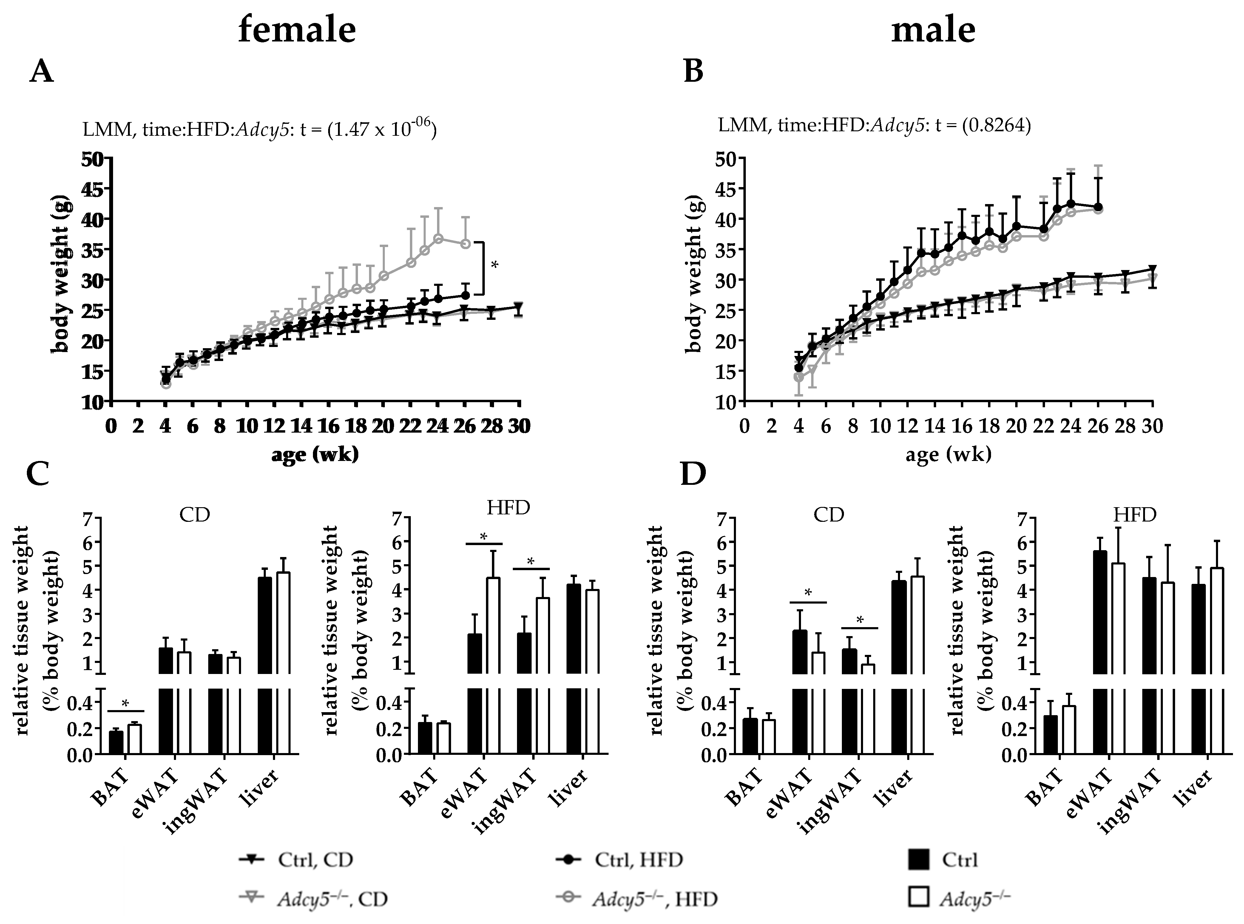
2.1. Adcy5–/– Mice Are Not Protected against Obesity

Under CD conditions Adcy5–/– and Ctrl mice of both sexes exhibited indistinguishable body weight gain until the age of 30 weeks (Figure 1A,B). In male mice, a 20-week HFD challenge started at 6 weeks of age caused obesity without significant differences between the body weight curves of Adcy5–/– and Ctrl mice (Figure 1B). We found that female Ctrl mice did not respond as male mice to HFD and gained significantly less weight than female Adcy5–/– mice (Figure 1A). Starting from 10 weeks until the end of HFD, female Adcy5–/– mice exhibited up to 30% higher body weight gain compared to Ctrl mice (Figure 1A). Since we did not use littermate controls, we performed a Mouse Genome Scan SNP analysis to exclude genetic background effects as causative for the observed weight differences. Genome scans revealed a 99.21% identity between our Ctrl and Adcy5–/– mice.
Except for a slight increase in brown adipose tissue (BAT) in Adcy5–/–, body composition in females fed on CD was comparable for both genotypes (Figure 1C). On the other hand, Adcy5–/– mice gained significantly more epigonadal white adipose tissue (eWAT) and inguinal WAT (ingWAT) upon HFD (Figure 1C, Figure S2A). Accordingly, relative lean mass was significantly lower in female Adcy5–/– compared to controls under both diets (Figure S2B). Observed body weight differences in females fed on HFD could not be explained by differences in body length (Figure S2C) or by a significantly increased food intake, despite Adcy5–/– females tending to feed more than their Ctrl (Figure S2D). In contrast to females, male Adcy5–/– and Ctrl mice did not differ in their response to HFD (Figure 1B), but Adcy5–/– mice had lower eWAT and ingWAT mass under CD (Figure 1D). Male Adcy5–/– mice showed a trend for lower body fat mass and higher relative lean body mass compared to Ctrl animals (Figure S2E,F). Body length was negligibly different for both genotypes upon HFD (Figure S2G). Surprisingly, food intake was not different between female Adcy5–/– and Ctrl mice for both diets, (Figure S2D) whereas male Adcy5–/– mice consumed significantly less under HFD conditions (Figure S2H).
Taken together, Adcy5–/– mice were not protected against HFD-induced obesity. The higher degree of adiposity in female Adcy5–/– mice is most likely explained by insufficient response to HFD in Ctrl mice.

2.2. Adcy5 Deletion Effects on Glucose Tolerance, Insulin Sensitivity and Serum Parameters

To detect potential disorders in glucose metabolism resulting from the Adcy5 knockout, we performed intraperitoneal glucose tolerance tests (ipGTTs). Under CD conditions, female Adcy5–/– mice responded with a higher increase in blood glucose 15 min after glucose injection (2 g glucose per kg body weight). Regarding the area under the curve (AUC) for both diets, Adcy5–/– females exhibited a trend to develop glucose intolerance (Figure 2A). On the contrary, CD male Adcy5–/– mice exhibited lower circulating glucose concentrations after the intraperitoneal glucose challenge, which did not result in significantly lower AUC glucose during the ipGTT course (Figure 2B). Challenged with a HFD this trend became inverted, resulting in higher AUC glucose in Adcy5–/– male mice (Figure 2B).
Fasting blood glucose levels were not different between female Adcy5–/– and control mice fed on CD, but there was a ~20% increase for Adcy5–/– upon HFD, which was not significant (Table 1). Female Adcy5–/– mice were further characterized by significantly lower HbA1c levels under CD (Table 1). Interestingly, lean male Adcy5–/– mice exhibited significantly lower HbA1c levels, fasting blood glucose and C-peptide concentrations compared to Ctrl (Table 2). However, these differences were not observed under HFD conditions (Table 2).
In hyperinsulinemic-euglycemic clamp studies under CD conditions, we did not find significantly impaired whole-body insulin sensitivity in Adcy5–/– compared to Ctrl mice (Figure 2C–F).
Stimulated with 11 and 22 mmol glucose, pancreatic islets of Adcy5–/– females secreted lower amounts of insulin into medium compared to islets of Ctrl animals (Figure 2G). There were no genotype differences in glucose-stimulated insulin secretion at other glucose concentrations in females (Figure 2G) or at any glucose concentration in males (Figure 2H). In both sexes, total insulin content per islet after glucose stimulation was not significantly different between Ctrl and Adcy5–/– except for male mice stimulated with 5.5 mmol glucose (Figure 2I,J).
Regarding serum lipids, Adcy5–/– females were characterized by significantly lower total and HDL-cholesterol compared to Ctrl under CD (Table 1). In contrast, total and HDL-cholesterol were significantly higher in female Adcy5–/– after HFD. In line with females, male Adcy5–/– had higher total and HDL-cholesterol serum concentrations under HFD, and significantly lower LDL-cholesterol serum levels under CD (Table 2). Leptin and adiponectin serum concentrations were not different between male Adcy5–/– and Ctrl mice under both dietary interventions (Table 2). In female Adcy5–/– mice we found higher leptin levels after HFD (Table 1). Additionally, microarrays performed with RNA isolated from eWAT of mice on HFD did not show differentially expressed pro-inflammatory genes such as Il6, Tnfa, Nfkb1 or Ccl2 between Adcy5–/– and control animals of both sexes (Figure 4).
In summary, lean Adcy5–/– mice exhibited lower HbA1c levels compared to controls independent of their sex. In contrast, glucose tolerance was in both sexes not significantly different between the genotypes (Table S3), but Adcy5–/– mice showed a trend for developing glucose intolerance under HFD. Furthermore, insulin sensitivity was not affected by deletion of Adcy5. Compared to Ctrl, 20 weeks of HFD challenge resulted in higher total and HDL-cholesterol serum levels for Adcy5–/– mice of both sexes.

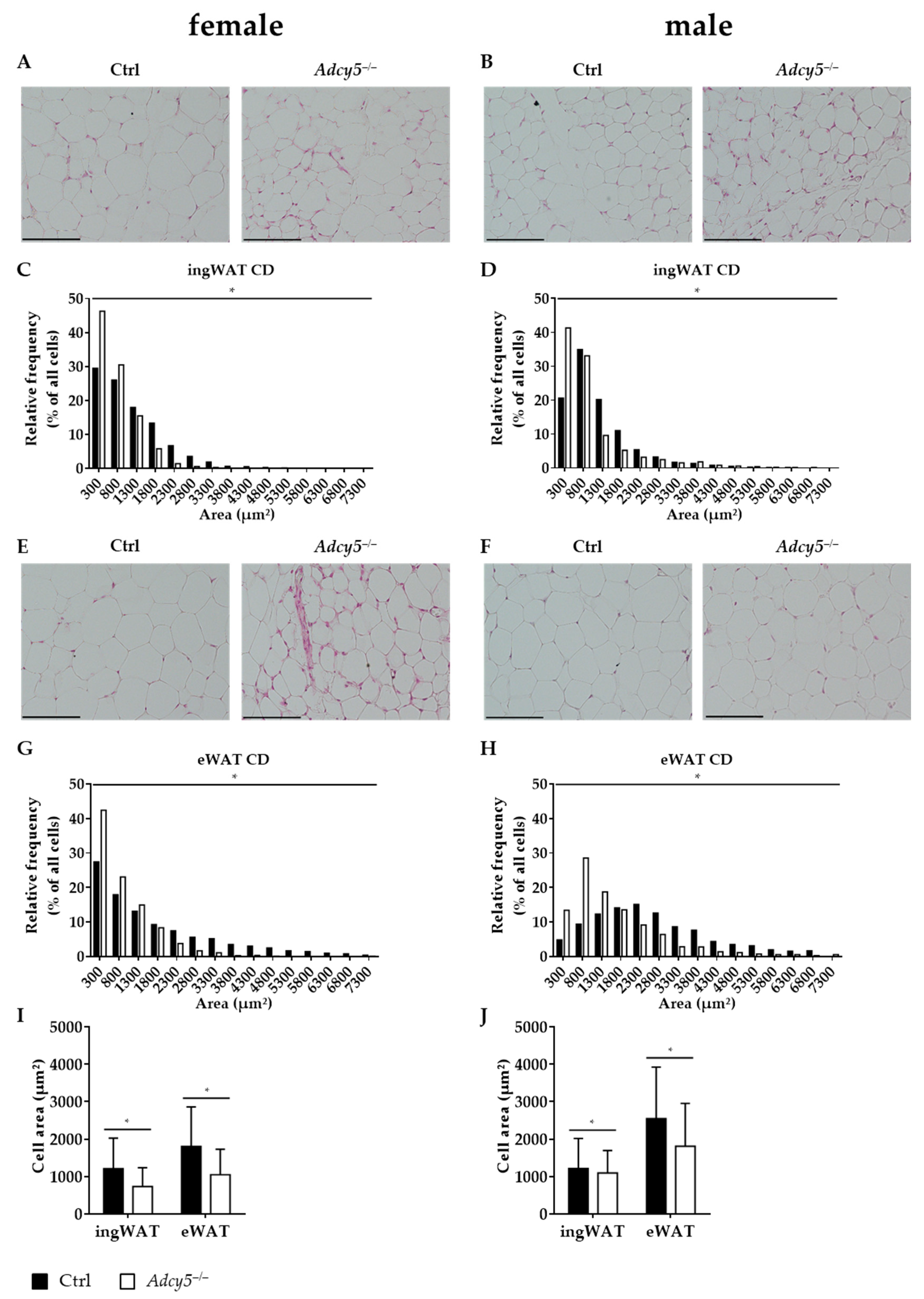
2.3. Consequences of Adcy5–/– on AT Morphology

Histological analyses of ingWAT and eWAT revealed that mean and maximal adipocyte sizes were significantly lower in Adcy5–/– compared to control mice of both sexes under CD (Figure 3). Particularly, the subfraction of small adipocytes was enriched in Adcy5–/– compared to Ctrl mice both in ingWAT (Figure 3A–D) and eWAT (Figure 3E–H). Smaller adipocytes in Adcy5–/– vs. Ctrl mice were further reflected by lower mean adipocyte areas per given analysis area of whole adipose tissue sections (Figure 3I,J). We did not find evidence for differences in adipose tissue immune cell infiltration between Adcy5–/– and Ctrl animals. The AT expression of immune cell markers CD68, CD40, CD11c and CD163 was not different between Adcy5–/– and Ctrl mice indicated by non-significant gene expression changes in microarray investigations (Figure 4A–D). HFD challenge resulted in the formation of larger adipocytes in female Adcy5–/– compared to Ctrl mice (Figure S3A,C,E,G,I). In contrast, male Adcy5–/– mice revealed smaller adipocytes under HFD challenge (Figure S3B,D,F,H,J).
In summary, Adcy5 knockout affected AT morphology and was characterized by smaller adipocytes except for females fed on HFD, but not by differences in AT inflammation markers.

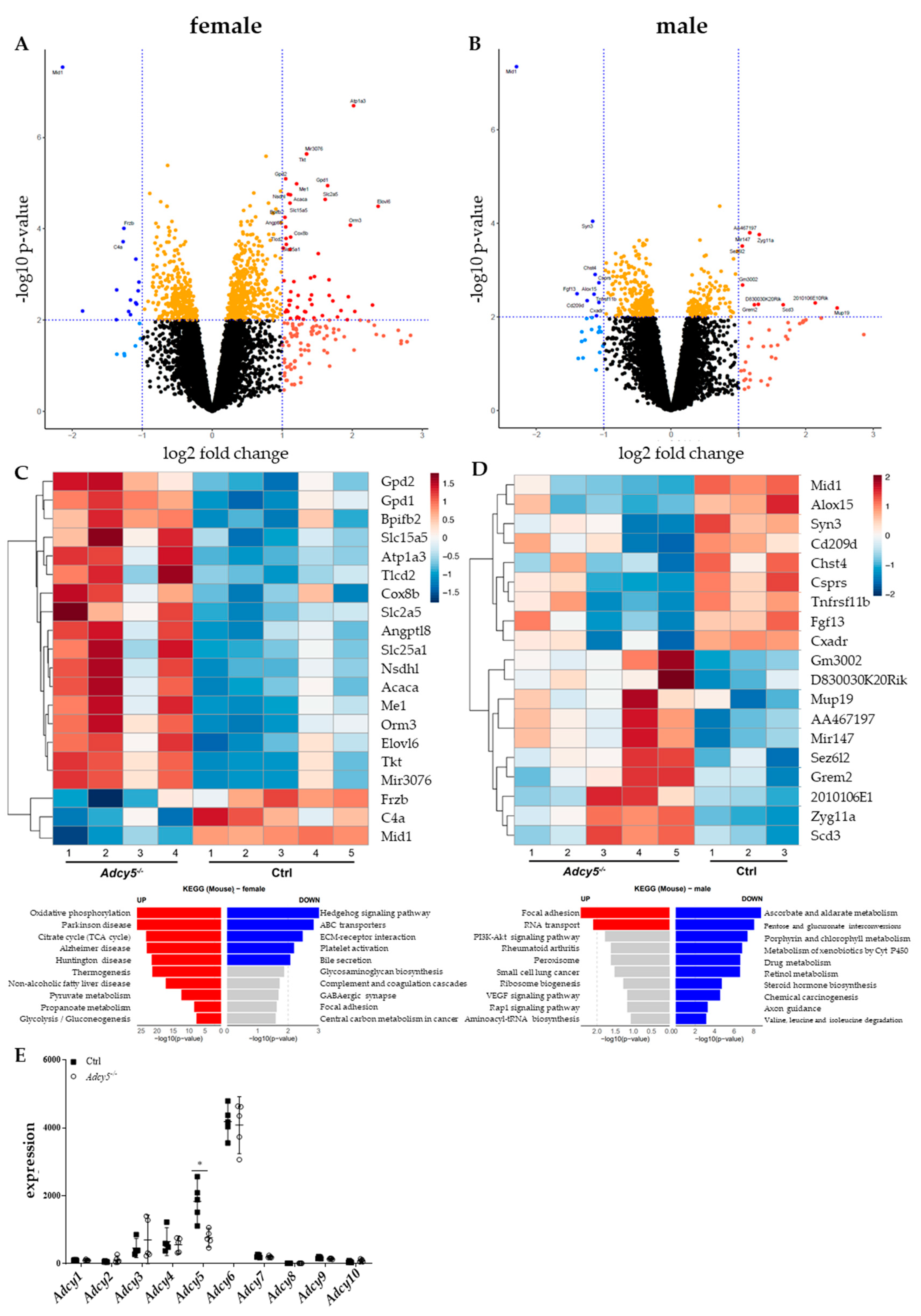
2.4. Adcy5–/– Mice Are Characterized by a Distinct EWAT Gene Expression Signature

Global microarray gene expression analyses in eWAT of CD mice revealed 64 (females) and 17 (males) differentially expressed genes (p-value < 0.01; |FC| ≥ 2) between Adcy5–/– and Ctrl animals (Figure 4A–D; Table S2). In both sexes, we found midline 1 (Mid1) as most significantly (FDR < 0.05) downregulated gene (logFC = –2.14 in females, males: –2.30) in Adcy5–/– vs. Ctrl mice. In female Adcy5–/– mice sodium/potassium-transporting ATPase subunit alpha-3 (Atp1a3) was one of the most significantly upregulated genes (logFC = 2.02) (Figure 4A). Interestingly, several genes of lipid (e.g., Elovl6 [logFC = 2.37], Fasn [logFC = 1.40]) and glucose metabolism (e.g., Gpd1 [logFC = 1.65], Slc2a5 [logFC = 1.61]) were significantly higher expressed in AT of Adcy5–/– compared to Ctrl mice (Table S2). According to KEGG analysis of our microarray data obtained for AT gene expression analyses, the most upregulated genes contributed to metabolic pathways like oxidative phosphorylation, citrate cycle, thermogenesis or glycolysis and gluconeogenesis in females. Interestingly, we found pathways relevant to the pathogenesis of Parkinson’s, Alzheimer´s, Huntington´s or non-alcoholic fatty liver disease to be upregulated in female Adcy5–/– mice (Figure 4C).
Global AT gene expression analysis confirmed the downregulation of Adcy5 expression and demonstrated that no other adenylyl cyclase gene expression was affected by targeting Adcy5 at the whole-body level (Figure 4E). To sum up, deletion of Adcy5 was not compensated by higher expression of other adenylyl cyclases. However, Adcy5–/– mice presented an altered expression of genes involved in metabolic pathways.

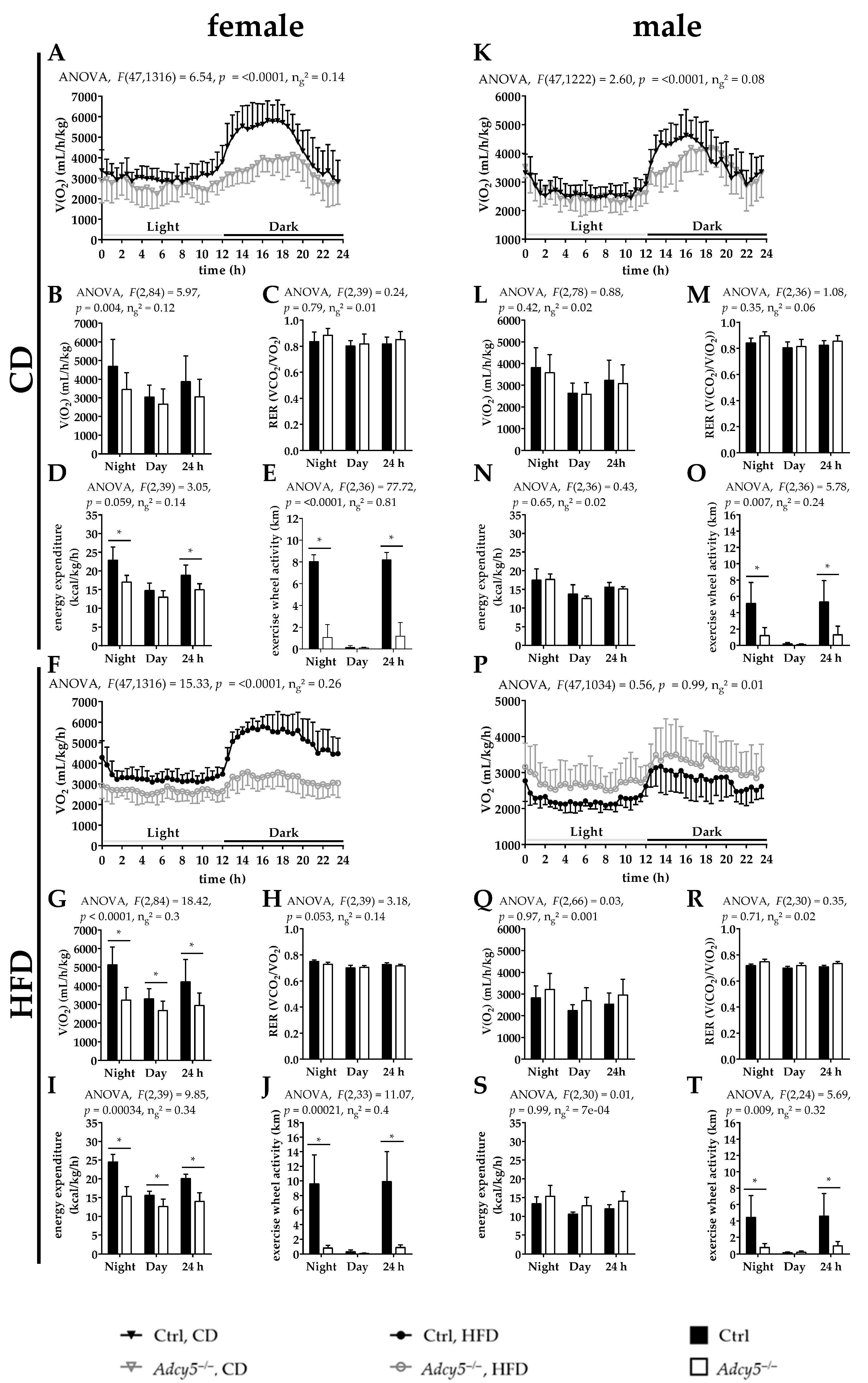
2.5. Adcy5–/– Affects Activity and Energy Expenditure

In previously characterized Adcy5 knockout mice, higher energy expenditure (EE) was reported together with beneficial effects on body fat mass, glucose metabolism and insulin sensitivity [13]. We therefore measured daily food intake, energy expenditure (EE) and running wheel activity over 48 h (Figure 5 and Figure S2). Except for male mice under HFD, food intake was not significantly different between Adcy5–/– and Ctrl mice in both sexes and diet interventions (Figure S2D,H). Female Adcy5–/– mice of both diets consumed significantly less oxygen and had a significantly lower EE compared to control mice under HFD (Figure 5A,B,D,F,G,I). Male Adcy5–/– and control mice were not significantly different in these parameters of oxygen consumption and energy expenditure (Figure 5K,L,N,P,Q,S). RER values were not found to be different in any sex or diet (Figure 5C,H,M,R). Importantly, in both male and female Adcy5–/– mice, exercise running wheel activity was significantly lower under both CD and HFD conditions (Figure 5E,J,O,T). Reduced exercise wheel activity was the most striking behavioral difference between the genotypes.

3. Discussion

Our study was inspired by previous reports that mice with a disruption of Adcy5 are protected against the development of high fat feeding-induced obesity, glucose intolerance, insulin resistance [13], heart failure [15], have reduced oxidative stress [18] and increased longevity [11]. In addition, deletion of Adcy5 caused reduced fat mass, particularly in visceral depots and changes in adipose tissue morphology mimicking the effects of caloric restriction [13,18]. Mechanisms linking Adcy5 deficiency to these health benefits (e.g., decreased glycogen storage) seemed to be so similar to the effects of caloric restriction that Adcy5 knockout mice have a shortened life span upon calorie restriction [18]. We previously showed that mice with a conditional disruption of the insulin receptor in adipose tissue have increased longevity [12,19] and share similarities to long-lived mice and rats in response to caloric restriction [20,21,22]. Altered AT function is one important aspect linking impaired insulin [12] or insulin-like growth factor-1 (IGF-1) signaling to longevity [23]. Based on these data, we hypothesized that reduced Adcy5 expression causes changes in AT that contribute to the reported healthier cardio-metabolic phenotype of Adcy5 knockout mice, which ultimately led to prolonged lifespan. We therefore generated Adcy5–/– mice with the same targeting strategy as the previously reported Adcy5 knockout mice [11,13,24].
Unexpectedly, we found that Adcy5–/– mice of both sexes were not protected against obesity and impaired glucose metabolism. Moreover, the previously described effects of Adcy5 deletion on improved insulin sensitivity, increased oxygen consumption, respiratory exchange ratio, and energy expenditure could not be confirmed in our model. In our model, ablation of Adcy5 at the whole-body level did not protect mice against obesity under high-fat diet conditions. Our model was further complicated by the observation that only female wild-type controls did not respond as expected to HFD with the development of obesity. The inappropriate response to HFD in control mice led to significantly higher body weight, impaired glucose tolerance and insulin sensitivity only in female Adcy5–/– mice.
It is well known that males and females are differentially susceptible to diet-induced obesity (DIO). Female mice have been shown to develop obesity later and to a lesser extent than males [25]. Comparing male mice with ovariectomized and non-ovariectomized females, Stubbins et al. found that ovary removal eliminated protection of female mice against DIO [26]. Here, we showed that females lacking Adcy5 developed severe obesity under HFD, comparable to male mice. In mice, Adcy5 is most highly expressed in subcutaneous fat and the ovary [27]. The lack of Adcy5 results in decreased levels of cAMP. Beside other effects, cAMP was shown to highly increase aromatase expression, the key enzyme in estrogen production, in stromal cells [28]. In summary, our Adcy5 knockout mice are similar to estrogen-lacking ovariectomized mice described by Stubbins et al. [26]. Unfortunately, we were not able to assess circulating estrogen concentrations in our model and could therefore only speculate that genotype differences in estrogen levels may contribute to the distinct phenotype of our female mice under HFD.
For our studies, littermate controls from the Adcy5–/– breeding were not available so we used wild-type mice of the same C57BL/6NTac background as controls. To investigate whether differences in the genetic background between Adcy5–/– and control mice may underlie the observed differences in HFD response of female mice, we performed Genome Scan SNP analyses. These analyses showed a 99.21% identity between Ctrl and Adcy5–/– animals, suggesting that the control mice sufficiently represent the background of Adcy5–/– mice. The analysis revealed that both Adcy5–/– and Ctrl mice are homozygous wild-type for nicotinamide nucleotide transhydrogenase (Nnt), a gene that we and others found to be associated with different DIO-susceptibility in C57BL/6 substrains [29,30]. Additionally, we previously showed that only four heterogeneous sex-specific genetic variants underlie the differences in HFD-induced weight gain between the otherwise genetically identical C57BL/6NTac and C57BL/6JRj substrains [31]. Since our Adcy5 knockout on C57BL/6NTac background differed from previously described ones, e.g., 129/Sv [18], C57BL/6J [13], or 129/SvJ-C57BL/6 mixed [14], we could not exclude that even small genetic background differences may have caused our contrasting findings compared to previous Adcy5 deletion models (reviewed in [16]). Importantly, the comparison of our model with previous data [13] suggested that interactions between Adcy5 deletion and the genetic background may play a previously unrecognized role determining the phenotype. If this assumption is true, it would turn ADCY5 into a less likely therapeutic target for the treatment of obesity and diabetes.
Individual variation in Adcy5 interaction with the genetic background may also explain the heterogeneous findings from genome-wide association studies and candidate gene approaches reporting different directions of the associations of ADCY5 genetic variants with obesity, diabetes-related quantitative traits and the risk of type 2 diabetes [1,2,5,8,9].
Additionally, the different dietary compositions between our HFD and HFDs used in previous studies could explain the various phenotypes [32,33].
However, the unexpected lack of a difference in body weight, fat mass, energy expenditure, glucose metabolism and insulin sensitivity between our male Adcy5–/– and control mice could also be an advantage, because it allowed us to investigate the effects of Adcy5 depletion on AT independently of lower body weight and a metabolically healthier phenotype. We therefore studied AT morphology and gene expression signatures to test the hypothesis that ablation of Adcy5 may have weight-independent effects on AT. In humans, we showed that AT ADCY5 expression is related to obesity and fat distribution, but not with impaired glucose metabolism and T2D [17]. In addition, altered ADCY5 expression in AT does not seem to underlie the association between the ADCY5 SNP rs11708067 and increased T2D risk [17].
It has been previously suggested that ACDY5 couples glucose to insulin secretion in human islets because reduced ACDY5 expression was associated with impaired insulin secretion after a glucose challenge [2]. Accordingly, islets of our Adcy5-deficient females exhibited lower glucose-stimulated insulin secretion into the medium after stimulation with high glucose concentrations. Importantly, we could not confirm these potential Adcy5 knockout-related effects at lower glucose concentrations in females or at any given glucose level in male mice. In addition, euglycemic-hyperinsulinemic clamp studies did not reveal an effect of Adcy5 deficiency on systemic insulin sensitivity. In contrast, we found just minor and non-significant effects on glucose tolerance upon depletion of Adcy5 in our model. In male Adcy5–/– mice, HOMA-ß was significantly higher under CD conditions, further supporting an unexpectedly better beta cell function despite reduced Adcy5 expression.
Except for females on HFD, Adcy5–/– mice exhibited an increased number of smaller adipocytes and lower mean adipocyte size. Smaller adipocyte size is a consistent finding compared to previous studies [13], suggesting that Adcy5 ablation affects AT independently of reduced fat mass. An increased number of smaller adipocytes may link Adcy5 knockout to the previously reported effects on longevity and reduced diabetes risk, because small adipocytes are strongly related to BMI-independent retained insulin sensitivity [34]. Both animal [12] and human studies [35] have suggested that protection against adipocyte hyperplasia is associated with a lower risk of hyperglycemia and type 2 diabetes. Nevertheless, in our model the smaller adipocyte size did not translate into better insulin sensitivity. In the context of glucose metabolism, we found HbA1c levels marginally reduced in lean Adcy5–/– mice of both sexes (~4% in males, ~6% in females), whereas fasting blood glucose levels tended to be enhanced after HFD challenge (~17% in males, ~23% in females). In contrast to males, Adcy5–/– females developed larger adipocytes under HFD and higher circulating leptin levels. We found a potentially important diet–genotype interaction in distinct parameters of cholesterol metabolism. Since female Adcy5–/– mice had significantly lower total and HDL-cholesterol serum concentrations under chow fed conditions, but significantly higher levels of these lipid metabolism parameters after HFD challenge, our data suggest a previously unrecognized role of Adcy5 in the regulation of cholesterol metabolism as demonstrated for Adcy1. In THP-1 foam cells, Adcy1 works as a signaling switch for the apolipoprotein A1-mediated cholesterol efflux pathway. The authors concluded that cAMP, produced by Adcy1, may regulate effectors for cholesterol exocytosis to remove excessive cellular lipids [36]. Additionally, we observed surprisingly high FFA serum concentrations in both genotypes. However, our data reflected previously reported FFA levels in mice after 16 h of fasting [37].
Using global gene expression analyses, we identified Mid1, Atp1a3 and several genes of glucose and lipid metabolism (e.g., Elovl6, Gpd1, Gpd2, Slc2a5) as the most significantly differentially expressed genes between female Adcy5–/– and Ctrl mice. Mid1 is an E3 ubiquitin ligase of the Tripartite Motif (TRIM) subfamily of RING-containing proteins and is therefore also known as Trim18 [38]. Mid1 is involved in several essential biological processes, especially during embryonic development. Binding to Mid1-interacting protein 1 (Mid1ip1, also known as Mig12), resulting Mid1/Mig12 heterodimers stabilizes microtubules in cell division and migration [39]. Interestingly, Mig12 depletion was shown to activate the master regulator of fatty acid oxidation or synthesis, AMP-activated protein kinase (Ampk) [40,41,42]. Taken together, reduced Mid1 expression in AT of Adcy5–/– mice could explain the observed morphological changes in AT, especially under HFD. However, to investigate a potential regulatory function of Mid1-Mig12/Thrsp-Acc1/Acc2 or Mid1-Mig12-Ampk on AT metabolism another study would be necessary.
We further found that both male and female Adcy5–/– mice were characterized by significantly lower voluntary running wheel activity in metabolic chambers. As mutations in the Atp1a3 gene are known to be related to dyskinesia, epilepsies and Parkinson’s disease [43] this could be a relevant finding in the context of other reports describing a connection between the loss of Adcy5 and movement disorders like dyskinesia or autism-like behavior [44,45,46]. Furthermore, we cannot exclude that lower activity in our Adcy5–/– mice was caused by previously reported movement disorders and contributed to the phenotype differences compared to previous Adcy5–/– models [11,13,24]. In addition to lower exercise activity, we did not observe any behavioral abnormality in Adcy5–/– mice. Noteworthy, independent groups reported that mice with a genetic disruption of Adcy5 exhibited increased repetitive behaviors and sociability deficits similar to autism-like behavior [47]. In humans, mutations of the ADCY5 gene have been associated with early-onset autosomal dominant chorea and dystonia [45].
In summary, ablation of the Adcy5 gene did not confer any physiological or biochemical benefits, under neither normal nor high-fat diet challenged conditions. Adcy5–/– mice were characterized by considerably lower physical activity. In contrast to previous independent models of Adcy5 deficiency, we found that targeting ADCY5 did not improve insulin sensitivity.

4. Materials and Methods

4.1. Animal Studies

All animal studies were approved by the local authorities of the state of Saxony, Germany (ethic approval code TVV60/16, 24.05.2017, Animal Welfare Committee of the Landesdirektion Sachsen, Germany). Mice were housed in pathogen-free facilities in groups of three to five at 22 ± 2 °C on a 12 h light/dark cycle. Animals were bred and kept in the animal laboratories at Leipzig University and were fed a standard chow diet containing 10% calories from fat, 36% from protein and 54% from carbohydrates (CD, Ssniff Spezialdiäten, Soest, Germany, V1324-300). Diet-induced obesity was achieved by feeding a high-fat diet (HFD) containing 54% calories from fat, 20% from protein and 26% from carbohydrates (HFD, Ssniff Spezialdiäten, E15772-34) starting at 6 weeks of age. Both diets were matched with respect to their crude protein content (21.2% vs. 21.6%). All animals had access to food and water ad libitum, except for experiments where a fasting state was required.

4.2. Generation of Adenylyl Cyclase Type 5 Knockout Mice

Adenylyl cyclase type 5 knockout (Adcy5–/–) mice were produced via VelociGene® (Regeneron Pharmaceuticals, INC, Tarrytown, NY, USA) similar to the approach reported by Okumura et al. [24]. Adcy5 was disrupted by an insertion of a loxP-hUBp-em7-Neo-polyA-loxP cassette into exon 1 (Figure S1). All mice were genotyped by DNA isolation from the tail tip or ear stamp isolated using DirectPCR (Tail) Lysis Reagent (Viagen Biotech Inc., Los Angeles, CA, USA). Primer pairs (biomers.net, Ulm, Germany) are listed in Table S1. On 2% agarose gels, wild-type (WT) mice showed a 202 bp band, whereas Adcy5–/– mice were identified by a 152 bp band.
Furthermore, KO efficiency was confirmed in several tissues on both DNA (Figure S1B) and mRNA level (Figure S1C). Animals were maintained on a C57BL/6NTac background and both sexes were included in all experiments. Due to mice interbreeding, no littermate controls could be generated. Mouse Genome Scan SNP analysis was performed by Taconic (Ms Genome Scan Panel, Taconic Biosciences, Rensselaer, NY, USA) and revealed a 99.21% identity between Ctrl and Adcy5–/– animals.

4.3. Phenotypic Characterization

In this study, 12 mice of each sex and genotype were studied up to age 30 weeks under chow diet (CD) conditions. In addition, a subgroup of 11 female Adcy5–/– and 14 control mice as well as 13 male Adcy5–/– and 14 control mice underwent an HFD for 20 weeks starting at age 6 weeks. We recorded body weight weekly and determined whole-body fat and lean mass with the EchoMRI700™ instrument (Echo Medical Systems, Houston, TX, USA) at the study end. Intraperitoneal glucose tolerance tests (ipGTTs; 2 g glucose per kg body weight) were performed at age 25 weeks as previously described [48]. Pancreatic islet isolation and glucose-stimulated insulin secretion experiments were performed as previously described [49]. In brief, islets of all animals were pooled per genotype and sex (n = 3). In groups of five, islets were incubated overnight in glucose-free Gibco RPMI 1640 medium (ThermoFisher Scientific, Waltham, MA, USA) supplemented with 5.5 mmol glucose, 10% fetal calf serum and 100 U/mL penicillin and 100 g/mL streptomycin (Sigma-Aldrich, Munich, Germany) at 37 °C and 5% CO2. For insulin secretion, RPMI 1640 medium was supplemented with 1.5, 5.5, 11 or 22 mmol glucose, respectively, and islets were incubated for 2 h at 37 °C and 5% CO2. Supernatant was collected and islets were sonicated with 0.04 M phosphate buffer. Insulin contents were measured with Ultra Sensitive Mouse Insulin ELISA kit (Crystal Chem, Downers Grove, IL, USA). Hyperinsulinemic-euglycemic clamps were performed at 23 to 25 weeks of age as previously described [50]. In brief, to determine initial GIR rate, mice received insulin continuously infused through an implanted catheter. When blood glucose levels dropped, 20% glucose solution was infused to stabilize blood glucose levels. Short interval blood glucose measurements were performed, and glucose infusion rate adjusted accordingly to reach physiological blood glucose levels of 5.6 mmol/L. When glucose levels were constant for 30 min, the clamp started. In subgroups, whole-body energy metabolism as well as food intake were investigated using an indirect metabolic chamber system at age 25 weeks (HFD) or 29 weeks (CD) (CaloSys V2.1, TSE Systems, Bad Homburg, Germany). In brief, 7 to 10 Adcy5–/– and Ctrl mice of each sex and diet were housed for 72 h (adaptation time included) in metabolic chambers as previously described (19). Naso-anal length and rectal body temperature were measured at the end of observation period. In EDTA-containing tubes, 20 µL of whole blood was collected for HbA1c analyses (COBAS 7000, Roche, Basel, Switzerland). At age 30 weeks (CD) or 26 weeks (HFD) mice were sacrificed by an overdose of anesthetic (Isofluran, Baxter, Unterschleißheim, Germany). Immediately, inguinal (ingWAT) and epigonadal white adipose tissue (eWAT) as well as liver and brown adipose tissue (BAT) were taken, weighed and stored in liquid nitrogen. For those tissues, relative organ weights were calculated in relation to whole-body weight. ELISA was used to analyze insulin (Mouse Ultrasensitive Insulin ELISA, ALPCO, Salem, NH, USA), C-peptide (Mouse C-peptide ELISA, ALPCO, Salem, NH, USA), leptin (Mouse Leptin ELISA Kit, Crystal Chem Inc., Downers Grove, IL, USA), and adiponectin (Adiponectin (mouse) ELISA Kit, AdipoGen® LIFE SCIENCES, Liestal, Switzerland) serum concentrations using mouse serum according to the manufacturer’s guidelines. In randomly selected subgroups of 3 to 4 mice per sex and genotype, serum protein levels were determined by OLINK proteomics (Uppsala, Sweden).

4.4. Adipose Tissue Histology

For histologic investigations, adipose tissue was fixed in 4% buffered formaldehyde for at least 48 h. Afterwards, tissue containing cassettes were rinsed with water and dehydrated in a graded series of 70–100% ethanol followed by ROTI®Histol (Carl Roth GmbH, Karlsruhe, Germany) and paraffin. Both, ingWAT and eWAT pads were cut in 5 µm sections and H&E stained afterwards. Systematic analyses with respect to adipocyte size were performed using Keyence BZ-X800 microscope and BZ-X800 Analyzer software (Keyence Corp., Osaka, Japan). At least 1900 (CD) or 2400 (HFD) adipocytes were analyzed from 4 animals for each genotype and sex to determine cell size distribution.

4.5. RNA Isolation and Tissue-Specific mRNA Expression

Frozen AT was lysed using QIAzol Lysis Reagent (Qiagen GmbH, Hilden, Germany) and Precellys Homogenizer (Bertin Technologies, Montigny-le-Bretonneux, France). RNA was isolated from tissue homogenates with RNeasy Lipid Tissue Mini Kit (Qiagen). For all other tissues, AllPrep DNA/RNA/Protein Mini Kit (Qiagen) was used for simultaneous purification of DNA, RNA and protein. RNA was reverse-transcribed using Random Primers (Invitrogen, Carlsbad, CA, USA) and Superscript II Reverse Transcriptase (Invitrogen). Quantitative real-time PCR (qRT-PCR) was performed with the LightCycler 480 system and LightCycler 480 SYBR Green I Master (Hoffmann-La Roche AG, Basel, Switzerland) with identical cDNA amounts for each reaction. Adcy5 mRNA expression was calculated relative to ribosomal protein, large, P0 (Rplp0 or 36b4) mRNA. Primers are listed in Table S1.

4.6. Adipose Tissue Microarray Analyses

RNA was isolated from frozen eWAT of Adcy5–/– (n = 10, 5 females, 5 males) and control (n = 10, 5 females, 5 males) CD mice. Before microarray analysis, RNA integrity and concentration were examined on an Agilent Fragment Analyzer (Agilent Technologies, Palo Alto, CA, USA) using the HS RNA Kit (Agilent Technologies) according to the manufacturer’s instructions. cRNA was prepared from 100 ng of total RNA hybridized to GeneChip Clariom S arrays (ThermoFisher Scientific). Arrays were scanned with a third generation Affymetrix GeneChip Scanner 3000 (ThermoFisher Scientific). Affymetrix GeneChip data were extracted from fluorescence intensities and were scaled to normalize data for inter-array comparison using Transcriptome Analysis Console (TAC) 4.0.2 software.
Raw data were preprocessed applying the oligo Bioconductor R package (v1.50.0, [51]), which performs a deconvolution method for background correction, quantile normalization and uses the Robust Multichip Average (RMA) algorithm for summarization [52]. Quality control of the raw and normalized data was performed using the Biobase (v2.46, [53]) and oligo R Bioconductor packages [54]. Based on the outlier detection test of the arrayQualityMetrics (v3.42, [54]) Bioconductor R package, two male Ctrl and one female Adcy5–/– samples were excluded. The differentially expressed genes (DEGs) for the comparisons (i) female Adcy5–/– vs. female Ctrl and (ii) male Adcy5–/– vs. male Ctrl were screened using the Linear Models for Microarray data (LIMMA) method (v3.42, [55]). To increase the signal-to-noise ratios array weights were considered. Since only a few genes (36 female; 1 male, see Table S2) survived multiple testing (FDR < 0.05) applying the Benjamini–Hochberg procedure [56], we set the threshold for identification of DEGs as p-value < 0.01 and |FC| ≥ 2. This allowed us to be exploratory about the results, especially in the pathway analysis. A broad gene list functional enrichment analysis for KEGG mouse pathways was performed using Enrichr (www.amppharm.mssm.edu/Enrichr accessed on 19th April 2021) [57,58]. All differentially expressed genes (p-value < 0.01) irrespective of FC were used. Microarray data were deposited in the ArrayExpress database at EMBL-EBI (www.ebi.ac.uk/arrayexpress accessed on 19th April 2021) under accession number E-MTAB-9417.

4.7. Statistical Analyses

Prism 6.0 software (GraphPad Software Inc., San Diego, CA, USA) was used for graph design. Data are shown as means ± SD or SEM. Multiple t-tests were performed only when no interactions of a grouping variable on a continuous outcome measure were of interest. Otherwise, the residual analysis was conducted to test for the assumption of the two-way ANOVA using the rstatix [59] R package. Here, post hoc tests using pairwise t-test were computed for significant interactions only and corrected applying the Bonferroni method [60].
For modeling continuous measurements over a time course of multilevel data we performed repeated-measure two-way ANOVAs (rstatix, [59]) when the data contained a balanced number of replicates or no missing data for some data points. Otherwise, we applied repeated-measure linear mixed-effects models (LMMs) because they account for variability and imbalance in the data. The LMMs were fitted to the data by the formula weight~time*diet*strain+(time|patient ID) applying the lme4 R package [61]. Outliers were assessed by box plot method and normality was assessed using Shapiro–Wilk’s normality test [62]. Homogeneity of variances was assessed by Levene’s test for the two-way ANOVAs and by Mauchly’s test of sphericity [63] for the repeated measurement analyses.
p-values < 0.05 were considered as statistically significant. The used statistical method for the single analyses is indicated in each figure legend.

5. Conclusions

In conclusion, our data did not support beneficial effects on obesity and insulin sensitivity described for other Adcy5-deficient mouse models. Since effects resulting from Adcy5 reduction were both sex and genetic background-dependent, this may therefore limit the relevance of ADCY5 as potential drug target. Additionally, our Adcy5–/– model was characterized by a significantly lower voluntary exercise activity in both sexes and diets, suggesting that lower activity may underlie the unexpected phenotype without protection against obesity and its associated cardio-metabolic abnormalities. We find that Adcy5 plays a role in the regulation of adipocyte size, and smaller adipocytes in Adcy5–/– mice may be caused by or at least reflect distinct AT gene expression signatures.

Supplementary Materials

Supplementary Materials can be found at https://www.mdpi.com/article/10.3390/ijms22094353/s1.

Author Contributions

M.B., A.K. and N.K. designed the studies. S.D., C.B. and M.K. performed experiments. S.D. and A.H. analyzed the data. S.D. and A.H. designed figures and tables. S.D., A.H. and M.B. wrote the paper with input from all authors. M.B. is the guarantor of this work and, as such, had full access to all data in the study and takes responsibility for the integrity of the data and the accuracy of the data analysis. All authors have read and agreed to the published version of the manuscript.

Funding

This work was founded by the Deutsche Forschungsgemeinschaft (DFG, German Research Foundation) through CRC 1052, project number 209933838, subproject B1 to M.B.

Institutional Review Board Statement

All animal studies were approved by the local authorities of the state of Saxony, Germany (ethic approval code TVV60/16, 24.05.2017, Animal Welfare Committee of the Landesdirektion Sachsen, Germany).

Informed Consent Statement

Not applicable.

Data Availability Statement

Data is contained within the supplementary material.

Acknowledgments

The authors thank PD Knut Krohn for support with the microarray studies and Jenny Schuster, Lisa Gärtner and Elisabeth Langer for excellent assistance at our animal facility. Further, we thank Viola Döbel for assistance with cell culture experiments.

Conflicts of Interest

A.K. was an employee at Sanofi. M.B. received honoraria as a consultant and speaker from Amgen, AstraZeneca, Bayer, Boehringer-Ingelheim, Lilly, Novo Nordisk, Novartis, and Sanofi. All other authors do not have any conflict of interest. For all other authors: no potential conflicts of interest relevant to this article were reported.

Abbreviations

36b4Rplp0, ribosomal protein, large, P0
ACCAcetyl-CoA carboxylase
AdcyAdenylyl cyclase
AMPKAMP-activated protein kinase
ATAdipose tissue
Atp1a3Sodium/potassium-transporting ATPase subunit alpha-3
BATBrown adipose tissue
BMIBody mass index
cAMPCyclic adenosine monophosphate
CDChow diet
CD11c, CD40, CD68, CD163Cluster of differentiation 11c, 40, 68 and 163
CtrlControl
DIODiet-induced obesity
EDTAEthylenediaminetetraacetic acid
EEEnergy expenditure
Elovl6Elongation of very long chain fatty acids protein 6
eWATEpigonadal white adipose tissue
FasnFatty acid synthase
Gpd1Glycerol-3-phosphate dehydrogenase
GTTGlucose tolerance test
HbA1cGlycated hemoglobin A1c
HDLHigh density lipoprotein
HFDHigh-fat diet
i.p.Intraperitoneal
IGF-1Insulin-like growth factor 1
IL-17fInterleukin 17f
ingWATInguinal white adipose tissue
ITTInsulin tolerance test
LDLLow density lipoprotein
LIMMALinear models for microarray data
Mid1Midline 1
Mid1ip1Mid1-interacting protein 1, also Mig12 or Spot14R
NntNicotinamide nucleotide transhydrogenase
PKAProtein kinase A
PPARgPeroxisome proliferator-activated receptor gamma
qRT-PCRQuantitative real-time polymerase chain reaction
RERRespiratory exchange ratio
RINGReally interesting new gene
RMARobust multichip average
Slc2a5Solute carrier family 2, facilitated glucose transporter member 5
SNPSingle nucleotide polymorphism
T2DType 2 diabetes mellitus
TACTranscriptome analysis console
ThrspThyroid hormone-inducible hepatic protein
TktTransketolase
TRIMTripartite motive family

References

  1. Dupuis, J.; DIAGRAM Consortium; Langenberg, C.; Prokopenko, I.; Saxena, R.; Soranzo, N.; Jackson, A.U.; Wheeler, E.; Glazer, N.L.; Bouatia-Naji, N.; et al. New genetic loci implicated in fasting glucose homeostasis and their impact on type 2 diabetes risk. Nat. Genet. 2010, 42, 105–116. [Google Scholar] [CrossRef] [PubMed]
  2. Hodson, D.J.; Mitchell, R.K.; Marselli, L.; Pullen, T.J.; Brias, S.G.; Semplici, F.; Everett, K.L.; Cooper, D.M.; Bugliani, M.; Marchetti, P.; et al. ADCY5 Couples Glucose to Insulin Secretion in Human Islets. Diabetes 2014, 63, 3009–3021. [Google Scholar] [CrossRef] [PubMed] [Green Version]
  3. Anderson, E.K.; Gutierrez, D.A.; Hasty, A.H. Adipose tissue recruitment of leukocytes. Curr. Opin. Lipidol. 2010, 21, 172–177. [Google Scholar] [CrossRef]
  4. Huopio, H.; Cederberg, H.; Vangipurapu, J.; Hakkarainen, H.; Pääkkönen, M.; Kuulasmaa, T.; Heinonen, S.; Laakso, M. Association of risk variants for type 2 diabetes and hyperglycemia with gestational diabetes. Eur. J. Endocrinol. 2013, 169, 291–297. [Google Scholar] [CrossRef] [PubMed] [Green Version]
  5. Windholz, J.; Kovacs, P.; Tönjes, A.; Dittrich, K.; Blüher, S.; Kiess, W.; Stumvoll, M.; Körner, A. Effects of Genetic Variants in ADCY5, GIPR, GCKR and VPS13C on Early Impairment of Glucose and Insulin Metabolism in Children. PLoS ONE 2011, 6, e22101. [Google Scholar] [CrossRef] [Green Version]
  6. Rees, S.D.; Hydrie, M.Z.I.; O’Hare, J.P.; Kumar, S.; Shera, A.S.; Basit, A.; Barnett, A.H.; Kelly, M.A. Effects of 16 Genetic Variants on Fasting Glucose and Type 2 Diabetes in South Asians: ADCY5 and GLIS3 Variants May Predispose to Type 2 Diabetes. PLoS ONE 2011, 6, e24710. [Google Scholar] [CrossRef]
  7. Wagner, R.; Dudziak, K.; Herzberg-Schäfer, S.A.; Machicao, F.; Stefan, N.; Staiger, H.; Häring, H.-U.; Fritsche, A. Glucose-Raising Genetic Variants in MADD and ADCY5 Impair Conversion of Proinsulin to Insulin. PLoS ONE 2011, 6, e23639. [Google Scholar] [CrossRef]
  8. Lin, R.; Yuan, Z.; Zhang, C.; Ju, H.; Sun, Y.; Huang, N.; Chen, L.; Jin, L. Common genetic variants in ADCY5 and gestational glycemic traits. PLoS ONE 2020, 15, e0230032. [Google Scholar] [CrossRef] [Green Version]
  9. Roman, T.S.; Cannon, M.E.; Vadlamudi, S.; Buchkovich, M.L.; Wolford, B.N.; Welch, R.P.; Morken, M.A.; Kwon, G.J.; Varshney, A.; Kursawe, R.; et al. A Type 2 Diabetes–Associated Functional Regulatory Variant in a Pancreatic Islet Enhancer at the ADCY5 Locus. Diabetes 2017, 66, 2521–2530. [Google Scholar] [CrossRef] [Green Version]
  10. Cash, T.P.; Pita, G.; Domínguez, O.; Alonso, M.R.; Moreno, L.T.; Borrás, C.; Rodríguez-Mañas, L.; Santiago, C.; Garatachea, N.; Lucia, A.; et al. Exome sequencing of three cases of familial exceptional longevity. Aging Cell 2014, 13, 1087–1090. [Google Scholar] [CrossRef] [Green Version]
  11. Yan, L.; Vatner, D.E.; O’Connor, J.P.; Ivessa, A.; Ge, H.; Chen, W.; Hirotani, S.; Ishikawa, Y.; Sadoshima, J.; Vatner, S.F. Type 5 Adenylyl Cyclase Disruption Increases Longevity and Protects Against Stress. Cell 2007, 130, 247–258. [Google Scholar] [CrossRef] [Green Version]
  12. Blüher, M.; Dodson Michael, M.; Peroni, O.D.; Ueki, K.; Carter, N.; Kahn, B.B.; Kahn, C. Adipose Tissue Selective Insulin Receptor Knockout Protects against Obesity and Obesity-Related Glucose Intolerance. Dev. Cell 2002, 3, 25–38. [Google Scholar] [CrossRef] [Green Version]
  13. Ho, D.; Zhao, X.; Yan, L.; Yuan, C.; Zong, H.; Vatner, D.E.; Pessin, J.E.; Vatner, S.F. Adenylyl Cyclase Type 5 Deficiency Protects Against Diet-Induced Obesity and Insulin Resistance. Diabetes 2015, 64, 2636–2645. [Google Scholar] [CrossRef] [PubMed] [Green Version]
  14. Okumura, S.; Kawabe, J.; Yatani, A.; Takagi, G.; Lee, M.-C.; Hong, C.; Liu, J.; Takagi, I.; Sadoshima, J.; Vatner, D.E.; et al. Type 5 Adenylyl Cyclase Disruption Alters Not Only Sympathetic But Also Parasympathetic and Calcium-Mediated Cardiac Regulation. Circ. Res. 2003, 93, 364–371. [Google Scholar] [CrossRef] [PubMed] [Green Version]
  15. Iwatsubo, K.; Bravo, C.; Uechi, M.; Baljinnyam, E.; Nakamura, T.; Umemura, M.; Lai, L.; Gao, S.; Yan, L.; Zhao, X.; et al. Prevention of heart failure in mice by an antiviral agent that inhibits type 5 cardiac adenylyl cyclase. Am. J. Physiol. Circ. Physiol. 2012, 302, H2622–H2628. [Google Scholar] [CrossRef]
  16. Vatner, S.F.; Park, M.; Yan, L.; Lee, G.J.; Lai, L.; Iwatsubo, K.; Ishikawa, Y.; Pessin, J.; Vatner, D.E. Adenylyl cyclase type 5 in cardiac disease, metabolism, and aging. Am. J. Physiol. Circ. Physiol. 2013, 305, H1–H8. [Google Scholar] [CrossRef] [Green Version]
  17. Knigge, A.; Klöting, N.; Schön, M.R.; Dietrich, A.; Fasshauer, M.; Gärtner, D.; Lohmann, T.; Dreßler, M.; Stumvoll, M.; Kovacs, P.; et al. ADCY5 Gene Expression in Adipose Tissue Is Related to Obesity in Men and Mice. PLoS ONE 2015, 10, e0120742. [Google Scholar] [CrossRef] [Green Version]
  18. Yan, L.; Park, J.Y.; Dillinger, J.-G.; De Lorenzo, M.S.; Yuan, C.; Lai, L.; Wang, C.; Ho, D.; Tian, B.; Stanley, W.C.; et al. Common mechanisms for calorie restriction and adenylyl cyclase type 5 knockout models of longevity. Aging Cell 2012, 11, 1110–1120. [Google Scholar] [CrossRef] [Green Version]
  19. Blüher, M.; Kahn, B.B.; Kahn, C.R. Extended Longevity in Mice Lacking the Insulin Receptor in Adipose Tissue. Science 2003, 299, 572–574. [Google Scholar] [CrossRef] [PubMed] [Green Version]
  20. Wiesenborn, D.S.; Menon, V.; Zhi, X.; Do, A.; Gesing, A.; Wang, Z.; Bartke, A.; Altomare, D.A.; Masternak, M.M. The effect of calorie restriction on insulin signaling in skeletal muscle and adipose tissue of Ames dwarf mice. Aging 2014, 6, 900–912. [Google Scholar] [CrossRef] [PubMed] [Green Version]
  21. Weindruch, R.; Walford, R. Dietary restriction in mice beginning at 1 year of age: Effect on life-span and spontaneous cancer incidence. Science 1982, 215, 1415–1418. [Google Scholar] [CrossRef] [PubMed] [Green Version]
  22. Walford, R.L.; Crew, M. How dietary restriction retards aging: An integrative hypothesis. Growth Dev. Aging 1989, 53, 139–140. [Google Scholar] [PubMed]
  23. Holzenberger, M.; Dupont, J.; Ducos, B.; Leneuve, P.; Géloën, A.; Even, P.C.; Cervera, P.; Le Bouc, Y. IGF-1 receptor regulates lifespan and resistance to oxidative stress in mice. Nat. Cell Biol. 2002, 421, 182–187. [Google Scholar] [CrossRef] [PubMed]
  24. Okumura, S.; Takagi, G.; Kawabe, J.-I.; Yang, G.; Lee, M.-C.; Hong, C.; Liu, J.; Vatner, D.E.; Sadoshima, J.; Vatner, S.F.; et al. Disruption of type 5 adenylyl cyclase gene preserves cardiac function against pressure overload. Proc. Natl. Acad. Sci. USA 2003, 100, 9986–9990. [Google Scholar] [CrossRef] [Green Version]
  25. Hong, J.; Stubbins, R.E.; Smith, R.R.; Harvey, A.E.; Núñez, N.P. Differential susceptibility to obesity between male, female and ovariectomized female mice. Nutr. J. 2009, 8, 11. [Google Scholar] [CrossRef] [PubMed] [Green Version]
  26. Stubbins, R.E.; Holcomb, V.B.; Hong, J.; Núñez, N.P. Estrogen modulates abdominal adiposity and protects female mice from obesity and impaired glucose tolerance. Eur. J. Nutr. 2011, 51, 861–870. [Google Scholar] [CrossRef]
  27. NCBI. Adcy5 Adenylate Cyclase 5 [Mus Musculus (House Mouse)]—Gene. Available online: https://www.ncbi.nlm.nih.gov/gene/224129 (accessed on 19 October 2020).
  28. Simpson, E.R.; Mahendroo, M.S.; Means, G.D.; Kilgore, M.W.; Hinshelwood, M.M.; Graham-Lorence, S.; Amarneh, B.; Ito, Y.; Fisher, C.R.; Michael, M.D.; et al. Aromatase Cytochrome P450, The Enzyme Responsible for Estrogen Biosynthesis. Endocr. Rev. 1994, 15, 342–355. [Google Scholar] [CrossRef]
  29. Freeman, H.C.; Hugill, A.; Dear, N.T.; Ashcroft, F.M.; Cox, R.D. Deletion of Nicotinamide Nucleotide Transhydrogenase: A New Quantitive Trait Locus Accounting for Glucose Intolerance in C57BL/6J Mice. Diabetes 2006, 55, 2153–2156. [Google Scholar] [CrossRef] [Green Version]
  30. Kunath, A.; Heiker, J.T.; Kern, M.; Kosacka, J.; Flehmig, G.; Stumvoll, M.; Kovacs, P.; Blüher, M.; Klöting, N. Nicotinamide Nucleotide Transhydrogenase (Nnt) is Related to Obesity in Mice. Horm. Metab. Res. 2020, 52, 877–881. [Google Scholar]
  31. Heiker, J.T.; Kunath, A.; Kosacka, J.; Flehmig, G.; Knigge, A.; Kern, M.; Stumvoll, M.; Kovacs, P.; Blüher, M.; Klöting, N. Identification of genetic loci associated with different responses to high-fat diet-induced obesity in C57BL/6N and C57BL/6J substrains. Physiol. Genom. 2014, 46, 377–384. [Google Scholar] [CrossRef]
  32. Madsen, L.; Myrmel, L.S.; Fjære, E.; Liaset, B.; Kristiansen, K. Links between Dietary Protein Sources, the Gut Microbiota, and Obesity. Front. Physiol. 2017, 8, 1047. [Google Scholar] [CrossRef] [Green Version]
  33. Fjære, E.; Myrmel, L.S.; Lützhøft, D.O.; Andersen, H.; Holm, J.B.; Kiilerich, P.; Hannisdal, R.; Liaset, B.; Kristiansen, K.; Madsen, L. Effects of exercise and dietary protein sources on adiposity and insulin sensitivity in obese mice. J. Nutr. Biochem. 2019, 66, 98–109. [Google Scholar] [CrossRef]
  34. Roberts, R.; Hodson, L.; Dennis, A.L.; Neville, M.J.; Humphreys, S.M.; Harnden, K.E.; Micklem, K.J.; Frayn, K.N. Markers of de novo lipogenesis in adipose tissue: Associations with small adipocytes and insulin sensitivity in humans. Diabetologia 2009, 52, 882–890. [Google Scholar] [CrossRef] [Green Version]
  35. Cotillard, A.; Poitou, C.; Torcivia, A.; Bouillot, J.-L.; Dietrich, A.; Klöting, N.; Grégoire, C.; Lolmede, K.; Blüher, M.; Clément, K. Adipocyte Size Threshold Matters: Link with Risk of Type 2 Diabetes and Improved Insulin Resistance After Gastric Bypass. J. Clin. Endocrinol. Metab. 2014, 99, E1466–E1470. [Google Scholar] [CrossRef]
  36. Tang, W.; Ma, W.; Ding, H.; Lin, M.; Xiang, L.; Lin, G.; Zhang, Z. Adenylyl cyclase 1 as a major isoform to generate cAMP signaling for apoA-1-mediated cholesterol efflux pathway. J. Lipid Res. 2018, 59, 635–645. [Google Scholar] [CrossRef] [Green Version]
  37. Bergman, B.C.; Jensen, D.R.; Pulawa, L.K.; Ferreira, L.D.-B.; Eckel, R.H. Fasting decreases free fatty acid turnover in mice overexpressing skeletal muscle lipoprotein lipase. Metabolism 2006, 55, 1481–1487. [Google Scholar] [CrossRef] [PubMed]
  38. Baldini, R.; Mascaro, M.; Meroni, G. The MID1 gene product in physiology and disease. Gene 2020, 747, 144655. [Google Scholar] [CrossRef] [PubMed]
  39. Berti, C.; Fontanella, B.; Ferrentino, R.; Meroni, G. Mig12, a novel Opitz syndrome gene product partner, is expressed in the embryonic ventral midline and co-operates with Mid1 to bundle and stabilize microtubules. BMC Cell Biol. 2004, 5, 9. [Google Scholar] [CrossRef] [PubMed] [Green Version]
  40. Kim, M.J.; Sim, D.Y.; Lee, H.M.; Lee, H.-J.; Kim, S.-H. Hypolipogenic Effect of Shikimic Acid Via Inhibition of MID1IP1 and Phosphorylation of AMPK/ACC. Int. J. Mol. Sci. 2019, 20, 582. [Google Scholar] [CrossRef] [Green Version]
  41. Hardie, D.G. AMP-Activated Protein Kinase: Maintaining Energy Homeostasis at the Cellular and Whole-Body Levels. Annu. Rev. Nutr. 2014, 34, 31–55. [Google Scholar] [CrossRef] [PubMed] [Green Version]
  42. Hardie, D.G.; Pan, D.A. Regulation of fatty acid synthesis and oxidation by the AMP-activated protein kinase. Biochem. Soc. Trans. 2002, 30, 1064–1070. [Google Scholar] [CrossRef]
  43. Papandreou, A.; Danti, F.R.; Spaull, R.; Leuzzi, V.; McTague, A.; A Kurian, M. The expanding spectrum of movement disorders in genetic epilepsies. Dev. Med. Child Neurol. 2019, 62, 178–191. [Google Scholar] [CrossRef] [PubMed]
  44. Carecchio, M.; Mencacci, N.E.; Iodice, A.; Pons, R.; Panteghini, C.; Zorzi, G.; Zibordi, F.; Bonakis, A.; Dinopoulos, A.; Jankovic, J.; et al. ADCY5-related movement disorders: Frequency, disease course and phenotypic variability in a cohort of paediatric patients. Park. Relat. Disord. 2017, 41, 37–43. [Google Scholar] [CrossRef] [PubMed] [Green Version]
  45. Carapito, R.; Paul, N.; Untrau, M.; Le Gentil, M.; Ott, L.; Alsaleh, G.; Jochem, P.; Radosavljevic, M.; Le Caignec, C.; David, A.; et al. A de novo ADCY5 mutation causes early-onset autosomal dominant chorea and dystonia. Mov. Disord. 2015, 30, 423–427. [Google Scholar] [CrossRef]
  46. Vijiaratnam, N.; Newby, R.; Kempster, P.A. Depression and psychosis in ADCY5-related dyskinesia—Part of the phenotypic spectrum? J. Clin. Neurosci. 2018, 57, 167–168. [Google Scholar] [CrossRef] [PubMed]
  47. Kim, H.; Lee, Y.; Park, J.-Y.; Kim, J.-E.; Kim, T.-K.; Choi, J.; Lee, J.-E.; Lee, E.-H.; Kim, D.; Kim, K.-S.; et al. Loss of Adenylyl Cyclase Type-5 in the Dorsal Striatum Produces Autistic-Like Behaviors. Mol. Neurobiol. 2017, 54, 7994–8008. [Google Scholar] [CrossRef]
  48. Kern, M.; Kosacka, J.; Hesselbarth, N.; Brückner, J.; Heiker, J.T.; Flehmig, G.; Klöting, I.; Kovacs, P.; Matz-Soja, M.; Gebhardt, R.; et al. Liver-Restricted Repin1 Deficiency Improves Whole-Body Insulin Sensitivity, Alters Lipid Metabolism, and Causes Secondary Changes in Adipose Tissue in Mice. Diabetes 2014, 63, 3295–3309. [Google Scholar] [CrossRef] [Green Version]
  49. Heiker, J.T.; Klöting, N.; Kovacs, P.; Kuettner, E.B.; Sträter, N.; Schultz, S.; Kern, M.; Stumvoll, M.; Blüher, M.; Beck-Sickinger, A.G. Vaspin inhibits kallikrein 7 by serpin mechanism. Cell. Mol. Life Sci. 2013, 70, 2569–2583. [Google Scholar] [CrossRef] [Green Version]
  50. Kern, M.; Klöting, N.; Niessen, H.G.; Thomas, L.; Stiller, D.; Mark, M.; Klein, T.; Blüher, M. Linagliptin improves insulin sensitivity and hepatic steatosis in diet-induced obesity. PLoS ONE 2012, 7, e38744. [Google Scholar] [CrossRef] [Green Version]
  51. Carvalho, B.S.; Irizarry, R.A. A framework for oligonucleotide microarray preprocessing. Bioinformatics 2010, 26, 2363–2367. [Google Scholar] [CrossRef]
  52. Irizarry, R.A.; Hobbs, B.; Collin, F.; Beazer-Barclay, Y.D.; Antonellis, K.J.; Scherf, U.; Speed, T.P. Exploration, normalization, and summaries of high density oligonucleotide array probe level data. In Selected Works of Terry Speed; Springer: New York, NY, USA, 2012; Volume 4, pp. 601–616. ISBN 9781461413479. [Google Scholar]
  53. Huber, W.; Carey, V.J.; Gentleman, R.; Anders, S.; Carlson, M.; Carvalho, B.S.; Bravo, H.C.; Davis, S.; Gatto, L.; Girke, T.; et al. Orchestrating high-throughput genomic analysis with Bioconductor. Nat. Methods 2015, 12, 115–121. [Google Scholar] [CrossRef]
  54. Kauffmann, A.; Gentleman, R.; Huber, W. arrayQualityMetrics—a bioconductor package for quality assessment of microarray data. Bioinformatics 2008, 25, 415–416. [Google Scholar] [CrossRef] [Green Version]
  55. Ritchie, M.E.; Phipson, B.; Wu, D.; Hu, Y.; Law, C.W.; Shi, W.; Smyth, G.K. limma powers differential expression analyses for RNA-sequencing and microarray studies. Nucleic Acids Res. 2015, 43, e47. [Google Scholar] [CrossRef]
  56. Benjamini, Y.; Hochberg, Y. Controlling the False Discovery Rate: A Practical and Powerful Approach to Multiple Testing. J. R. Stat. Soc. Ser. B Stat. Methodol. 1995, 57, 289–300. [Google Scholar] [CrossRef]
  57. Kuleshov, M.V.; Jones, M.R.; Rouillard, A.D.; Fernandez, N.F.; Duan, Q.; Wang, Z.; Koplev, S.; Jenkins, S.L.; Jagodnik, K.M.; Lachmann, A.; et al. Enrichr: A comprehensive gene set enrichment analysis web server 2016 update. Nucleic Acids Res. 2016, 44, W90–W97. [Google Scholar] [CrossRef] [Green Version]
  58. Chen, E.Y.; Tan, C.M.; Kou, Y.; Duan, Q.; Wang, Z.; Meirelles, G.V.; Clark, N.R.; Ma’ayan, A. Enrichr: Interactive and collaborative HTML5 gene list enrichment analysis tool. BMC Bioinform. 2013, 14, 128. [Google Scholar] [CrossRef] [PubMed] [Green Version]
  59. Kassambara, A. Pipe-Friendly Framework for Basic Statistical Tests—Rstatix. Available online: https://rpkgs.datanovia.com/rstatix/ (accessed on 12 March 2021).
  60. Haynes, W. Bonferroni Correction. In Encyclopedia of Systems Biology; Springer: New York, NY, USA, 2013; p. 154. [Google Scholar]
  61. Bates, D.; Mächler, M.; Bolker, B.M.; Walker, S.C. Fitting linear mixed-effects models using lme4. J. Stat. Softw. 2015, 67, 1–48. [Google Scholar] [CrossRef]
  62. Shapiro, S.S.; Wilk, M.B. An analysis of variance test for normality (complete samples). Biometrika 1965, 52, 591–611. [Google Scholar] [CrossRef]
  63. Mauchly, J.W. Significance Test for Sphericity of a Normal n-Variate Distribution. Ann. Math. Stat. 1940, 11, 204–209. [Google Scholar] [CrossRef]
Figure 1. Adcy5 deficiency did not protect against obesity. (A) In females, body weight gain under chow diet (CD) was not different between Adcy5–/– and wild-type control (Ctrl) mice, whereas 20 weeks of a high-fat diet (HFD) challenge revealed an inefficient response of Ctrl compared to Adcy5–/– mice resulting in a weight gain difference between both genotypes. (B) In male mice, both CD and HFD conditions did not reveal differences in body weight dynamics between Adcy5–/– and Ctrl mice. Statistical analysis to evaluate the effect of diet and genotype on body weight over time was performed using a linear mixed effect model (Table S3). Tissue weights of brown adipose tissue (BAT), epigonadal white adipose tissue (eWAT), inguinal WAT (ingWAT) and liver were calculated relative to whole-body weight in female (C) and male (D) Adcy5–/– and Ctrl mice. Genotype-related differences were proofed for statistical significance using multiple t-tests with FDR correction of 5% separately for each diet. Results are expressed as mean ± SD. * Significantly different between Ctrl and Adcy5–/– of the same diet with p < 0.05. Number of included animals: n (A,C) = 12 (CD per group), 14 (Ctrl, HFD) and 11 (Adcy5–/–, HFD). n (B,D) = 12 (CD, both genotypes), 14 (HFD, Ctrl) and 11 (HFD, Adcy5–/–).
Ijms 22 04353 g001
Figure 2. Adcy5 deficiency (Adcy5–/–) did not protect against glucose intolerance and insulin resistance. (A, left) Compared with control (Ctrl) mice, Adcy5–/– females had normal glucose tolerance under chow diet (CD), but tended to develop impaired glucose tolerance after high-fat diet (HFD). Areas under curve (AUC) of intraperitoneal glucose tolerance tests (ipGTT) were not significantly different between female Adcy5–/– and Ctrl under both CD and HFD conditions. (B, left) Male Adcy5–/– mice exhibited lower glucose concentrations 30, 60 and 120 min after the i.p. glucose challenge upon CD, but after HFD this effect was vice versa. Statistical significance to evaluate the effect of diet and genotype on blood glucose over time (A,B) was calculated using a linear mixed effect model (Table S3). However, these differences did not translate into significant differences in AUC, after both CD and HFD. In euglycemic-hyperinsulinemic clamps Adcy5–/– mice of both sexes (females: C and E; males: D and F) under CD conditions were not significantly different in insulin sensitivity as measured by the curves of glucose infusion rate (GIR, C and D) during the steady state (E,F). Statistical significance was determined using a two-way ANOVA with p < 0.05. Glucose-stimulated insulin secretion (GSIS) of isolated islets (G–J). Insulin concentrations secreted into the medium (G,H) were lower in islets of female Adcy5–/– mice upon 2 h stimulation with 11 and 22 mmol glucose (G). In males, GSIS was indistinguishable between Adcy5–/– and Ctrl mice at all given glucose concentrations (H). After glucose stimulation, intrinsic insulin levels of homogenized islets were not different between Adcy5–/– and Ctrl mice in both females and males (I,J). Results are expressed as mean ± SD Statistical significance (*) in GJ were determined using multiple t-test with p < 0.05 and FDR of 5%. Number of included animals: n (A,B) = 12 (CD per group), 14 (Ctrl, HFD) and 11 (Adcy5–/–, HFD). n (C,D) = 12 (CD per group), 14 (Ctrl, HFD) and 13 (Adcy5–/–, HFD). n (E,F,I) = 8 Ctrl and 9 Adcy5–/–, n (G,H,J) = 9 Ctrl and 13 Adcy5–/–.
Ijms 22 04353 g002
Figure 3. Whole-body Adcy5 deletion caused smaller adipocytes in white adipose tissue (WAT) after chow diet (CD). Representative inguinal WAT (ingWAT) histology sections of female (A) and male (B) Adcy5–/– and control (Ctrl) mice under CD displayed that Adcy5–/– mice have smaller adipocytes. Subfractions of small adipocytes were enriched in female (C) and male (D) Adcy5–/– compared to Ctrl mice. (E,F) Histological slides from epigonadal WAT (eWAT) confirmed smaller adipocytes in female (E) and male (F) Adcy5–/– mice. Subfractions of small adipocytes were enriched in female (G) and male (H) Adcy5–/– compared to Ctrl mice. Cell area measurements of female (I) and male (J) Adcy5–/– and Ctrl mice revealed lower mean adipocyte size in both ingWAT and eWAT under CD. Scale bar = 100 µm. Data represent mean ± SD. Statistical significance (*) for relative frequencies (C,D,G,H) were calculated by using a Chi2-test. Statistical significance (*) in I and J were determined using a multiple t-test with p < 0.05 and FDR = 5%.
Ijms 22 04353 g003
Figure 4. Adcy5–/– mice had a distinct gene expression signature in epigonadal white adipose tissue (eWAT). Comparison of eWAT gene expression profiles between female (A) and male (B) Adcy5–/– and control (Ctrl) mice at age 30 weeks after chow diet. Microarray gene expression data in the volcano plot are displayed as log2 fold change (FC) vs. the –log10 of the p-value. Lower expressed genes in Adcy5–/– (log FC ≤ –1; p-value < 0.01) compared to Ctrl mice are shown in blue, whereas red color codes higher expressed genes in Adcy5–/– (logFC ≥ 1; p-value < 0.01). Thresholds are shown as dashed lines. The top 20 genes (sorted by log-odds) are labeled with gene symbols. Top 20 differentially expressed genes in eWAT of female (C) and male (D) mice are presented as a heat map. Red indicates genes higher expressed in Adcy5–/– compared to Ctrl, whereas blue represents lower expressed genes. Additionally, top 10 functional KEGG mouse 2019 pathways of the up- and downregulated differentially expressed genes (DEGs) are provided below the heat map. The vertical axis represents the KEGG pathway terms significantly enriched by the DEGs; the horizontal axis indicates –log10 (p-value). Pathways above a threshold –log10 (p-value) ≥ 2 are highlighted in red (for upregulated) and blue (for downregulated). (E) mRNA expression of adenylyl cyclase types 1–10 in female mice. Only Adcy5 was significantly lower expressed in female Adcy5–/– compared to Ctrl. Data in (E) are expressed as single values with mean ± SD. Statistical significance (*) was determined using multiple t-tests with p < 0.05 and FDR = 5%.
Ijms 22 04353 g004
Figure 5. Characterization of energy metabolism in Adcy5 knockout (Adcy5–/–) and control mice (Ctrl) under chow (CD) and high-fat diets (HFD). All mice were housed in a metabolic chamber for 72 h. Data are expressed as mean ± SD of two consecutive dark and light phases (in total, 48 h) after a 24-h adaptation phase. Under CD, oxygen consumption (V(O2), A and B) was lower in female Adcy5–/– compared to Ctrl mice during the dark phase. Respiratory exchange ratio (RER) was not significantly different between the genotypes in female (C) mice under CD. Energy expenditure (D) was not, but running wheel activity (E) was significantly lower in CD-fed female Adcy5–/– mice compared to Ctrl. Under HFD, V(O2) (F,G) was significantly lower in female Adcy5–/– mice. RER was not changed in female Adcy5–/– under HFD (H). Under HFD, energy expenditure (I) and running wheel activity (J) were lower in female Adcy5–/– than in Ctrl mice. (K) Male Adcy5–/– mice showed partly lower (night) V(O2) without significance during mean observation periods (L) under CD. RER (M) and energy expenditure (N) were not significantly different between Adcy5–/– and control mice, whereas running wheel activity (O) was significantly lower in male Adcy5–/– mice under CD. Under HFD, V(O2) (P,Q) was not significantly altered in male Adcy5–/– mice. RER (R) and energy expenditure (S) were unaltered by the loss of Adcy5 in males. In comparison to females, running wheel activity (T) was reduced in male Adcy5–/– mice. Oxygen consumption over time (A,F,K,P) was analyzed for statistically significant differences between genotypes using a repeated-measured two-way ANOVA. In all bar graphs, statistically significant (*) differences between the genotypes were determined using a two-way ANOVA and pairwise t-tests were calculated for only significant interactions. P-values were corrected using the Bonferroni method. Significance was determined by p < 0.05. Number of included animals: n (AE): Ctrl = 6, Adcy5–/– = 9, n (FJ): Ctrl = 8, Adcy5–/– = 7. n (KO) Ctrl = 8, Adcy5–/– = 6, n (PT): Ctrl = 5, Adcy5–/– = 7.
Ijms 22 04353 g005
Table 1. Serum concentrations of parameters of lipid metabolism and glucose homeostasis. Measured in female mice at the age of 30 (CD) or 26 weeks (HFD).
CDHFD
CtrlAdcy5–/–nCtrlAdcy5–/–n
Serum Lipids
Triglycerides (mmol/L)1.07 ± 0.321.10 ± 0.204 vs. 51.03 ± 0.471.31 ± 0.225 vs. 5
Cholesterol (mmol/L)2.16 ± 0.221.53 ± 0.235 vs. 52.98 ± 0.283.50 ± 0.305 vs. 5
HDL-cholesterol (mmol/L)1.67 ± 0.201.21 ± 0.205 vs. 52.23 ± 0.272.78 ± 0.265 vs. 5
LDL-cholesterol (mmol/L)0.35 ± 0.070.18 ± 0.035 vs. 50.73 ± 0.070.58 ± 0.075 vs. 5
FFA (mmol/L)1.50 ± 0.301.67 ± 0.355 vs. 51.71 ± 0.231.82 ± 0.265 vs. 5
OLINK Protein serum Analysis (Normalized Protein Expression (AU))
Il17f3.43 ± 0.201.84 ± 0.263 vs. 45.05 ± 0.802.79 ± 0.233 vs. 3
Glucose Homeostasis
C-peptide (ng/mL)0.60 ± 0.100.40 ± 0.195 vs. 51.18 ± 0.531.11 ± 0.355 vs. 5
Insulin (µg/L)0.28 ± 0.120.19 ± 0.035 vs. 50.69 ± 0.630.65 ± 0.485 vs. 5
Adiponectin (µg/mL)208.5 ± 56.1242.5 ± 105.75 vs. 5140.1 ± 8.3145.6 ± 4.35 vs. 5
Leptin (ng/mL)3.51 ± 1.123.96 ± 5.555 vs. 57.02 ± 2.8322.80 ± 10.615 vs. 5
Leptin/body weight (ng/mL/g)0.15 ± 0.04 0.17 ± 0.245 vs. 50.27 ± 0.100.69 ± 0.315 vs. 5
Fasting glucose (mmol/L)4.57 ± 1.144.23 ± 1.3412 vs. 124.96 ± 1.436.10 ± 1.8514 vs. 11
HbA1c (%)4.20 ± 0.123.94 ± 0.1812 vs. 114.09 ± 0.144.09 ± 0.1413 vs. 11
All values were obtained after a 16-h overnight fasting period. Significantly different values in animals of the same diet are highlighted in bold. Statistical significance was determined using a two-way ANOVA with p < 0.05. CD—chow diet, HFD—high-fat diet, Ctrl—C57Bl/6NTac, Adcy5–/–—adenylyl cyclase 5 knockout, HDL—high density lipoprotein, LDL—low density lipoprotein, FFA—free fatty acids, HbA1c—glycated hemoglobin, AU—arbitrary units.
Table 2. Serum concentrations of parameters of lipid metabolism and glucose homeostasis. Measured in male mice at an age of 30 (CD) or 26 weeks (HFD).
CDHFD
CtrlAdcy5–/–nCtrlAdcy5–/–n
Serum Lipids
Triglycerides (mmol/L)1.08 ± 0.091.11 ± 0.225 vs. 5 1.54 ± 0.105 vs. 5
Cholesterol (mmol/L)2.62 ± 0.492.07 ± 0.105 vs. 54.30 ± 0.155.05 ± 0.405 vs. 5
HDL-cholesterol (mmol/L)2.07 ± 0.331.77 ± 0.095 vs. 53.28 ± 0.143.77 ± 0.205 vs. 5
LDL-cholesterol (mmol/L)0.39 ± 0.100.15 ± 0.035 vs. 50.83 ± 0.201.04 ± 0.155 vs. 5
FFA (mmol/L)1.53 ± 0.091.47 ± 0.265 vs. 51.86 ± 0.081.99 ± 0.195 vs. 5
OLINK Protein Serum Analysis (Normalized Protein Expression (AU))
Il17f3.17 ± 0.281.56 ± 0.233 vs. 45.21 ± 1.393.14 ± 0.893 vs. 4
Glucose Homeostasis
C-peptide (ng/mL)1.08 ± 0.410.45 ± 0.115 vs. 52.28 ± 1.374.56 ± 1.475 vs. 5
Insulin (µg/L)0.34 ± 0.150.17 ± 0.065 vs. 51.47 ± 1.051.81 ± 0.754 vs. 5
Adiponectin (µg/mL)135.2 ± 13.1107.9 ± 20.75 vs. 588.3 ± 7.985.4 ± 10.75 vs. 5
Leptin (ng/mL)7.69 ± 4.402.80 ± 1.265 vs. 439.8 ± 1.3040.4 ± 1.175 vs. 5
Leptin/body weight (ng/mL/g)0.25 ± 0.120.10 ± 0.055 vs. 41.00 ± 0.050.92 ± 0.025 vs. 5
Fasting glucose (mmol/L)4.94 ± 1.443.44 ± 0.5512 vs. 125.28 ± 2.206.18 ± 1.3714 vs. 13
HbA1c (%)4.48 ± 0.154.29 ± 0.1312 vs. 124.31 ± 0.174.34 ± 0.158 vs. 13
HOMA-β (%)86.6 ± 38.3190.2 ± 15.35 vs. 5318 ± 265281 ± 118 4 vs. 5
All values were obtained after a 16-h overnight fasting period. Significantly different values in animals of the same diet are highlighted in bold. Statistical significance was determined using a two-way ANOVA with p < 0.05. CD—chow diet, HFD—high-fat diet, CtrlC57Bl/6NTac, Adcy5–/–—adenylyl cyclase 5 knockout, HDL—high density lipoprotein, LDL—low density lipoprotein, FFA—free fatty acids, HbA1c—glycated hemoglobin, AU—arbitrary units.
Publisher’s Note: MDPI stays neutral with regard to jurisdictional claims in published maps and institutional affiliations.

