Article

Molecular Identification of Endophytic Bacteria in Leucojum aestivum In Vitro Culture, NMR-Based Metabolomics Study and LC-MS Analysis Leading to Potential Amaryllidaceae Alkaloid Production

1
Université de Lorraine, CNRS, L2CM, F-54000 Nancy, France
2
Department of Plant Breeding, Physiology and Seed Science, University of Agriculture in Krakow, Łobzowska 24, 31-140 Krakow, Poland
3
Université de Lorraine, INRAE, LAE, F-54000 Nancy, France
4
Université de Lorraine, CNRS, CRM2, F-54000 Nancy, France
5
Université de Lorraine, CNRS, LEMTA, F-54000 Nancy, France
*
Authors to whom correspondence should be addressed.
Int. J. Mol. Sci. 2021, 22(4), 1773; https://doi.org/10.3390/ijms22041773
Submission received: 21 December 2020 / Revised: 21 January 2021 / Accepted: 5 February 2021 / Published: 10 February 2021
(This article belongs to the Section Molecular Plant Sciences)

Abstract

:
In this study, endophytic bacteria belonging to the Bacillus genus were isolated from in vitro bulblets of Leucojum aestivum and their ability to produce Amaryllidaceae alkaloids was studied. Proton Nuclear Magnetic Resonance (1H NMR)-based metabolomics combined with multivariate data analysis was chosen to compare the metabolism of this plant (in vivo bulbs, in vitro bulblets) with those of the endophytic bacteria community. Primary metabolites were quantified by quantitative 1H NMR (qNMR) method. The results showed that tyrosine, one precursor of the Amaryllidaceae alkaloid biosynthesis pathway, was higher in endophytic extract compared to plant extract. In total, 22 compounds were identified including five molecules common to plant and endophyte extracts (tyrosine, isoleucine, valine, fatty acids and tyramine). In addition, endophytic extracts were analyzed using Liquid Chromatography-Mass Spectrometry (LC-MS) and Gas Chromatography-Mass Spectrometry (GC-MS) for the identification of compounds in very low concentrations. Five Amaryllidaceae alkaloids were detected in the extracts of endophytic bacteria. Lycorine, previously detected by 1H NMR, was confirmed with LC-MS analysis. Tazettine, pseudolycorine, acetylpseudolycorine, 1,2-dihydro-chlidanthine were also identified by LC-MS using the positive ionization mode or by GC-MS. In addition, 11 primary metabolites were identified in the endophytic extracts such as tyramine, which was obtained by decarboxylation of tyrosine. Thus, Bacillus sp. isolated from L. aestivum bulblets synthesized some primary and specialized metabolites in common with the L. aestivum plant. These endophytic bacteria are an interesting new approach for producing the Amaryllidaceae alkaloid such as lycorine.

1. Introduction

Amaryllidaceae plants produce a huge arsenal of chemical compounds known as related isoquinoline alkaloids, which include the well-known galanthamine, lycorine and tazettine. These specialized metabolites show very complex structures and possess various biological activities.
Galanthamine is used for the treatment of Alzheimer’s disease [1]. Lycorine possesses the ability to inhibit in vitro replication of the poliomyelitis virus, herpes simplex virus, Bunyamwera virus, West Nile virus, dengue virus and Severe Acute Respiratory Syndrome-Coronavirus (SARS-CoV) [2,3,4,5]. This alkaloid also exhibits antimalarial [6] and cytotoxic activities, which have led to clinical trials to determine the use of lycorine in the treatment of cancer [7].
In the last decade, several studies made evidence that a plant has to be considered as a holobiont, comprising the host plant and its associated microbiota. Every living plant on Earth is host to one or more endophytes: bacteria or fungi that colonize living plant tissue producing a wide variety of specialized metabolites without causing disease [8]. However, their biodiversity within the plant kingdom has not yet been assessed. Likewise, information concerning plant–endophyte interactions is nonexistent or poorly understood.
Several endophytic microbes present in plants were also described to be able to synthesize such biomolecules alone or in close interaction with plants, [9,10]. It has been recently reported in our group that the Curvularia papendorfii endophytic fungus can produce both known and novel structures [11]. Some strains of endophytes could potentially modulate the level of production of metabolites of interest in the model plant. A complex molecular dialogue between endophytic microorganisms and plants can modulate the growth and tolerance of plants to stress, which will lead to the increased production of a large diversity of specialized metabolites.
Concerning the Amaryllidaceae plants, only few studies have reported the isolation of endophytic fungi and bacteria from plants of this family: Lycoris radiata [12], Crinum macowanii [13,14] and Narcissus tazetta [15]. Some endophytes isolated from L. radiata are able to promote the concentration of alkaloids and plant growth [12]; however comprehensive studies about their metabolic capacity has not been conducted.
Currently, to study the metabolism of plants and microorganisms, the main approaches rely on Nuclear Magnetic Resonance (NMR) and Mass Spectrometry (MS) [16,17,18,19,20,21]. In the literature, Amaryllidaceae plants are investigated using metabolomic approaches. A metabolomic analysis by Gas Chromatography-Mass Spectrometry (GC-MS) was developed for the determination of Amaryllidaceae alkaloids, sugar and amino acids and quantification of galanthamine in Leucojum aestivum and Narcissus ssp. The authors have used unsupervised multivariate data analysis technique to separate different species [22].
A metabolomic analysis by Ultra Performance Liquid Chromatography-Quadrupole Time-of-Flight Mass Spectrometry(UPLC-QTOF-MS) was realized using the bulbs, roots, leaves and stems of Narcissus pseudonarcissus ‘King Alfred’ to present a profile of Amaryllidaceae alkaloids and create a metabolome database [23].
Some examples in Amaryllidaceae plants using 1H NMR metabolomics studies are the authentication of Narcissus pseudonarcissus in in vivo bulbs [24,25]; the metabolic profiles of alkaloids in bulbs, leaves and roots depending of seasonal variations [26]; the evaluation of alkaloid variation related to the age of Narcissus bulbs [27]; and the profile of Amaryllidaceae alkaloids in the tissue culture of Narcissus pseudonarcissus cv. Carlton [28].
Leucojum aestivum L. plant synthesizes Amaryllidaceae alkaloids such as galanthamine and lycorine. Different biotechnological approaches were reported in the literature with a view to producing these alkaloids with a high added value [29].
A clear correlation between the accumulation of these alkaloids and cell differentiation as has been previously reported concerning the induction of bulblets with auxins [30]) and the elicitation of biosynthesis Amaryllidaceae alkaloids in L. aestivum L. plants cultured with carbohydrates in a temporary immersion system (RITA® bioreactor, Vitropic, Saint-Mathieu-de-Tréviers, France) has been described [31].
However, the quantities of alkaloids produced in tissue cultures remain insufficient to consider large-scale production. Endophytic bacteria or fungi can synthesize specialized metabolites like their host plants and are considered to be an interesting source of biomolecules [8]. In addition, in plant tissue cultures like Echinacea [32], or in some genotypes of the Juglans species [33] endophytic bacteria have been identified. These endophytic bacteria communities often act on plant growth promoting effectors [34,35].
In our study, for the first time, Leucojum aestivum bulblets induced in in vitro conditions were investigated to discover the capacity of the endophytic bacteria community to synthesize specialized metabolites.
NMR-based metabolomics was chosen as the method for comparing the metabolism of L. aestivum in vivo bulbs, in vitro bulblets and the isolated endophyte obtained from the in vitro bulblet. The 1H NMR-based metabolomics method with multivariate analysis provides a metabolomic fingerprint in the biological samples, which describes both qualitatively and quantitatively the metabolites present in a living organism at a precise moment. This is especially true of the L. aestivum in vivo bulb and in vitro bulblet and isolated endophytic bacteria, so that the specific metabolic signatures and the quantification of identified metabolites can be established. The ability of endophyte bacterium isolate from in vitro bulblets of L. aestivum to produce Amaryllidaceae alkaloids was investigated. The enriched extract of isolated endophyte, obtained after Solid-Phase Extraction (SPE) was extensively studied by NMR, LC-MS and GC-MS analysis. To our knowledge, the investigation of the presence of Amaryllidaceae alkaloids and the identification of specialized metabolites in the endophyte from bulblets of L. aestivum has never been realized.

2. Results and Discussion

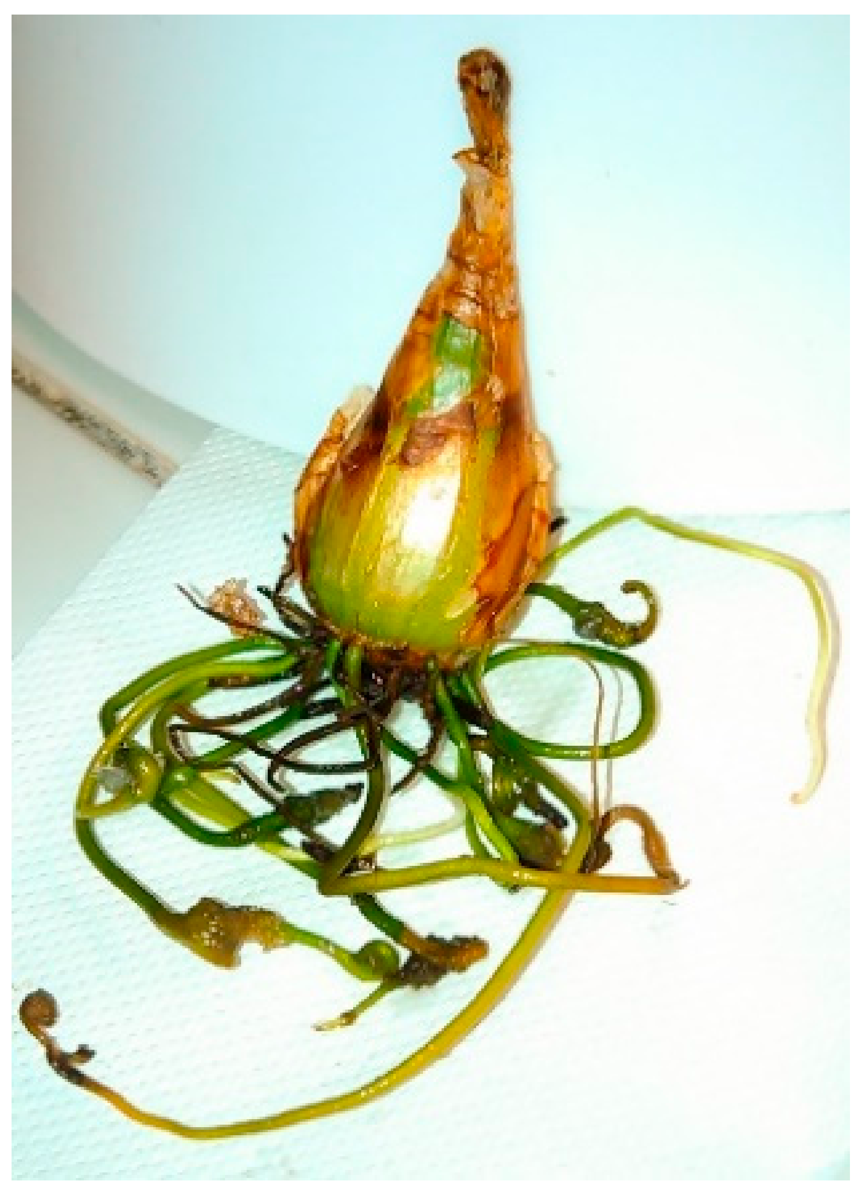
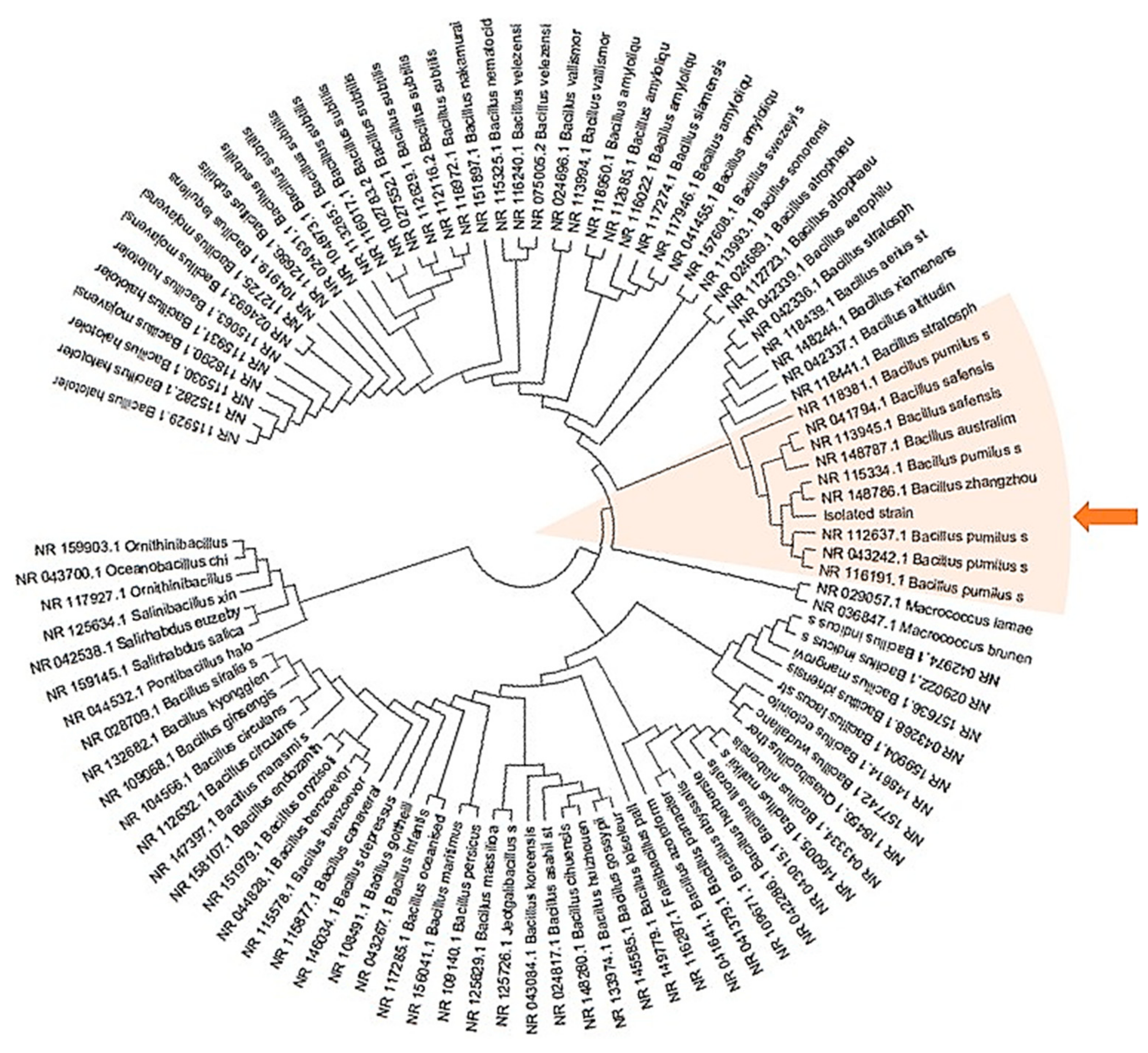
2.1. Isolation and Identification of Endophytic Bacteria from Leucojum aestivum In Vitro Bulblets

The potential presence of bacterial endophytes was investigated in in vitro bulblets of L. aestivum (Figure 1). This research had never been described before.
Of the 300 explants obtained from bulblets, 20 bacterial isolates and four endophytic bacteria were determined to be morphologically unique. These bacteria were designated as endophytes because they are microorganisms that do not visibly harm the host but live in the inner parts of the plants and can be isolated from in vitro bulblets. It is important to note that the bulblets were never in contact with the microorganisms of the rhizosphere; however, they were regenerated after several propagation cycles from primary explants isolated from the leaves inside in vivo bulbs. The endophytic microorganisms survive in microplantlets undetected [36], and their identification is important since they may have positive, neutral or negative impacts on their host plants [35]. The Gram stain reaction indicated that they are Gram-positive bacilli. Polymerase Chain reaction (PCR) analyses, targeted at 16S Ribonucleic acid (RNA) sequences, was carried out on the four bacteria strains.
The results obtained indicate 100% identity of the four isolates with the targeted sequence. A search for sequence similarity carried out on a RNA16S database shows that the four isolates belong to the Bacillus genus and are closest to the Bacillus safensis, pumilus and zhangzhouensis species (Figure 2, orange arrow).
The isolated strain (highlighted by an orange arrow) has been compared to all bacteria presented in the Figure 2 and described in the insertion S1 in supplementary materials. The tree with the highest log likelihood (−13485.60) is shown. Initial tree(s) for the heuristic search were obtained automatically by applying Neighbor-Join and BioNJ algorithms to a matrix of pairwise distances estimated using the Maximum Composite Likelihood (MCL) approach, and then selecting the topology with superior log likelihood value. This analysis involved 101 nucleotide sequences. There were a total of 1600 positions in the final dataset.
Bacillus spp. are frequently isolated from seeds and plant tissues. Antagonistic activities against pathogenic fungi are described for different strains belonging to many species of Bacillus notably B. pumilus and B. subtilis. B. pumilus is described as a biocontrol agent and commercialized for these properties by a life sciences company [37]. B. pumilus is also an endophytic bacteria isolated from different species of Echinacea [32] and Citrus plants [38]. Only the genus Bacillus was isolated in in vitro bulblets of L. aestivum as compared with endophytic bacteria that were isolated from the Amaryllidaceae bulbs of Crinum macowanii of which three to seven bacterial genera were identified [13,14]. In Lycoris radiata 30 bacterial genera were identified [12]. These various results could be associated with the selected plant tissue [39].

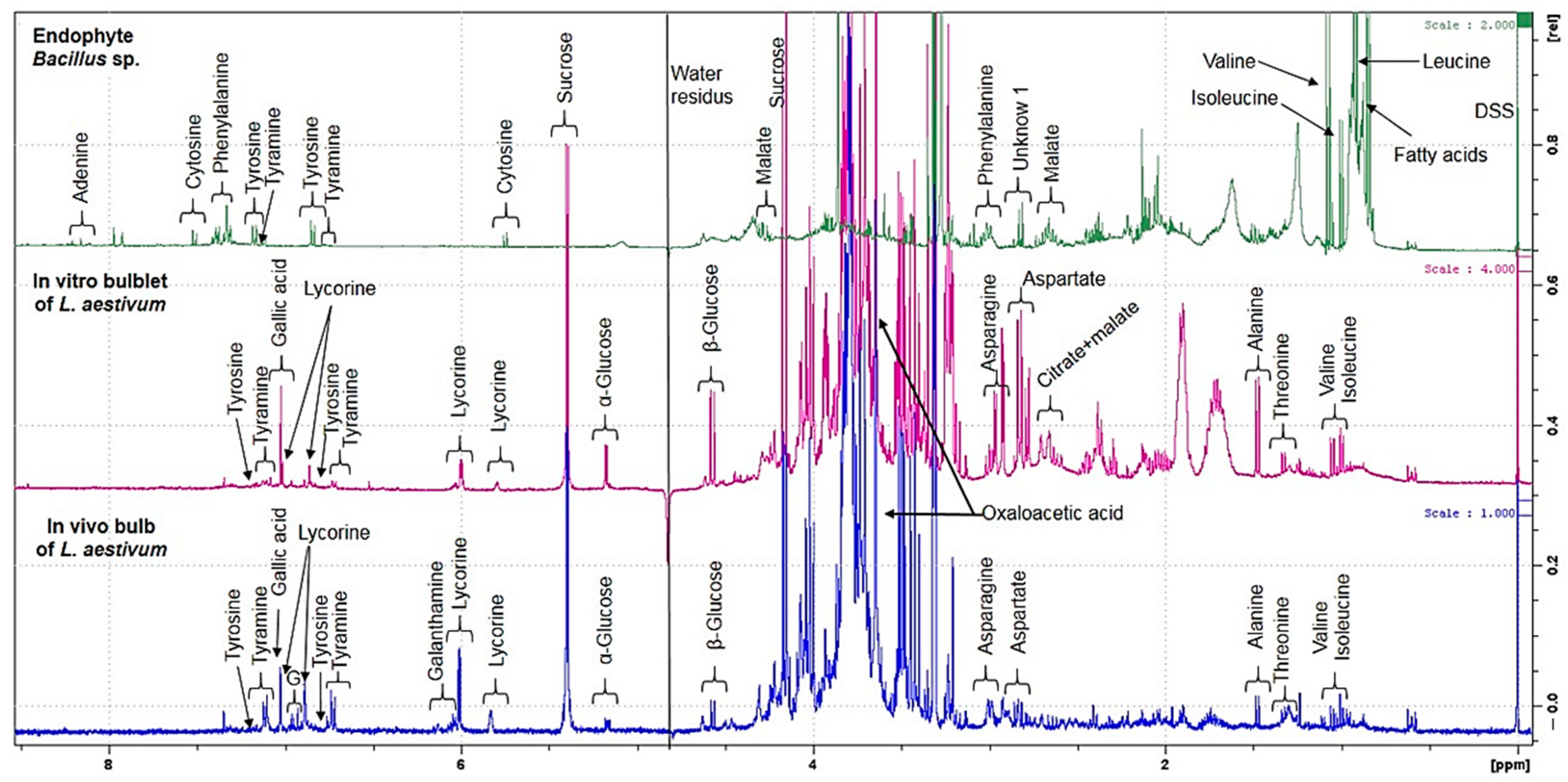
2.2. Untargeted 1H NMR-Based Metabolomics: Identification and Quantification

The 1H NMR spectra of the three biological samples––in vivo bulb and in vitro bulblets of L. aestivum and Bacillus sp. endophyte isolated from in vitro bulblets––were performed. The representative 1H NMR spectra of methanol–water extracts, are shown in Figure 3.
By untargeted 1H NMR-based metabolomics, it was shown that all the extracts have in common four primary metabolites (tyrosine, isoleucine, valine, fatty acids) and a monoamine, tyramine.
An inspection of the proton spectra of L. aestivum in vivo bulbs revealed the presence of seven amino acids: tyrosine, asparagine, aspartate, alanine, threonine, valine and isoleucine. In addition, the signals of phenylethylamine such as tyramine and organic acids such as gallic acid and oxaloacetic acid were observed. Regarding Amaryllidaceae alkaloids, the characteristic signals of both lycorine and galanthamine were identified in the in vivo bulb. These results were confirmed by comparison of original standards recorded in the same conditions of analysis.
An inspection of the proton spectra of the L. aestivum in vitro bulblets, revealed a similar spectrum profile to that of the in vivo bulbs, except for the presence of citrate and malate, which were only observed in the tissue cultures. However, the characteristic signals of galanthamine were not present in the in vitro bulblets while lycorine was observed.
In both in vitro bulblets and in vivo bulbs, the sugars sucrose, α-glucose and β-glucose were present. It is known that sugars are involved in the expression of genes implicated in both primary and specialized metabolisms [40].
The presence of tyrosine and tyramine was emphasized in the in vivo bulbs [26] and in different tissue cultures obtained from Narcissus pseudonarcissus [28].
In the extract of Bacillus sp. endophyte, the 1H NMR spectrum revealed the presence of six amino acids: phenylalanine, tyrosine, alanine, isoleucine, valine and leucine. The characteristic signals of a nitrogenous base, adenosine and cytosine, are visible. In the endophyte Bacillus sp. extract, it is interesting to note the presence of tyramine, which can be synthesized by decarboxylation of tyrosine via the enzyme tyrosine decarboxylase. Tyrosine and tyramine are two of the biosynthetic precursors of Amaryllidaceae alkaloids and tyramine is one starting point in the biosynthesis pathway [41].
Using 1H NMR-based metabolomic analysis it was possible to identify in a single step 22 metabolites with differences in polarity, chemical structure, stability and concentration. The detailed information for the assigned peaks can be found in Table 1.

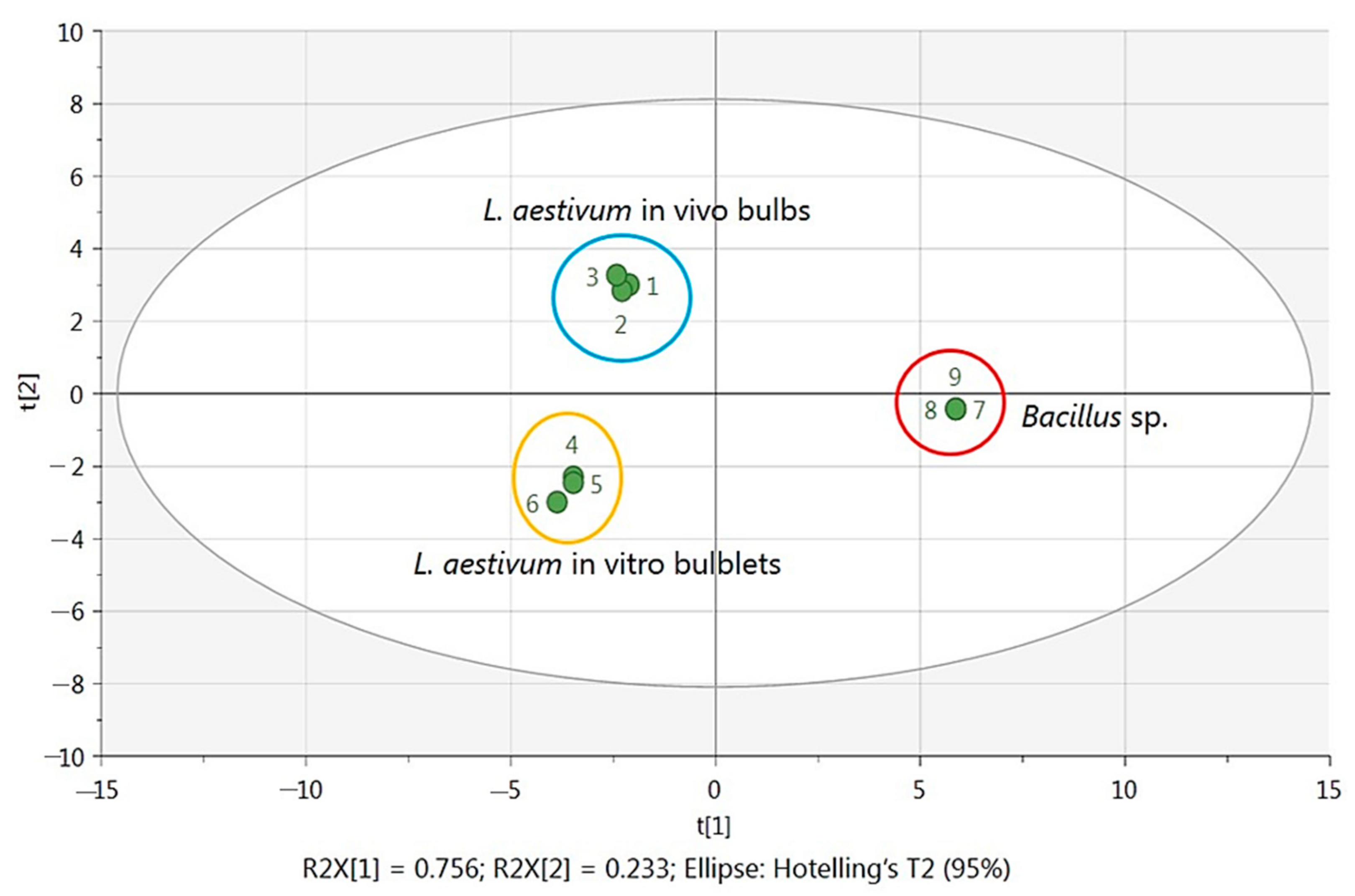
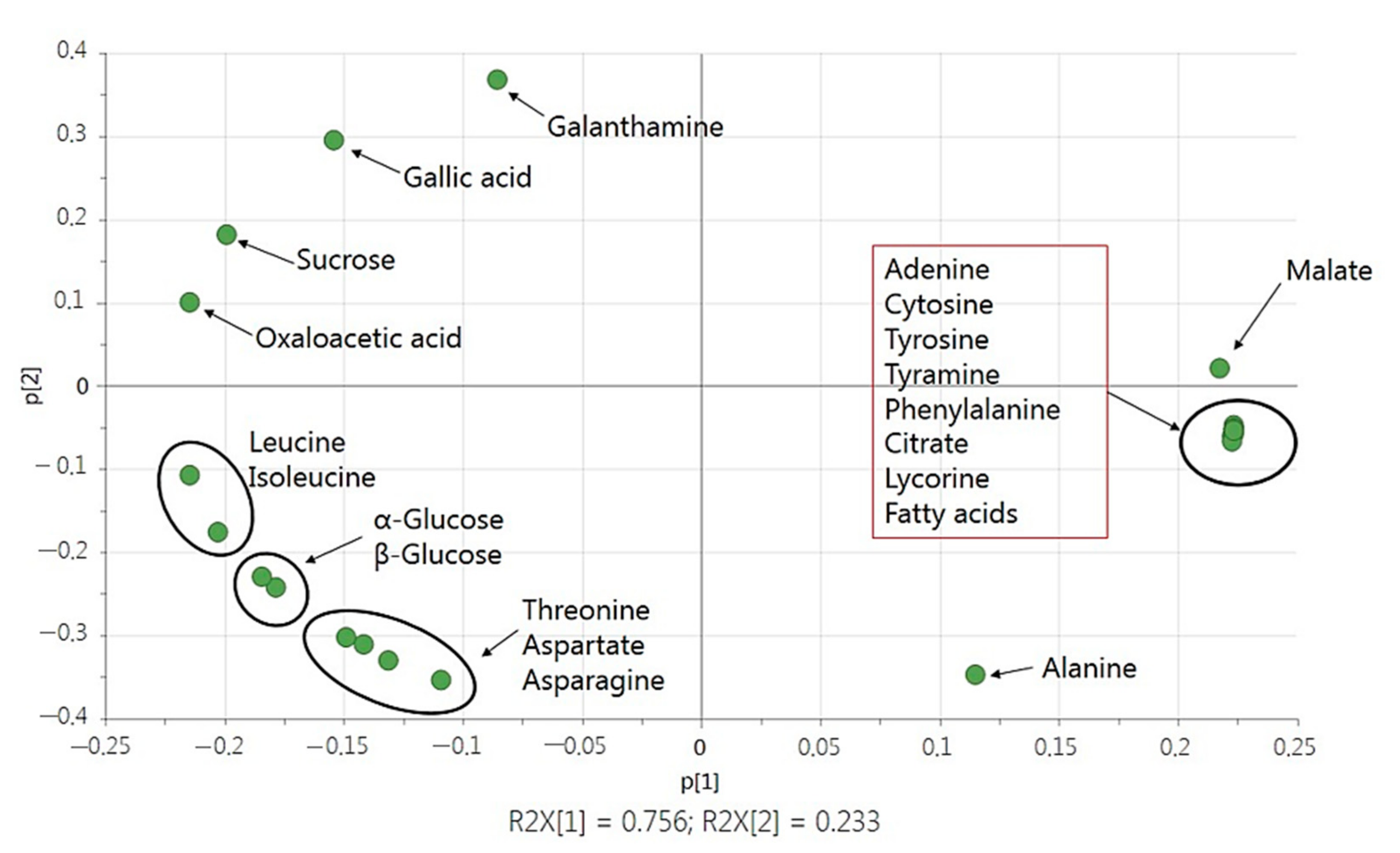
2.3. Statistical Analysis with Multivariate Data Analysis

A Principal Components Analysis (PCA) was chosen to represent the obtained data and to explore the correlation of the samples. The PCA score plot (Figure 4) revealed that the first and second principal axis accounted for 75.6% and 23.3% of the total variability, respectively.
The PCA ordination clearly discriminated the biological extracts according to their biological origin (L. aestivum in vivo bulbs, in vitro bulblets and Bacillus sp.) with weak variability between the three replicates of each origin.
The loading plot shows the contribution of the respective metabolites to the discrimination. The correlation circle shows on plane 1–2 that the metabolites adenine, cytosine, tyrosine, tyramine, phenylalanine, citrate, fatty acids and lycorine form a separate group. It was opposed to the group represented by the variables galanthamine, gallic acid, sucrose and oxaloacetic acid. The other three groups are represented by the pair leucine and isoleucine, α-glucose and β-glucose and the three amino acids threonine, aspartate and asparagine. From these identified signals, it was clear that adenine, cytosine, tyrosine, tyramine, phenylalanine, citrate as well as lycorine and fatty acids were more predominant in Bacillus sp. Endophytes were located on the positive side of PC1. Meanwhile, metabolites located on the negative side of PC1 were identified exclusively in plant extracts of L. aestivum. Metabolites that discriminated more in in vitro bulblets were leucine, isoleucine, α-glucose, β-glucose, threonine, aspartate and asparagine while galanthamine, gallic acid, sucrose and oxaloacetic acid were more discriminated in in vivo bulbs (Figure 5).

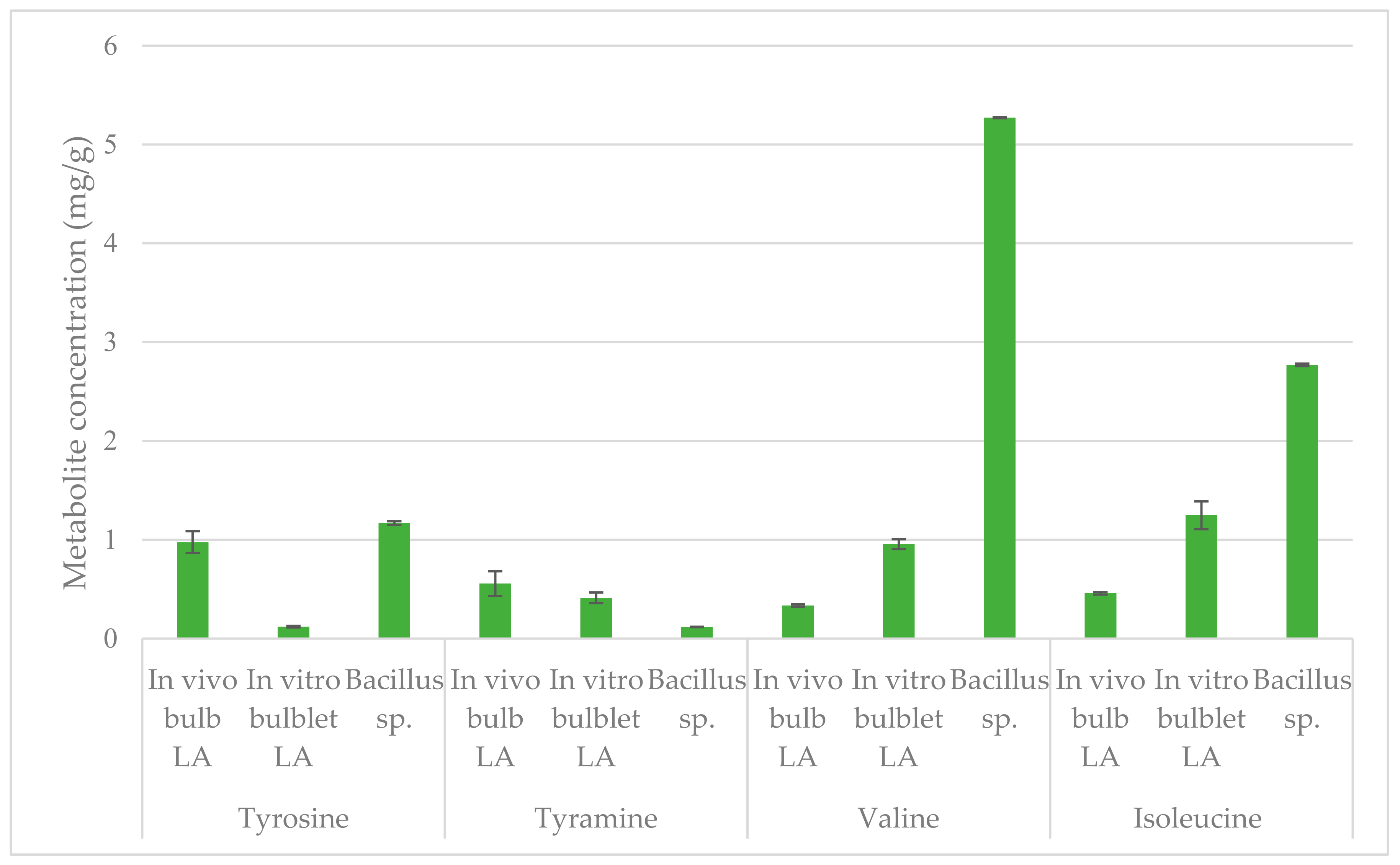
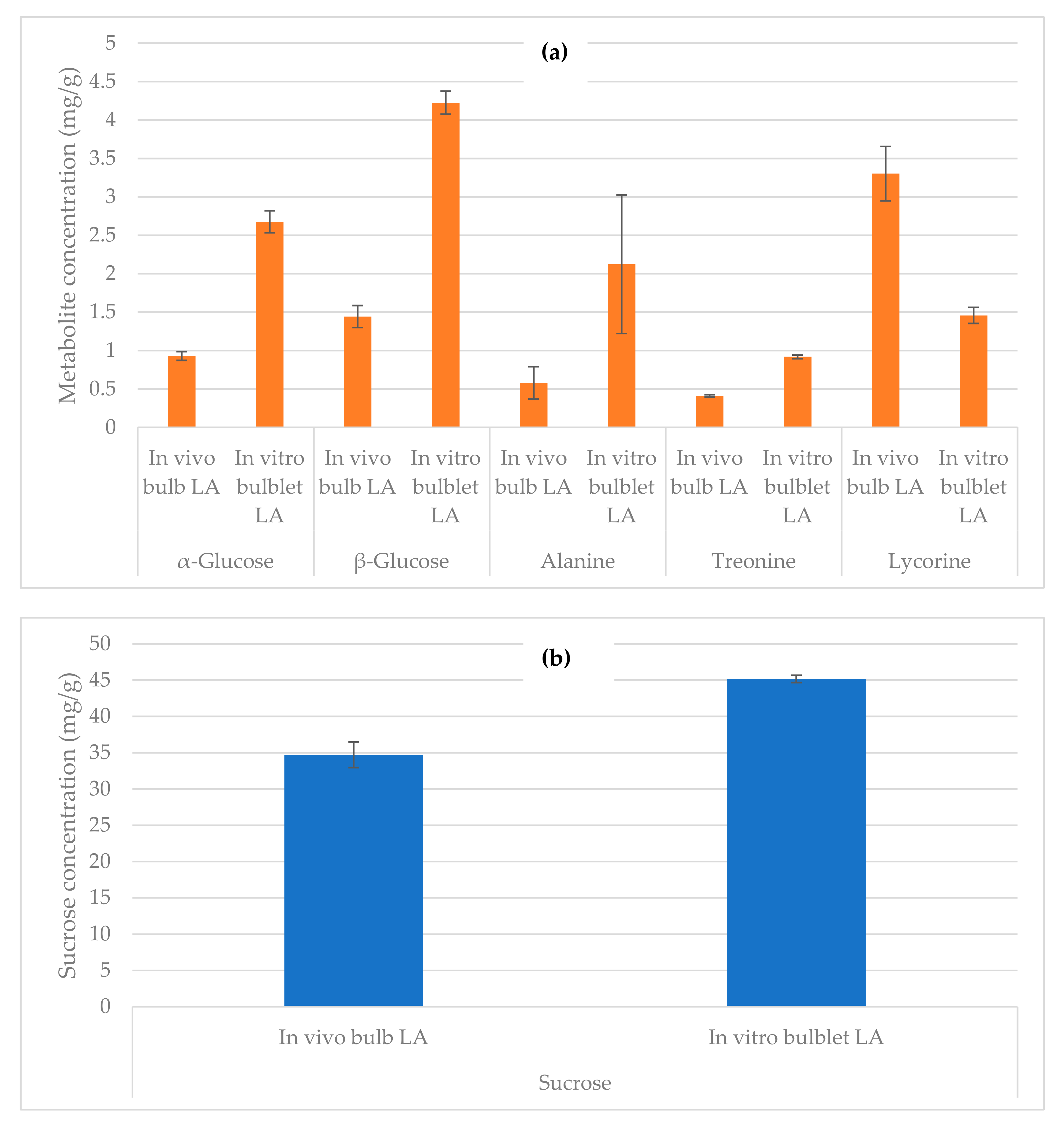
2.4. Quantification by NMR

In this study 10 metabolites (tyrosine, tyramine, valine, isoleucine, threonine, alanine, α-glucose, β-glucose, sucrose and lycorine) were quantified using relative NMR quantification (qNMR). Three amino acids (tyrosine, valine and isoleucine) and the monoamine tyramine (Figure 6) were quantified in in vivo bulbs, in vitro bulblets of L. aestivum and endophytic extracts. The characteristic signal of tyrosine (6.84 ppm), tyramine (6.72 ppm), valine (1.05 ppm), and isoleucine (0.96 ppm), were chosen for the quantification.
Higher concentrations of two amino acids are observed, 5.27 mg/g of dry weight (DW) for valine and 2.77 mg/g of DW for isoleucine, in endophyte Bacillus extract comparatively to the others amino acids in the different extracts tested. Interestingly, the concentration of tyrosine was higher in endophytic extract with 1.17 mg/g of DW by comparison to in vivo extract (0.97 mg/g of DW) and in vitro extract (0.12 mg/g of DW). However, the amount of the decarboxylated compound, tyramine, present in the Bacillus sp. extract (0.12 mg/g of DW) was the lowest comparatively to the others extracts.
Other metabolites in in vivo bulbs and in vitro bulblets of L. aestivum extracts were quantified (Figure 7).
Three sugars (α-glucose, β-glucose and sucrose), two amino acids (threonine, alanine) and one Amaryllidaceae alkaloid (lycorine) were distinguished. The characteristic signal of α-glucose (5.17 ppm), β-glucose (4.57 ppm), sucrose (5.39 ppm), threonine (1.33 ppm), alanine (1.46 ppm) and lycorine (6.85 ppm), were chosen for the quantification.
In the in vivo bulb of L. aestivum, the concentration of α-glucose was 0.93 mg/g of DW and β-glucose of 1.44 mg/g of DW. In contrast, the highest amounts of α-glucose (2.67 mg/g of DW) and β-glucose (4.22 mg/g of DW) were observed in in vitro bulblets. Sucrose was the sugar most abundant in both tissues. The concentration of sucrose was 34.7 mg/g of DW and 45.2 mg/g of DW respectively in in vivo bulb and in vitro bulblets. Alanine was present in in vivo bulb with a concentration of 0.58 mg/g of DW and in vitro bulblets 2.12 mg/g of DW. The concentration of threonine was 0.41 mg/g of DW and 0.92 mg/g of DW in in vivo bulb and in in vitro bulblets respectively.
The amount of lycorine was 3.30 mg/g of DW in in vivo extract and 1.46 mg/g of DW in in vitro extract of L. aestivum.
The main source of lycorine is plant extraction, which is limited by the low concentration in Amaryllidaceae plants (varying from trace to 1.73 mg/g), seasonal variations [42] and depletions of wild Amaryllidaceae populations [43]. In vitro cultures offer an alternative approach for obtaining galanthamine and lycorine metabolites and several methods are described in a recent review [44]. By in vitro cultures the concentration of lycorine up to 1.13 mg/g of DW in Leucojum aestivum ‘Gravity Giant’ bulblets [29]. In L. aestivum, lycorine content is known to vary between in vivo bulbs and in vitro bulblets regenerated in various conditions of plant tissue cultures [29,45].
A negative correlation can be observed between the presence of lycorine and the presence of amino acids (alanine and threonine) and sugars (α-glucose and β-glucose). Also lycorine biosynthesis decreased by the addition of various concentrations of glucose in the culture medium of L. aestivum plants [31]. Using qNMR, in a single step of analysis, the quantification of the primary and specialized metabolites is realized and a global overview of our three biological samples (in vivo bulb, in vitro bulblets and isolated endophyte) has been presented.

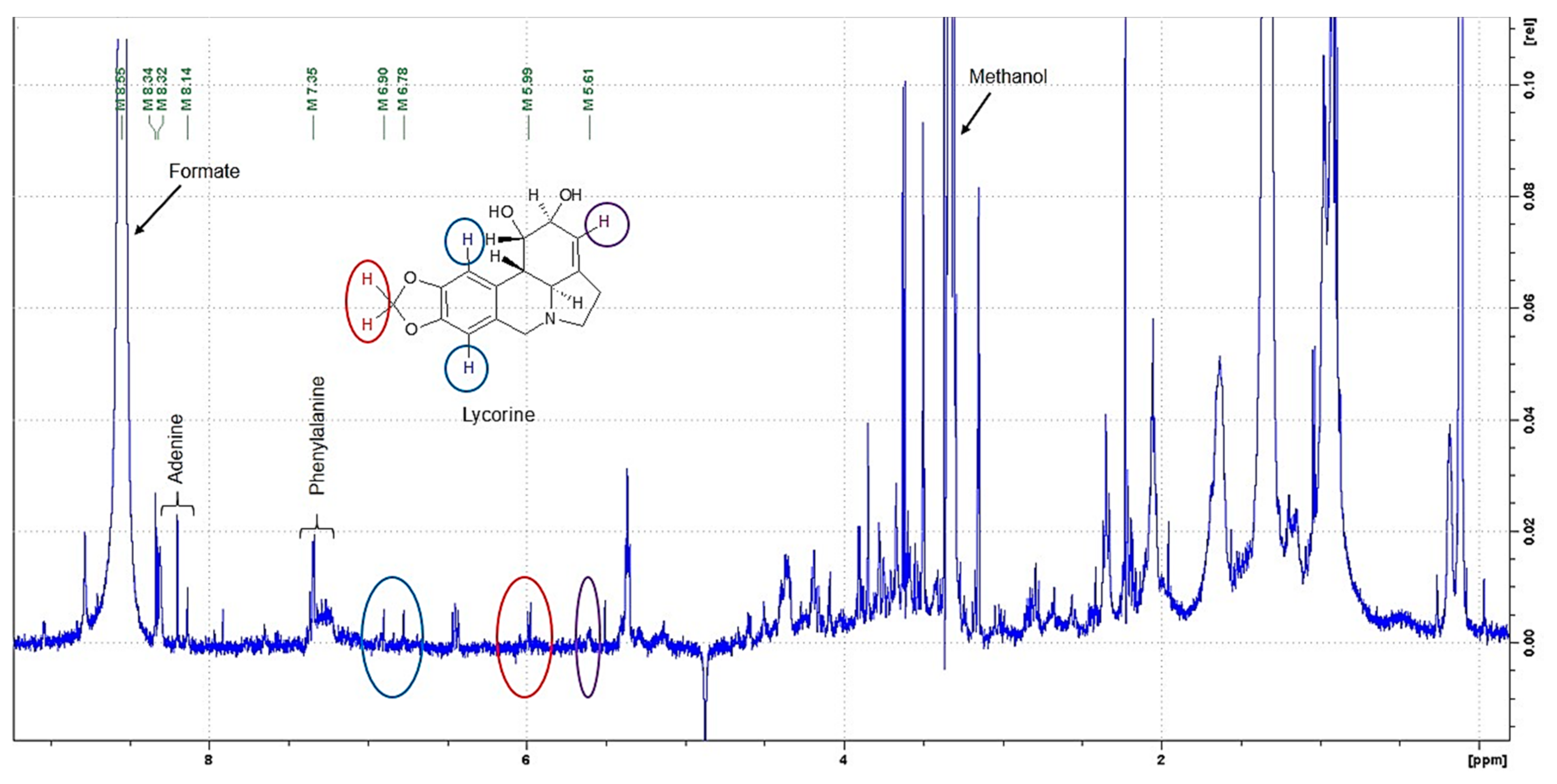
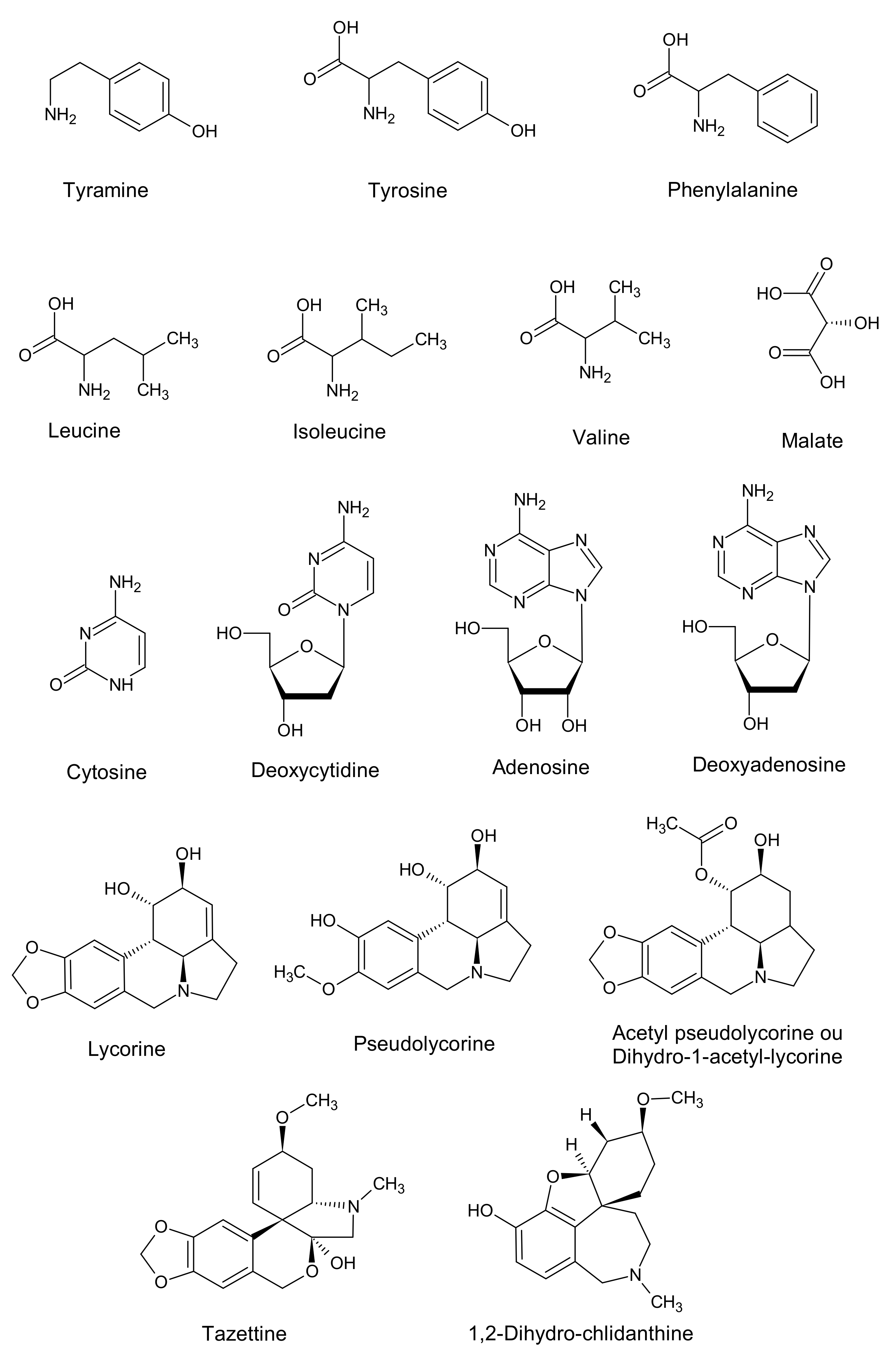
2.5. Target Research of Amaryllidaceae Alkaloids by 1H NMR, LC-MS and GC-MS Analysis in Bacterial Endophyte Bacillus sp.

A selective purification for the research of Amarylidaceae alkaloids, by a solid-phase extraction (SPE) was used. The methanolic extract of bacterial endophyte Bacillus sp. was subjected to SPE for the removal of sugars and to concentrate the sample. The purified extract was evaporated. The dry residues were dissolved in deuterated methanol solvent. The enriched extract was analyzed by NMR using the same sequence used for the 1H NMR metabolomic analysis.
Inspection of the 1H NMR spectrum (Figure 8) showed signals of a purine derivative, adenine, at 8.32 ppm, the aromatic signals at 7.35 ppm of phenylalanine and the characteristic signals of lycorine never described before. Clearly, the signals at 6.90 ppm and 6.78 ppm in blue, 5.99 ppm in red and 5.61 ppm in violet are in an uncrowded region without interference from other signals in the mixtures.
Liquid Chromatography coupled with high-resolution Mass Spectrometry (LC-MS) analysis was realized using a purified methanolic extract of endophytic bacteria Bacillus sp. using the same conditions of analysis as in our previous works. The bulbs and bulblets of L. aestivum were extensively investigated by LC-MS to show the presence of Amaryllidaceae alkaloids [29].
LC-MS profiling generated an extensive mass list corresponding to a wide variety of metabolites in the endophytic extract of Bacillus sp. from the L. aestivum in vitro bulblets. The LC-MS analysis performed in positive-ion mode confirmed the presence of lycorine previously detected by 1H NMR analysis. Additional information was obtained such as the detection of eight compounds including four Amaryllidaceae alkaloids (lycorine, tazettine, pseudolycorine and acetyl pseudolycorine) (Table 2), and four other molecules (methyl-phenylalanine, deoxyadenosine, adenosine, deoxycytidine). These results were confirmed using different databases such as GNPS [46], Metlin [47], Massbank [48] and Sirius [49]. Also a house-made data base (DataNat database, n° IDDN: FR.001.480019.000.S.P.2020.000.10300) was used to confirm the chemical identification.
Lycorine was confirmed in the endophytic extract by the analysis of the mass spectrum (C16H17NO4, [M + H]+ 288.1230) and the retention time (10.1 ± 0.2 min). The same result was obtained for commercial standard lycorine (Supplementary Figure S1).
The alkaloid tazettine (C18H21NO4 [M + H]+ 332.1486) was identified in bulbs of different varieties of Narcissus tazetta [50], Galanthus nivalis [51] but not in L. aestivum. However, tazettine was detected in this endophytic extract of Bacillus and in plant tissue culture of L. aestivum [31]. In the literature, bacterial endophytes are recognized as a rich and diverse source of alkaloids [52] such as camptothecin [53]. In general, Bacillus spp. are the most abundant metabolite-producing bacteria endophytes [54]. Bacillus endophytes have been reported to produce aromatic compounds, polysaccharides and plant hormones, thus representing higher potential in crop management strategies [55]. Endophytic Bacillus sp. was able to induce the highest level of diosgenin biosynthesis in Trigonella foenum-graecum [39].
The GC-MS analysis was also used to investigate the chemical composition of purified extract from endophytic bacteria. The results are shown in the Table 3. Two compounds showed MS fragmentation patterns characteristic of Amaryllidaceae alkaloids, specifically tazettine and 1,2-dihydro-chlidanthine. The alkaloids were identified by comparing the measured data with published data [46] and using the NIST library [56]. Tazettine and 1,2-dihydro-chlidanthine were identified in N. tazetta [50].
To resume our investigation, for the first time the metabolic screening showed the presence of Amaryllidaceae alkaloids in endophytic Bacillus sp. isolated from in vitro bulblets of L. aestivum. The endophyte was able to synthesize lycorine and its derivatives from an ortho-para’ phenol oxidative coupling of precursor 4′-O-methyl-norbelladine, tazettine from a para-para’ phenol oxidative coupling and 1,2-dihydro-chlidanthine from a para-ortho’ phenol oxidative coupling of this precursor [57].
By metabolic investigation using targeted and untargeted 1H NMR-based metabolomics, LC-MS and GC-MS analysis in Bacillus sp. endophyte extracts, a total of 11 primary and 6 specialized metabolites were identified. All chemical structures identified in the extract of Bacillus sp. are presented in Figure 9. The identification of each of these compounds depended on the method of analysis considered as reported by Tarakemeh et al. [50]. The liquid chromatography coupled with high resolution mass spectrometry (LC-HRMS) was the most sensitive method comparatively to GC-MS and NMR and was able to detect the compounds in amounts lower than 0.02 μg/mL.
The obtained results revealed that the Bacillus sp. endophytic bacterium was a potential source of lycorine, an Amaryllidaceae alkaloid of interest.
It is to be noted that NMR metabolomic studies had not been previously used to describe the relation between bulbs, bulblets and endophytes in L. aestivum plants. The combination of NMR-based metabolomics and mass spectrometry is a very useful tool for describing the metabolism of Amarylidaceae plants and other biological systems [26,28]
However, large-scale production of lycorine by cultivating this promising endophytic bacterium strain and the investigation of enzymatic systems present in endophytes are in our future projects.

3. Materials and Methods

3.1. Chemicals

Standards (gallic acid, lycorine, galanthamine, tyramine, tyrosine, α-glucose, β-glucose, sucrose, alanine, valine) are purchased by Sigma Aldrich (Sigma-Aldrich, St. Quentin Fallavier, France). All solvents used are analytical profile grade, deuterated methanol (CD3OD), 3-(trimethylsilyl)-1-propanesulfonic acid sodium salt (DSS sodium salt), sodium deuteroxide solution (NaOD) 40 wt.% in deuterium oxide (D2O), 99.5 atom % D, ammonium hydroxide solution (NH4OH) containing ≥25% NH3 basis, were purchased from Sigma Aldrich (Sigma-Aldrich, St. Quentin Fallavier, France). All chemicals for in vitro culture are listed in the article Saliba et al. [29]. SCX cation exchanger column are purchased by Interchim® (Interchim, Montluçon, France).

3.2. Plant Materials, Preparation of In Vitro Culture of L. aestivum

L. aestivum bulbs were purchased from a local French market (Graines Baumax, Mazirot, France) and voucher specimens were deposited at the herbarium of the Botanic Garden Jean-Marie Pelt (N° 911371) (Nancy, France).
Under sterilized conditions in the laminar air flow, the leaves isolated from the bulbs of this Amaryllidaceae plant were surface sterilized, and initial explants were cultivated on a Murashige and Skoog (MS) culture medium [58] as previously reported by Ptak et al. [59]. For somatic embryo induction, the medium was supplemented with 0.5 µM benzyladenine (BA) combined with 5 µM 4-amino-3,5,6-trichloro-2-pyridinecarboxylic acid (picloram) in accordance to previous results [60,61]. In addition, 3% w/v sucrose and 0.8 % w/v agar (purified agar from Difco) were added to the medium. All supplements were added before autoclaving. The pH was adjusted to 5.5. Bulblets were regenerated after transferring plants obtained from somatic embryos successively to the MS solid medium containing 5 µM zeatin during 16 weeks, then to the MS solid medium without growth regulators and supplemented with 6% w/v sucrose [31]. Cultures were placed in a growth chamber and maintained at 25 ± 2 °C under a 16/8-h (light/dark) photoperiod (Tungsram lamp, 40 WF, 90 µmol m−2 s−1), and subcultured once every 4 weeks under the same conditions.

3.3. Isolation of Endophytic Bacteria

In vitro bulblets of L. aestivum were used for the isolation of endophytes. Thin slices about 3–5 mm in length, were obtained from in vitro bulblets. All these explants were placed on nutrient agar (NA) in Petri dishes and incubated in the dark at 28 °C. Growth was monitored periodically for 5 days. Distinct colonies were selected and subcultured on NA to obtain pure isolates. Pure bacterial isolates were preserved in 50% glycerol in a ratio of 500 μL glycerol: 500 μL overnight broth culture and kept at −80 °C.

3.4. Identification of Endophytic Bacteria and PCR Amplification

Pure colonies were subjected to Gram staining to establish morphological characteristics such as Gram-stain reaction using a compound bright-field microscope (OLYMPUS CH20BIMF200, Rungis, France) with 100× magnification.
The 16S rDNA genes were amplified from single colonies of isolated strains in a final volume of 25 mL containing 2.5 mL of Taq Polymerase buffer 10× (MP Biomedicals, Illkirch-Graffenstaden, France), 200 mM of each dNTP, 1.5 mM magnesium chloride (MgCl2), 0.5 mM of each universal primer 27f (50-GAGAGT TTG ATC CTG GCT CAG-30, positions 8–27 of Escherichia coli 16S rDNA) and 1492r (50-CTA CGG CTA CCT TGT TAC GA-30, positions 1492–1513 of E. coli 16S rDNA) [62], and 0.625 U of Taq Polymerase (MP Biomedicals, Illkirch-Graffenstaden, France). DNA amplification was carried out in a thermocycler using the following conditions: 4 min at 94 °C, 35 cycles of 1 min at 94 °C, 1 min at 55 °C, and 2 min at 72 °C, plus an additional 15-min cycle at 72 °C.

3.5. In Silico Analysis

Using the BLASTx tool, the sequences were compared to a database available on NCBI containing sequences of RNA16S from bacteria (https://blast.ncbi.nlm.nih.gov/Blast.cgi, accessed on 17 July 2020). The top 100 hits were aligned with Clustal X and the resulting alignment was used to create a phylogenetic tree with MEGA-X. The evolutionary history was inferred by using the Maximum Likelihood method and Tamura-Nei model [63]. Evolutionary analyses were conducted in MEGA X [64].

3.6. Preparation of Plants Samples and Endophytic Bacteria for Untargeted NMR-Based Metabolomics

The procedure for obtaining plants samples used in 1H NMR based metabolomics entails in vivo bulbs and in vitro bulblets of L. aestivum being catted in small pieces, frozen at −80 °C overnight and then lyophilized. The dry materials were blended to obtain a finer powder.
For the endophytic bacterium, a preculture in 2 mL of an NA medium was performed at 32 °C overnight. Twenty Erlenmeyer flasks containing 50 mL of an NA medium were inoculated with the preculture, shaken at 120 rpm and incubated at 32 °C. After 72 h of cultivation, the cultures were centrifuged at 10 °C for 10 min at 4000 rpm, and the bacterial pellets were recovered. The pellets and the liquid media, were frozen and lyophilized for 48 h.
Fifty mg of each sample (in vivo bulbs, in vitro bulblets and bacterial pellets) were directly extracted with a mixture of 0.75 mL of potassium dihydrogenphosphate (KH2PO4) buffer in deuterium oxide (D2O) at pH 6.0 containing 0.1% of 3-(trimethylsilyl)-1-propanesulfonic acid sodium salt (DSS sodium salt), and 0.75 mL CD3OD. The mixture was then placed in a vortex mixer. The extraction was realized in an ultrasonic bath for 30 min of sonication. Each sample was centrifuged 2 times for 10 min at 3400 rpm. We use a closed protocol as described in the article by Kim et al. [65]. All deuterated supernatants from in vivo bulbs and in vitro bulblets of L. aestivum were transferred into NMR tubes (5 mm) for measurement of their 1H spectra. The extraction and analysis were repeated three times.

3.7. Preparation of Enriched Endophytic Bacteria Sample for NMR, LC-MS and GC-MS Analysis

For NMR, LC-MS and GC-MS analysis, 50 mg of dried bacterial pellets were extracted with 3 mL of 60% MeOH for 30 min by sonication in an ultrasonic bath followed by maceration for 2 h. The extract was purified using the solid-phase extraction (SPE) purification protocol as previously described by Saliba et al. [29].
The cartridge was washed with 3 mL of alkalized water (2% ammonium hydroxide NH4OH, pH = 8) and then eluted with 15 mL of the mobile phase constituted by methanol, distillated water and formic acid (MeOH–H2O–HCOOH, 85:10:5 v/v). The obtained eluate was then transferred on a SCX cation exchanger column (preconditioned with 2 mL of 2% formic acid in methanol). The cartridge was then rinsed successively with 2 mL of 2% formic acid in methanol, 3 mL of 2% formic acid in acetonitrile and 3 mL of 2% formic acid in acetonitrile. The alkaloids were finally eluted as free bases with 15 mL of 5% NH4OH in acetonitrile. The purified extracts were then concentrated. The extraction and analysis were repeated three times.
The purified extract was evaporated and reconstituted in 250 µL of deuterated MeOH. The extract was transferred into Shigemi NMR tubes matched with methanol (Cortecnet Europe, Voisins-Le-Bretonneux, France) for measurement of its 1H spectrum.
Then, after evaporation, the purified extract residue was dissolved in 250 µL of acetonitrile and analyzed by LC-MS and GC-MS. The experiments and analysis were repeated three times.

3.8. NMR Equipment and Experimental Conditions

1H NMR spectra were recorded on a Bruker Avance III 400 spectrometer (Bruker BioSpin, Rheinstetten, Germany), operating at a frequency of 400.13 M + Hz for 1 h and at a temperature of 26 °C using a BBFO Probe and a Bruker sample changer. NMR analyses were performed on the «Plateforme de RMN de l’Institut Jean Barriol», University of Lorraine, France. For quantification, a pulse sequence with presaturation for water suppression was used to acquire 1H NMR spectra from all extracts.
For each sample, 512 scans were acquired with 32,768 data points using a spectral width of 4800 Hz, a relaxation delay of 10 s and a pulse width of 14.2 μs. The relaxation delay was chosen to be largely superior to 5 times the longest T1 in the molecules in order to ensure quantitative measurements. The estimated saturation bandwidth is about 60 Hz, as calculated from the presaturation pulse power level. The coupling constant (J) is expressed in Hz.
All spectra were manually phased and baseline corrected using the TOPSPIN 4.0.6 (Bruker Biospin GmbH, Rheinstetten, Germany).
DSS sodium salt (at 0.0 ppm) was used as an internal standard for the chemical shift reference and intensity scaling for all 1H-NMR signals, as well as a reference in the quantitation of metabolites. Calibration of the data was carried out using TOPSPIN 4.0.6 software (Bruker Biospin GmbH, Rheinstetten, Germany).
The assignments of metabolites in the NMR spectra were made by comparing the proton chemical shifts with literature [66,67,68,69,70,71] or by a comparison with the spectra of authentic compounds recorded in the same solvent conditions (in-house library).
For the quantification [72,73], Equation (1) was used
mX = mSt × (AX/ASt) × (MwX/MwSt) × (NSt/NX)
mX is the unknown mass of the targeted metabolite; mSt is the mass of the DSS; AX and ASt are the integral areas for the selected signals; MwX and MwSt are the molecular weights of the targeted metabolite and DSS; NX and NSt are the number of protons generating the integral signals.

3.9. Softwares and the Statistical Analysis

For the quantification, the analyses were repeated three times, and the data were expressed as a mean ± SD, p < 0.01.
For the 1H NMR spectra processing, bucketing and data export, the interactive tool NMRProcFlow v1.3 was used [74]. In the downloaded 1H NMR spectra (Supplementary Figure S2) the baseline correction, ppm calibration and re-alignment were applied. The NMR spectra were reduced in several buckets using the intelligent bucketing method according to De Meyer et al. [75], in a range from 8.2 to 0.5 ppm, excluding the methanol signals (3.34–3.26 ppm).
The obtained bucket table was used to create a data matrix. Normalization, using the option Constant Sum Normalization, was used.
The data matrix, with columns representing the content of each identified metabolite and the lines of each replicate of the three biological origins tested in the study, was imported to SIMCA software version 16.0.2 (Umetrics, Umeå, Sweden), and the PCA was realized using the Pareto scaling method.

3.10. LC-MS and GC-MS Equipments and Analysis

The LC system consisted of a U3000-Dionex apparatus with an injector comprising a 1 µL loop and a UV detector at 280 nm. The analytical column used was an Acclaim mixed-mode HILIC-1 (Thermo Scientific, Bellefonte, PA, USA) ID 2.1 mm column (150 mm × 5 µm × 120 Å) and eluted at a flow rate of 200 µL/min using a gradient ranging from 2% solvent B to 25% solvent B in a time span of 26 min and 30 s. Solvent A consisted of pure methanol and solvent B consisted of 10 mM ammonium formate in water at pH 6.8. The Electrospray Ionization—High Resolution Mass Spectrometry (ESI-HRMS) was a micrOTOFQTM apparatus (Bruker Daltonics, Bruker, Bremen, Germany).
The GC system consisted to QP2010-Shimadzu equipment (Shimadzu, Kyoto, Japan) operating in the EI mode at 70 eV. An SLB5 column DB-5 ms (30 m, 0.25 mm film thickness) was employed with a 36 min temperature program of 60–320 at 10 °C/min followed by a 10 min hold at 320 °C. The injector temperature was 250 °C; the flow rate of the carrier gas (helium) was 1 mL/min; and the split ratio was 1:50. The interval of the scan m/z was between 35 and 900 and the identification of the compounds was based on an individual spectrum comparison of each compound in the Shimadzu NIST08 data. The retention index was calculated using the mixture of alkanes standard from C10 to C40 (Merck KGaA, Darmstadt, Germany).

4. Conclusions

For the first time, endophytic bacteria were isolated from Leucojum aestivum in in vitro bulblets. To date, no other studies have investigated the presence of endophytic bacteria in Leucojum species (neither bulbs nor bulblets). The discovery of endophytic bacteria belonging to the Bacillus genus living in tissue cultures of this plant and their capacity to synthesize Amaryllidaceae alkaloids opens new prospects for producing these added-value specialized metabolites, which are accumulated at low levels in plants. The coupling between NMR-based metabolomics and mass spectrometry combined with multivariate statistical analysis was a reliable method for comparing the metabolism of L. aestivum and its endophytes. For several years, researchers tried to enhance the biosynthesis of Amaryllidaceae-specialized metabolites, especially galanthamine and lycorine production via in vitro cultures (a precursor addition to the culture medium for in vitro culture conditions modulation and optimization, bioreactors RITA®). These techniques showed interesting results but neither offered an economically viable protocol for an effective scale-up due to the complexity of managing plant bioreactors and the relatively high costs of such technologies. Bacterial cultures can be scaled-up because culturing conditions, specialized metabolite extraction, culturing costs and time scales offer an economically viable biotechnology.

Supplementary Materials

Supplementary Materials can be found at https://www.mdpi.com/1422-0067/22/4/1773/s1. Insertion S1: Bacteria strains compared to isolated strain (Bacillus sp.) from L. aestivum in vitro bulblet; Figure S1: MSMS of lycorine derived from endophytic Bacillus sp. isolated from L. aestivum in vitro bulblet; Figure S2: 1H NMR Spectra in NMRProcFlow.

Author Contributions

Conceptualization, R.S., S.S. and D.L.-M.; formal analysis, R.S., S.S., F.D., A.P., S.P. (Sophie Poinsignon) and S.L.; investigation, R.S. and F.D.; resources, R.S., S.S. A.P. and D.L.-M.; data curation, R.S., S.S., F.D. and D.L.-M.; writing—original draft preparation, R.S. and D.L.-M.; writing—review and editing, R.S., S.S., F.D., A.P., A.H., S.P. (Séverine Piutti), S.P. (Sophie Poinsignon), S.B.-B. S.L. and D.L.-M.; supervision, R.S., A.P., A.H., S.P. (Séverine Piutti), S.B.-B. and D.L.-M.; funding acquisition, R.S., A.P. S.B.-B. and D.L.-M. All authors have read and agreed to the published version of the manuscript.

Funding

The study was funded by Institut Jean Barriol (IJB) at the University of Lorraine and project “Interdisciplinarité” (number: INTER-2019-029) of the University of Lorraine. The APC was funded by L2CM laboratory.

Institutional Review Board Statement

Not applicable.

Informed Consent Statement

Not applicable.

Data Availability Statement

Not applicable.

Acknowledgments

The authors acknowledge: the support of “L2CM UMR 7053” by the “Impact Biomolecules” project of the “Lorraine Université d’Excellence” (Investissements d’avenir—ANR), the support PHC Polonium project (number: 40524YB), the support of the Ministry of Science and Higher Education of the Republic of Poland. The authors are grateful to Masse Plateform of L2CM and the “Plateforme de RMN de l’Institut Jean Barriol” both at the University of Lorraine.

Conflicts of Interest

The authors declare no conflict of interest.

Glossary

MeOHMethanol
CH3CNAcetonitrile
HCOOHFormic acid
NH4OHAmmonium hydroxide
LA Leucojum aestivum
MSMurashige and Skoog
NANutrient agar
DWDry weight
SPESolid phase extraction
NMRNuclear Magnetic Resonance
ESI-HRMSElectrospray Ionization—High Resolution Mass Spectrometry
MSMass spectrometry
LCLiquid chromatography
GCGas chromatography

References

  1. Rinner, U.; Dank, C.; Hudlicky, T. Galanthamine. Targets Heterocycl. Syst. 2016, 283. [Google Scholar] [CrossRef]
  2. He, M.; Qu, C.; Gao, O.; Hu, X.; Hong, X. Biological and Pharmacological Activities of Amaryllidaceae Alkaloids. Rsc Adv. 2015, 5, 16562–16574. [Google Scholar] [CrossRef]
  3. Szlávik, L.; Gyuris, Á.; Minárovits, J.; Forgo, P.; Molnár, J.; Hohmann, J. Alkaloids from Leucojum vernum and Antiretroviral Activity of Amaryllidaceae Alkaloids. Planta Med. 2004, 70, 871–873. [Google Scholar] [CrossRef] [PubMed]
  4. Khalifa, M.F.; Attia, E.Z.; Fahim, J.R.; Kamel, M.S. An Overview on the Chemical and Biological Aspects of Lycorine Alkaloid. J. Adv. Biomed. Pharm. Sci. 2018, 1, 41–49. [Google Scholar] [CrossRef]
  5. Cao, Z.; Yang, P.; Zhou, Q. Multiple Biological Functions and Pharmacological Effects of Lycorine. Sci. China Chem. 2013, 56, 1382–1391. [Google Scholar] [CrossRef]
  6. Osorio, E.J.; Robledo, S.M.; Bastida, J. Chapter 2, Alkaloids with Antiprotozoal Activity. In The Alkaloids: Chemistry and Biology; Cordell, G.A., Ed.; Academic Press: Cambridge, MA, USA, 2008; Volume 66, pp. 113–190. [Google Scholar]
  7. Ying, X.; Huang, A.; Xing, Y.; Lan, L.; Yi, Z.; He, P. Lycorine Inhibits Breast Cancer Growth and Metastasis via Inducing Apoptosis and Blocking Src/FAK-Involved Pathway. Sci. China Life Sci. 2017, 60, 417–428. [Google Scholar] [CrossRef]
  8. Gouda, S.; Das, G.; Sen, S.K.; Shin, H.-S.; Patra, J.K. Endophytes: A Treasure House of Bioactive Compounds of Medicinal Importance. Front. Microbiol. 2016, 7. [Google Scholar] [CrossRef] [Green Version]
  9. Khiralla, A.; Spina, R.; Saliba, S.; Laurain-Mattar, D. Diversity of Natural Products of the Genera Curvularia and Bipolaris. Fungal Biol. Rev. 2019, 33, 101–122. [Google Scholar] [CrossRef]
  10. Na, R.; Jiajia, L.; Dongliang, Y.; Yingzi, P.; Juan, H.; Xiong, L.; Nana, Z.; Jing, Z.; Yitian, L. Indentification of Vincamine Indole Alkaloids Producing Endophytic Fungi Isolated from Nerium indicum, Apocynaceae. Microbiol. Res. 2016, 192, 114–121. [Google Scholar] [CrossRef] [PubMed]
  11. Khiralla, A.; Spina, R.; Varbanov, M.; Philippot, S.; Lemiere, P.; Slezack-Deschaumes, S.; André, P.; Mohamed, I.; Yagi, S.M.; Laurain-Mattar, D. Evaluation of Antiviral, Antibacterial and Antiproliferative Activities of the Endophytic Fungus Curvularia papendorfii, and Isolation of a New Polyhydroxyacid. Microorganisms 2020, 8, 1353. [Google Scholar] [CrossRef]
  12. Liu, Z.; Zhou, J.; Li, Y.; Wen, J.; Wang, R. Bacterial Endophytes from Lycoris radiata Promote the Accumulation of Amaryllidaceae Alkaloids. Microbiol. Res. 2020, 239, 126501. [Google Scholar] [CrossRef]
  13. Sebola, T.E.; Uche-Okereafor, N.C.; Tapfuma, K.I.; Mekuto, L.; Green, E.; Mavumengwana, V. Evaluating Antibacterial and Anticancer Activity of Crude Extracts of Bacterial Endophytes from Crinum macowanii Baker Bulbs. MicrobiologyOpen 2019, 8, e914. [Google Scholar] [CrossRef] [PubMed] [Green Version]
  14. Rebotiloe, F.M.; Eunice, U.-J.; Mahloro, H.S.-D. Isolation and Identification of Bacterial Endophytes from Crinum macowanii Baker. Afr. J. Biotechnol. 2018, 17, 1040–1047. [Google Scholar] [CrossRef]
  15. Wang, W.-X.; Kusari, S.; Sezgin, S.; Lamshöft, M.; Kusari, P.; Kayser, O.; Spiteller, M. Hexacyclopeptides Secreted by an Endophytic Fungus Fusarium solani N06 Act as Crosstalk Molecules in Narcissus tazetta. Appl. Microbiol. Biotechnol. 2015, 99, 7651–7662. [Google Scholar] [CrossRef] [PubMed]
  16. Deborde, C.; Moing, A.; Roch, L.; Jacob, D.; Rolin, D.; Giraudeau, P. Plant Metabolism as Studied by NMR Spectroscopy. Prog. Nucl. Magn. Reson. Spectrosc. 2017, 102–103, 61–97. [Google Scholar] [CrossRef] [PubMed]
  17. Vignoli, A.; Ghini, V.; Meoni, G.; Licari, C.; Takis, P.G.; Tenori, L.; Turano, P.; Luchinat, C. High-Throughput Metabolomics by 1D NMR. Angew. Chem. Int. Ed. 2019, 58, 968–994. [Google Scholar] [CrossRef]
  18. Courant, F.; Antignac, J.-P.; Dervilly-Pinel, G.; Bizec, B.L. Basics of Mass Spectrometry Based Metabolomics. Proteomics 2014, 14, 2369–2388. [Google Scholar] [CrossRef]
  19. Covington, B.C.; McLean, J.A.; Bachmann, B.O. Comparative Mass Spectrometry-Based Metabolomics Strategies for the Investigation of Microbial Secondary Metabolites. Nat. Prod. Rep. 2017, 34, 6–24. [Google Scholar] [CrossRef]
  20. Kostidis, S.; Addie, R.D.; Morreau, H.; Mayboroda, O.A.; Giera, M. Quantitative NMR Analysis of Intra- and Extracellular Metabolism of Mammalian Cells: A Tutorial. Anal. Chim. Acta 2017, 980, 1–24. [Google Scholar] [CrossRef]
  21. Kosmides, A.K.; Kamisoglu, K.; Calvano, S.E.; Corbett, S.A.; Androulakis, I.P. Metabolomic Fingerprinting: Challenges and Opportunities. Crit. Rev. Biomed. Eng. 2013, 41, 205–221. [Google Scholar] [CrossRef] [PubMed]
  22. Berkov, S.; Bastida, J.; Viladomat, F.; Codina, C. Development and Validation of a GC–MS Method for Rapid Determination of Galanthamine in Leucojum aestivum and Narcissus ssp.: A Metabolomic Approach. Talanta 2011, 83, 1455–1465. [Google Scholar] [CrossRef]
  23. Singh, A.; Desgagné-Penix, I. Transcriptome and Metabolome Profiling of Narcissus pseudonarcissus ‘King Alfred’ Reveal Components of Amaryllidaceae Alkaloid Metabolism. Sci. Rep. 2017, 7, 17356. [Google Scholar] [CrossRef]
  24. Holmes, E.; Tang, H.; Wang, Y.; Seger, C. The Assessment of Plant Metabolite Profiles by NMR-Based Methodologies. Planta Med. 2006, 72, 771–785. [Google Scholar] [CrossRef] [PubMed] [Green Version]
  25. Lubbe, A.; Pomahačová, B.; Choi, Y.H.; Verpoorte, R. Analysis of Metabolic Variation and Galanthamine Content in Narcissus Bulbs by 1H NMR. Phytochem. Anal. 2010, 21, 66–72. [Google Scholar] [CrossRef]
  26. Lubbe, A.; Gude, H.; Verpoorte, R.; Choi, Y.H. Seasonal Accumulation of Major Alkaloids in Organs of Pharmaceutical Crop Narcissus Carlton. Phytochemistry 2013, 88, 43–53. [Google Scholar] [CrossRef] [PubMed]
  27. Akram, M.N.; Verpoorte, R.; Pomahačová, B. Effect of Bulb Age on Alkaloid Contents of Narcissus pseudonarcissus Bulbs. S. Afr. J. Bot. 2021, 136, 182–189. [Google Scholar] [CrossRef]
  28. Ferdausi, A.; Chang, X.; Hall, A.; Jones, M. Galanthamine Production in Tissue Culture and Metabolomic Study on Amaryllidaceae Alkaloids in Narcissus pseudonarcissus cv. Carlton. Ind. Crop. Prod. 2020, 144, 112058. [Google Scholar] [CrossRef]
  29. Saliba, S.; Ptak, A.; Boisbrun, M.; Spina, R.; Dupire, F.; Laurain-Mattar, D. Stimulating Effect of Both 4’-O-Methylnorbelladine Feeding and Temporary Immersion Conditions on Galanthamine and Lycorine Production by Leucojum aestivum L. Bulblets. Eng. Life Sci. 2016, 16, 731–739. [Google Scholar] [CrossRef]
  30. Ptak, A.; El Tahchy, A.; Skrzypek, E.; Wójtowicz, T.; Laurain-Mattar, D. Influence of Auxins on Somatic Embryogenesis and Alkaloid Accumulation in Leucojum aestivum Callus. Cent. Eur. J. Biol. 2013, 8, 591–599. [Google Scholar] [CrossRef]
  31. Ptak, A.; Morańska, E.; Skrzypek, E.; Warchoł, M.; Spina, R.; Laurain-Mattar, D.; Simlat, M. Carbohydrates Stimulated Amaryllidaceae Alkaloids Biosynthesis in Leucojum aestivum L. Plants Cultured in RITA® Bioreactor. PeerJ 2020, 8, e8688. [Google Scholar] [CrossRef] [Green Version]
  32. Lata, H.; Li, X.C.; Silva, B.; Moraes, R.M.; Halda-Alija, L. Identification of IAA-Producing Endophytic Bacteria from Micropropagated Echinacea Plants Using 16S RRNA Sequencing. Plant Cell Tissue Organ Cult. 2006, 85, 353–359. [Google Scholar] [CrossRef]
  33. Pham, N.T.; Meier-Dinkel, A.; Höltken, A.M.; Quambusch, M.; Mahnkopp, F.; Winkelmann, T. Endophytic Bacterial Communities in in Vitro Shoot Cultures Derived from Embryonic Tissue of Hybrid Walnut (Juglans × intermedia). Plant Cell Tissue Organ Cult. 2017, 130, 153–165. [Google Scholar] [CrossRef]
  34. Quambusch, M.; Brümmer, J.; Haller, K.; Winkelmann, T.; Bartsch, M. Dynamics of Endophytic Bacteria in Plant in Vitro Culture: Quantification of Three Bacterial Strains in Prunus avium in Different Plant Organs and in Vitro Culture Phases. Plant Cell Tissue Organ Cult. 2016, 126, 305–317. [Google Scholar] [CrossRef]
  35. Orlikowska, T.; Nowak, K.; Reed, B. Bacteria in the Plant Tissue Culture Environment. Plant Cell Tissue Organ Cult. 2017, 128, 487–508. [Google Scholar] [CrossRef]
  36. Thomas, P. A Three-Step Screening Procedure for Detection of Covert and Endophytic Bacteria in Plant Tissue Cultures. Curr. Sci. 2004, 87, 67–72. [Google Scholar]
  37. Fongicides Sonata®: Bayer-Agri, Traitement Phytopharmaceutique Pour La Protection Des Cultures—Sonata®. Available online: https://www.bayer-agri.fr/produits/fiche/fongicides-sonata/ (accessed on 26 August 2020).
  38. Araújo, W.L.; Marcon, J.; Maccheroni, W.; van Elsas, J.D.; van Vuurde, J.W.L.; Azevedo, J.L. Diversity of Endophytic Bacterial Populations and Their Interaction with Xylella fastidiosa in Citrus Plants. Appl. Environ. Microbiol. 2002, 68, 4906–4914. [Google Scholar] [CrossRef] [Green Version]
  39. Jasim, B.; Joseph, A.A.; John, C.J.; Mathew, J.; Radhakrishnan, E.K. Isolation and Characterization of Plant Growth Promoting Endophytic Bacteria from the Rhizome of Zingiber officinale. 3 Biotech 2014, 4, 197–204. [Google Scholar] [CrossRef] [Green Version]
  40. Ali, M.; Abbasi, B.H.; Ahmad, N.; Ali, S.S.; Ali, S.; Ali, G.S. Sucrose-Enhanced Biosynthesis of Medicinally Important Antioxidant Secondary Metabolites in Cell Suspension Cultures of Artemisia absinthium L. Bioprocess Biosyst. Eng. 2016, 39, 1945–1954. [Google Scholar] [CrossRef] [PubMed]
  41. Bastida, J.; Berkov, S.; Torras, L.; Pigni, N.B.; de Andrade, J.P.; Martínez, V.; Codina, C.; Viladomat, F. 3. Chemical and Biological Aspects of Amaryllidaceae Alkaloids. In Recent Advances in Pharmaceutical Sciences; Transworld Research Network: Trivandrum, India, 2011; pp. 65–100. ISBN 978-81-7895-528-5. [Google Scholar]
  42. Petruczynik, A.; Misiurek, J.; Tuzimski, T.; Uszyński, R.; Szymczak, G.; Chernetskyy, M.; Waksmundzka-Hajnos, M. Comparison of Different HPLC Systems for Analysis of Galantamine and Lycorine in Various Species of Amaryllidaceae Family. J. Liq. Chromatogr. Relat. Technol. 2016, 39, 574–579. [Google Scholar] [CrossRef]
  43. Gussev, C.; Bosseva, Y.; Pandova, B.; Yanev, S.; Stanilova, M. Resource Assessment of Leucojum aestivum L. (Amaryllidaceae) Populations in Bulgaria. Bocconea 2007, 21, 405–411. [Google Scholar]
  44. Georgiev, V.; Ivanov, I.; Pavlov, A. Recent Progress in Amaryllidaceae Biotechnology. Molecules 2020, 25, 4670. [Google Scholar] [CrossRef] [PubMed]
  45. Laurain-Mattar, D.; Ptak, A. Amaryllidaceae Alkaloid Accumulation by Plant In Vitro Systems. In Bioprocessing of Plant In Vitro Systems; Pavlov, A., Bley, T., Eds.; Reference Series in Phytochemistry; Springer: Cham, Switzerland, 2018; pp. 203–223. ISBN 978-3-319-54600-1. [Google Scholar]
  46. Wang, M.; Carver, J.J.; Phelan, V.V.; Sanchez, L.M.; Garg, N.; Peng, Y.; Nguyen, D.D.; Watrous, J.; Kapono, C.A.; Luzzatto-Knaan, T.; et al. Sharing and Community Curation of Mass Spectrometry Data with Global Natural Products Social Molecular Networking. Nat. Biotechnol. 2016, 34, 828–837. [Google Scholar] [CrossRef] [Green Version]
  47. Guijas, C.; Montenegro-Burke, J.R.; Domingo-Almenara, X.; Palermo, A.; Warth, B.; Hermann, G.; Koellensperger, G.; Huan, T.; Uritboonthai, W.; Aisporna, A.E.; et al. METLIN: A Technology Platform for Identifying Knowns and Unknowns. Anal. Chem. 2018, 90, 3156–3164. [Google Scholar] [CrossRef] [Green Version]
  48. MassBank Japan. Mass Spectral DataBase. Available online: http://www.massbank.jp/ (accessed on 16 April 2020).
  49. Dührkop, K.; Fleischauer, M.; Ludwig, M.; Aksenov, A.A.; Melnik, A.V.; Meusel, M.; Dorrestein, P.C.; Rousu, J.; Böcker, S. SIRIUS 4: A Rapid Tool for Turning Tandem Mass Spectra into Metabolite Structure Information. Nat. Methods 2019, 16, 299–302. [Google Scholar] [CrossRef] [PubMed] [Green Version]
  50. Tarakemeh, A.; Azizi, M.; Rowshan, V.; Salehi, H.; Spina, R.; Dupire, F.; Arouie, H.; Laurain-Mattar, D. Screening of Amaryllidaceae Alkaloids in Bulbs and Tissue Cultures of Narcissus papyraceus and Four Varieties of N. tazetta. J. Pharm. Biomed. Anal. 2019, 172, 230–237. [Google Scholar] [CrossRef] [PubMed]
  51. Berkov, S.; Codina, C.; Viladomat, F.; Bastida, J. Alkaloids from Galanthus nivalis. Phytochemistry 2007, 68, 1791–1798. [Google Scholar] [CrossRef] [PubMed]
  52. Singh, M.; Kumar, A.; Singh, R.; Pandey, K.D. Endophytic Bacteria: A New Source of Bioactive Compounds. 3 Biotech 2017, 7, 315. [Google Scholar] [CrossRef] [PubMed]
  53. Soujanya, K.N.; Siva, R.; Mohana Kumara, P.; Srimany, A.; Ravikanth, G.; Mulani, F.A.; Aarthy, T.; Thulasiram, H.V.; Santhoshkumar, T.R.; Nataraja, K.N.; et al. Camptothecin-Producing Endophytic Bacteria from Pyrenacantha volubilis Hook. (Icacinaceae): A Possible Role of a Plasmid in the Production of Camptothecin. Phytomedicine Int. J. Phytother. Phytopharm. 2017, 36, 160–167. [Google Scholar] [CrossRef]
  54. Ek-Ramos, M.J.; Gomez-Flores, R.; Orozco-Flores, A.A.; Rodríguez-Padilla, C.; González-Ochoa, G.; Tamez-Guerra, P. Bioactive Products From Plant-Endophytic Gram-Positive Bacteria. Front. Microbiol. 2019, 10. [Google Scholar] [CrossRef]
  55. Villarreal-Delgado, M.F.; Villa-Rodríguez, E.D.; Cira-Chávez, L.A.; Estrada-Alvarado, M.I.; Parra-Cota, F.I.; de los Santos-Villalobos, S.; Villarreal-Delgado, M.F.; Villa-Rodríguez, E.D.; Cira-Chávez, L.A.; Estrada-Alvarado, M.I.; et al. The Genus Bacillus as a Biological Control Agent and Its Implications in the Agricultural Biosecurity. Rev. Mex. Fitopatol. 2018, 36, 95–130. [Google Scholar] [CrossRef]
  56. NIST Standard Reference Databases: Analytical Chemistry. Available online: https://www.nist.gov/srd/nist-standard-reference-databases-analytical-chemistry (accessed on 25 April 2020).
  57. Berkov, S.; Osorio, E.; Viladomat, F.; Bastida, J. Chapter Two—Chemodiversity, chemotaxonomy and chemoecology of Amaryllidaceae alkaloids. In The Alkaloids: Chemistry and Biology; Knölker, H.-J., Ed.; Academic Press: Cambridge, MA, USA, 2020; Volume 83, pp. 113–185. [Google Scholar]
  58. Murashige, T.; Skoog, F. A Revised Medium for Rapid Growth and Bio Assays with Tobacco Tissue Cultures. Physiol. Plant. 1962, 15, 473–497. [Google Scholar] [CrossRef]
  59. Ptak, A.; El Tahchy, A.; Dupire, F.; Boisbrun, M.; Henry, M.; Chapleur, Y.; Moś, M.; Laurain-Mattar, D. LCMS and GCMS for the Screening of Alkaloids in Natural and in vitro Extracts of Leucojum aestivum. J. Nat. Prod. 2009, 72, 142–147. [Google Scholar] [CrossRef]
  60. El Tahchy, A.; Ptak, A.; Boisbrun, M.; Barre, E.; Guillou, C.; Dupire, F.; Chrétien, F.; Henry, M.; Chapleur, Y.; Laurain-Mattar, D. Kinetic Study of the Rearrangement of Deuterium-Labeled 4′-O-Methylnorbelladine in Leucojum aestivum Shoot Cultures by Mass Spectrometry. Influence of Precursor Feeding on Amaryllidaceae Alkaloid Accumulation. J. Nat. Prod. 2011, 74, 2356–2361. [Google Scholar] [CrossRef]
  61. Ptak, A.; Simlat, M.; Kwiecień, M.; Laurain-Mattar, D. Leucojum aestivum Plants Propagated in in vitro Bioreactor Culture and on Solid Media Containing Cytokinins. Eng. Life Sci. 2013, 13, 261–270. [Google Scholar] [CrossRef]
  62. Gürtler, V.; Stanisich, V.A. New Approaches to Typing and Identification of Bacteria Using the 16S-23S RDNA Spacer Region. Microbiol. Read. Engl. 1996, 142, 3–16. [Google Scholar] [CrossRef] [PubMed] [Green Version]
  63. Tamura, K.; Nei, M. Estimation of the Number of Nucleotide Substitutions in the Control Region of Mitochondrial DNA in Humans and Chimpanzees. Mol. Biol. Evol. 1993, 10, 512–526. [Google Scholar] [CrossRef] [PubMed]
  64. Kumar, S.; Stecher, G.; Li, M.; Knyaz, C.; Tamura, K. MEGA X: Molecular Evolutionary Genetics Analysis across Computing Platforms. Mol. Biol. Evol. 2018, 35, 1547–1549. [Google Scholar] [CrossRef] [PubMed]
  65. Kim, H.K.; Choi, Y.H.; Verpoorte, R. NMR-Based Metabolomic Analysis of Plants. Nat. Protoc. 2010, 5, 536–549. [Google Scholar] [CrossRef]
  66. Verpoorte, R.; Choi, Y.H.; Kim, H.K. NMR-Based Metabolomics at Work in Phytochemistry. Phytochem. Rev. 2007, 6, 3–14. [Google Scholar] [CrossRef] [Green Version]
  67. Larive, C.K.; Barding, G.A.; Dinges, M.M. NMR Spectroscopy for Metabolomics and Metabolic Profiling. Anal. Chem. 2015, 87, 133–146. [Google Scholar] [CrossRef]
  68. Ullrich, S.F.; Averesch, N.J.H.; Castellanos, L.; Choi, Y.H.; Rothauer, A.; Kayser, O. Discrimination of Wild Types and Hybrids of Duboisia myoporoides and Duboisia leichhardtii at Different Growth Stages Using 1H NMR-Based Metabolite Profiling and Tropane Alkaloids-Targeted HPLC-MS Analysis. Phytochemistry 2016, 131, 44–56. [Google Scholar] [CrossRef]
  69. Pan, Q.; Dai, Y.; Nuringtyas, T.R.; Mustafa, N.R.; Schulte, A.E.; Verpoorte, R.; Choi, Y.H. Investigation of the Chemomarkers Correlated with Flower Colour in Different Organs of Catharanthus roseus Using NMR-Based Metabolomics. Phytochem. Anal. 2014, 25, 66–74. [Google Scholar] [CrossRef]
  70. Palama, T.L.; Canard, I.; Rautureau, G.J.P.; Mirande, C.; Chatellier, S.; Elena-Herrmann, B. Identification of Bacterial Species by Untargeted NMR Spectroscopy of the Exo-Metabolome. Analyst 2016, 141, 4558–4561. [Google Scholar] [CrossRef]
  71. Pariyani, R.; Ismail, I.S.; Ahmad Azam, A.; Abas, F.; Shaari, K. Identification of the Compositional Changes in Orthosiphon stamineus Leaves Triggered by Different Drying Techniques Using 1H NMR Metabolomics. J. Sci. Food Agric. 2017, 97, 4169–4179. [Google Scholar] [CrossRef]
  72. Liu, Y.; Fan, G.; Zhang, J.; Zhang, Y.; Li, J.; Xiong, C.; Zhang, Q.; Li, X.; Lai, X. Metabolic Discrimination of Sea Buckthorn from Different Hippophaë Species by 1H NMR Based Metabolomics. Sci. Rep. 2017, 7, 1585. [Google Scholar] [CrossRef] [PubMed]
  73. Bharti, S.K.; Roy, R. Quantitative 1H NMR Spectroscopy. TrAC Trends Anal. Chem. 2012, 35, 5–26. [Google Scholar] [CrossRef]
  74. Jacob, D.; Deborde, C.; Lefebvre, M.; Maucourt, M.; Moing, A. NMRProcFlow: A Graphical and Interactive Tool Dedicated to 1D Spectra Processing for NMR-Based Metabolomics. Metab. Off. J. Metab. Soc. 2017, 13, 36. [Google Scholar] [CrossRef] [PubMed] [Green Version]
  75. De Meyer, T.; Sinnaeve, D.; Van Gasse, B.; Tsiporkova, E.; Rietzschel, E.R.; De Buyzere, M.L.; Gillebert, T.C.; Bekaert, S.; Martins, J.C.; Van Criekinge, W. NMR-Based Characterization of Metabolic Alterations in Hypertension Using an Adaptive, Intelligent Binning Algorithm. Anal. Chem. 2008, 80, 3783–3790. [Google Scholar] [CrossRef] [PubMed]
Figure 1. L. aestivum in vitro bulblet.
Ijms 22 01773 g001
Figure 2. Evolutionary analysis by Maximum Likelihood method and identification of Bacillus genus.
Ijms 22 01773 g002
Figure 3. Selected 1H NMR spectra of methanol–water extracts of in vivo bulb and in vitro bulblet of L. aestivum and endophyte Bacillus sp. All analyses were performed in triplicate.
Ijms 22 01773 g003
Figure 4. PCA score plot of 1H NMR spectra from Bacillus sp. endophyte, in vivo bulbs and in vitro bulblets of L. aestivum.
Ijms 22 01773 g004
Figure 5. PCA loading plot of 1H NMR spectra of the variables from Bacillus sp. endophyte, in vivo bulbs and in vitro bulblets of L. aestivum.
Ijms 22 01773 g005
Figure 6. Metabolites quantified by qNMR present in in vivo bulb, in vitro bulblet of L. aestivum (LA) and endophyte Bacillus sp. extracts. Data are given as mean ± SD, replicates are n = 3, p < 0.01. The concentration is expressed as mg/g DW.
Ijms 22 01773 g006
Figure 7. Metabolites quantified by qNMR present in in vivo bulbs and in vitro bulblets of L. aestivum (LA). (a) α-glucose, β-glucose, alanine, threonine and lycorine contents expressed as mg/g of dry weight (DW); (b) sucrose contents expressed as mg/g DW. Data are given as mean ± SD, p < 0.01.
Ijms 22 01773 g007
Figure 8. Representative one-dimensional 1H NMR spectrum of Bacillus sp. isolated from in vitro bulblets of L. aestivum after purification by solid-phase extraction (SPE) strong cation exchange (SCX) cartridge. The colors (blue, red and violet) display some characteristic signals of a lycorine structure.
Ijms 22 01773 g008
Figure 9. Chemical structures of characteristic compounds found in Bacillus sp. endophytic bacterium extract involved in NMR, LC-MS and GC-MS metabolomics analysis.
Ijms 22 01773 g009
Table 1. Identified molecules by metabolic fingerprinting using 1H NMR spectra in in vivo bulbs, in vitro bulblets of L. aestivum and endophytes Bacillus sp.
MoleculesMolecular WeightMolecular FormulaChemical Shifts, Multiplicity (ppm), J (Hz)Extract from In Vivo
L. aestivum
Bulb
Extract from In Vitro
L. aestivum
Bulblet
Extract from Bacillus sp.
Adenine135,113C5H5N58.2 (s), 8.21 (s)Y
Cytosine111.1C4H5N3O7.52 (s), 7.51 (s)Y
Phenylalanine165.19C9H11NO27.42–7.33 (m), 3.09 (dd, J = 14.8, 8.3)Y
Tyrosine181.19C9H11NO37.16 (d, J = 8.4),
6.83 (d, J = 8.5)
YYY
Tyramine137.18C8H11NO7.11 (d, J = 8.6)
6.71,(d, J = 8.5)
YYY
Gallic acid170.12C7H6O57.02 (s)YY
Lycorine287.32C16H17NO47.00 (s), 6.85 (s)YY
Galanthamine287.35C17H21NO36.95 (d, J = 8.5)
6.89 (d, J = 8.5)
Y
Sucrose342.30C12H22O115.39 (d, J = 3.8)YY
α-Glucose180.16C6H12O65.17 (d, J = 3.8)YY
β-Glucose180.16C6H12O64.57 (d, J = 7.9)YY
Malate134.09C4H6O54.34 (dd, J = 6.6 Hz, 4.7 Hz)Y
Oxaloacetic acid132.07C4H4O53.64 (s)YY
Asparagine132.11C4H8N2O33.94 (dd, J = 8.0, 4.0)YY
Aspartate133.10C4H7NO42.78 (dd, J = 1.7, 5.3)YY
Citrate192.12C6H8O72.74 (d, J = 17.6 Hz),Y
Alanine89.09C3H7NO21.46 (d, J = 7.2)YY
Threonine119.12C4H9NO31.33 (d, J = 6.6)YY
Valine117.15C5H11NO21.00 (d, J = 6.8)
1.05 (d, J = 6.8)
YYY
Isoleucine131.17C6H13NO20.96 (t, J = 7.4)
1.03 (d, J = 6.8)
YYY
Leucine131.17C6H13NO21.05 (d, J = 7)
1.01 (t, J = 7)
Y
Fatty acids (as oleic acid)282.5C18H34O20.89 (d, J = 7.1)YYY
400 M + Hz, DSS = 3-(trimethylsilyl)-1-propanesulfonic acid sodium salt is used as chemical shift reference. The symbol (−) means that compound was not observed, the symbol (Y) means the presence of compound.
Table 2. Amaryllidaceae alkaloids and other compounds identified in endophytic bacteria of in vitro bulblets of L. aestivum by LC-MS using positive ionization mode.
Compounds
Name
Retention Time (min)Molecular
Formula
Molecular Mass [M+H]+Identification Methods
Methyl-phenylalanine1.9C10H13NO2180.1025b, d, e, f
Tazettine3.2C18H21NO5332.1486c, e, f
Deoxyadenosine4.6C10H13N5O3252.1097c, d, e
Adenosine4.9C10H13N5O4268.10481b, c, d, e
Lycorine6.1C16H17NO4288.1231a, c, e, f
Deoxycytidine6.4C9H13N3O4228.0997c, d, e
Pseudolycorine8C16H19NO4290.14e, f
Acetyl pseudolycorine11.5C18H21NO5332.1498c, e
(a) original standard, (b) GNPS [46], (c) Metlin [47], (d) Massbank [48], (e) Sirius [49], (f) manualy or house database (DataNat). In black bold are presented Amaryllidaceae alkaloids.
Table 3. Amaryllidaceae alkaloids identified by GC-MS in purified endophytic Bacillus sp. extract.
Compound NameRetention Time (min)Molecular MassMolecular FormulaRetention Index
Tazettine12.84331C18H21NO52415
1,2-Dihydro-chlidanthine15.15289C17H23NO32211
Publisher’s Note: MDPI stays neutral with regard to jurisdictional claims in published maps and institutional affiliations.

