Article

Tolerance Screening for Phytophthora capsici and Fusarium solani f. sp. cucurbitae in Cucurbita spp. and Gene Expression Responses in Mutant Families

by
Alejandro Ayala-Doñas
,
Pedro Gómez
and
Miguel de Cara-García
*
Institute for Research and Training in Agriculture and Fisheries, IFAPA La Mojonera, Camino San Nicolás, 1, 04745 Almeria, Spain
*
Author to whom correspondence should be addressed.
Horticulturae 2022, 8(3), 191; https://doi.org/10.3390/horticulturae8030191
Submission received: 25 January 2022 / Revised: 17 February 2022 / Accepted: 18 February 2022 / Published: 22 February 2022
(This article belongs to the Special Issue Plant Disease Management: Latest Advances and Prospects)

Abstract

:
Cucurbita species can be affected by soil-borne pathogens, such as Phytopthora capsici and Fusarium solani f. sp. cucurbitae (Fsc). Diverse commercial and conserved lines of Cucurbita spp. were tested. C. pepo subsp. pepo genotypes showed the highest susceptibility to both pathogens. The tolerance to P. capsici and Fsc was then screened in a zucchini mutant population. Two M3 mutant lines (Cp107 and Cp116) with a high occurrence of tolerant individuals to Phytophthora capsici were obtained from a screening of 160 M2 mutant lines. The M3 lines presented higher tolerance than the background MUCU-16. Furthermore, in the inoculated samples, both mutants overexpressed CpDEF and expressed more CpPAL and CpChiIV than the susceptible control. It has been previously shown that this expression pattern could be associated with tolerance in the P. capsici - Cucurbita spp. pathosystem. The M3 lines obtained could be applied in breeding programs, as they are likely to be compatible with the highly susceptible C. pepo subsp. pepo genotype.

Graphical Abstract

1. Introduction

The Cucurbitaceae family includes a group of cultivable species within the Cucurbita genus, such as C. moschata, C. maxima, and C. pepo. There are several morphotypes for each species, with the zucchini morphotype (C. pepo subsp. pepo) being one of the most economically relevant. Cucurbita production can be affected by soil-borne pathogens, which can lead to plant death. Two important pathogens are the oomycete Phytophthora capsici [1] and the fungus Fusarium solani f. sp. cucurbitae (Fsc) [2]. Both Fsc and P. capsici are able to affect a wide range of cucurbit species and have analogous symptomatology, initially causing damage localized to the crown that leads to constriction and plant wilting due to the inability to transport water and nutrients [3,4,5]. These pathogens can remain in soil for long periods of time [1,5], and their management is currently through chemical treatment, crop rotation, or bio/solarization [5,6,7].
Screening of resistance to soil-borne pathogenic fungi and oomycetes (SBPFO) in cucurbits, mainly through existing germplasms and wild species, has been successfully used to inform breeding via rootstock candidates or introgression programs [3]. Grafting has proven to be very valuable in cucurbits, but plant resistance through resistance genes will always be the preferred objective, because it is an easy-handling solution, despite its complications [8]. Most studies on SBPFO in cucurbits have been focused on the Cucumis genus and the different formae speciales of Fusarium oxysporum, although there have been some focused on Cucurbita spp. with P. capsici [9,10,11,12,13,14,15,16,17] and Fsc [2,18]. Cucurbita spp. accessions have been identified with high frequency of individuals with resistance to Fsc [18]. Also, some C. lundelliana and C. okeechobeensis wild species [15], C. pepo [9], and C. moschata [11] genotypes showed reduced crown rot damage caused by P. capsici.
Currently, for cucurbits, artificially induced mutants are usually oriented towards viral diseases and the use of targeted strategies, such as CRISPR [19,20,21]. However, for pathogens with unknown resistance genes, such strategies are difficult to implement. In these circumstances, the use of mutagenic agents that produce high variability in numerous genes in a large population can be worthy. For this purpose, the ethyl methanesulfonate EMS mutagenic agent is especially useful in plant breeding [22]. New trait selection derived from mutations can be performed directly through screening, or indirectly by localizing the DNA changes in the genes of interest [23]. Previously, a method for generating zucchini EMS-mutants that exhibit high genetic variability and plant viability was developed [23,24]. Within Cucurbita spp. EMS–mutant collections, individuals with higher tolerances to abiotic [25,26] and biotic stresses [27] have been identified via screening, and the function of the altered genes with phenotypes has been obtained [28]. However, this has never been undertaken for soil-borne pathogen tolerance.
The tolerance to SBPFO phenotyping could be complemented by gene expression studies. Gene expression changes in CpChiIV (EP3 endochitinase) [29], CpLPOX (lignin-forming peroxidase) [30,31], CpDEF (J1-2-like defensin) [31,32], CpPAL (phenylalanine ammonia-lyase) [31,33], and CpACO (1-aminocyclopropane-1-carboxylate oxidase) [31] were previously tested in the Cucurbita spp. and P. capsici pathosystem [34]. Differential responses in CpDEF (higher upregulation with tolerance) and CpPAL (down-regulation with susceptibility) were found between tolerant and susceptible Cucurbita phenotypes inoculated with P. capsici [34].
The aim of this study was to evaluate different mutant cucurbit materials as a source of tolerance to P. capsici and Fsc, which could be suitable rootstocks or used for introgression programs in susceptible zucchini genotypes. For this, we carried out tolerance screenings with diverse commercial and conserved Cucurbita spp. genotypes, including a second-generation mutant (M2) population derived from the MUCU-16 (C. pepo subsp. pepo) genotype.

2. Materials and Methods

2.1. Plant Materials and Pathogen Isolates
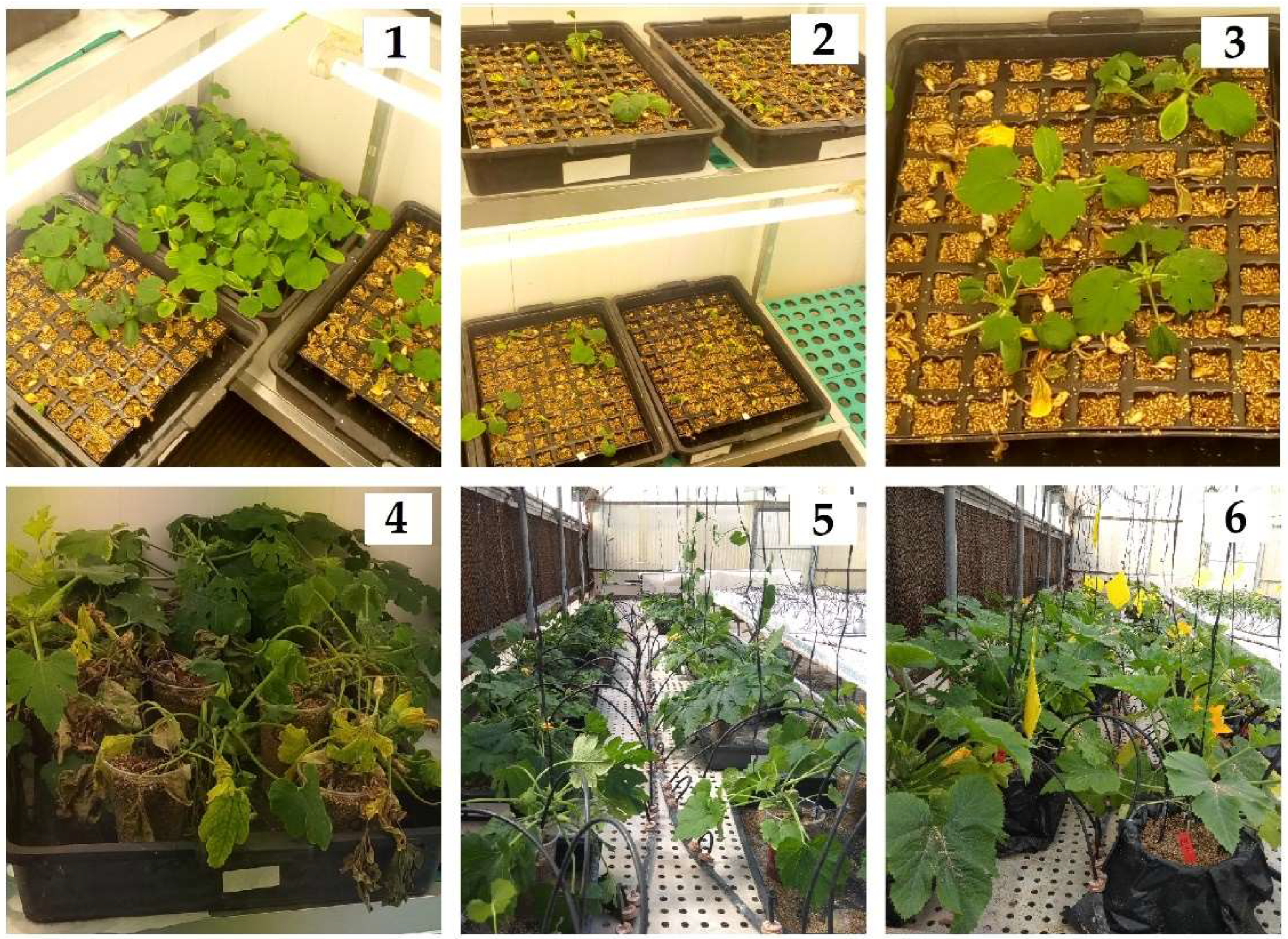
A total of 18 Cucurbita genotypes were evaluated initially. These included C. pepo subsp. ovifera SCA (Scallop, Germplasm Bank Comunidad Valenciana BGV-5382); C. pepo subsp. pepo morphotype zucchini MUC (MUCU-16, IFAPA-La Mojonera collection); AMA (Amalthée, Gautier seeds), JED (Jedida, HM Clause), MIL (Milenio, Fitó seeds), NAT (Natura, Enza Zaden), VIC (Victoria, HM Clause), 2 marrow PIC (Piccolo, Tozer seeds), and ZEB (Zebra Cross, Tozer seeds); C. moschata M64 (64-064, Rijk Zwaan), M63 (64-063, Rijk Zwann), TZ3 (TZ-3380, Tozer seeds), and TER (Butternut Sprinter, Tozer seeds); and hybrids of C. maxima × C. moschata, ROU (Routpower, Sakata seeds), SHI (Shintoza, Fitó seeds), CAM (Shintosa Camelforce, Bayer CropScience), AZM (Azman, Rick Zwaan), and HER (Hércules, Ramiro Arnedo).
Mutant germplasms were obtained from a second-generation mutant (M2) population of MUCU-16 C. pepo subsp. pepo [23]. The mutations were randomly distributed [23] in the 160 mutant families tested. Currently, the M2 and M3 families are conserved in the IFAPA-La Mojonera germplasm collection.
Phytophthora capsici (MI0211, GenBank MG012233) [6] and Fsc race 1 (Fs511, GenBank AM940070) isolates [35], obtained from infected plants in greenhouses in Almería province (Spain) were morphologically and molecularly described in previous studies [6,35]. Both pathogens were preserved in the IFAPA-La Mojonera laboratories and subsequently inoculated on C. pepo, and re-isolated and identified again from dead plants prior to inoculation.

2.2. Inoculation

P. capsici and Fsc inocula followed the same inoculation procedures. Two inoculation methods were used: root dipping and substrate infestation (Table 1). The first screening with different Cucurbita spp. genotypes and the initial M2 mutant germplasm were inoculated by root dipping (Table 1). In this method, the radicles were immersed for 30 s in a suspension containing the pathogen propagules. This suspension was obtained by grinding several colonies fully covering the petri dish surface with each isolate, which had been previously grown for 10 days in PDA (1 petri dish per 400 mL of standard nutrient solution). The inocula concentrations were calculated by means of the dilution plate technique on PDA, obtaining ca. 106 UFC mL−1 for Fsc and ca. 104 UFC mL−1 for P. capsici.
The same procedure was used to obtain the inoculum for the subsequent mutant screenings and gene expression quantification analysis (test 3–5, Table 1). However, instead of radicle immersion, the inoculum was added by irrigation to the substrate at 2 cm depth near the plant with 50 mL of the inoculum suspension (substrate infestation method) once the plants reached the development stage of 2–3 true leaves (Table 1). The presence of Fsc macroconidia and Phytophthora sporangia on the dead plants’ roots and crown was checked under a microscope, and they were reisolated for the following inoculations after each test. Control plants were watered with standard nutrient solution.

2.3. Test Conditions

All experiments were performed in a growth chamber with a 14 h photoperiod (>12,000 lux), 23–33 °C, and 40–75% relative humidity. The temperature and humidity were measured using a HOBO data logger. Before planting, the seeds were disinfected by immersing them in a sodium hypochlorite (35 g L−1 active chloride) solution 1:1 for 20 min, and subsequently they were rinsed with autoclaved (30 min, 121 °C) sterile water and incubated at 28 °C in the dark until the roots reached 3–5 cm in length. Only germinated seeds were used.
In order to inoculate all plants at the same age, different sowing dates were applied depending on previous knowledge about the early development of each genotype. This step is important, because different plant development stages could affect their resistance responses [36]. The germination rate after one week for commercial and non-mutated germplasms was higher than 90%, while the M2 mutant germplasms had a germination rate between 25–95%, depending on the mutant family. Once the seeds had germinated, the seedlings reached 2–3 true leaves after 10–15 days. The substrate used for all of the experiments was vermiculite at field capacity, with a standard nutrient solution (1.5 dS m−1). For all tests, all samples were randomly distributed in separate trays in the chamber beside non-inoculated controls. Non-inoculated controls were included in all of the tests. For test 1, 8 control replicates per genotype were used. At least one plant per mutant family served as the control for tests 2, 3, and 4. Test 5 was completed with the same number (32) of control repetitions.
For the different Cucurbita spp. genotype tolerance screenings (test 1), two plants per 1 L pot (eight plants per pathogen, two trials) were used, and these plants were inoculated by the root-dipping method (Table 1). Susceptible and tolerant reference controls for tests 4 and 5 were chosen based on the responses of these plants (test 1).
The mutant screening and the selection of tolerant families consisted of three steps (Table 1): initial screening with seedlings (test 2), second screening with developed plants (test 3), and final selection (test 4). Initial mutant screening (test 2) was performed with 160 mutant families (three replicates) in 60 mL 96-well trays (Table 1). The mutant families that showed less symptomatology or that had tolerant individuals by day 14 post-inoculation were selected for the next test. Subsequent screenings were undertaken in 1 L pots (two plants per pot) with a decreasing number of selected families but increasing numbers of replicates per family (Table 1). For the next test, 50 selected families for P. capsici and 26 for Fsc were inoculated when the plants reached 2–3 true leaves (test 3). For the final mutant selection test (test 4), six families were selected for each pathogen, and genotypes M64 and AMA acted as the tolerant and susceptible controls, respectively. The mutant individuals that survived the P. capsici substrate infestation were reinoculated, transplanted into a 25 L pot, and developed in a greenhouse. This allowed the plants to reach a large enough size to produce, after self-pollination, a mature zucchini fruit with grown M3 seeds. The two M3 families with fertile seeds obtained in the study were used in the expression analysis test. For the gene expression analysis (test 5), the M3 mutant lines Cp107 and Cp116, and genotypes M63 (tolerant control) and MUCU-16 (susceptible control) were used (Table 1).

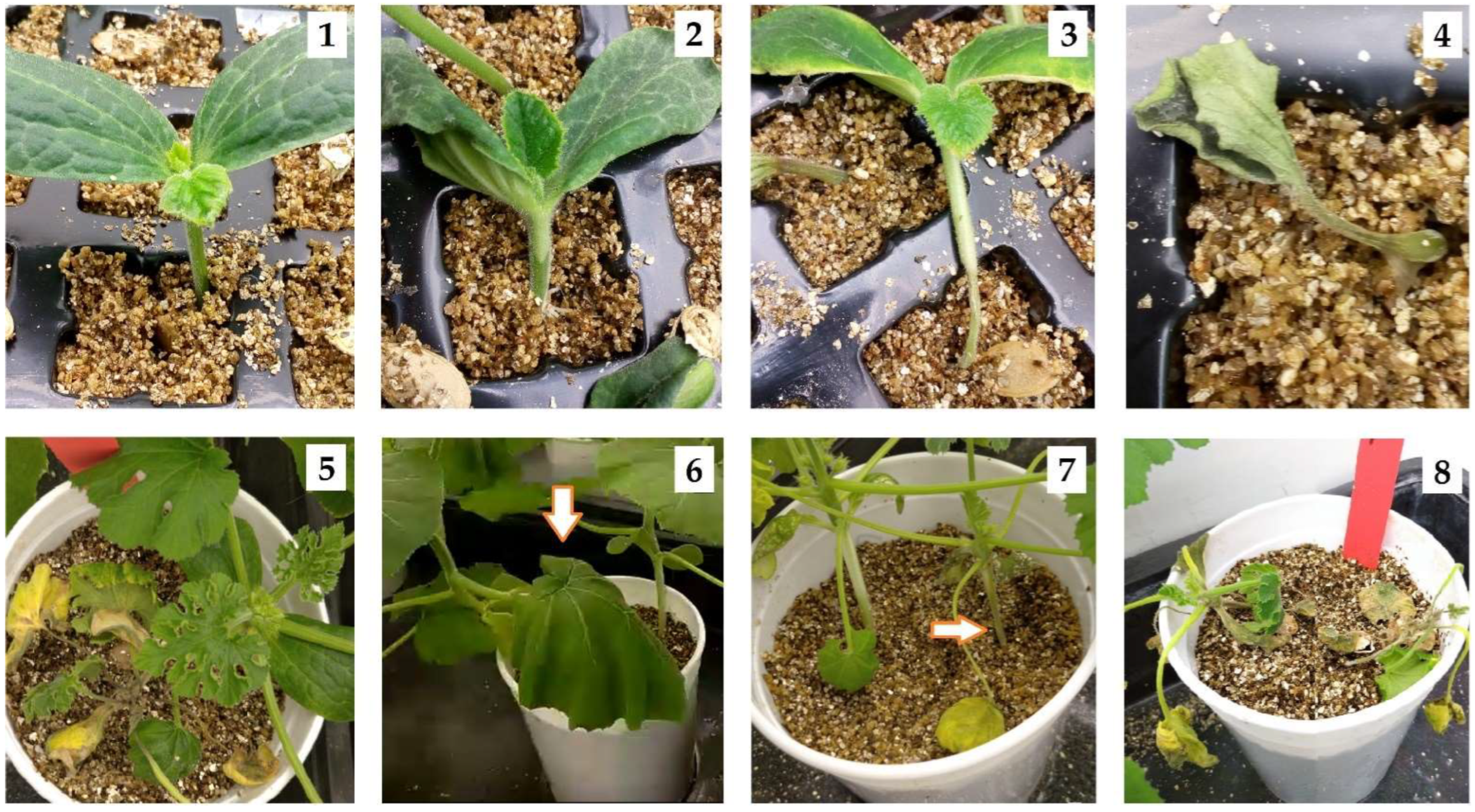
2.4. Tolerance Evaluation

The tolerance evaluation was based on three parameters: the disease severity index (DSI), percentage of plants with symptomatology, and relativized area under the disease progress curve (RAUDPC).
The DSI is based on a visual non-parametric scale ranging from 0 to 4: 0 = no damage, 1 = leaf epinasty/soaked crown, 2 = constricted/girdling crown, 3 = wilt/chlorosis, and 4 = death; this is analogous to other authors [6,9,18]. The percentage of plants with symptomatology plotted against time produced an area under the disease progress curve (AUDPC) that represented the disease evolution. The AUDPC was relativized (RAUDPC) using the sample with the highest AUDPC value in each trial [2,6,14]. In test 1, plants that had an RAUDPC between 0–10% were considered tolerant, 11–30% low susceptibility, 31–70% intermediate susceptibility, and 71–100% high susceptibility. The RAUDPC was submitted to an analysis of variance (ANOVA); when significant differences (p < 0.05) were found, Tukey’s HSD was carried out. For two-level comparison, Student’s t-test was used. The data were tested for a normal distribution and homoscedasticity by the Shapiro–Wilk and Levene tests. Statistix 9.0 software (Tallahassee, FL, USA) was used for all statistical analyses. Tests 1 and 5 were duplicated (Table 1), and the data were pooled. Additionally, the damage level caused by both pathogens was analyzed once the plant died (Figure A1).

2.5. qPCR Sample Collection and Conditions

The test conditions are shown in Table 1 (test 5). Root and crown samples were collected on 0, 3, 10, and 14 days post inoculation (dpi), as the disease evolution was observed. Each sample was extracted from eight different plants, which decreased the number of plants in the test by eight after each sample collection day. Sample collection, RNA extraction, and cDNA obtention were performed as described in previous work [34]. The primers (Sigma–Aldrich, St. Louis, MO, USA), except for the UFP and EFP-1α reference genes [37], were obtained from previous work [34]. The qPCR cycling conditions were: 50 °C 2′; 95 °C 10′; 40× (95 °C 15”, 55 °C 15′, and 60 °C 1”); and melting (95 °C 15”, 60 °C 1′, and 95 °C 1”). Gene expression was relativized with UFP (ubiquitin fusion protein) and EFP-1α (elongation factor-1α) reference genes [37] following data processing with efficiency correction, multiple reference gene normalization, and the use of error propagation rules [38,39]. The normalized relative quantities (NRQ) for each sample were obtained and transformed with LOG10(NRQinoculated/NRQnon-inoculated). Sample data were pooled (3, 10, and 14 dpi and crown and root tissues) by genotype and compared against the susceptible control by applying Student’s t-test (p < 0.005). Homoscedasticity and normal distribution were checked with Statistix 9.0 software.

3. Results

3.1. Cucurbita Genotype Tolerance Screening to P. capsici and F. solani f. sp. cucurbitae

The RAUDPC caused by P. capsici and Fsc for different Cucurbita spp. genotypes after 37 dpi is shown in Table 2. Both pathogens, inoculated by root-dipping, showed damage to all genotypes tested. However, the C. moschata species showed less damage for both Fsc and P. capsici (Table 2). According to the phenotype classification, M64 and M63 were considered tolerant to P. capsici, while TZ3 was tolerant to Fsc (Table 2). In contrast, many C. pepo plants showed high susceptibility to both pathogens, although the two marrow morphotypes analyzed (PIC and ZEB) presented intermediate susceptibility to Fsc (Table 2). Just one hybrid of C. maxima × C. moschata showed high susceptibility to both pathogens, HER, while SHI showed intermediate susceptibility (Table 2). Significant differences in the RAUDPC were observed between the TZ3 tolerant and AZM, CAM, HER, MUC, MIL and NAT susceptible genotypes after Fsc inoculation (Table 2). For P. capsici, significant differences also appeared between M63, M64, TZ3, TER, and ZEB with the highly susceptible MIL, MUC, NAT, SCA, and VIC genotypes (Table 2).

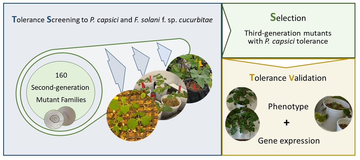
3.2. Mutant Tolerance Screening to P. capsici and Fusarium solani f. sp. cucurbitae

The first mutant screening included 160 families (test 2, Figure 1). Both P. capsici and Fsc produced symptomatology (Figure 2) in the entire inoculated population after 27 dpi. In some cases, plants were unable to develop true leaves due to the disease. Families with less damage or with individuals without damage on day 14 post-inoculation were selected for the next test (Figure 1). For P. capsici, 50 families were selected, while for Fsc, 26 families were selected. Fewer families were selected for Fsc because this fungus produced more damage by day 14 than P. capsici. The selected families were inoculated in a new test (test 3). In this test, the six families with the lowest RAUDPC by day 27 post-inoculation were selected for the subsequent screening (test 4, Figure 1).
For P. capsici, all the previously selected families from test 3 had individuals that survived to the inoculation of test 4 (Table 3); these plants were re-inoculated at 34 dpi and transferred to a greenhouse (Figure 1). In total, 15 plants from families Cp45 (1), Cp107 (2), Cp116 (3), Cp136 (3), Cp139 (2), and Cp144 (4) remained at 97 dpi. Mature fruits were obtained from families Cp107, Cp116, Cp139, and Cp144, but only Cp107 and Cp116 fruits had M3 fertile seeds. On the other hand, none of the six families selected for Fsc showed tolerance to the pathogen (Table 3). The tolerant (M64) and susceptible (AMA) controls responded (Table 3) as in the previous test for both pathogens (Table 2).
The undergrown plant lesions evaluated after plant death indicated that both pathogens produced damage to the crown and root tissues. All dead plants analyzed for P. capsici and for Fsc presented more accentuated symptomatology in the crown, with at least soaking damage, while the secondary roots were the least affected tissue (Figure A1).

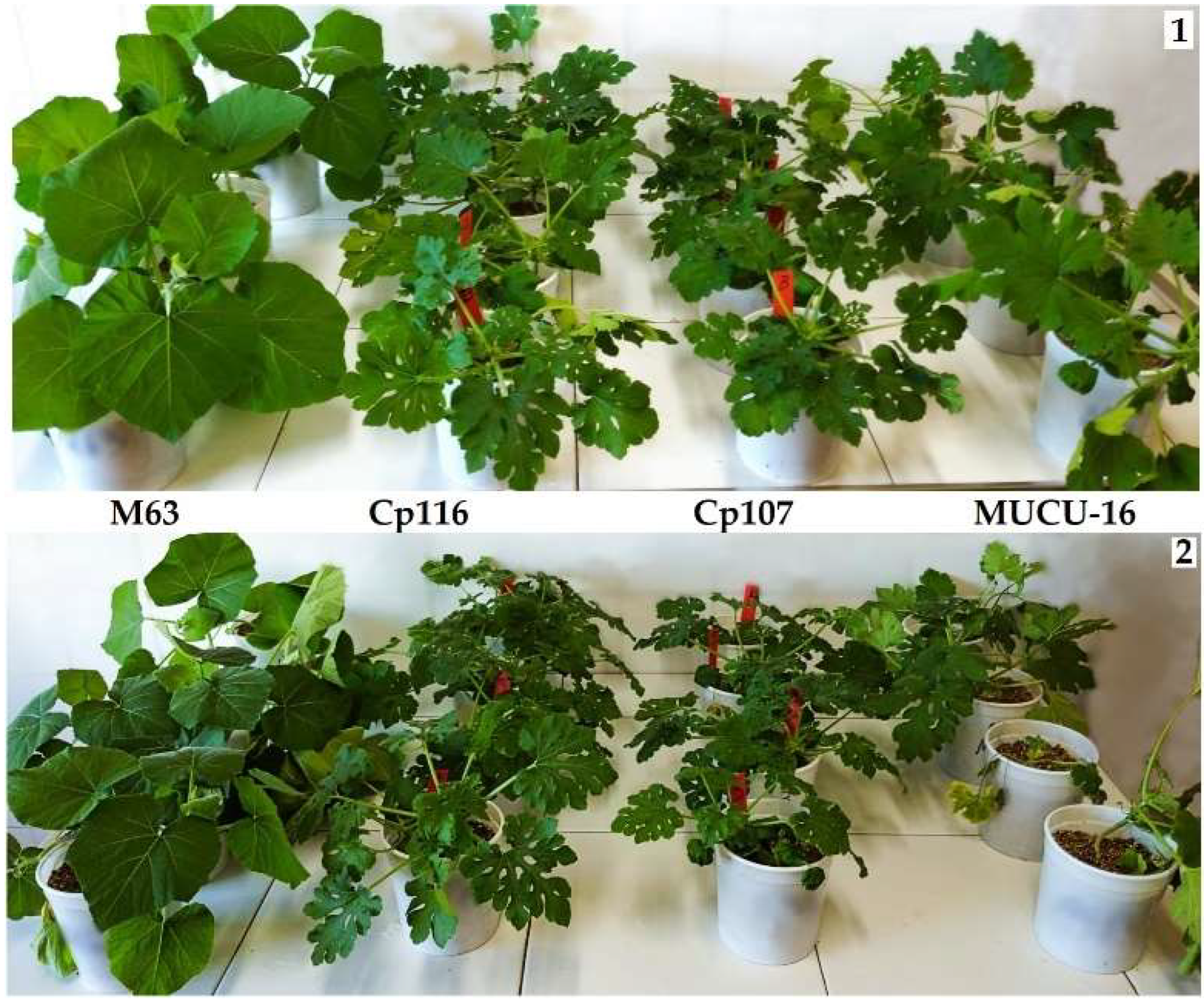
3.3. M3 Mutant Tolerance Analysis

The M3 Cp107 and Cp116 mutant families (derived from self-pollinated M2 plants that showed tolerance) presented less damage caused by P. capsici than the non-mutated MUCU-16 (M0) (Figure 3). The mutants showed significant differences in the RAUDPC against the susceptible control 14 dpi (Table 4). No damage was observed until day 10 after inoculation for Cp107, Cp116, and M63 while, for MUCU-16, the symptoms started on day 3 (Table 4).

3.4. M3 Gene Expression

The differential expression between inoculated and non-inoculated samples for all pooled samples (dpi: 3, 10, and 14) showed significant differences in MUCU-16 for the CpDEF, CpChiIV, and CpPAL genes. On the other hand, the CpACO and CpLPOX genes remained similar to the control (Figure 4). Mainly due to down-regulation of CpPAL in the susceptible after inoculation, the genotypes M63 (p = 0.021), Cp107 (p = 0.042) and Cp116 (p = 0.043) were expressed CpPAL more than MUCU-16 (Figure 4). No significant differences were found between the C. pepo genotypes (MUCU-16, Cp107, and Cp116) in CpDEF expression in the inoculated samples, while the tolerant control showed higher up-regulation (p = 0.0117, Figure 4) with respect to the susceptible control. However, only the Cp107 (p = 0.046) and Cp116 (p = 0.035) mutant families expressed the CpChiIV gene more in the inoculated samples versus the control (Figure 4).

4. Discussion

4.1. Tolerant Sources for Fusarium solani f. sp. cucurbitae and Phytophthora capsici in the Cucurbita Genus

Enzenbacher and Hausbeck [14] tested different isolates of P. capsici on C. pepo (zucchini and straightneck) and C. moschata (butternut) genotypes. Their results pointed out that the C. pepo straightneck morphotypes showed higher susceptibility and the C. moschata genotype showed higher tolerance [14]. Similarly, differences in the disease severity index were observed between the morphotypes of C. pepo and between the subspecies C. pepo subsp. pepo and C. pepo subsp. ovifera [4]. In this study, C. pepo subsp. pepo marrow morphotypes showed more tolerance to P. capsici, and the C. pepo subsp. ovifera were highly susceptible [4]. Our results indicated that the plant responses to these pathogens were also dependent on the species and morphotype. In our study, C. moschata was the most tolerant, while C. pepo subsp. ovifera was highly susceptible to P. capsici. The C. pepo zucchini morphotypes were the most susceptible, while the marrows were the least susceptible among the C. pepo subsp. pepo morphotypes. Furthermore, Perez-Hernandez et al. [2] tested some pathogenic isolates of Fsc that were shown to be highly aggressive to zucchini plants, while other cucurbit hosts showed differential susceptibility to the pathogen. These results support ours, since, in this study, zucchini genotypes were found to be highly susceptible to Fsc, while some genotypes of C. moschata showed tolerance to the fungus.
It has been observed in different screenings for resistance to P. capsici that some genotypes of C. lundelliana and C. okeechobeensis wild species [15], C. pepo [9], and C. moschata [11] show reduced crown damage. Padley et al. [9] obtained, from a resistance screening in C. pepo, 16 accessions that were less damaged by P. capsici, and selected two of them because they were more resistant [9]. All these accessions showed high genetic proximity, but one line was also close to the susceptible C. pepo subsp. pepo and C. pepo subsp. texana genotypes [16]. The backcrossing and its phenotypic segregation analysis indicated that the resistance to P. capsici was controlled by three dominant genes (R4, R5, and R6) [17]. Furthermore, resistance to P. capsici has been introduced into C. moschata from wild C. lundelliana and C. okeechobeensis, and its segregation also suggests that this trait is controlled by three dominant genes (Crr-1, Crr-2, Crr-3) [10].
In addition, the genotypes that present more tolerance could be used for grafting other compatible cucurbits. Cucurbita spp. germplasms that present high frequency of individuals with tolerance to Fsc were identified as rootstock candidates [18]. Additionally, grafted Citrullus lanatus on Ferro, GV 100 (C. maxima × C. moschata), and Just (Cucurbita spp.) rootstocks showed tolerance to Fsc, although this response depended on the plant development at the inoculation moment [41]. For watermelon, the Lagenaria siceraria (bottle gourd) rootstocks FR-Strong, Emphasis, Macis, and WMXP-3938 were resistant to crown and root rot caused by P. capsici [42]. Our results suggest that there are rootstock candidates for tolerance to P. capsici (M63 and M64) and Fsc (TZ3), but compatibility studies with zucchini are needed.

4.2. Mutant Selection

To our knowledge, none of the previous SBPFO resistance screening studies in cucurbits were performed with artificially induced mutants. All were based on germplasm bank collections of wild, conserved, commercial, or hybrid genotypes [2,9,14,15,18]. This study presents the first results for mutant C. pepo subsp. pepo lines with tolerance to P. capsici.
Few studies have been conducted in the Cucurbitaceae family on biotic stress with EMS-mutated populations, despite this being an excellent tool for breeding [3,27]. For example, one bottle gourd (Lagenaria siceraria) M4 EMS-induced inbred line (117-8) resistant to zucchini yellow mosaic virus (ZYMV) was obtained and showed stable resistance and no virus presence among inoculated plants [27]. Our selected families (Cp107 and Cp116) presented more tolerance to P. capsici than the highly susceptible C. pepo subsp. pepo from which they originated (Table 4). However, some characteristics, such as plant architecture, seemed to be affected with respect to the MUCU-16 background M0 (Figure 3), although specific phenotyping analyses still have to be performed.
Genes for resistance to SBPFO have been reported in cucurbits [3,43]. However, the mutant tolerance to P. capsici could be due to diverse causes, given the nature (random and numerous) of mutations caused by EMS. Plant development, which affects plant tolerance [36], plant resistance genes that respond to pathogen virulence factors [3,30,31], or plant susceptibility genes, which are targeted by the pathogen [33,44,45,46,47], may have been affected by the mutations.
During soilborne disease-resistance screening with a large population, it is common to find individuals that show more tolerance, although the family as a whole may seem susceptible [18]. We also observed this situation, especially in the mutant population. In an M1 population, most of the randomly mutated genes would be in heterozygosis, and its segregation (M2) could show different phenotypes [23]. The individuals tolerant to P. capsici were self-fertilized and M3 seeds were obtained. This reduced the family phenotypic heterogeneity, although segregation studies should be performed to confirm this fact. It should be taken into account that, despite obtaining a mutant of interest, sometimes it will not be possible to produce seeds due to the difficulty some mutants have in generating fertile seeds, as random mutations can alter their formation [23]. Finding a mutant material with tolerance to a disease in the species to be cultivated would reduce the steps in introgression programs or grafting. Nevertheless, the method for producing a mutant population for each crop must be previously known to avoid the loss of desirable characteristics of the background M0, while still allowing it to acquire new ones. Vicente-Dólera et al. [23] observed that, with the mutagenic agent EMS at a concentration of 0.4%, plants had more viability than at higher concentrations. Also, with this concentration, the plant population incorporated a mutation frequency of 1/135 kb [23]. For this reason, we chose this concentration so that the plants would not lose the useful characteristics acquired during domestication.

4.3. qPCR

The CpDEF gene was highly overexpressed in the inoculated plants, and CpPAL down-regulation occurred only in the susceptible genotypes, which is in agreement with previous studies [34]. Moreover, The mutants Cp107 and Cp116 expressed more CpChiIV than MUCU-16 after inoculation. The mutant population may not have directly affected these genes, since this differential expression between the susceptible and tolerant phenotypes observed may be the consequence, rather than the cause, of tolerance.
Gene expression analysis has been demonstrated to be a useful approach to complement studies where the plant responds to biotic stress. Genes such as CaPBR1, CaPO1, CaDEF1 [48], chitinase [29], lignin-forming peroxidase, ethylene, and jasmonic related genes [30], were found to be differentially expressed in the P. capsici and Capsicum annuum pathosystem. Additionally, defensins (ClPDF2.1 and ClPDF2.4), PAL, chitinase, and ascorbate peroxidase were significantly induced in roots in watermelon during Fusarium oxysporum f. sp. niveum infection [32]. Also, a resistant genotype of Cucumis spp. showed up-regulation of ACO, PAL, defensins, and lignin-forming peroxidase genes in P. capsici inoculated roots [31].

5. Conclusions

The screening of an EMS-mutated population was proven to be a reliable method for obtaining new traits in complex phenomena, such as C. pepo subsp. pepo tolerance to P. capsici. The tolerance trait was found in the phenotype and gene expression analyses of the two M3 families obtained. These families presented significant differences in P. capsici tolerance, and in the expression of CpPAL and CpChiIV genes compared with the susceptible background.

Author Contributions

Conceptualization, A.A.-D. and M.d.C.-G.; methodology, A.A.-D. and M.d.C.-G.; software, A.A.-D. and M.d.C.-G.; validation, M.d.C.-G. and P.G.; formal analysis, M.d.C.-G.; investigation, A.A.-D.; resources, M.d.C.-G. and P.G.; data curation, M.d.C.-G.; writing—original draft preparation, A.A.-D.; writing—review and editing, A.A.-D., M.d.C.-G., and P.G.; visualization, A.A.-D.; supervision, M.d.C.-G.; project administration, M.d.C.-G.; funding acquisition, M.d.C.-G. and P.G. All authors have read and agreed to the published version of the manuscript.

Funding

This research was funded by the European Regional Development Fund (ERDF) through the research project PP.AVA.AVA2019.015 “Control sostenible de plagas y enfermedades en hortícolas protegidas”.

Acknowledgments

Julio Gómez, for facilitating the Fs511 isolate. A. Ayala-Doñas thanks INIA for economic support through a pre-doctoral grant (FPI-INIA2016 CPD2016-0199).

Conflicts of Interest

The authors declare no conflict of interest.

Appendix A

Figure A1. Underground symptomatology of P. capsici and Fsc. P. capsici (left) and Fsc (right) 14 dpi in seedlings (1). P. capsici (left) and Fsc (right) 21 dpi in plants inoculated when they reached 2–3 true leaves (2,3). Soaked and constricted crown and main root disaggregation caused by Fsc (4), main root browning caused by P. capsici (5) 14 dpi. Note that these cucurbits had a fibrous root system (generate more than one main root), but, in the young plants, a main root is clearly observable.
Horticulturae 08 00191 g0a1

References

  1. Babadoost, M. Oomycete Diseases of Cucurbits: History, Significance, and Management. In Horticultural Reviews; Wiley backwell: Hoboken, NJ, USA, 2016; Volume 44, pp. 279–314. [Google Scholar]
  2. Pérez-Hernández, A.; Rocha, L.O.; Porcel-Rodríguez, E.; Summerell, B.A.; Liew, E.C.Y.; Gómez-Vázquez, J.M. Pathogenic, Morphological, and Phylogenetic Characterization of Fusarium solani f. sp. cucurbitae Isolates From Cucurbits in Almería Province, Spain. Plant Dis. 2020, 104, 1465–1476. [Google Scholar] [CrossRef] [PubMed]
  3. Ayala-Doñas, A.; de Cara-García, M.; Talavera-Rubia, M.; Verdejo-Lucas, S. Management of Soil-Borne Fungi and Root-Knot Nematodes in Cucurbits through Breeding for Resistance and Grafting. Agronomy 2020, 10, 1641. [Google Scholar] [CrossRef]
  4. Krasnow, C.S.; Hammerschmidt, R.; Hausbeck, M.K. Characteristics of Resistance to Phytophthora Root and Crown Rot in Cucurbita pepo. Plant Dis. 2017, 101, 659–665. [Google Scholar] [CrossRef] [PubMed] [Green Version]
  5. Pérez-Hernández, A.; Porcel-Rodríguez, E.; Gómez-Vázquez, J. Survival of Fusarium solani f. sp. cucurbitae and Fungicide Application, Soil Solarization, and Biosolarization for Control of Crown and Foot Rot of Zucchini Squash. Plant Dis. 2017, 101, 1507–1514. [Google Scholar] [CrossRef] [Green Version]
  6. de Cara García, M.; Plaza, M.F.; Vázquez, J.M.G. Pathogenic and Biological Characterization of Phytophthora capsici Isolates from Zucchini and Pepper in Southeast Spain. Span. J. Agric. Res. 2018, 16, 22. [Google Scholar] [CrossRef]
  7. Sanogo, S.; Ji, P. Integrated Management of Phytophthora capsici on Solanaceous and Cucurbitaceous Crops: Current Status, Gaps in Knowledge and Research Needs. Can. J. Plant Pathol. 2012, 34, 479–492. [Google Scholar] [CrossRef]
  8. Davis, A.R.; Perkins-Veazie, P.; Sakata, Y.; López-Galarza, S.; Maroto, J.V.; Lee, S.-G.; Huh, Y.-C.; Sun, Z.; Miguel, A.; King, S.R.; et al. Cucurbit Grafting. Crit. Rev. Plant Sci. 2008, 27, 50–74. [Google Scholar] [CrossRef]
  9. Padley, L.D.; Kabelka, E.A.; Roberts, P.D. Evaluation of Cucurbita pepo Accessions for Crown Rot Resistance to Isolates of Phytophthora capsici. HortScience 2008, 43, 1996–1999. [Google Scholar] [CrossRef] [Green Version]
  10. Padley, L.D.; Kabelka, E.A.; Roberts, P.D. Inheritance of Resistance to Crown Rot Caused by Phytophthora capsici in Cucurbita. HortScience 2009, 44, 211–213. [Google Scholar] [CrossRef] [Green Version]
  11. Chavez, D.J.; Kabelka, E.A.; Chaparro, J.X. Screening of Cucurbita moschata Duchesne Germplasm for Crown Rot Resistance to Floridian Isolates of Phytophthora capsici Leonian. HortScience 2011, 46, 536–540. [Google Scholar] [CrossRef] [Green Version]
  12. Ramos, A.; Fu, Y.; Michael, V.; Meru, G. QTL-Seq for Identification of Loci Associated with Resistance to Phytophthora Crown Rot in Squash. Sci. Rep. 2020, 10, 5326. [Google Scholar] [CrossRef] [PubMed]
  13. Vogel, G.; LaPlant, K.E.; Mazourek, M.; Gore, M.A.; Smart, C.D. A Combined BSA-Seq and Linkage Mapping Approach Identifies Genomic Regions Associated with Phytophthora Root and Crown Rot Resistance in Squash. Theor. Appl. Genet. 2020, 134, 1015–1031. [Google Scholar] [CrossRef]
  14. Enzenbacher, T.B.; Hausbeck, M.K. An Evaluation of Cucurbits for Susceptibility to Cucurbitaceous and Solanaceous Phytophthora capsici Isolates. Plant Dis. 2012, 96, 1404–1414. [Google Scholar] [CrossRef] [Green Version]
  15. Kabelka, E.; Les Padley, J.; Roberts, P.; Ramos, L.; Martinez, M.; Klassen, W. Resistance to Phytophthora capsici within Winter Squash (Cucurbita moschata) Derived from a Wild Cucurbita Species. In Proceedings of the HortScience; American Society for Horticultural Science: Alexandria, VA, USA, 2007; Volume 42, p. 1014. [Google Scholar]
  16. Vincent Michael, N.; Moon, P.; Fu, Y.; Meru, G. Genetic Diversity Among Accessions of Cucurbita pepo Resistant to Phytophthora Crown Rot. HortScience 2019, 54, 17–22. [Google Scholar] [CrossRef] [Green Version]
  17. Michael, V.N.; Fu, Y.; Meru, G. Inheritance of Resistance to Phytophthora Crown Rot in Cucurbita pepo. HortScience 2019, 54, 1156–1158. [Google Scholar] [CrossRef] [Green Version]
  18. Santos, J.S.D.; Dias, R.D.C.S.; Almeida, K.B.D.; Ribeiro Junior, P.M.; Nascimento, T.L.D. Resistance of Cucurbita spp. to Fusarium solani for Use as Rootstock. Rev. Caatinga 2020, 33, 384–394. [Google Scholar] [CrossRef]
  19. Ji, X.; Zhang, H.; Zhang, Y.; Wang, Y.; Gao, C. Establishing a CRISPR–Cas-like Immune System Conferring DNA Virus Resistance in Plants. Nat. Plants 2015, 1, 15144. [Google Scholar] [CrossRef]
  20. Pyott, D.E.; Sheehan, E.; Molnar, A. Engineering of CRISPR/Cas9-Mediated Potyvirus Resistance in Transgene-Free Arabidopsis Plants: CRISPR/Cas9-Induced Potyvirus Resistance. Mol. Plant Pathol. 2016, 17, 1276–1288. [Google Scholar] [CrossRef] [Green Version]
  21. Chandrasekaran, J.; Brumin, M.; Wolf, D.; Leibman, D.; Klap, C.; Pearlsman, M.; Sherman, A.; Arazi, T.; Gal-On, A. Development of Broad Virus Resistance in Non-Transgenic Cucumber Using CRISPR/Cas9 Technology: Virus Resistance in Cucumber Using CRISPR/Cas9. Mol. Plant Pathol. 2016, 17, 1140–1153. [Google Scholar] [CrossRef] [Green Version]
  22. Chaudhary, J.; Deshmukh, R.; Sonah, H. Mutagenesis Approaches and Their Role in Crop Improvement. Plants 2019, 8, 467. [Google Scholar] [CrossRef] [Green Version]
  23. Vicente-Dólera, N.; Troadec, C.; Moya, M.; del Río-Celestino, M.; Pomares-Viciana, T.; Bendahmane, A.; Picó, B.; Román, B.; Gómez, P. First TILLING Platform in Cucurbita pepo: A New Mutant Resource for Gene Function and Crop Improvement. PLoS ONE 2014, 9, e112743. [Google Scholar] [CrossRef] [PubMed] [Green Version]
  24. García, A.; Aguado, E.; Parra, G.; Manzano, S.; Martínez, C.; Megías, Z.; Cebrián, G.; Romero, J.; Beltrán, S.; Garrido, D.; et al. Phenomic and Genomic Characterization of a Mutant Platform in Cucurbita pepo. Front. Plant Sci. 2018, 9, 1049. [Google Scholar] [CrossRef] [PubMed]
  25. García, A.; Manzano, S.; Megías, Z.; Aguado, E.; Martínez, C.; Garrido, D.; Rebolloso, M.M.; Valenzuela, J.L.; Jamilena, M. Use of Mutant Platforms to Discover Novel Postharvest Fruit-Quality Traits in Cucurbita pepo. Acta Hortic. 2018, 1194, 367–374. [Google Scholar] [CrossRef]
  26. Chen, X.; Guo, W.; Jiang, L.; Hayat, S.; Chen, B.; Yang, P.; Zhou, J.; Li, X.; Bai, Y. Screening of EMS-Induced NaCl-Tolerant Mutants in Cucurbita moschata Duchesne Ex Poir. Pak. J. Bot. 2018, 50, 1305–1312. [Google Scholar]
  27. Al-Kubati, A.M.S.; Kang, B.; Liu, L.; Abbas, A.; Gu, Q. Development of Bottle Gourd Lines Resistant to Zucchini Yellow Mosaic Virus Using Ethyl Methanesulfonate Mutagenesis. HortScience 2021, 56, 909–914. [Google Scholar] [CrossRef]
  28. Cebrián, G.; Iglesias-Moya, J.; García, A.; Martínez, J.; Romero, J.; Regalado, J.J.; Martínez, C.; Valenzuela, J.L.; Jamilena, M. Involvement of Ethylene Receptors in the Salt Tolerance Response of Cucurbita pepo. Hortic. Res. 2021, 8, 73. [Google Scholar] [CrossRef]
  29. Liu, Z.; Shi, L.; Yang, S.; Lin, Y.; Weng, Y.; Li, X.; Hussain, A.; Noman, A.; He, S. Functional and Promoter Analysis of ChiIV3, a Chitinase of Pepper Plant, in Response to Phytophthora capsici Infection. Int. J. Mol. Sci. 2017, 18, 1661. [Google Scholar] [CrossRef] [Green Version]
  30. Wang, P.; Liu, X.; Guo, J.; Liu, C.; Fu, N.; Shen, H. Identification and Expression Analysis of Candidate Genes Associated with Defense Responses to Phytophthora capsici in Pepper Line “PI 201234”. Int. J. Mol. Sci. 2015, 16, 11417–11438. [Google Scholar] [CrossRef] [Green Version]
  31. Wang, P.; Wu, H.; Zhao, G.; He, Y.; Kong, W.; Zhang, J.; Liu, S.; Liu, M.; Hu, K.; Liu, L.; et al. Transcriptome Analysis Clarified Genes Involved in Resistance to Phytophthora capsici in Melon. PLoS ONE 2020, 15, e0227284. [Google Scholar] [CrossRef]
  32. Zhang, M.; Xu, J.H.; Liu, G.; Yao, X.F.; Li, P.F.; Yang, X.P. Characterization of the Watermelon Seedling Infection Process by Fusarium oxysporum f. sp. niveum. Plant Pathol. 2015, 64, 1076–1084. [Google Scholar] [CrossRef]
  33. Li, Q.; Chen, Y.; Wang, J.; Zou, F.; Jia, Y.; Shen, D.; Zhang, Q.; Jing, M.; Dou, D.; Zhang, M. A Phytophthora capsici Virulence Effector Associates with NPR1 and Suppresses Plant Immune Responses. Phytopathol. Res. 2019, 1, 1–11. [Google Scholar] [CrossRef] [Green Version]
  34. Ayala-Doñas, A.; Gómez, P.; de Cara-García, M. Gene Expression in Cucurbita spp. Root and Crown during Phytophthora capsici Infection. Plants 2021, 10, 2718. [Google Scholar] [CrossRef] [PubMed]
  35. Gomez, J.; Guerra-Sanz, J.M.; Sanchez-Guerrero, M.C.; Serrano, Y.; Melero-Vara, M. Crown Rot of Zucchini Squash Caused by Fusarium solani f. sp. cucurbitae in Almería Province, Spain. Plant Dis. 2008, 92, 1137. [Google Scholar] [CrossRef]
  36. Meyer, M.D.; Hausbeck, M.K. Age-Related Resistance to Phytophthora Fruit Rot in ‘Dickenson Field’ Processing Pumpkin and ‘Golden Delicious’ Winter Squash Fruit. Plant Dis. 2013, 97, 446–552. [Google Scholar] [CrossRef] [PubMed] [Green Version]
  37. Obrero, Á.; Die, J.V.; Román, B.; Gómez, P.; Nadal, S.; González-Verdejo, C.I. Selection of Reference Genes for Gene Expression Studies in Zucchini (Cucurbita pepo) Using QPCR. J. Agric. Food Chem. 2011, 59, 5402–5411. [Google Scholar] [CrossRef] [Green Version]
  38. Hellemans, J.; Mortier, G.; Paepe, A.D.; Speleman, F.; Vandesompele, J. QBase Relative Quantification Framework and Software for Management and Automated Analysis of Real-Time Quantitative PCR Data. Genome Biol. 2007, 8, 1–14. [Google Scholar] [CrossRef] [PubMed] [Green Version]
  39. Pomares-Viciana, T.; Die, J.; Del Río-Celestino, M.; Román, B.; Gómez, P. Auxin Signalling Regulation during Induced and Parthenocarpic Fruit Set in Zucchini. Mol. Breed. 2017, 37, 56. [Google Scholar] [CrossRef]
  40. Krasnow, C.S.; Naegele, R.P.; Hausbeck, M.K. Evaluation of Fruit Rot Resistance in Cucurbita Germplasm Resistant to Phytophthora capsici Crown Rot. HortScience 2014, 49, 285–288. [Google Scholar] [CrossRef] [Green Version]
  41. Boughalleb, N.; Mhamdi, M.; El Assadi, B.; El Bourgi, Z.; Tarchoun, N.; Romdhani, M.S. Resistance Evaluation of Grafted Watermelon (Citrullus lanatus L.) Against Fusarium Wilt and Fusarium Crown and Root Rot. Asian J. Plant Pathol. 2008, 2, 24–29. [Google Scholar] [CrossRef]
  42. Kousik, C.S.; Donahoo, R.S.; Hassell, R. Resistance in Watermelon Rootstocks to Crown Rot Caused by Phytophthora capsici. Crop Prot. 2012, 39, 18–25. [Google Scholar] [CrossRef]
  43. Tezuka, T.; Waki, K.; Kuzuya, M.; Ishikawa, T.; Takatsu, Y.; Miyagi, M. Development of New DNA Markers Linked to the Fusarium Wilt Resistance Locus Fom-1 in Melon: New DNA Markers Linked to Fom-1 in Melon. Plant Breed. 2011, 130, 261–267. [Google Scholar] [CrossRef]
  44. Win, K.T.; Zhang, C.; Lee, S. Genome-Wide Identification and Description of MLO Family Genes in Pumpkin (Cucurbita maxima Duch.). Hortic. Environ. Biotechnol. 2018, 59, 397–410. [Google Scholar] [CrossRef]
  45. Stam, R.; Jupe, J.; Howden, A.J.M.; Morris, J.A.; Boevink, P.C.; Hedley, P.E.; Huitema, E. Identification and Characterisation CRN Effectors in Phytophthora capsici Shows Modularity and Functional Diversity. PLoS ONE 2013, 8, e59517. [Google Scholar] [CrossRef]
  46. Li, Q.; Wang, J.; Bai, T.; Zhang, M.; Jia, Y.; Shen, D.; Zhang, M.; Dou, D. A Phytophthora capsici Effector Suppresses Plant Immunity via Interaction with EDS1. Mol. Plant Pathol. 2020, 21, 502–511. [Google Scholar] [CrossRef] [PubMed] [Green Version]
  47. Chen, X.-R.; Zhang, Y.; Li, H.-Y.; Zhang, Z.-H.; Sheng, G.-L.; Li, Y.-P.; Xing, Y.-P.; Huang, S.-X.; Tao, H.; Kuan, T.; et al. The RXLR Effector PcAvh1 Is Required for Full Virulence of Phytophthora capsici. Mol. Plant-Microbe Interact. 2019, 32, 986–1000. [Google Scholar] [CrossRef]
  48. Bellini, A.; Pugliese, M.; Guarnaccia, V.; Meloni, G.R.; Gullino, L.M. Calcium Oxide, Potassium Phosphite and a Trichoderma Enriched Compost Water Suspension Protect Capsicum annuum against Phytophthora capsici by Priming the Immune System. Pest. Manag. Sci. 2021, 77, 3484–3490. [Google Scholar] [CrossRef] [PubMed]
Figure 1. Tolerance screenings in the mutant population of MUCU-16. Screening with seedlings 14 days post-inoculation (dpi) (13): control tray (center) without inoculation (1); Fusarium solani f. sp. cucurbitae (left) and Phytophthora capsici (right) inoculated trays (2); individuals showing tolerance (3). Screening with selected plants (46): plant symptomatology 21 dpi—surviving individuals were moved to a greenhouse after 34 dpi and reinoculated (5); surviving plant growth until 97 dpi for fruit production (6).
Horticulturae 08 00191 g001
Figure 2. Disease symptoms caused by Phytophthora capsici and Fusarium solani f. sp. cucurbitae in Cucurbita pepo. Shoot symptomatology in seedlings 14 dpi (14): DSI 0 = no symptoms (1); DSI 1 = soaked crown (2); DSI 2 = constricted crown (3); DSI 4 = death (4). Shoot symptomatology 14 dpi in plants inoculated when they reached 2–3 true leaves (14). In some cases, pathogen damage was different between plants in the same pot (5): dead plant (left) and plant show no symptoms (right). Leaf epinasty (6), soaked and constricted crown (7), and wilted and dead (8) plants.
Horticulturae 08 00191 g002
Figure 3. Randomly selected plants for the sample collection of the gene expression analysis 14 days post-inoculation. A total of eight roots and eight crowns per genotype for each non-inoculated (1) and inoculated (2) sample were collected from four pots (two plants/pot). Plants were inoculated with P. capsici. The symptomatology severity was higher in MUCU-16 (2, right) than that for the rest of the genotypes, although the damage was not homogeneous for all individuals.
Horticulturae 08 00191 g003
Figure 4. Differential gene expression between genotypes in inoculated and non-inoculated samples Phytopthora capsici for each genotype. Each bar represents the genotype (MUCU-16, M63, Cp117, or Cp106) mean of the normalized relative quantity transformations between the inoculated and non-inoculated samples (LOG10[NRQinoculated/NRQnon-inoculated]) from pooled samples of root and crown tissues and the days 3, 10 and 14 post-inoculation. Positive values are up-regulation and negative are down-regulation for the inoculated samples. Significant differences (*) from the MUCU-16 control were determined with Student’s t-test (p < 0.05, number of samples compared = 12). Error bars represent the standard errors (n = 6).
Horticulturae 08 00191 g004
Table 1. Summary of the inoculation tests in chronological order.
TestDescriptionInoculation MethodInoculation
Plant Stage
PathogenCucurbita spp. MaterialDuration (Days) 1N 2
(rep. 3)
1 4Cucurbita spp. screeningRoot dippingSeedlingsPc5Commercial and
Conserved genotypes
3718 (16)
Fsc618 (16)
2Mutant first
screening
Root dippingSeedlingsPcM2 mutant populations27160 (3)
Fsc160 (3)
3Mutant second
screening
Substrate
infestation
2–3
true leaves
PcM2 selected mutants2750 (3)
Fsc26 (3)
4Mutant third
screening.
Final selection
Substrate
infestation
2–3
true leaves
PcM2 selected mutants34
(97 8)
6 (10)
AMA1 (4)
M641 (4)
FscM2 selected mutants6 (10)
AMA 71 (4)
M641 (4)
5 4M3 tolerance and RNA expressionSubstrate
infestation
2–3
true leaves
PcCp106 (M3)141 (32)
Cp117 (M3)1 (32)
MUCU-161 (32)
M631 (32)
1 Test duration after inoculation. 2 Number of different genotypes tested. 3 Number of total inoculated plant replicates for each genotype. 4 Test conducted twice, and results pooled. 5 Phytophthora capsici. 6 Fusarium solani f. sp. cucurbitae. 7 AMA susceptible control, M63 tolerant control. 8 Until day 34 in the chamber and 97 in the greenhouse for seed production.
Table 2. Cucurbita spp. genotype responses to Phytophthora capsici and Fusarium solani f. sp. cucurbitae.
Genotypecv.Fusarium solani f. sp. cucurbitaePhytophthora capsici
RAUDPC 1Phenotype 2RAUDPCPhenotype
Cucurbita moschataM630.29abLS0.09cT
M640.18abLS0.08cT
TZ30.04bT0.27bcLS
TER0.23abLS0.25bcLS
C. maxima
×
C. moschata
SHI0.67abIS0.62abcIS
ROU0.61abIS0.81abHS
AZM0.79aHS0.50abcIS
CAM0.74aHS0.64abcIS
HER0.84aHS0.88abHS
C. pepo subsp. ovifera SCA0.86abHS1.00aHS
C. pepo subsp. pepo
(marrow)
PIC0.63abIS0.74abcHS
ZEB0.67abIS0.29bcLS
C. pepo subsp. pepo
(zucchini)
AMA0.72abHS0.72abcHS
JED0.46abIS0.71abcHS
NAT1.00aHS1.00aHS
MUC0.91aHS0.97aHS
MIL0.97aHS0.99aHS
VIC0.72abHS0.97aHS
1 Relative area under the disease progress curve for 37 days (mean of two trials). Significant differences among genotypes for Fsc (ANOVA, p = 0.0008, replicates = 4) and P. capsici (ANOVA, p < 0.0001, replicates = 4) are grouped by letter (Tukey HSD). 2 Plants with RAUDPC between 0.71–1 were considered highly susceptible (HS); 0.31–0.7 intermediately susceptible (IS); 0.11–0.30 low susceptibility (LS); and 0.0–0.10 tolerant (T).
Table 3. Phytophthora capsici and Fusarium solani f. sp. cucurbitae disease evolution in selected mutant families.
PathogenGenotypeDays Post-InoculationRAUDPC 4
7152734
DSI 1%s.p.2DSI%s.p.DSI%s.p.DSI%s.p.
Fusarium solani f. sp. cucurbitaeCp005 30.800.803.7013.9014.0010.99
Cp0591.4013.7014.0014.0011.00
Cp0601.110.893.6714.0014.0010.99
Cp1090.750.752.500.883.0014.0010.91
Cp1382.0013.8014.0014.0011.00
Cp1571.130.753.1314.0014.0010.99
AMA10.753.2514.0014.0010.99
M6400000.300.250.500.250.24
Phytophthora capsiciCp0450.220.220.440.331.440.561.890.780.95
Cp10700002.000.502.000.500.63
Cp11600000.250.130.500.130.10
Cp1360.170.171.170.331.670.671.670.670.90
Cp13900000.170.170.830.330.18
Cp144000.140.140.570.5710.570.55
AMA000.500.252.500.754.0011.00
M6400000.250.2510.500.26
1 Disease severity index mean: 0 = no symptoms, 1 = leaf epinasty/soaked crown, 2 = constricted/girdling crown, 3 = wilt/chlorosis, 4 = death [40]. 2 Percentage of the inoculated population with symptoms. 3 Mutant family (M2) code (CpXXX), susceptible control (AMA), and tolerant control (M64). 4 Relative area under the disease progress curve for 34 days.
Table 4. Phytophthora capsici disease evolution in M3 mutant families.
GenotypesCucurbita SpeciesDpi 1 3 (n 2 ≥ 32)dpi 10 (n ≥ 24)dpi 14 (n ≥ 16)RAUDPC 5
DSI 3%s.p.4DSI%s.p.DSI%s.p.
M63Cucurbita moschata000.080.080.500.210.34b
Cp116Cucurbita pepo000.360.180.800.200.56b
Cp107Cucurbita pepo000.210.131.120.290.48b
MUCU-16Cucurbita pepo0.060.030.350.191.280.441.00a
1 Days post-inoculation. 2 Number of plants analyzed. 3 Disease severity index mean: 0 = no symptoms, 1 = leaf epinasty/crown scar, 2 = soaked/girdling crown, 3 = wilt/chlorosis, 4 = death [40]. 4 Percentage of affected plants in the inoculated population. 5 Relative area under the disease progress curve for 19 days, with significant differences (ANOVA p = 0.0004, n = 3) indicated with letters (Tukey’s HSD).
Publisher’s Note: MDPI stays neutral with regard to jurisdictional claims in published maps and institutional affiliations.

