Article

Diachronic Comparison of Three Historical Skeletal Series from Croatia with Regard to Mandibular Bone Quality

by
Ivana Savić Pavičin
1,2,
Anita Adamić Hadžić
3,
Tadej Čivljak
4,
Jelena Dumančić
1,2,*,
Mario Šlaus
3,
Tomislav Lauc
5 and
Ajla Zymber Çeshko
4,6
1
Department of Dental Anthropology, School of Dental Medicine, University of Zagreb, 10000 Zagreb, Croatia
2
Department of Dental Medicine, University Hospital Centre Zagreb, 10000 Zagreb, Croatia
3
Anthropological Centre of Croatian Academy of Sciences and Arts, 10000 Zagreb, Croatia
4
Doctoral Study, School of Dental Medicine, University of Zagreb, 10000 Zagreb, Croatia
5
Study of Anthropology, Faculty of Social Sciences and Humanities, University of Zagreb, 10000 Zagreb, Croatia
6
Alma Mater Europaea Campus College “Rezonanca”, 10000 Pristina, Kosovo
*
Author to whom correspondence should be addressed.
Heritage 2024, 7(1), 162-174; https://doi.org/10.3390/heritage7010008
Submission received: 28 November 2023 / Revised: 21 December 2023 / Accepted: 28 December 2023 / Published: 31 December 2023
(This article belongs to the Special Issue Advances in Archaeology and Anthropology of the Ancient World)

Abstract

:
The aim of this study was to assess the quality of mandibular bone using CBCT images in archeological populations that inhabited Croatia from the medieval to the Early Modern Period. A total of 88 human skulls (45 male and 43 female) from three samples (pre-Ottoman (N = 27), Ottoman (N = 32), and Vlach (N = 29)) were analyzed by using CBCT. The mental index, gonion index, antegonion index, panoramic mandibular index, degree of resorption of the alveolar ridge, and cortical index of the mandible were evaluated using the OnDemand3DApp. The results showed an expected higher value of cortex thickness in males when compared to females. Females in the younger groups had higher values of cortical thickness than those in the older age group. The Ottoman sample had significantly lower values of mandibular indices than the other two samples. There were no age-related differences in bone thickness in males, suggesting that hormonal changes have a stronger influence in females. Lower values of mandibular indices in the Ottoman sample may be an indicator of specific factors that influenced this population. Our study of bone changes in archeological populations with different living conditions may contribute to a better understanding of impact of biocultural factors on physiological and pathological processes, which are extremely complex in bone tissue.

1. Introduction

Morphometric indices of the mandible have been shown as useful indicators of bone quality. Their role in identifying individuals with low bone mineral density has been described previously [1,2,3]. Mandibular bone is constantly remodeled under significant influence of age and dental status [4,5,6]. The bone tissue changes that occur under the influence of various factors have not been fully elucidated, and we often find contradictory results in the literature.
Until recently, research on bone quality has been conducted mainly in modern populations, focusing on osteoporotic changes of the mandibular bone, particularly in females. The study of bone changes in archaeological populations with different living circumstances and conditions can contribute to a better understanding of the biocultural factors on the physiological and pathological processes. Agarwal and Grynpas reviewed 19 papers on bone quantity and quality in past populations from different geographic regions [7]. They found many reports on bone loss in both sexes; however, the pattern of bone loss related to ageing and postmenopausal osteoporosis was not present. Also, a prevalence of osteoporotic fractures was absent. They concluded that reduced bone quality may reflect the effect of nutritional stress with potential compensatory mechanisms that protected bone quality, like higher activity levels and physical demands of lifestyle.
The focus of this study was on the mandibular bones of archaeological samples. The aim was to determine whether significant changes in the mandibular bone were present in archaeological populations living on Croatian territory in the period from the Early Middle Ages to the Early Modern Period and whether their different living habits, e.g., dietary habits, could influence the appearance of the mandibular cortex.
This study was conducted on three samples from the Early Middle Ages, the Late Middle Ages, and the Early Modern Period. The first, a pre-Ottoman sample, relates to a Croatian site from the Early Middle Ages (9th to 11th centuries). In the Early Middle Ages, the Croats were involved in battles with the Hungarians, Franks, and Bulgarians as well as with the Venetians and the Byzantine Empire to gain control over the eastern Adriatic coast [8]. Any interruption in the fighting was short-lived, so the situation with the Venetians and the Byzantine Empire continued throughout the developed Middle Ages. In the 13th century, the Mongols invaded Croatia but did not conquer any major fortified cities [8]. The second sample, i.e., the Ottoman sample, refers to the Dugopolje site. It is believed that the necropolis of Dugopolje belonged to the village of Crisii (Križice) [9,10,11]. According to Gjurašin, the village of Križice probably disappeared after the Ottoman conquests that began in Croatia in the 14th century. Like the rest of the Croatian population, the inhabitants of Križice sought safety in the towns and villages on the coast as well as on the islands. Few of the remaining inhabitants accepted Ottoman rule [10]. The third sample, the Vlach sample, dates to the Early Modern Period (the 16th to 18th centuries). In the vast, depopulated mountainous regions of Croatia, the Ottomans settled the Vlachs [8], an ethnically heterogeneous community united solely by common interests, a nomadic or semi-nomadic lifestyle, and a subsistence strategy based on transhumant pastoralism [12,13]. Osteological remains from the Vlach sample were found in the Koprivno site.
The aim of this study was: (1) to assess the quality of the mandibular bone using high-quality CBCT images for measuring the radiomorphometric indices (mental—MI, gonial—GI, antegonial—AI, and panoramic mandibular index—PMI), (2) to determine the extent of alveolar ridge resorption (M/M), and (3) to evaluate the mandibular cortical index (MCI) for these three chronologically consecutive samples. We aimed to estimate if there were significant differences in bone quality between the samples according to the age and sex and to relate potential deviations with their different living conditions.

2. Materials and Methods

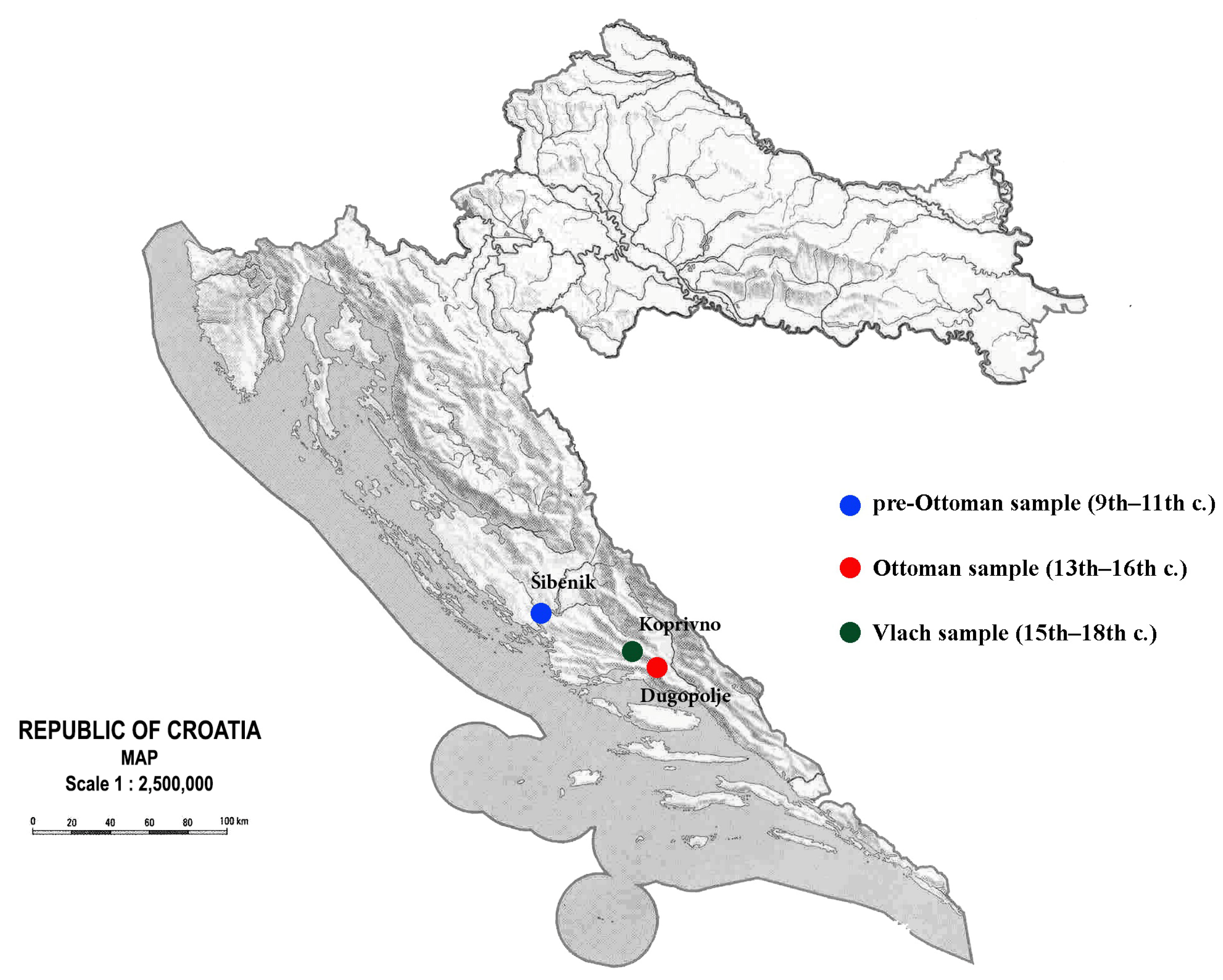
2.1. Sample Selection

This study was approved by the Institutional Research Ethics Committee of the School of Dental Medicine, University of Zagreb, Croatia (approval number: 5-PA-26-12/2016).
The research was conducted on 88 skulls from three chronologically consecutive samples, i.e., archaeological sites (Figure 1). Sample distribution by age and sex is presented in Table 1. The pre-Ottoman sample was excavated from the archaeological site Šibenik St. Lovre, which is located about 9 km east of the town of Šibenik. In all archaeological campaigns together, 350 skeletons were found [14], but this study included only skeletons from the earlier phase of the cemetery (from the 9th to the 11th century). The architecture of the tombs consisted of irregular stone slabs that form the floor and walls and cover the tomb [15]. Grave goods were rare and mostly consisted of rings, earrings, knives, and anklets [15,16,17].
The Ottoman sample (Table 1) originated from the site of Dugopolje—Vučipolje, which is located 15 km from the city of Split. The grave goods consisted of buttons, earrings, rings, pins, textiles, and beads and proved the use of the necropolis between the 13th and 16th centuries [10,13].
The Vlachs sample (Table 1) was from the village of Koprivno—kod Križa. The village of Koprivno is located northwest of Dugopolje and northeast of Klis (Figure 1). The graves were dated to the Early Modern Period (from the late 15th to the early 18th century). According to Gjurašin, most of the graves had an oval or rectangular crown of dry-stone masonry and contained goods that were quite common and modest, including mainly iron pins, buttons, parts of clothing, etc. Considering the historical sources, the characteristic grave architecture, and the fact that some of the graves contained Ottoman silver coins, it can be assumed that most of the graves belonged to the Vlach population [11,18].
For the purposes of this study, only adult specimens were analyzed. In this study, adults were defined as individuals older than 15 years of age. They were classified into one of two age categories: younger adults (15.0–39.9 years) and older adults (40+ years). Since the individuals included in the analysis also had a preserved postcranial skeleton, sex was determined based on pelvic [19] and cranial [20] morphology. Age at death was determined by using several methods that included the degree of ectocranial suture closure [21,22], dental wear [23], pubic symphysis morphology [23,24], pelvic auricular surface morphology [23], and sternal costal end morphology [25].

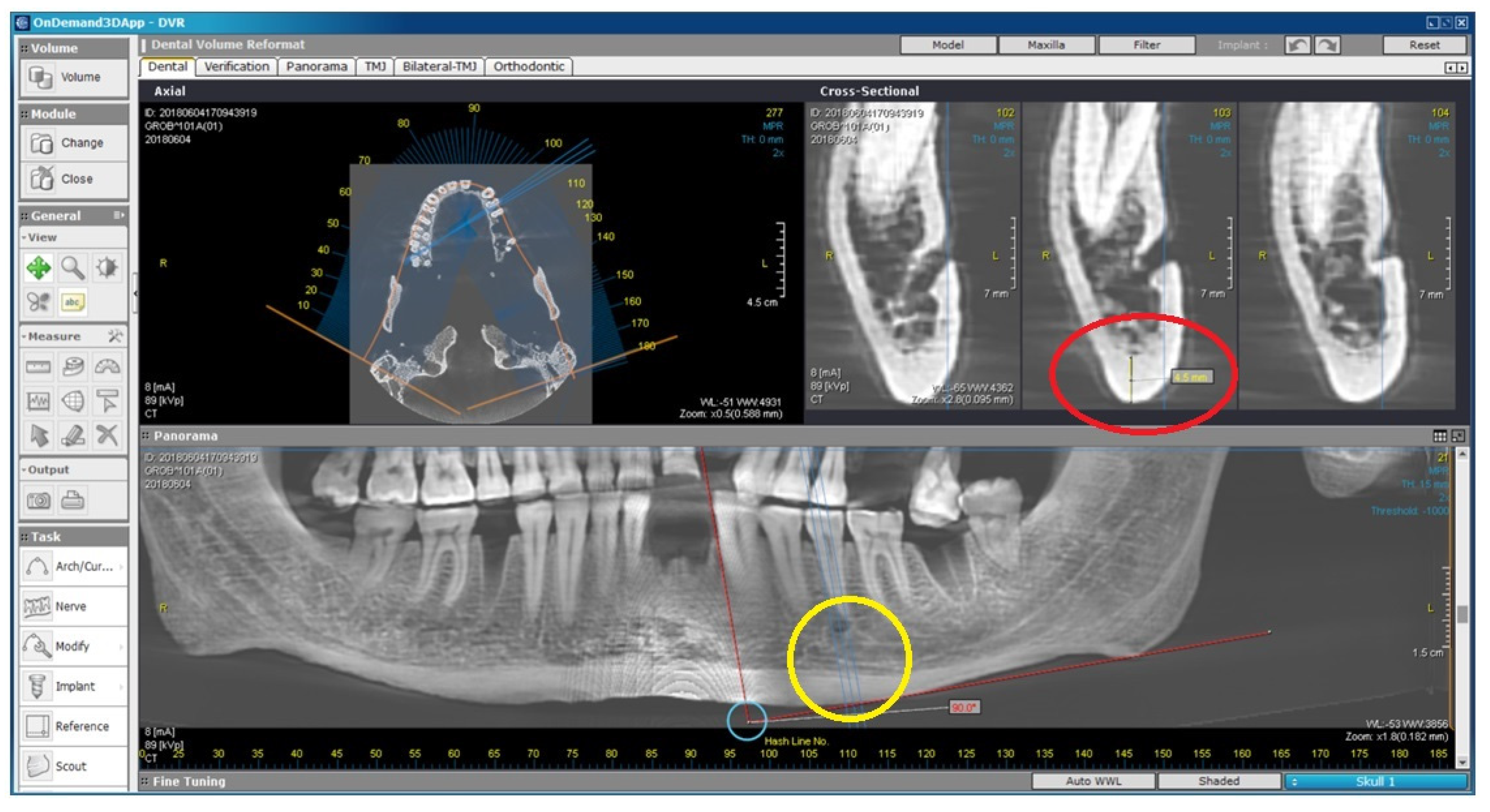
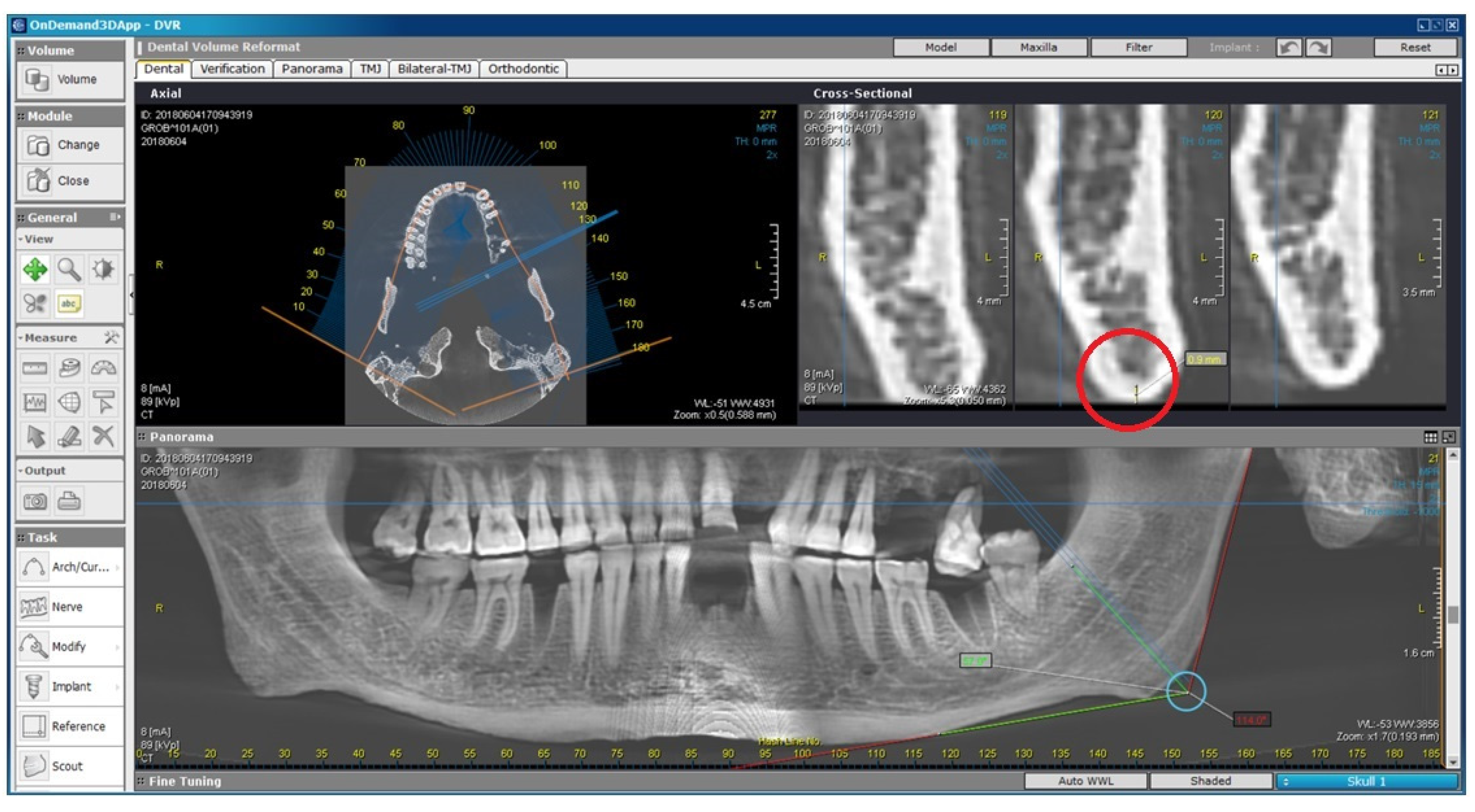
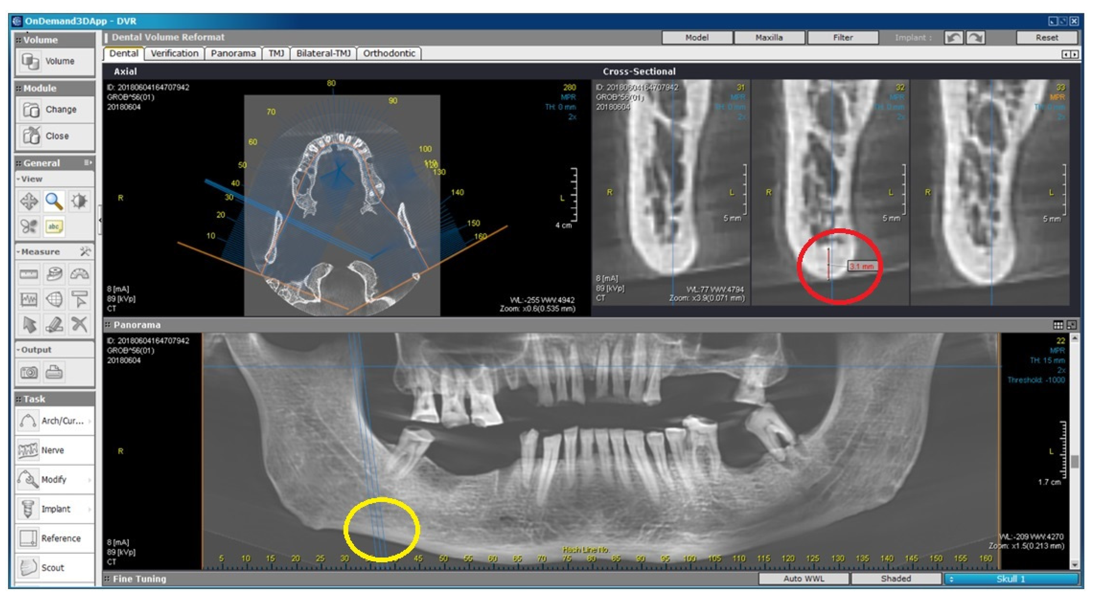
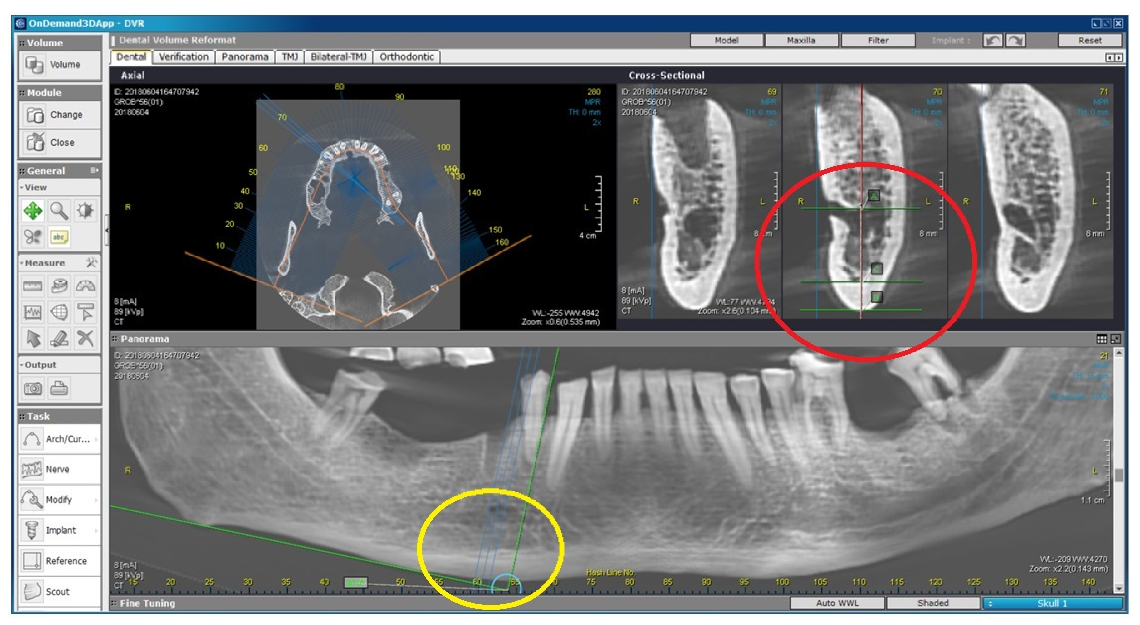
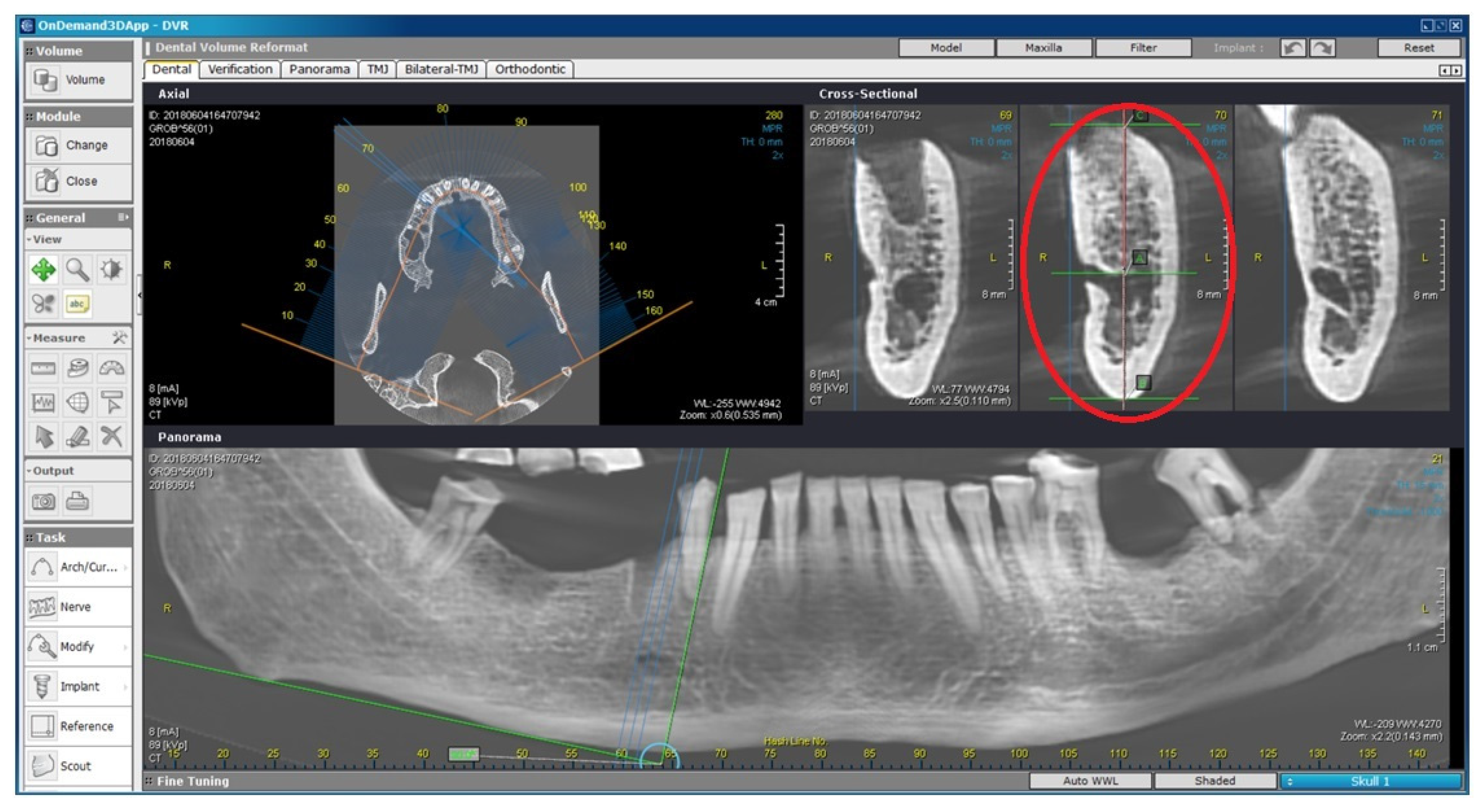
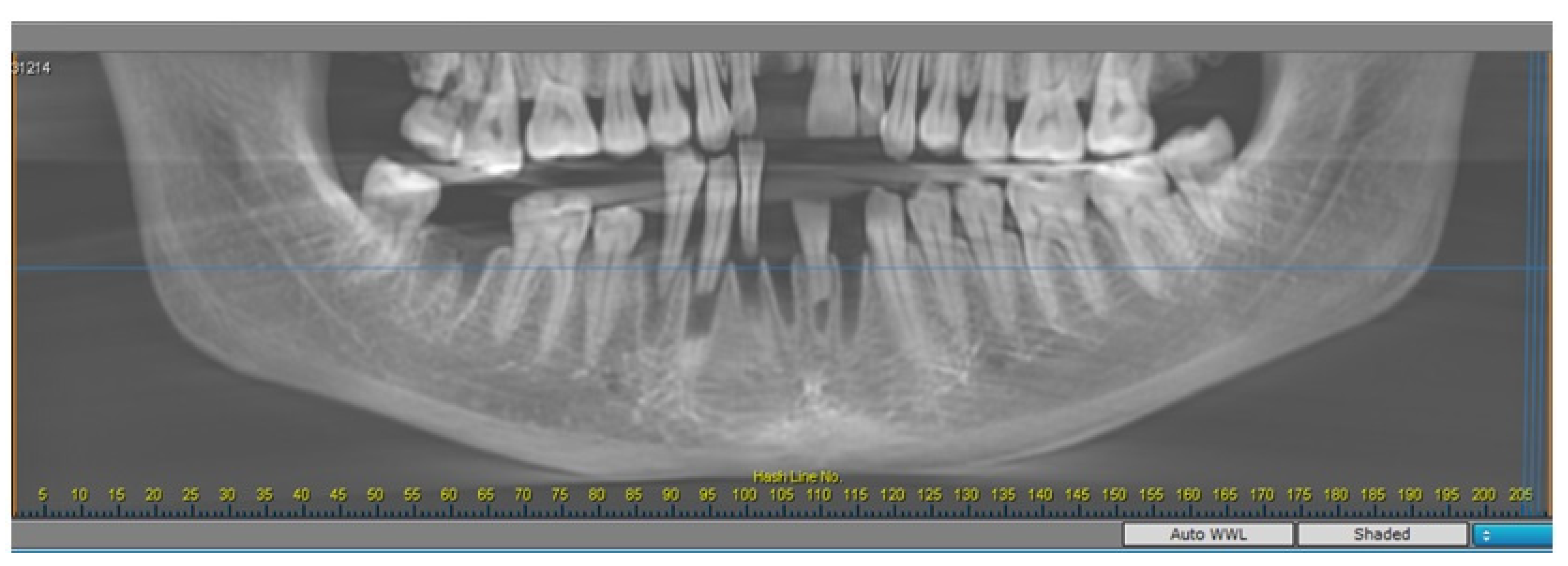
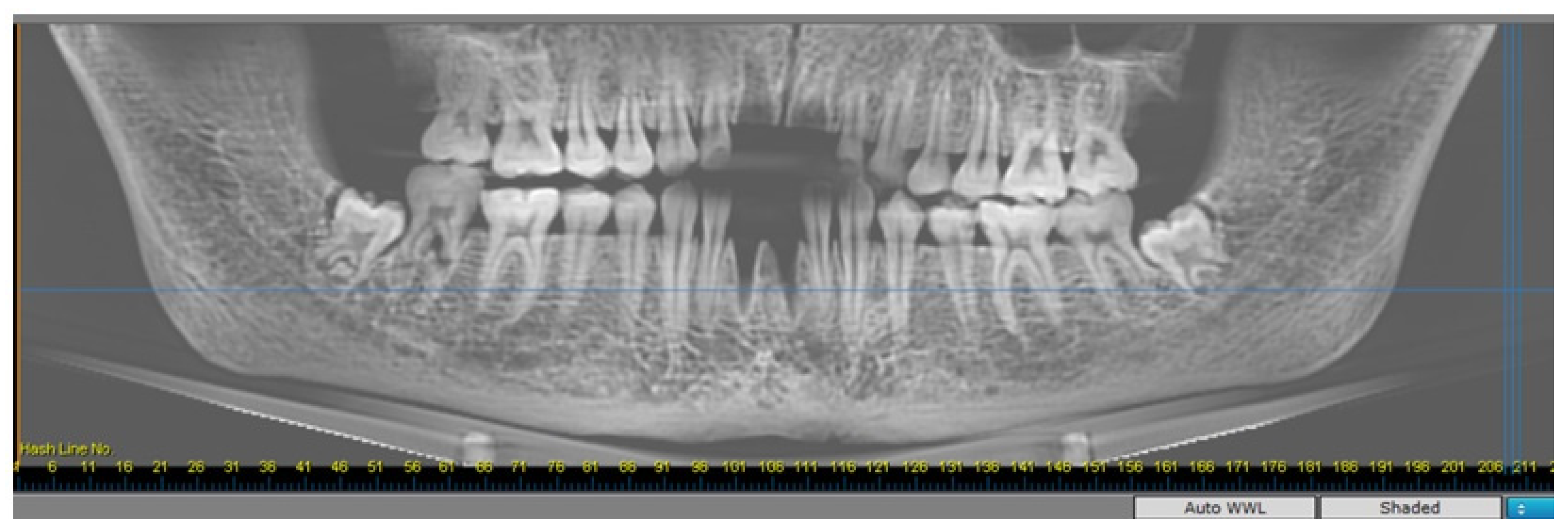
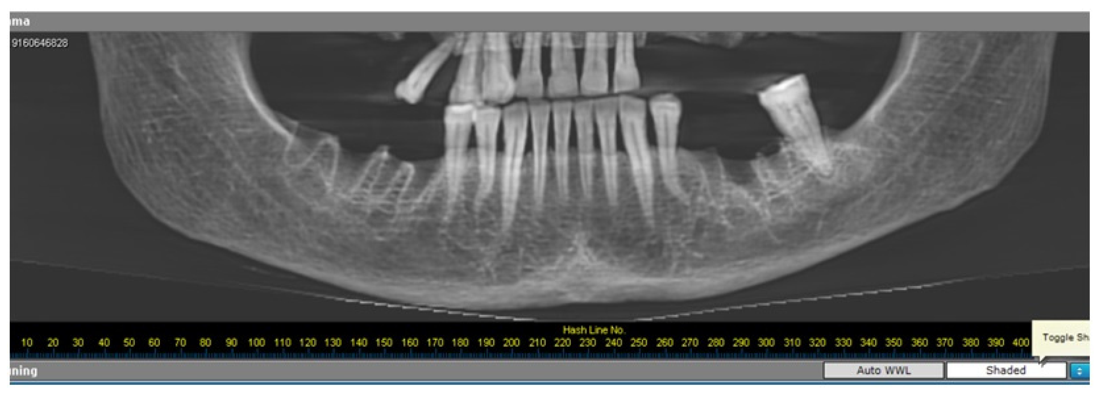
2.2. Radiomorphometric Assessments

The following measurements were made on the CBCT images of the mandibles: the mental index, gonion index, antegonion index, panoramic mandibular index, degree of resorption of the alveolar ridge, and the cortical index. CBCT images of the skulls were obtained with the Cranex3Dx device (2015, Soredex, Tuusula, Finland), and indices were measured bilaterally via the OnDemand3D software (Cybermed, Seoul, South Korea). Values that could not be measured accurately due to mandibular fracture, fragment misalignment, or a missing reference point were excluded. Three weeks after the first measurement, all indices on fifteen skulls (16.2% of the total number of skulls) were measured repeatedly by the same person (T.Č.) to evaluate the reliability of the measurements. All numerical variables had a high intraclass correlation score (above 0.8 and 0.9), which indicates good reliability of repeated measurements. With a nominal variable (MCI), there was no difference at all between the two measurements.
The mental index (MI) is the thickness of the mandibular cortex in millimeters measured on a line perpendicular to a tangent parallel to the inferior edge of the mandible and passing through the middle of the mental foramen (Figure 2) [26].
The gonion index (GI) is the thickness of the mandibular cortex in millimeters at the mandibular angle (Figure 3) [27].
The antegonion index (AI) is the thickness of the mandibular cortical bone in millimeters measured on a line parallel to the anterior edge of the mandibular ramus (Figure 4) [28].
The panoramic mandibular index (PMI) is the ratio between the thickness of the mandibular cortex and the distance between the center of the mental foramen and the inferior border of the mandible (Figure 5) [26].
The level of resorption of the alveolar ridge (M/M) is the ratio between the total height of the mandible and the distance between the center of the mental foramen and the lower edge of the mandible (Figure 6) [29].
The mandibular cortical (Klemetti) index (MCI) is a classification of the appearance of the inferior edge of the mandible distal to the mental foramen [30]. Three classes are distinguished: C1—the endosseous margin of the cortical bone is clear and sharp (Figure 7); C2—the endosseous margin contains semilunar defects and the appearance of fragmented parts of the margin (Figure 8); and C3—a remarkable porosity of the cortical bone layer (Figure 9). This index was evaluated in panoramic projection.

2.3. Statistical Analysis

Data were analyzed using the Statistical Package for Social Sciences (SPSS) version 20 (IBM Inc., Chicago, IL, USA). Parametric and nonparametric independent t tests, paired t tests, and one-way ANOVA were used to analyze the data, with statistical significance set at p < 0.05.

3. Results

There were no statistically significant differences in the values for the MI, GI, AI, PMI, and M/M between the left (L) and right (R) sides of the mandibles in the three sample groups. Regarding sex, statistically significant differences were found between males and females in the pre-Ottoman sample for MI (R) (t = 2.185046, df = 24, p < 0.05), in the Ottoman sample for MI (R) and MI (L) (Z = −2.595, p < 0.01; t = 4.276355, df = 28, p < 0.001) and M/M (L) (t = −3.040, df = 29, p = 0.005), and in the Vlach sample for MI (L) (t = 2.140975, df = 24, p < 0.05). In all cases, males had statistically significantly higher values than females except in the Ottoman sample for the M/M (L) value.
When comparing two age groups within samples, females from the younger age group in the Ottoman sample had significantly higher scores for MI (R) and AI (L) than females from the older age group (Z = −2.459, p < 0.05; t = 2.2449, df = 12, p < 0.05). Younger females from the Vlach sample had higher scores of borderline significance for MI (L) (t = 2.222, df = 0, p = 0.05) and a statistically significant higher GI (R) (t = 3.430, df = 6.848, p < 0.05) than older females. In contrast, there were no significant differences between age groups in men.
Comparison between samples showed that younger males from the pre-Ottoman sample had higher scores for AI (L) than younger males from the Ottoman sample but of borderline significance (p = 0.05). Similarly, males in the 40+ age category from the pre-Ottoman and the Vlach sample had significantly higher scores for AI (R) than males from the Ottoman sample and the same age category (p < 0.05 and p = 0.05, respectively). Younger females from the Vlach sample had significantly higher scores for GI (R) than younger females from the Ottoman sample (p < 0.05). Younger females from the pre-Ottoman sample also had significantly higher scores for GI (L) than younger females from the Ottoman sample (p < 0.05). Older females from the pre-Ottoman sample had significantly higher scores for GI (R) than older females from the Ottoman sample (p < 0.05).
Comparison of MCI categories between samples showed that the Vlach sample had significantly more C1 values than the pre-Ottoman (χ2 = 3.807, p = 0.05) and Ottoman samples (χ2 = 4.608, p < 0.05) (Table 2).
When comparing within samples, it is noticeable that in the pre-Ottoman and Ottoman samples, the mandibular cortical (Klemetti) index (MCI) predominantly showed the C2 category. In contrast, in the Vlach sample, there were significantly more mandibles with C1 categories than C3 categories (χ2 = 13.614, p < 0.01) and more C2 categories than C3 categories (χ2 = 6.345, p = 0.01). An analysis of MCI categories according to sex and age was not performed because of the limited sample size.

4. Discussion

Bone quality is a complex indicator of the overall condition of the body, as it is influenced by a variety of factors such as age, sex, hormonal status, diet, and others. Research on bone quality using mandibular indices has been applied in a variety of studies, mostly in the recent population. Because these measurements have often been shown to have a significant correlation with overall bone quality and are relatively simple and readily available on orthopantomographic images (radiomorphometry), they have numerous advantages over other methods that are destructive [31] or not so reliable for archaeological specimens (DEXA) [32]. In this study, we used the CBCT radiology technique, which offers the possibility of precise three-dimensional analysis of anatomical and pathological structures and allows more accurate measurements of mandibular indices compared with standard two-dimensional orthopantomograms.
As mentioned earlier, research on bone quality using mandibular indices is conducted mainly in recent populations. Mandibular indices can tell us a lot about the diet and subsistence strategies of people. Therefore, it seemed interesting to expand the knowledge about earlier populations from turbulent times such as the Middle Ages and the Early Modern Period on Croatian territory. To properly interpret the results, it is important to understand the political, social, and economic environment of the observed periods.
According to Goldstein, the Early Middle Ages in Croatia can be briefly described as a turbulent period marked by constant fighting. The battles with Venice and Byzantium continued into the Late Middle Ages. In the 13th century, a new enemy appeared on Croatia’s doorstep—the Mongols [8]. Although powerful and aggressive, they did not conquer any major fortified cities. The Late Middle Ages did not bring any relief either, as in 1463, the Ottomans conquered the medieval kingdom of Bosnia and made Croatia the next stop of their campaign [33]. From that moment on, Croatia fought with the Ottoman Empire for three centuries. The Ottoman strategy was for the Akinji to invade new areas before the advance of the regular Ottoman forces. The Akinji were an irregular light cavalry whose goal was to terrorize the local population into leaving their home territory [34]. They raided, captured livestock, took prisoners who were then sold into slavery, and attacked routes and trading centers to prevent food supplies and transportation [33,34]. In addition to the Akinji, the Martologs were also a major threat to the local population. According to Mažuran, they were a special Ottoman military order composed mainly of conquered Christians and Vlach shepherds from Bosnia. Their sources of income were also robbery and the capture of people [11].
After the emigration of the local population, the abandoned mountain regions were repopulated by the Vlach population. According to Kursar, the Vlachs were largely descendants of the autochthonous, Romanized, pre-Slavic Balkan populations such as Thracians, Illyrians, and Dacians who inhabited the mountainous regions of the central Balkans and practiced a nomadic or semi-nomadic lifestyle with transhumant pastoralism as their primary subsistence strategy [12].
A significantly different lifestyle (also regarding the dominant type of diet) that could be reflected in bone quality was characteristic for the pre-Ottoman population. The population of the Early Middle Ages was mainly engaged in agriculture and subsisted on cereals such as soft wheat, durum wheat, and barley [13,35]. According to Fabijanec, the coastal population also raised livestock [35]. It can be assumed that the subsistence strategy remained the same in the High and Late Middle Ages [36].
As mentioned above, the population of Vlach primarily engaged in transhumant pastoralism. Valentić claims that they raised cattle, sheep, and goats and made seasonal migrations with livestock. They also practiced agriculture, but this was only on a small scale. Their lack of grain production was compensated by increased meat and milk production [37].
Research by Adamić and Šlaus also confirmed that the late medieval population of Dugopolje and Vlach Koprivno had different diets—the population of Dugopolje consumed more carbohydrates and that of Koprivno more proteins [33].
The results of this study showed the expected higher value of cortex thickness in males compared to females. In addition, a statistically significant difference was confirmed between females of different age groups in the Ottoman and Vlach samples, with females in the older group having lower mandibular cortical thickness. These results indicate a possible trend that was present and that is consistent with the previously demonstrated relationship between aging and bone loss. [38]. Interestingly, this difference was not present in the male subjects, suggesting that age-related hormonal changes have stronger influence in females. The consequences are seen in a decrease in bone mass, which may also be reflected in the thickness of the mandibular cortex [39]. Previous studies of modern populations have shown that cortical thickness does not decrease as much with age in males as in females [40].
Among the males, an interesting significant difference in cortex thickness was found, with those in the younger age group from the pre-Ottoman and Vlach samples and those in the older age group from the Vlach sample having higher values than the Ottoman samples. This suggests a probable negative influence of some systemic or environmental factors on mandibular bone quality that were specific to the Ottoman sample.
In the Vlach sample, females had a higher alveolar ridge resorption (M/M) score. The alveolar ridge tends to remodel under the direct influence of physical stress. For this reason, pronounced resorption of the ridge occurs after tooth loss due to the lack of functional stimulation of the alveolar bone. In this study, we did not consider tooth loss due to the limited sample size, so this result may also be a consequence of this factor, which was not included in research.
The finding of higher GI values in the pre-Ottoman and Vlach samples compared to the Ottoman samples in groups of younger females again suggests some factors influencing the Ottoman sample that could be responsible for lower bone quality. This is supported by the statistically significant lower GI scores in the older sample of Ottoman females compared to the pre-Ottoman group. The negative Ottoman influences on political, social, economic, and demographic changes in medieval Croatia were described in research by Grgin (2002), in which the possible influence of food shortages at the end of the 15th century is mentioned [41].
The degree of erosion of the inner edge of the mandibular cortex (MCI) has been recognized as an indicator of higher risk for osteoporosis [42] and fractures [43] in modern populations. The present research analysis of MCI scores showed that the Vlach sample had the highest quality of cortical bone, with significantly more individuals in category C1 compared with the other two populations. This is consistent with the other findings of this research, confirming the possible beneficial effect of a protein-rich diet on bone quality.
Mays investigated cortical bone loss in a skeletal sample from a British medieval site using metacarpal radiographs [44]. Significant bone loss in females compared to males and in older females compared to younger individuals was found, which is concordant with our findings. As in our research, age-related changes were not found in males. In comparison to the recent population, lower cortical bone values were found; however, the same trend in bone loss in postmenopausal females was found.
In later research, Mays compared the British medieval sample with a modern Spitalfields sample from the 18th/19th century and found no difference in cortical thickness, although lifestyle significantly changed [45].
In recent research, Šerstņova et al. used novel methods including micro-CT and immunohistochemistry to analyze the bone quality of skeletons (humerus, radius, and ulna) from medieval and Late Modern Period Latvia. Lower bone quality was found in the medieval sample, which is consistent with our findings [46].
In our research, the modern digital technology of CBCT was used as noninvasive method for studying bone quality in archaeological samples. CBCT allowed for bone analysis in sagittal and panoramic projections, providing new insights and results that can be compared to modern populations.

5. Conclusions

This study, which measured the thickness and assessed the quality of mandibular cortical bone in three different archaeological samples, confirmed the significant influence of age and hormonal changes on bone quality in females, while in males, there were no age-related differences in bone thickness. Pre-Ottoman and Vlach populations had better mandibular bone quality in comparison to the Ottoman population, probably reflecting different lifestyles and dietary habits that influenced bone-remodeling processes, largely due to functional requirements. The lower values of the mandibular indices in the Ottoman sample may be an indicator of a specific factors related to general negative political, social, economic, and demographic changes in medieval Croatia that influenced this population.

Supplementary Materials

The following supporting information can be downloaded at: https://www.mdpi.com/article/10.3390/heritage7010008/s1, Research dataset “Datasheet Savic Pavicin Diachronic comparison of three historical skeletal series from Croatia with regard to mandibular bone quality”; Table S1: Mandibular index scores: the mental index (MI), gonion index (GI), antegonion index (AI), panoramic mandibular index (PMI), degree of resorption of the alveolar ridge (M/M), and cortical index (MCI). F cells are missing data.

Author Contributions

Conceptualization, I.S.P., A.A.H., J.D. and M.Š.; Formal analysis, A.A.H. and T.Č.; Funding acquisition, I.S.P., J.D. and T.L.; Investigation, I.S.P., A.A.H., T.Č., M.Š. and T.L.; Methodology, I.S.P., A.A.H., T.Č. and T.L.; Project administration, I.S.P.; Supervision, M.Š.; Validation, I.S.P. and T.Č.; Writing—original draft, I.S.P., A.A.H., T.Č., J.D. and A.Z.Ç.; Writing—review and editing, I.S.P., A.A.H., T.Č., J.D., M.Š., T.L. and A.Z.Ç. All authors have read and agreed to the published version of the manuscript.

Funding

This research was funded by the University of Zagreb, Croatia.

Data Availability Statement

The original dataset was uploaded as Supplementary Materials to this publication.

Conflicts of Interest

The authors declare no conflict of interest.

References

  1. Passos, J.S.; Gomes Filho, I.S.; Sarmento, V.A.; Sampaio, D.S.; Gonçalves, F.P.; Coelho, J.M.F.; Cruz, S.S.; Trindade, S.C.; Cerqueira, E.M. Women with Low Bone Mineral Density and Dental Panoramic Radiography. Menopause 2012, 19, 704–709. [Google Scholar] [CrossRef]
  2. Taguchi, A.; Suei, Y.; Ohtsuka, M.; Otani, K.; Tanimoto, K.; Ohtaki, M. Usefulness of Panoramic Radiography in the Diagnosis of Postmenopausal Osteoporosis in Women: Width and Morphology of Inferior Cortex of the Mandible. Dentomaxillofacial Radiol. 1996, 25, 263–267. [Google Scholar] [CrossRef]
  3. Savić Pavičin, I.; Dumančić, J.; Jukić, T.; Badel, T. The Relationship between Periodontal Disease, Tooth Loss and Decreased Skeletal Bone Mineral Density in Ageing Women. Gerodontology 2017, 34, 441–445. [Google Scholar] [CrossRef]
  4. Barr, C.; Sharafieh, R.; Schwarz, G.; Wu, R.; Klueh, U.; Kreutzer, D. Noninflammatory Stress-Induced Remodeling of Mandibular Bone: Impact of Age and Pregnancy. J. Oral Maxillofac. Surg. Off. J. Am. Assoc. Oral Maxillofac. Surg. 2021, 79, 1147–1155. [Google Scholar] [CrossRef]
  5. Robling, A.G.; Castillo, A.B.; Turner, C.H. Biomechanical and Molecular Regulation of Bone Remodeling. Annu. Rev. Biomed. Eng. 2006, 8, 455–498. [Google Scholar] [CrossRef]
  6. Pearson, O.M.; Lieberman, D.E. The Aging of Wolff’s ”Law”: Ontogeny and Responses to Mechanical Loading in Cortical Bone. Am. J. Phys. Anthropol. 2004, 125, 63–99. [Google Scholar] [CrossRef]
  7. Agarwal, S.C.; Grynpas, M.D. Bone Quantity and Quality in Past Populations. Anat. Rec. 1996, 246, 423–432. [Google Scholar] [CrossRef]
  8. Goldstein, I. Hrvatski Rani Srednji Vijek; Historiae; Novi Liber: Zagreb, Croatia, 1995. [Google Scholar]
  9. Kružić, K. Povijest Dugopolja i Njegovih Sela. In Zbornik Radova Općine Dugopolje; Gulin, A., Ed.; Općina Dugopolje: Zagreb/Dugopolje, Croatia, 2001; pp. 149–248. [Google Scholar]
  10. Gjurašin, H. Dugopolje—Vučipolje—Crkvine, Kasnosrednjovjekovno Groblje (Istraživanja 2004./2005. Godine). Starohrv. Prosvj. 2010, 3, 111–133. [Google Scholar]
  11. Adamić Hadžić, A. Comparative Analysis of Trauma on the Human Osteological Material from Croatia from Early Medieval to Early Modern Period. Ph.D. Thesis, University of Zadar, Zadar, Croatia, 2021. [Google Scholar]
  12. Kursar, V. Being an Ottoman Vlach. On Vlach Identity (Ies), Role and Status in Western Parts of the Ottoman Balkans (15th–18th Centuries). OTAM Ank. Üniversitesi Osman. Tarihi Araşt. Uygul. Merk. Derg. 2013, 24, 115–161. [Google Scholar]
  13. Adamić Hadžić, A. Izloženost Koštanim Ozljedama Ratarskih i Stočarskih Populacija Srednjovjekovne i Ranosrednjovjekovne Hrvatske. Archaeol. Adriat. 2022, 16, 233–265. [Google Scholar] [CrossRef]
  14. Vyroubal, V. Antropološka Analiza Zdravlja i Kvalitete Života u Kontekstu Socijalnog Statusa. Ph.D.Thesis, University of Zadar, Zadar, Croatia, 2014. [Google Scholar]
  15. Krnčević, Ž. Sveti Lovre–Šibensko Donje Polje, Sustavna Arheološka Istraživanja u Godini 1986. Obavijesti HAD-A 1997, 2, 70–72. [Google Scholar]
  16. Krnčević, Ž. Stoljeća Arheologije Na Šibenskom Području; Muzej grada Šibenika: Šibenik, Croatia, 1995; 62p. [Google Scholar]
  17. Krnčević, Ž. Područje Šibenske Županije Od Pretpovijesti Do Srednjeg Vijeka: Srednjovjekovna Arheološka Nalazišta Na Šibenskom Području. Izdanja HAD-a 1998, 19, 197–226. [Google Scholar]
  18. Gjurašin, H. Nastavak Zaštitnih Istraživanja Arheoloških Lokaliteta u Selu Koprivno—Općina Dugopolje—Sjeveroistočno Od Klisa u Godini 2002. Obavijesti Hrvat. Arheol. Druš. 2002, 3, 151–153. [Google Scholar]
  19. Phenice, T.W. A Newly Developed Visual Method of Sexing the Os Pubis. Am. J. Phys. Anthropol. 1969, 30, 297–301. [Google Scholar] [CrossRef]
  20. Krogman, W.; Iscan, M. The Human Skeleton in Forensic Medicine; Charles C Thomas Publisher, Ltd.: Springfield, IL, USA, 1986. [Google Scholar]
  21. Meindl, R.S.; Lovejoy, C.O.; Mensforth, R.P.; Carlos, L.D. Accuracy and Direction of Error in the Sexing of the Skeleton: Implications for Paleodemography. Am. J. Phys. Anthropol. 1985, 68, 79–85. [Google Scholar] [CrossRef]
  22. Lovejoy, C.O. Dental Wear in the Libben Population: Its Functional Pattern and Role in the Determination of Adult Skeletal Age at Death. Am. J. Phys. Anthropol. 1985, 68, 47–56. [Google Scholar] [CrossRef]
  23. Lovejoy, C.O.; Meindl, R.S.; Pryzbeck, T.R.; Mensforth, R.P. Chronological Metamorphosis of the Auricular Surface of the Ilium: A New Method for the Determination of Adult Skeletal Age at Death. Am. J. Phys. Anthropol. 1985, 68, 15–28. [Google Scholar] [CrossRef]
  24. Brooks, S.; Suchey, J.M. Skeletal Age Determination Based on the Os Pubis: A Comparison of the Acsádi-Nemeskéri and Suchey-Brooks Methods. Hum. Evol. 1990, 5, 227–238. [Google Scholar] [CrossRef]
  25. Işcan, M.Y.; Loth, S.R.; Wright, R.K. Age Estimation from the Rib by Phase Analysis: White Males. J. Forensic Sci. 1984, 29, 1094–1104. [Google Scholar]
  26. Horner, K.; Devlin, H. The Relationship between Mandibular Bone Mineral Density and Panoramic Radiographic Measurements. J. Dent. 1998, 26, 337–343. [Google Scholar] [CrossRef]
  27. Devlin, H.; Horner, K. Mandibular Radiomorphometric Indices in the Diagnosis of Reduced Skeletal Bone Mineral Density. Osteoporos. Int. 2002, 13, 373–378. [Google Scholar] [CrossRef]
  28. Ledgerton, D.; Horner, K.; Devlin, H.; Worthington, H. Radiomorphometric Indices of the Mandible in a British Female Population. Dentomaxillofacial Radiol. 1999, 28, 173–181. [Google Scholar] [CrossRef]
  29. Taguchi, A.; Tanimoto, K.; Suei, Y.; Otani, K.; Wada, T. Oral Signs as Indicators of Possible Osteoporosis in Elderly Women. Oral Surg. Oral Med. Oral Pathol. Oral Radiol. Endod. 1995, 80, 612–616. [Google Scholar] [CrossRef]
  30. Klemetti, E.; Vainio, P.; Lassila, V.; Alhava, E. Cortical Bone Mineral Density in the Mandible and Osteoporosis Status in Postmenopausal Women. Scand. J. Dent. Res. 1993, 101, 219–223. [Google Scholar] [CrossRef]
  31. Gonzalez-Reimers, E.; Mas-Pascual, M.A.; Arnay-de-la-Rosa, M.; Velasco-Vázquez, J.; Santolaria-Fernández, F.; Machado-Calvo, M. Noninvasive Estimation of Bone Mass in Ancient Vertebrae: Noninvasive Diagnosis of Osteopenia. Am. J. Phys. Anthropol. 2004, 125, 121–131. [Google Scholar] [CrossRef]
  32. Kneissel, M.; Boyde, A.; Hahn, M.; Teschler-Nicola, M.; Kalchhauser, G.; Plenk, H. Age- and Sex-Dependent Cancellous Bone Changes in a 4000y BP Population. Bone 1994, 15, 539–545. [Google Scholar] [CrossRef]
  33. Adamić, A.; Šlaus, M. Comparative Analysis of Dental Health in Two Archaeological Populations from Croatia: The Late Medieval Dugopolje and Early Modern Vlach Population from Koprivno. Bull. Int. Assoc. Paleodont. 2017, 11, 11–22. [Google Scholar]
  34. Goodwin, G. The Janissaries; Saqi essentials; Saqi: London, UK, 2006. [Google Scholar]
  35. Fabijanec, F.S. Gospodarstvo, Nova Zraka u Europskom Svjetlu. In Hrvatske Zemlje u Ranome Srednjem Vijeku (oko 550–oko 1150); Nikolić Jakus, Z., Ed.; Matica Hrvatska: Zagreb, Croatia, 2016; pp. 133–158. [Google Scholar]
  36. Šlaus, M.; Novak, M.; Bedić, Ž.; Strinović, D. Bone Fractures as Indicators of Intentional Violence in the Eastern Adriatic from the Antique to the Late Medieval Period (2nd–16th Century AD). Am. J. Phys. Anthropol. 2012, 149, 26–38. [Google Scholar] [CrossRef]
  37. Valentić, M. Osmanski Ratovi i Hrvatska Dijaspora. Senj. Zb. 1990, 17, 45–60. [Google Scholar]
  38. Khosla, S. Pathogenesis of Age-Related Bone Loss in Humans. J. Gerontol. A. Biol. Sci. Med. Sci. 2013, 68, 1226–1235. [Google Scholar] [CrossRef]
  39. Savic Pavicin, I.; Dumancic, J.; Jukic, T.; Badel, T.; Badanjak, A. Digital Orthopantomograms in Osteoporosis Detection: Mandibular Density and Mandibular Radiographic Indices as Skeletal BMD Predictors. Dentomaxillofacial Radiol. 2014, 43, 20130366. [Google Scholar] [CrossRef]
  40. Roberts, M.; Yuan, J.; Graham, J.; Jacobs, R.; Devlin, H. Changes in Mandibular Cortical Width Measurements with Age in Men and Women. Osteoporos. Int. 2011, 22, 1915–1925. [Google Scholar] [CrossRef]
  41. Grgin, B. The Ottoman Influences on Croatia in the Second Half of the Fifteenth Century. Povij. Pril. 2002, 21, 87–102. [Google Scholar]
  42. Yamada, S.; Uchida, K.; Iwamoto, Y.; Sugino, N.; Yoshinari, N.; Kagami, H.; Taguchi, A. Panoramic Radiography Measurements, Osteoporosis Diagnoses and Fractures in Japanese Men and Women. Oral Dis. 2015, 21, 335–341. [Google Scholar] [CrossRef]
  43. Jonasson, G.; Hassani-Nejad, A.; Hakeberg, M. Mandibular Cortical Bone Structure as Risk Indicator in Fractured and Non-Fractured 80-Year-Old Men and Women. BMC Oral Health 2021, 21, 468. [Google Scholar] [CrossRef]
  44. Mays, S.A. Age-Dependent Cortical Bone Loss in a Medieval Population. Int. J. Osteoarchaeol. 1996, 6, 144–154. [Google Scholar] [CrossRef]
  45. Mays, S. Age-Dependent Cortical Bone Loss in Women from 18th and Early 19th Century London. Am. J. Phys. Anthropol. 2000, 112, 349–361. [Google Scholar] [CrossRef]
  46. Šerstņova, K.; Edelmers, E.; Zolovs, M.; Pilmane, M. Comparison of Bone Quality in Middle Ages and Late Modern Period Human Skeletons from Latvia. Heritage 2023, 6, 5329–5346. [Google Scholar] [CrossRef]
Figure 1. Map of Croatia with locations showing origins of all three samples.
Heritage 07 00008 g001
Figure 2. Measurement of the mental index (MI) on the CBCT scan. The MI was measured by drawing a line in the panoramic projection denoting the inferior border of the mandible and then a second line in the sagittal projection perpendicular to it and passing through the center of the mental foramen (circled in yellow). The cortical bone of the mandible was measured in the sagittal projection on this line (circled in red).
Heritage 07 00008 g002
Figure 3. Measurement of the gonion index (GI) on the CBCT scan. The GI was measured along the line bisecting the angle formed by the tangent parallel to the inferior edge of the mandible and the tangent parallel to the posterior edge of the ramus of the mandible (circled in blue). The thickness of the mandibular cortex at the mandibular angle was measured in the sagittal projection (circled in red).
Heritage 07 00008 g003
Figure 4. Measurement of the antegonion index (AI) on the CBCT scan. The AI was measured on a line parallel to the anterior edge of the mandibular ramus (circled in yellow) in the sagittal projection (circled in red).
Heritage 07 00008 g004
Figure 5. Measurement of the panoramic mandibular index (PMI) on the CBCT scan. The PMI was measured so that the line drawn in the panoramic projection was perpendicular to the inferior edge of the mandible and passed through the center of the mental foramen (circled in yellow). Then, the thickness of the mandibular cortex (BC) and the distance between the line passing through the center of the mental foramen and the inferior edge of the mandible (AB) were measured in the sagittal projection (circled in red). Finally, the ratio of the two assessments was determined as PMI = BC/AB.
Heritage 07 00008 g005
Figure 6. Measurement of the level of resorption of the alveolar ridge (M/M) on the CBCT scan. The M/M was calculated as the ratio between the total height of the mandible (BC) and the distance between the center of the mental foramen and the lower edge of the mandible (AB) (circled in red). M/M = BC/AB.
Heritage 07 00008 g006
Figure 7. C1 class of the MCI classification: clear and sharp endosseous margin of the cortical bone.
Heritage 07 00008 g007
Figure 8. C2 class of the MCI classification: endosseous margin contains semilunar defects.
Heritage 07 00008 g008
Figure 9. C3 class of the MCI classification: significant degradation in the cortical bone layer.
Heritage 07 00008 g009
Table 1. Age and sex distribution of samples.
SampleAgeSexTotal
MaleFemale
Pre-Ottoman15.0–39.97714
40+6713
Total131427
Ottoman15.0–39.991120
40+6612
Total151732
Vlach15.0–39.9369
40+14620
Total171229
Total15.0–39.9192443
40+261945
Total454388
Table 2. Distribution according to sex and sample of categories of MCI for left side (L).
MCI (L)
Pre-Ottoman Sample
C1C2C3Total
Males5 (38.5%)5 (38.5%)3 (23.1%)13 (48.1%)
Females2 (14.3%)9 (64.3%)3 (21.4%)14 (51.9%)
Total7 (25.9%)14 (51.9%)6 (22.2%)27 (100.0%)
Ottoman sample
C1C2C3Total
Males2 (13.3%)8 (53.3%)5 (33.3%)15 (46.9%)
Females6 (35.3%)10 (58.8%)1 (5.9%)17 (53.1%)
Total8 (25.0%)18 (56.3%)6 (18.8%)32 (100.0%)
Vlach sample
C1C2C3Total
Males10 (58.8%)7 (41.2%)0 (0.0%)17 (58.6%)
Females6 (50.0%)4 (33.3%)2 (16.7%)12 (41.4%)
Total16 (55.2%)11 (37.9%)2 (6.9%)29 (100.0%)
Disclaimer/Publisher’s Note: The statements, opinions and data contained in all publications are solely those of the individual author(s) and contributor(s) and not of MDPI and/or the editor(s). MDPI and/or the editor(s) disclaim responsibility for any injury to people or property resulting from any ideas, methods, instructions or products referred to in the content.

