Article

Biological Deterioration of an Inca Monument at High Altitude in the Andean Range: A Case Study from Ingapirca’s Temple of the Sun (Ecuador)

by
Luis Andrés Yarzábal
1,2,*,
Lenys Buela
1,
Asunción de los Ríos
3,
Diana Peláez
4,
Martha Romero
5,
Fernando Espinoza
5,
Alisson Samantha Torres
1,
Gina Maritza Medina
1,
Jaqueline Gabriela Landi
1 and
Marco Vinicio Tapia
1
1
Carrera de Bioquímica y Farmacia, Unidad de Salud y Bienestar, Universidad Católica de Cuenca, Av. Las Américas, Cuenca 010101, Ecuador
2
Laboratorio de Microbiología Molecular y Biotecnología, Facultad de Ciencias, Universidad de Los Andes, Av. Alberto Carnevalli, Mérida 5101, Venezuela
3
Department of Biogeochemistry and Microbial Ecology, National Museum of Natural Sciences (MNCN-CSIC), 28006 Madrid, Spain
4
Centro de Investigación, Innovación y Transferencia de Tecnología (CIITT), Universidad Católica de Cuenca, Ricaurte 010162, Ecuador
5
Unidad de Laboratorio y Análisis, Instituto Nacional del Patrimonio Cultural, Quito 170520, Ecuador
*
Author to whom correspondence should be addressed.
Heritage 2022, 5(3), 2504-2518; https://doi.org/10.3390/heritage5030130
Submission received: 9 August 2022 / Revised: 24 August 2022 / Accepted: 29 August 2022 / Published: 4 September 2022
(This article belongs to the Section Archaeological Heritage)

Abstract

:
Scientific studies concerning the causes and consequences of the biodeterioration of stone monuments located at high altitudes in permanently cold, mountainous regions are scarce. For that reason, this study aimed to detect and identify the bacteria involved in the deterioration of this type of monument. To achieve this goal, we focused on the most important archeological Inca site in the Ecuadorian Andes: Ingapirca’s Temple of the Sun, built approximately 500 years ago at 3.100 m.a.s.l. We first examined the stone surfaces of the temple by scanning electron microscopy and showed the detrimental impact on the mineral structure of the green andesite mineral used to build the temple, caused by crustose lichen thalli and heterotrophic bacteria. Then, we isolated, characterized, and identified several of these bacteria. Most of them multiplied at a wide range of temperatures, from 4 °C to 30 °C, and were thus considered eurypsychrophiles. Actinobacteria and Proteobacteria species dominated the culturable fraction of this community. Several isolates produced metabolites that solubilized mineral phosphates at low temperatures; others solubilized iron-containing mineral fractions in the green andesite rock when tested in vitro. To the best of our knowledge, this is the first report related to the biodeterioration of an Inca monument at such an altitude in the Andes range.

1. Introduction

Biodeterioration refers to the damage caused by micro- and macroorganisms to the materials used to produce or build any kind of artifact or monument [1]. In the particular context of historic or archaeological monuments, this process is responsible for the gradual modification and destruction of the aesthetics and chemical structure of their constituent mineral blocks [2,3].
Many studies have explored the causes and consequences of the biodeterioration of archaeological monuments, as well as the ways of preventing microbes from exerting their detrimental effects [4,5,6]. These studies provide information concerning the main groups of microorganisms involved in these processes and the mechanisms through which they are able to colonize the stone substrates and fragilize their structure in order to penetrate deep inside massive blocks of stone.
Among the most studied stone monuments in Latin America, from a biodeterioration point of view, are those located in humid tropical and sub-tropical areas [7]. These include, for instance, Mayan and Aztec monuments (in Mexico and Central America) [8,9] or colonial monuments in Brazil and Venezuela [10,11]. On the contrary, with the exception of a few data concerning the biodeterioration of Machu Picchu [12,13], Inca monuments located at high altitude in permanently cold, mountainous regions have been much less studied. This is the case for Ingapirca, considered the most important archeological site in the Ecuadorian Andes and built 3.100 m.a.s.l. approximately 500 years ago. In this complex of buildings—which include observatories, storage rooms, and several chambers—stands out an oval-shaped structure: the “Temple of the Sun”, also named “The Castle” or “The Elipse”.
This temple was built as the result of a joint effort between the Incas and the Cañaris, the original inhabitants of this region. The temple was built with green andesite blocks, following the distinctive style of Inca monuments and featuring precisely cut and shaped massive stones, closely fitted together without mortar.
Ingapirca’s Temple of the Sun has suffered from the detrimental activity of microbes growing on its surfaces and exerting their chemical and mechanical activity for centuries. Several campaigns have been deployed since the 1990s to try to prevent further deterioration of the Temple of the Sun; the treatments applied include the rigorous cleaning of the blocks’ surfaces using either abrasive tools (wire hand brushes) or wood spatulas, to remove all visible traces of lichens and mosses. Unfortunately, and contrarily to what was expected, the results of these efforts were even more destructive to the structure of the temple (INPC Report). Therefore, in order to explore the causes of the biodeterioration process of the Temple of the Sun, we performed a survey to detect and identify the microbial actors behind the biodeterioration of Ingapirca’s main monument. We present here the first report concerning the potential detrimental effect of the culturable bacterial fraction of this community on the structure of the rocks used to build the Temple of the Sun.

2. Materials and Methods

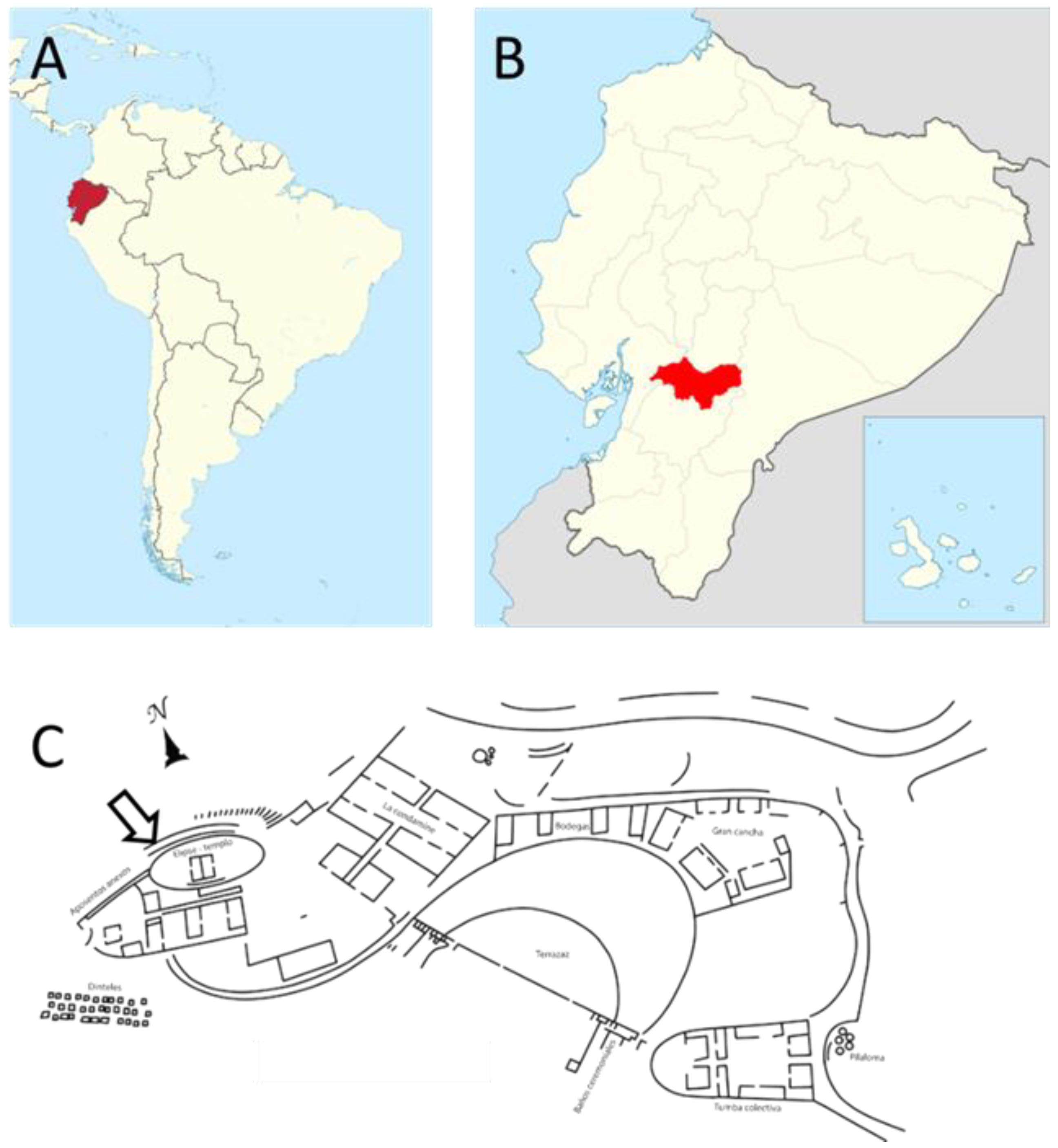
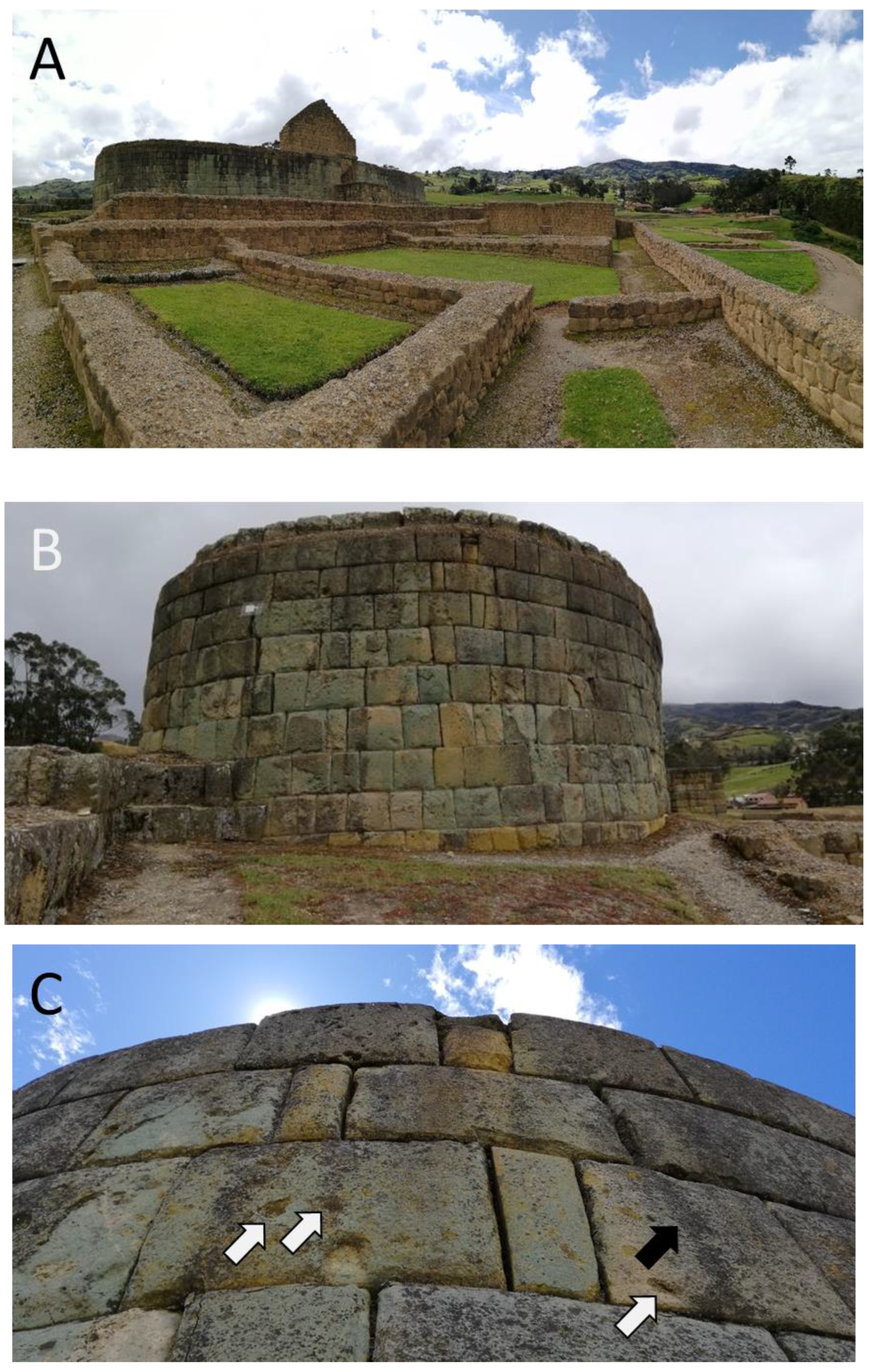
2.1. Field Location and Microclimate

Ingapirca’s archaeological complex is located in the province of Cañar, some 80 km from Cuenca city (2°32′54.9″ S and 78°52′18.7″ W) (Figure 1). The site is characterized by a large diurnal temperature range (from 10 to 36 °C), with a mean annual temperature of 12.3 °C. The humidity may also vary daily across a wide range, from 11% to 77%, which means that the dew point can be reached often, mainly at sunset and before sunrise [14]. Thus, in addition to the frequent rainfall, water condenses very often on the walls of the monuments and contributes to their deterioration.
Ingapirca’s complex was built approximately 500 years ago, and it lies 3.100 m above sea level. Its most important structure, the Temple of the Sun, is an elliptical structure built on top of an ancient Cañari culture ceremonial rocky outcrop. The monument was built in the Inca way, without mortar, by positioning together green andesite blocks chiseled and fashioned to fit together perfectly (Figure 2). Andesite is an extrusive rock of volcanic origin, mainly composed of plagioclase and pyroxene [14,15]. The rock is porphyritic in texture and presents crystals of various sizes. The open porosity of the constituent blocks varies between 4% and 12%, and the majority of these blocks are highly weathered on their surfaces due to a combination of climatic, anthropic, and microbial factors.

2.2. Rock Sample Collection

Fallen fragments of rock (green andesite), detached recently from the green andesite blocks used to build the Temple of the Sun, were collected during field studies. The small-sized samples (i.e., 1.5–8 cm length; 1–4 cm width) were detected a few centimeters away from the monument’s walls, on the soil surface, and many were heavily colonized by lichens, as were the surfaces of many stone blocks (Figure 2C). The collected samples were used to perform optical and electron microscopy observations (see below).

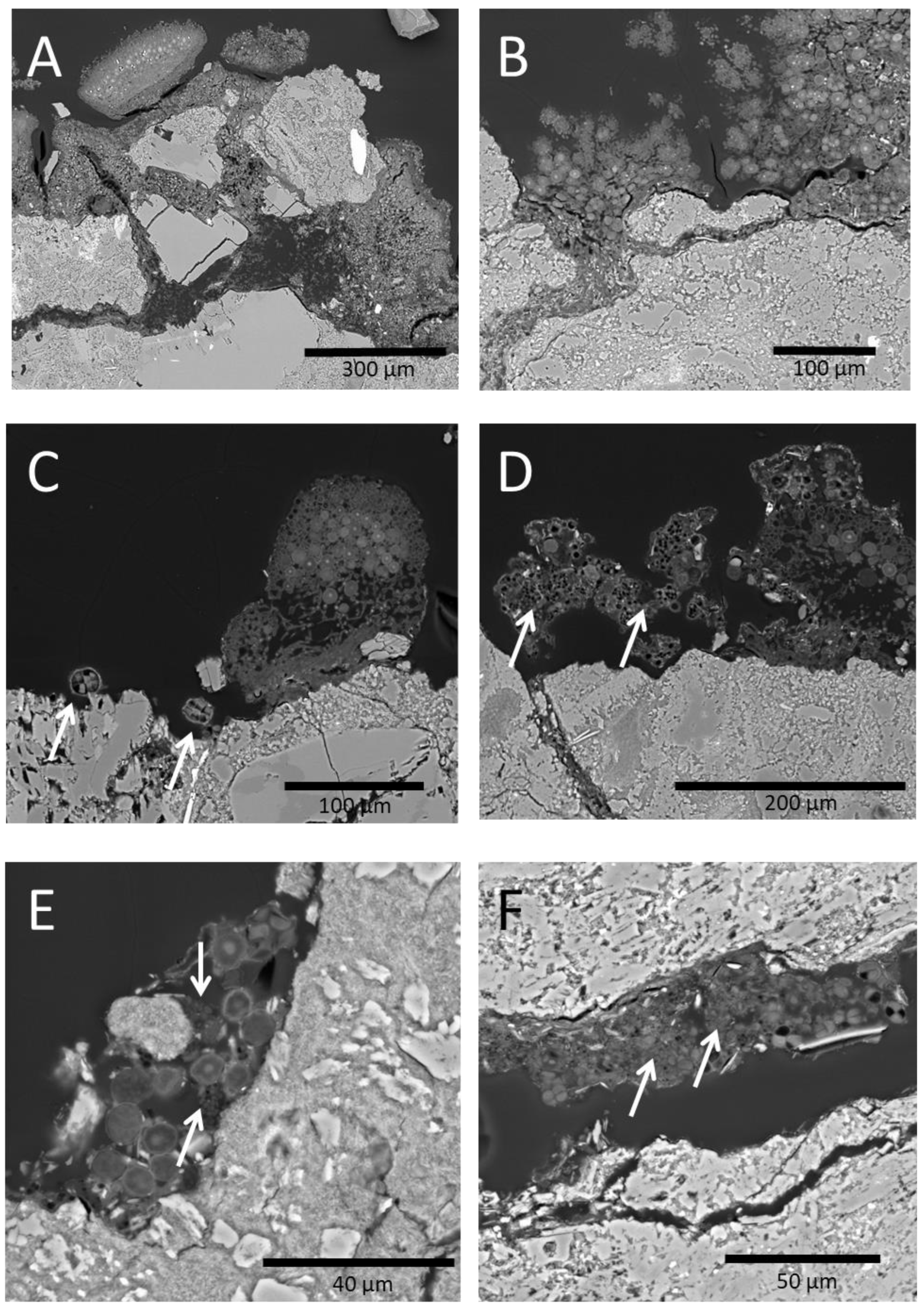
2.3. Scanning Electron Microscopy Analysis of Stone Samples

The characterization of microscale biological colonization was achieved using the SEM-BSE technique [16]. Briefly, colonized rock fragments were fixed in glutaraldehyde (3% v/v.) and osmium tetroxide solutions (1% w/v), dehydrated in a graded ethanol series, embedded in LR white resin, and finely polished. The samples were observed in back scattered electron mode using a FEI INSPECT microscope after being carbon coated.

2.4. Sampling and Isolation of Bacteria Colonizing Andesite Blocks’ Surface

Cotton swabs, moistened with sterile saline solution, were used to collect samples by gently rubbing them over a surface area of approximately 7.4 cm2 (equivalent to the surface area of a half-dollar coin) on different blocks in the walls of the Temple of the Sun. Sites exhibiting signs of deterioration that were located in the walls oriented towards the four cardinal points were chosen for this sample collection. The biological material collected was suspended in 2 mL of sterile saline solution and transported to the lab (located 90 min away) in a cooler at 4–8 °C. Once arrived, the suspensions were serially diluted and plated onto nutrient agar (¼ strength), Czapek’s medium, and potato dextrose agar (PDA). The plates were incubated at 12 °C for several days, until colonies appeared on the surface. Each colony was purified by re-streaking several times in the same medium. Once their purity had been verified, each isolate was stored in nutrient agar slants at 4 °C and in 20% glycerol at −20 °C.

2.5. Characterization of Bacterial Isolates

Morphological characteristics of bacterial colonies and cells were recorded for each selected isolate. Additionally, several standard tests were conducted, including Gram staining, a catalase test, an oxidative/fermentative test, and a fluorescence test. The temperature growth range of the isolates was determined by spotting 10 µL of each bacterial suspension, adjusted to 0.5 McFarland units, onto the surface of nutrient agar plates, followed by incubation at 4, 12, 20, and 30 °C and visual inspection of the plates.

2.6. Identification of Bacterial Isolates

Culture and morphological characteristics of bacterial isolates were determined following standard microbiological procedures. Bacteria were grouped and partially identified on the basis of Gram staining and biochemical tests described in Bergey’s Manual of Systematic Bacteriology [17,18].

2.7. Screening of the Mineral-Solubilization Abilities of Bacterial Isolates

The ability of the bacterial isolates to solubilize inorganic minerals such as tri-calcium phosphate (TCP) was monitored at different temperatures. For this, we used a double-layer agarized NBRIP medium (glucose, 10 gL−1; Ca3(PO4)2, 5 gL−1; MgCl2·6H2O, 5 gL−1; MgSO4·7H2O, 0.25 gL−1; KCl, 0.2 gL−1; (NH4)2SO4, 0.1 gL−1; Agar 20 gL−1) [19]. Bacterial suspensions of pure isolates, previously adjusted to 0.5 McFarland units, were spotted onto the surface of each plate (10 µL), and the plates were incubated for 3–5 days at 4, 12, 20, and 30 °C. Isolates producing clear halos around the colonies were considered to have TCP-solubilizing activity.

2.8. In Vitro Simulation of Green Andesite Deterioration Process

Twenty milliliters of liquid NBRIP salts (i.e., NBRIP medium devoid of calcium phosphate), supplemented with 10 gL−1 glucose as the sole source of carbon and 1.0 g green andesite particles (0.075–0.150 mm), were inoculated with selected bacterial strains that had been previously shown to solubilize inorganic minerals (see Section 2.7). Inocula were prepared by growing the isolates for five days in NBRIP medium at 20 °C, followed by washing the cells twice via centrifugation with NBRIP salts (1×) (to remove most traces of soluble P); the resuspension of the bacteria in the same salts; and inoculation with a 1:50 inoculum (v/v), previously adjusted to 0.5 O.D. units at 600 nm. The cultures were incubated at 20 °C for six days, with sporadic agitation several times a day. At the end of the incubation period, the supernatant was collected by centrifugation at 5000× g for 15 min, filtered through 0.2 µm Millipore ultrafilters, acidified with HCl, and subjected to FAAS (see below). All experiments were performed in triplicate.

2.9. Flame Atomic Absorption Spectroscopy (FAAS) Analysis of Solubilized Minerals

Analysis of metal concentration in the supernatant of the previous cultures was performed by flame atomic absorption spectroscopy (FAAS) with a AA-7000 spectrophotometer (Shimadzu Corporation, Tokyo, Japan), equipped with a hollow monoelement cathode lamp (Hollow Cathode Lamp) for each element analyzed (Na, Ca, Mg, and Fe). An air–acetylene flame was used with a ratio ranging from 0.8 Lmin−1 to 4.0 Lmin−1 and from 13.5 Lmin−1 to 17.5 Lmin−1 for air and acetylene, respectively, to measure Na, Mg, and Fe. A nitrous oxide–acetylene flame was used with a ratio ranging from 5.8 Lmin−1 to –9.0 Lmin−1 and from 10.0 Lmin−1 to 12.5 Lmin−1 for nitrous oxide and acetylene, respectively, to measure Ca. Chemicals used in this study to prepare stock solutions were of analar grade.

2.10. Statistical Analysis

When necessary, the assays were conducted in triplicate and the results were reported as average values ± standard deviation. One-way analysis of variance (ANOVA) and post hoc Tukey’s honest significant difference tests for multiple comparisons were performed with R (version 4.2.0) [20], to evaluate differences in the concentration of soluble metals in assays described in Section 2.8. A p-value less than 0.05 was considered statistically significant.

3. Results

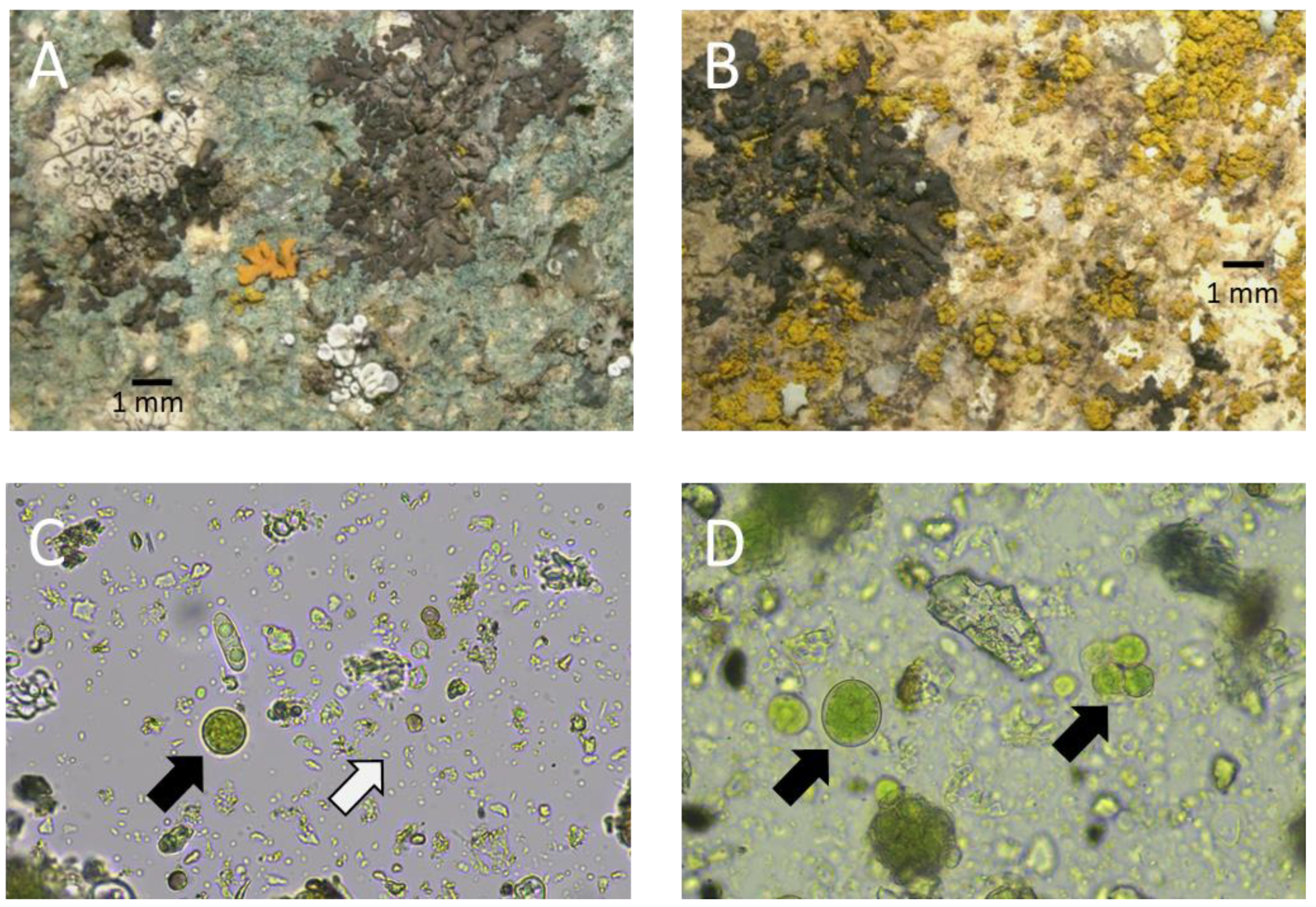
3.1. Biological Characterization of Andesite Blocks’ Surfaces

The green andesite blocks used to build the Temple of the Sun at Ingapirca’s site were often colonized on their surfaces by numerous saxicolous crustose lichens, mainly from the following genera: Caloplaca, Lecanora, Candelaria, Buellia, and Hyperphyscia [21] (Figure 3A,B). In some areas of the collected fragments, dark-colored patinas, possibly dominated by cyanobacteria and/or black fungi, were also present.
The SEM-BSE technique revealed the presence of extensive colonization by crustose lichen closely attached to the lithic substrate (Figure 4). Symbiont cells reached deep into the andesite slabs by growing through microfissures and intergranular spaces (Figure 4A,B). Besides the evident impact of crustose lichens on the stone surface (Figure 4A), which will be presented and discussed elsewhere, free-living cyanobacteria (arrows in Figure 4C) and heterotrophic bacteria (arrows in Figure 4D,E) were also detected on the rock surface and within rock fissures (arrows in Figure 4F). Both types of bacteria are frequently associated with the presence of lichen thalli (Figure 4C,D), free-living algae (Figure 4E), and cyanobacteria colonies (Figure 4F).
Optical microscopy observations of suspensions, prepared by submersing the cotton swabs in sterile saline solution, confirmed the presence on the stone surface of a mix of diverse microorganisms including abundant bacteria, filamentous fungi, microalgae, and cyanobacteria (Figure 3C,D). The inoculation of NA and PDA with these microbial suspensions, followed by incubation at 12 °C for several days, allowed us to isolate and purify several dozen bacterial morphotypes (Figure 5; Table 1). We also isolated and purified up to 13 morphologically distinct filamentous fungi (not shown).

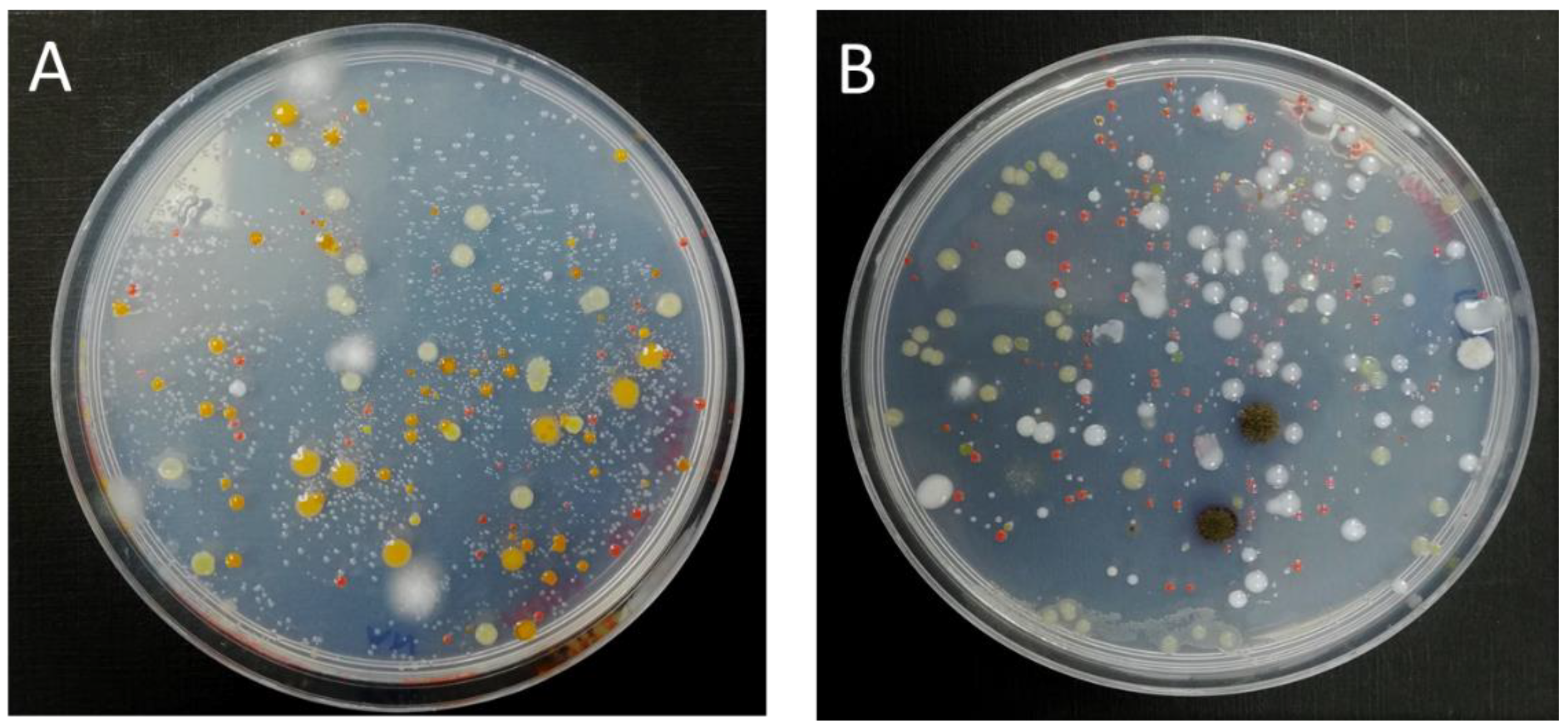
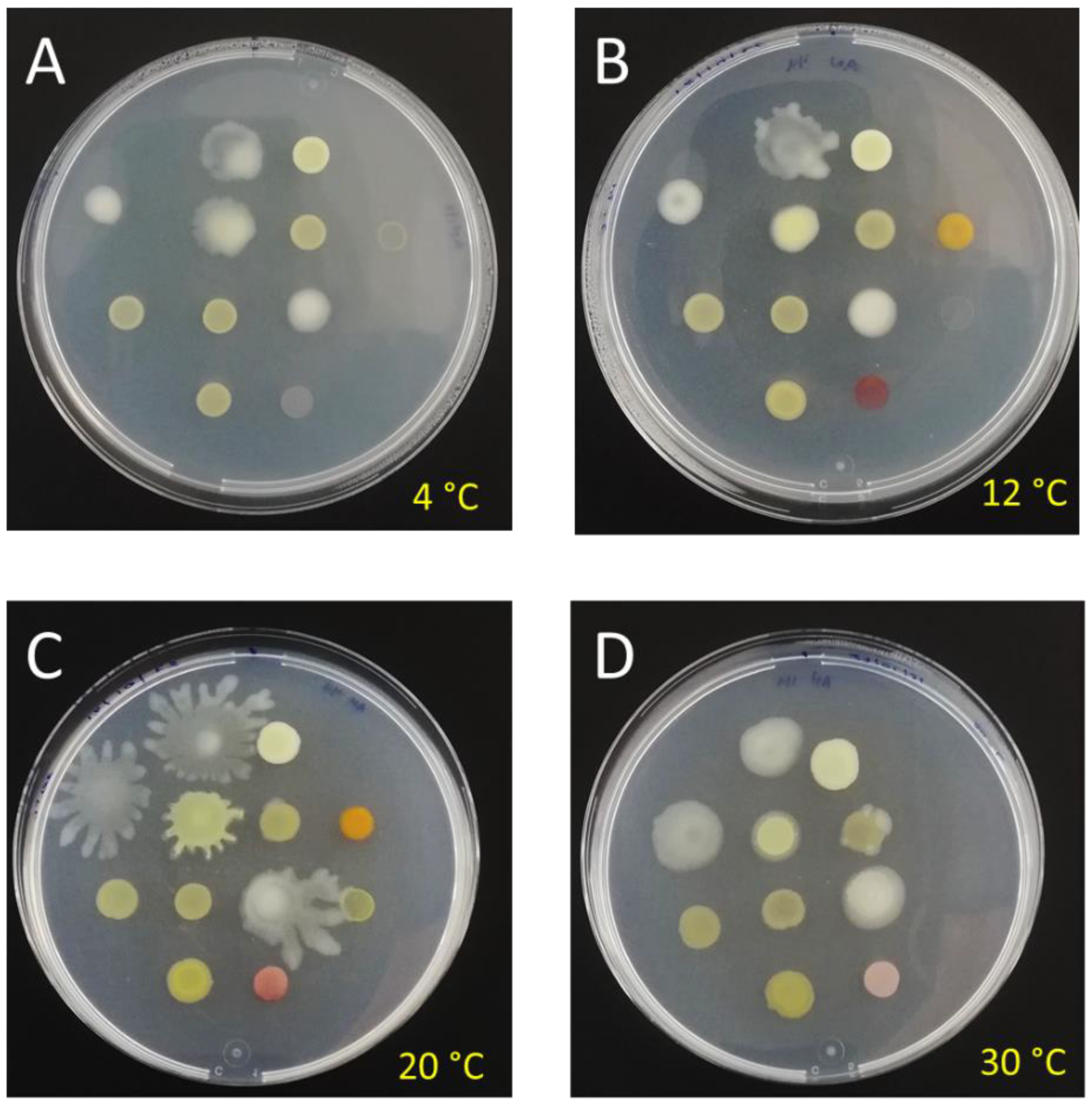
3.2. Morphological and Physiological Characterization of Bacterial Isolates

More than 50 pure isolates were preserved in NA slants at 4 °C. A sub-group of 30 isolates, obtained from each of the four samples collected by swabbing and showing different and similar colony morphologies, were selected to be thoroughly characterized by traditional microbiological methods (i.e., Gram staining, colony morphology, pigmentation, and growth temperature) (Table 1). Most of them were considered eurypsychrophiles, as they were able to grow at a wide range of temperatures, ranging from 4 to 30 °C. Two isolates (namely IP003.3 and IP003.18) grew between 4 and 20 °C; strikingly, one isolate (namely IP004.22) exhibited a narrower temperature range of growth, from 12 to 20 °C.
Noticeably, some isolates were able to synthesize reddish to yellowish pigments while others were highly mucoid, particularly when grown at lower temperatures (i.e., 4–12 °C); other isolates exhibited a conspicuous swarming phenotype when grown above 20 °C (Figure 6 and Table 1).
Based on morphology, culture characteristics, and biochemical tests, the isolates were identified as members of two bacterial phyla: Proteobacteria and Actinobacteria (Table 1). Pseudomonas was the most frequent genera among the isolates, followed by Curtobacterium and Pseudoarthrobacter.

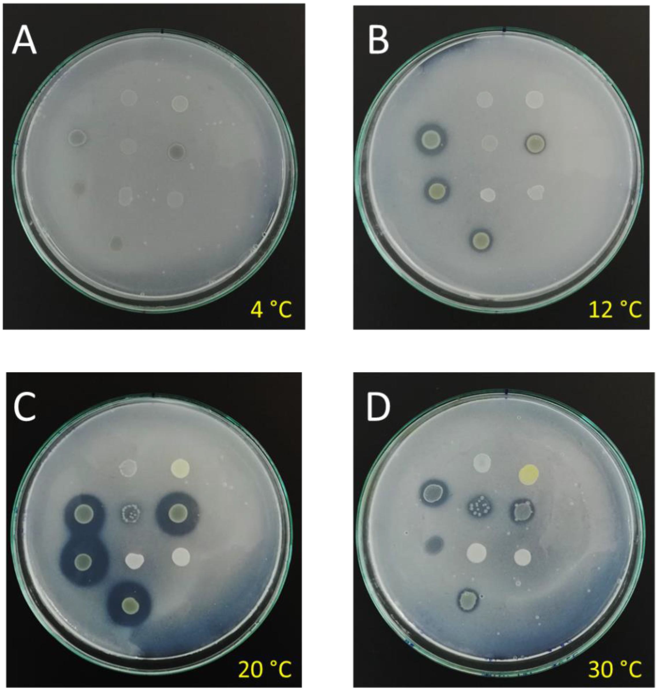
3.3. Mineral Solubilization Assays

One of our main goals in this work was to identify the bacterial strains that are able to dissolve sparingly soluble minerals at low temperatures and, thus, potentially participate in the degradation of green andesite blocks. Most of the isolates tested in the preliminary assays exhibited their greatest P-solubilization efficiencies at 20 °C, as compared with 4, 12, and 30 °C, respectively (Figure 7). At increasing incubation times, the size of the solubilization halos increased concomitantly and allowed us to detect a few more mineral-solubilizing isolates. Therefore, we performed a qualitative estimation of the ability to dissolve tri-calcium phosphate at 20 °C in agarized NBRIP medium, showing that half of the isolates (15 out of 30) were able to do so (Table 1). Preliminary tests of medium acidification revealed that the majority of these halo-producing isolates produced and released acids (not shown). On the basis of these results, we selected a few P-solubilizing isolates to perform in vitro simulations of green andesite solubilization (see below).

3.4. Simulation of Green Andesite Bioweathering

The assays performed in liquid NBRI medium supplemented with finely ground green andesite dust revealed that a few selected isolates (i.e., isolates 1.1, 1.4, 1.11. 1.8.2, and 1.28) were able to solubilize a mineral fraction containing iron. Indeed, after several days at 12 °C, a visual inspection of the culture supernatants allowed us to detect a significant change in their color, from transparent to brownish (not shown). The AAS analysis of these supernatants detected significant amounts of soluble Fe in the presence of bacteria, as opposed to what was found in the uninoculated control experiments (Table 2).
On the contrary, the concentration of other cations did not vary significantly when andesite particles were incubated in the presence of bacteria.

4. Discussion

Archaeological monuments located worldwide are suffering from continuous deterioration due, among other causes, to the detrimental biological activity of microbes that are able to grow actively on their surfaces. To date, a significant amount of information has been obtained from studies focused on monuments located in different geographic areas, from dry Mediterranean deserts to humid tropical forests [7,8,9]. Strikingly, almost no studies have been conducted in the high Andean mountains, where climatic factors play an important role in shaping the microbial communities that are able to grow under the prevailing harsh conditions. These environmental challenges to microbial life include low (sometimes sub-zero) temperatures, high doses of UV radiation, strong winds, and periodic freezing–thawing cycles. In the present work, we showed that a diverse community of microorganisms actively and densely colonized the Ingapirca Temple of the Sun, contributing to the deterioration of its constituent blocks. To the best of our knowledge, this is the first report concerning the causes of the biodeterioration of an Inca monument at such an altitude in the Andean mountain range.
Heterotrophic bacteria were frequently detected by electron microscopy, forming independent colonies or associated with lichens, in the andesite blocks that showed biodeterioration signs. Even though it is nowadays acknowledged that heterotrophic bacteria are among the most important players in the deterioration of stone monuments, their role in these processes has not been as thoroughly studied as that of other microorganisms (e.g., fungi or cyanobacteria). Much less is known about the bacterial communities that are able to thrive under harsh or extreme conditions, such as those prevailing in the Andean mountains.
The culturable fraction of the bacterial community colonizing the surface of the green andesite blocks included isolates belonging to two bacterial phyla, namely Proteobacteria and Actinobacteria. This is not surprising, since it is well-known that members of these two taxa are dominant in many rock types, particularly in extreme environments [6,22,23]. It has been proposed that, besides their natural tolerance to harsh conditions (such as low temperatures, aridness, and high UV radiation), these bacteria find shelter inside small fissures below the rock surface to protect themselves and multiply [24,25].
The preliminary tests conducted in this study to monitor the temperature growth range of the bacterial isolates—a meaningful way to determine their ability to multiply in the cold, although a contested one [26]—suggested that many of them were eurypsychrophiles (formerly known as psychrotolerants or psychrotrophs), i.e., they are able to multiply at low temperatures ranging from ≤0 °C to more than 20 °C [26,27]. In fact, in the present work, several isolates were not only able to grow at 4 °C, but also reached higher amounts of biomass at 12 °C than they did at higher temperatures. Altogether, these results emphasize the ability of these isolates to thrive at temperatures well below those considered to be characteristic of mesophiles (i.e., 20–40 °C). This was not completely unexpected, since the mean annual temperature of Ingapirca’s site is 12.3 °C [14]; in addition, during long periods of the year, the site may reach temperatures around or below the freezing point of water. Thus, it was far from surprising to find that microorganisms able to grow in the cold were members of the microbial community that colonized the surface of Ingapirca’s green andesite blocks.
As we have seen, the solubilization of mineral phosphates could be achieved by several of the bacterial isolates at a wide range of temperatures. In some cases, the solubilization was maximal at temperatures below 20 °C, which emphasized the detrimental potential of these bacteria under field conditions on the mineral structure of the andesite rocks. The ability to solubilize minerals at low temperatures was also anticipated, since it has already been established that many environmental bacterial isolates, particularly those found in the soil or in the surface of rocks, are able to exhibit this phenotype in the cold [28,29]. This is the case for Pseudomonads, whose solubilizing ability is related to the production of organic acids, mainly gluconic, through the direct oxidation pathway [30,31,32]. The production and release of acids by microorganisms is the best-known mechanism of the bioweathering of inorganic matrices [1,33], with two different mechanisms underlying this process: (i) the acidification of the milieu due to the accumulation of protons and their consequent effect on the dissolution of susceptible materials; and (ii) the chelation of cations by anions to form stable complexes [2,6].
As proposed recently by da Silva et al. [34], the solubilization of minerals by saxicolous, lichen-associated bacteria might be related to the nutrient acquisition of the lichen ecosystem (and their associated lichensphere) from oligotrophic substrates. Incidentally, Pseudomonas was also the most predominant genera among the isolates able to solubilize phosphate at low temperatures, retrieved by the same authors from Antarctic lichens. Even though we did not expand upon this aspect, our preliminary results indicated that the acidification of the cell surroundings might contribute to the solubilization of green andesite. In addition, the presence of bacterial colonies associated with lichens was frequent in the samples analyzed in this study. We also noticed that solubilization occurred at 4 °C, which not only suggested the synthesis of cold-active enzymes, but also highlighted a potential mechanism of biodeterioration functioning under extreme conditions. This was an interesting observation and raised questions about the relevance of cold-adapted bacteria in the biodeterioration of stone monuments in cold environments.
In line with these observations, we also showed that some specific mineral phases of green andesite may be solubilized at low temperatures by means of microbial metabolites. Since Fe ions were detected on the supernatant of bacterial cultures grown in the presence of finely ground andesite rocks, and considering that ferromagnesians are among the most prominent minerals in the composition of this rock, we believe that the release of Fe is related to their weathering. Indeed, it has already been proposed that this kind of mineral can be released from ultramafic rocks as a consequence of bacterial growth [35].
The weathering of ferromagnesians might be related to: (i) the active colonization of the andesite rock by bacteria through microfractures (as we have shown to occur in this study); (ii) the production and release of organic acids; (iii) the production of other bacterial metabolites such as siderophores, which act as iron-chelating substances; and (iv) the production of high levels of exo-polysacharides (EPSs), such as those observed when growing bacteria at low temperatures (see Section 3). The production of these EPS has been proposed to be a physiological adaptation to protect cells from the deleterious effect of ice crystals [36]. However, EPSs also allow bacteria to bind to other cells, adhere on the substratum surface, retain water, and trap metal ions and nutrients [33,37,38].
Among the results reported here, particularly intriguing was the finding that the swarming phenotype exhibited by several isolates when grown at specific temperatures. In some cases, the ability of bacteria to spread far away from the point of application was particularly evident (see, for instance, Figure 6C). In the context of stone biodeterioration, this behavior might be crucial, since it would allow cells to actively penetrate the mineral substratum through pre-existing microscopic pores and fractures. This, in turn, would extend the microbial-mediated weathering of stones far away from the rock surface, contributing to the damage exerted at deeper levels within the rock. In fact, even though it is usually believed that bacteria cannot penetrate pores smaller than 0.2 µm, several studies have shown that they can move to, penetrate, and surpass very narrow constrictions (sub- µm width, i.e., smaller than the bacterial diameter) and grow thereafter [39].
As we observed in this study, several isolates produced pigments ranging from deep red to light yellow (see Figure 6B,C). The production of these pigments, most probably carotenes and carotenoids, depended on the temperature of culture: in some cases, maximal production was registered at low temperatures (i.e., <12 °C), while in other cases the pigmentation increased at higher temperatures (i.e., <20 °C). As stated above, pigment production by microorganisms colonizing the surface of stones may be extremely detrimental to historical monuments, since it does not merely affect the aesthetics of the monuments but can also modify the physicochemical characteristics of the minerals [2,33]. This aspect deserves to be further explored in the future in order to determine the real impact of pigment production by heterotrophic bacteria when monitored in situ under more realistic conditions.
Among the limitations of our study, we should mention that we focused on neither phototrophic microorganisms (such as cyanobacteria and algae), chemolithotrophs, nor fungi. All three groups of microorganisms are important players in biodeterioration processes and are crucial for depicting the actual impact of microbes on the deterioration of the Temple of the Sun, as we observed via electron microscopy. Another limitation is that we concentrated only on the culturable fraction of bacteria, even though it is currently acknowledged that this fraction only represents between 1 and 10% of the actual diversity of a microbial community. However, many of these microbes fall into the “unculturable” category and are extremely hard to grow in vitro, thus preventing their characterization in the laboratory. It is therefore important to conduct further studies, with the aid of metagenomics techniques, to shed more light on the actual composition and structure of these communities and the ecological roles played by their members.
In summary, the surface and interior of microfractures of the mineral used to build the Temple of the Sun at the Ingapirca’ site are extensive and heavily colonized by a community of bacterial species, some of which are in intimate contact with lichens. Several of these bacterial strains are psyhcrophiles, which are able to fragilize the chemical structure of the green andesite blocks through currently unknown solubilization mechanisms and remain active at low temperatures. Other strains also modified the aesthetics of the mineral surface by producing pigments. Even though this work provides a preliminary picture of the actual biogeochemical processes that take place on the surface of this monument, our results are the first to be published concerning an Inca monument at such an altitude and in permanently cold conditions in the Andean mountains. Therefore, we believe they will be important for designing and orienting the necessary actions needed to prevent and control the biodeterioration process of this and other similar monuments.

5. Conclusions

The results presented here provide information concerning the possible role played by heterotrophic, eurypsichrophilic bacteria on the biodeterioration of an archaeological monument of the Inca culture, located at high altitude in the Ecuadorian Andes. The mechanisms responsible—at least in part—for this phenomenon are active at low temperatures. Among the culturable heterotrophic bacteria isolated from the walls of the Temple of the Sun (Ingapirca’s archaeological complex), Actinobacteria and Proteobacteria species dominate and produce metabolites that can solubilize mineral substrates, such as tri-calcium phosphate. Furthermore, some isolates could also solubilize certain iron-containing mineral fractions present in the green andesite rock used to build the Temple of the Sun.

Author Contributions

Conceptualization, L.A.Y.; methodology, L.B., D.P., A.d.l.R., M.R., F.E., A.S.T., G.M.M., J.G.L. and M.V.T.; investigation, L.B., D.P., A.d.l.R., M.R., F.E., A.S.T., G.M.M., J.G.L. and M.V.T.; resources, L.A.Y.; data curation, L.A.Y. and A.d.l.R.; writing—original draft preparation, L.A.Y. and A.d.l.R.; writing—review and editing, L.A.Y., A.d.l.R. and M.R.; visualization, L.A.Y. and A.d.l.R.; project administration L.A.Y.; funding acquisition, L.A.Y., A.d.l.R. and M.R. All authors have read and agreed to the published version of the manuscript.

Funding

This project was partially funded by the Catholic University of Cuenca, grant number PICCIITT19-21. The project also received financial support from MNCN-CSIC under a TOP Heritage project, grant number P2018/NMT-4372 of the Community of Madrid, and from the Unidad de Laboratorio y Análisis (Instituto Nacional del Patrimonio Cultural, Quito, Ecuador).

Institutional Review Board Statement

Not applicable.

Informed Consent Statement

Not applicable.

Data Availability Statement

Not applicable.

Acknowledgments

We are deeply indebted to Marco Velecela Serrano, former Director of Centro de Investigación e Interpretación Z6, INPC (Ingapirca’s Archaeological Complex), for the logistic support provided in situ and for sharing his extensive knowledge of Ingapirca’s history. We also thank the technical assistance offered by Esther Rodríguez (MNCN-CSIC) and the staff of the Non-destructive Techniques Service of MNCN-CSIC during the electron microscopy study. Finally, we express our gratitude to J. E. Torracchi for statistical analysis with R. The collection of biological specimens was authorized by Ministerio del Ambiente, Agua y Tansición Ecológica, No. MAATE-ARSFC-2022-2305.

Conflicts of Interest

The authors declare no conflict of interest. The funders had no role in the design of the study; in the collection, analyses, or interpretation of data; in the writing of the manuscript; or in the decision to publish the results.

References

  1. Liu, X.; Koestler, R.J.; Warscheid, T.; Katayama, Y.; Gu, J.D. Microbial deterioration and sustainable conservation of stone monuments and buildings. Nat. Sustain. 2020, 3, 991–1004. [Google Scholar] [CrossRef]
  2. Scheerer, S.; Ortega-Morales, O.; Gaylarde, C. Microbial deterioration of stone monuments—An updated overview. Adv. Appl. Microbiol. 2009, 66, 97–139. [Google Scholar] [CrossRef] [PubMed]
  3. Schröer, L.; Boon, N.; De Kock, T.; Cnudde, V. The capabilities of bacteria and archaea to alter natural building stones—A review. Int. Biodeterior. Biodegrad. 2021, 165, 105329. [Google Scholar] [CrossRef]
  4. Sohrabi, M.; Favero-Longo, S.E.; Pérez-Ortega, S.; Ascaso, C.; Haghighat, Z.; Talebian, M.H.; Fadaei, H.; de los Ríos, A. Lichen colonization and associated deterioration processes in Pasargadae, UNESCO world heritage site, Iran. Int. Biodeterior. Biodegrad. 2017, 117, 171–182. [Google Scholar] [CrossRef]
  5. Tonon, C.; Favero-Longo, S.E.; Matteucci, E.; Piervittori, R.; Croveri, P.; Appolonia, L.; Meirano, V.; Serino, M.; Elia, D. Microenvironmental features drive the distribution of lichens in the House of the Ancient Hunt, Pompeii, Italy. Int. Biodeterior. Biodegrad. 2019, 136, 71–81. [Google Scholar] [CrossRef]
  6. Nir, I.; Barak, H.; Kramarsky-Winter, E.; Kushmaro, A.; de los Ríos, A. Microscopic and biomolecular complementary approaches to characterize bioweathering processes at petroglyph sites from the Negev Desert, Israel. Environ. Microbiol. 2022, 24, 967–980. [Google Scholar] [CrossRef]
  7. Ortega-Morales, O.; Montero-Munoz, J.L.; Baptista Neto, J.A.; Beech, I.B.; Sunner, J.; Gaylarde, C. Deterioration and microbial colonization of cultural heritage stone buildings in polluted and unpolluted tropical and subtropical climates: A metaanalysis. Int. Biodeterior. Biodegrad. 2019, 143, 104734. [Google Scholar] [CrossRef]
  8. Ortega-Morales, B.O.; Narváez-Zapata, J.A.; Schmalenberger, A.; Sosa-López, A.; Tebbe, C.C. Biofilms fouling ancient limestone Mayan monuments in Uxmal, Mexico: A cultivation-independent analysis. Biofilms 2004, 1, 79–90. [Google Scholar] [CrossRef]
  9. McNamara, C.J.; Perry, T.D.; Bearce, K.A.; Hernandez-Duque, G.; Mitchell, R. Epilithic and endolithic bacterial communities in limestone from a Maya archaeological site. Microb. Ecol. 2006, 51, 51–64. [Google Scholar] [CrossRef]
  10. Gaylarde, C.; Ogawa, A.; Beech, I.; Kowalski, M.; Baptista Neto, J.A. Analysis of dark crusts on the church of Nossa Senhora do Carmo in Rio de Janeiro, Brazil, using chemical, microscope and metabarcoding microbial identification techniques. Int. Biodeterior. Biodegrad. 2017, 117, 60–67. [Google Scholar] [CrossRef]
  11. Lemus-Espinoza, D.; Maniscalchi-Badaoui, M.T.; Quereguan, R.; Morales, Y. Biodeterioro del Monumento Religioso Arquitectónico “San Antonio de Padua”, Clarines, Anzoátegui, Venezuela. SABER 2012, 24, 98–102. Available online: https://www.redalyc.org/pdf/4277/427739447010.pdf (accessed on 28 August 2022).
  12. Gaylarde, C.C.; Gaylarde, P.M.; Neilan, B.A. Endolithic phototrophs in built and natural stone. Curr. Microbiol. 2012, 65, 183–188. [Google Scholar] [CrossRef] [PubMed]
  13. Morillas, H.; Maguregui, M.; Gallego-Cartagena, E.; Huallparimachi, G.; Marcaida, I.; Salcedo, I.; Silva, L.F.O.; Astete, F. Evaluation of the role of biocolonizations in the conservation state of Machu Picchu (Peru): The Sacred Rock. Sci. Total Environ. 2019, 654, 1379–1388. [Google Scholar] [CrossRef]
  14. Instituto Nacional del Patrimonio Cultural (INPC) Report. Diagnóstico del Estado de Conservación. Complejo Arqueológico Ingapirca. Available online: https://nube.patrimoniocultural.gob.ec/owncloud/index.php/s/XX6fv76QqxZ6Eht#pdfviewer (accessed on 4 August 2022).
  15. Romero Bastidas, M.; Díaz Benalcázar, R.; Espinoza, F. Estado de Conservación del Complejo Arqueológico Ingapirca. PAQU Instituto Nacional del Patrimonio Cultural. 2020. Available online: https://paqu.patrimoniocultural.gob.ec/investigacion/diagnostico-estado-conservacion-ingapirca/ (accessed on 4 August 2022).
  16. Wierzchos, J.; Ascaso, C. Application of back-scattered electron imaging to study the lichen–rock interface. J. Microsc. 1994, 175, 54–59. [Google Scholar] [CrossRef]
  17. Brenner, D.J.; Krieg, N.R.; Staley, J.T.; Garrity, G.M. The Proteobacteria, Part A Introductory Essays. In Bergey’s Manual of Systematic Bacteriology; Williams & Wilkins: Baltimore, MD, USA; London, UK, 2012; Volume 2. [Google Scholar]
  18. Whitman, W.B.; Goodfellow, M.; Kämpfer, P.; Busse, H.J.; Trujillo, M.E.; Suzuki, K.I.; Ludwig, W.; Whitman, W.B. The Actinobacteria. In Bergey’s Manual of Systematic Bacteriology; Williams & Wilkins: Baltimore, MD, USA; London, UK, 2012; Volume 5. [Google Scholar]
  19. Nautiyal, C.S. An efficient microbiological growth medium for screening phosphate solubilizing microorganisms. FEMS Microbiol. Lett. 1999, 170, 265–270. [Google Scholar] [CrossRef]
  20. R Core Team. R: A Language and Environment for Statistical Computing; R Foundation for Statistical Computing: Vienna, Austria, 2020; Available online: https://www.R-project.org/ (accessed on 11 July 2022).
  21. Torracchi, J.E.; Universidad Católica de Cuenca, Cuenca, Azuay, Ecuador. Personal communication, 2022.
  22. Wong, F.K.Y.; Lau, M.C.Y.; Lacap, D.C.; Aitchison, J.C.; Cowan, D.A.; Pointing, S.B. Endolithic microbial colonization of limestone in a high-altitude arid environment. Microb. Ecol. 2010, 59, 689–699. [Google Scholar] [CrossRef]
  23. Choe, Y.H.; Kim, M.; Woo, J.; Lee, M.J.; Lee, J.I.; Lee, E.J.; Lee, Y.K. Comparing rock-inhabiting microbial communities in different rock types from a high arctic polar desert. FEMS Microbiol. Ecol. 2018, 94, fiy070. [Google Scholar] [CrossRef]
  24. de Los Ríos, A.; Wierzchos, J.; Ascaso, C. The lithic microbial ecosystems of Antarctica’s McMurdo Dry Valleys. Antarct. Sci. 2014, 26, 459–477. [Google Scholar] [CrossRef]
  25. Garrido-Benavent, I.; Pérez-Ortega, S.; Durán, J.; Ascaso, C.; Pointing, S.B.; Rodríguez-Cielos, R.; Navarro, F.; de Los Ríos, A. Differential Colonization and Succession of Microbial Communities in Rock and Soil Substrates on a Maritime Antarctic Glacier Forefield. Front. Microbiol. 2020, 11, 126. [Google Scholar] [CrossRef]
  26. Cavicchioli, R. On the concept of a psychrophile. ISME J. 2016, 10, 793–795. [Google Scholar] [CrossRef]
  27. Feller, G.; Gerday, C. Psychrophilic enzymes: Hot topics in cold adaptation. Nat. Rev. Microbiol. 2003, 1, 200–208. [Google Scholar] [CrossRef] [PubMed]
  28. Pandey, A.; Yarzábal, L.A. Bioprospecting cold-adapted plant growth promoting microorganisms from mountain environments. Appl. Microbiol. Biotechnol. 2019, 103, 643–657. [Google Scholar] [CrossRef]
  29. Yarzábal, L.A. Perspectives for using glacial and periglacial microorganisms for plant growth promotion at low temperatures. Appl. Microbiol. Biotechnol. 2020, 104, 3267–3278. [Google Scholar] [CrossRef] [PubMed]
  30. Lessie, T.G.; Phibbs, P.V. Alternative pathways of carbohydrate utilization in pseudomonads. Annu. Rev. Microbiol. 1984, 38, 359–388. [Google Scholar] [CrossRef]
  31. Goldstein, A.H. Future trends in research on microbial phosphate solubilization: 100 years of insolubility. In First International Meeting on Microbial Phosphate Solubilization; Velazquez, E., Rodrıguez-Barrueco, C., Eds.; Springer: Dordrhecht, The Netherlands, 2007; pp. 91–96. [Google Scholar] [CrossRef]
  32. Rodríguez, H.; Fraga, R. Phosphate solubilizing bacteria and their role in plant growth promotion. Biotechnol. Adv. 1999, 17, 319–339. [Google Scholar] [CrossRef]
  33. Warscheid, T.; Braams, J. Biodeterioration of stone: A review. Int. Biodeterior. Biodegrad. 2000, 46, 343–368. [Google Scholar] [CrossRef]
  34. da Silva, A.V.; de Oliveira, A.J.; Tanabe, I.S.B.; Silva, J.V.; da Silva Barros, T.W.; da Silva, M.K.; França, P.H.B.; Leite, J.; Putzke, J.; Montone, R.; et al. Antarctic lichens as a source of phosphate-solubilizing bacteria. Extremophiles 2021, 25, 181–191. [Google Scholar] [CrossRef]
  35. Becerra-Castro, C.; Kidd, P.; Kuffner, M.; Prieto-Fernández, Á.; Hann, S.; Monterroso, C.; Sessitsch, A.; Wenzel, W.; Puschenreiter, M. Bacterially induced weathering of ultramafic rock and its implications for phytoextraction. Appl. Env. Microbiol. 2013, 79, 5094–5103. [Google Scholar] [CrossRef]
  36. De Maayer, P.; Anderson, D.; Cary, C.; Cowan, D.A. Some like it cold: Understanding the survival strategies of psychrophiles. EMBO Rep. 2014, 15, 508–517. [Google Scholar] [CrossRef]
  37. de los Ríos, A.; Wierzchos, J.; Sancho, L.G.; Ascaso, C. Exploring the physiological state of continental Antarctic endolithic microorganisms by microscopy. FEMS Microbiol. Ecol. 2004, 50, 143–152. [Google Scholar] [CrossRef] [Green Version]
  38. Nichols, C.A.M.; Guezennec, J.; Bowman, J.P. Bacterial exopolysaccharides from extreme marine environments with special consideration of the Southern Ocean, sea ice, and deep-sea hydrothermal vents: A review. Mar. Biotechnol. 2005, 7, 253–271. [Google Scholar] [CrossRef] [PubMed]
  39. Männik, J.; Driessen, R.; Galajda, P.; Keymer, J.E.; Dekker, C. Bacterial growth and motility in sub-micron constrictions. Proc. Natl. Acad. Sci. USA 2009, 106, 14861–14866. [Google Scholar] [CrossRef] [PubMed] [Green Version]
Figure 1. Location and map of the Ingapirca’s archeological complex (IAC) (Cañar, Ecuador). (A) Ecuador in the South American context (in red); (B) Location of Cañar Province in Ecuador (in red). (C) Map of Ingapirca’s archeological complex, showing the location of the Temple of the Sun (white arrow) (reproduced from Wikimedia Commons under Creative Commons licences 3.0 and 4.0; by DeDuijn—own work, CC BY-SA 4.0, https://commons.wikimedia.org/w/index.php?curid=47668112 accessed on 28 August 2022).
Heritage 05 00130 g001
Figure 2. Ingapirca’s Temple of the Sun. (A) The temple (or Elipse) view from outside. (B) A closer view of the walls of the Temple of the Sun (lateral movement of the blocks occurred over time due to frequent earthquakes). (C) The building blocks of green andesite, fitting together perfectly and showing signs of weathering and biodeterioration, such as lichen-colonized alveoli or pits (white arrows) or cyanobacteria/black fungi biofilms (black arrow).
Heritage 05 00130 g002
Figure 3. Lichen and microbial colonization on the surface of green andesite used to build the Temple of the Sun. (A,B) Rock fragments detached from the green andesite blocks, heavily colonized by crustose lichens. (C,D) Optical microscopy of cell suspensions of microbes released from the rock surface by gentle rubbing with moistened cotton swabs. Bacteria (white arrow) and algae (black arrows) can be seen.
Heritage 05 00130 g003
Figure 4. SEM-BSE images of green andesite fragments detached from the walls and colonized by lichens and associated microorganisms. (A,B) Cells growing and actively penetrating the andesite substrate through microfissures. Small mineral fragments separated from the mineral substrate can be seen, surrounded by microbial cells. (C) Free-living cyanobacteria (arrows) visible on the rock surface. (DF) Heterotrophic bacteria were also present on the surface, within rock fissures, and associated with free-living algae as well as lichen thalli.
Heritage 05 00130 g004
Figure 5. Isolation of heterotrophic bacteria and fungi from the surface of the Temple of the Sun walls. Primary isolation cultures, showing pigmented and non-pigmented colonies after 48–72 h growth at 12 °C on nutrient agar (1/4 strength). Some mucoid colonies and filamentous fungi can also be seen in both plates. Samples inoculated in plates shown in (A,B) were collected from different sites in the monument walls.
Heritage 05 00130 g005
Figure 6. Colony phenotype of bacterial isolates growing at different temperatures on the surface of nutrient agar (1/4 strength). (A) 4 °C; (B) 12° C; (C) 20 °C; and (D) 30 °C. Each isolate was spotted in exactly the same place on the surface of the agarized medium. The production of pigments depending on the growth temperature can be clearly seen. Some isolates also show a conspicuous swarming phenotype, mainly at 20 °C.
Heritage 05 00130 g006
Figure 7. Solubilization of mineral phosphate by heterotrophic bacterial isolates at different temperatures. Heterotrophic bacterial isolates were grown on the surface of NBRIP medium and incubated at different temperatures for 48–72 h. (A) 4 °C; (B) 12 °C; (C) 20 °C; (D) 30 °C. A clear halo of mineral solubilization can be seen surrounding colonies of isolates that were able to dissolve tri-calcium phosphate. The isolates were spotted in the same place on the surface of the agarized medium, and thus the efficiency of solubilization depended on the incubation temperature.
Heritage 05 00130 g007
Table 1. Characteristics of heterotrophic bacteria isolated from the exterior surface of the green andesite blocks used to build Ingapirca’s Temple of the Sun.
Origin 1Colony Description 2Growth Temp. RangeNBRIP 3Identification
IP001.1 NAWsRS4–30++Pseudarthrobacter sp.
IP001.4NAWsRS4–30++Pseudomonas sp.
IP001.5NAYsRS4–30-Paenarthrobacter sp.
IP001.7NACsRS4–30+++Pseudomonas sp.
IP001.8.2NAYsRS4–30++Frigoribacterium sp.
IP001.11PDACsRS4–30+++Pseudomonas sp.
IP001.21CzWoRM4–30+++Curtobacterium sp.
IP001.23CzWoRS4–30+++Pseudomonas sp.
IP001.25NAWsRM4–30+++Arthrobacter sp.
IP001.28NAYsRM4–30-Curtobacterium sp.
IP002.4.1NATsRS4–30+++Pseudomonas sp.
IP002.6.1NATsRS4–30+++Pseudomonas sp.
IP002.9PDACmRM4–30-Pantoea sp.
IP002.20NAYsIM4–30-Curtobacterium sp.
IP002.21CzCsRS4–30+++Pseudomonas sp.
IP003.3NAOsRS4–20-Sphingomonas sp.
IP003.5NACsRS4–30+++Pseudomonas sp.
IP003.6NAWsRS4–30-Pseudomonas sp.
IP003.11PDACsRS4–30-Pseudomonas sp.
IP003.15PDAYmRS4–30+Pseudomonas sp.
IP003.18NAOsRS4–20-Sphingomonas sp.
IP003.19PDAYsRS4–30-Pseudomonas sp.
IP003.21NACsRS4–30-Plantibacter sp.
IP004.4NACsRS4–30++Pseudomonas sp.
IP004.5NAWmIL4–30-Pseudarthrobacter sp.
IP004.8NAWsRS4–30+++Pseudarthrobacter sp.
IP004.22PDAYsRS12–20-Frondihabitans sp.
IP004.24NACsRS4–30-Microbacterium sp.
IP004.27NAYsRM4–30-Curtobacterium sp.
IP004.29NAPsRS4–30-Pseudarthrobacter sp.
1 Primary isolation medium: NA = nutrient agar (1/4 strenght); PDA = potato dextrose agar; Cz = Czapek’s medium. 2 Colony phenotype (as determined in nutrient agar medium at 20 °C). Color: W = white; C = cream; P = pink; O = orange; Y = yellow; T = transparent. Texture: s = shiny; o = opaque; m = mucoid. Margin: R = regular; I = irregular. Size: S = small; M = medium; L = large. 3 NBRIP solubilization halo diameter: - = no halo; + = between 6 and 9 mm; ++ = between 9 and 16 mm; +++ = larger than 16 mm.
Table 2. In vitro lixiviation of green andesite blocks in the presence of bacterial isolates.
TreatmentIron (ppm)Calcium (ppm)Sodium (ppm)Magnesium (ppm)
Control0.00035.384 ± 1.6336.424 ± 0.06222.415 ± 0.032
Isolate IP001.10.00036.020 ± 2.0985.851 ± 0.06822.509 ± 0.169
Isolate IP001.476.025 ± 1.532 **34.687 ± 1.6906.513 ± 0.34122.411 ± 0.131
Isolate IP001.1179.756 ± 1.856 **34.672 ± 1.5906.543 ± 0.35722.512 ± 0.069
Isolate IP001.8.269.205 ± 9.595 **34.990 ± 1.2116.632 ± 0.45422.415 ± 0.094
Isolate IP001.2878.013 ± 2.046 **34.869 ± 0.5846.404 ± 0.28622.460 ± 0.151
** p = 0 in Tukey’s post hoc test.
Publisher’s Note: MDPI stays neutral with regard to jurisdictional claims in published maps and institutional affiliations.

