Communication

STS and PUDP Deletion Identified by Targeted Panel Sequencing with CNV Analysis in X-Linked Ichthyosis: A Case Report and Literature Review

1
Department of Laboratory Medicine, Jeonbuk National University Medical School and Hospital, Jeonju 54907, Republic of Korea
2
Department of Pediatrics, Jeonbuk National University Medical School and Hospital, Jeonju 54907, Republic of Korea
3
Research Institute of Clinical Medicine, Jeonbuk National University-Biomedical Research Institute, Jeonbuk National University Hospital, Jeonju 54907, Republic of Korea
*
Author to whom correspondence should be addressed.
Genes 2023, 14(10), 1925; https://doi.org/10.3390/genes14101925
Submission received: 12 September 2023 / Revised: 5 October 2023 / Accepted: 7 October 2023 / Published: 10 October 2023
(This article belongs to the Special Issue Genetics of Skin Disorders (2.0 Edition))

Abstract

:
X-linked recessive ichthyosis (XLI) is clinically characterized by dark brown, widespread dryness with polygonal scales. We describe the identification of STS and PUDP deletions using targeted panel sequencing combined with copy-number variation (CNV) analysis in XLI. A 9-month-old infant was admitted for genetic counseling. Since the second day after birth, the infant’s skin tended to be dry and polygonal scales had accumulated over the abdomen and upper extremities. The infant’s maternal uncle and brother (who had also exhibited similar skin symptoms from birth) presented with polygonal scales on their trunks. CNV analysis revealed a hemizygous deletion spanning 719.3 Kb on chromosome Xp22 (chrX:7,108,996–7,828,312), which included a segment of the STS gene and exhibited a Z ratio of −2 in the proband. Multiplex ligation-dependent probe amplification (MLPA) confirmed this interstitial Xp22.31 deletion. Our report underscores the importance of implementing CNV screening techniques, including sequencing data analysis and gene dosage assays such as MLPA, to detect substantial deletions that encompass the STS gene region of Xq22 in individuals suspected of having XLI.

1. Introduction

Ichthyosis is a condition characterized by the excessive growth of the outermost layer of the skin, the stratum corneum, leading to dry skin with scaling. Congenital ichthyosis, which presents during the neonatal period, often poses significant health risks, as the skin’s protective function is compromised [1]. Syndromic ichthyosis is classified based on its genetic origin, including ichthyosis vulgaris, which is inherited in an autosomal dominant manner, and X-linked recessive ichthyosis (XLI). XLI occurs in approximately 1 in 1500 to 6000 males and typically manifests within the first three months of life. Clinically, XLI is characterized by dark brown, widespread dryness with polygonal scales. These cutaneous symptoms appear shortly after birth and tend not to improve with age. The affected areas primarily include the neck, trunk, and extensor side of the limbs. Unlike other forms of ichthyosis, XLI also affects the flexion side of limbs [2]. Histopathologically, XLI typically exhibits mild acanthosis and dense hyperkeratosis with a normal granular layer [3].
Several genetic studies of Korean patients with XLI attributed to STS mutations have been documented [4,5,6,7]. In this report, we describe the identification of STS and PUDP deletions using targeted panel sequencing combined with copy-number variation (CNV) analysis in XLI. To our knowledge, this was the first report of Korean XLI screening through targeted panel sequencing with CNV analysis.

2. Materials and Methods
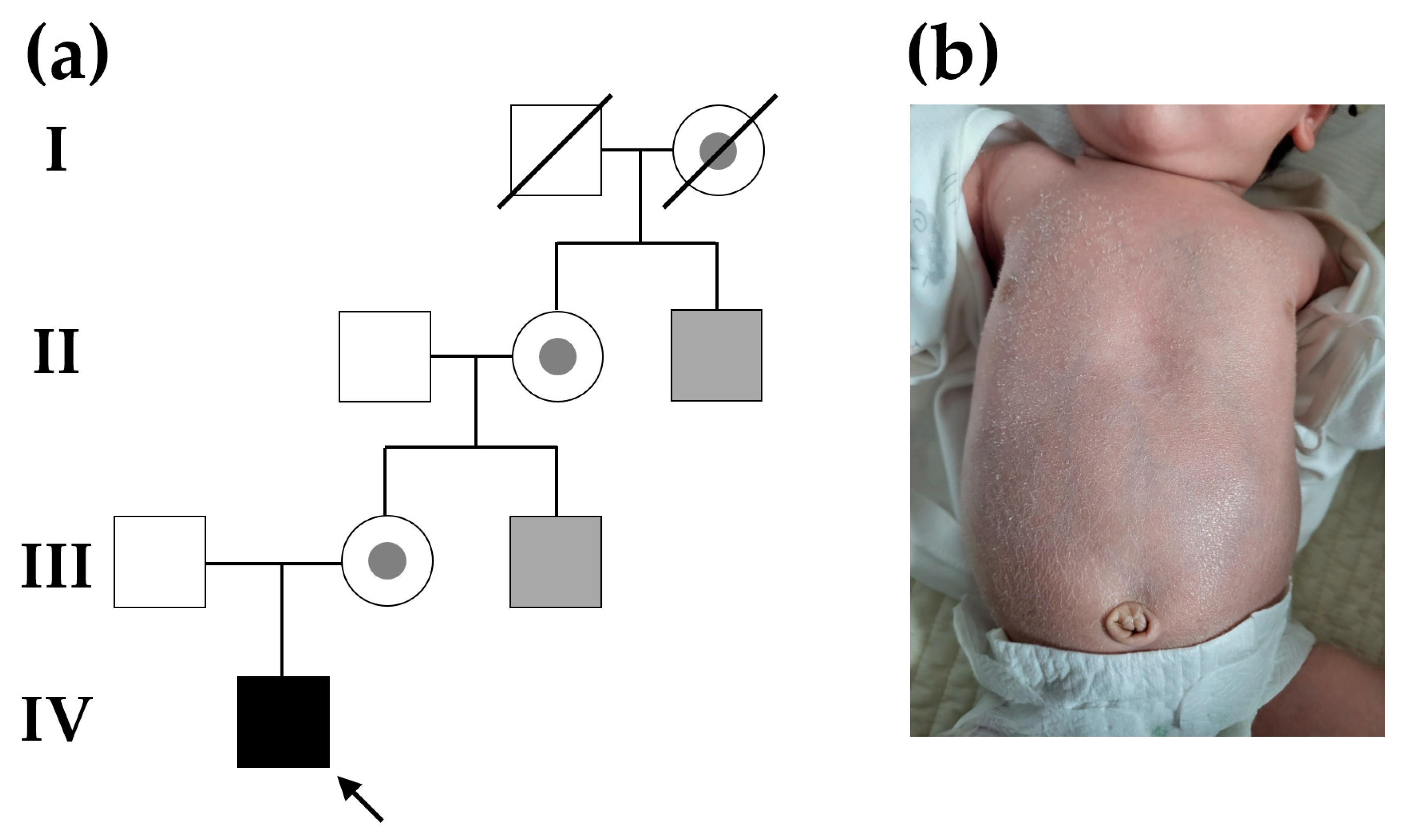
2.1. Patient

A 9-month-old infant (IV-1 in Figure 1a) was vaginally born at a gestational age of 39 weeks and 5 days. The birth weight was 3300 g, and the 1- and 5-min Apgar score was 10/10, which was good. The mother had no diagnosed diseases before pregnancy or pregnancy-related diseases during pregnancy, and this was her first delivery. The patient visited the Department of Pediatrics at Jeonbuk National University Hospital (Jeonju, Republic of Korea) with skin lesions accompanied by scales all over his body since the second day of birth. The skin tended to be dry, and polygonal scales had accumulated over the abdomen and upper extremities (Figure 1b). Blood count, electrolytes, liver function tests, blood urea nitrogen, creatinine, and C-reactive protein levels were all normal. In the pedigree of three generations, the patient’s maternal uncle had the same skin symptoms as those of the infant, and the other ancestral family members had no skin symptoms. The patient’s maternal uncle and brother, who also exhibited similar skin symptoms from birth, presented with polygonal scales on their trunks. Their symptoms were alleviated within the first year of life by maintaining intensive skin moisturization. They experienced intermittent skin manifestations throughout adulthood, but applying topical moisturizers consistently improved symptoms. Hereditary skin disease was suspected, considering the family history and the disease that occurred early after birth, and related genetic tests were performed. X-linked inheritance was suspected in genetic counseling, because only male family members presented with similar clinical symptoms associated with ichthyosis. G-banding karyotyping showed a normal male karyotype of 46, XY. Then, a targeted panel sequencing was performed that included genes associated with genetic disorders of the skin.

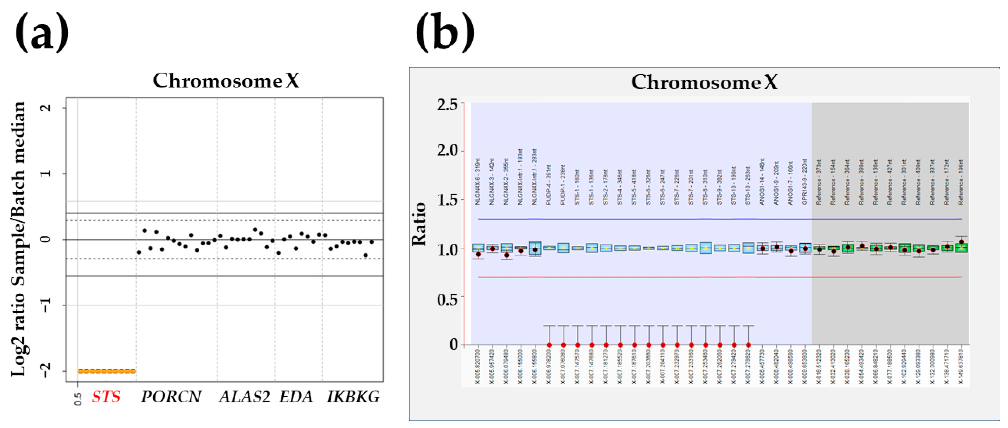
2.2. Targeted Panel Sequencing

Genomic DNA extracted from the proband was subjected to analysis using a customized hereditary dermatology panel (Green Cross Genome, Yongin, Republic of Korea). This panel was designed to target the exons and adjacent regions of 95 genes associated with epidermolysis bullosa and ichthyosis (see Table 1 for details). Sequencing was conducted as paired-end (PE) sequencing with a high-output flow cell, involving 300 cycles PE (150 bp × 2) on a NextSeq500 instrument (Illumina, San Diego, CA, USA) at the Green Cross Genome. Data were analyzed following the Genome Analysis Tool Kit best practice pipeline workflow (https://gatk.broadinstitute.org/hc/en-us (accessed on 18 January 2023)), which encompassed processes such as base-calling, base alignment, variant calling, annotation, and quality control reporting.
Medical laboratory geneticists independently reviewed the interpretation of sequence variants, adhering to the standards and guidelines established by the Joint Consensus Recommendation of the American College of Medical Genetics and Genomics and the Association for Molecular Pathology to ensure accuracy [8]. Raw sequencing data generated from targeted panel sequencing underwent CNV analysis using VisCap [9]. In brief, this involved calculating the specimen-specific fractional coverage for each site, which was then divided by the median value for that region across the entire batch. A boxplot interquartile range multiplier of 3 was applied as a cutoff for detecting gains and losses. Specifically, a gain was defined by a minimum log2 ratio of 0.40, while a loss was defined by a maximum log2 ratio of −0.55, based on the distribution of log2 ratios for each specimen.

2.3. Multiplex Ligation-Dependent Probe Amplification

We employed SALSA® MLPA® Probemix P160-C1 STS reagent (MRC-Holland, Amsterdam, The Netherlands) to assess large exon deletions in the STS gene identified through targeted panel sequencing with CNV analysis. The procedure followed the manufacturer’s protocol for multiplex ligation-dependent probe amplification (MLPA). Subsequently, capillary electrophoresis and fragment analysis were performed consecutively using a 3500XL Dx Genetic Analyzer (Applied Biosystems, Carlsbad, CA, USA). Coffalyser.Net software version 220401.000 (MRC-Holland) was utilized for the comparative analysis of CNV data in accordance with the manufacturer’s instructions. Three sex-matched normal DNA samples were employed to normalize the resulting peak intensities. Manufacturer control probes served as references for the analysis. Generally, probe-to-peak ratios ranging from 0.7 to 1.3 were considered indicative of a normal copy number (wild-type). Conversely, a loss of one copy number was defined by probe-to-peak ratios between 0.4 and 0.7. A gain of one copy number was inferred from probe results for probe-to-peak ratios between 1.3 and 1.6.

3. Results

No (likely) pathogenic variants, such as single nucleotide variants (SNVs) or small indels, were identified through targeted panel sequencing. However, CNV analysis revealed a hemizygous deletion spanning 719.3 Kb on chromosome Xp22 (chrX:7,108,996–7,828,312), which included a segment of the STS gene and exhibited a Z ratio of −2 in the proband (Figure 2a). MLPA confirmed this hemizygous deletion, encompassing exons 1 to 4 of the PUDP gene and the entire region of the STS gene, as identified by CNV analysis in targeted panel sequencing (Figure 2b). Consequently, our proband received a diagnosis of XLI resulting from the deletion of both the STS and PUDP genes. Unfortunately, other family members declined to participate in the segregation analysis. The patient’s skin symptoms showed partial improvement following regular and intensive moisturization. No deterioration in symptoms was noted at the latest follow-up at 9 months of age.

4. Discussion

Genomic instability is a notable characteristic of the human Xp22.31 region. Deletions occurring in this region have been linked to a range of conditions, including XLI, mental retardation, and attention-deficit/hyperactivity disorder (ADHD). This instability is partly attributed to the presence of low-copy repeats (LCRs), which are DNA sequences that are duplicated in the genome. LCRs mediate recurrent deletions at this locus, leading to associated disorders. Additionally, the Xp22.31 region is enriched with a putative homologous recombinant hotspot motif. This motif likely plays a role in facilitating recombination events that lead to these recurrent deletions [10].
Homologous recombination is a genetic mechanism in which similar or identical DNA sequences recombine with one another, potentially leading to DNA rearrangements, such as deletions. The presence of this recombinant hotspot motif in the region may contribute to the genetic instability observed in Xp22.31 and the associated disorders. [10]. Most XLI cases (up to 90%) stem from a deletion that completely encompasses the STS gene [3]. Typically, the deletion in Xp22.31 spans approximately 1.6 Mb and includes the STS, HDHD1/PUDP, PNPLA4, and VCX genes’ encoding proteins. This deletion is classified as pathogenic, based on the recommendations of the American College of Medical Genetics and Genomics [11], and results in ichthyosis, a condition primarily affecting males.
Our patient visited the hospital with symptoms of ichthyosis vulgaris, and initial genetic testing was performed through targeted panel sequencing according to the symptoms. No (likely) pathogenic SNV or small indel variants were detected. However, CNV analysis using massively parallel sequencing (MPS) data generated from targeted panel sequencing identified a hemizygous 719.3 Kb deletion in chromosome Xp22. Subsequent MLPA confirmed this large deletion, enabling the early diagnosis of a congenital disease caused by STS mutations related to recessive ichthyosis in the X chain during the neonatal period.
Ichthyosis identified early in life shows varying severity depending on the causative disease [1]. If the symptoms are not severe, they can be confused with temporary skin changes in the neonatal period. Therefore, targeted panel sequencing tests can be applied in the neonatal intensive care unit soon after birth if screening tests to exclude syndromic ichthyosis are warranted. Since the panel tests the proven genes according to the symptoms of the patient, it can provide the genetic confirmation of a neonatologist’s clinical diagnosis [12]. Based on symptoms and family history, additional analysis using sequencing depth confirmed a hemizygous deletion in the region, including STS, leading to the final diagnosis.
Clinical manifestations are closely linked to the content of neighboring genes. Extensive deletions that encompass a larger number of nearby genes are correlated with severe conditions. These conditions manifest as ocular albinism, epilepsy, abnormal electroencephalographic readings, intellectual disabilities, reduced sense of smell (hyposmia), ADHD, autism, and language development disorders [13,14]. Some male patients may exhibit only minor skin lesions, such as widespread dry and scaly skin and scaling [15,16]. In addition to ichthyosis, benign corneal opacities affect approximately 10–50% of males with XLI [17,18], and roughly 20% of males with XLI experience cryptorchidism [19]. Male carriers of typical XLI-associated deletions approximately 1.6 Mb in size were reported to exhibit developmental delays [20], intellectual disabilities [21], autism [22], epilepsy [21,23], and kidney abnormalities [24]. Despite the established association, the skin symptoms can be considered relatively mild compared to other inherited dermatologic conditions or phenotypes linked to other pathogenic CNVs that might be identified prenatally. Thus, the primary focus is, typically, on neurodevelopmental outcomes and structural anomalies.
Recent studies reported differences from other cases involving Xp22.31 CNVs and attempts to establish correlations between disease severity, the size of the chromosomal deletion, and gene content [25,26]. In clinical and molecular findings in 35 Italian patients [25], the frequency of cryptorchidism (12.9%) corresponded to previous XLI studies. However, the absence of corneal opacities in these individuals can be attributed to their youth. Out of 33 patients, nine (27%) displayed symptoms of ADHD, which aligns with recent literature reporting the heightened occurrence of these behavioral traits in patients with XLI. ADHD has been documented in XLI patients, stemming from both STS deletions and point mutations. These findings suggest that STS deficiency directly contributed to the development of inattentive and hyperactivity symptoms. In genotype-phenotype correlations in prenatal and postnatal follow-ups of 87 fetuses [26], while skin disorders in male fetuses associated with Xp22.31 deletions could be improved with appropriate treatment, the presence of this deletion in males may also be considered a potential susceptibility factor for neurodevelopmental disorders.
A literature review of Korean patients with XLI found that six cases were genetically confirmed by chromosomal microarray (CMA) or MLPA. Depending on the size of the Xp22 deletion, only skin symptoms or other combinations of symptoms were seen. The larger the size of the Xp22 deletion, the more diverse and serious symptoms tended to appear in these cases (Table 2).
We depicted our patient’s deletion through a schematic representation, which was compared to previously documented cases covering the interstitial 4.4 Mb of chromosome Xp, as visualized using DECIPHER’s genome browser (https://www.deciphergenomics.org/browser#q/X:5000000-9400000/location/X:4875000-9525000; accessed on 11 September 2023) (Figure 3). The skin-barrier function remains intact, and there is a high occurrence of kallikrein 7 gene polymorphism among Korean patients with XLI. Specifically, the histopathological examination of ichthyosis epidermis did not reveal acanthosis, and there were no significant differences in the levels of pro-inflammatory cytokines within the skin corneal layer of XLI patients compared to the control group, whether the corneal layer was lesional or non-lesional [5].
On the other hand, MPS is widely used as a screening test for rare diseases [27]. Screening tests, including neonatal screening tests, tandem mass spectrometry, and the automated auditory brainstem response test, are conducted in the neonatal period to detect diseases related to poor prognosis and mortality. As MPS technology develops and the demand for early diagnosis increases, attempts are being made to utilize newborn screening tests for MPS, such as in neonatal intensive care units [28]. Targeted panel sequencing can be used according to symptoms and is used in the clinical field, and a genetic approach to a diagnosis can be tried from the beginning of the evaluation process. Newborns with congenital diseases diagnosed early in the neonatal period can be managed through continuous care and the follow-up of related diseases, so a good prognosis can be expected.
In this case, a genetic diagnosis based on MPS was able to diagnose XLI in the early stages after birth, and it was possible to consider the continuous treatment of skin symptoms and related disease testing. The majority of XLI patients, around 85–90%, exhibit a defect characterized by a complete deletion encompassing the entire STS gene and its adjacent sequences. However, about 10% of cases result from point mutations or partial deletions [25]. The current best practices for CNV detection often necessitate the use of MLPA for detecting CNVs in gene size or CMA for identifying larger CNVs exceeding 50 Kb.
In previous studies, suspected XLI was screened initially or diagnosed molecularly by fluorescence in situ hybridization (FISH) [29,30,31] or CMA [32]. FISH can provide visual confirmation of the presence or absence of the STS gene or its deletion in XLI. It may be advantageous when there is a known familial deletion or when rapid results are needed. However, FISH can be more targeted and may require fewer resources than MLPA. In contrast, MLPA is a PCR-based technique that can detect CNVs across multiple genes simultaneously. It can also identify deletions, duplications, and other CNVs, including those of smaller sizes. MLPA may provide higher resolution and quantitative information about CNVs. The choice between FISH and MLPA for the genetic diagnosis of XLI may depend on various factors, including the specific objectives of the analysis, available resources, and laboratory expertise. In some cases, both techniques may be used to complement each other. FISH can be valuable for initial screening and confirming large deletions, while MLPA can provide more detailed information on CNVs and is useful for broader genetic testing. Ultimately, the decision should be made in consultation with geneticists or clinical laboratory experts who can assess the specific needs of the patient and the clinical context.
In addition to sequencing a clinically enhanced exome to enable the targeted analysis of disease-specific variants, the implementation of CNV detection algorithms using various tools within routine targeted panel sequencing diagnostic protocols can lead to immediate improvements in clinical care for individuals affected by genetically diverse diseases [33,34]. Therefore, sequencing data generated through MPS can serve as a valuable initial step for CNV screening in genetic diagnostic settings. This screening step has the potential to enhance genetic diagnoses of hereditary skin diseases.

5. Conclusions

In this review, we successfully conducted a genetic diagnosis, identifying a hemizygous deletion involving the STS and PUDP genes through targeted panel sequencing with CNV analysis in a patient with XLI. The application of targeted panel sequencing based on neonatal skin symptoms proved to be an effective approach in the clinical context. Our report underscores the importance of implementing CNV screening techniques, including sequencing data analysis and gene dosage assays such as MLPA, to detect substantial deletions encompassing the STS gene region of Xq22 in individuals suspected of having XLI.

Author Contributions

Conceptualization, H.H.K.; methodology, J.P.; software, J.P.; validation, Y.G.C.; formal analysis, J.P.; investigation, Y.G.C.; resources, H.H.K.; data curation, J.P.; writing—original draft preparation, J.P. and H.H.K.; writing—review and editing, J.K.K. and H.H.K.; visualization, Y.G.C.; supervision, J.K.K.; project administration, H.H.K.; funding acquisition, J.P. All authors have read and agreed to the published version of the manuscript.

Funding

This article was supported by research funds for newly appointed professors of Jeonbuk National University in 2020. This research was supported by the Bio&Medical Technology Development Program of the National Research Foundation (NRF) funded by the Korean government (MSIT) (No. RS-2023-00236157).

Institutional Review Board Statement

This study was approved by the Institutional Review Board (IRB) of Jeonbuk National University Hospital (approval number: 2022-12-031; date of approval: 12 December 2022).

Informed Consent Statement

Written informed consent was obtained from the parents on behalf of their child before blood sampling and for the publication of any potentially identifiable images or data included in this article, and for the use of the clinical data of the proband’s family.

Data Availability Statement

Not applicable.

Acknowledgments

We thank Sung Su Park, M.T. (Jeonbuk National University Hospital, Jeonju, Republic of Korea) for providing technical assistance and help with data collection.

Conflicts of Interest

No potential conflict of interest are reported by the authors.

References

  1. Craiglow, B.G. Ichthyosis in the newborn. Semin. Perinatol. 2013, 37, 26–31. [Google Scholar] [CrossRef] [PubMed]
  2. Traupe, H. Revealing the mysteries of X-linked recessive ichthyosis. Br. J. Dermatol. 2018, 179, 821–822. [Google Scholar] [CrossRef] [PubMed]
  3. Takeichi, T.; Akiyama, M. Inherited ichthyosis: Non-syndromic forms. J. Dermatol. 2016, 43, 242–251. [Google Scholar] [CrossRef] [PubMed]
  4. Cho, E.H.; Kim, S.Y.; Kim, J.K. A case of 9.7 Mb terminal Xp deletion including OA1 locus associated with contiguous gene syndrome. J. Korean Med. Sci. 2012, 27, 1273–1277. [Google Scholar] [CrossRef]
  5. Lee, N.R.; Yoon, N.Y.; Jung, M.; Kim, J.-Y.; Seo, S.J.; Wang, H.-y.; Lee, H.; Sohn, Y.B.; Choi, E.H. Skin Barrier Function Is Not Impaired and Kallikrein 7 Gene Polymorphism Is Frequently Observed in Korean X-linked Ichthyosis Patients Diagnosed by Fluorescence in Situ Hybridization and Array Comparative Genomic Hybridization. J. Korean Med. Sci. 2016, 31, 1307–1318. [Google Scholar] [CrossRef]
  6. Yun, J.M.; Na, K.S.; Kim, M.S.; Kim, H.S.; Hwang, H.B. Two Cases of Pre-descemet Corneal Dystrophy Associated with X-linked Ichthyosis: A Case Report by Genetic Analysis. J. Korean Ophthalmol. Soc. 2017, 58, 993–997. [Google Scholar] [CrossRef]
  7. Park, S.J.; Park, K.H.; Bae, M.H.; Kim, Y.M. Developmental Delay in Children with X-Linked Ichthyosis: A Case Series. Ann. Child. Neurol. 2021, 29, 186–189. [Google Scholar] [CrossRef]
  8. Richards, S.; Aziz, N.; Bale, S.; Bick, D.; Das, S.; Gastier-Foster, J.; Grody, W.W.; Hegde, M.; Lyon, E.; Spector, E.; et al. Standards and guidelines for the interpretation of sequence variants: A joint consensus recommendation of the American College of Medical Genetics and Genomics and the Association for Molecular Pathology. Genet. Med. 2015, 17, 405–424. [Google Scholar] [CrossRef]
  9. Pugh, T.J.; Amr, S.S.; Bowser, M.J.; Gowrisankar, S.; Hynes, E.; Mahanta, L.M.; Rehm, H.L.; Funke, B.; Lebo, M.S. VisCap: Inference and visualization of germ-line copy-number variants from targeted clinical sequencing data. Genet. Med. 2016, 18, 712–719. [Google Scholar] [CrossRef]
  10. Liu, P.; Erez, A.; Nagamani, S.C.; Bi, W.; Carvalho, C.M.; Simmons, A.D.; Wiszniewska, J.; Fang, P.; Eng, P.A.; Cooper, M.L.; et al. Copy number gain at Xp22.31 includes complex duplication rearrangements and recurrent triplications. Hum. Mol. Genet. 2011, 20, 1975–1988. [Google Scholar] [CrossRef]
  11. Riggs, E.R.; Andersen, E.F.; Cherry, A.M.; Kantarci, S.; Kearney, H.; Patel, A.; Raca, G.; Ritter, D.I.; South, S.T.; Thorland, E.C.; et al. Technical standards for the interpretation and reporting of constitutional copy-number variants: A joint consensus recommendation of the American College of Medical Genetics and Genomics (ACMG) and the Clinical Genome Resource (ClinGen). Genet. Med. 2020, 22, 245–257. [Google Scholar] [CrossRef] [PubMed]
  12. Daoud, H.; Luco, S.M.; Li, R.; Bareke, E.; Beaulieu, C.; Jarinova, O.; Carson, N.; Nikkel, S.M.; Graham, G.E.; Richer, J.; et al. Next-generation sequencing for diagnosis of rare diseases in the neonatal intensive care unit. Cmaj 2016, 188, E254–E260. [Google Scholar] [CrossRef]
  13. Lonardo, F.; Parenti, G.; Luquetti, D.V.; Annunziata, I.; Della Monica, M.; Perone, L.; De Gregori, M.; Zuffardi, O.; Brunetti-Pierri, N.; Andria, G.; et al. Contiguous gene syndrome due to an interstitial deletion in Xp22.3 in a boy with ichthyosis, chondrodysplasia punctata, mental retardation and ADHD. Eur. J. Med. Genet. 2007, 50, 301–308. [Google Scholar] [CrossRef] [PubMed]
  14. Ma, W.; Mao, J.; Wang, X.; Duan, L.; Song, Y.; Lian, X.; Zheng, J.; Liu, Z.; Nie, M.; Wu, X. Novel Microdeletion in the X Chromosome Leads to Kallmann Syndrome, Ichthyosis, Obesity, and Strabismus. Front. Genet. 2020, 11, 596. [Google Scholar] [CrossRef] [PubMed]
  15. Xie, W.; Zhou, H.; Zhou, L.; Gong, Y.; Lin, J.; Chen, Y. Clinical features and genetic analysis of two Chinese families with X-linked ichthyosis. J. Int. Med. Res. 2020, 48, 300060520962292. [Google Scholar] [CrossRef]
  16. Zhang, M.; Huang, H.; Lin, N.; He, S.; An, G.; Wang, Y.; Chen, M.; Chen, L.; Lin, Y.; Xu, L. X-linked ichthyosis: Molecular findings in four pedigrees with inconspicuous clinical manifestations. J. Clin. Lab. Anal. 2020, 34, e23201. [Google Scholar] [CrossRef]
  17. Boere, P.M.; Bonnet, C.; Frausto, R.F.; Fung, S.S.M.; Aldave, A.J. Multimodal Imaging of Pre-Descemet Corneal Dystrophy Associated with X-Linked Ichthyosis and Deletion of the STS Gene. Cornea 2020, 39, 1442–1445. [Google Scholar] [CrossRef]
  18. Williams, D.; Onyia, O.; Chung, D.D.; Kirakosyan, A.; Hovakimyan, A.; Payne, C.; Moshirfar, M.; Aldave, A.J. Identification of a novel partial deletion of STS associated with pre-Descemet corneal dystrophy and X-linked ichthyosis. Mol. Vis. 2023, 29, 25–30. [Google Scholar]
  19. Brcic, L.; Underwood, J.F.; Kendall, K.M.; Caseras, X.; Kirov, G.; Davies, W. Medical and neurobehavioural phenotypes in carriers of X-linked ichthyosis-associated genetic deletions in the UK Biobank. J. Med. Genet. 2020, 57, 692–698. [Google Scholar] [CrossRef]
  20. Ben Khelifa, H.; Soyah, N.; Ben-Abdallah-Bouhjar, I.; Gritly, R.; Sanlaville, D.; Elghezal, H.; Saad, A.; Mougou-Zerelli, S. Xp22.3 interstitial deletion: A recognizable chromosomal abnormality encompassing VCX3A and STS genes in a patient with X-linked ichthyosis and mental retardation. Gene 2013, 527, 578–583. [Google Scholar] [CrossRef]
  21. Gao, K.; Zhang, Y.; Zhang, L.; Kong, W.; Xie, H.; Wang, J.; Wu, Y.; Wu, X.; Liu, X.; Zhang, Y.; et al. Large De Novo Microdeletion in Epilepsy with Intellectual and Developmental Disabilities, with a Systems Biology Analysis. Adv. Neurobiol. 2018, 21, 247–266. [Google Scholar] [CrossRef] [PubMed]
  22. Labonne, J.D.J.; Driessen, T.M.; Harris, M.E.; Kong, I.K.; Brakta, S.; Theisen, J.; Sangare, M.; Layman, L.C.; Kim, C.H.; Lim, J.; et al. Comparative Genomic Mapping Implicates LRRK2 for Intellectual Disability and Autism at 12q12, and HDHD1, as Well as PNPLA4, for X-Linked Intellectual Disability at Xp22.31. J. Clin. Med. 2020, 9, 274. [Google Scholar] [CrossRef] [PubMed]
  23. Myers, K.A.; Simard-Tremblay, E.; Saint-Martin, C. X-Linked Familial Focal Epilepsy Associated With Xp22.31 Deletion. Pediatr. Neurol. 2020, 108, 113–116. [Google Scholar] [CrossRef]
  24. Schierz, I.A.M.; Giuffrè, M.; Cimador, M.; D’Alessandro, M.M.; Serra, G.; Favata, F.; Antona, V.; Piro, E.; Corsello, G. Hypertrophic pyloric stenosis masked by kidney failure in a male infant with a contiguous gene deletion syndrome at Xp22.31 involving the steroid sulfatase gene: Case report. Ital. J. Pediatr. 2022, 48, 19. [Google Scholar] [CrossRef]
  25. Diociaiuti, A.; Angioni, A.; Pisaneschi, E.; Alesi, V.; Zambruno, G.; Novelli, A.; El Hachem, M. X-linked ichthyosis: Clinical and molecular findings in 35 Italian patients. Exp. Dermatol. 2019, 28, 1156–1163. [Google Scholar] [CrossRef]
  26. Hu, H.; Huang, Y.; Hou, R.; Xu, H.; Liu, Y.; Liao, X.; Xu, J.; Jiang, L.; Wang, D. Xp22.31 copy number variations in 87 fetuses: Refined genotype-phenotype correlations by prenatal and postnatal follow-up. BMC Med. Genom. 2023, 16, 69. [Google Scholar] [CrossRef] [PubMed]
  27. Souche, E.; Beltran, S.; Brosens, E.; Belmont, J.W.; Fossum, M.; Riess, O.; Gilissen, C.; Ardeshirdavani, A.; Houge, G.; van Gijn, M.; et al. Recommendations for whole genome sequencing in diagnostics for rare diseases. Eur. J. Hum. Genet. 2022, 30, 1017–1021. [Google Scholar] [CrossRef]
  28. de Castro, M.J.; González-Vioque, E.; Barbosa-Gouveia, S.; Salguero, E.; Rite, S.; López-Suárez, O.; Pérez-Muñuzuri, A.; Couce, M.L. Rapid Phenotype-Driven Gene Sequencing with the NeoSeq Panel: A Diagnostic Tool for Critically Ill Newborns with Suspected Genetic Disease. J. Clin. Med. 2020, 9, 2362. [Google Scholar] [CrossRef]
  29. Hosomi, N.; Oiso, N.; Fukai, K.; Hanada, K.; Fujita, H.; Ishii, M. Deletion of distal promoter of VCXA in a patient with X-linked ichthyosis associated with borderline mental retardation. J. Dermatol. Sci. 2007, 45, 31–36. [Google Scholar] [CrossRef]
  30. Abdel-Hamed, M.F.; Hussein, H.A.; Helmy, N.A.; Elsaie, M.L. The detection of steroid sulfatase gene deletion (STS) in Egyptian males with X-linked ichthyosis. J. Drugs Dermatol. 2010, 9, 1192–1196. [Google Scholar]
  31. Ramesh, R.; Chen, H.; Kukula, A.; Wakeling, E.L.; Rustin, M.H.; McLean, W.H. Exacerbation of X-linked ichthyosis phenotype in a female by inheritance of filaggrin and steroid sulfatase mutations. J. Dermatol. Sci. 2011, 64, 159–162. [Google Scholar] [CrossRef] [PubMed]
  32. Zhang, H.; Xi, Q.; Liu, X.; Yue, F.; Zhang, H.; Sun, M.; Liu, R. Prenatal Diagnosis and Molecular Cytogenetic Characterization of Copy Number Variations on 4p15.2p16.3, Xp22.31, and 12p11.1q11 in a Fetus with Ultrasound Anomalies: A Case Report and Literature Review. Biomed. Res. Int. 2020, 2020, 1761738. [Google Scholar] [CrossRef] [PubMed]
  33. Yuan, X.; Zhang, J.; Yang, L.; Bai, J.; Fan, P. Detection of Significant Copy Number Variations From Multiple Samples in Next-Generation Sequencing Data. IEEE Trans. Nanobiosci. 2018, 17, 12–20. [Google Scholar] [CrossRef] [PubMed]
  34. Royer-Bertrand, B.; Cisarova, K.; Niel-Butschi, F.; Mittaz-Crettol, L.; Fodstad, H.; Superti-Furga, A. CNV Detection from Exome Sequencing Data in Routine Diagnostics of Rare Genetic Disorders: Opportunities and Limitations. Genes 2021, 12, 1427. [Google Scholar] [CrossRef] [PubMed]
Figure 1. Pedigree analysis and skin image of the proband (indicated by black arrow). (a) Family pedigree shows hereditary ichthyosis inherited in an X-linked recessive manner. Carrier status was not genetically confirmed. The asymptomatic mother of the proband was an obligate heterozygote. The gray symbol indicates that the family member was clinically suspected to have ichthyosis but that was not confirmed genetically. (b) A 9-month-old infant with ichthyosis due to an STS mutation showed abnormal shedding, a tendency toward dry skin, and the accumulation of polygonal scales on the abdomen.
Genes 14 01925 g001
Figure 2. Hemizygous STS deletion identified by targeted panel sequencing with copy-number variation (CNV) analysis and multiplex ligation-dependent probe amplification (MLPA) in the proband with X-linked ichthyosis. (a) CNV analysis with VisCap illustrated a hemizygous 719.3 Kb deletion in chromosome Xp22 (chrX:7,108,996–7,828,312), including a region of the STS gene with a Z ratio of −2 in the proband. (b) MLPA confirmed the same hemizygous deletion between exons 1–4 of the PUDP gene and the whole region of the STS gene screened by CNV analysis.
Genes 14 01925 g002
Figure 3. Schematic representation of our patient’s deletion compared to previously reported cases spanning the interstitial 4.4 Mb of chromosome Xp displayed by DECIPHER’s genome browser (https://www.deciphergenomics.org/browser#q/X:5000000-9400000/location/X:4875000-9525000; accessed on 11 September 2023). Red and blue bars indicate pathogenic/like pathogenic losses and gains, respectively, of any size (Mb) by Decipher.
Genes 14 01925 g003
Table 1. List of 95 genes included in the customized hereditary dermatology panel.
AAGAB, ABCA12, ALAS2, ALOX12B, ALOXE3, ANAPC1, APCDD1, AQP5, CARD14, CAST, CDH3, CDSN, CERS3, COL17A1, COL7A1, CSTA, CTSC, CYP4F22, DSC2, DSG1, DSG4, DSP, DST, EDA, EDAR, EDARADD, ENPP1, EXPH5, FECH, FERMT1, GJA1, GJB2, GJB3, GJB4, GJB6, HOXC13, HR, IKBKG, ITGA3, ITGA6, ITGB4, JUP, KDSR, KLHL24, KRT1, KRT10, KRT14, KRT16, KRT17, KRT5, KRT6A, KRT6B, KRT6C, KRT74, KRT85, KRT9, LAMA3, LAMB3, LAMC2, LIPH, LPAR6, LSS, MSX1, NECTIN1, NECTIN4, NFKB2, NFKBIA, NIPAL4, PKP1, PLEC, PNPLA1, PORCN, RHBDF2, RMRP, RSPO1, SDR9C7, SERPINB7, SERPINB8, SLC27A4, SLURP1, SNAP29, ST14, STS, SULT2B1, TAT, TGM1, TGM5, TP63, TRPV3, TSPEAR, WNT10A, ALDH3A2, C3orf52, SMARCAD1, SNRPE
Table 2. Reported Korean patients with X-linked ichthyosis and variable symptoms caused by Xp22 deletion.
Case 1Case 2Case 3Case 4Case 5Case 6Case 7
Reference[4][6][6][7][7][7]This study
MethodCMAMLPAMLPACMACMACMAMPS,MLPA
Deletion size9.7 Mb0.720 Mb?0.720 Mb?1.686 Mb1.654 Mb1.670 Mb0.719 Mb
Deletion rangeXpter to 9,786,2977,108,996 to 7,828,312?7,108,996 to 7,828,312?6,455,151 to 8,141,0766,473,896 to 8,127,5796,458,939 to 8,143,5097,108,996 to 7,828,312
Age at Dx13.5 yr19 yr19 yr7 yr17 mo3 yr1 yr
SexMaleMaleMaleMaleMaleFemaleMale
Symptoms
Skin
Dry skin PPPPPPP
IchthyosisPPPPPPP
Eye
MyopiaPNNPNNN
StrabismusPNNNNPN
Macular hypoplasiaPNNNNNN
Hypopigmented fundusPNNNNNN
Pre-dCDNPPNNNN
Neurological
Cognitive impairmentPNNPPPN
Delayed GMDPNNPPPN
GDDPNNPPPN
EpilepsyNNNPNNN
IAHNNNPNPN
Face
DysmorphismPNNNNNN
Cleft lipPNNNNNN
Bone & growth
Short staturePNNNNNN
Skeletal malformationPNNNNNN
ScoliosisNNNPNNN
Others
CoANNNPNNN
IUGRNNNPPNN
Premature birthNNNPNNN
CMA, chromosomal microarray; MLPA, multiplex ligation-dependent probe amplification; MPS, massively parallel sequencing; Dx, diagnosis; Pre-dCD, pre-descemet corneal dystrophy; GMD, gross motor development; GDD, global developmental delay; IHA, infantile axial hypotonia; CoA, coarctation of aorta; IUGR, intrauterine growth retardation.
Disclaimer/Publisher’s Note: The statements, opinions and data contained in all publications are solely those of the individual author(s) and contributor(s) and not of MDPI and/or the editor(s). MDPI and/or the editor(s) disclaim responsibility for any injury to people or property resulting from any ideas, methods, instructions or products referred to in the content.

