Article

CCND1 Overexpression in Idiopathic Dilated Cardiomyopathy: A Promising Biomarker?

1
Department of Cardiology, Jahrom University of Medical Sciences, Jahrom 7414846199, Iran
2
Department and Clinic of Internal Medicine, Angiology and Physical Medicine, Faculty of Medical Sciences in Zabrze, Medical University of Silesia, Batorego 15 Street, 41-902 Bytom, Poland
3
Department of Biology, Faculty of Sciences, University of Mohaghegh Ardabili, Ardabil 5619911367, Iran
4
Faculty of Medicine, Jahrom University of Medical Sciences, Jahrom 7414846199, Iran
5
Department of Agricultural Biotechnology, College of Agriculture, Isfahan University of Technology, Isfahan 8415683111, Iran
6
Department of Molecular and Cellular Sciences, Faculty of Advanced Sciences & Technology, Tehran Medical Sciences, Islamic Azad University, Tehran 1916893813, Iran
7
Research Center, Department of Medical Laboratory Sciences, Faculty of Medicine, Jahrom University of Medical Sciences, Jahrom 7414846199, Iran
8
Molecular Study and Diagnostic Center, Jahrom University of Medical Sciences, Jahrom 7414846199, Iran
9
Medical Biology Research Center, Health Technology Institute, Kermanshah University of Medical Sciences, Kermanshah 6714415185, Iran
10
Department of Biology, Jahrom Branch, Islamic Azad University, Jahrom 7414785318, Iran
*
Authors to whom correspondence should be addressed.
These authors contributed equally to this work.
Genes 2023, 14(6), 1243; https://doi.org/10.3390/genes14061243
Submission received: 4 May 2023 / Revised: 5 June 2023 / Accepted: 8 June 2023 / Published: 10 June 2023
(This article belongs to the Special Issue New Advances in Immunogenetics of Disease)

Abstract

:
Cardiomyopathy, a disorder of electrical or heart muscle function, represents a type of cardiac muscle failure and culminates in severe heart conditions. The prevalence of dilated cardiomyopathy (DCM) is higher than that of other types (hypertrophic cardiomyopathy and restrictive cardiomyopathy) and causes many deaths. Idiopathic dilated cardiomyopathy (IDCM) is a type of DCM with an unknown underlying cause. This study aims to analyze the gene network of IDCM patients to identify disease biomarkers. Data were first extracted from the Gene Expression Omnibus (GEO) dataset and normalized based on the RMA algorithm (Bioconductor package), and differentially expressed genes were identified. The gene network was mapped on the STRING website, and the data were transferred to Cytoscape software to determine the top 100 genes. In the following, several genes, including VEGFA, IGF1, APP, STAT1, CCND1, MYH10, and MYH11, were selected for clinical studies. Peripheral blood samples were taken from 14 identified IDCM patients and 14 controls. The RT-PCR results revealed no significant differences in the expression of the genes APP, MYH10, and MYH11 between the two groups. By contrast, the STAT1, IGF1, CCND1, and VEGFA genes were overexpressed in patients more than in controls. The highest expression was found for VEGFA, followed by CCND1 (p < 0.001). Overexpression of these genes may contribute to disease progression in patients with IDCM. However, more patients and genes need to be analyzed in order to achieve more robust results.

1. Introduction

Cardiovascular diseases (CVDs) are among the leading causes of mortality and morbidity globally, representing a considerable burden to families and society [1]. As statistics indicate, the prevalence of CVDs has virtually doubled during the past two decades, and, worryingly, deaths from CVDs have risen from 12.1 to 18.6 million throughout this period [1]. Among all CVDs, cardiomyopathies are the most common and represent cardiac muscle failure, culminating in serious heart conditions. Dilated cardiomyopathy (DCM) refers to heart muscle failure characterized by ventricular enlargement, systolic dysfunction [2], and impaired contraction, resulting in a lessened ability to pump blood [3]. Most contributors to DCM are genetic and acquired factors, such as mutations, infections, autoimmunity, metabolic or endocrine dysfunction, exposure to toxins, neuromuscular diseases, and pregnancy [4].
Idiopathic dilated cardiomyopathy (IDCM) is a type of DCM for which the underlying causes are unclear [5]. However, genetics play a crucial role in this disease, and many genes have contributed to its progression. In a study among African Americans, the IDCM heritability was 33%. In this ethnic group, a variant in a novel intronic locus in the gene CACNB4 contributes to IDCM [6]. In another study, IL1RL1, TIMP-1, and TIMP-4 have been reported as similarly altered markers in adult and pediatric IDCM hearts. Furthermore, microRNAs 29a–c are substantially downregulated in pediatric IDCM patients [7].
Given the high prevalence of IDCM and the mortality caused by it, it is becoming increasingly critical to identify high-risk patients at earlier stages so as to start therapy before the late phase of the disease [8]. Numerous studies have examined the data to identify the best screening and prognostication indices [9], but they have been inconclusive. Currently, a direct biopsy of the myocardium achieves the most exact diagnosis, but it is invasive. As an alternative, the peripheral blood sample provides a non-invasive diagnosis [10]. In fact, more than 84% of genes expressed in heart tissue overlap with genes expressed in blood cells [11], and peripheral blood gene expression profiles have the potential to identify genetic signatures associated with cardiovascular diseases [10,12,13,14,15]. Particularly, it has been shown that gene expression changes in the peripheral blood of IDCM patients can reflect changes in left ventricular function [16]. In addition, several reports have indicated that gene expression alterations in blood cells occur in the early stages of the disease and before the condition worsens [17,18,19,20]. Considering that aberrant molecular variations and environmental modifications leading to cardiovascular disease occur before tissue morphological abnormalities [21], the study of molecular variations can serve as a valuable tool for the early diagnosis of asymptomatic patients. Moreover, numerous studies have reported that gene expression changes in peripheral blood cells are associated with coronary artery disease severity [22,23,24], and gene expression analysis is a practical method capable of reflecting genetic predisposition, disease activity, environmental modifier effects, and therapeutic responses [23,24,25,26,27]. In addition to its scientific value, the blood gene expression profile offers a cost-effective and less-invasive alternative to invasive measurements [10]. Therefore, in this study, by analyzing the gene expression in cardiac tissues of patients who were in advanced stages of the disease (GSE5406 dataset), we determined the genes that have high expression in the advanced stages of the disease. Afterwards, we used blood samples (the present study) to know which of these advanced-stage genes have increased expression in the early stages of the disease (suitable for introduction as biomarkers and prognosticators).
In this research, we conducted a comprehensive bioinformatics analysis, including the identification of DEGs, gene ontology (GO) enrichment, and the construction of a PPI network. Furthermore, we validated the expression of several genes identified through bioinformatics analysis as having increased expression in IDCM patients using the RT-PCR method. It is important to note that our study differs from previous research in that we employed distinct thresholds and parameters for identifying DEGs and conducted multiple rounds of analysis to ensure the robustness of our findings.

2. Materials and Methods

2.1. Data Collection

The gene expression profile dataset GSE5406 was downloaded from the National Center for Biotechnology Information (NCBI) and the Gene Expression Omnibus (GEO) database (http://www.ncbi.nlm.gob/geo/ (accessed on 10 August 2022)). The dataset contained 210 RNA samples, as follows: 108 cases of human explanted left ventricular (LV) myocardial with systolic heart failure (HF) due to ischemic cardiomyopathy (IC) (IC-HF), 86 cases of explanted LV myocardial with systolic HF due to idiopathic dilated cardiomyopathy (IDC) (IDC-HF), and 16 non-failing controls with a normally functioning LV myocardium from the unused donor heart. Based on the report by Hannenhalli et al. [28], all heart failure patients had New York Heart Association class 3 to 4 symptoms and LV systolic dysfunction, with a mean ± SD ejection fraction of 14 ± 8%. Nonfailing controls had normal left ventricular function with a mean ejection fraction of 56 ± 7% (p = 0.0001 versus failing). Ages were comparable in subjects with heart failure (57 ± 12 years) and nonfailing controls (54 ± 12; p = 0.3) [28]. Further information on sample characteristics was not reported. For further studies, 102 samples of IDC-HF and non-failing controls were selected in total. The data were generated using an Affymetrix Human Genome U133A Array (HG-U133A). In addition, GPL96 platform annotation was performed to map gene probes to gene names.

2.2. Data Preprocessing

Series matrix files and associated annotations for the dataset were acquired from the GEO database [29] using the R programming language package “GEOquery” (https://www.r-project.org/ (accessed on 1 August 2022)). All arrays were pre-processed and normalized together using the robust multichip average (RMA) method (http://www.bioconductor.org/). For those multiple expression values that corresponded to an individual gene symbol, a maximum level was adopted.

2.3. DEG Identification

Empirical Bayes statistics (eBayes) were used via the Linear Models for Microarray Data “limma” package [30] of R to uncover the DEGs in the IDC-HF samples compared to the normal LV samples. DEGs were selected with the following cut-off:|log2 FC| > 0.3 and p-value < 0.05. To avoid gene name corruption in Microsoft Excel, the Escape Excel [31] plug-in was implemented. Using the “pheatmap” package, a hierarchical clustering heatmap for DEGs was plotted.

2.4. Enrichment and Functional Analysis

The R package “ClusterProfiler” [32] was used to carry out KEGG pathway analysis [33] and GO enrichment analysis [34]. The cut-off was set at an adjusted p-value < 0.05 (obtained using the BH procedure), and this work was conducted to discover the related fundamental processes and biological pathways.

2.5. PPI Network Construction and HUB Detection

Utilizing the Search Tool for the Retrieval of Interacting Genes (STRING, https://stringdb.org/, accessed on 15 August 2022) v11.5 database [35], the PPI network was built to investigate the interactions between upregulated genes in IDC-HF. Only interactions with a medium confidence of a combined score > 0.4 were acquired. Moreover, Cytoscape (v3.8.2) [36] was used to evaluate network parameters for further HUB identification. All topological parameters were computed using the “CentiScaPe 2.2” plug-in.

2.6. Sampling

Fourteen IDCM patients, aged 45–55 years, comprising seven men and seven women, referred to healthcare centers affiliated with Jahrom University of Medical Sciences (Jahrom, Iran), were investigated against fourteen controls with the same age and gender distribution. Patients were diagnosed with IDCM by echocardiography using visual and Simpson methods. All patients underwent coronary angiography (CAG) to reject HF ischemic and other secondary causes. Patients were clinically stable for at least a month after receiving diuretics, angiotensin-converting inhibitors, digoxin, and β-blockers. Inclusion criteria included having a left ventricular ejection fraction < 45% (LVEF < 45%) and undergoing left ventricular catheterization (LVC) and CAG with normal results or minimal coronary artery involvement. Exclusion criteria were having CVDs, significant coronary artery stenosis (> or =50%), a prior history of MI, severe heart valve disease, restrictive or hypertrophic cardiomyopathy, prolonged or uncontrolled systemic diseases such as acute and chronic cardiac involvement, myocarditis, thyroid disease, drug abuse, HIV, chronic kidney dysfunction, and consuming drugs with myocardial toxicity. The patients did not report any familial history of DCM. All the participants were informed of the study procedures and asked to sign a consent form before entering the study. Whole blood samples (10 mL from each participant) were obtained and stored at −80 °C for further examinations. The JUMS’s research council approved the study under the ethical code IR.JUMS.REC.1398.122.

2.7. Total RNA Isolation, cDNA Synthesis, and RT-PCR Validation

Whole blood samples were collected into ethylenediaminetetraacetic acid (EDTA) collection tubes. Peripheral blood mononuclear cells were isolated by histopaque-ficoll (SIGMA) centrifugation. Total RNA was isolated using Invitrogen™ TRIzol™ Reagent (Kimia Gostar Pooyesh Co., Ltd., Tehran, Iran). The RNA purity and quantity were estimated by a colorimetric assay. The OD values at 260, 280, and 320 nm were read using a UV/Vis spectrophotometer (Eppendorf, Germany), and the RNA concentration was measured as RNA.Conc. = (OD260OD280) × 40 × 100. The 260/230 absorbance ratio was 1.8, indicating the acceptable purity of the RNA extracted. cDNA was constructed using random hexamer primers and the Fermentas cDNA Synthesis Kit (Sankt Leon-Rot, Germany), which has RevertAid H Minus Reverse Transcriptase. This enzyme has a point mutation, enabling it to inhibit RNase H activity entirely. It further prevents RNA decomposition and allows the synthesis of a full-length cDNA from the primary strand. Additionally, the kit contains Ribolock RNase Inhibitor, which prevents RNA decomposition up to 55 °C. Primers were designed according to PREMIER Biosoft International Inc. (San Francisco, CA, USA) (Table 1). All the primers were investigated for the possibility of the formation of secondary structures, primer-dimer (PD) formation during the PCR, primer melting temperature (Tm), and proper Tm ranges. The mRNA sequences of target and housekeeping genes were obtained from NCBI, and all primers were precisely investigated for exact attachment to the sequences obtained. The NCBI Blast tool was used to investigate the likely primer attachment to other sequences.
The RT-PCR was carried out in an ABI thermocycler (ABI Co., Chiyoda City, Tokyo) using the 2–∆∆Ct method. The reaction was performed using SYBR® Green Fluorescent as a marker. The Ct:Ct values for all eight target genes and the housekeeping gene (GAPDH) were estimated from RT-PCR graphs. Similarly, ∆∆Ct was obtained by subtracting the ∆Ct of each state from the ∆Ct of controls. Furthermore, the relative increase in gene expression level was calculated using the 2–∆∆Ct method.

2.8. Over-Representation ANALYSIS

We utilized the “DOSE” package in R [37] to apply over-representation analysis for the disease ontology [38] and DisGeNET [39] databases using enrichDO and enrichDGN commands, respectively. We set the cut-off at an adjusted p-value < 0.05 (obtained using the BH procedure). This approach led us to find significant gene-disease associations among our validated hub genes.

3. Results

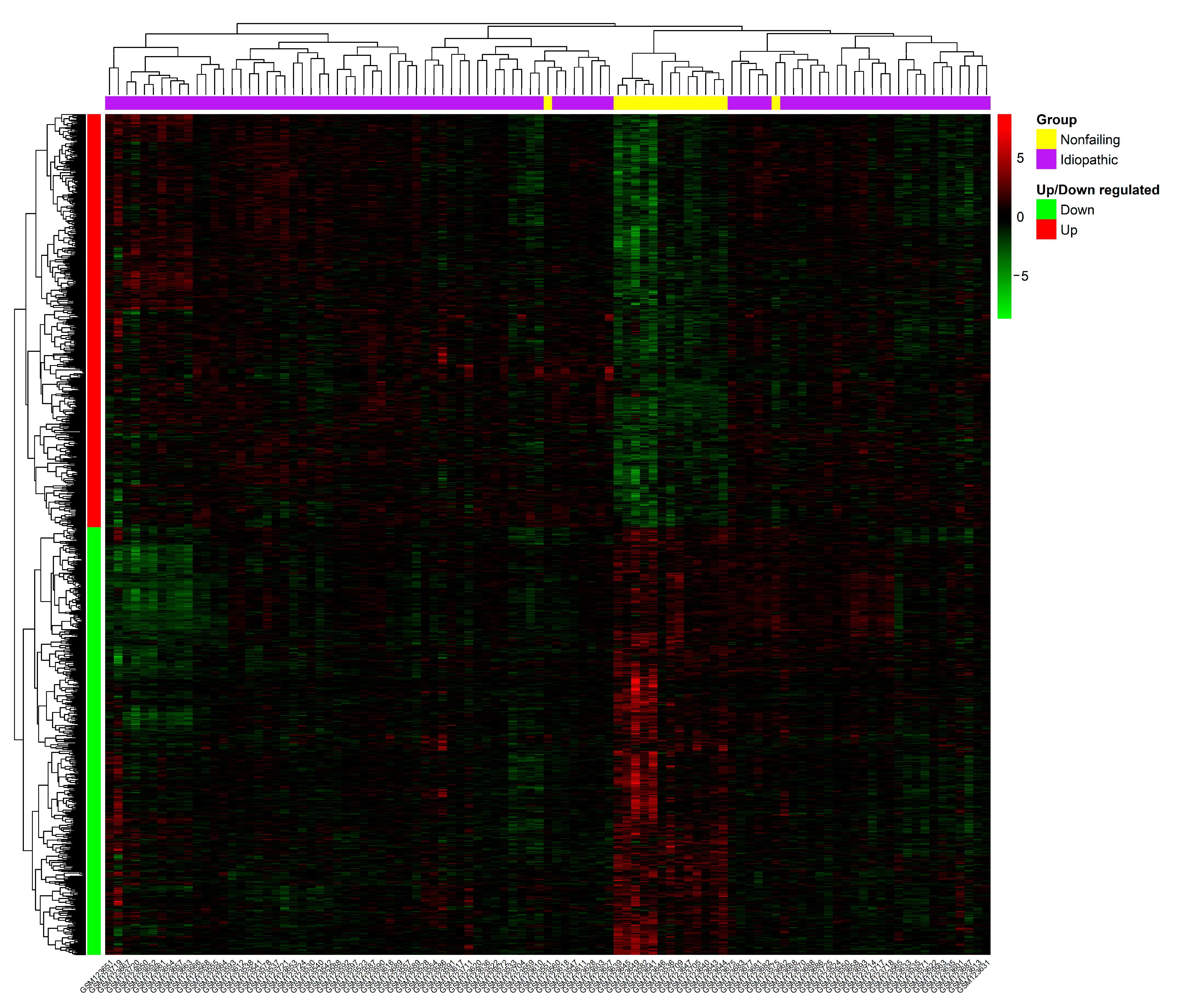
3.1. Data Preprocessing and Identification of DEGs

The GSE5406, a microarray expression dataset, includes mRNA expression data from 102 samples, 86 IDCM samples, and 16 normal LV samples. For our analysis, we selected IDCM and normal samples and downloaded the series matrix file, which was pre-processed and normalized together using the robust multichip average (RMA) method. Using the limma R program, the DEGs were filtered (criteria: |log2 FC| > 0.3 and p-value < 0.05). Finally, 1287 DEGs (Table S1) were extracted from the IDCM samples and compared to the normal left ventricle function samples, comprising 632 upregulated and 655 downregulated DEGs (Figure 1 depicts the heatmap for upregulated and downregulated DEGs).

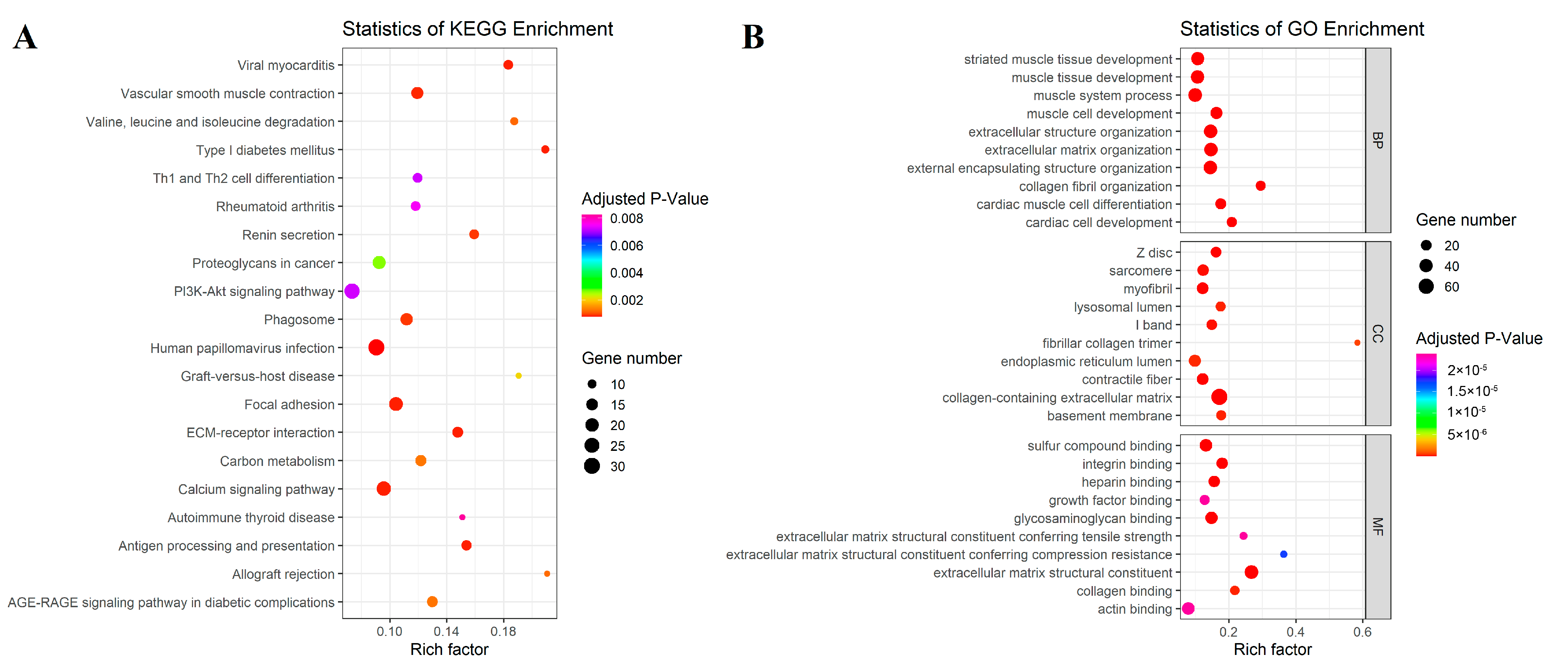
3.2. Differentially Expressed Gene Enrichment Analysis

We performed KEGG pathway analysis by using the ClusterProfiler package for upregulated DEGs. KEGG analysis demonstrated that human papillomavirus infection, viral myocarditis, focal adhesion, ECM receptor interaction, and the calcium signaling pathway were linked to our DEGs (Table 2). The top 20 enriched KEGG pathways are shown in Figure 2A.
Furthermore, GO analysis was performed by using the ClusterProfiler package for upregulated DEGs. The result revealed that the most significant GO terms under the biological processes category (in descending order of adjusted p-value) were extracellular matrix organization (GO:0030198), extracellular structure organization (GO:0043062), and external encapsulating structure organization (GO:0045229). Furthermore, the most significant GO terms under the molecular functions category were extracellular matrix structural constituent (GO:0005201), glycosaminoglycan binding (GO:0005539), and integrin binding (GO:0005178). Finally, the most significant GO terms under the cellular component category were collagen-containing extracellular matrix (GO:0062023), contractile fiber (GO:0043292), and Z disc (GO:0030018) (Figure 2B).

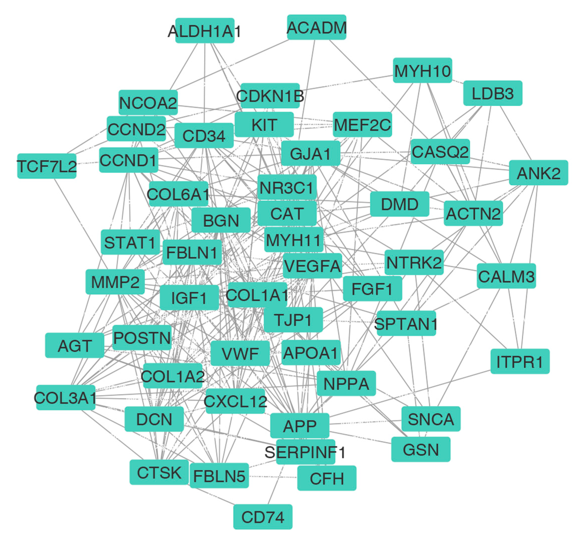
3.3. PPI Network Construction and HUB Selection

For this article, we selected the most upregulated DEGs and used the STRING online database to produce a protein-protein interaction (PPI) network. According to the STRING analysis, our network consisted of 615 nodes and 2543 edges. The top 100 genes, ranked based on the intersections between four topological algorithms (i.e., degree, betweenness, closeness, and centroid), were expected to play a significant role in biological processes and were set as HUBs (Figure 3, Table S2). Although in vivo examination and validation of all these genes can be of particular importance in understanding the pathogenesis of IDCM, due to experimental limitations, we selected seven of the top 100 genes (nominated as “HUB of HUBs”) based on the bioinformatics analysis results and the literature review. In fact, one of the common methods for selecting genes (i.e., seven HUB of HUBs in this study) out of the top ones in the PPI network (to introduce them as biomarkers or potential therapeutic targets and consider further experimental studies) is the results of patient survival analysis. Despite the abundance of survival data for cancer patients, the absence of such information for DCM patients presents a technical limitation. Consequently, we relied on an extensive literature review as an alternative and considerable method for gene selection. Thus, due to these limitations, five genes (VEGF-A, STAT1, APP, CCND1, and IGF1) with the best betweenness ranking were selected based on the bioinformatics analysis, and two genes related to heart disease were selected according to the literature review (MYH 10 and MYH 11): 1. Vascular endothelial growth factor-A (VEGF-A) can regulate angiogenesis, vascular permeability, and inflammation. Based on our bioinformatics analysis, this gene was among the top ten genes, but there were few documents and pieces of evidence examining its expression in IDCM patients. 2. Signal transducer and activator of transcription 1 (STAT1) is a member of the STAT family, and it has been proposed that STAT1 promotes the generation of larger infarcts, which can lead to heart failure, by enhancing apoptosis and negatively regulating autophagy [40]. 3. The amyloid precursor protein (APP) is known as a precursor protein of Alzheimer’s disease (AD)-related amyloid β-protein (Aβ) [41]. Recent studies identified Aβ aggregates in the hearts of patients with dilated cardiomyopathy [42]. It was important and exciting for us to examine the expression of APP in patients without a history of Alzheimer’s. 4. CCND1 is an important cell cycle gene involved in three pathways (the cell cycle, Hedgehog (Hh) signaling, and Wnt signaling). Limited previous observations have reported its upregulation in IDCM [43,44]. 5. Insulin-like growth factor-1 (IGF-1) is a peptide hormone that activates canonical and non-canonical signaling pathways in the heart. It has a direct growth-promoting effect on cardiomyocyte hypertrophy. 6. Myosin heavy chain (MYH10), the only non-muscle myosin, is expressed in the heart and required for normal heart development. Recently, the upregulation of MYH10 in the cardiomyocytes of adults with heart failure has been reported [45]. 7. Myosin heavy chain 11 (MYH11) belongs to the MYH family and is a myocardial contractile protein. Hydrolyzed proteins are involved in muscle contraction through adenosine triphosphate. Myocardial cytoskeleton proteins are important for maintaining the structural and functional integrity of the myocardium [46]. However, there are few studies on the changes in cytoskeletal proteins associated with dilated cardiomyopathy. Figure 4 represents the violin plots of selected HUB of HUBs expression patterns.

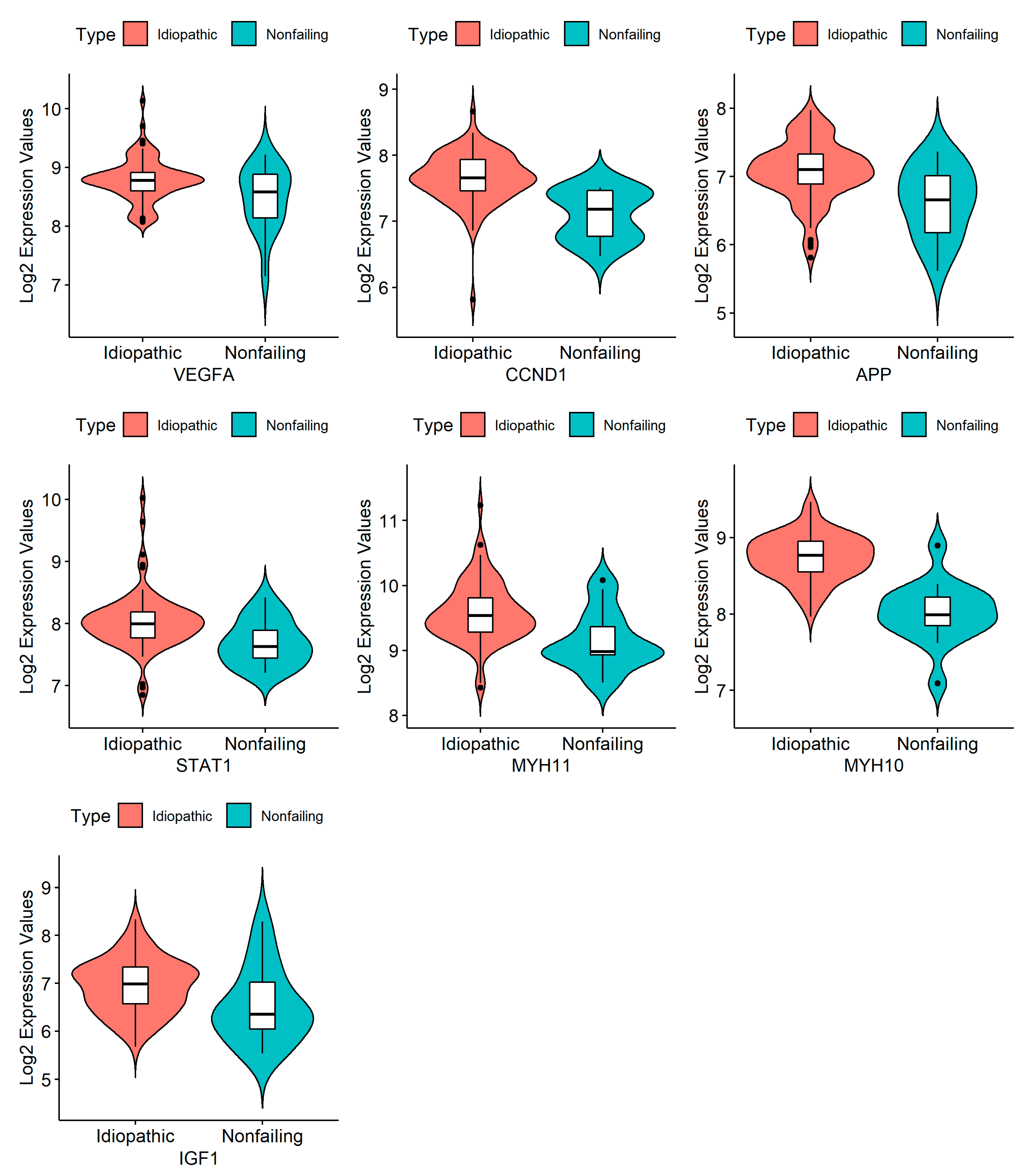
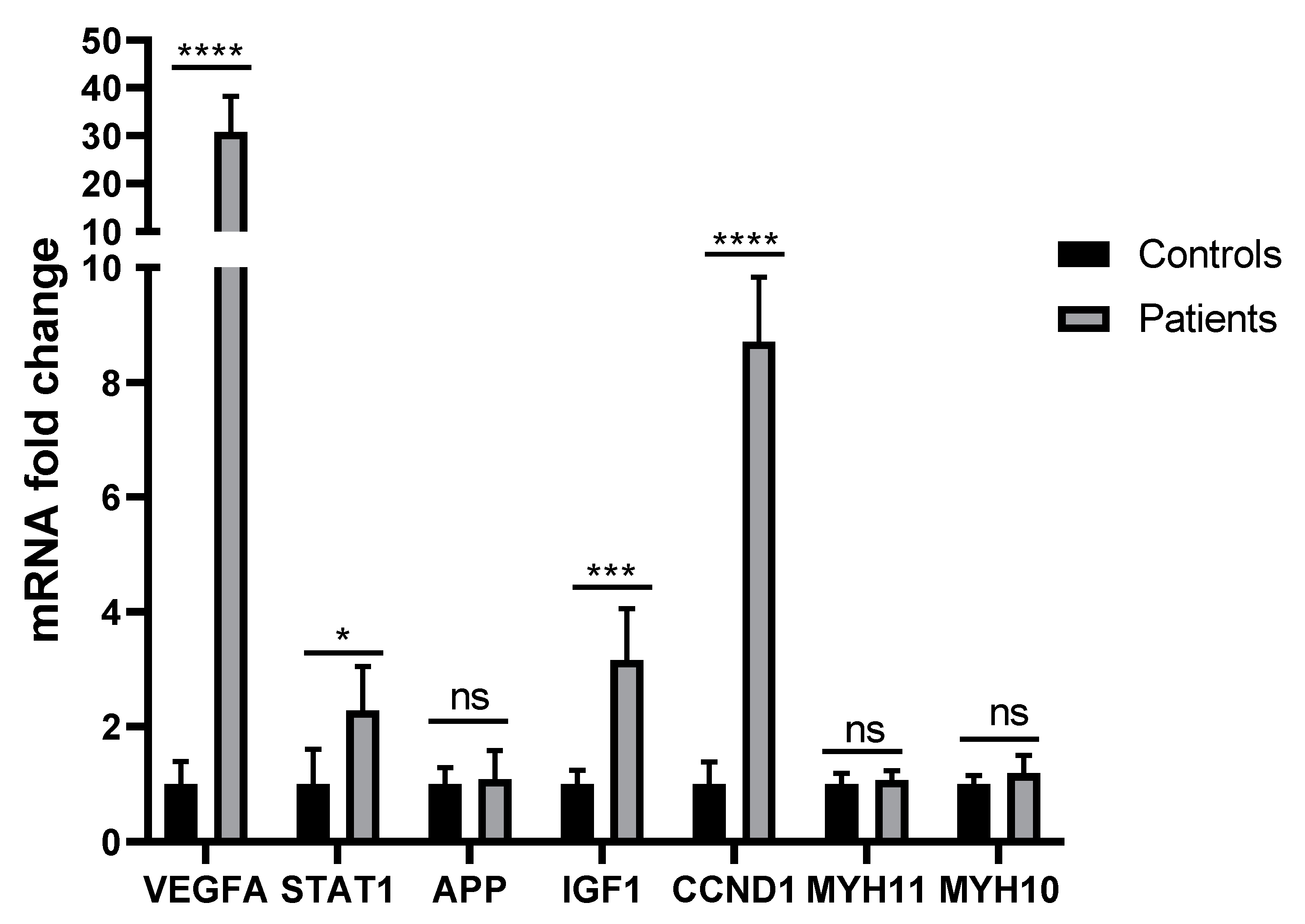
3.4. RNA Isolation and Quantification, cDNA Synthesis, and RT-PCR

Isolated RNA was quantified with NanoDrop (Biotech. ENG). The OD 260/280 and OD 260/230 ratios of 1.8 to 2 and 1.7 to 2 indicated acceptable RNA quantities. The synthesized cDNA was similarly quantified with NanoDrop (Biotech. ENG). The melting curve was investigated at 60 to 96 °C. The highest expression in patients compared to controls was found for the gene VEGFA (p < 0.001), followed by the genes CCND1 (p < 0.001) and IGF1 (p = 0.001). STAT1 was also overexpressed in patients (p = 0.017). Contrarily, the genes APP (p = 0.823), MYH11 (p = 0.781), and MYH10 (p = 0.502) were not significantly more expressed in patients compared to controls (Figure 5).

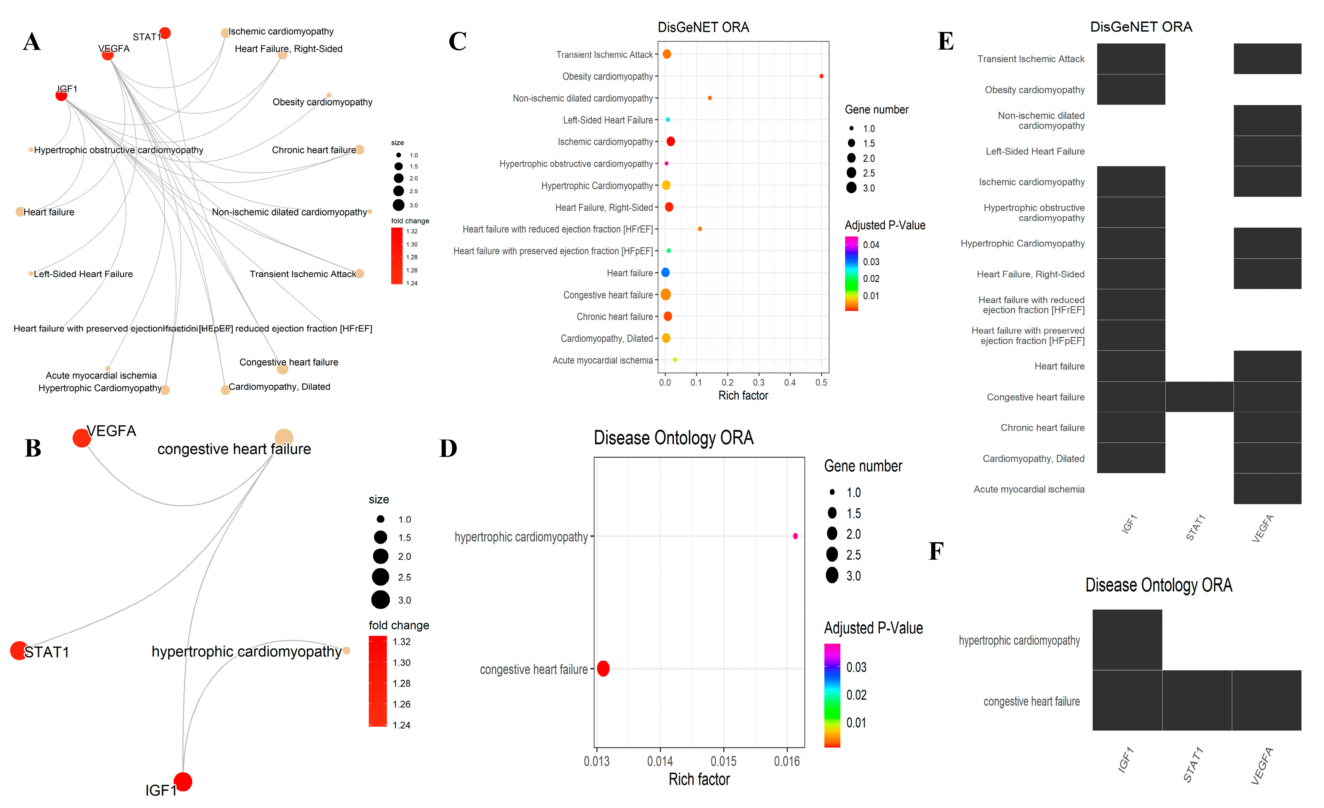
3.5. Over-Representation Analysis for the DisGeNET and DO

Over-representation analysis (ORA) on four significantly expressed HUBs of HUBs (VEGFA, IGF1, CCND1, and STAT1) was applied by the DOSE package for two databases of the disease gene network (DisGeNET) and disease ontology (DO). However, it is essential to interpret these results cautiously and consider the databases’ constraints. DisGeNET integrates data from expert-curated repositories, scientific literature, animal models, and GWAS catalogs. Nevertheless, it is important to recognize that the data contained in these databases may have inherent limitations, such as errors in text mining or insufficient annotations.
In this study, three keywords were used to filter out diseases that are related to this project: “cardiomyopathy,” “heart failure,” and “ischemic.” Some genes were also involved in “cardiovascular” diseases, but this keyword was ignored to prevent generalization. According to the DisGeNET and DO analyses, two genes (IGF1 and VEGFA) were significantly related to IDCM, while there is no link between STAT1 and CCND1 with IDCM (Figure 6, Table 3).
It should be noted that CCND1’s role was detected by DisGeNET in two related diseases. The former was “Cardiomyopathy, Familial Idiopathic,” while after studying the stated PMIDs, it was found that all of them were related to “invasive ductal carcinoma (IDC)” incorrectly, instead of idiopathic dilated cardiomyopathy (IDC), due to possible errors in text mining. The latter was “myocardial ischemia,” given only one PMID, which was not enough to conclude the role of CCND1 in that disease. To sum up, there is insufficient cardio-relational experimental evidence for CCND1. However, it is important to emphasize that the absence of significant associations in the current analysis does not preclude the existence of a potential relationship between CCND1 and IDCM. A deeper exploration of the potential involvement of CCND1 in IDCM is needed through rigorous experiments and validation.

4. Discussion

DCM is a consequence of various pathogenic factors, such as genetic, infectious, hormonal, and environmental factors [47]. However, knowledge about the genotype-phenotype relationship is still unknown. Therefore, DCM is almost always diagnosed late, which in turn causes a poor prognosis. In recent years, gene sequencing and bioinformatics methods have resulted in new ideas for understanding the mechanisms of disease development, disease diagnosis, and personalized precision medicine development and presented some meaningful results. Sun and Li (2023) used WGCNA and the machine learning algorithm to identify biomarkers of IDCM and obtained two hub genes, AQP3 and CYP2J2, which have the potential to serve as targets for the diagnosis and management of IDCM [48]. Liu et al. (2022) investigated the candidate genes and pathways involved in DCM patients (data sets GSE3585 and GSE5406) and predicted the microRNAs (miRNAs) targeting the hub genes. Their screening criteria were set as p < 0.05 and |log fold change (FC)| > 0.589. Furthermore, they investigated the pattern of immune cell infiltration in DCM. The top 10 hub genes included collagen type III alpha 1 chain (COL3A1), COL1A2, signal transducer and activator of transcription 3 (STAT3), C-C motif chemokine ligand 2 (CCL2), fibromodulin (FMOD), aspirin (ASPN), C-X-C motif chemokine ligand 12 (CXCL12), lumican (LUM), heat shock protein 90 alpha family class A member 1 (HSP90AA1), and osteoglycin (OGN) [49]. Si (2023) has screened the associated genes and biological pathways of inflammatory dilated cardiomyopathy (DCMi) [50]. Luo et al. (2020) identified novel long non-coding RNA (lncRNA) biomarkers associated with DCM and revealed the potential molecular mechanisms of DCM development using bioinformatics approaches (dataset GSE5406 from GEO) [51]. Zhang et al. (2022) screened diagnostic biomarkers and identified the landscape of immune infiltration in DCM (dataset GSE141910 from Geo). They found that ASPN, CD163, IL10, and LUM could predict the occurrence of DCM [52]. Huang et al. (2018) found that Fos proto-oncogene, AP-1 transcription factor subunit, tissue inhibitor of metalloprotease-1, and serpin family E member 1 may serve as therapeutic targets in DCM [25].
In this study, we investigated which of the DEGs related to the advanced stages of the disease (confirmed by bioinformatics methods using the GSE5406 dataset) have increased expression in the early stages of the disease (using the RT-PCR method in blood samples of IDCM patients participating in this study). It is noteworthy that the dataset used in this study (GSE5406) has already been analyzed by several researchers to predict potential targets [25,53], but in this study we set different thresholds for some parameters compared to previous studies. For instance, a similar paper for this dataset used |log2 FC| > 0.589 to detect DEGs, while we used |log2 FC| > 0.3. FC is a main parameter to show the change in expression levels of genes. FC analysis is a very intuitive method to identify DEGs [54]. Although in most cases, |log2 FC| is set to ≥1, sometimes these cutoff values can significantly change the interpretation of the microarray due to the deletion of some hub genes [55]. For instance, a minor change in transcription factor expression may alter the entire system and induce a high effect across the network of genes [56]. Thus, we covered more genes in our study that could possibly have significant impacts on cellular processes while having a minor gene expression level. Furthermore, we took additional steps to ensure the reliability of our findings and performed multiple rounds of analysis with different thresholds and parameters (e.g., different topological centrality parameters for detecting HUBs in our PPI network and then selecting HUBs of HUBs) and used different software packages for the analysis to confirm the robustness of our results (e.g., using DOSE and enrichplot packages to perform and visualize over-representation analysis (ORA) on significantly expressed HUBs of HUBs). Finally, from the DEGs identified by bioinformatics methods, seven genes were selected to check their expression in the blood of patients participating in this study.
RT-PCR tests showed that four of the seven selected genes (VEGFA, IGF1, CCND1, and STAT1) have increased expression in blood samples from patients. Some studies, in accordance with the results of our RT-PCR experiments, showed an increase in the expression of some genes in a variety of heart diseases. However, for genes whose expression changes were not observed in our study, we did not find any reports on the expression of these genes in patients with IDCM or other heart diseases. The literature review revealed that, in general, there is little information on the expression changes of the HUB genes selected in this study in IDCM patients.
VEGFA is one of the HUB genes whose increased expression in patients was observed in our study. The generation of six isoforms, including VEGF111, VEGF121, VEGF145, VEGF165, VEGF189, and VEGF206, from VEGFA mRNA under the alternative splicing process has been reported [57]. VEGFA and its two receptors, VEGFR-1 and VEGFR-2, play fundamental roles in angiogenesis under physiological and pathological conditions, as well as in neurological and cardiac growth and morphogenesis [46,58]. Disturbance in angiogenesis has been shown to contribute to the development of myocardial interstitial fibrosis in IDCM [59,60,61,62]. Some evidence has confirmed that serum VEGF-A [61] or total VEGF [60] is significantly higher in patients with IDCM compared to controls. Moreover, significant increases in serum VEGF levels were reported in DCM patients compared to ischemic cardiomyopathy patients [63]. However, another study showed that, in the heart tissue of patients with end-stage DCM, the mRNA transcript levels of VEGF165 and VEGF189 and the protein level of VEGF-A decreased compared to controls [64]. In fact, it has been indicated that in the disease’s acute stage, the expression of genes and receptors may be altered significantly, with varying intensities and at different times. Accordingly, the concentration of VEGF-A may fluctuate significantly [65]. In this regard, it has been reported that VEGF-A is strongly expressed in the acute and subacute stages of CNS damage, but its expression decreases over time [66].
CCND1 was one of the HUB genes that our RT-PCR studies confirmed to be overexpressed in IDCM patients. CCND1 is a proto-oncogene that manages progression via the G1-S phase of the cell cycle [67], and its involvement in heart diseases has been reported [68]. In a study reported on children and adults with IDCM in the end-stages, it was shown that the CCND1 gene is not overexpressed in adults, but it is overexpressed in children [42]. A significant association of microRNAs (miRNAs) with heart diseases such as cardiac arrhythmia, myocardial infarction, and cardiac hypertrophy has been confirmed [69]. For instance, it has been revealed that several miRNAs (e.g., miR-34a, miR-28, miR-148, and miR-93) are differentially expressed in patients with left ventricular hypertrophy (LVH) and normal controls [70]. Moreover, more recent studies confirmed that miR-93 is reduced in LVH patients and that CCND1 is the direct target of miR-93 [68]. In mouse models, the MYCN gene has been revealed to induce cardiomyocyte proliferation, at least in part by upregulating CCND1, CCND2, and the inhibitor of DNA binding 2 (ID2). MYCN is a multifunctional transcription factor that plays an essential role in the development of disease [71]. MYCN has been indicated to be the major disease-causing gene for Feingold syndrome, a developmental disorder characterized in part by congenital heart abnormalities [72]. However, some other reports showed that the upregulation of CCND1 and CCND2 suppressed DCM caused by TTN gene insufficiency [73]. Truncating variants of TTN are common in patients with IDCM [74,75,76]. Titin, encoded by the gene TTN, is a giant sarcomeric protein that is critical for cardiac contraction and relaxation [77].
IGF-1 is a peptide hormone homologous to pro-insulin, which is expressed in most tissues and is involved in cell proliferation, apoptosis, migration, and differentiation [78]. The results of epidemiological studies on the relationship between IGF-1 levels and the risk of cardiovascular disease were inconsistent [79], as various reports indicated that both an increase and decrease in serum IGF-1 levels were involved in heart disease [80]. Similarly, a meta-analysis conducted in 2015 showed that both low and high IGF-1 levels were associated with an increased risk of cardiovascular disease [79].
Indeed, more recent studies have also not reached a consensus on the association of IGF-1 with heart disease (including dilated cardiomyopathy): while some have reported a decrease in IGF-1, others have reported an increase, and some studies have not even found an association between IGF-1 levels and heart failure. In this regard, some evidence associates the decline in IGF-1 expression with heart disease. For instance, in a cohort study of 337 patients, IGF-1 was reported as a predictor of cardiovascular mortality in patients with heart failure, so older patients with lower serum IGF-1 levels showed higher mortality [81]. Another report described that IGF-1 signaling was inhibited in end-stage DCM [82]. Moreover, a study on DCM rats revealed that the intramuscular injection of human umbilical cord-derived mesenchymal stem cells (hUCMSC) improved cardiac function, and the same study suggested that the expression of IGF-1, HGF, and VEGF in the myocardium of DCM rats was remarkably increased by hUCMSC injection [83]. In contrast, other evidence suggests that increased IGF-1 expression is involved in heart disease. Free IGF-1 levels have been shown to be higher in patients with myocardial infarction compared to controls [64]. Furthermore, IGF-1 levels in patients with HFpEF (heart failure with preserved ejection fraction) were significantly higher than the IGF-1 levels of their HFrEF (heart failure with reduced ejection fraction) counterparts [84]. Moreover, it was shown that the concentration of IGF-binding protein (IGFBP)-1 and the ratio of IGFBP-1/IGF-1 in patients with heart failure were significantly lower than in a control group, and they can be used to easily identify patients with and without heart failure [85]. Due to the fact that IGFBP1 and IGFBP2 have inhibitory effects on the biological activity of IGF-1, it can be concluded that, in this group of patients, although the amount of IGF-1 did not increase due to the decrease in the inhibitory effect of its binding proteins, in fact, IGF-1 had increased activity [86]. Furthermore, some evidence suggests that elevated IGF-1 levels may be causally correlated with a higher risk of type 2 diabetes [68], and another report indicated that patients with type 2 diabetes were notably associated with an increased risk of IDCM in all age and sex categories, with the exception of men over 64 years of age [87]. On the other hand, some studies have linked an increase in IGF-1 system activity to a decrease in lifespan [88], while other reports have revealed that an increase in IGF-1 is associated with increased survival in people over 65, unlike younger people [89]. In general, it can be concluded that an increase in IGF-1 in the under-65 population can be dangerous, which is highly consistent with the data of our study, in which the subjects were under 65 years of age.
STAT1 is another gene whose high expression was observed in the IDCM patients who participated in our study. STAT1, encoding a signal transducer and activator of transcription factor, is involved in apoptosis and the interferon response [90]. Some reports have suggested that this gene is involved in dilated cardiomyopathy. It has been shown that mutations of sodium voltage-gated channel alpha subunit 5 (SCN5A) are involved in dilated cardiomyopathy [91,92]. Furthermore, mice with the SCN5A N1325S mutation in the heart (TG-NS mice) exhibited the phenotype of dilated cardiomyopathy and heart failure [93], and other studies have indicated the upregulation of STAT1 in the hearts of TG-NS mice [90]. Moreover, in vitro studies on engineered myocardial tissues stated that interferon-γ (IFN-γ) damages myofibrillar organization and contractile force production in human cardiomyocytes through upregulation of the JAK/STAT signaling pathway and downregulation of multiple sarcomeric proteins. In fact, circulating IFN-γ (a pro-inflammatory cytokine) is enhanced in numerous clinical conditions, including acute coronary syndrome, autoimmune and inflammatory diseases, sepsis, and viral infections, which are correlated with a high risk of myocardial dysfunction [94]. In addition, Trypanosoma cruzi, a protozoan parasite, causes zoonotic Chagas disease, which is a chronic and systemic infection that typically brings about progressive dilated cardiomyopathy and gastrointestinal manifestations [95]. Some in vitro studies revealed that in response to T. cruzi, intracellular STAT1 was increased both at the mRNA and protein levels [96].
Further analysis was performed through the DisGeNET database to find the correlation between four significant HUB of HUBs genes (VEGFA, IGF1, CCND1, and STAT1) and IDCM. According to DisGeNET data, confirmatory findings were reported for two genes (IGF1 and VEGFA) with an IDCM link in DisGeNET. These results suggest that IGF1 and VEGFA are also associated with other heart diseases similar to IDCM. Furthermore, DisGeNET data show that two genes, including STAT1 and CCND1, have not been associated with IDCM, but STAT1 is related to congestive heart failure. Although, based on DisGeNET results, CCND1 is not related to IDCM, its upregulation in IDCM has been previously reported in children [44]. The same report has shown that CCND1 is not overexpressed in adults at the end stage of IDCM [44]. Thus, our study is the first evidence of CCND1’s upregulation in IDCM adult patients at an early stage, and CCND1 might be a potential newfound gene for cardiomyopathy.
In contrast to the genes that exhibited increased expression in the patients who participated in our study, some upregulated genes (based on analysis of GEO data) did not show significant changes in the patients compared to the control group, based on the RT-PCR results. These genes included MYH10, MYH11, and APP. There are several points to be made regarding this issue. As mentioned in the previous paragraphs, the expression of some genes depends on the stage of the disease or the age of the patients. It is clear from the data of the present study and the GSE5406 dataset that the patients participating in the present study were in the early stages of the disease, while the patients in the GSE5406 dataset were in the final stages. It can be the reason for the difference in APP, MYH10, and MYH11 gene expressions in these two groups of patients. Moreover, the number of samples used in this study was limited; therefore, future studies can include larger groups of participants to investigate these genes’ status and identify other genes associated with the disease. Due to the limited available studies regarding the expression of these genes in IDCM patients, further studies are needed for a more detailed discussion. Finally, the lack of sample characteristics and clinical and survival information in the GSE5406 dataset were other limitations in this research.

5. Conclusions

In this study, seven genes that have increased expression in IDCM patients (obtained from bioinformatics analysis on the GSE5406 dataset) were selected for further investigation in patients referred to Jahrom Hospital. The RT-PCR results confirmed the overexpression of the STAT1, IGF1, CCND1, and VEGFA genes in blood samples of patients with IDCM, which was consistent with our GEO analysis. Among these four genes, CCND1 was the only one not linked to IDCM or related heart disease in DisGeNET; therefore, according to our investigation, CCND1 may be introduced as a potential biomarker for IDCM. Conversely, there were no significant differences in the expression of three DEGs (APP, MYH10, and MYH11) between patients and controls, which was inconsistent with GEO analysis. Considering the limited available studies on the expression of these genes in IDCM patients and the lack of detailed information on the GSE5406 dataset, more studies are needed for a more detailed discussion. Nevertheless, the present study may advance our understanding of IDCM pathogenesis and provide new targets for clinical diagnosis.

Supplementary Materials

The following are available online at https://www.mdpi.com/article/10.3390/genes14061243/s1, Table S1: DEGS(1287), Table S2: HUB genes.

Author Contributions

Conceptualization, K.D., A.S. and S.Z.; methodology, A.B., A.H. and S.Z.; validation, S.Z. and S.B.; formal analysis, A.B., F.A. and P.H.; investigation, A.H. and F.A.; data curation, F.A., P.H., M.F. and A.B.; writing—original draft preparation, A.S., S.B., S.Z., A.B., M.B., A.H. and K.D.; writing—review and editing, K.D., A.S., S.B., S.Z. and A.B.; visualization, S.Z.; supervision, A.S., S.Z. and S.B. All authors have read and agreed to the published version of the manuscript.

Funding

This research received no external funding.

Institutional Review Board Statement

The present study conforms to the Declaration of Helsinki for the use of human tissue or subjects. The study was approved by the Ethics Committee of the Medical Sciences University of Jahrom (permission number: IR.JUMS.REC.1398.112, Iran, date of approval: 4 March 2020).

Informed Consent Statement

All the patients gave written informed consent for their participation in the study.

Data Availability Statement

No new data were created or analyzed in this study. Data sharing does not apply to this article.

Conflicts of Interest

The authors declare no conflict of interest.

References

  1. Roth, G.A.; Mensah, G.A.; Johnson, C.O.; Addolorato, G.; Ammirati, E.; Baddour, L.M.; Barengo, N.C.; Beaton, A.Z.; Benjamin, E.J.; Benziger, C.P.; et al. Global Burden of Cardiovascular Diseases and Risk Factors, 1990–2019: Update From the GBD 2019 Study. J. Am. Coll. Cardiol. 2020, 76, 2982–3021. [Google Scholar] [CrossRef] [PubMed]
  2. Jefferies, J.L.; Towbin, J.A. Dilated cardiomyopathy. Lancet 2010, 375, 752–762. [Google Scholar] [CrossRef] [PubMed]
  3. Phelan, D.; Wilson, G.; James, P.; Lockhart, P. The genetics of cardiomyopathy, new technologies and the path to personalised medicine. OA Genet. 2013, 1, 1–10. [Google Scholar]
  4. Schultheiss, H.-P.; Fairweather, D.; Caforio, A.L.; Escher, F.; Hershberger, R.E.; Lipshultz, S.E.; Liu, P.P.; Matsumori, A.; Mazzanti, A.; McMurray, J. Dilated cardiomyopathy. Nat. Rev. Dis. Prim. 2019, 5, 32. [Google Scholar] [CrossRef]
  5. Bailly, C.; Henriques, S.; Tsabedze, N.; Krause, A. Role of family history and clinical screening in the identification of families with idiopathic dilated cardiomyopathy in Johannesburg, South Africa. S. Afr. Med. J. 2019, 109, 673–678. [Google Scholar] [CrossRef] [Green Version]
  6. Xu, H.; Dorn, I.I.G.W.; Shetty, A.; Parihar, A.; Dave, T.; Robinson, S.W.; Gottlieb, S.S.; Donahue, M.P.; Tomaselli, G.F.; Kraus, W.E. A genome-wide association study of idiopathic dilated cardiomyopathy in African Americans. J. Pers. Med. 2018, 8, 11. [Google Scholar] [CrossRef] [Green Version]
  7. Woulfe, K.C.; Siomos, A.K.; Nguyen, H.; SooHoo, M.; Galambos, C.; Stauffer, B.L.; Sucharov, C.; Miyamoto, S. Fibrosis and fibrotic gene expression in pediatric and adult patients with idiopathic dilated cardiomyopathy. J. Card. Fail. 2017, 23, 314–324. [Google Scholar] [CrossRef] [Green Version]
  8. Wong, C.M.; Hawkins, N.M.; Jhund, P.S.; MacDonald, M.R.; Solomon, S.D.; Granger, C.B.; Yusuf, S.; Pfeffer, M.A.; Swedberg, K.; Petrie, M.C. Clinical characteristics and outcomes of young and very young adults with heart failure: The CHARM programme (Candesartan in Heart Failure Assessment of Reduction in Mortality and Morbidity). J. Am. Coll. Cardiol. 2013, 62, 1845–1854. [Google Scholar] [CrossRef] [Green Version]
  9. Merlo, M.; Pyxaras, S.A.; Pinamonti, B.; Barbati, G.; Di Lenarda, A.; Sinagra, G. Prevalence and prognostic significance of left ventricular reverse remodeling in dilated cardiomyopathy receiving tailored medical treatment. J. Am. Coll. Cardiol. 2011, 57, 1468–1476. [Google Scholar] [CrossRef] [Green Version]
  10. Aziz, H.; Zaas, A.; Ginsburg, G.S. Peripheral blood gene expression profiling for cardiovascular disease assessment. Genom. Med. 2007, 1, 105–112. [Google Scholar] [CrossRef] [Green Version]
  11. Liew, C.-C.; Ma, J.; Tang, H.-C.; Zheng, R.; Dempsey, A.A. The peripheral blood transcriptome dynamically reflects system wide biology: A potential diagnostic tool. J. Lab. Clin. Med. 2006, 147, 126–132. [Google Scholar] [CrossRef]
  12. Basu, M.; Wang, K.; Ruppin, E.; Hannenhalli, S. Predicting tissue-specific gene expression from whole blood transcriptome. Sci. Adv. 2021, 7, eabd6991. [Google Scholar] [CrossRef] [PubMed]
  13. Cappuzzello, C.; Napolitano, M.; Arcelli, D.; Melillo, G.; Melchionna, R.; Di Vito, L.; Carlini, D.; Silvestri, L.; Brugaletta, S.; Liuzzo, G. Gene expression profiles in peripheral blood mononuclear cells of chronic heart failure patients. Physiol. Genom. 2009, 38, 233–240. [Google Scholar] [CrossRef]
  14. Kontaraki, J.E.; Marketou, M.E.; Zacharis, E.A.; Parthenakis, F.I.; Vardas, P.E. Early cardiac gene transcript levels in peripheral blood mononuclear cells in patients with untreated essential hypertension. J. Hypertens. 2011, 29, 791–797. [Google Scholar] [CrossRef]
  15. Kurano, M.; Darestani, S.G.; Shinnakasu, A.; Yamamoto, K.; Dochi, Y.; Uemura, K.; Ikeda, Y.; Kikuchi, A.; Hashiguchi, H.; Deguchi, T. mRNA expression of platelet activating factor receptor (PAFR) in peripheral blood mononuclear cells is associated with albuminuria and vascular dysfunction in patients with type 2 diabetes. Diabetes Res. Clin. Pract. 2018, 136, 124–133. [Google Scholar] [CrossRef] [PubMed]
  16. Kontaraki, J.E.; Parthenakis, F.I.; Nyktari, E.G.; Patrianakos, A.P.; Vardas, P.E. Myocardial gene expression alterations in peripheral blood mononuclear cells of patients with idiopathic dilated cardiomyopathy. Eur. J. Heart Fail. 2010, 12, 541–548. [Google Scholar] [CrossRef]
  17. Marketou, M.E.; Kontaraki, J.; Patrianakos, A.; Kanoupakis, E.; Plevritaki, A.; Mavrakis, H.; Kallergis, E.; Koutalas, E.; Anastasiou, I.; Chlouverakis, G. Long-term prognostic value of myocardin expression levels in non-ischemic dilated cardiomyopathy. Heart Vessel. 2021, 36, 1841–1847. [Google Scholar] [CrossRef] [PubMed]
  18. Xu, H.; Xie, X.; Li, J.; Zhang, Y.; Xu, C.; Yang, J. Early right ventricular apical pacing-induced gene expression alterations are associated with deterioration of left ventricular systolic function. Dis. Markers 2017, 2017, 8405196. [Google Scholar] [CrossRef] [PubMed] [Green Version]
  19. Simantirakis, Ε.; Arkolaki, E.; Kontaraki, J.; Chlouverakis, G.; Mavrakis, H.; Kallergis, E.; Parthenakis, F.; Vardas, P. The impact of paced QRS duration on the expression of genes related to contractile function of the left ventricle in chronically paced patients from the right ventricular apex. Hell. J. Cardiol. 2020, 61, 274–278. [Google Scholar] [CrossRef]
  20. Arkolaki, E.G.; Simantirakis, E.N.; Kontaraki, J.E.; Chrysostomakis, S.I.; Patrianakos, A.P.; Chlouverakis, G.I.; Nakou, E.S.; Vardas, P.E. Alterations in the expression of genes related to contractile function and hypertrophy of the left ventricle in chronically paced patients from the right ventricular apex. Ep Eur. 2015, 17, 1563–1570. [Google Scholar] [CrossRef] [Green Version]
  21. Martin-Lorenzo, M.; Gonzalez-Calero, L.; Maroto, A.S.; Martinez, P.J.; Zubiri, I.; de la Cuesta, F.; Mourino-Alvarez, L.; Barderas, M.G.; Heredero, A.; Aldamiz-Echevarría, G. Cytoskeleton deregulation and impairment in amino acids and energy metabolism in early atherosclerosis at aortic tissue with reflection in plasma. Biochim. Biophys. Acta (BBA) Mol. Basis Dis. 2016, 1862, 725–732. [Google Scholar] [CrossRef]
  22. Anand, S.S.; Yusuf, S.; Vuksan, V.; Devanesen, S.; Teo, K.K.; Montague, P.A.; Kelemen, L.; Yi, C.; Lonn, E.; Gerstein, H. Differences in risk factors, atherosclerosis, and cardiovascular disease between ethnic groups in Canada: The Study of Health Assessment and Risk in Ethnic groups (SHARE). Lancet 2000, 356, 279–284. [Google Scholar] [CrossRef]
  23. Liu, M.; Jiang, S.; Ma, Y.; Ma, J.; Hassan, W.; Shang, J. Peripheral-blood gene expression profiling studies for coronary artery disease and its severity in Xinjiang population in China. Lipids Health Dis. 2018, 17, 154. [Google Scholar] [CrossRef] [Green Version]
  24. Sinnaeve, P.R.; Donahue, M.P.; Grass, P.; Seo, D.; Vonderscher, J.; Chibout, S.-D.; Kraus, W.E.; Sketch, M., Jr.; Nelson, C.; Ginsburg, G.S. Gene expression patterns in peripheral blood correlate with the extent of coronary artery disease. PLoS ONE 2009, 4, e7037. [Google Scholar] [CrossRef]
  25. Huang, H.; Luo, B.; Wang, B.; Wu, Q.; Liang, Y.; He, Y. Identification of Potential Gene Interactions in Heart Failure Caused by Idiopathic Dilated Cardiomyopathy. Med. Sci. Monit. Int. Med. J. Exp. Clin. Res. 2018, 24, 7697–7709. [Google Scholar] [CrossRef]
  26. Laguna, J.C.; Alegret, M. Regulation of gene expression in atherosclerosis: Insights from microarray studies in monocytes/macrophages. Pharmacogenomics 2012, 13, 477–495. [Google Scholar] [CrossRef]
  27. Nührenberg, T.G.; Langwieser, N.; Binder, H.; Kurz, T.; Stratz, C.; Kienzle, R.-P.; Trenk, D.; Zohlnhöfer-Momm, D.; Neumann, F.-J. Transcriptome analysis in patients with progressive coronary artery disease: Identification of differential gene expression in peripheral blood. J. Cardiovasc. Transl. Res. 2013, 6, 81–93. [Google Scholar] [CrossRef]
  28. Hannenhalli, S.; Putt, M.E.; Gilmore, J.M.; Wang, J.; Parmacek, M.S.; Epstein, J.A.; Morrisey, E.E.; Margulies, K.B.; Cappola, T.P. Transcriptional genomics associates FOX transcription factors with human heart failure. Circulation 2006, 114, 1269–1276. [Google Scholar] [CrossRef] [PubMed] [Green Version]
  29. Barrett, T.; Wilhite, S.E.; Ledoux, P.; Evangelista, C.; Kim, I.F.; Tomashevsky, M.; Marshall, K.A.; Phillippy, K.H.; Sherman, P.M.; Holko, M.; et al. NCBI GEO: Archive for functional genomics data sets—Update. Nucleic Acids Res. 2013, 41, D991–D995. [Google Scholar] [CrossRef] [PubMed] [Green Version]
  30. Ritchie, M.E.; Phipson, B.; Wu, D.; Hu, Y.; Law, C.W.; Shi, W.; Smyth, G.K. Limma powers differential expression analyses for RNA-sequencing and microarray studies. Nucleic Acids Res. 2015, 43, e47. [Google Scholar] [CrossRef] [PubMed]
  31. Welsh, E.A.; Stewart, P.A.; Kuenzi, B.M.; Eschrich, J.A. Escape Excel: A tool for preventing gene symbol and accession conversion errors. PLoS ONE 2017, 12, e0185207. [Google Scholar] [CrossRef] [Green Version]
  32. Yu, G.; Wang, L.-G.; Han, Y.; He, Q.-Y. clusterProfiler: An R Package for Comparing Biological Themes Among Gene Clusters. OMICS A J. Integr. Biol. 2012, 16, 284–287. [Google Scholar] [CrossRef]
  33. Kanehisa, M.; Goto, S. KEGG: Kyoto Encyclopedia of Genes and Genomes. Nucleic Acids Res. 2000, 28, 27–30. [Google Scholar] [CrossRef]
  34. Chen, L.; Zhang, Y.-H.; Wang, S.; Zhang, Y.; Huang, T.; Cai, Y.-D. Prediction and analysis of essential genes using the enrichments of gene ontology and KEGG pathways. PLoS ONE 2017, 12, e0184129. [Google Scholar] [CrossRef] [Green Version]
  35. Szklarczyk, D.; Gable, A.L.; Nastou, K.C.; Lyon, D.; Kirsch, R.; Pyysalo, S.; Doncheva, N.T.; Legeay, M.; Fang, T.; Bork, P.; et al. The STRING database in 2021: Customizable protein–protein networks, and functional characterization of user-uploaded gene/measurement sets. Nucleic Acids Res. 2021, 49, D605–D612. [Google Scholar] [CrossRef] [PubMed]
  36. Shannon, P.; Markiel, A.; Ozier, O.; Baliga, N.S.; Wang, J.T.; Ramage, D.; Amin, N.; Schwikowski, B.; Ideker, T. Cytoscape: A Software Environment for Integrated Models of Biomolecular Interaction Networks. Genome Res. 2003, 13, 2498–2504. [Google Scholar] [CrossRef] [PubMed]
  37. Yu, G.; Wang, L.-G.; Yan, G.-R.; He, Q.-Y. DOSE: An R/Bioconductor package for disease ontology semantic and enrichment analysis. Bioinformatics 2015, 31, 608–609. [Google Scholar] [CrossRef] [PubMed] [Green Version]
  38. Schriml, L.M.; Arze, C.; Nadendla, S.; Chang, Y.-W.W.; Mazaitis, M.; Felix, V.; Feng, G.; Kibbe, W.A. Disease Ontology: A backbone for disease semantic integration. Nucleic Acids Res. 2012, 40, D940–D946. [Google Scholar] [CrossRef] [PubMed] [Green Version]
  39. Piñero, J.; Queralt-Rosinach, N.; Bravo, À.; Deu-Pons, J.; Bauer-Mehren, A.; Baron, M.; Sanz, F.; Furlong, L.I. DisGeNET: A discovery platform for the dynamical exploration of human diseases and their genes. Database 2015, 2015, bav028. [Google Scholar] [CrossRef] [PubMed] [Green Version]
  40. Bourke, L.T.; Knight, R.A.; Latchman, D.S.; Stephanou, A.; Mccormick, J. Signal transducer and activator of transcription-1 localizes to the mitochondria and modulates mitophagy. Jak-Stat 2013, 2, e25666. [Google Scholar] [CrossRef] [PubMed] [Green Version]
  41. Kang, J.; Lemaire, H.-G.; Unterbeck, A.; Salbaum, J.M.; Masters, C.L.; Grzeschik, K.-H.; Multhaup, G.; Beyreuther, K.; Müller-Hill, B. The precursor of Alzheimer’s disease amyloid A4 protein resembles a cell-surface receptor. Nature 1987, 325, 733–736. [Google Scholar] [CrossRef]
  42. Troncone, L.; Luciani, M.; Coggins, M.; Wilker, E.H.; Ho, C.-Y.; Codispoti, K.E.; Frosch, M.P.; Kayed, R.; del Monte, F. Aβ Amyloid Pathology Affects the Hearts of Patients With Alzheimer’s Disease: Mind the Heart. J. Am. Coll. Cardiol. 2016, 68, 2395–2407. [Google Scholar] [CrossRef] [PubMed]
  43. Chen, S.N.; Lombardi, R.; Karmouch, J.; Tsai, J.-Y.; Czernuszewicz, G.; Taylor, M.R.; Mestroni, L.; Coarfa, C.; Gurha, P.; Marian, A.J. DNA damage response/TP53 pathway is activated and contributes to the pathogenesis of dilated cardiomyopathy associated with LMNA (Lamin A/C) mutations. Circ. Res. 2019, 124, 856–873. [Google Scholar] [CrossRef] [PubMed]
  44. Tatman, P.D.; Woulfe, K.C.; Karimpour-Fard, A.; Jeffrey, D.A.; Jaggers, J.; Cleveland, J.C.; Nunley, K.; Taylor, M.R.; Miyamoto, S.D.; Stauffer, B.L. Pediatric dilated cardiomyopathy hearts display a unique gene expression profile. JCI Insight 2017, 2, e94249. [Google Scholar] [CrossRef] [Green Version]
  45. Vigil-Garcia, M.; Demkes, C.J.; Eding, J.E.C.; Versteeg, D.; de Ruiter, H.; Perini, I.; Kooijman, L.; Gladka, M.M.; Asselbergs, F.W.; Vink, A.; et al. Gene expression profiling of hypertrophic cardiomyocytes identifies new players in pathological remodelling. Cardiovasc. Res. 2021, 117, 1532–1545. [Google Scholar] [CrossRef] [PubMed]
  46. Wang, F.; Li, Z.; Song, T.; Jia, Y.; Qi, L.; Ren, L.; Chen, S. Proteomics study on the effect of silybin on cardiomyopathy in obese mice. Sci. Rep. 2021, 11, 7136. [Google Scholar] [CrossRef]
  47. Merlo, M.; Cannata, A.; Gobbo, M.; Stolfo, D.; Elliott, P.M.; Sinagra, G. Evolving concepts in dilated cardiomyopathy. Eur. J. Heart Fail. 2018, 20, 228–239. [Google Scholar] [CrossRef] [Green Version]
  48. Sun, M.; Li, L. Identification of Biomarkers Associated with Heart Failure Caused by Idiopathic Dilated Cardiomyopathy Using WGCNA and Machine Learning Algorithms. Int. J. Genom. 2023, 2023, 2250772. [Google Scholar] [CrossRef]
  49. Liu, Z.; Song, Y.-N.; Chen, K.-Y.; Gao, W.-L.; Chen, H.-J.; Liang, G.-Y. Bioinformatics prediction of potential mechanisms and biomarkers underlying dilated cardiomyopathy. World J. Cardiol. 2022, 14, 282–296. [Google Scholar] [CrossRef]
  50. Si, W. Analysis of Associated Genes and Biological Pathways Between Inflammatory Dilated Cardiomyopathy and Ischemic Cardiomyopathy by Bioinformatics. Eur. J. Cardiovasc. Med. 2023, 11, 31–38. [Google Scholar] [CrossRef]
  51. Luo, X.; Luo, P.; Zhang, Y. Identification of differentially expressed long non-coding RNAs associated with dilated cardiomyopathy using integrated bioinformatics approaches. Drug Discov. Ther. 2020, 14, 181–186. [Google Scholar] [CrossRef] [PubMed]
  52. Zhang, Q.; Fan, M.; Cao, X.; Geng, H.; Su, Y.; Wu, C.; Pan, H.; Pan, M. Integrated Bioinformatics Algorithms and Experimental Validation to Explore Robust Biomarkers and Landscape of Immune Cell Infiltration in Dilated Cardiomyopathy. Front. Cardiovasc. Med. 2022, 9, 809470. [Google Scholar] [CrossRef] [PubMed]
  53. Sheng, X.; Jin, X.; Liu, Y.; Fan, T.; Zhu, Z.; Jin, J.; Zheng, G.; Chen, Z.; Lu, M.; Wang, Z. The Bioinformatical Identification of Potential Biomarkers in Heart Failure Diagnosis and Treatment. Genet. Res. 2022, 2022, 8727566. [Google Scholar] [CrossRef] [PubMed]
  54. Mohammad, T.; Singh, P.; Jairajpuri, D.S.; Al-Keridis, L.A.; Alshammari, N.; Adnan, M.; Dohare, R.; Hassan, M.I. Differential Gene Expression and Weighted Correlation Network Dynamics in High-Throughput Datasets of Prostate Cancer. Front. Oncol. 2022, 12, 881246. [Google Scholar] [CrossRef]
  55. Zhao, B.; Erwin, A.; Xue, B. How many differentially expressed genes: A perspective from the comparison of genotypic and phenotypic distances. Genomics 2018, 110, 67–73. [Google Scholar] [CrossRef]
  56. Dalman, M.R.; Deeter, A.; Nimishakavi, G.; Duan, Z.-H. Fold change and p-value cutoffs significantly alter microarray interpretations. BMC Bioinform. 2012, 13, S11. [Google Scholar] [CrossRef] [Green Version]
  57. Peach, C.J.; Mignone, V.W.; Arruda, M.A.; Alcobia, D.C.; Hill, S.J.; Kilpatrick, L.E.; Woolard, J. Molecular pharmacology of VEGF-A isoforms: Binding and signalling at VEGFR2. International journal of molecular sciences 2018, 19, 1264. [Google Scholar] [CrossRef] [Green Version]
  58. Feucht, M.; Christ, B.; Wilting, J. VEGF induces cardiovascular malformation and embryonic lethality. Am. J. Pathol. 1997, 151, 1407–1416. [Google Scholar]
  59. Arif, M.; Alam, P.; Ahmed, R.P.H.; Pandey, R.; Faridi, H.M.; Sadayappan, S. Upregulated Angiogenesis Is Incompetent to Rescue Dilated Cardiomyopathy Phenotype in Mice. Cells 2021, 10, 771. [Google Scholar] [CrossRef]
  60. Ohtsuka, T.; Inoue, K.; Hara, Y.; Morioka, N.; Ohshima, K.; Suzuki, J.; Ogimoto, A.; Shigematsu, Y.; Higaki, J. Serum markers of angiogenesis and myocardial ultrasonic tissue characterization in patients with dilated cardiomyopathy. Eur. J. Heart Fail. 2005, 7, 689–695. [Google Scholar] [CrossRef]
  61. Roura, S.; Planas, F.; Prat-Vidal, C.; Leta, R.; Soler-Botija, C.; Carreras, F.; Llach, A.; Hove-Madsen, L.; Pons Lladó, G.; Farré, J.; et al. Idiopathic dilated cardiomyopathy exhibits defective vascularization and vessel formation. Eur. J. Heart Fail. 2007, 9, 995–1002. [Google Scholar] [CrossRef]
  62. Tham, E.; Wang, J.; Piehl, F.; Weber, G. Upregulation of VEGF-A without angiogenesis in a mouse model of dilated cardiomyopathy caused by mitochondrial dysfunction. J. Histochem. Cytochem. Off. J. Histochem. Soc. 2002, 50, 935–944. [Google Scholar] [CrossRef] [PubMed] [Green Version]
  63. Saygili, E.; Noor-Ebad, F.; Schröder, J.W.; Mischke, K.; Saygili, E.; Rackauskas, G.; Marx, N.; Kelm, M.; Rana, O.R. Autoantibodies in dilated cardiomyopathy induce vascular endothelial growth factor expression in cardiomyocytes. Biochem. Biophys. Res. Commun. 2015, 465, 119–124. [Google Scholar] [CrossRef] [PubMed]
  64. Abraham, D.; Hofbauer, R.; Schäfer, R.; Blumer, R.; Paulus, P.; Miksovsky, A.; Traxler, H.; Kocher, A.; Aharinejad, S. Selective downregulation of VEGF-A(165), VEGF-R(1), and decreased capillary density in patients with dilative but not ischemic cardiomyopathy. Circ. Res. 2000, 87, 644–647. [Google Scholar] [CrossRef] [Green Version]
  65. Kowalczyk, J.; Domal-Kwiatkowska, D.; Mazurek, U.; Zembala, M.; Michalski, B.; Zembala, M. Post-transcriptional modifications of VEGF-A mRNA in non-ischemic dilated cardiomyopathy. Cell. Mol. Biol. Lett. 2007, 12, 331–347. [Google Scholar] [CrossRef] [PubMed] [Green Version]
  66. Mecollari, V.; Nieuwenhuis, B.; Verhaagen, J. A perspective on the role of class III semaphorin signaling in central nervous system trauma. Front. Cell. Neurosci. 2014, 8, 328. [Google Scholar] [CrossRef] [PubMed] [Green Version]
  67. Wang, Q.; He, G.; Hou, M.; Chen, L.; Chen, S.; Xu, A.; Fu, Y. Cell cycle regulation by alternative polyadenylation of CCND1. Scientific reports 2018, 8, 6824. [Google Scholar] [CrossRef] [PubMed] [Green Version]
  68. Zhang, J.; Qin, L.; Han, L.; Zhao, Y.; Jing, H.; Song, W.; Shi, H. Role of MicroRNA-93 I in pathogenesis of left ventricular remodeling via targeting cyclin-D1. Med. Sci. Monit. Int. Med. J. Exp. Clin. Res. 2017, 23, 3981. [Google Scholar] [CrossRef] [PubMed] [Green Version]
  69. Ruetten, H.; Dimmeler, S.; Gehring, D.; Ihling, C.; Zeiher, A.M. Concentric left ventricular remodeling in endothelial nitric oxide synthase knockout mice by chronic pressure overload. Cardiovasc. Res. 2005, 66, 444–453. [Google Scholar] [CrossRef] [Green Version]
  70. Wang, J.; Xu, R.; Lin, F.; Zhang, S.; Zhang, G.; Hu, S.; Zheng, Z. MicroRNA: Novel regulators involved in the remodeling and reverse remodeling of the heart. Cardiology 2009, 113, 81–88. [Google Scholar] [CrossRef]
  71. Harmelink, C.; Peng, Y.; DeBenedittis, P.; Chen, H.; Shou, W.; Jiao, K. Myocardial Mycn is essential for mouse ventricular wall morphogenesis. Dev. Biol. 2013, 373, 53–63. [Google Scholar] [CrossRef] [PubMed] [Green Version]
  72. Cognet, M.; Nougayrede, A.; Malan, V.; Callier, P.; Cretolle, C.; Faivre, L.; Genevieve, D.; Goldenberg, A.; Heron, D.; Mercier, S.; et al. Dissection of the MYCN locus in Feingold syndrome and isolated oesophageal atresia. Eur. J. Hum. Genet. 2011, 19, 602–606. [Google Scholar] [CrossRef] [PubMed] [Green Version]
  73. Liao, D.; Chen, W.; Tan, C.Y.; Wong, J.X.; Chan, P.S.; Tan, L.W.; Foo, R.; Jiang, J. Upregulation of Yy1 Suppresses Dilated Cardiomyopathy caused by Ttn insufficiency. Sci. Rep. 2019, 9, 16330. [Google Scholar] [CrossRef] [Green Version]
  74. Deo, R.C. Alternative Splicing, Internal Promoter, Nonsense-Mediated Decay, or All Three: Explaining the Distribution of Truncation Variants in Titin. Circ. Cardiovasc. Genet. 2016, 9, 419–425. [Google Scholar] [CrossRef] [Green Version]
  75. Haggerty, C.M.; Damrauer, S.M.; Levin, M.G.; Birtwell, D.; Carey, D.J.; Golden, A.M.; Hartzel, D.N.; Hu, Y.; Judy, R.; Kelly, M.A.; et al. Genomics-first evaluation of heart disease associated with Titin-truncating variants. Circulation 2019, 140, 42–54. [Google Scholar] [CrossRef]
  76. Herman, D.S.; Lam, L.; Taylor, M.R.G.; Wang, L.; Teekakirikul, P.; Christodoulou, D.; Conner, L.; DePalma, S.R.; McDonough, B.; Sparks, E.; et al. Truncations of titin causing dilated cardiomyopathy. New Engl. J. Med. 2012, 366, 619–628. [Google Scholar] [CrossRef] [Green Version]
  77. Linke, W.A. Titin gene and protein functions in passive and active muscle. Annu. Rev. Physiol. 2018, 80, 389–411. [Google Scholar] [CrossRef]
  78. Foncea, R.; Andersson, M.; Ketterman, A.; Blakesley, V.; Sapag-Hagar, M.; Sugden, P.H.; LeRoith, D.; Lavandero, S. Insulin-like growth factor-I rapidly activates multiple signal transduction pathways in cultured rat cardiac myocytes. J. Biol. Chem. 1997, 272, 19115–19124. [Google Scholar] [CrossRef] [Green Version]
  79. Jing, Z.; Hou, X.; Wang, Y.; Yang, G.; Wang, B.; Tian, X.; Zhao, S.; Wang, Y. Association between insulin-like growth factor-1 and cardiovascular disease risk: Evidence from a meta-analysis. Int. J. Cardiol. 2015, 198, 1–5. [Google Scholar] [CrossRef] [PubMed]
  80. Carlzon, D.; Svensson, J.; Petzold, M.; Karlsson, M.K.; Ljunggren, Ö.; Tivesten, A.; Mellström, D.; Ohlsson, C. Both low and high serum IGF-1 levels associate with increased risk of cardiovascular events in elderly men. J. Clin. Endocrinol. Metab. 2014, 99, E2308–E2316. [Google Scholar] [CrossRef] [PubMed] [Green Version]
  81. De Giorgi, A.; Marra, A.M.; Iacoviello, M.; Triggiani, V.; Rengo, G.; Cacciatore, F.; Maiello, C.; Limongelli, G.; Masarone, D.; Perticone, F.; et al. Insulin-like growth factor-1 (IGF-1) as predictor of cardiovascular mortality in heart failure patients: Data from the T.O.S.CA. registry. Intern. Emerg. Med. 2022, 17, 1651–1660. [Google Scholar] [CrossRef]
  82. Cheng, X.; Jiang, H. Long non-coding RNA HAND2-AS1 downregulation predicts poor survival of patients with end-stage dilated cardiomyopathy. J. Int. Med. Res. 2019, 47, 3690–3698. [Google Scholar] [CrossRef]
  83. Mao, C.; Hou, X.; Wang, B.; Chi, J.; Jiang, Y.; Zhang, C.; Li, Z. Intramuscular injection of human umbilical cord-derived mesenchymal stem cells improves cardiac function in dilated cardiomyopathy rats. Stem Cell Res. Ther. 2017, 8, 18. [Google Scholar] [CrossRef] [PubMed] [Green Version]
  84. Anastasilakis, A.D.; Koulaxis, D.; Upadhyay, J.; Pagkalidou, E.; Kefala, N.; Perakakis, N.; Polyzos, S.A.; Economou, F.; Mantzoros, C.S. Free IGF-1, intact IGFBP-4, and PicoPAPP-A are altered in acute myocardial infarction compared to stable coronary artery disease and healthy controls. Horm. Metab. Res. 2019, 51, 112–119. [Google Scholar] [CrossRef]
  85. Guo, S.; Gong, M.; Tse, G.; Li, G.; Chen, K.-Y.; Liu, T. The Value of IGF-1 and IGFBP-1 in Patients With Heart Failure With Reduced, Mid-range, and Preserved Ejection Fraction. Front. Cardiovasc. Med. 2021, 8, 772105. [Google Scholar] [CrossRef]
  86. Larsson, S.C.; Michaëlsson, K.; Burgess, S. IGF-1 and cardiometabolic diseases: A Mendelian randomisation study. Diabetologia 2020, 63, 1775–1782. [Google Scholar] [CrossRef] [PubMed]
  87. Chen, H.F.; Chang, Y.H.; Lo, H.J.; Isfandiari, M.A.; Martini, S.; Hou, W.H.; Li, C.Y. Incidence of idiopathic cardiomyopathy in patients with type 2 diabetes in Taiwan: Age, sex, and urbanization status-stratified analysis. Cardiovasc. Diabetol. 2020, 19, 177. [Google Scholar] [CrossRef] [PubMed]
  88. Schüler, R.; Markova, M.; Osterhoff, M.A.; Arafat, A.; Pivovarova, O.; Machann, J.; Hierholzer, J.; Hornemann, S.; Rohn, S.; Pfeiffer, A.F.H. Similar dietary regulation of IGF-1- and IGF-binding proteins by animal and plant protein in subjects with type 2 diabetes. Eur. J. Nutr. 2021, 60, 3499–3504. [Google Scholar] [CrossRef]
  89. Levine, M.E.; Suarez, J.A.; Brandhorst, S.; Balasubramanian, P.; Cheng, C.W.; Madia, F.; Fontana, L.; Mirisola, M.G.; Guevara-Aguirre, J.; Wan, J.; et al. Low protein intake is associated with a major reduction in IGF-1, cancer, and overall mortality in the 65 and younger but not older population. Cell Metab. 2014, 19, 407–417. [Google Scholar] [CrossRef] [Green Version]
  90. Wu, L.; Archacki, S.R.; Zhang, T.; Wang, Q.K. Induction of high STAT1 expression in transgenic mice with LQTS and heart failure. Biochem. Biophys. Res. Commun. 2007, 358, 454. [Google Scholar] [CrossRef] [Green Version]
  91. Olson, T.M.; Michels, V.V.; Ballew, J.D.; Reyna, S.P.; Karst, M.L.; Herron, K.J.; Horton, S.C.; Rodeheffer, R.J.; Anderson, J.L. Sodium channel mutations and susceptibility to heart failure and atrial fibrillation. JAMA 2005, 293, 447–454. [Google Scholar] [CrossRef] [PubMed]
  92. Zicha, S.; Maltsev, V.A.; Nattel, S.; Sabbah, H.N.; Undrovinas, A.I. Post-transcriptional alterations in the expression of cardiac Na+ channel subunits in chronic heart failure. J. Mol. Cell. Cardiol. 2004, 37, 91–100. [Google Scholar] [CrossRef] [PubMed]
  93. Zhang, T.; Yong, S.L.; Drinko, J.K.; Popović, Z.B.; Shryock, J.C.; Belardinelli, L.; Wang, Q.K. LQTS Mutation N1325S in Cardiac Sodium Channel Gene SCN5A Causes Cardiomyocyte Apoptosis, Cardiac Fibrosis and Contractile Dysfunction in Mice. Int. J. Cardiol. 2011, 147, 239–245. [Google Scholar] [CrossRef] [PubMed] [Green Version]
  94. Zhan, R.-Z.; Rao, L.; Chen, Z.; Strash, N.; Bursac, N. Loss of sarcomeric proteins via upregulation of JAK/STAT signaling underlies interferon-γ-induced contractile deficit in engineered human myocardium. Acta Biomater. 2021, 126, 144–153. [Google Scholar] [CrossRef]
  95. Boscardin, S.B.; Torrecilhas, A.C.T.; Manarin, R.; Revelli, S.; Rey, E.G.; Tonelli, R.R.; Silber, A.M. Chagas’ disease: An update on immune mechanisms and therapeutic strategies. J. Cell. Mol. Med. 2010, 14, 1373–1384. [Google Scholar] [CrossRef] [Green Version]
  96. Stahl, P.; Ruppert, V.; Schwarz, R.T.; Meyer, T. Trypanosoma cruzi Evades the Protective Role of Interferon-Gamma-Signaling in Parasite-Infected Cells. PLoS ONE 2014, 9, e110512. [Google Scholar] [CrossRef] [Green Version]
Figure 1. The hierarchical heatmap (based on Pearson correlation) of the DEGs. Each sample is represented by a column, and each gene is represented by a row. The change in color from green to red reflects the pattern of expression, from downregulation to upregulation.
Genes 14 01243 g001
Figure 2. Scatter plot functional and pathway enrichment of upregulated DEGs. (A) Plot of the top 20 enriched KEGG pathways. (B) Plot of the top 10 enriched GO terms for molecular functions (MF), biological processes (BP), and cellular components (CC).
Genes 14 01243 g002
Figure 3. The protein–protein interaction (PPI) network of HUB genes.
Genes 14 01243 g003
Figure 4. The violin plots of selected HUB of HUBs expression patterns. The vertical axes are not scaled.
Genes 14 01243 g004
Figure 5. RT-PCR of the genes under study. Threshold cycle (Ct) values were obtained for each target and GAPDH as a control for normalization, and fold inductions were measured using the 2 −∆∆Ct method. * p-value < 0.05, *** p-value < 0.001, and **** p-value < 0.0001 vs. controls. “ns” stands for not-significant.
Genes 14 01243 g005
Figure 6. An overview of ORA for DisGeNET and DO generated by the “enrichplot” package. (A) and (B) gene-concept networks for DisGeNET and DO gene-disease associations, respectively. (C) and (D) dot plots illustrating these relationships with adj. p-value and Rich factor. (E,F) Heatmap-like functional classification representing the role and association of each gene among different diseases. Compared to all gene numbers involved in a disease term, the richest factor is the proportion of selected gene numbers (four HUBs). As the rich factor increases, the degree of disease increases.
Genes 14 01243 g006
Table 1. The sequences of primers used in real-time PCR.
PrimerSequence (5′→3′)
VEGFAFGGCAGAAGGAGGAGGGCAGAAT
RCATCGCATCAGGGGCACACA
CCND1FAGGCGGAGGAGAACAAACAGA
RTGAGGCGGTAGTAGGACAGGA
MYH11FGGACAAACTGCAAGCAAAGGTGA
RTTGTGTGAACCTCCCTGCTCT
MYH10FATCTCGGCGTAAACTCCAGCG
RTCTGTGTCATCGTCGGAGAGC
STAT1FGTGGCAGGATGTCTCAGTGGT
RAACATCATTGGCAGCGTGCTC
IGF1FACCATGTCCTCCTCGCATCTCT
RACTGCTGGAGCCATACCCTGT
APPFTGGTGGGCGGTGTTGTCATAG
RGCCGTTCTGCTGCATCTTGGA
GAPDH
(Housekeeping)
FATTATTCTCTGATTTGGTCGTAT
RCTCCTGGAAGATGGTGAT
Table 2. Top 5 results of KEGG pathway enrichment analysis of DEGs.
IDDescriptionAdjusted
p-Value
Gene Symbol
hsa05165Human papillomavirus infection0.000771289ATP6V0E2/CCND1/CCND2/CDKN1B/COL1A1/COL1A2/COL6A1/COMP/EIF2AK2/FZD1/FZD7/HEY1/HLA-B/HLA-E/ITGB5/JAG1/LAMA4/LAMB1/LAMB2/MX1/NOTCH2/NOTCH3/PRKACB/STAT1/TCF7L2/THBS2/THBS4/TNXB/VEGFA/VWF
hsa05416Viral myocarditis0.000833702CCND1/DMD/HLA-B/HLA-DMA/HLA-DMB/HLA-DPA1/HLA-DPB1/HLA-DRA/HLA-DRB1/HLA-E/SGCG
hsa04510Focal adhesion0.000833702CCND1/CCND2/COL1A1/COL1A2/COL6A1/COMP/IGF1/ITGB5/LAMA4/LAMB1/LAMB2/MYLK3/PDGFC/PDGFD/PPP1R12B/THBS2/THBS4/TNXB/VEGFA/VEGFB/VWF
hsa04512ECM receptor interaction0.000833702AGRN/COL1A1/COL1A2/COL6A1/COMP/ITGB5/LAMA4/LAMB1/LAMB2/THBS2/THBS4/TNXB/VWF
hsa04020Calcium signaling pathway0.000833702ASPH/CALM3/CAMK2B/CASQ2/EDNRA/F2R/FGF1/GRM1/HRC/ITPR1/MYLK3/NTRK2/PDE1A/PDE1C/PDGFC/PDGFD/PLCB4/PLCE1/PRKACB/TPCN1/VDAC3/VEGFA/VEGFB
Table 3. Detailed information on ORA for DisGeNET and DO.
IDDescriptionGene SymbolAdjusted p-ValueDatabase
C0349782Ischemic cardiomyopathyIGF1/VEGFA0.000957133DisGeNET
C0235527Right-sided heart failureIGF1/VEGFA0.001407601DisGeNET
C4552322Obesity cardiomyopathyIGF10.001552258DisGeNET
C0264716Chronic heart failureIGF1/VEGFA0.002243913DisGeNET
C1168330Non-ischemic dilated cardiomyopathyVEGFA0.003639825DisGeNET
C0007787Transient ischemic attackIGF1/VEGFA0.003972576DisGeNET
C4509223Heart failure with reduced ejection fraction (HFrEF)IGF10.004298851DisGeNET
C0018802Congestive heart failureIGF1/STAT1/VEGFA0.004923199DisGeNET
C0007193Cardiomyopathy, dilatedIGF1/VEGFA0.006671016DisGeNET
C0007194Hypertrophic cardiomyopathyIGF1/VEGFA0.007424915DisGeNET
C0746731Acute myocardial ischemiaVEGFA0.009968188DisGeNET
C4509226Heart failure with preserved ejection fraction (HFpEF)IGF10.020442736DisGeNET
C0023212Left-sided heart failureVEGFA0.026246253DisGeNET
C0018801Heart failureIGF1/VEGFA0.029611829DisGeNET
C4551472Hypertrophic obstructive cardiomyopathyIGF10.044684056DisGeNET
DOID:6000Congestive heart failureIGF1/STAT1/VEGFA0.000875442DO
DOID:11984Hypertrophic cardiomyopathyIGF10.038275847DO
Disclaimer/Publisher’s Note: The statements, opinions and data contained in all publications are solely those of the individual author(s) and contributor(s) and not of MDPI and/or the editor(s). MDPI and/or the editor(s) disclaim responsibility for any injury to people or property resulting from any ideas, methods, instructions or products referred to in the content.

