Article

Molecular Genetic Characteristics of FANCI, a Proposed New Ovarian Cancer Predisposing Gene

by
Caitlin T. Fierheller
1,2,
Wejdan M. Alenezi
1,2,3,
Corinne Serruya
2,
Timothée Revil
1,4,
Setor Amuzu
1,4,
Karine Bedard
5,6,
Deepak N. Subramanian
7,
Eleanor Fewings
8,
Jeffrey P. Bruce
9,
Stephenie Prokopec
9,
Luigi Bouchard
10,11,12,
Diane Provencher
13,14,
William D. Foulkes
1,2,15,16,
Zaki El Haffaf
17,
Anne-Marie Mes-Masson
13,18,
Marc Tischkowitz
8,
Ian G. Campbell
7,19,
Trevor J. Pugh
9,
Celia M. T. Greenwood
1,15,20,21,
Jiannis Ragoussis
1,4 and
Patricia N. Tonin
1,2,16,*
add Show full author list remove Hide full author list
1
Department of Human Genetics, McGill University, Montreal, QC H3A 0C7, Canada
2
Cancer Research Program, The Research Institute of the McGill University Health Centre, Montreal, QC H4A 3J1, Canada
3
Department of Medical Laboratory Technology, Taibah University, Medina 42353, Saudi Arabia
4
McGill Genome Centre, McGill University, Montreal, QC H3A 0G1, Canada
5
Laboratoire de Diagnostic Moléculaire, Centre Hospitalier de l’Université de Montréal (CHUM), Montreal, QC H2X 3E4, Canada
6
Département de Pathologie et Biologie Cellulaire, Université de Montréal, Montreal, QC H3T 1J4, Canada
7
Cancer Genetics Laboratory, Peter MacCallum Cancer Centre, Melbourne, VIC 3000, Australia
8
Department of Medical Genetics, National Institute for Health Research Cambridge Biomedical Research Centre, University of Cambridge, Cambridge CB2 1TN, UK
9
Princess Margaret Cancer Centre, University Health Network, Toronto, ON M5G 2C1, Canada
10
Department of Biochemistry and Functional Genomics, Université de Sherbrooke, Sherbrooke, QC J1K 2R1, Canada
11
Department of Medical Biology, Centres Intégrés Universitaires de Santé et de Services Sociaux du Saguenay-Lac-Saint-Jean Hôpital Universitaire de Chicoutimi, Saguenay, QC G7H 7K9, Canada
12
Centre de Recherche du Centre Hospitalier l’Université de Sherbrooke, Sherbrooke, QC J1K 2R1, Canada
13
Centre de Recherche du Centre Hospitalier de l’Université de Montréal and Institut du Cancer de Montréal, Montreal, QC H2X 0A9, Canada
14
Division of Gynecologic Oncology, Université de Montréal, Montreal, QC H3T 1J4, Canada
15
Lady Davis Institute for Medical Research, Jewish General Hospital, Montreal, QC H3T 1E2, Canada
16
Department of Medicine, McGill University, Montreal, QC H3G 2M1, Canada
17
Centre de Recherche du Centre Hospitalier de l’Université de Montréal, Montreal, QC H2X 0A9, Canada
18
Department of Medicine, Université de Montréal, Montreal, QC H3T 1J4, Canada
19
Sir Peter MacCallum Department of Oncology, University of Melbourne, Melbourne, VIC 3010, Australia
20
Gerald Bronfman Department of Oncology, McGill University, Montreal, QC H4A 3T2, Canada
21
Department of Epidemiology, Biostatistics & Occupational Health, McGill University, Montreal, QC H3A 1Y7, Canada
*
Author to whom correspondence should be addressed.
Genes 2023, 14(2), 277; https://doi.org/10.3390/genes14020277
Submission received: 1 December 2022 / Revised: 11 January 2023 / Accepted: 16 January 2023 / Published: 20 January 2023
(This article belongs to the Section Molecular Genetics and Genomics)

Abstract

:
FANCI was recently identified as a new candidate ovarian cancer (OC)-predisposing gene from the genetic analysis of carriers of FANCI c.1813C>T; p.L605F in OC families. Here, we aimed to investigate the molecular genetic characteristics of FANCI, as they have not been described in the context of cancer. We first investigated the germline genetic landscape of two sisters with OC from the discovery FANCI c.1813C>T; p.L605F family (F1528) to re-affirm the plausibility of this candidate. As we did not find other conclusive candidates, we then performed a candidate gene approach to identify other candidate variants in genes involved in the FANCI protein interactome in OC families negative for pathogenic variants in BRCA1, BRCA2, BRIP1, RAD51C, RAD51D, and FANCI, which identified four candidate variants. We then investigated FANCI in high-grade serous ovarian carcinoma (HGSC) from FANCI c.1813C>T carriers and found evidence of loss of the wild-type allele in tumour DNA from some of these cases. The somatic genetic landscape of OC tumours from FANCI c.1813C>T carriers was investigated for mutations in selected genes, copy number alterations, and mutational signatures, which determined that the profiles of tumours from carriers were characteristic of features exhibited by HGSC cases. As other OC-predisposing genes such as BRCA1 and BRCA2 are known to increase the risk of other cancers including breast cancer, we investigated the carrier frequency of germline FANCI c.1813C>T in various cancer types and found overall more carriers among cancer cases compared to cancer-free controls (p = 0.007). In these different tumour types, we also identified a spectrum of somatic variants in FANCI that were not restricted to any specific region within the gene. Collectively, these findings expand on the characteristics described for OC cases carrying FANCI c.1813C>T; p.L605F and suggest the possible involvement of FANCI in other cancer types at the germline and/or somatic level.

1. Introduction

Since the first reports of pathogenic variants in BRCA1 and BRCA2, the breast- and ovarian cancer-predisposing genes (CPGs), almost 30 years ago [1,2], it is increasingly evident that there are unlikely to be other major high-risk genes contributing to these cancers. For ovarian cancer (OC), pathogenic variants in BRIP1 [3,4], RAD51C [5,6,7], RAD51D [8], and PALB2 [9] (p. 2) have been identified but each accounts for less than 2% of sporadic OC cases [10]. Other genes, such as the mismatch DNA repair genes MLH1 [11,12], MSH2 [13], MSH6 [14], and PMS2 [15] featured in colorectal cancer (Lynch syndrome) families, have been associated with OC risk as well, although carriers are also rare, cumulatively less than 1% of sporadic OC cases [10]. Other genes have been proposed for OC risk, such as ATM [16,17,18,19], BARD1 [20], [21] (p. 1), and FANCM [22,23], although penetrance is not yet established. Our strategy for identifying new risk genes has focused on individuals from an ancestrally defined population exhibiting genetic drift, which allowed us to identify frequently occurring variants as potential candidates [24]. In contrast to the general population, a few pathogenic variants in BRCA1 [25,26], BRCA2 [25,26], PALB2 [27,28], RAD51C [29], and RAD51D [29,30] are found to frequently occur in French Canadians (FCs) in Quebec, Canada [24]. Using this approach and applying biological assays, our group reported the candidacy of FANCI c.1813C>T; p.L605F as a new OC-predisposing gene [31]. Heterozygous carriers of FANCI c.1813C>T were identified more commonly in OC families negative for BRCA1 and BRCA2 pathogenic variants compared to FC cancer-free controls. FANCI c.1813C>T cancer-free control carriers were more likely to have a first-degree relative with OC, suggesting a role in the risk of OC. In addition, this variant and other candidate variants in FANCI were more common in familial OC compared to sporadic OC cases from another (Australian) population. FANCI, a member of the Fanconi anemia (FA) DNA repair pathway involved in the repair of interstrand crosslinks, was shown to have abrogated function in cells expressing p.L605F. FANCI p.L605F showed reduced protein expression, was destabilized upon treatment with DNA-damaging agents (mitomycin C and formaldehyde), and exhibited sensitivity to cisplatin. Although the expression of FANCI protein was variable in OC tumours, it was shown to be highly expressed in normal fallopian tube epithelium, a purported tissue of origin for high-grade serous ovarian carcinoma (HGSC) [32,33,34,35,36,37], the most common histopathological subtype of OC. Although its role in conferring a risk on OC remains to be determined, the number of FANCI c.1813C>T; p.L605F carriers identified in OC cases affords an opportunity to investigate the molecular genetic features of carriers.
This study applies a bioinformatic approach to assess the genetic background in which FANCI carriers were identified in the context of familial OC and investigates the molecular genomic landscape of ovarian tumours from carriers using available whole-exome sequencing (WES) data. We also investigate FANCI variant carriers in the context of other cancer types by taking advantage of The Cancer Genome Atlas (TCGA) molecular genetic data sets. We relate our findings to current knowledge concerning moderate-to-high-risk OC-predisposing genes and other established CPGs and the biological role of FANCI.

2. Methods

2.1. Study Subjects

The study groups are described in Table S1. Family F1528 was previously reported in a study on the histopathology of FC hereditary BC and OC families [38] and was updated with clinical data in the identification of FANCI as a new candidate OC-predisposing gene [31]. WES analyses were previously described [31]. Briefly, DNA extracted from peripheral blood lymphocytes was exome captured, followed by 100 bp paired-end sequencing. Variants were aligned to human genome assembly hg19 for germline variant calling.
WES data available from familial OC cases negative for pathogenic variants in BRCA1, BRCA2, BRIP1, RAD51C, and RAD51D (n = 13 cases (12 families)) and OC cases harbouring FANCI c.1813C>T; p.L605F (n = 10) have been previously described [25,26,29,31,39]. Four additional HGSC cases of FANCI c.1813C>T were also included, which were obtained from the Réseau de recherche sur le cancer (RRCancer) Tumour and Data biobank. All cases were self-reported FC ancestry. FANCI c.1813C>T-harbouring cases are also included in familial OC cases with (n = 1) or without (n = 2) BRCA1, BRCA2, BRIP1, RAD51C, or RAD51D pathogenic variants. There are no other cases known to be ascertained to more than one FC study group. For cancer-free FC controls, we used WES data available from CARTaGENE (n = 171) [40,41,42] (cartagene.qc.ca) and whole-genome sequencing (WGS) data from the Genetics of Glucose regulation in Gestation and Growth project (Gen3G) (n = 422) [43] to survey the germline genetic landscape variants.
WES data available from Australian HGSC cases (n = 516) [31,44] were surveyed for genetic landscape variants.
TCGA PanCancer Atlas cancer cases from the general population were investigated for germline FANCI c.1813C>T carriers (n = 10,389) [45] and somatic FANCI variants (n = 10,434) [46,47]. Age at diagnosis and sex for cancer cases is available on cbioportal.org. Cancer-free cases from the general population Genome Aggregation Database (gnomAD; gnomad.broadinstitute.org) [48] were used as a comparator for FANCI c.1813C>T carrier frequency.
All biological samples and associated clinical information were obtained from biobanks, where participants were recruited in accordance with ethical guidelines and approved Institutional Research Ethics Boards (Table S1). FC OC samples were anonymized at the source by providers and were assigned unique PT identifiers to further protect anonymity. This project received approval from and was conducted in accordance with The McGill University Health Centre Research Ethics Board (MP-37-2019-4783 and 2017-2722).

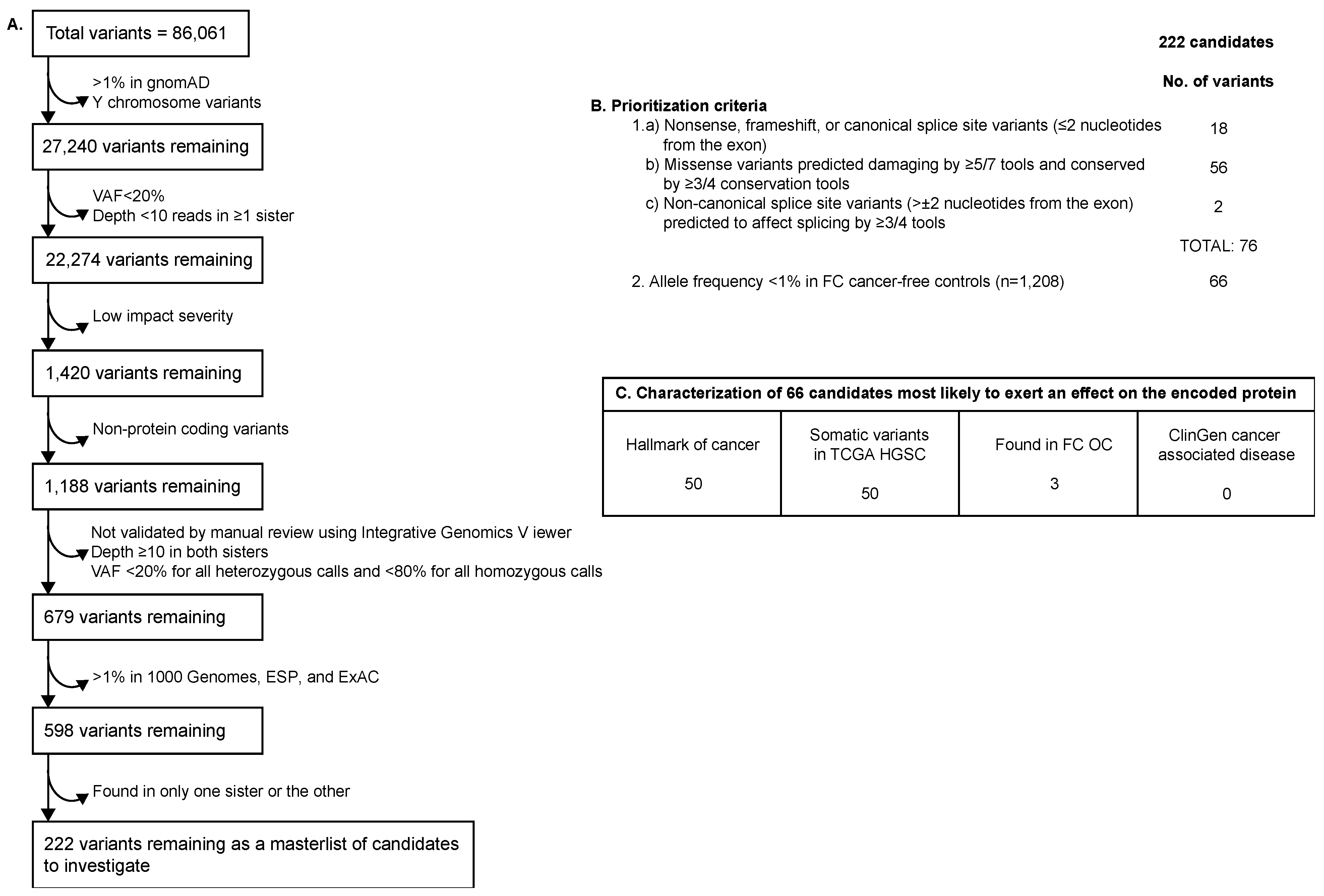
2.2. WES Filtering and Prioritization of Variants Identified in Family F1528

Sequencing data from FANCI c.1813C>T carrier sisters from family F1528 were sequentially filtered (Figure 1a) for (a) rare (minor allele frequency (MAF)     1%) variants in the general population database gnomAD [48]; (b) variants within autosomes and the X chromosome only; (c) variant allele frequency ( 20% in at least one sister); (d) variant depth ( 10 reads in at least one sister); and (e) protein-coding variants. The remaining variants were visually inspected and confirmed by Integrative Genomics Viewer (IGV) [49] and filtered, as shown in Figure 1a, where variants in both sisters had variant depth 10 reads, variant allele frequency 20% for variants called heterozygous, and variant allele frequency 80% for variants called homozygous. The final filter applied included a survey for rare (MAF     1%) variants in the general population from the 1000 Genomes Project [50]; the National Heart, Lung, and Blood Institute (NHLBI) Exome Sequencing Project (ESP) ESP6500SI-V2 (https://evs.gs.washington.edu/EVS/); and the Exome Aggregation Consortium [51] (ExAC). These filtering steps led to a list of variants for further annotation and prioritization. The genetic landscape variants were annotated for the type of variant effect (nonsense, frameshift, splice site, or missense) and the results from applying in silico tools that predict if the variant is located at a conserved locus; whether the variant is deleterious to the protein; or if the variant has the potential to affect splicing. These in silico tools were selected based on their best predictive performance [52]. The tools used to determine the predicted conservation of variants were Genomic Evolutionary Rate Profiling (GERP++) [53], Site-Specific Phylogenetic Analysis (SiPhy) [54], Phylogenetic P-values (PhyloP) 100-way vertebrates [55], and Phylogenetic Analysis with Space/Time Models Conservation (phastCons) v1.5 [56]. Tools used to predict the ability of the amino acid change to affect protein function (deleterious or not) were Combined Annotation Dependent Depletion (CADD) v1.4 [57], Eigen v1.1 [58], Protein Variant Effect Analyzer (PROVEAN) v1.1 [59], Meta-Logistic Regression (MetaLR) [60], Meta-Support Vector Machine (MetaSVM) [60], Rare Exome Variant Ensemble Learner (REVEL) [61], and the Variant Effect Scoring Tool (VEST) v4.0 [62]. Tools used to predict the potential of variants to affect splicing were the database of splicing consensus regions (dbscSNV)’s adaptive boosting (ADA) and random forest (RF) [63], MaxEntScan [64], and SpliceAI [65]. These tools were applied in our previous studies where the biological function of proteins aligned with in silico tool prediction [29,31].
Variants were then prioritized if they were identified in both sisters and predicted to be inherited as an autosomal dominant (heterozygous) or autosomal recessive (homozygous or compound heterozygous) trait. Variants were then prioritized if they were (a) nonsense, frameshift, or canonical splice site variants ( ± 1–2 nucleotides away from the exon), (b) missense variants predicted to affect protein function by 5/7 in the in silico tools and highly conserved by 3/4 in the in silico tools, or (c) non-canonical splice site variants (> ± 2 nucleotides away from the exon) predicted to affect splicing by 3/4 in the in silico tools, as these variants either will not encode a protein product (nonsense-mediated decay) or could affect protein function. Variants were further prioritized if they had an MAF < 1% in cancer-free controls of FC ancestry (n = 1208 alleles) because pathogenic variants are more likely to be rare based on the rare allele hypothesis [66]. The resulting variants are henceforth referred to as genetic landscape variants. FANCI c.1813C>T; p.L605F met all filtering and prioritization criteria and is included in the tables as a reference but was not included in the total variant counts.

2.3. Investigation of Genetic Landscape Variants

Genes associated with genetic landscape variants that were identified in the FANCI c.1813C>T; p.L605F carriers were annotated for biological function, cellular location, encoded protein function, associated disease(s), and RNA expression in the ovaries and fallopian tubes using the Human Protein Atlas [67] (proteinatlas.org). These genes were annotated using the Cancer Hallmarks Analytics Tool [68] and a list of previously identified genes associated with hallmarks of cancer [69], which are defined as various abilities or characteristics acquired by cells in the development of cancer [70,71,72]. Genes were also characterized based on being catalogued as having any somatic variants regardless of their location in the same gene in the TCGA PanCancer OC cases [46,47] (cbioportal.org) and for their association with disease in ClinGen [73].
The genetic landscape variants were annotated for carrier or allele frequency from the available WES data of familial OC cases of FC ancestry negative for pathogenic variants in BRCA1, BRCA2, BRIP1, RAD51C, and RAD51D (n = 13). We also investigated this OC study group for other variants in genes where genetic landscape variants had been identified, as there may be allelic, as well as genetic, heterogeneity among CPGs. The same filtering and prioritization criteria were applied to OC cases of FC ancestry.
Genetic landscape variants were annotated for carrier frequency from the available WES data of the Australian HGSC cases. Other variants in genes where genetic landscape variants were identified were not investigated in this study group, as these samples were previously reported using a landscape approach [44].

2.4. Loss of Heterozygosity Analyses of FANCI c.1813C>T in OC Tumour DNA from Candidate Variant Carriers

Loss of heterozygosity analysis of FANCI c.1813C>T was performed by Sanger sequencing of OC tumour DNA from carriers. Extracted DNA from fresh-frozen tumours was provided by the RRCancer biobank. Previously reported primers were used [31]. Sequencing chromatograms were inspected using 4peaks (nucleobytes.com/4peaks/index.html) visualization software.

2.5. Somatic Genetic Landscape of FANCI c.1813C>T Carriers

Extracted DNA from fresh-frozen HGSC tumours from FANCI c.1813C>T carriers (n = 7) was provided by the RRCancer biobank. WES was performed at the McGill Genome Centre, as previously described [31]. Annotated Variant Call Format files were inspected for variants in genes most commonly altered somatically in HGSC: TP53, BRCA1, BRCA2, RB1, NF1, FAT3, CSMD3, GABRA6, and CDK12 [74].
Somatic copy number alteration (CNA) profiles were generated from WES data from tumour samples and corresponding matched-normal samples using Fraction and Allele-Specific Copy Number Estimates from Tumour-Normal Sequencing (FACETS) version 0.61 [75]. Total and allele-specific read counts were extracted from tumour and normal samples based on common, polymorphic SNV loci from dbSNP version 150 [76]. The following parameters were used for copy number segmentation: a minimum total sample depth of 20, a critical value for segmentation of 350, and a minimum number of heterozygous SNPs to cluster segments of 100. Focal amplification of CCNE1 was assessed, as it has been identified in over 20% of HGSC cases [74] and may be a therapeutic target for cyclin-dependent kinase (CDK) inhibitors [77].
DeconstructSigs version 1.8.0 [78] was used to determine the contribution of known mutational signatures associated with OC in each tumour sample. Catalogue of Somatic Mutations in Cancer (COSMIC) Single-Base Substitution (SBS) version 3.2 signatures were used as a reference (cancer.sanger.ac.uk/signatures/documents/452/COSMIC_v3.2_SBS_GRCh37.txt: accessed on 16 May 2022). Mutational signatures were compared to those associated with OC [79]. Synonymous and non-synonymous single nucleotide variants (SNVs) with at least three alternate reads were used for mutational signature analysis. The number of SNVs per sample ranged from 65 to 2560.
The somatic genetic landscape of FANCI c.1813C>T carrier tumours from the TCGA (n = 6) was assessed for genes most commonly altered somatically in HGSC and focal amplification of CCNE1 using cBioPortal [46,47] (cbioportal.org).

2.6. FANCI c.1813C>T Germline Carrier Frequency across Different Cancer Types from TCGA PanCancer Atlas

Data from the analysis of germline pathogenic variants in TCGA PanCancer Atlas cancer cases were downloaded [45]. A Variant Call Format file was generated with all FANCI c.1813C>T events identified at the germline level.
The clinical and genetic characteristics, including age at diagnosis and sex, of cancer cases harbouring germline FANCI c.1813C>T were retrieved from cBioPortal [46,47]. These characteristics were compared to the entire TCGA PanCancer Atlas study group [45].

2.7. Identification of Somatic FANCI Variants in Different Cancer Types from TCGA PanCancer Atlas

All somatic FANCI variants were retrieved from cBioPortal [46,47] TCGA PanCancer Atlas studies. The clinical and genetic characteristics of cancer cases where somatic FANCI variants had been identified were also retrieved. These included the total variant count, microsatellite instability (MSI) score from microsatellite analysis for normal tumour instability (MANTIS) [80], age at cancer diagnosis, and sex. These characteristics were compared to the entire TCGA PanCancer Atlas study group.

2.8. Investigation of Missense Variants in FANCI Reported in Public Databases

All missense variants in FANCI were retrieved from ClinVar [81] in March 2022. The variants were investigated using bioinformatic criteria established for FANCI c.1813C>T, including those uncommon in gnomAD non-cancer controls (MAF 0.1–1%), highly conserved by 3/4 in the in silico tools, and predicted to affect protein function by 5/7 in the in silico tools. The in silico tools used were the same as those mentioned above.

2.9. Identification of Variants in the FANCI Protein Interactome

Familial OC cases of FC ancestry negative for pathogenic variants in BRCA1, BRCA2, BRIP1, RAD51C, RAD51D, and FANCI (n = 11) were investigated for variants in genes involved in the FANCI protein interactome. To construct a list of FANCI protein interactome genes in Homo sapiens, we searched STRING [82], BioGRID [83], Protein Interaction Network Online Tool (PINOT) [84], Signalling Network Open Resource (SIGNOR) [85], Molecular Interaction Database (MINT) [86], Protein Interaction Knowledgebase (PICKLE) [87], Database of Interacting Proteins (DIP) [88], and IntAct [89] (Table S2). The literature was also searched for proteins shown to directly interact with FANCI protein experimentally by using “FANCI” as the search term (Table S2). As germline variants in DNA repair pathway genes have already been investigated in this study group (P.N. Tonin unpublished data), we focused on genes that were part of the FANCI interactome but not involved in DNA repair pathways (n = 115). Variants were filtered and prioritized using the same criteria as described above (Figure 1a,b).

3. Results

3.1. Candidate Variants Identified in Family F1528

We reported the discovery of FANCI c.1813C>T; p.L605F in two siblings in family F1528 as the most plausible OC-predisposing candidate based on the association of FANCI in the Fanconi anemia homologous recombination (FA-HR) DNA repair pathway [31]. The only other variant shared between these siblings and other OC carriers of FANCI c.1813C>T (n = 14) is POLG c.2492A>G; p.Y831C, a marker found in linkage disequilibrium with carriers of FANCI c.1813C>T in all the populations that we studied, which remains an unlikely candidate OC-predisposing allele based on its purported function, as reported previously [31]. Using bioinformatic tools and the most recent annotation from genetic databases, we re-evaluated the WES germline data of these siblings to further investigate the genetic landscape of carrier siblings, reasoning that there may be other co-occurring potentially pathogenic variants of interest that could also be investigated in other OC families of the same FC ancestry. We only applied this strategy to this OC family (F1528), as we did not have any other examples of familial cases, especially sibling pairs affected by OC where both siblings harboured the same FANCI variant, which would facilitate the identification of candidate variants associated with the disease.
Using the same generated WES data used in the discovery of FANCI c.1813C>T, we performed a new bioinformatic analysis and applied the best-performing predictive tools to identify the germline genetic landscape variants that were shared among the siblings in family F1528 (Figure 1a). From a master list of 86,061 variants identified in both sisters, we used a filtering strategy to identify rare (MAF     1%), high-quality variants that were most likely to affect the protein-coding regions, which generated a list of 222 variants that were shared among these siblings. These variants were identified in 214 different genes and, as expected, included POLG c.2492A>G (Table S3). The variants were present in the same genetic state in both siblings: heterozygous (n = 196 variants), compound heterozygous (n = 14 variants in 7 genes) or homozygous (n = 1 variant), and heterozygous X-chromosome linked (n = 11). The MAFs of these variants varied from 4 × 10⁻⁶ to 9.7 × 10⁻³ in gnomAD, except for 14 variants that were not found in this database. These comprised 186 missense, 14 non-canonical splice site, 9 frameshift, 6 nonsense, 3 inframe, 3 canonical splice site, and 1 stop loss variant.
To further refine the list of plausible candidates, we applied criteria to the list of 222 variants to select those of interest for further investigation, focusing on the in silico tools, type of variant, and allele frequency in FC cancer-free controls (Figure 1b). Using these criteria, we identified 18 variants with nonsense, frameshift, or canonical splice site effects. We selected 57 missense candidates predicted to be damaging based on 5 of 7 of our best-performing in silico tools, in keeping with the rationale that the most likely biologically relevant candidates would be predicted by the majority of the in silico tools [90]. We also selected an additional two non-canonical splice site variants that were predicted to affect splicing. As expected, this excluded POLG c.2492A>G as a variant of interest but not FANCI c.1813C>T. From this list of 76 variants, we selected 66 variants for further investigation based on their allele frequency (MAF < 1%) in population-matched FC controls. Excluding the FANCI variant, the 66 variants of interest were identified in 66 different genes (Table S4).
A large proportion of the 66 variants were identified at least once in various cancer contexts, such as hallmarks of cancer [68,69] (83%), or somatically mutated in OC cases from the TCGA [46,47] (83%), as summarized in Figure 1c. Although some (9/66.14%) variants have been reported in other clinical contexts, none of the variants were found to be associated with a cancer context in ClinGen [73] (Table S4). As shown in Table S4, some of these genes were found in more than one group, as defined in Figure 1c. None of the 66 variants were identified in genes associated with DNA repair pathways.

3.2. Genetic Analyses of Variants Identified in FANCI Carrier Siblings in FC Study Groups

To further characterize our 66 variants, we reviewed the available WES data from 13 familial OC cases negative for pathogenic variants in BRCA1, BRCA2, BRIP1, RAD51C, and RAD51D for carrier status. These cases have been well characterized and are of FC ancestry [25,26,28,29,30,31,38,91,92,93,94,95,96,97,98]. We identified a total of four carriers from three families of variants in PTPN22, GPD1, and SEC14L4 (Table 1). Notable is that none of the three FANCI c.1813C>T carriers from the independently ascertained cases harboured any of the 66 variants. Thus, although there may be shared FC ancestry among the carriers, none of the five independently ascertained familial OC cases in our FC study group of FANCI carriers harboured other potentially deleterious alleles initially identified in the index FANCI c.1813C>T OC cases.
Given the possibility of allelic heterogeneity, even within the FC population, as we have previously demonstrated with established OC-predisposing genes [24], we screened the same 13 familial OC cases for other plausible deleterious variants in the 66 candidate genes. Applying our bioinformatic filtering and prioritization criteria, we identified 10 different variants that were not present in our index family F1528. There were six carriers of variants in PIWIL3, SCN10A, PCDH15, TEX2, DNAH3, DNAH1, IQCA1, CACNA1S, and MYO7A (Table 1). Four cases were found to carry variants in two different genes: F1085-PT0134 (CACNA1S and MYO7A); F845-PT0196 (PCDH15 and TEX2); F1506-PT0136 (PIWIL3 and DNAH1); and F1543-PT0137 (DNAH1 and IQCA1). Both variants in F1085-PT0134 were not carried by their sibling F1085-PT0135 and thus did not segregate with the disease. Each variant was harboured by only one case; two different variants were identified in the same gene, DNAH1.

3.3. Genetic Analyses of Variants Identified in FANCI Carrier Siblings in Non-FC Study Groups

We screened our 66 variants in the available WES data from the Australian HGSC study group regardless of FANCI variant carrier status. We identified 70 carriers of 21 variants among 516 HGSC cases, where the majority harboured only one variant (Table 2). None of these variants were identified in any of the previously identified 10 Australian carriers of FANCI c.1813C>T [31]. Two carriers of other likely pathogenic FANCI variants (c.1264G>A; p.G422R and c.3635T>C; p.F1212S) were found to harbour variants in ALDH16A1 and NBAS, respectively. Although different variants were identified in OC cases of FC ancestry in DNAH3, IQCA1, and PCDH15, the same variants as those found in the family F1528 sisters were identified in these genes in five Australian HGSC cases.

3.4. Genetic Analyses of Germline FANCI Interactome Variants Identified in FC OC Cases

Based on our previous analyses, FANCI c.1813C>T is the strongest candidate OC-predisposing variant identified in family F1528. Therefore, we used a candidate gene approach to investigate the germline variants in genes that encode proteins that are part of the FANCI interactome. We reviewed the available WES data from 11 familial OC cases negative for pathogenic variants in BRCA1, BRCA2, BRIP1, RAD51C, RAD51D, and FANCI to search for other candidate predisposition variants. Variants in DNA repair pathway genes were excluded, as this has been previously reported by our lab (P.N. Tonin unpublished data). We identified a total of three carriers from three families of missense variants in EZH2, ANKRD55, MOV10, and LRRK2 (Table 3). Variants in ANKRD55 and MOV10 were identified in the same case F1506-PT0136.

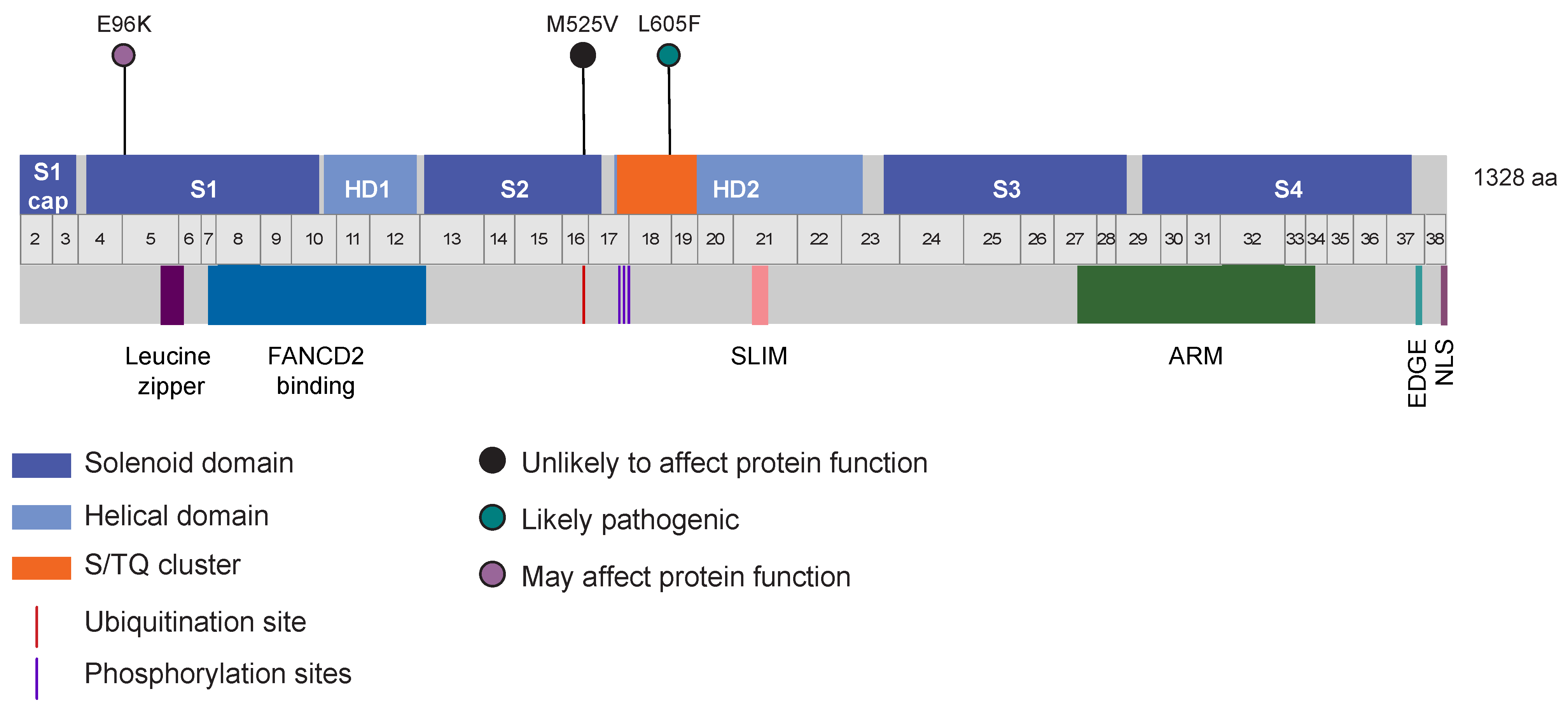
3.5. Identification of Other Germline Potentially Deleterious Variants in FANCI

To identify additional germline potentially pathogenic variants in FANCI, we used ClinVar [81], which aggregates information about genomic variation and its relationship to human health. We focused our analyses on the missense variants, as the loss-of-function variants (i.e., frameshift, nonsense, and canonical splice site) were previously reported by our group [31]. We investigated 319 missense variants using bioinformatic criteria established for selecting variants of interest: uncommon (0.1–1% MAF) in non-cancer controls from gnomAD, highly conserved, and predicted to affect protein function by in silico tools. Three missense variants met these criteria (Figure 2): FANCI c.286G>A; p.E96K, c.1573A>G; p.M525V, and our candidate variant c.1813C>T; p.L605F. FANCI c.1573A>G; p.M525V was previously reported by our group; however, based on in cellulo assays, we concluded that this variant did not affect protein function [31]. Thus, the only FANCI variant to investigate further was c.286G>A; p.E96K. In the gnomAD non-cancer population, the allele frequency of c.286G>A is 0.17%, which is less common than the allele frequency of c.1813C>T of 0.67%. We investigated the available genetic data or genotyped FC, Australian, and TCGA study groups for carriers of FANCI c.286G>A; p.E96K. FANCI c.286G>A was not identified in OC (n = 527) or BC (n = 220) cases or controls (n = 171) of FC ancestry, but was previously reported by our group in an Australian OC case (1/516, 0.2%), controls (5/4878, 0.1%), and TCGA OC cases (1/412, 0.2%) [31].

3.6. Loss of Heterozygosity Analyses of FANCI c.1813C>T in OC Tumour DNA from Carriers

We previously reported loss of the wild-type allele in bilateral OC tumours from FANCI c.1813C>T carriers, suggesting that loss of FANCI function was an early event in tumourigenesis [31]. We extended our analysis to investigate tumour samples from other carriers, although only DNA from FC cases was available for these analyses. The inspection of Sanger sequencing chromatograms from OC tumour DNA from nine carriers revealed three cases exhibiting loss of the wild-type allele and retention of the FANCI c.1813C>T allele. One case showed loss of the variant allele and retention of the wild-type allele, and the remaining cases retained heterozygosity with little evidence of allelic imbalance.

3.7. Somatic Genetic Analyses of OC Tumours from FANCI c.1813C>T Carriers

The somatic genetic landscape of HGSC cases has been well characterized, where there is a long tail of uncommon somatic variants and extensive genome-wide CNAs, with the exception of TP53 (which harbours driver mutations that cause cells to become cancerous) being the most altered gene (>95% of cases) [74]. To determine if HGSC cases carrying FANCI c.1813C>T exhibit similar somatic genetic characteristics to HGSC cases, we performed WES analyses or surveyed the available genetic data from seven FC cases and six TCGA cases, respectively. We focused our analyses on the most altered genes reported for HGSCs, TP53 (96%), BRCA1 (3.5%), CSMD3 (6%), NF1 (4%), CDK12 (3%), FAT3 (6%), GABRA6 (2%), BRCA2 (3%), and RB1 (2%) [74]. Somatic variants were identified in CDK12 (1/13, 8%), FAT3 (3/13, 23%), BRCA2 (3/13, 23%), and TP53 (11/13, 85%) (Table 4), at frequencies comparable to those HGSC cases [74]. As expected, most deleterious variants identified in our HGSC cases carrying FANCI c.1813C>T in TP53 were missense [74,107,108] (n = 7), with the remainder being frameshift (n = 2), splice (n = 1), or inframe indel (n = 1). Extensive and unremarkable genome-wide CNAs were evident across tumours from FANCI c.1813C>T carriers, which was consistent with those seen for HGSC tumours (Figure S1). Amplification of CCNE1, reported to occur in approximately 20% of HGSC cases [74], was exhibited in two FANCI carrier cases (2/11, 18%), PT0006 from our FC study group and sample TCGA-25-2393 from the TGCA project.
DNA from HGSC tumours has been shown to exhibit global DNA mutational signatures, reflecting disruptions in specific DNA repair pathways, aging, and other processes that have accumulated during tumourigenesis [79]. We performed a somatic mutational signature analysis using WES data derived from FC OC tumour DNA from FANCI c.1813C>T carriers, using COSMIC SBS signatures as a reference. The signatures identified in tumours from FANCI c.1813C>T carriers were compared to those exhibited by HGSC tumours, as there have been no reports attributing mutational signatures to cancers harbouring deleterious FANCI variants. We were able to profile seven OC tumour samples from FC carriers due to the availability of WES data for these samples (Figure S2). The mutational profiles were indicative of the presence of extensive and complex mutational patterns typified by HGSC tumours (https://signal.mutationalsignatures.com/explore/tissueType/15: accessed on 16 May 2022). The homologous recombination deficiency signature (referred to as SBS3) was identified in 6/7 (86%) cases. The sample that did not exhibit this SBS3 signature, PT0003, exhibited signature pattern SBS8, a signature whose etiology is unknown, but it has been proposed to be associated with homologous recombination deficiency (cancer.sanger.ac.uk/signatures/sbs/sbs8/: accessed on 16 May 2022). PT0003 also exhibited the largest contribution of signature SBS6, which has been attributed to defective mismatch repair and MSI. The aging signature (SBS1) was identified in 5 out of 7 (71%) tumours and the contribution was consistent with age at diagnosis [31]. All tumours exhibited varying proportions of SBS18, a signature indicative of damage due to reactive oxygen species. A signature with a proposed etiology associated with prior treatment with platinum chemotherapy drugs (SBS35) was evident in 5 out of 7 (71%) tumours, although not in sample PT0007, which was from a patient who received neoadjuvant chemotherapy with the platinum compound carboplatin.

3.8. Germline FANCI c.1813C>T Carriers Identified in Other Cancer Types

We previously reported carriers of FANCI c.1813C>T and other potentially pathogenic FANCI variants in BC cases, a disease associated with OC risk genes [109], and a review of the literature also indicated that there were FANCI carriers in other cancer types [31]. To further investigate the role of FANCI in other cancer types we investigated the germline carrier frequency of FANCI c.1813C>T in 10,389 cancer cases from the TCGA PanCancer data set [45]. We focused on this variant to further investigate its association with familial OC and our in cellulo assays, demonstrating abrogated protein function [31]. The highest carrier frequency was observed in adrenocortical carcinoma cases (3.3%, 3/92); there were no carriers identified in cases with diffuse large B-cell carcinoma (n = 41), kidney renal papillary cell carcinoma (n = 289), thymoma (n = 123), or uterine carcinosarcoma (n = 57) (Table 5). The median age of diagnosis (59 ± 14.7 years) and the number of females (48.5%) of FANCI c.1813C>T carriers were comparable to the total study group (59.2 ± 14.4 [45] and 52% [45], respectively). Interestingly, the overall carrier frequency of FANCI c.1813C>T at 1.6% (171/10,389) was significantly higher in the TCGA PanCancer cases than in non-cancer individuals in gnomAD (1.3%, 1787/134,164; Pearson’s χ 2 = 7.3, p = 0.007).

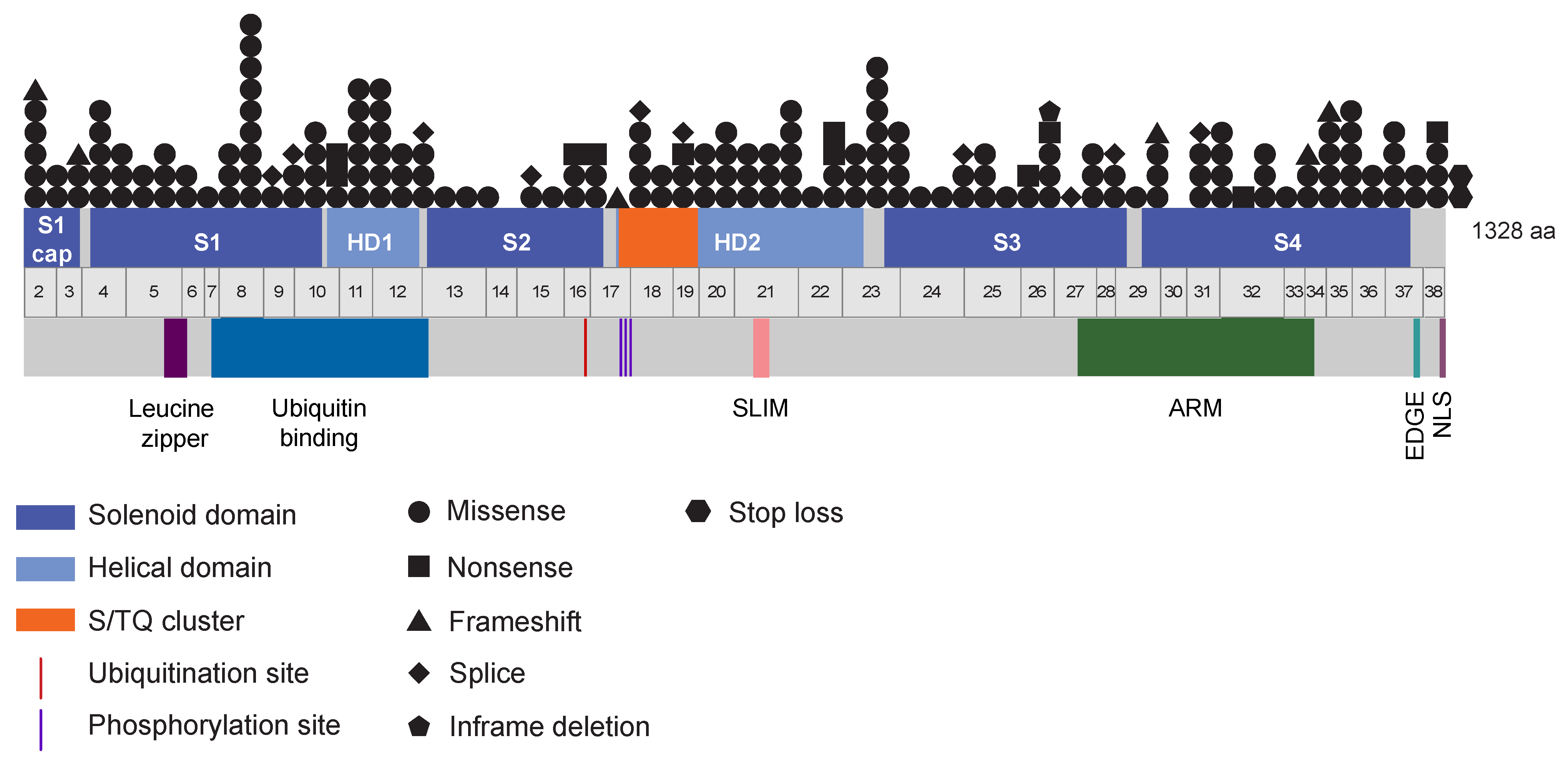
3.9. A Wide Spectrum of Somatic FANCI Variants Identified in a Variety of Cancer Types

Approximately 40% of germline CPGs have been found with somatic variants in tumours as drivers and some of these genes have been shown to play a role in tumourigenesis, with RB1 being the classical example [110]. From the above analysis of the germline FANCI variant, there were four FANCI c.1813C>T germline carriers with different somatic FANCI variants, two bladder urothelial carcinomas and two lung squamous cell carcinomas. These observations prompted us to investigate the spectrum and frequency of somatic variants in FANCI in TCGA PanCancer tumours (n = 10,434) from cBioPortal [46,47]. We identified 198 different variants in 172 tumours (1.65%, 172/10,434) in 28 different cancer types, comprising a variety of genetic abnormalities: 168 missense, 11 nonsense, 10 splice, 6 frameshift, 2 stop loss, and 1 inframe deletion variant (Table 6 and Table S5). Noteworthy, FANCI c.1813C>T was not among the variants identified at the somatic level. There appears to be no mutational hotspot identified and variants were distributed across the gene (Figure 3). The highest total number of variants was identified in uterine corpus endometrial carcinoma tumours (8.32%, 43/517); no variants were identified in tumours from cases with cholangiocarcinoma (n = 36), diffuse large B-cell carcinoma (n = 41), kidney chromophobe (n = 65), pancreatic adenocarcinoma (n = 179), or uveal melanoma (n = 80). The median age at diagnosis and sex of cases with somatic FANCI variants were comparable to those without somatic FANCI variants (60 ± 13.5 vs. 60 ± 14.4 and 58% vs. 52%, respectively). Tumours with somatic FANCI variants had a higher mutational load (p = 2.2 × 10⁻¹⁶) and MSI score (p = 6.8 × 10⁻¹⁰) compared to tumours without somatic FANCI variants.

4. Discussion

Genetic analyses of the germline of two sisters with OC from family F1528 that are heterozygous for FANCI c.1813C>T; p.L605F revealed 222 variants of interest, of which 66 were most likely to exert a function on the encoded protein (genetic landscape variants). Of note is the number of loss-of-function (frameshift, nonsense, and canonical splice site) variants identified in both sisters after filtering and prioritization (n = 15). Increasing evidence shows that all individuals carry more potentially deleterious variants than previously suspected (approximately 24–100 heterozygous variants per individual [111,112]), although some may not impact gene function [113,114]. As adequate in silico tools with high predictive performance, such as those for missense and splice site variants, have not yet been developed for loss-of-function variants, it is difficult to further interpret these variants in the absence of laboratory experiments by examining their biological effect. The investigation of the genetic landscape variants in OC cases of FC ancestry revealed four carriers of variants in PTPN22, GPD1, and SEC14L4 in three different families, although two of these families were previously identified by our group to harbour likely pathogenic variants in known or putative DNA repair pathway genes (P.N. Tonin unpublished data). When we assessed the same study group for carrier status, we identified six carriers of variants in nine different genes, where all but two families were previously identified to harbour likely pathogenic variants in DNA repair genes (P.N. Tonin unpublished data). Families F845 and F1543 were the only cases not found to harbour likely pathogenic variants in DNA repair genes. Although the previously identified DNA repair pathway variants are plausible OC-predisposing variants, they have yet to be verified independently. Although it is possible that the genetic variants identified in this study may independently affect the risk of OC, FANCI c.1813C>T; p.L605F remains the most likely candidate for an OC-predisposing gene in family F1528.
We also investigated the 66 genetic landscape variants for carrier frequency in the Australian OC cases (regardless of FANCI variant carrier status) and identified several carriers of variants in these genes, mainly missense variants. These variants had not been reported in a previous independent analysis of WES data from these cases, as the study focused primarily on loss-of-function variants [44]. Thus, it is possible that these missense variants are relevant in these cases, which are notably negative for pathogenic variants in BRCA1 and BRCA2. Also notable is that we did not identify any carriers of any of our genetic landscape variants among the 10 previously identified FANCI c.1813C>T; p.L605F carriers from this study group. Based on principal component analysis (PCA), the Australian OC cases are likely of white European ancestry, which is similar to the ancestral origins of the FC population [31].
The role of the 66 genetic variants identified in the FANCI carriers in modifying the risk of OC in family F1528, which harbours FANCI c.1813C>T; p.L605F, remains to be determined. Interestingly, additional potentially pathogenic variants were identified in FANCI using the criteria established for c.1813C>T and it would be of interest to further assess their effect on gene function. The most interesting variant, FANCI c.286G>A; p.E96K, was identified in a BC case (diagnosed at 44 and 50 years), with OC at age 53 years, was of Ukrainian ancestry, and who reported a grandmother with OC (Figure S3). This individual did not harbour any pathogenic variants in BRCA1, BRCA2, BRIP1, RAD51C, or RAD51D by WES analyses. FANCI c.286G>A; p.E96K was reported in BC cases (2/133, 1.5%) [115] and OC cases (1/6385, 0.02% (0/6115 controls)) [116] (p. 2) in the literature. FANCI p.E96K may affect the ubiquitination of FANCD2, the heterodimeric binding partner of FANCI, and/or the Van der Waals forces between FANCI and FANCD2 [115].
HGSC tumours from cases harbouring germline FANCI c.1813C>T exhibited features consistent with tumours from HGSC cases. The majority (85%) of tumours had identifiable pathogenic variants in TP53 and two cases were identified with CCNE1 amplification (18%). The somatic mutational signatures characteristic of HGSC tumour cells were also present in HGSC samples harbouring FANCI c.1813C>T. We also identified signatures SBS35 (platinum associated) and SBS6 (mismatch repair deficiency) in 5 out of 7 and 6 out of 7 cases, respectively, which are less commonly observed in HGSC cases (<10%). This may be the result of the small sample size, although they could also be the result of harbouring FANCI c.1813C>T in the germline. Interestingly, the two cases with the highest contributions of the SBS6 signature, samples PT0004 and PT0005, have germline or somatic variants in mismatch repair genes, which may contribute to the presence of this signature. In a study involving Caenorhabditis elegans, it has recently been reported that genotoxic agents tended to have a stronger influence on the mutational signature than a DNA repair-deficient background [117]. Moreover, the signature attributed to defects in HR DNA repair may be identified in the absence of an identifiable DNA repair pathway gene pathogenic variant [118]. This could be because the signature is more attributed to global defects and is distinct from other signatures that have characteristic nucleotide changes such as the aging signature [118]. Although the sample size was small, there appeared to be no identifiable FANCI-specific signature from the analyses of tumours from FANCI c.1813C>T carriers.
The contribution of cancer cases attributed to CPGs is approximately 3%, although this varies based on cancer type [110]. Some CPGs predispose to multiple primary cancer types, such as BRCA1 with OC and BC, although there is often a preferential predisposition to certain histological subtypes such as the association of BRCA1 with HGSC. Germline FANCI c.1813C>T was initially identified in HGSC cases [31], but as shown in our study, it can be observed across many cancer types in the TGCA PanCancer Atlas. It is unknown if FANCI c.1813C>T contributes to the risk of these cancers, as the variant is more common in the general population compared to other high-risk CPGs (0.6% vs. 0.0001%) [48]. The moderately increased carrier frequency of FANCI c.1813C>T in TCGA cancer cases (1.6%) compared to gnomAD cancer-free controls (1.3%) is intriguing, suggesting a role for this variant in risk (p = 0.007). Additionally, TCGA cancer cases were identified with somatic variants in FANCI across cancer types. As variants were identified in cases with significantly higher mutational load and/or MSI scores, it is possible that these somatic FANCI variants arose as a consequence of either of these processes, although they could be drivers of these processes through mechanisms that remain to be elucidated. FANCI is not currently included among the 733 cancer-driving genes in the Cancer Gene Census [119], but a number of other genes involved in the FA pathway, such as BRCA1 (FANCS), BRCA2 (FANCD1), BRIP1 (FANCJ), FANCA, FANCC, FANCD2, PALB2 (FANCN), and RAD51C (FANCO), are implicated as cancer drivers in this census. Further assessment of cancers in individuals harbouring germline FANCI c.1813C>T or somatic FANCI variants may elucidate a genetic signature associated with FANCI, as has been reported for FA-associated squamous cell carcinomas [120].
Many CPGs are also associated with non-cancer phenotypes, the spectrum of which is broad. Homozygous or compound heterozygous variants in FANCI were associated with FA complementation group I in 2007 [99,100,121]. FA is a rare disease characterized by congenital defects, progressive bone marrow failure, and an increased risk of cancers (mainly acute myeloid leukemia and squamous cell carcinomas) [122]. FA-I cases comprise approximately 1% of all FA cases and have been associated with at least three features of VACTERL-H [123], a rare disease that affects multiple body systems. Recently, an eight-year-old male with aplasia referred for a diagnosis of FA was reported to harbour germline homozygous FANCI c.1813C>T [124]. FANCD2 ubiquitination was not detected in peripheral blood cells from this patient and increased chromosomal breakage was observed, suggesting abrogation of the FA pathway. These data are consistent with our previous observations that FANCI c.1813C>T abrogates FANCI protein function [31]. FANCI is an integral member of the FA pathway and acts as the molecular switch to activate the pathway [101]. FANCI also functions outside the FA pathway such as in dormant origin firing [125], negative regulation of Akt signalling [126], and ribosome biogenesis [127].
The risk of cancer has been assessed in heterozygous relatives of individuals with FA, and although no association with cancer risk was found, few FANCI families (n = 4) have been investigated due to the paucity of FANCI carriers [128,129,130]. FANCI c.1813C>T; p.L605F was previously identified by our group in the germline and was associated with a suspected autosomal dominant mode of inheritance of OC [31]. This is consistent with more than half of CPGs, which are associated with an autosomal dominant mode of inheritance [110]. The majority of CPGs also act as tumour suppressors, where many are classical tumour suppressors that require biallelic inactivation for tumour development and/or progression, although some CPGs may exert their effect through haploinsufficiency or in a dominant-negative manner [110]. Here, we have shown that biallelic inactivation of FANCI c.1813C>T may occur through loss of the wild-type allele. As we showed that not all carriers exhibited this loss, it is possible that loss of the wild-type allele may not be required for OC tumourigenesis. This is consistent with the OC-predisposing genes BRCA1 and BRCA2, where loss of the wild-type allele is not always observed in tumour cells from carriers of pathogenic variants in these CPGs [22,131]. Beyond assessing the biological effect of a variant using cell-line models, as we have shown with FANCI c.1813C>T [31], there are no suitable animal models to evaluate OC risk alleles.
The identification of CPGs has had a significant clinical impact on diagnosis and management, targeted therapies, and screening and prevention. The clinical utility of FANCI for diagnosis and management cannot be determined until penetrance for cancer risk is established. Currently, there are no effective cancer screening methods for OC or prevention strategies to reduce OC risk in the general population, although prophylactic salpingo-oophorectomy has been proven to reduce risk in carriers of pathogenic BRCA1 or BRCA2 variants [132]. Given the identification of FANCI variants in other cancers, particularly gynecological cancers, future research should investigate the risks of these variants in view of prevention and management strategies, including childbearing considerations [133,134]. Although there are no targeted therapies for FANCI, 73 chemicals interact with this gene, including cisplatin and mitomycin C, which is concordant with our previous findings that loss of FANCI sensitizes cells to these drugs [31]. There are 12 cancer-related drugs that interact with FANCI, 7 of which are chemotherapies and 5 of which are targeted therapies [135]. These chemicals present opportunities for future investigation for the treatment of cancer cases with FANCI variants.

5. Conclusions

This study has expanded on the molecular genetic characteristics of FANCI c.1813C>T; p.L605F in OC, which were first reported in OC families of FC ancestry [31]. These data suggest FANCI c.1813C>T carrier HGSC tumours show characteristics known to be exhibited by HGSC cases. The identification of germline FANCI c.1813C>T carriers and various somatic FANCI variants across cancer types suggests a possible involvement of FANCI in other cancers and an avenue for future research.

Supplementary Materials

The following supporting information can be downloaded at: https://www.mdpi.com/article/10.3390/genes14020277/s1, Figure S1: Copy number alterations per chromosome across the genome of tumours from French Canadian cases harbouring germline FANCI c.1813C>T. The top panel shows total copy number log-ratio, the middle panel shows allele-specific copy number log-ratio, and the bottom panel shows the corresponding integer copy number calls (red is the minor allele); Figure S2: Proportional representation of defined somatic mutational signatures observed in each ovarian tumour from French Canadian cases harbouring germline FANCI c.1813C>T. SBS1: Ageing; SBS3: Homologous recombination deficiency; SBS6: Mismatch repair deficiency; SBS8: Unknown (putative homologous recombination deficiency); SBS13: Activation of APOBEC family; SBS14: Polymerase epsilon mutation and mismatch repair deficiency; SBS18: Damage due to reactive oxygen species; SBS35: Platinum chemotherapy treatment. Figure S3: Pedigree of a Ukrainian breast and ovarian cancer family negative for BRCA1 and BRCA2 pathogenic variants where FANCI c.286G>A; p.E96K was identified by WES in the proband, indicated by the arrow. Age at diagnosis and death are shown where known. Cancer type (OC: ovarian, BC: breast, Pro: prostate, Col: colon, and Leu: leukemia) and age of diagnosis are shown. DCIS: ductal carcinoma in situ; IDC: infiltrating ductal carcinoma. Table S1: Description of study groups investigated; Table S2: List of genes encoding proteins involved in the FANCI interactome selected from our search of databases and the literature (n = 219); Table S3: List of variants (n = 222) that passed our filtering and were further prioritized and characterized; Table S4: List of variants (n = 66) that passed our prioritization criteria; Table S5: Somatic FANCI variants identified in tumours from different cancer cases from TCGA PanCancer Atlas.

Author Contributions

C.T.F. performed all genetic variant analyses and wrote the initial draft of the manuscript. J.R. developed the sequencing data analysis pipelines. C.T.F., W.M.A., S.A. and T.R. performed bioinformatic analyses. K.B. performed initial genetic analyses of family F1528. Study samples and clinical data were provided by Z.E.H., W.D.F., A.-M.M.-M., D.P., E.F., M.T., D.N.S. and I.G.C. C.T.F. and C.S. collated the study samples and clinical data. TCGA PanCancer Atlas germline FANCI c.1813C>T carrier data were provided by J.P.B., S.P. and T.J.P. Gen3G data were provided by L.B. Statistical analyses were overseen by C.M.T.G. Supervision of bioinformatic analyses was provided by J.R. and P.N.T.; whole-exome sequencing by J.R.; and genetic analysis by C.T.F. and P.N.T. P.N.T. conceived, designed, and oversaw all aspects of the project. All authors have read and agreed to the published version of the manuscript.

Funding

C.T.F. was supported by The Research Institute of the McGill University Health Centre Studentship (RI-MUHC) Award, Ovarian Cancer Canada Trainee Travel (Research) Award, and James O. and Maria Meadows Award. W.M.A. was supported by a Scholarship Award from The Ministry of Education, Saudi Arabia. The research was supported by the following: The Canadian Institute for Health Research (CIHR) operating grants (PCC-156736 to P.N.T., Celia M.T. Greenwood, J.R.; PJT-156124 to P.N.T., J.R.); Cancer Research Society and Ovarian Cancer Canada partnership grant (21123 to P.N.T.); FRQS and Quebec Breast Cancer Foundation network grants to P.N.T.; and Compute Canada resource allocation project wst-164 and Genome Canada Genome Technology Platform award to J.R. The ovarian tumour banking was supported by the Banque de tissus et de données of the Réseau de recherche sur le cancer of the FRQS, affiliated with the Canadian Tumor Repository Network (CTRNet). The RI-MUHC and CRCHUM receive support from the FRQS. The sequencing of the Gen3G offspring was sponsored by Fonds de recherche du Québec, McGill University, and Université de Sherbrooke. M.T. was supported by the NIHR Cambridge Biomedical Research Centre (BRC-1215-20014).

Institutional Review Board Statement

The project received approval from The McGill University Health Centre (MUHC) REB (MP-37-2019-4783 and 2017-2722).

Informed Consent Statement

Not applicable.

Data Availability Statement

The data sets generated and/or analyzed during the current study are not publicly available due to ongoing analyses as part of current projects but are available from the corresponding author on reasonable request.

Acknowledgments

We thank Mary Fujiwara for her assistance in editing the drafts and CARTaGENE (cartagene.qc.ca) for providing the French-Canadian cancer-free control data. The data used for this research originated from the Gen3G Biobank. Gen3G was supported by Le Fonds de Recherche du Québec en Santé (#20697) and the Canadian Institutes of Health Research (#MOP-115071; #PJT-152989). We want to acknowledge the Centre de Recherche du Centre Hospitalier Universitaire de Sherbrooke, l’Université de Sherbrooke, and the women and their children who agreed to participate in the Gen3G cohort. We also acknowledge Patrice Peron, Luigi Bouchard, and Marie-France Hivert, who established the Gen3G cohort and sequencing. Pierre-Étienne Jacques also contributed to the generation and analysis of the sequencing data in Gen3G. We thank Simon Gravel for his assistance in the use of data from the Gen3G biobank. The results published here are in whole or part based upon data generated by The Cancer Genome Atlas and managed by the NCI and NHGRI. Information about the TCGA can be found at http://cancergenome.nih.gov.

Conflicts of Interest

The authors declare that they have no competing interests.

References

  1. Miki, Y.; Swensen, J.; Shattuck-eidens, D.; Futreal, P.A.; Harshman, K.; Tavtigian, S.; Liu, Q.; Cochran, C.; Bennett, L.M.; Ding, W.; et al. Strong candidate for the breast and ovarian cancer susceptibility gene BRCA1. Science 1994, 266, 66–71. [Google Scholar] [CrossRef] [PubMed] [Green Version]
  2. Wooster, R.; Bignell, G.; Lancaster, J.; Swift, S.; Seal, S.; Mangion, J.; Collins, N.; Gregory, S.; Gumbs, C.; Micklem, G. Identification of the breast cancer susceptibility gene BRCA2. Nature 1995, 378, 789–792. [Google Scholar] [CrossRef] [PubMed]
  3. Song, H.; Ramus, S.J.; Kjaer, S.K.; Hogdall, E.; Dicioccio, R.A.; Whittemore, A.S.; McGuire, V.; Hogdall, C.; Jacobs, I.J.; Easton, D.F.; et al. Tagging single nucleotide polymorphisms in the BRIP1 gene and susceptibility to breast and ovarian cancer. PLoS ONE 2007, 2, e268. [Google Scholar] [CrossRef] [PubMed] [Green Version]
  4. Rafnar, T.; Gudbjartsson, D.F.; Sulem, P.; Jonasdottir, A.; Sigurdsson, A.; Jonasdottir, A.; Besenbacher, S.; Lundin, P.; Stacey, S.N.; Gudmundsson, J.; et al. Mutations in BRIP1 confer high risk of ovarian cancer. Nat. Genet. 2011, 43, 1104–1107. [Google Scholar] [CrossRef]
  5. Meindl, A.; Hellebrand, H.; Wiek, C.; Erven, V.; Wappenschmidt, B.; Niederacher, D.; Freund, M.; Lichtner, P.; Hartmann, L.; Schaal, H.; et al. Germline mutations in breast and ovarian cancer pedigrees establish RAD51C as a human cancer susceptibility gene. Nat. Genet. 2010, 42, 410–414. [Google Scholar] [CrossRef]
  6. Zheng, Y.; Zhang, J.; Hope, K.; Niu, Q.; Huo, D.; Olopade, O.I. Screening RAD51C nucleotide alterations in patients with a family history of breast and ovarian cancer. Breast Cancer Res. Treat. 2010, 124, 857–861. [Google Scholar] [CrossRef]
  7. Somyajit, K.; Subramanya, S.; Nagaraju, G. RAD51C: A novel cancer susceptibility gene is linked to Fanconi anemia and breast cancer. Carcinogenesis 2010, 31, 2031–2038. [Google Scholar] [CrossRef] [Green Version]
  8. Loveday, C.; Turnbull, C.; Ramsay, E.; Hughes, D.; Ruark, E.; Frankum, J.R.; Bowden, G.; Kalmyrzaev, B.; Warren-perry, M.; Snape, K.; et al. Germline mutations in RAD51D confer susceptibility to ovarian cancer. Nat. Genet. 2011, 43, 879–882. [Google Scholar] [CrossRef]
  9. Yang, X.; Leslie, G.; Doroszuk, A.; Schneider, S.; Allen, J.; Decker, B.; Dunning, A.M.; Redman, J.; Scarth, J.; Plaskocinska, I.; et al. Cancer Risks Associated With Germline PALB2 Pathogenic Variants: An International Study of 524 Families. J. Clin. Oncol. 2020, 38, 674–685. [Google Scholar] [CrossRef]
  10. Pavanello, M.; Chan, I.H.Y.; Ariff, A.; Pharoah, P.D.P.; Gayther, S.A.; Ramus, S.J. Rare germline genetic variants and the risks of epithelial ovarian cancer. Cancers 2020, 12, 3046. [Google Scholar] [CrossRef]
  11. Papadopoulos, N.; Nicolaides, N.C.; Wei, Y.F.; Ruben, S.M.; Carter, K.C.; Rosen, C.A.; Haseltine, W.A.; Fleischmann, R.D.; Fraser, C.M.; Adams, M.D. Mutation of a mutL homolog in hereditary colon cancer. Science 1994, 263, 1625–1629. [Google Scholar] [CrossRef] [PubMed]
  12. Bronner, C.E.; Baker, S.M.; Morrison, P.T.; Warren, G.; Smith, L.G.; Lescoe, M.K.; Kane, M.; Earabino, C.; Lipford, J.; Lindblom, A. Mutation in the DNA mismatch repair gene homologue hMLH1 is associated with hereditary non-polyposis colon cancer. Nature 1994, 368, 258–261. [Google Scholar] [CrossRef] [PubMed]
  13. Fishel, R.; Lescoe, M.K.; Rao, M.R.; Copeland, N.G.; Jenkins, N.A.; Garber, J.; Kane, M.; Kolodner, R. The human mutator gene homolog MSH2 and its association with hereditary nonpolyposis colon cancer. Cell 1993, 75, 1027–1038. [Google Scholar] [CrossRef]
  14. Miyaki, M.; Konishi, M.; Tanaka, K.; Kikuchi-Yanoshita, R.; Muraoka, M.; Yasuno, M.; Igari, T.; Koike, M.; Chiba, M.; Mori, T. Germline mutation of MSH6 as the cause of hereditary nonpolyposis colorectal cancer. Nat. Genet. 1997, 17, 271–272. [Google Scholar] [CrossRef] [PubMed]
  15. Nicolaides, N.C.; Papadopoulos, N.; Liu, B.; Wei, Y.F.; Carter, K.C.; Ruben, S.M.; Rosen, C.A.; Haseltine, W.A.; Fleischmann, R.D.; Fraser, C.M. Mutations of two PMS homologues in hereditary nonpolyposis colon cancer. Nature 1994, 371, 75–80. [Google Scholar] [CrossRef]
  16. Pennington, K.P.; Walsh, T.; Harrell, M.I.; Lee, M.K.; Pennil, C.C.; Rendi, M.H.; Thornton, A.; Norquist, B.M.; Casadei, S.; Nord, A.S.; et al. Germline and Somatic Mutations in Homologous REcombination Genes Predict Platinum Response and Survival in Ovarian, Fallopian Tube, and Peritoneal Carcinomas. Clin. Cancer Res. 2014, 20, 764–775. [Google Scholar] [CrossRef] [Green Version]
  17. Lu, H.M.; Li, S.; Black, M.H.; Lee, S.; Hoiness, R.; Wu, S.; Mu, W.; Huether, R.; Chen, J.; Sridhar, S.; et al. Association of breast and ovarian cancers with predisposition genes identified by large-scale sequencing. JAMA Oncol. 2019, 5, 51–57. [Google Scholar] [CrossRef]
  18. Lilyquist, J.; LaDuca, H.; Polley, E.; Davis, B.T.; Shimelis, H.; Hu, C.; Hart, S.N.; Dolinsky, J.S.; Couch, F.J.; Goldgar, D.E. Frequency of mutations in a large series of clinically ascertained ovarian cancer cases tested on multi-gene panels compared to reference controls. Gynecol. Oncol. 2017, 147, 375–380. [Google Scholar] [CrossRef]
  19. Norquist, B.M.; Harrell, M.I.; Brady, M.F.; Walsh, T.; Lee, M.K.; Gulsuner, S.; Bernards, S.S.; Casadei, S.; Yi, Q.; Burger, R.A.; et al. Inherited Mutations in Women With Ovarian Carcinoma. JAMA Oncol. 2016, 2, 482–490. [Google Scholar] [CrossRef] [Green Version]
  20. Weber-Lassalle, N.; Borde, J.; Weber-Lassalle, K.; Horváth, J.; Niederacher, D.; Arnold, N.; Kaulfuß, S.; Ernst, C.; Paul, V.G.; Honisch, E.; et al. Germline loss-of-function variants in the BARD1 gene are associated with early-onset familial breast cancer but not ovarian cancer. Breast Cancer Res. BCR 2019, 21, 55. [Google Scholar] [CrossRef]
  21. Alenezi, W.M.; Fierheller, C.T.; Recio, N.; Tonin, P.N. Literature review of BARD1 as a cancer predisposing gene with a focus on breast and ovarian cancers. Genes 2020, 11, 856. [Google Scholar] [CrossRef]
  22. Kanchi, K.L.; Johnson, K.J.; Lu, C.; Mclellan, M.D.; Leiserson, D.M.; Wendl, M.C.; Zhang, Q.; Koboldt, D.C.; Kandoth, C.; Mcmichael, J.F.; et al. Integrated analysis of germline and somatic variants in ovarian cancer. Nat. Commun. 2014, 5, 3156. [Google Scholar] [CrossRef] [PubMed] [Green Version]
  23. Dicks, E.; Song, H.; Ramus, S.J.; Van Oudenhove, E.; Tyrer, J.P.; Intermaggio, M.P.; Kar, S.; Harrington, P.; Bowtell, D.D.; Study Group, A.; et al. Germline whole exome sequencing and large-scale replication identifies FANCM as a likely high grade serous ovarian cancer susceptibility gene. Oncotarget 2017, 8, 50930–50940. [Google Scholar] [CrossRef] [PubMed]
  24. Fierheller, C.T.; Alenezi, W.M.; Tonin, P.N. The Genetic Analyses of French Canadians of Quebec Facilitate the Characterization of New Cancer Predisposing Genes Implicated in Hereditary Breast and/or Ovarian Cancer Syndrome Families. Cancers 2021, 13, 3406. [Google Scholar] [CrossRef]
  25. Tonin, P.N.; Mes-Masson, A.M.; Futreal, P.A.; Morgan, K.; Mahon, M.; Foulkes, W.D.; Cole, D.E.; Provencher, D.; Ghadirian, P.; Narod, S.A. Founder BRCA1 and BRCA2 mutations in French Canadian breast and ovarian cancer families. Am. J. Hum. Genet. 1998, 63, 1341–1351. [Google Scholar] [CrossRef] [PubMed] [Green Version]
  26. Oros, K.K.; Ghadirian, P.; Greenwood, C.M.T.; Perret, C.; Shen, Z.; Paredes, Y.; Arcand, S.L.; Mes-Masson, A.M.; Narod, S.A.; Foulkes, W.D.; et al. Significant proportion of breast and/or ovarian cancer families of French Canadian descent harbor 1 of 5 BRCA1 and BRCA2 mutations. Int. J. Cancer 2004, 112, 411–419. [Google Scholar] [CrossRef] [PubMed]
  27. Foulkes, W.D.; Ghadirian, P.; Akbari, M.R.; Hamel, N.; Giroux, S.; Sabbaghian, N.; Darnel, A.; Royer, R.; Poll, A.; Fafard, E.; et al. Identification of a novel truncating PALB2 mutation and analysis of its contribution to early-onset breast cancer in French-Canadian women. Breast Cancer Res. 2007, 9, R83. [Google Scholar] [CrossRef] [Green Version]
  28. Tischkowitz, M.; Xia, B.; Sabbaghian, N.; Reis-filho, J.S.; Hamel, N.; Li, G.; Beers, E.H.V.; Li, L.; Khalil, T.; Quenneville, L.A.; et al. Analysis of PALB2/FANCN-associated breast cancer families. Proc. Natl. Acad. Sci. USA 2007, 104, 6788–6793. [Google Scholar] [CrossRef] [PubMed] [Green Version]
  29. Alenezi, W.M.; Milano, L.; Fierheller, C.T.; Serruya, C.; Revil, T.; Oros, K.K.; Behl, S.; Arcand, S.L.; Nayar, P.; Spiegelman, D.; et al. The Genetic and Molecular Analyses of RAD51C and RAD51D Identifies Rare Variants Implicated in Hereditary Ovarian Cancer from a Genetically Unique Population. Cancers 2022, 14, 2251. [Google Scholar] [CrossRef]
  30. Rivera, B.; Di Iorio, M.; Frankum, J.; Nadaf, J.; Fahiminiya, S.; Arcand, S.L.; Burk, D.L.; Grapton, D.; Tomiak, E.; Hastings, V.; et al. Functionally null RAD51D missense mutation associates strongly with ovarian carcinoma. Cancer Res. 2017, 77, 4517–4529. [Google Scholar] [CrossRef]
  31. Fierheller, C.T.; Guitton-Sert, L.; Alenezi, W.M.; Revil, T.; Oros, K.K.; Gao, Y.; Bedard, K.; Arcand, S.L.; Serruya, C.; Behl, S.; et al. A functionally impaired missense variant identified in French Canadian families implicates FANCI as a candidate ovarian cancer-predisposing gene. Genome Med. 2021, 13, 186. [Google Scholar] [CrossRef] [PubMed]
  32. Callahan, M.J.; Crum, C.P.; Medeiros, F.; Kindelberger, D.W.; Elvin, J.A.; Garber, J.E.; Feltmate, C.M.; Berkowitz, R.S.; Muto, M.G. Primary fallopian tube malignancies in BRCA-positive women undergoing surgery for ovarian cancer risk reduction. J. Clin. Oncol. 2007, 25, 3985–3990. [Google Scholar] [CrossRef] [PubMed]
  33. Carlson, J.W.; Miron, A.; Jarboe, E.A.; Parast, M.M.; Hirsch, M.S.; Lee, Y.; Muto, M.G.; Kindelberger, D.; Crum, C.P. Serous tubal intraepithelial carcinoma: Its potential role in primary peritoneal serous carcinoma and serous cancer prevention. J. Clin. Oncol. 2008, 26, 4160–4165. [Google Scholar] [CrossRef] [PubMed] [Green Version]
  34. Finch, A.; Shaw, P.; Rosen, B.; Murphy, J.; Narod, S.A.; Colgan, T.J. Clinical and pathologic findings of prophylactic salpingo-oophorectomies in 159 BRCA1 and BRCA2 carriers. Gynecol. Oncol. 2006, 100, 58–64. [Google Scholar] [CrossRef]
  35. Labidi-galy, S.I.; Papp, E.; Hallberg, D.; Niknafs, N.; Adleff, V.; Noe, M.; Bhattacharya, R.; Novak, M.; Jones, S.; Phallen, J.; et al. High grade serous ovarian carcinomas originiate in the fallopian tube. Nat. Commun. 2017, 8, 1093. [Google Scholar] [CrossRef] [Green Version]
  36. Leeper, K.; Garcia, R.; Swisher, E.; Goff, B.; Greer, B.; Paley, P. Pathologic findings in prophylactic oophorectomy specimens in high-risk women. Gynecol. Oncol. 2002, 87, 52–56. [Google Scholar] [CrossRef]
  37. Powell, C.B.; Kenley, E.; Chen, L.; Crawford, B.; Mclennan, J.; Zaloudek, C.; Komaromy, M.; Beattie, M.; Ziegler, J. Risk-reducing salpingo-oophorectomy in BRCA mutation carriers: Role of seial sectioning in the setection of occult malignancy. J. Clin. Oncol. 2005, 23, 127–132. [Google Scholar] [CrossRef]
  38. Tonin, P.N.; Maugard, C.M.; Perret, C.; Mes-Masson, A.M.; Provencher, D.M. A review of histopathological subtypes of ovarian cancer in BRCA-related French Canadian cancer families. Fam. Cancer 2007, 6, 491–497. [Google Scholar] [CrossRef]
  39. Tonin, P.N.; Mes-Masson, A.-M.; Narod, S.A.; Ghadirian, P.; Provencher, D. Founder BRCA1 and BRCA2 mutations in French Canadian ovarian cancer cases unselected for family history. Clin. Genet. 1999, 55, 318–324. [Google Scholar] [CrossRef]
  40. Hodgkinson, A.; Idaghdour, Y.; Gbeha, E.; Grenier, J.-C.; Hip-Ki, E.; Bruat, V.; Goulet, J.-P.; de Malliard, T.; Awadalla, P. High-resolution genomic analysis of human mitochondrial RNA sequence variation. Science 2014, 344, 413–415. [Google Scholar] [CrossRef]
  41. Hussin, J.G.; Hodgkinson, A.; Idaghdour, Y.; Grenier, J.-C.; Goulet, J.-P.; Gbeha, E.; Hip-Ki, E.; Awadalla, P. Recombination affects accumulation of damaging and disease-associated mutations in human populations. Nat. Genet. 2015, 47, 400–404. [Google Scholar] [CrossRef]
  42. Peischl, S.; Dupanloup, I.; Foucal, A.; Jomphe, M.; Bruat, V.; Grenier, J.-C.; Gouy, A.; Gilbert, K.J.; Gbeha, E.; Bosshard, L.; et al. Relaxed Selection During a Recent Human Expansion. Genetics 2018, 208, 763–777. [Google Scholar] [CrossRef] [PubMed] [Green Version]
  43. Guillemette, L.; Allard, C.; Lacroix, M.; Patenaude, J.; Battista, M.-C.; Doyon, M.; Moreau, J.; Ménard, J.; Bouchard, L.; Ardilouze, J.-L.; et al. Genetics of Glucose regulation in Gestation and Growth (Gen3G): A prospective prebirth cohort of mother–child pairs in Sherbrooke, Canada. BMJ Open 2016, 6, e010031. [Google Scholar] [CrossRef] [PubMed]
  44. Subramanian, D.N.; Zethoven, M.; Mcinerny, S.; Morgan, J.A.; Rowley, S.M.; Lee, J.E.A.; Li, N.; Gorringe, K.L.; James, P.A.; Campbell, I.G. Exome sequencing of familial high-grade serous ovarian carcinoma reveals heterogeneity for rare candidate susceptibility genes. Nat. Commun. 2020, 11, 1640. [Google Scholar] [CrossRef] [PubMed] [Green Version]
  45. Huang, K.-L.; Mashl, R.J.; Wu, Y.; Ritter, D.I.; Wang, J.; Oh, C.; Paczkowska, M.; Reynolds, S.; Wyczalkowski, M.A.; Oak, N.; et al. Pathogenic germline variants in 10,389 adult cancers. Cell 2018, 173, 355–370.e14. [Google Scholar] [CrossRef] [Green Version]
  46. Cerami, E.; Gao, J.; Dogrusoz, U.; Gross, B.E.; Onur, S.; Larsson, E.; Antipin, Y.; Reva, B.; Goldberg, A.P.; Sander, C. The cBio Cancer Genomics Portal: An open platform for exploring multidimensional cancer genomics data. Cancer Discov. 2014, 2, 401–404. [Google Scholar] [CrossRef] [Green Version]
  47. Gao, J.; Aksoy, B.A.; Dogrusoz, U.; Dresdner, G.; Gross, B.; Sumer, S.O.; Sun, Y.; Jacobsen, A.; Sinha, R.; Larsson, E.; et al. Integrative analysis of complex cancer genomics and clinical profiles using the cBioPortal. Sci. Signal. 2013, 6, l1. [Google Scholar] [CrossRef] [Green Version]
  48. Karczewski, K.J.; Francioli, L.C.; Tiao, G.; Cummings, B.B.; Alföldi, J.; Wang, Q.; Collins, R.L.; Laricchia, K.M.; Ganna, A.; Birnbaum, D.P.; et al. The mutational constraint spectrum quantified from variation in 141,456 humans. Nature 2020, 581, 434–443. [Google Scholar] [CrossRef]
  49. Robinson, J.T.; Thorvaldsdóttir, H.; Winckler, W.; Guttman, M.; Lander, E.S.; Getz, G.; Mesirov, J.P. Integrative genomics viewer. Nat. Biotechnol. 2011, 29, 24–26. [Google Scholar] [CrossRef] [Green Version]
  50. 1000 Genomes Project Consortium; Auton, A.; Brooks, L.D.; Durbin, R.M.; Garrison, E.P.; Kang, H.M.; Korbel, J.O.; Marchini, J.L.; McCarthy, S.; McVean, G.A.; et al. A global reference for human genetic variation. Nature 2015, 526, 68–74. [Google Scholar] [CrossRef]
  51. Lek, M.; Karczewski, K.J.; Minikel, E.V.; Samocha, K.E.; Banks, E.; Fennell, T.; O’Donnell-Luria, A.H.; Ware, J.S.; Hill, A.J.; Cummings, B.B.; et al. Analysis of protein-coding genetic variation in 60,706 humans. Nature 2016, 536, 285–291. [Google Scholar] [CrossRef] [PubMed] [Green Version]
  52. Ghosh, R.; Oak, N.; Plon, S.E. Evaluation of in silico algorithms for use with ACMG/AMP clinical variant interpretation guidelines. Genome Biol. 2017, 18, 225. [Google Scholar] [CrossRef] [PubMed] [Green Version]
  53. Davydov, E.V.; Goode, D.L.; Sirota, M.; Cooper, G.M.; Sidow, A. Identifying a high fraction of the human genome to be under selective constraint using GERP++. PLoS Comput. Biol. 2010, 6, e1001025. [Google Scholar] [CrossRef] [PubMed] [Green Version]
  54. Garber, M.; Guttman, M.; Clamp, M.; Zody, M.C.; Friedman, N.; Xie, X. Identifying novel constrained elements by exploiting biased substitution patterns. Bioinformatics 2009, 25, i54–i62. [Google Scholar] [CrossRef] [Green Version]
  55. Pollard, K.S.; Hubisz, M.J.; Rosenbloom, K.R.; Siepel, A. Detection of nonneutral substitution rates on mammalian phylogenies. Genome Res. 2010, 20, 110–121. [Google Scholar] [CrossRef] [Green Version]
  56. Siepel, A.; Bejerano, G.; Pedersen, J.S.; Hinrichs, A.S.; Hou, M.; Rosenbloom, K.; Clawson, H.; Spieth, J.; Hillier, L.W.; Richards, S.; et al. Evolutionarily conserved elements in vertebrate, insect, worm, and yeast genomes. Genome Res. 2005, 15, 1034–1050. [Google Scholar] [CrossRef] [Green Version]
  57. Rentzsch, P.; Witten, D.; Cooper, G.M.; Shendure, J.; Kircher, M. CADD: Predicting the deleteriousness of variants throughout the human genome. Nucleic Acids Res. 2019, 47, D886–D894. [Google Scholar] [CrossRef]
  58. Ionita-Laza, I.; McCallum, K.; Xu, B.; Buxbaum, J.D. A spectral approach integrating functional genomic annotations for coding and noncoding variants. Nat. Genet. 2016, 48, 214–220. [Google Scholar] [CrossRef] [Green Version]
  59. Choi, Y.; Sims, G.E.; Murphy, S.; Miller, J.R.; Chan, A.P. Predicting the Functional Effect of Amino Acid Substitutions and Indels. PLoS ONE 2012, 7, e46688. [Google Scholar] [CrossRef] [Green Version]
  60. Dong, C.; Wei, P.; Jian, X.; Gibbs, R.; Boerwinkle, E.; Wang, K.; Liu, X. Comparison and integration of deleteriousness prediction methods for nonsynonymous SNVs in whole exome sequencing studies. Hum. Mol. Genet. 2015, 24, 2125–2137. [Google Scholar] [CrossRef]
  61. Ioannidis, N.M.; Rothstein, J.H.; Pejaver, V.; Middha, S.; McDonnell, S.K.; Baheti, S.; Musolf, A.; Li, Q.; Holzinger, E.; Karyadi, D.; et al. REVEL: An Ensemble Method for Predicting the Pathogenicity of Rare Missense Variants. Am. J. Hum. Genet. 2016, 99, 877–885. [Google Scholar] [CrossRef] [PubMed] [Green Version]
  62. Douville, C.; Masica, D.L.; Stenson, P.D.; Cooper, D.N.; Gygax, D.M.; Kim, R.; Ryan, M.; Karchin, R. Assessing the Pathogenicity of Insertion and Deletion Variants with the Variant Effect Scoring Tool (VEST-Indel). Hum. Mutat. 2016, 37, 28–35. [Google Scholar] [CrossRef] [Green Version]
  63. Jian, X.; Boerwinkle, E.; Liu, X. In silico prediction of splice-altering single nucleotide variants in the human genome. Nucleic Acids Res. 2014, 42, 13534–13544. [Google Scholar] [CrossRef] [PubMed] [Green Version]
  64. Shamsani, J.; Kazakoff, S.H.; Armean, I.M.; McLaren, W.; Parsons, M.T.; Thompson, B.A.; O’Mara, T.A.; Hunt, S.E.; Waddell, N.; Spurdle, A.B. A plugin for the Ensembl Variant Effect Predictor that uses MaxEntScan to predict variant spliceogenicity. Bioinformatics 2019, 35, 2315–2317. [Google Scholar] [CrossRef] [PubMed]
  65. Jaganathan, K.; Kyriazopoulou Panagiotopoulou, S.; McRae, J.F.; Darbandi, S.F.; Knowles, D.; Li, Y.I.; Kosmicki, J.A.; Arbelaez, J.; Cui, W.; Schwartz, G.B.; et al. Predicting Splicing from Primary Sequence with Deep Learning. Cell 2019, 176, 535–548.e24. [Google Scholar] [CrossRef] [PubMed] [Green Version]
  66. Gibson, G. Rare and common variants: Twenty arguments. Nat. Rev. Genet. 2012, 13, 135–145. [Google Scholar] [CrossRef] [PubMed] [Green Version]
  67. Uhlen, M.; Fagerberg, L.; Hallstrom, B.M.; Lindskog, C.; Oksvold, P.; Mardinoglu, A.; Sivertsson, A.; Kampf, C.; Sjostedt, E.; Asplund, A.; et al. Tissue-based map of the human proteome. Science 2015, 347, 1260419. [Google Scholar] [CrossRef]
  68. Baker, S.; Ali, I.; Silins, I.; Pyysalo, S.; Guo, Y.; Högberg, J.; Stenius, U.; Korhonen, A. Cancer Hallmarks Analytics Tool (CHAT): A text mining approach to organize and evaluate scientific literature on cancer. Bioinformatics 2017, 33, 3973–3981. [Google Scholar] [CrossRef] [Green Version]
  69. Menyhárt, O.; Harami-Papp, H.; Sukumar, S.; Schäfer, R.; Magnani, L.; de Barrios, O.; Győrffy, B. Guidelines for the selection of functional assays to evaluate the hallmarks of cancer. Biochim. Biophys. Acta 2016, 1866, 300–319. [Google Scholar] [CrossRef] [Green Version]
  70. Hanahan, D.; Weinberg, R.A. The hallmarks of cancer. Cell 2000, 100, 57–70. [Google Scholar] [CrossRef]
  71. Hanahan, D.; Weinberg, R.A. Hallmarks of cancer: The next generation. Cell 2011, 144, 646–674. [Google Scholar] [CrossRef] [PubMed] [Green Version]
  72. Hanahan, D. Hallmarks of Cancer: New Dimensions. Cancer Discov. 2022, 12, 31–46. [Google Scholar] [CrossRef] [PubMed]
  73. Rehm, H.L.; Berg, J.S.; Brooks, L.D.; Bustamante, C.D.; Evans, J.P.; Landrum, M.J.; Ledbetter, D.H.; Maglott, D.R.; Martin, C.L.; Nussbaum, R.L.; et al. ClinGen—The Clinical Genome Resource. N. Engl. J. Med. 2015, 372, 2235–2242. [Google Scholar] [CrossRef] [Green Version]
  74. Bell, D.; Berchuck, A.; Birrer, M.; Chien, J.; Cramer, D.W.; Dao, F.; Dhir, R.; Disaia, P.; Gabra, H.; Glenn, P.; et al. Integrated genomic analyses of ovarian carcinoma. Nature 2011, 474, 609–615. [Google Scholar] [CrossRef]
  75. Shen, R.; Seshan, V.E. FACETS: Allele-specific copy number and clonal heterogeneity analysis tool for high-throughput DNA sequencing. Nucleic Acids Res. 2016, 44, e131. [Google Scholar] [CrossRef] [Green Version]
  76. Sherry, S.T.; Ward, M.H.; Kholodov, M.; Baker, J.; Phan, L.; Smigielski, E.M.; Sirotkin, K. dbSNP: The NCBI database of genetic variation. Nucleic Acids Res. 2001, 29, 308–311. [Google Scholar] [CrossRef] [Green Version]
  77. Etemadmoghadam, D.; Au-Yeung, G.; Wall, M.; Mitchell, C.; Kansara, M.; Loehrer, E.; Batzios, C.; George, J.; Ftouni, S.; Weir, B.A.; et al. Resistance to CDK2 inhibitors is associated with selection of polyploid cells in CCNE1-amplified ovarian cancer. Clin. Cancer Res. 2013, 19, 5960–5971. [Google Scholar] [CrossRef] [Green Version]
  78. Rosenthal, R.; McGranahan, N.; Herrero, J.; Taylor, B.S.; Swanton, C. DeconstructSigs: Delineating mutational processes in single tumors distinguishes DNA repair deficiencies and patterns of carcinoma evolution. Genome Biol. 2016, 17, 31. [Google Scholar] [CrossRef] [Green Version]
  79. Degasperi, A.; Zou, X.; Amarante, T.D.; Martinez-Martinez, A.; Koh, G.C.C.; Dias, J.M.L.; Heskin, L.; Chmelova, L.; Rinaldi, G.; Wang, V.Y.W.; et al. Substitution mutational signatures in whole-genome-sequenced cancers in the UK population. Science 2022, 376, 6591. [Google Scholar] [CrossRef]
  80. Kautto, E.A.; Bonneville, R.; Miya, J.; Yu, L.; Krook, M.A.; Reeser, J.W.; Roychowdhury, S. Performance evaluation for rapid detection of pan-cancer microsatellite instability with MANTIS. Oncotarget 2017, 8, 7452–7463. [Google Scholar] [CrossRef]
  81. Landrum, M.J.; Lee, J.M.; Benson, M.; Brown, G.R.; Chao, C.; Chitipiralla, S.; Gu, B.; Hart, J.; Hoffman, D.; Jang, W.; et al. ClinVar: Improving access to variant interpretations and supporting evidence. Nucleic Acids Res. 2018, 46, 1062–1067. [Google Scholar] [CrossRef] [PubMed] [Green Version]
  82. Szklarczyk, D.; Gable, A.L.; Nastou, K.C.; Lyon, D.; Kirsch, R.; Pyysalo, S.; Doncheva, N.T.; Legeay, M.; Fang, T.; Bork, P.; et al. The STRING database in 2021: Customizable protein-protein networks, and functional characterization of user-uploaded gene/measurement sets. Nucleic Acids Res. 2021, 49, D605–D612. [Google Scholar] [CrossRef] [PubMed]
  83. Stark, C.; Breitkreutz, B.-J.; Reguly, T.; Boucher, L.; Breitkreutz, A.; Tyers, M. BioGRID: A general repository for interaction datasets. Nucleic Acids Res. 2006, 34, D535–D539. [Google Scholar] [CrossRef] [Green Version]
  84. Tomkins, J.E.; Ferrari, R.; Vavouraki, N.; Hardy, J.; Lovering, R.C.; Lewis, P.A.; McGuffin, L.J.; Manzoni, C. PINOT: An intuitive resource for integrating protein-protein interactions. Cell Commun. Signal. 2020, 18, 92. [Google Scholar] [CrossRef]
  85. Licata, L.; Lo Surdo, P.; Iannuccelli, M.; Palma, A.; Micarelli, E.; Perfetto, L.; Peluso, D.; Calderone, A.; Castagnoli, L.; Cesareni, G. SIGNOR 2.0, the SIGnaling Network Open Resource 2.0: 2019 update. Nucleic Acids Res. 2020, 48, D504–D510. [Google Scholar] [CrossRef] [PubMed]
  86. Calderone, A.; Iannuccelli, M.; Peluso, D.; Licata, L. Using the MINT Database to Search Protein Interactions. Curr. Protoc. Bioinform. 2020, 69, e93. [Google Scholar] [CrossRef]
  87. Gioutlakis, A.; Klapa, M.I.; Moschonas, N.K. PICKLE 2.0: A human protein-protein interaction meta-database employing data integration via genetic information ontology. PLoS ONE 2017, 12, e0186039. [Google Scholar] [CrossRef] [Green Version]
  88. Salwinski, L.; Miller, C.S.; Smith, A.J.; Pettit, F.K.; Bowie, J.U.; Eisenberg, D. The Database of Interacting Proteins: 2004 update. Nucleic Acids Res. 2004, 32, D449–D451. [Google Scholar] [CrossRef] [Green Version]
  89. Orchard, S.; Ammari, M.; Aranda, B.; Breuza, L.; Briganti, L.; Broackes-Carter, F.; Campbell, N.H.; Chavali, G.; Chen, C.; del-Toro, N.; et al. The MIntAct project—IntAct as a common curation platform for 11 molecular interaction databases. Nucleic Acids Res. 2014, 42, D358–D363. [Google Scholar] [CrossRef] [Green Version]
  90. Cubuk, C.; Garrett, A.; Choi, S.; King, L.; Loveday, C.; Torr, B.; Burghel, G.J.; Durkie, M.; Callaway, A.; Robinson, R.; et al. Clinical likelihood ratios and balanced accuracy for 44 in silico tools against multiple large-scale functional assays of cancer susceptibility genes. Genet. Med. 2021, 23, 2096–2104. [Google Scholar] [CrossRef]
  91. Tischkowitz, M.; Sabbaghian, N.; Hamel, N.; Pouchet, C.; Foulkes, W.D.; Mes-Masson, A.-M.; Provencher, D.M.; Tonin, P.N. Contribution of the PALB2 c.2323C>T [p. Q775X] Founder mutation in well-defined breast and/or ovarian cancer families and unselected ovarian cancer cases of French Canadian descent. BMC Med. Genet. 2013, 14, 5. [Google Scholar] [CrossRef] [PubMed] [Green Version]
  92. Oros, K.K.; Ghadirian, P.; Maugard, C.M.; Perret, C.; Paredes, Y.; Mes-Masson, A.-M.; Foulkes, W.D.; Provencher, D.; Tonin, P.N. Application of BRCA1 and BRCA2 mutation carrier prediction models in breast and/or ovarian cancer families of French Canadian descent. Clin. Genet. 2006, 70, 320–329. [Google Scholar] [CrossRef] [PubMed]
  93. Tonin, P.N.; Perret, C.; Lambert, J.A.; Paradis, A.J.; Kantemiroff, T.; Benoît, M.H.; Martin, G.; Foulkes, W.D.; Ghadirian, P. Founder BRCA1 and BRCA2 mutations in early-onset french Canadian breast cancer cases unselected for family history. Int. J. Cancer 2001, 95, 189–193. [Google Scholar] [CrossRef] [PubMed]
  94. Arcand, S.L.; Akbari, M.R.; Provencher, D.; Foulkes, W.D.; Narod, S.A.; Tonin, P.N. Germline TP53 mutational spectrum in French Canadians with breast cancer. BMC Med. Genet. 2015, 16, 24. [Google Scholar] [CrossRef] [Green Version]
  95. Cote, S.; Arcand, S.L.; Royer, R.; Nolet, S.; Mes-Masson, A.; Ghadirian, P.; Foulkes, W.D.; Tischkowitz, M.; Narod, S.A.; Provencher, D.; et al. The BRCA2 c.9004G>A (E2002K) [corrected] variant is likely pathogenic and recurs in breast and/or ovarian cancer families of French Canadian descent. Breast Cancer Res. Treat. 2012, 131, 333–340. [Google Scholar] [CrossRef]
  96. Osher, D.J.; Leeneer, K.D.; Michils, G.; Hamel, N.; Tomiak, E.; Poppe, B.; Leunen, K.; Legius, E.; Shuen, A.; Smith, E.; et al. Mutation analysis of RAD51D in non-BRCA1/2 ovarian and breast cancer families. Br. J. Cancer 2012, 106, 1460–1463. [Google Scholar] [CrossRef] [Green Version]
  97. Arcand, S.L.; Maugard, C.M.; Ghadirian, P.; Robidoux, A.; Perret, C.; Zhang, P.; Fafard, E.; Mes-Masson, A.M.; Foulkes, W.D.; Provencher, D.; et al. Germline TP53 mutations in BRCA1 and BRCA2 mutation-negative French Canadian breast cancer families. Breast Cancer Res. Treat. 2008, 108, 399–408. [Google Scholar] [CrossRef]
  98. Alenezi, W.M.; Fierheller, C.T.; Revil, T.; Serruya, C.; Mes-Masson, A.-M.; Foulkes, W.D.; Provencher, D.; El Haffaf, Z.; Ragoussis, J.; Tonin, P.N. Case Review: Whole-Exome Sequencing Analyses Identify Carriers of a Known Likely Pathogenic Intronic BRCA1 Variant in Ovarian Cancer Cases Clinically Negative for Pathogenic BRCA1 and BRCA2 Variants. Genes 2022, 13, 697. [Google Scholar] [CrossRef]
  99. Sims, A.E.; Spiteri, E.; Sims, R.J.; Arita, A.G.; Lach, F.P.; Landers, T.; Wurm, M.; Freund, M.; Neveling, K.; Hanenberg, H.; et al. FANCI is a second monoubiquitinated member of the Fanconi anemia pathway. Nat. Struct. Mol. Biol. 2007, 14, 564–567. [Google Scholar] [CrossRef]
  100. Smogorzewska, A.; Matsuoka, S.; Vinciguerra, P.; McDonald III, E.R.; Hurov, K.E.; Luo, J.; Ballif, B.A.; Gygi, S.P.; Hofmann, K.; D’Andrea, A.D.; et al. Identification of the Fanconi anemia (FANC) I protein, a monoubiquitinated FANCD2 paralog required for crosslink repair. Cell 2007, 129, 289–301. [Google Scholar] [CrossRef]
  101. Ishiai, M.; Kitao, H.; Smogorzewska, A.; Tomida, J.; Kinomura, A.; Uchida, E.; Saberi, A.; Kinoshita, E.; Kinoshita-Kikuta, E.; Koike, T.; et al. FANCI phosphorylation functions as a molecular switch to turn on the Fanconi anemia pathway. Nat. Struct. Mol. Biol. 2008, 15, 1138–1146. [Google Scholar] [CrossRef] [PubMed]
  102. Tan, W.; van Twest, S.; Murphy, V.J.; Deans, A.J. ATR-Mediated FANCI Phosphorylation Regulates Both Ubiquitination and Deubiquitination of FANCD2. Front. Cell Dev. Biol. 2020, 8, 2. [Google Scholar] [CrossRef] [PubMed] [Green Version]
  103. Yuan, F.; El Hokayem, J.; Zhou, W.; Zhang, Y. FANCI protein binds to DNA and interacts with FANCD2 to recognize branched structures. J. Biol. Chem. 2009, 284, 24443–24452. [Google Scholar] [CrossRef] [PubMed] [Green Version]
  104. Wang, R.; Wang, S.; Dhar, A.; Peralta, C.; Pavletich, N.P. DNA clamp function of the monoubiquitinated Fanconi anaemia ID complex. Nature 2020, 580, 278–282. [Google Scholar] [CrossRef] [PubMed]
  105. Yang, K.; Moldovan, G.-L.; Vinciguerra, P.; Murai, J.; Takeda, S.; D’Andrea, A.D. Regulation of the Fanconi anemia pathway by a SUMO-like delivery network. Genes Dev. 2011, 25, 1847–1858. [Google Scholar] [CrossRef] [PubMed] [Green Version]
  106. Colnaghi, L.; Jones, M.J.K.; Cotto-Rios, X.M.; Schindler, D.; Hanenberg, H.; Huang, T.T. Patient-derived C-terminal mutation of FANCI causes protein mislocalization and reveals putative EDGE motif function in DNA repair. Blood 2011, 117, 2247–2256. [Google Scholar] [CrossRef] [PubMed] [Green Version]
  107. Ahmed, A.A.; Etemadmoghadam, D.; Temple, J.; Lynch, A.G.; Riad, M.; Sharma, R.; Stewart, C.; Fereday, S.; Caldas, C.; DeFazio, A.; et al. Driver mutations in TP53 are ubiquitous in high grade serous carcinoma of the ovary. J. Pathol. 2010, 221, 49–56. [Google Scholar] [CrossRef] [Green Version]
  108. Wojnarowicz, P.M.; Oros, K.K.; Quinn, M.C.J.; Arcand, S.L.; Gambaro, K.; Madore, J.; Birch, A.H.; de Ladurantaye, M.; Rahimi, K.; Provencher, D.M.; et al. The genomic landscape of TP53 and p53 annotated high grade ovarian serous carcinomas from a defined founder population associated with patient outcome. PLoS ONE 2012, 7, e45484. [Google Scholar] [CrossRef]
  109. PDQ Cancer Genetics Editorial Board. Genetics of Breast and Gynecologic Cancers (PDQ®): Health Professional Version; National Cancer Institute: Bethesda, MD, USA, 2022. [Google Scholar]
  110. Rahman, N. Realizing the promise of cancer predisposition genes. Nature 2014, 505, 302–308. [Google Scholar] [CrossRef] [Green Version]
  111. MacArthur, D.G.; Balasubramanian, S.; Frankish, A.; Huang, N.; Morris, J.; Walter, K.; Jostins, L.; Habegger, L.; Pickrell, J.K.; Montgomery, S.B.; et al. A systematic survey of loss-of-function variants in human protein-coding genes. Science 2012, 335, 823–828. [Google Scholar] [CrossRef]
  112. Halldorsson, B.V.; Eggertsson, H.P.; Moore, K.H.S.; Hauswedell, H.; Eiriksson, O.; Ulfarsson, M.O.; Palsson, G.; Hardarson, M.T.; Oddsson, A.; Jensson, B.O.; et al. The sequences of 150,119 genomes in the UK Biobank. Nature 2022, 607, 732–740. [Google Scholar] [CrossRef] [PubMed]
  113. MacArthur, D.G.; Tyler-Smith, C. Loss-of-function variants in the genomes of healthy humans. Hum. Mol. Genet. 2010, 19, R125–R130. [Google Scholar] [CrossRef] [PubMed] [Green Version]
  114. Sulem, P.; Helgason, H.; Oddson, A.; Stefansson, H.; Gudjonsson, S.A.; Zink, F.; Hjartarson, E.; Sigurdsson, G.T.; Jonasdottir, A.; Jonasdottir, A.; et al. Identification of a large set of rare complete human knockouts. Nat. Genet. 2015, 47, 448–452. [Google Scholar] [CrossRef] [PubMed]
  115. Lin, P.-H.; Kuo, W.-H.; Huang, A.-C.; Lu, Y.-S.; Lin, C.-H.; Kuo, S.-H.; Wang, M.-Y.; Liu, C.-Y.; Cheng, F.T.-F.; Yeh, M.-H.; et al. Multiple gene sequencing for risk assessment in patients with early-onset or familial breast cancer. Oncotarget 2016, 7, 8310–8320. [Google Scholar] [CrossRef] [PubMed] [Green Version]
  116. Song, H.; Dicks, E.M.; Tyrer, J.; Intermaggio, M.; Chenevix-Trench, G.; Bowtell, D.D.; Traficante, N.; Group, A.; Brenton, J.; Goranova, T.; et al. Population-based targeted sequencing of 54 candidate genes identifies PALB2 as a susceptibility gene for high-grade serous ovarian cancer. J. Med. Genet. 2021, 58, 305–313. [Google Scholar] [CrossRef] [PubMed]
  117. Volkova, N.V.; Meier, B.; González-Huici, V.; Bertolini, S.; Gonzalez, S.; Vöhringer, H.; Abascal, F.; Martincorena, I.; Campbell, P.J.; Gartner, A.; et al. Mutational signatures are jointly shaped by DNA damage and repair. Nat. Commun. 2020, 11, 2169. [Google Scholar] [CrossRef]
  118. Degasperi, A.; Amarante, T.D.; Czarnecki, J.; Shooter, S.; Zou, X.; Glodzik, D.; Morganella, S.; Nanda, A.S.; Badja, C.; Koh, G.; et al. A practical framework and online tool for mutational signature analyses show inter-tissue variation and driver dependencies. Nat. Cancer 2020, 1, 249–263. [Google Scholar] [CrossRef]
  119. Sondka, Z.; Bamford, S.; Cole, C.G.; Ward, S.A.; Dunham, I.; Forbes, S.A. The COSMIC Cancer Gene Census: Describing genetic dysfunction across all human cancers. Nat. Rev. Cancer 2018, 18, 696–705. [Google Scholar] [CrossRef]
  120. Webster, A.L.H.; Sanders, M.A.; Patel, K.; Dietrich, R.; Noonan, R.J.; Lach, F.P.; White, R.R.; Goldfarb, A.; Hadi, K.; Edwards, M.M.; et al. Fanconi Anemia Pathway Deficiency Drives Copy Number Variation in Squamous Cell Carcinomas. Biorxiv 2021, 82, 6196. [Google Scholar] [CrossRef]
  121. Dorsman, J.C.; Levitus, M.; Rockx, D.; Rooimans, M.A.; Oostra, A.B.; Haitjema, A.; Bakker, S.T.; Steltenpool, J.; Schuler, D.; Mohan, S.; et al. Identification of the Fanconi anemia complementation group I gene, FANCI. Cell. Oncol. 2007, 29, 211–218. [Google Scholar] [CrossRef]
  122. Mehta, P.A.; Tolar, J. Fanconi Anemia. In GeneReviews(®) [Internet]; Pagon, R., Adam, M., Ardinger, H., Eds.; University of Washington: Seattle, WA, USA, 2002. [Google Scholar]
  123. Savage, S.A.; Ballew, B.J.; Giri, N.; Dceg, N.C.I.; Genomics, C.; Chandrasekharappa, S.C.; Ameziane, N.; Winter, J.D.; Alter, B.P.; Dceg, N.C.I.; et al. Novel FANCI Mutations in Fanconi Anemia with VACTERL Association. Am. J. Med. Genet. Part A 2015, 170A, 386–391. [Google Scholar] [CrossRef]
  124. George, M.; Solanki, A.; Chavan, N.; Rajendran, A.; Raj, R.; Mohan, S.; Nemani, S.; Kanvinde, S.; Munirathnam, D.; Rao, S.; et al. A comprehensive molecular study identified 12 complementation groups with 56 novel FANC gene variants in Indian Fanconi anemia subjects. Hum. Mutat. 2021, 42, 1648–1665. [Google Scholar] [CrossRef] [PubMed]
  125. Chen, Y.; Jones, M.J.K.; Yin, Y.; Crist, S.B.; Colnaghi, L.; Sims III, R.J.; Rothenberg, E.; Jallepalli, P.V.; Huang, T.T. ATR-mediated phosphorylation of FANCI regulates dormant origin firing in response to replication stress. Mol. Cell 2016, 58, 323–338. [Google Scholar] [CrossRef] [PubMed] [Green Version]
  126. Zhang, X.; Lu, X.; Akhter, S.; Georgescu, M.; Legerski, R.J. FANCI is a negative regulator of Akt activation. Cell Cycle 2016, 15, 1134–1143. [Google Scholar] [CrossRef] [PubMed] [Green Version]
  127. Sondalle, S.B.; Longerich, S.; Ogawa, L.M.; Sung, P.; Baserga, S.J. Fanconi anemia protein FANCI functions in ribosome biogenesis. Proc. Natl. Acad. Sci. USA 2019, 116, 2561–2570. [Google Scholar] [CrossRef]
  128. McReynolds, L.J.; Giri, N.; Leathwood, L.; Risch, M.O.; Carr, A.G.; Alter, B.P. Risk of cancer in heterozygous relatives of patients with Fanconi anemia. Genet. Med. 2022, 24, 245–250. [Google Scholar] [CrossRef]
  129. Berwick, M.; Satagopan, J.M.; Ben-Porat, L.; Carlson, A.; Mah, K.; Henry, R.; Diotti, R.; Milton, K.; Pujara, K.; Landers, T.; et al. Genetic heterogeneity among fanconi anemia heterozygotes and risk of cancer. Cancer Res. 2007, 67, 9591–9596. [Google Scholar] [CrossRef] [Green Version]
  130. Swift, M.; Caldwell, R.J.; Chase, C. Reassessment of cancer predisposition of Fanconi anemia heterozygotes. J. Natl. Cancer Inst. 1980, 65, 863–867. [Google Scholar]
  131. Maxwell, K.N.; Wubbenhorst, B.; Wenz, B.M.; Sloover, D.D.; Pluta, J.; Emery, L.; Barrett, A.; Kraya, A.A.; Anastopoulos, I.N.; Yu, S.; et al. BRCA locus-specific loss of heterozygosity in germline BRCA1 and BRCA2 carriers. Nat. Commun. 2017, 8, 319. [Google Scholar] [CrossRef] [Green Version]
  132. Ovarian, Fallopian Tube, and Primary Peritoneal Cancer Prevention (PDQ®): Health Professional Version. In PDQ Cancer Information Summaries; National Cancer Institute: Bethesda, MD, USA, 2002.
  133. Guidi, S.; Berghella, V.; Scambia, G.; Fagotti, A.; Vidiri, A.; Restaino, S.; Vizzielli, G.; Inzani, F.; Cavaliere, A.F. Adult Granulosa Cell Tumor in Pregnancy: A New Case and a Review of the Literature. Healthcare 2021, 9, 1455. [Google Scholar] [CrossRef] [PubMed]
  134. Cavaliere, A.F.; Perelli, F.; Zaami, S.; D’Indinosante, M.; Turrini, I.; Giusti, M.; Gullo, G.; Vizzielli, G.; Mattei, A.; Scambia, G.; et al. Fertility Sparing Treatments in Endometrial Cancer Patients: The Potential Role of the New Molecular Classification. Int. J. Mol. Sci. 2021, 22, 12248. [Google Scholar] [CrossRef] [PubMed]
  135. Davis, A.P.; Grondin, C.J.; Johnson, R.J.; Sciaky, D.; McMorran, R.; Wiegers, J.; Wiegers, T.C.; Mattingly, C.J. The Comparative Toxicogenomics Database: Update 2019. Nucleic Acids Res. 2019, 47, D948–D954. [Google Scholar] [CrossRef] [PubMed]
Figure 1. Criteria used for filtering and prioritizing variants identified across the genetic landscape of sisters from family F1528. (A) Filtering strategy to identify genetic landscape variants; (B) prioritization of variants to identify those most likely to exert an effect on the encoded protein; and (C) characterization of variants most likely to affect protein function using various characteristics of cancer-associated genes. gnomAD: Genome Aggregation Database; VAF: variant allele frequency; ESP: Exome Sequencing Project; ExAC: Exome Aggregation Consortium; FC: French Canadian; TCGA: The Cancer Genome Atlas; HGSC: high-grade serous ovarian carcinoma; OC: ovarian cancer.
Genes 14 00277 g001
Figure 2. Schema showing location of variants in FANCI gene and protein identified from ClinVar using the established criteria for c.1813C>T; p.L605F. FANCI domains were adapted from pfam (https://pfam.xfam.org). FANCI exon locations were adapted from the University of California Santa Cruz Genome Browser (https://genome.ucsc.edu). Solenoid domain: antiparallel pairs of α-helices that form α-α superhelix segments; Helical domain: α-helices; Ubiquitination site: site of monoubiquitination by the FA core complex to allow downstream FA pathway function, located at K523 [99,100]; S/TQ cluster: location of conserved phosphorylation sites [101]. Phosphorylation sites (556, 559, and 565aa): sites of phosphorylation that stabilize the association of FANCI with DNA and FANCD2 [102]. Leucine zipper (130–151aa): may be related to protein–protein interactions, DNA binding, or RNA binding, but the leucine zipper found at the N-terminus of FANCI has been shown not to bind to DNA [103]. Ubiquitin binding (175–377aa): this region binds to the ubiquitin on FANCD2 [104]. SUMO-like domain-interacting motif (SLIM; 682–696aa): binds to the SUMO-like domain 2 (SLD2) of UAF1 promoting FANCD2 deubiquitination, which is required for FA pathway function [105]. Armadillo repeat (ARM; 985–1207aa): forms a superhelix of helices, which can also be found in FANCD2 [100]. EDGE motif (1300–1303aa): this motif consists of Glutamic acid (E)–Aspartic acid (D)–Glycine (G)–Glutamic acid (E) and is required for DNA crosslink repair function [99,100,106]. Nuclear localization site (NLS; 1323–1238aa): required for localization to the nucleus, where subsequent function in the FA pathway can occur [106].
Genes 14 00277 g002
Figure 3. Schema showing the location of all somatic variants in the FANCI gene and protein identified in tumours from the TCGA PanCancer Atlas.
Genes 14 00277 g003
Table 1. Genetic landscape variants and other variants identified in genes where genetic landscape variants were identified in OC cases of FC ancestry negative for pathogenic variants in BRCA1, BRCA2, BRIP1, RAD51C, and RAD51D, including FANCI c.1813C>T carrier status.
Gene Coding ChangeProtein Change F1528-PT0056 1F1528-PT0057 1F1085-PT0134 2F1085-PT0135 2F1601-PT0138F845-PT0196F1490-PT0047F1620-PT0100F1506-PT0136F1543-PT0137F1288-PT0158F1617-PT0090F694-PT0128F439-PT0184F1650-PT0142
FANCIc.1813C>Tp.L605Fxx xx
PTPN22c.993-1G>ANAxxxx
GPD1c.431T>Cp.M144Txx x
SEC14L4c.364C>Tp.R122Wxx x
PIWIL3c.2023T>Gp.C675Rxx
PIWIL3c.1932+1G>ANA x
CACNA1Sc.4340G>Ap.R1447Qxx
CACNA1Sc.773G>Ap.G258D x
MYO7Ac.5866G>Ap.V1956Ixx
MYO7Ac.1078G>Tp.E360 * x
SCN10Ac.3776G>Ap.R1259Qxx
SCN10Ac.2972C>Tp.P991L x
PCDH15c.3127C>Tp.P1043Sxx
PCDH15c.2581G>Ap.V861M x
TEX2c.73G>Tp.V25Lxx
TEX2c.3040G>Ap.E1014K x
DNAH3c.10382C>Gp.P3461Rxx
DNAH3c.5368A>Tp.I1790F x
DNAH1c.1941_1944delp.N648Afs * 36xx
DNAH1c.2717A>Gp.D906G x
DNAH1c.10216G>Ap.V3406I x
IQCA1c.29G>Ap.W10 *xx
IQCA1c.979G>Cp.A327P x
x: heterozygous; NA: not applicable; 1 From the same family F1528; 2 From the same family F108. * Denotes a change to a stop codon.
Table 2. Genetic landscape variants identified in Australian HGSC cases (n = 516).
GeneCoding ChangeProtein ChangeNo. of Carriers (%)
CACNA1Sc.4340G>Ap.R1447Q1 (0.2)
NBASc.3217C>Tp.R1073C6 (1.2)
ANKARc.3815G>Ap.R1272H2 (0.4)
PARD3Bc.365T>Cp.I122T1 (0.2)
TNS1c.1333G>Cp.G445R4 (0.8)
IQCA1c.29G>Ap.W10 *3 (0.6)
CXCL6c.239dupp.V81Gfs * 449 (1.7)
CEP120c.2134C>Tp.L712F8 (1.6)
KCNU1c.2731G>Ap.A911T1 (0.2)
NUP188c.3974G>Ap.R1325H4 (0.8)
CREMc.677C>Tp.S226L5 (1)
PCDH15c.3127C>Tp.P1043S1 (0.2)
NPFFR1c.8-2A>GNA6 (1.2)
MYO7Ac.5866G>Ap.V1956I3 (0.6)
PWP1c.1402G>Ap.E468K7 (1.4)
PAQR5c.20C>Gp.P7R3 (0.6)
DNAH3c.10382C>Gp.P3461R1 (0.2)
PLIN4c.3260_3263dupp.F1089Pfs * 323 (0.6)
CYP2A6c.289G>Ap.E97K1 (0.2)
ALDH16A1c.1376A>Tp.D459V5 (1)
MYH9c.4396C>Tp.R1466W2 (0.4)
* Denotes a change to a stop codon.
Table 3. FANCI interactome candidate variants identified in FC OC cases negative for pathogenic variants in BRCA1, BRCA2, BRIP1, RAD51C, RAD51D, and FANCI.
GeneCoding ChangeProtein ChangeF1601-PT0138F1506-PT0136F845-PT0196F1085-PT0134 1F1085-PT0135 1F1617-PT0090F694-PT0128F1288-PT0158F1543-PT0137F439-PT0184F1650-PT0142
EZH2c.1786G>Ap.Ala596Thrx
ANKRD55c.1126T>Cp.Ser376Pro x
MOV10c.2501G>Ap.Arg834Gln x
LRRK2c.356T>Cp.Leu119Pro x
1 From the same family F1085.
Table 4. Somatic variants in the nine most frequently altered genes in HGSC identified in cases harbouring FANCI c.1813C>T (n = 13).
Sample IDTP53BRCA1CSMD3NF1CDK12FAT3GABRA6BRCA2RB1
PT0001
PT0002
PT0003
PT0004
PT0006
PT0005
PT0007
TCGA-04-1336
TCGA-24-1603
TCGA-25-2393
TCGA-29-2431
TCGA-61-1903
TCGA-61-2009
Total (%)11 (85%)0001 (8%)3 (23%)03 (23%)0
Colors legend: green means missense; yellow means splice; violet means frameshift; blue means in-frame.
Table 5. Carrier frequency of FANCI c.1813C>T in TCGA PanCancer cases (n = 10,389).
Cancer Type (TCGA Acronym)Total No. CasesNo. of FANCI c.1813C>T CarriersCarrier Frequency of FANCI c.1813C>T (%)
Adrenocortical carcinoma (ACC)9233.3
Kidney chromophobe (KICH)6623
Lung squamous cell carcinoma (LUSC)499142.8
Skin cutaneous melanoma (SKCM)470132.8
Kidney renal clear cell carcinoma (KIRC)387102.6
Colon adenocarcinoma (COAD)419102.4
Cholangiocarcinoma (CHOL)4512.2
Esophageal carcinoma (ESCA)18442.2
Brain lower-grade glioma (LGG)515112.1
Liver hepatocellular carcinoma (LIHC)37582.1
Head and neck squamous cell carcinoma (HNSC)52691.7
Uterine corpus endometrial carcinoma (UCEC)54391.7
Breast invasive carcinoma (BRCA) 11076171.6
Cervical squamous cell carcinoma (CESC)30551.6
Sarcoma (SARC)25541.6
Stomach adenocarcinoma (STAD)44371.6
Ovarian serous cystadenocarcinoma (OV)41261.5
Testicular germ cell tumours (TGCT)13421.5
Lung adenocarcinoma (LUAD)51871.4
Rectum adenocarcinoma (READ)14521.4
Uveal melanoma (UVM)8011.3
Bladder urothelial carcinoma (BLCA)41251.2
Mesothelioma (MESO)8211.2
Thyroid carcinoma (THCA)49961.2
Pancreatic adenocarcinoma (PAAD)18521.1
Pheochromocytoma and paraganglioma (PCPG)17921.1
Glioblastoma multiforme (GBM)39341
Prostate adenocarcinoma (PRAD)49851
Acute myeloid leukemia (LAML)14210.7
Diffuse large B-cell carcinoma (DLBC)4100
Kidney renal papillary cell carcinoma (KIRP)28900
Thymoma (THYM)12300
Uterine carcinosarcoma (UCS)5700
Total10,3891711.6
gnomAD non-cancer overall 2134,16417871.3
1 1 homozygous carrier; 2 17 homozygous carriers.
Table 6. Frequency of somatic FANCI variants identified in TCGA PanCancer tumours.
Cancer type (TCGA Acronym)Total No. of Cases No. of Tumours Harbouring FANCI Variants Frequency of Tumours Harbouring FANCI Variants (%)
Uterine corpus endometrial carcinoma (UCEC)517438.32
Skin cutaneous melanoma (SKCM)438204.57
Bladder urothelial carcinoma (BLCA)410133.17
Colon adenocarcinoma (COAD)/Rectum adenocarcinoma (READ)534142.62
Stomach adenocarcinoma (STAD)43692.06
Cervical squamous cell carcinoma (CESC)29162.06
Uterine carcinosarcoma (UCS)5711.75
Lung squamous cell carcinoma (LUSC)48481.65
Lung adenocarcinoma (LUAD)56691.59
Head and neck squamous cell carcinoma (HNSC)51581.55
Acute myeloid leukemia (LAML)20031.50
Mesothelioma (MESO)8611.16
Adrenocortical carcinoma (ACC)9111.10
Esophageal carcinoma (ESCA)18221.10
Glioblastoma multiforme (GBM)39141.02
Breast invasive carcinoma (BRCA)1066100.94
Thymoma (THYM)12310.81
Sarcoma (SARC)25520.78
Ovarian serous cystadenocarcinoma (OV)52340.76
Kidney renal clear cell carcinoma (KIRC)40230.75
Testicular germ cell tumours (TGCT)14910.67
Pheochromocytoma and paraganglioma (PCPG)17810.56
Liver hepatocellular carcinoma (LIHC)36620.55
Prostate adenocarcinoma (PRAD)49420.40
Brain lower-grade glioma (LGG)51420.39
Kidney renal papillary cell carcinoma (KIRP)27610.36
Thyroid carcinoma (THCA)48910.20
Cholangiocarcinoma (CHOL)3600
Diffuse large B-cell carcinoma (DLBC)4100
Kidney chromophobe (KICH)6500
Pancreatic adenocarcinoma (PAAD)17900
Uveal melanoma (UVM)8000
Total10,4341721.65
Disclaimer/Publisher’s Note: The statements, opinions and data contained in all publications are solely those of the individual author(s) and contributor(s) and not of MDPI and/or the editor(s). MDPI and/or the editor(s) disclaim responsibility for any injury to people or property resulting from any ideas, methods, instructions or products referred to in the content.

