Article

Growth Performance, Feed Utilisation, Digestive and Metabolic Enzyme Activity, and Liver Morphohistology in Hybrid Tilapia (Oreochromis mossambicus × Oreochromis niloticus) Juveniles Fed with the Inclusion of Chitosan in Their Diet

by
Yuniel Méndez-Martínez
1,*,
Alan Rodrigo Vera-Veliz
1,
Edilmar Cortés-Jacinto
2,
Yanis Cruz-Quintana
3,
Aroldo Botello-Leon
4,
Pedro Daniel Mendoza-Carranza
1 and
Natalia S. Calvo
5
1
Facultad de Ciencias Pecuarias y Biológicas, Universidad Técnica Estatal de Quevedo (UTEQ), Av. Quito Km. 1 1/2 via a Santo Domingo de los Tsachilas, Quevedo 120301, Los Rios, Ecuador
2
Programa de Acuicultura, Centro de Investigaciones Biológicas del Noroeste (CIBNOR), Av. Instituto Politecnico Nacional #195, Playa Palo de Santa Rita Sur, La Paz 23096, Baja California Sur, Mexico
3
Grupo de Investigación en Sanidad Acuícola, Inocuidad y Salud Ambiental (SAISA), Departamento de Acuicultura, Pesca y Recursos Naturales Renovables, Facultad de Acuicultura y Ciencias del Mar, Universidad Técnica de Manabí (UTM), c/Gonzalo Loor Velasco s/n, Bahia de Caraquez 130104, Manabi, Ecuador
4
Facultad de Ciencias Agropecuarias, Universidad Técnica Luis Vargas Torres de Esmeraldas (UTLVTE), Km 18 via Aeropuerto, San Mateo 080150, Esmeraldas, Ecuador
5
Instituto Nacional de Limnología (UNL-CONICET), Ciudad Universitaria, Paraje El Pozo s/n, Santa Fe 3000, Argentina
*
Author to whom correspondence should be addressed.
Fishes 2023, 8(11), 546; https://doi.org/10.3390/fishes8110546
Submission received: 17 September 2023 / Revised: 13 October 2023 / Accepted: 22 October 2023 / Published: 9 November 2023
(This article belongs to the Special Issue Feed Additives in Aquaculture)

Abstract

:
This study aimed to evaluate the growth performance, feed utilisation, digestive and metabolic enzyme activity, and liver histology in juveniles of hybrid red tilapia (Oreochromis mossambicus × Oreochromis niloticus) fed with the inclusion of chitosan in their diet. Six dietary chitosan levels (0 “control’’, 10, 20, 30, 40, and 50 g kg−1) were used to feed juvenile fish (initial weight 7.50 ± 0.20 g) that were cultured for eight weeks in 18 tanks at a density of 15 fish/tank. The growth performance presented significant differences (p ˂ 0.05) for weight gain, specific growth rate, feed conversion ratio, hepatosomatic index, and survival rate. The digestive and metabolic enzyme activities were significantly (p < 0.05) influenced by the levels of chitosan in the administered diet. Histologically, no damage was found in the liver; however, morphometrically, a significant difference (p < 0.05) was found in the hepatocyte area and sinusoid area. Vacuolisation of hepatocytes was found in treatments with 40 and 50 g kg−1 of chitosan in the diet. Treatments with doses of chitosan showed a better response (p < 0.05) compared to the control treatment in most of the different groups of variables analysed. The result of the principal component analysis suggests that a diet containing 40 g kg−1 of chitosan is optimal for tilapia growth performance.
Key Contribution: This experiment investigated the effects of dietary chitosan levels on growth performance, feed utilization, digestive and metabolic enzyme activity, and liver morphohistology in hybrid red tilapia juveniles.

1. Introduction

Globally, aquaculture has grown faster than capture fisheries in recent decades, and it is expected to continue to do so over the next decade. Global aquatic animal production was estimated at 178 million tons in 2022 [1].
One of the main factors that affect the success of this industry is the selection of species with high productive potential, which are rich in proteins and a variety of unsaturated fatty acids and organoleptic conditions (good flavour, texture, and colour) [2]. Such is the case of red tilapia, of which there are hybrids such as Oreochromis mossambicus × O. niloticus. The red tilapia is one of the favourite fish in America for consumption due to its features such as the absence of intramuscular spines, mild flavour, and easy preparation [3].
In Ecuador, red tilapia was introduced in 1993, and it is the second most important species in aquaculture after white shrimp (Penaeus vannamei). The commercial culture of tilapia in Ecuador was developed due to the appearance of the white spot syndrome virus (WSSV) that affected shrimp production, leaving available infrastructure [3]. In less than ten years, the production of red tilapia on the Ecuadorian coast covered approximately 10% of the income that Ecuador previously received from shrimp exports [2]. However, the growth in red tilapia production has led to outbreaks of infections, resulting in substantial harm to the yield [4]. Hence, various growth promoters that do not cause damage to ecosystems are being used to enhance feed efficiency and increase productivity. However, products such as antibiotics result in the spread of antibiotic resistance among pathogens and environmental damage. Due to this, fish nutritionists have stepped up their efforts to develop safe feed supplements that can enhance the health and productivity of farmed red tilapia [5].
In this sense, chitosan has become a candidate, since it is a natural alkaline polysaccharide (b-1,4-N-acetylglucosamine) and is derived from chitin via deacetylation under alkaline conditions. Chitin is the second most abundant polysaccharide in nature after cellulose and is obtained from the external skeleton and skin of arthropods and insects [6]. Chitosan is biodegradable and non-toxic, contains amino groups and a hydroxyl group, and has various properties, including hemostatic, anti-inflammatory, antitumor, antimicrobial, antioxidant, hypoglycemic and hypocholesterolemic, and anabolic effects, respectively [7,8]. Chitosan is widely used in food and bioengineering industries for enzyme immobilisation, the encapsulation of active food components, and growth promotion in living organisms [9]. In addition to applications in the food industry, chitosan is also an eco-friendly solution to the contamination caused by the processing of seafood. An estimated 60,000–80,000 tons of arthropod shell byproducts are produced annually worldwide. This high amount of waste degrades very slowly and represents an environmental concern. The conversion of shell waste to chitin, which is then de-acetylated to chitosan, is a valuable solution to this problem [10].
Several reports have shown the roles of dietary chitosan in enhancing the growth rates and boosting the immune system in fish and shrimp, thus protecting cultured animals [5,11,12], strengthening the intestinal histomorphometry of grey mullet (Liza ramada) [13], improving haematology and blood biochemical indices of grey mullet (Mugil cephalus) [14], and decreasing the total bacterial counts sharply in European sea bass (Dicentrarchus labrax) [15]. However, chitosan has also been documented to negatively affect the growth of tilapia, which could be associated with the absorption of nutrients and the size of chitosan particles [16]. The strategy for chitosan dietary supplementation in fish requires extensive investigation according to the species and the growth stages of fish. Data on the effects of dietary chitosan on growth performance, intestinal digestive and metabolic enzymes, and liver histology in hybrid red tilapia (O. mossambicus × O. niloticus) are limited.
Hence, the present investigation was developed to evaluate the growth performance, feed utilisation, digestive and metabolic enzyme activity, and liver histology in hybrid red tilapia (O. mossambicus × O. niloticus) juveniles fed with the inclusion of chitosan in their diet.

2. Materials and Methods

2.1. Experimental Location

The research was carried out in the Aquaculture Laboratory of the Quevedo State Technical University (UTEQ), located in Quevedo, Los Ríos, Ecuador, whose geographical coordinates are 01°06′13″ south latitude and 79°29′22″ west longitude with a height of 73 masl.

2.2. Formulation and Preparation of Experimental Diets

The formulations of the experimental diets are presented in Table 1 and contain six different levels (0 “control”, 10, 20, 30, 40, and 50 g kg−1) of chitosan medium molecular weight (Sigma-Aldrich, Saint Louis, MO, USA). Formulation was performed with software (LINDO Systems, Inc., Chicago, IL, USA). Macroingredients were sifted with a 250 µm mesh. All ingredients were weighed with a digital balance, and every diet was prepared by mixing all the macroingredients in an industrial blender (A200, Hobart 20 Qt, Troy, OH, USA) until a uniform mixture or mass was obtained. The microingredients were also mixed individually before being added to the mixture. Soy and fish oil were mixed until a homogeneous blend was obtained. Then, water that was equivalent to 30% of the weight of the ingredients in the diet was added. The food was passed twice through a meat mill (Tor-Rey MJ22 JR, N.L., MX, Houston, TX, USA) to form 2 mm-diameter granules or pellets, which were then dried for 8 h at 45 °C in an air flux oven (HS1600, Sheldon Manufacturing Inc., Cornelius, OR, USA). All diets were sealed and stored at −4 °C in plastic bags [17,18].

2.3. Chemical Analysis of Diets

The proximate composition of experimental diets (Table 2) was determined using the methods of the Association of Official Analytical Chemists [19]. All analyses were performed three times. The moisture content was analysed by drying the samples to a constant weight at 105 °C. The ash was incinerated in a muffle oven for 8 h at 550 °C. Crude protein (CP) (N × 6.25) was determined by using the Kjeldahl of combustion nitrogen method (Foss, Hillerød, Denmark). The ether extract content was determined using the ether extraction method and the Soxtec system (HT6, Tecator, Sweden, UK, USA). The fibre content was determined according to Weende’s method; after being digested with solutions of sulfuric acid and sodium hydroxide, the residue was collected. The nitrogen-free extract (NFE) was determined according to difference. The digestible energy (DE) was theoretically estimated according to Ramanathan et al. [20] from the conversion factors of 4.25 kcal g−1 for animal protein, 3.8 kcal g−1 for vegetable protein, 8.0 kcal g−1 for lipids, 2.0 kcal g−1 for carbohydrates (legume), and 3.0 kcal g−1 for carbohydrates (non-legume).

2.4. Experimental Design and Rearing Conditions

The juvenile fish (7.53 ± 0.50 g) were donated by the Aquaculture Program, UTEQ. The fish were acclimated for a week in plastic tanks. Then, they were randomly placed in 18 tanks (n = 3 tanks per treatment), which were operated at 100 L of water and at a density of 15 fish/tank. They were fed with six experimental diets with three replicates each (tanks). The assay lasted eight weeks. Water temperature was measured with a mercury thermometer (0 to 50 °C), O2 with a digital oximeter (DO55, YSI Incorporated, Yellow Springs, OH, USA), and pH and NH4 with a colourimetric kit (Saltwater Master Test, Chalfont, OH, USA). Water quality indicators were determined daily. Water DO was maintained at 5.45 ± 0.75 mg L−1, temperature at 28.75 ± 0.74 °C, pH at 7.15 ± 0.45, and NH4 at 0.06 ± 0.02 mg L−1. The photoperiod was natural and between 12 h light and 12 h darkness.
All tanks were siphoned every morning before feeding to discard faeces and surplus food and the water was replaced. Fish were fed ad libitum, and their food was divided into two rations at 09:00 h and 17:00 h. Food intake was determined by feeding to apparent satiation. Food remains, which could be readily identified by their swollen pellet shape, were removed the next day in the morning and quantified by concentrating them through Whatman No.1 filter paper with a vacuum pump (Gast Manufacturing, Benton Harbor, MI, USA) before being dried at 50 °C for 18 h in an air flux oven (HS1600, Sheldon Manufacturing, OR, USA) [17,18]. Daily rations were adjusted every week to minimise the amount of surplus food [3].

2.5. Sampling

At the end of the experiment (eight weeks), all fish had been starved for 24 h and then were anaesthetised with 4-Allyl-2-methoxyphenol (1:10,000) before weighing and measuring. All fish were individually weighed on a digital balance of ±0.01 g (PE3600 Mettler–Toledo, Columbus, OH, USA), and their total length was determined with a vernier calliper of ±0.001 mm (GTMA15 Gester, Xiamen, China).
Then, five experimental juvenile red tilapias were randomly selected from each tank (n = 15 per treatment) and blood samples were drawn from the fish through caudal artery puncturing at the level of the haemal arch using disposable syringes (1 mL, Bio-In, Guayaquil, Ecuador). The blood samples were placed at 4 °C for 24 h for analysis of the activity of metabolic enzymes. Then, the middle intestine was dissected from the five juvenile red tilapia (n = 15 per treatment) and homogenised in solution buffer (30 mM tris-HCl, 12.5 mM CaCl2, pH 7.5), followed by double centrifugation at 14,000 rpm at 4 °C for 15 min. The supernatants were transferred to new tubes and used as a crude enzyme source for the analysis of digestive enzyme activity.
Then, three juvenile red tilapias were randomly collected from each tank (n = 9 per treatment) to examine the liver, and the samples were immediately fixed through immersion in 10% neutral formalin for 24 h for histological analysis.

2.6. Fish Growth Performance

The calculation formulas of weight gain rate (WG), feed conversion ratio (FCR), specific growth rate (SGR), hepatosomatic index (HSI), and survival rate (SR) are as follows [2,3]:
Weight gain (WG, %) = 100 × (Wx − Wi),
Specific growth rate (SGR) = [(lnWx − lnWi)]/t × 100,
Feed conversion ratio (FCR) = total feed consumed (g, dry weight)/total weight gain (g, wet weight),
Hepatosomatic index (HSI, %) = 100 × Liver weight/Wx,
Survival rate (SR, %) = (final number of fish/initial number of fish) × 100,
where Wx is the final body weight (g), Wi is the initial body weight (g) and t is the duration of the experiment (days), Lx is the final body length (cm), and Li is the initial body length (cm).

2.7. Digestive Enzyme Activity

The activity of proteases, lipases, and amylases was analysed. The protease analysis was performed according to the technique of Anson [21] with the following modifications: First, 20 μL of enzyme extract was added to 1 mL of haemoglobin (0.5%) in 0.1 M glycine–HCl buffer at pH 2.0. The extract was incubated for 30 min at 37 °C, and the reaction was stopped by adding 0.5 mL of trichloroacetic acid (20% TCA). After incubating the reaction mixture (15 to 30 min) at 4 °C, it was centrifuged at 12,000 rpm for 5 min. Using the supernatant, the amount of tyrosine released was measured using a UV/visible spectrophotometer with absorbance (ABS) at 280 nm. The specific activity of amylase was determined according to the modified method of Bernfeld [22], using starch as the substrate and maltose as the standard. The extract was evaluated via incubation at 37 °C as follows: First, 10 μL of the extract with 0.25 mL of 1% soluble starch (p/v) was combined with 0.25 mL of 0.1 M citrate–phosphate buffer at pH 7.0. After a 30-min incubation time, the reducing sugars were measured with ABS at 600 nm.
The specific activity of lipase was evaluated according to the method of Versaw et al. [23], in which 100 μL of sodium taurocholate (100 mM) and 1.9 mL of 50 mM Tris HCl (pH = 7.2) was added to 20 μL of enzyme extract and incubated at room temperature for 5 min, and the reaction was started with 20 μL of ß-naphthyl caprylate (200 mM) for 30 min at 37 °C. Then, 20 μL of fast blue (100 mM) was added and incubated for 5 min at room temperature. The reaction was stopped with 200 μL of TCA (0.72 N), and the reaction was clarified with 2.71 mL of ethanol ethyl acetate (1: 1 v/v). It was read using the spectrophotometer with ABS at 540 nm.
All these digestive enzyme activities were expressed as units (U) per mg of soluble protein. Protein concentration of intestinal crude extracts was measured according to Bradford [24], using bovine serum albumin (1 mg mL−1) as the standard.

2.8. Metabolic Enzyme Activity

Blood samples were centrifuged (Gemmy, PLC-05, Taipei, Taiwan) at 1200 rpm for 10 min to obtain plasma, and the metabolic enzyme activities of aspartate aminotransferase, (AST), alanine aminotransferase (ALT), and alkaline phosphatase (ALP) were determined following the procedure of Bergmeyer et al. [25] and using a kit (Diagnostics Worldwide, DE). Samples were incubated for 15 min at 37 °C for AST, 5 min at 37 °C for ALT, and 6 min at 35 °C for ALP. Absorbance readings were performed with a spectrophotometer (SunostIk Plus, Kunshan Road, Changchun, China) for three minutes at ABS 340 nm for AST and ALT and two minutes at ABS 405 nm for ALP. Enzyme activity was expressed as U L−1. All the assays were run in triplicate to avoid any errors as much as possible.

2.9. Liver Histology

The liver samples after neutral formalin immersion were dehydrated in a graded ethanol series from 70 to 100%; next, the sample slices were embedded in paraffin, and the samples were sectioned at 5 µm on a rotating microtome (Leica RM, San Diego, CA, USA). Then, they were stained with hematoxylin and eosin (H&E) and observed under the light of an optical microscope (Olympus, New York, NY, USA) using a colour digital camera attached to the microscope and a computer equipped with the software Image Scion 4.0.2 (National Institute of Health, Bethesda, MD, USA) [26]. The areas of the hepatocytes, nuclei, and cytoplasm of the liver tissues were measured: twenty values were randomly measured in each section, and the average value was taken as the measurement result [27,28].

2.10. Statistical Processing

All data are presented as mean ± standard error (SE). Statistical analysis was performed using the Statistic® v10.0 (StatSoft, Tulsa, OK, USA) software package. The effect of different dietary chitosan levels on growth performance, feed utilisation, digestive and metabolic enzymes, and liver histology was evaluated using a one-way analysis of variance (ANOVA) with a post-hoc test (Tukey multiple range test) at a level of p < 0.05. A principal component analysis (PCA), using the covariance matrix (to analyse large datasets containing a high number of dimensions/features per observation), was executed to explore differences among the growth performance, feed utilisation, digestive and metabolic enzyme activity, and liver morphohistology in hybrid red tilapia juveniles from different treatments as a function of chitosan in their diet [29].

3. Results

3.1. Growth and Production Rates

For the growth and production rates (Table 3), there were significant differences for p < 0.05: the highest values were obtained for final weight, weight gain, SGR rate, and survival when supplemented with 40 g kg−1 of chitosan (28.08 g, 20.39 g, 7.35, and 100%, respectively). The feed conversion factor of 1.24 was lower when supplemented with 40 g kg−1 of chitosan.

3.2. Digestive Enzyme Activity

The activity of the digestive enzymes of the red tilapia hybrids (Table 4) increased with the levels of chitosan up to 30–40 g kg−1 of supplementation and then decreased with 50 g kg−1; the highest values for proteases and amylases (54.26 and 77.89 U mg−1 protein, respectively) were observed with 40 g kg−1 of chitosan; meanwhile, for lipase, it was higher, with 41.77 U mg−1 protein for supplementation with 30 g kg−1.

3.3. Metabolic Enzyme Activity

Regarding the serum metabolic enzymes (Table 5), it was found that there were significant differences (p ˂ 0.05) in ALT, AST, and ALP levels among groups. Red tilapia groups fed diets supplemented with chitosan tended to have significantly higher ALP levels (p ˂ 0.05) compared with the control. Serum ALT and AST levels were significantly decreased in all fish groups fed with the supplemented diets with chitosan (p ˂ 0.05) when compared to the control (AST: 170.37 U L1 and AST: 45.33 U L1).

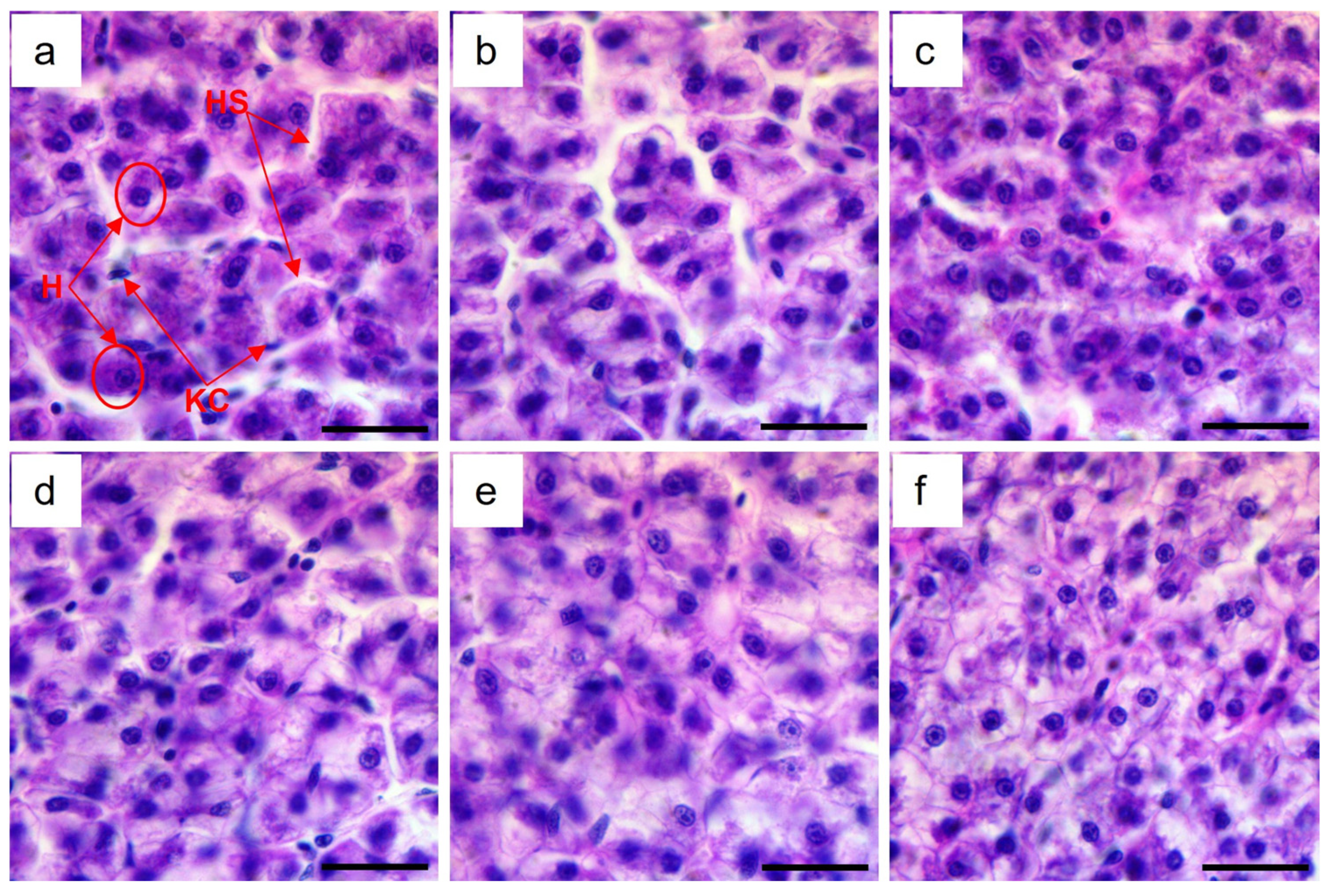
3.4. Liver Histology

When analysing the cross-section of the liver in juvenile red tilapia fed with different levels of chitosan in their respective diets (Figure 1), hepatocytes (H) with well-defined nuclei containing nucleoli, Kupffer cells (KCs) and liver sinusoid (HS) were observed. It is also worth noting that there was no liver damage for any treatment, although a slightly vacuolate appearance in the cytoplasm of hepatocytes was observed in fish that received the 40 g kg1 and 50 g kg1 chitosan inclusion levels. For the liver parameters (Table 6), differences were found between the treatments that used chitosan and the control: the highest values were reported for 40 g kg1 for HA, CA, and NA; meanwhile, HR was the highest for 20 g kg1 of chitosan.

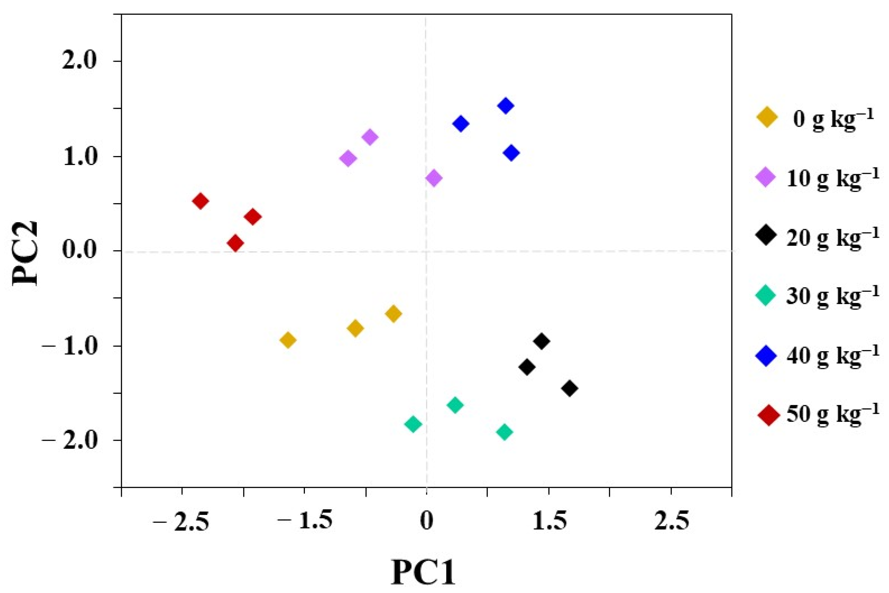
3.5. Principal Component Analysis (PCA)

Using the covariance matrix to explore differences in the response of juveniles, a PCA is presented in Figure 2. The plot PC1 vs. PC2 (using their multipliers as a variable) showed a clear pattern. PC1 (45.04%) and PC2 (21.54%), together, accounted for 66.58% of the total variance between the differences of the investigated indicators: performance response, feed utilisation, digestive and metabolic enzyme activity, and liver histology. For PC1, the main differences were observed in weight, length, SGR, FCF, transaminases, and hepatocyte morphometry. For PC2, the main differences were observed in enzymes, such as ALP, protease and amylases, and survival.
The diet with 40 g kg−1 of chitosan (blue diamond) is shown at the top-right of the plot and was highly positive for both PC1 and PC2; moreover, the diets with 20 and 30 g kg−1 were positive, as shown in the bottom-right of the PC1 plot (grey, black, and green diamonds, respectively); however, this was not the case for the other diets, as those with the 0, 10, and 50 g kg−1 chitosan inclusion levels in PC1 (grey, white, and red diamonds, respectively) were more negative. However, the diets with the 10 and 50 g kg−1 chitosan inclusion levels (white and red diamonds, respectively) were positive in the upper-right of the PC2 plot. From this PCA, it was possible to reduce the dimensionality in which the original set of treatments is expressed, showing that the diet with 40 g kg−1 of chitosan has better characteristics based on the variables analysed.

4. Discussion

Cichlids are a very diverse family and are distributed worldwide and occupy a wide variety of habitats in terms of substrate and available food. Hence, their feeding habits, digestive physiology, and metabolism are different among different species. Because of this, particular studies of each species are relevant. In the particular case of hybrid red tilapia (O. mossambicus × O. niloticus), they can be defined as omnivorous and are capable of digesting ingredients from different origins. However, it has become necessary to look for supplements that enhance the adsorption of nutrients, metabolism, growth, and survival of fish species in cultures [30].
Chitosan is an active growth promoter and can be considered to be an essential element for increasing the productive response of aquatic animals. Moreover, it is a natural polymer that is polycationic due to its multiple positively charged amine groups, and it has been recognised for its prebiotic effect. Chitosan inhibits the growth of harmful microorganisms and promotes the growth of beneficial bacteria (e.g., lactic acid bacteria and Bifidobacterium), which benefit the secretion and activity of digestive enzymes, as well as the digestion and nutrient adsorption of the diet [6,12,31].
In the present experiment (Table 3), the best survival rate (93.78–100%) was obtained after two months of rearing, and it was comparable to that reported for tilapia O. niloticus × O. aureus [16], grey mullet (Mugil cephalus) [14], European sea bass (Dicentrarchus labrax) [6], and Caspian kutum (Rutilus frisiikutum) [31], suggesting that chitosan, when incorporated into aquafeed, reduced fish mortality. In studies with a higher inclusion of chitosan than those previously mentioned, increases in growth, SGR, and FHR have also been reported. Akbary and Younesi [14] studied the effect of dietary chitosan at similar concentrations on the growth of grey mullet (Mugil cephalus), and the results showed that diets containing 10 and 15 g·kg−1 of chitosan improved growth rates. Similarly, the inclusion of chitosan at a higher level of 20 g·kg−1 in the diet of European carp (Cyprinus carpio) decreased fish mortality and enhanced growth under stress conditions [32].
Furthermore, a study was carried out to assess the effects of dietary chitosan on the growth performance, lipid metabolism, and gut microbiome of juvenile loach (Misgurnus anguillicaudatus), which were fed different levels of chitosan (0, 5, 10, 20, and 50 g·kg−1 diets) for 50 days; the results showed that high levels of supplemented chitosan (50 g·kg−1 diet) improved growth performance in loach [33]. Some authors [7,13,30,34], when employing chitosan nanoparticles in catfish (Clarias gariepinus), Nile tilapia (Oreochromis niloticus), and grey mullet (Liza ramada), found improvements in weight, weight gain, SGR, feeding efficiency, and survival. These beneficial effects on growth performance provide evidence that chitosan increases the absorption and assimilation of nutrients in freshwater fish. However, it is necessary to be mindful of the doses of chitosan, as it has been reported that high levels of chitosan in diets, when attacked by the gut microbiota, generate fermentation products, such as short-chain fatty acids, hydrogen (H2), carbon dioxide (CO2), and methane (CH4), which cause intestinal inflammation, permeability, and inadequate absorption of nutrients in the enterocytes, affecting the growth of the fish [5,8,12,31].
The study by Sun et al. [35] demonstrated the benefit of dietary supplementation with chitosan in diets, which improved the WG and SGR in tiger puffers (Takifugu rubripes) and decreased the FCR after eight weeks of feeding. Other studies have indicated that such increases in the growth of aquatic animals fed prebiotic diets may be attributed to improvements in digestive activity caused by enhancing the synthesis and secretion of pancreatic juices that are rich in zymogens, which are necessary for digestion [36], consequently improving food digestibility and increasing weight gain. Chitosan is possibly involved in the ability to foster a favourable intestinal bacterial population, promote digestive enzyme activity, and promote growth performance [37,38].
In our study (Table 4), an increase in digestive enzyme activity (pancreatic proteases, lipases, and amylases) was found in the treatments with chitosan compared to the control treatment. Increases in the activity of digestive enzymes when supplementing with chitosan have been previously reported by Sheikhzadeh et al. [39]; they found significant increases in growth performance and digestive enzymes when using chitosan and nanochitosan in combination with zeolite, which can partly be attributed to the enhanced activity of hydrolysis (protein, lipid, and carbohydrate) for digestive enzymes, gut microbiota, and the adsorption of macromolecules in the midgut.
Liu et al. [40], when studying the effect of chitosan on the larvae of Larimichthys crocea, found high differences in amylase activity between the diets of the control and chitosan concentration; moreover, they found that the behaviour of amylase may be related to chitosan’s ability to restore intestinal microflora balance and improve intestinal mucosal barrier function, with improved adsorption of carbohydrates in microvilli.
Changes in the activities of the intestinal digestive enzymes after the 60-day feeding period with chitosan in Paramisgurnus dabryanus were found by Zhang [37], wherein the oral administration of chitosan significantly improved the activities of protease, lipase, and amylase compared to those of the control group. Chitosan in the digestive tract of fish is hydrolysed and converted into oligosaccharides, which are water soluble and helpful for Ca2+ absorption, which works to promote enzymatic activity [31,38]. Due to its antibacterial activity, chitosan can decrease intestinal pathogenic flora in fish, stabilising the intestinal barrier, and also induce the expression of the intestinal digestive enzymes of the brush border of the enterocytes, increasing the hydrolysates that will be absorbed by the intestine [30,36].
The majority of ALP (more than 80%) is produced in the liver, bones, and—in small amounts—the intestine [41,42]. The ALP is associated with catalytic activity and the calcification process in bone tissue, as well as with fat transfer in the intestine [43]. The relationship between chitosan and ALP in this study suggests that increased levels of chitosan in the diet may affect the calcification process. Similar results were reported by Sheikhzadeh et al. [39], who conducted a study on the effect of chitosan on fish, which could enhance the level of ALP enzyme activity. Therefore, it is thought that increased chitosan absorption in the intestinal cells triggers the mechanisms involved in absorbing glucose [6,44]. Glucose acts as a substrate in the biosynthesis process of some macromolecules. Therefore, the presence of available glucose may be the starting point for increasing the activity of ALP, which is, structurally, a glycoprotein macromolecule: it is a part of the catalytic structure of the ALP enzyme [43].
The results of this study revealed that a significant decrease in AST and ALT activity levels depends on the chitosan dose in all treatment groups. Abdel El-Naby et al. [30] recorded a linear decrease in serum ALT and AST and an increase in chitosan levels in Nile tilapia diets compared to the control. Feed biostimulants have been found to reduce the oxidative stress induced by pollutants correlated with their antioxidant and free radical scavenging activity [44] and thus, consequently, to promote the health benefits reflected in hepatocyte function and structural improvements [45,46].
Mehrpak et al. [47] reported that the activities of AST and ALT were altered in Cyprinus carpio fish fed with 1 g kg−1 of chitosan per kg of feed. This effect might be attributable to the antioxidant activity of chitosan, which guards hepatocytes against oxidative damage [44]. This hepatoprotective effect of chitosan can be associated with its immunostimulating effect in fish because the liver plays a vital role in the generation of acute phase proteins and the elimination of pathogens, antigens, and molecules [48].
Additives, such as chitosan, used in fish supplementation exert synergistic effects on health in the promotion and prevention of diseases [44,48]. In recent studies, chitosan was found to act on liver nuclear receptors and as a regulator of cholesterol and phospholipids in the liver [49]. Our findings from the cross-section of the liver coincide with Salaah et al. [50], who reported positive effects on O. niloticus livers when using chitosan in the diet. In O. mossambicus, normal liver tissue was observed in groups of fish fed only with chitosan, which had less toxic effects and improved cellular changes like hypertrophy, vacuole formations, and cellular degenerations caused by heavy metals [51]. On the other hand, when using chitosan nanoparticles in a preventive way in fish, they foundin the histopathological studies that the therapeutic treatment group had the fewest signs of pathological lesions [6]. Systemic inflammatory signs were detected in different patterns in the liver in positive control groups but not for those that received preventive treatment with chitosan, where no affectations were observed.
The values obtained in the hepatocyte area (Table 6) in this study were higher than those reported by Rezende et al. [52]; in their study with biostimulants in juvenile red tilapia, they obtained a hepatocyte area value of 8.57 μm2. The hepatocytes observed in the present work were of round morphology (Figure 1), which coincides with what was stated by Li et al. [53], who indicated that the addition of chitosan produces, in hepatocytes, a round morphology with many microvilli, and improves metabolic activities such as protein synthesis, which is conducive to healthy cells.
Dawood et al. [13] reported that chitosan improved the histomorphometric characteristics in grey salmon, where they maintained that the inclusion of chitosan in fish diets helps to increase the size of the cells up to normal morphophysiological levels. When carrying out the histological and histochemical characterisation of the liver of creole perch (Percichthys trucha), they found a hepatocyte diameter value of 4.48 μm, which is considered normal [54]. Park et al. [55] evaluated the effect of galactosylated chitosan on hepatocytes using in vitro conditions and found that it improves the epidermal growth factor and survival of hepatocytes.
In our study, the addition of chitosan to the fish diet resulted in a slight increase in the hepatocyte vacuolisation of 40 g kg−1 and 50 g kg−1 chitosan inclusion levels, and this vacuolisation was mainly related to the fatty and glycogen types. This is consistent with the study by Chiu et al. [49], who proved that dietary supplementation with fish oil or chitosan, or a combination of both, can improve abnormal lipid accumulation in the liver of rats. According to Salam et al. [8], vacuolisation in cells may indicate stored energy in the form of glycogen or lipids; alternatively, it may represent a degenerative change in which there is a fluid distension of organelles, such as in the endoplasmic reticulum and Golgi apparatus, and/or an accumulation of free fluid in the cytoplasm. The histological analysis of juvenile red tilapia liver showed a positive effect of chitosan on liver morphology. In general, all these previously mentioned data evidence a lower oxidative stress level in fish fed with chitosan. In addition, optimal dietary chitosan content should be carefully analysed together with water quality, feeding rations, and feed quality.

5. Conclusions

In the present study, chitosan directly improved digestion and growth rate. The PCA results suggest that a dietary chitosan level of 40 g kg−1 promotes growth, digestive and metabolic enzyme activity, and proper nutrient use without affecting the liver in O. mossambicus × O. niloticus hybrid juveniles. Furthermore, this could be recommended for further studies in feed formulation for the red tilapia.

Author Contributions

Y.M.-M. contributed to project ideas, supervision, data analysis, and manuscript preparation; A.R.V.-V. Investigation, visualization and collected the date of researched; Y.C.-Q., contributed to data analysis and manuscript review preparation, review and editing original drafts; A.B.-L. contributed to data analysis, manuscript and review preparation and P.D.M.-C. contributed to visualization and collected the dates of research material; E.C.-J. and N.S.C. contributed to conceptualization and the review and editing of the original drafts. All authors have read and agreed to the published version of the manuscript.

Funding

This research was supported by the Universidad Técnica Estatal de Quevedo and the Projects: FOCYCYT-7ma/PFOC7-48-2020 and EMBLEMÁTICOS/PEMBL-003-2018.

Institutional Review Board Statement

This study was carried out without sacrificing organisms unnecessarily to reduce and perfect the use of animals used for scientific purposes. The proposed methods, use of animals, and research practice (approval number: CBA2022015) were examined and approved by the Animal Welfare and Ethics Committee at Universidad Técnica Estatal de Quevedo.

Data Availability Statement

Available after a reasonable request from the corresponding authors.

Acknowledgments

Our gratitude for the technical assistance of L. Ramos and W. Hidalgo. Thanks to the students of R. Zamora and M. Choez for their support in the investigation. This research was supported by the Universidad Técnica Estatal de Quevedo and the Projects: FOCYCYT-7ma/PFOC7-48-2020 and EMBLEMÁTICOS/PEMBL-003-2018. Thanks to K. Cienfuegos-Martinez for the support in the English edition.

Conflicts of Interest

The authors declare no conflict of interest.

References

  1. FAO. El Estado Mundial de la Pesca y la Acuicultura. Hacia la Transformación Azul; FAO: Rome, Italy, 2022; pp. 1–97. [Google Scholar] [CrossRef]
  2. Méndez-Martínez, Y.; Pacheco-Morales, G.K.; Del Barco-Ibarra, K.A.; Torres-Navarrete, Y.G.; Hernández-Vergara, M.P. Biochemical and immune response in red tilapia (Oreochromis mossambicus × O. niloticus) with dietary chitosan supplementation. Rev. Fac. Agron. 2021, 38, 1016–1034. [Google Scholar] [CrossRef]
  3. Méndez-Martínez, Y.; Narváez-Narváez, R.I.; Angulo, C.; Cortés-Jacinto, E.; Botello Leon, A.; Verdecia, D.; Torres-Navarrete, Y.G. Chemical composition of Tithonia diversifolia (Hemsl.) and its effect on growth performance, feed efficiency and metabolic biochemistry of juvenile hybrid tilapia, Oreochromis mossambicus × Oreochromis niloticus. Not. Bot. Horti Agrobot. Cluj-Napoca 2023, 51, 13337. [Google Scholar] [CrossRef]
  4. Ayyat, M.S.; Ayyat, A.M.N.; Al-Sagheer, A.A.; El-Hais, A.E.-A.M. Effect of some safe feed additives on growth performance, blood biochemistry, and bioaccumulation of aflatoxin residues of Nile tilapia fed aflatoxin-B1 contaminated diet. Aquaculture 2018, 495, 27–34. [Google Scholar] [CrossRef]
  5. Wu, Y.; Rashidpour, A.; Almajano, M.P.; Metón, I. Chitosan-Based Drug Delivery System: Applications in Fish Biotechnology. Polymers 2020, 12, 1177. [Google Scholar] [CrossRef]
  6. Saleh, M.; Essawy, E.; Shaalan, M.; Osman, S.; Ahmed, F.; El-Matbouli, M. Therapeutic Intervention with Dietary Chitosan Nanoparticles Alleviates Fish Pathological and Molecular Systemic Inflammatory Responses against Infections. Mar. Drugs 2022, 20, 425. [Google Scholar] [CrossRef]
  7. Udo, I.U.; Etukudo, U.; Anwana, U.I.U. Effects of chitosan and chitosan nanoparticles on water quality, growth performance, survival rate and meat quality of the African catfish, Clarias gariepinus. Nanoscience 2018, 1, 12–25. [Google Scholar] [CrossRef]
  8. Salam, M.A.; Rahman, M.A.; Paul, S.I.; Islam, F.; Barman, A.K.; Rahman, Z. Dietary chitosan promotes the growth, biochemical composition, gut microbiota, hematological parameters and internal organ morphology of juvenile Barbonymus gonionotus. PLoS ONE 2021, 16, e0260192. [Google Scholar] [CrossRef]
  9. Cheba, B.A. Chitin and chitosan: Marine biopolymers with unique properties and versatile applications. Glob. J. Biotechnol. Biochem. (GJBB) 2011, 6, 149–153. Available online: http://idosi.org/gjbb/gjbb6%283%2911/7.pdf (accessed on 10 August 2023).
  10. Divya, K.; Jisha, M. Chitosan nanoparticles preparation and applications. Environ. Chem. Lett. 2018, 16, 101–112. [Google Scholar] [CrossRef]
  11. Nikapitiya, C.; Dananjaya, S.H.S.; De Silva, B.C.J.; Heo, G.J.; Oh, C.; De Zoysa, M.; Lee, J. Chitosan nanoparticles: A positive immune response modulator as display in zebrafish larvae against Aeromonas hydrophila infection. Fish Shellfish Immunol. 2018, 76, 240–246. [Google Scholar] [CrossRef]
  12. Abdel-Tawwab, M.; Razek, N.A.; Abdel-Rahman, A.M. Immunostimulatory effect of dietary chitosan nanoparticles on the performance of Nile tilapia, Oreochromis niloticus (L.). Fish Shellfish Immunol. 2019, 88, 254–258. [Google Scholar] [CrossRef] [PubMed]
  13. Dawood, M.A.; Gewaily, M.S.; Soliman, A.A.; Shukry, M.; Amer, A.A.; Younis, E.M.; Fadl, S.E. Marine-derived chitosan nanoparticles improved the intestinal histo-morphometrical features in association with the health and immune response of grey mullet (Liza ramada). Mar. Drugs 2020, 18, 611. [Google Scholar] [CrossRef] [PubMed]
  14. Akbary, P.; Younesi, A. Effect of dietary supplementation of Chitosan on growth, hematology and innate immunity of grey Mullet (Mugil cephalus). Vet. Res. Biol. Prod. 2017, 30, 194–203. [Google Scholar] [CrossRef]
  15. Salem, M.E. Utilization of Some Feed Additives in Improving the Nutritive Value of Marine Fish Diet. Ph.D. Dissertation, Alexandria University, Alexandria, Egypt, 2015. [Google Scholar]
  16. Shiau, S.Y.; Yu, Y.P. Dietary supplementation of chitin and chitosan depresses growth in tilapia, Oreochromis niloticus × O. aureus. Aquaculture 1999, 179, 439–446. [Google Scholar] [CrossRef]
  17. Méndez-Martínez, Y.; García-Guerrero, M.U.; Arcos-Ortega, F.G.; Martínez-Córdova, L.R.; Yamasaki-Granados, S.; Pérez-Rodríguez, J.C.; Cortés-Jacinto, E. Effect of different ratios of dietary protein-energy on growth, body proximal composition, digestive enzyme activity, and hepatopancreas histology in Macrobrachium americanum (Bate, 1868) prawn juveniles. Aquaculture 2018, 485, 1–11. [Google Scholar] [CrossRef]
  18. Méndez-Martínez, Y.; Ceseña, C.E.; Luna-González, A.; García-Guerrero, M.U.; Martinez-Porchas, M.; Campa-Cordova, Á.I.; Cortés-Jacinto, E. Effects of different dietary protein-energy ratios on growth, carcass amino acid and fatty acid profile of male and female Cherax quadricarinatus (von Martens, 1868) pre-adults. Aquac. Nutr. 2021, 27, 2481–2496. [Google Scholar] [CrossRef]
  19. AOAC. Official Methods of Analysis of AOAC International, 21st ed.; AOAC: Gaithersburg, MD, USA, 2019. [Google Scholar]
  20. Ramanathan, G.; Ramalakshmi, P.; Gopperundevi, B.; Suresh, J.I. Production Characterization and Aqua Feed Supplementation of Astaxanthin from Halobacterium salinarium. Int. J. Curr. Microbiol. App. Sci. 2015, 4, 56–63. Available online: https://www.ijcmas.com/vol-4-3/G (accessed on 12 August 2023).
  21. Anson, M.L. The estimation of pepsin, trypsin, papain and cathepsin with hemoglobin. J. Gen. Physiol. 1938, 22, 79–89. [Google Scholar] [CrossRef]
  22. Bernfeld, P. Amylase, alpha and beta. Methods Enzymol. 1955, 1, 149–151. Available online: https://cir.nii.ac.jp/crid/1573668924362136704 (accessed on 29 June 2023).
  23. Versaw, W.; Cuppett, S.L.; Winters, D.D.; Williams, L.E. An improved colorimetric assay for bacterial lipase in nonfat dry milk. J. Food Sci. 1989, 54, 232–254. [Google Scholar] [CrossRef]
  24. Bradford, M.M. Rapid and sensitive method for quantitation of microgram quantities of protein utilizing principle of protein-dye binding. Anal. Biochem. 1976, 72, 248–254. [Google Scholar] [CrossRef] [PubMed]
  25. Bergmeyer, H.U.; Bowers, G.N., Jr.; Horder, M.; Moss, D.W. Provisional recommendations on IFCC methods for the measurement of catalytic concentrations of enzymes. Part 2. IFCC method for aspartate aminotransferase. Clin. Chim. Acta 1976, 70, F19–F29. [Google Scholar] [CrossRef] [PubMed]
  26. Xu, J.; Yu, Z.; Liu, G.; Li, S.; Zhou, G.; Wang, H.; Dong, Y.; You, C.; Bai, W.; Zhou, M. Effects of Dietary Lentinus edodes Fermentation Supplementation on Digestive Enzyme Activity, Antioxidant Capacity and Morphology of the Liver and Intestine in Largemouth Bass (Micropterus salmoides) Fed High Plant Protein Diets. Fishes 2023, 8, 329. [Google Scholar] [CrossRef]
  27. Naiel, M.A.E.; Ismael, N.E.M.; Abd El-hameed, S.A.A.; Amer, M.S. The antioxidative and immunity roles of chitosan nanoparticle and vitamin C-supplemented diets against imidacloprid toxicity on Oreochromis niloticus. Aquaculture 2020, 523, 735219. [Google Scholar] [CrossRef]
  28. De Souza Filho, J.; Pires, F.S.; Grisolia, C.K.; De Sabóia Morais, S.M.T. Toxicological effects of a glyphosate-based formulation on the liver of Poecilia reticulata. Curr Top. Toxicol. 2014, 9, 81–91. [Google Scholar] [CrossRef]
  29. Zar, J.H. Biostatistical Analysis, 2nd ed.; Prentice-Hall: Upper Saddle Creek, NJ, USA, 1984; pp. 236–346. [Google Scholar]
  30. Abdel El-Naby, F.; Naiel, M.; Al-Sagheer, A.A.; Negm, S. Dietary chitosan nanoparticles enhance the growth, production performance, and immunity in Oreochromis niloticus. Aquaculture 2019, 25, 82–89. [Google Scholar] [CrossRef]
  31. Najafabad, M.; Imanpoor, M.R.; Taghizadeh, V.; Alishahi, A. Effect of dietary chitosan on growth performance, hematological parameters, intestinal histology and stress resistance of Caspian kutum (Rutilus frisii kutum Kamenskii, 1901) fingerlings. Fish Physiol. Biochem. 2016, 42, 1063–1071. [Google Scholar] [CrossRef]
  32. Stanek, M.; Mazurkiewicz, J.; Rawski, M.; Bogucka, J.; Ziółkowska, E.; Dankowiakowska, A.; Kierończyk, B. Effect of chitosan on common carp (Cyprinus carpio) fry growth performance, feed utilization and nutriphysiological status. Aquac. Rep. 2023, 30, 101622. [Google Scholar] [CrossRef]
  33. Yan, J.; Guo, C.; Dawood, M.A.O.; Gao, J. Effects of dietary chitosan on growth, lipid metabolism, immune response and antioxidant-related gene expression in Misgurnus anguillicaudatus. Benef. Microbes 2017, 8, 439–449. [Google Scholar] [CrossRef]
  34. Fadl, S.E.; El-Gammal, G.A.; Abdo, W.S.; Barakat, M.; Sakr, O.A.; Nassef, E.; El-Sheshtawy, H.S. Evaluation of dietary chitosan effects on growth performance, immunity, body composition and histopathology of Nile tilapia (Oreochromis niloticus) as well as the resistance to Streptococcus agalactiae infection. Aquac. Res. 2020, 51, 1120–1132. [Google Scholar] [CrossRef]
  35. Su, P.; Han, Y.; Jiang, C.; Ma, Y.; Pan, J.; Liu, S.; Zhang, T. Effects of chitosan-oligosaccharides on growth performance, digestive enzyme and intestinal bacterial flora of tiger puffer (Takifugu rubripes Temminck et Schlegel, 1850). J. Appl. Ichthyol. 2017, 33, 458–467. [Google Scholar] [CrossRef]
  36. Hussain, M.A.; Sumon, T.A.; Mazumder, S.K.; Ali, M.M.; Jang, W.J.; Abualreesh, M.H.; Hasan, M.T. Essential oils and chitosan as alternatives to chemical preservatives for fish and fisheries products: A review. Food Control 2021, 129, 108244. [Google Scholar] [CrossRef]
  37. Zhang, B. Dietary chitosan oligosaccharides modulate the growth, intestine digestive enzymes, body composition and nonspecific immunity of loach Paramisgurnus dabryanus. Fish Shellfish Immunol. 2019, 88, 359–363. [Google Scholar] [CrossRef]
  38. Coutinho, F.; Castro, C.; Guerreiro, I.; Rangel, F.; Couto, A.; Serra, C.R.; Enes, P. Mealworm larvae meal in diets for meagre juveniles: Growth, nutrient digestibility and digestive enzymes activity. Aquaculture 2021, 535, 736362. [Google Scholar] [CrossRef]
  39. Sheikhzadeh, N.; Kouchaki, M.; Mehregan, M.; Tayefi-Nasrabadi, H.; Divband, B.; Khataminan, M.; Shabanzadeh, S. Influence of nanochitosan/zeolite composite on growth performance, digestive enzymes and serum biochemical parameters in rainbow trout (Oncorhynchus mykiss). Aquac. Res. 2017, 48, 5955–5964. [Google Scholar] [CrossRef]
  40. Liu, J.; Xu, W.; Liu, Y.; Wang, Y.; Zhang, J.; Wang, Z.; Ai, Q. Effects of chitosan-coated microdiet on dietary physical properties, growth performance, digestive enzyme activities, antioxidant capacity, and inflammation response of large yellow croaker (Larimichthys crocea) larvae. Aquac. Nutr. 2022, 2022, 4355182. [Google Scholar] [CrossRef] [PubMed]
  41. Coleman, J.E. Structure and mechanism of alkaline phosphatase. Annu. Rev. Biophys. Biomol. Struct. 1922, 21, 441–444. [Google Scholar] [CrossRef]
  42. Estaki, M.; DeCoffe, D.; Gibson, D.L. Interplay between intestinal alkaline phosphatase, diet, gut microbes and immunity. World J. Gastroenterol. 2014, 20, 15650. [Google Scholar] [CrossRef]
  43. Ray, C.S.; Singh, B.; Jena, I.; Behera, S.; Ray, S. Low alkaline phosphatase (ALP) in adult population an indicator of zinc (Zn) and magnesium (Mg) deficiency. Curr. Res. Nutr. Food Sci. J. 2017, 5, 347–352. [Google Scholar] [CrossRef]
  44. Abdel-Ghany, H.M.; Salem, M.E.S. Effects of dietary chitosan supplementation on farmed fish; a review. Rev. Aquac. 2020, 12, 438–452. [Google Scholar] [CrossRef]
  45. Dong, X.; Wang, Y.; Song, H.; Zou, X. Effects of in ovo injection of carbohydrate solution on small intestine development in domestic pigeons (Columba livia). J. Anim. Sci. 2013, 91, 3742–3749. [Google Scholar] [CrossRef] [PubMed]
  46. Ghasemi, Z.; Sourinejad, I.; Kazemian, H.; Rohani, S. Application of zeolites in aquaculture industry: A review. Rev. Aquac. 2018, 10, 75–95. [Google Scholar] [CrossRef]
  47. Mehrpak, M.; Banaee, M.; Nematdoost Haghi, B.; Noori, A. Protective effects of vitamin C and chitosan against cadmium-induced oxidative stress in the liver of common carp (Cyprinus carpio). Iran. J. Toxicol. 2015, 9, 1360–1367. Available online: https://ijt.arakmu.ac.ir/article-1-455-en.pdf (accessed on 20 June 2023).
  48. Ismael, N.E.M.; Abd El-Hameed, S.A.A.; Salama, A.M.; Naiel, M.A.E.; Abdel-Latif, H.M.R. The effects of dietary clinoptilolite and chitosan nanoparticles on growth, body composition, haemato-biochemical parameters, immune responses, and antioxidative status of Nile tilapia exposed to imidacloprid. Environ. Sci. Pollut. Res. Int. 2021, 28, 29535–29550. [Google Scholar] [CrossRef]
  49. Chiu, C.Y.; Chang, T.C.; Liu, S.H.; Chiang, M.T. The regulatory effects of fish oil and chitosan on hepatic lipogenic signals in high-fat diet-induced obese rats. J. Food Drug Anal. 2017, 25, 919–930. [Google Scholar] [CrossRef]
  50. Salaah, S.M.; Dalia, M.; Gaber, H.S. Potential effects of dietary chitosan against lead-induced innate immunotoxicity and oxidative stress in Nile tilapia (Oreochromis niloticus). Egypt. J. Aquat. Res. 2022, 48, 123–129. [Google Scholar] [CrossRef]
  51. Thilagar, G.; Samuthirapandian, R. Chitosan from crustacean shell waste and its protective role against lead toxicity in Oreochromis mossambicus. Toxicol. Rep. 2020, 7, 296–303. [Google Scholar] [CrossRef]
  52. Rezende, K.F.O.; Bergami, E.; Alves, K.V.B.; Corsi, I.; Barbieri, E. Titanium dioxide nanoparticles alter routine metabolism and cause histopathological alterations in Oreochromis niloticus. B. Inst. Pesca. 2018, 44, 1–11. [Google Scholar] [CrossRef]
  53. Li, J.; Pan, J.; Zhang, L.; Yu, Y. Culture of hepatocytes on fructose-modified chitosan scaffolds. Biomaterials 2003, 24, 2317–2322. [Google Scholar] [CrossRef]
  54. Palma Leotta, M.E.; Caliri, M.N.; Cáceres Gimenez, A.R.R. Caracterización Histológica e Histoquímica de Branquia, Hígado y Riñón de Perca Criolla (Percichthys trucha, Valenciennes, 1833) Para su uso en Biomonitoreo Ambiental. Acta Microsc. 2017, 26, 32–45. Available online: https://ri.conicet.gov.ar/handle/11336/50211 (accessed on 18 August 2023).
  55. Park, I.K.; Yang, J.; Jeong, H.J.; Bom, H.S.; Harada, I.; Akaike, T.; Cho, C.S. Galactosylated chitosan as a synthetic extracellular matrix for hepatocytes attachment. Biomaterials 2003, 24, 2331–2337. [Google Scholar] [CrossRef] [PubMed]
Figure 1. Cross-section of liver in juvenile hybrid red tilapia (O. mossambicus × O. niloticus) fed diets with different chitosan inclusion levels. Treatments: (a) 0 (control), (b) 10, (c) 20, (d) 30, (e) 40, and (f) 50 g kg−1 of chitosan in diet. H&E stain. Abbreviations: H, hepatocytes; KC, Kupffer cell; SH, liver sinusoid. Scale bar is 100 µm.
Fishes 08 00546 g001
Figure 2. Plot for the multipliers of the first two principal components of the PCA analysis of the growth performance, feed utilisation, digestive and metabolic enzyme activity, and liver histology in juvenile hybrid red tilapia (O. mossambicus × O. niloticus) fed diets with different chitosan inclusion levels after 8 weeks of culture. N = 3, yellow diamond (0 g kg−1), pink diamond (10 g kg−1), black diamond (20 g kg−1), green diamond (30 g kg−1), blue diamond (40 g kg−1), and red diamond (50 g kg−1) denote chitosan inclusion levels.
Fishes 08 00546 g002
Table 1. Formulations of experimental diets with the inclusion of chitosan at different levels.
IngredientsChitosan Levels (g kg−1)
01020304050
Fish meal 1250250250250250250
Soybean meal 2280280280280280280
Wheat flour 3204204204204204204
Corn meal 4190180170160150140
Chitosan 5-1020304050
Vegetable oil 6101010101010
Fish oil 7151515151515
Sodium alginate 8202020202020
Mineral premixes 9,10101010101010
Vitamin premixes 11,12202020202020
Vitamin C 13111111
1 Comercial El Gordillo, Santo Domingo of The Tsáchilas, Ecuador; 2 Ullón Poultry—Valencia, Ecuador; 3,4,6,13 Supermaxi—Quevedo; Ecuador, 5 Sigma Aldrich, USA; 7 Fortidex S.A—Santa Elena; 8 Supplies AZ, La Paz, BCS, México; 9,11,13 Super Éxito, Quevedo, Ecuador; 10 mg·kg−1: Magnesium sulfate 5.1; Sodium chloride 2.4; Potassium Chloride 2; Ferrous sulfate 1; Zinc sulfate 0.2; Cupric sulfate 0.0314; Manganous sulfate 0.1015; Cobalt sulfate 0.0191; Calcium iodate 0.0118; Chlorine Chloride 0.051. 12 mg·kg−1: Thiamine 60; Rivoflavin 25; Niacin 40; Vitamin B6 50; Pantothenic acid 75; Biotin 1; Folate 10; Vitamin B12 0.2; Hill 600; Myoinositol 400; Vitamin C 200; Vitamin A 5000 IU; Vitamin E 100; Vitamin D 0.1; Vitamin K5.
Table 2. Proximal composition (as-fed basis) of the experimental diets with the inclusion of chitosan at different levels.
Proximal Composition
(g kg−1 as Fed Basis) 1
Chitosan Levels (g kg−1)
01020304050
Dry matter (DM)946.8937.7940.2942.8938.0939.9
Crude protein (CP)334.2333.5333.8333.2333.0332.8
Crude lipid (CL)66.466.065.164.557.365.8
Crude fibre (CF)15.716.417.118.219.320.1
Ash114.3114.9115.2116.1117.0118.4
Nitrogen-free extract (NFE)416.2406.9408.9410.8411.4402.8
DE (MJ kg−1 feed)12.8512.8512.8512.8512.8512.85
CP DE−1 (mg PC MJ−1)26.0125.9525.9825.9325.9125.9
1 Data are expressed as the mean of three replicates.
Table 3. Growth performance and feed utilisation in juvenile hybrid red tilapia (O. mossambicus × O. niloticus) when fed diets with different chitosan inclusion levels.
Productive ParametersChitosan Levels (g kg−1)p
01020304050
Initial Weight, g7.47 ± 0.0527.62 ± 0.0357.53 ± 0.0407.46 ± 0.0647.69 ± 0.0307.41 ± 0.0750.066
Final Weight, g22.66 ± 0.133 c20.37 ± 0.185 d25.72 ± 0.202 b23.59 ± 0.162 c28.08 ± 0.150 a25.58 ± 0.191 b0.011
Final Length, cm10.05 ± 0.017 bc9.49 ± 0.040 c10.58 ± 0.075 ab10.14 ± 0.029 ab11.14 ± 0.104 a10.38 ± 0.110 ab0.030
Weight Gain, g15.19 ± 0.017 d12.75 ± 0.69 e18.19 ± 0.092 b16.23 ± 0.081 c20.39 ± 0.121 a18.27 ± 0.381 b0.010
SGR, %0.96 ± 0.006 cd0.55 ± 0.012 d1.30 ± 0.017 bc1.11 ± 0.014 c1.55 ± 0.029 a1.34 ± 0.006 b0.031
HIS, %2.03 ± 0.069 b2.25 ± 0.098 a2.38 ± 0.127 a1.75 ± 0.133 c2.15 ± 0.087 ab1.83 ± 0.064 c0.017
FCF1.63 ± 0.023 b1.80 ± 0.069 a1.29 ± 0.052 c1.60 ± 0.064 b1.24 ± 0.035 c1.68 ± 0.021 b0.024
Survival Rate, %98.67 ± 1.478 bc93.78 ± 2.182 d97.33 ± 1.663 c98.67 ± 1.490 bc100 ± 1.599 a97.33 ± 1.652 c0.010
The results are expressed as mean ± standard error from triplicate groups (n = 3). abcde Different letters between chitosan levels denote significant differences (Tukey test, p < 0.05). Abbreviations: SGR, specific growth rate; FCF, feed conversion factor; HIS, hepatosomatic index.
Table 4. Digestive enzyme activity in juvenile hybrid red tilapia (O. mossambicus × O. niloticus) fed diets with different chitosan inclusion levels.
Enzyme Activity
(U mg−1 Protein)
Chitosan Levels (g kg−1)p
01020304050
Proteases39.19 ± 1.091 bc42.99 ± 1.126 b53.31 ± 1.287 a54.05 ± 1.189 a54.26 ± 1.276 a38.38 ± 1.143 c0.012
Lipases34.97 ± 0.704 c31.15 ± 0.566 d40.33 ± 0.774 a41.77 ± 0.762 a38.22 ± 0.727 b37.65 ± 0.768 b0.001
Amylases69.41 ± 1.709 c76.20 ± 1.732 a63.97 ± 1.697 d72.67 ± 1.709 b77.89 ± 1.761 a77.10 ± 1.738 a0.010
The results are expressed as mean ± standard error from triplicate groups (n = 3). abcd Different letters between chitosan levels denote significant differences (Tukey test, p < 0.05).
Table 5. Metabolic enzyme activity in juvenile hybrid red tilapia (O. mossambicus × O. niloticus) fed diets with different chitosan inclusion levels.
Enzyme Activity
(U L−1)
Chitosan Levels (g kg−1)p
01020304050
AST170.37 ± 1.611 a109.71 ± 1.513 b182.22 ± 0.970 a74.48 ± 1.247 d77.49 ± 1.126 d93.25 ± 0.375 c0.022
ALT45.33 ± 1.201 a45.67 ± 0.883 a38.00 ± 1.178 bc37.67 ± 1.334 bc39.67 ± 2.188 b35.67 ± 1.455 c0.004
ALP89.67 ± 0.664 d112.00 ± 2.084 bc107.67 ± 1.669 c109.33 ± 1.767 c114.00 ± 1.531 ab118.02 ± 1.478 a 0.015
The results are expressed as mean ± standard error from triplicate groups (n = 3). abcd Different letters between chitosan levels denote significant differences (Tukey test, p < 0.05). Abbreviations: AST, aspartate aminotransferase; ALT, alanine aminotransferase; ALP, alkaline phosphatase.
Table 6. Morpho-histology of liver in juvenile hybrid red tilapia (O. mossambicus × O. niloticus) fed diets with different chitosan inclusion levels.
HepatocyteChitosan Levels, g kg−1p
01020304050
HA, µm266.17 ± 1.022 d62.68 ± 3.683 e79.38 ± 3.759 b70.75 ± 1.432 c88.54 ± 1.951 a83.55 ± 3.655 ab0.016
CA, µm257.07 ± 1.062 c54.59 ± 3.406 c71.11 ± 3.683 ab62.20 ± 1.339 b78.71 ± 1.732 a74.24 ± 0.433 a0.015
NA, µm29.09 ± 0.289 ab8.09 ± 0.237 c8.28 ± 0.092 bc8.55 ± 0.098 b9.83 ± 0.242 a9.31 ± 0.289 a0.036
HR, µm26.56 ± 0.225 cd6.49 ± 0.219 d8.86 ± 0.323 a6.93 ± 0.087 c7.86 ± 0.081 b7.64 ± 0.225 b0.024
The results are expressed as mean ± standard error from triplicate groups (n = 3). abcde Different letters between chitosan levels denote significant differences (Tukey test, p < 0.05). Abbreviations: HA, hepatocyte area; CA, hepatocyte cytoplasm area; NA, hepatocyte nucleus area; HR, ratio of cytoplasmic area and nuclear area of the hepatocyte.
Disclaimer/Publisher’s Note: The statements, opinions and data contained in all publications are solely those of the individual author(s) and contributor(s) and not of MDPI and/or the editor(s). MDPI and/or the editor(s) disclaim responsibility for any injury to people or property resulting from any ideas, methods, instructions or products referred to in the content.

