Article

Saudi Traditional Fermented Goat Milk Protects against Experimental Non-Alcoholic Fatty Liver Disease by Hypoglycaemic and Antioxidant Potentials

by
Khadijah M. Altamimy
1,
Ghedeir M. Alshammari
1,*,
Abu ElGasim A. Yagoub
1,
Norah A. Albekairi
2,
Samiyah Alshehri
2,
Ali Saleh
1 and
Mohammed Abdo Yahya
1
1
Department of Food Science and Nutrition, College of Food and Agricultural Sciences, King Saud University, Riyadh 11451, Saudi Arabia
2
Department of Pharmacology and Toxicology, College of Pharmacy, King Saud University, Riyadh 11451, Saudi Arabia
*
Author to whom correspondence should be addressed.
Fermentation 2022, 8(12), 735; https://doi.org/10.3390/fermentation8120735
Submission received: 20 November 2022 / Revised: 7 December 2022 / Accepted: 8 December 2022 / Published: 13 December 2022

Abstract

:
This study examined the effect of fermented goat milk (oggtt) against non-alcoholic fatty liver disease (NAFLD) in rats induced by chronic high-fat diet (HFD) treatments. Both control-fed and HFD-fed adult male rats received the same vehicle or treatment with two doses of freshly collected oggtt (2 mL or 5 mL) for 12 weeks (n = 8/group). The treatment of the control and HFD-fed rats with oggtt in both doses significantly reduced weight gain, but fasting serum glucose and insulin levels as well as HOMA-IR levels were lowered only in the HFD-fed rats. Treatment improved HFD-induced glucose and insulin homeostasis impairment as measured by the oral glucose tolerance test. Both doses of oggtt reduced serum levels of liver function markers and C-reactive protein (CRP) as well as hepatic levels of malondialdehyde (MDA), tumour necrosis factor-α (TNF-α), and in-terlukin-6 (IL-6) in HFD-fed rats. In addition, the oggtt doses reduced serum and hepatic levels of triglycerides (TGs) and cholesterol (CHOL) as well as serum levels of low-density lipoproteins (LDL) in these rats. These biochemical endpoints were reflected by the improvement in liver histology and reduction in the number of fatty vacuolated and pyknotic cells. In both the control and HFD-fed rats, oggtt at both doses stimulated levels of superoxide dismutase (SOD) and glutathione (GSH). All these effects were more profound with the highest dose of oggtt. In conclusion, the finding of this study strongly supports the use of oggtt as a functional food to treat NAFLD, as it has shown hypoglycaemic and antioxidant properties.

1. Introduction

Obesity, defined as an increase in body mass index (BMI), is a major health problem that results from increased calorie intake and reduced energy expenditure [1]. The prevalence of obesity is rapidly increasing worldwide across both genders and all ages [2]. Current records show that 24.7% of the Saudi Arabian population is obese, which is expected to increase over the next decades [3].
Yet, obesity is an independent risk factor for developing type-2 diabetes mellitus (T2DM) and metabolic syndrome [4]. In addition, obesity is a major contributor to the onset of non-alcoholic fatty liver disease (NAFLD), which impairs hepatic glucose and lipid metabolism and increases peripheral adipose tissue lipogenesis through the induction of T2DM and IR [4,5]. Uncontrolled de novo lipogenesis (DNL) and increased intrahepatic lipid accumulation are two features of NAFLD that can lead to steatosis, which can progress to non-alcoholic steatohepatitis fibrosis and liver failure [6].
The mechanism behind the development and progression of NAFLD is still complicated and involves numerous pathways. Increased lipid intake is the intimal trigger that accumulates lipids in the liver [6]. However, hepatic oxidative stress and inflammation is the second event that leads to a progression of the disease to NASH [6,7]. However, hyperglycaemia, increased influx of free fatty acids (FFAs) and inflammatory cytokines from the insulin-unresponsive adipose tissue, simultaneous activation of hepatic Kupfer cells, increased production of reactive oxygen species (ROS) due to impaired mitochondrial oxidative phosphorylation, FAs (β)-oxidation, endoplasmic reticulum (ER) stress, and scavenging of antioxidants are the best-known mechanisms responsible for the triggering of NAFLD [4,5,6,7].
Goat’s milk (GM) is one of the most consumed dairy products in the general population of developing countries and is considered a major source of dietary fats, calories, and proteins [8]. Several health benefits have been reported for GM due to its easy digestion and high content of short/medium fatty acids (e.g., omega-3 polyunsaturated fatty acids (n = 3 PUFA), oleic acid, stearic acid, etc.), minerals (Zn, Fe2+, Mg2+, and Ca2+), and oligosaccharides [9,10,11]. In addition, the antidiabetic activity of GM has been demonstrated in experimental studies. Indeed, daily consumption of GM reduced body weight and attenuated dyslipidaemia, hepatic IR, and steatosis in diabetic mice by regulating glucose/insulin haemostasis and improving liver antioxidant and anti-inflammatory mechanisms [12,13]. In the same line, GM prevented liver damage in non-diabetic animals that were exposed to the hepatotoxic drug [14,15].
Fermented animal milk from camels and cows can be considered a functional anti-oxidant and anti-inflammatory food that can attenuate metabolic disturbance as com-pared to crude milk [16]. Recently, it has been shown that fermented milk (laban) has several significant health-promoting properties and higher antioxidant activity than raw milk [17]. Indeed, the administration of fermented camel and soy products can reduce circulatory liver enzymes in patients with metabolic syndrome and attenuate IR and NAFLD in diabetic animal models by ameliorating hepatic oxidative stress and inflammation [18,19]. Along the same line, fermented soy milk was discovered to reduce obesity, improve peripheral insulin sensitivity, and prevent hepatic and renal damage in the T2DM animal model by attenuating tissue oxidative stress and inflammation [20]. Furthermore, kefir, an acidic and slightly alcoholic fermented milk from cows, prevented hepatic steatosis and renal injury in leptin-deficient ob/ob mice and diabetic rats by suppressing oxidative stress [21,22]. Water kefir has been explored as a promising source of natural antioxidants with high potency in health development due to its powerful ability to inhibit ascorbate and scavenge DPPH free radicals [23].
In Saudi Arabia and other Arabian Gulf countries, fermented goat milk (FGM), known as oggtt (gamid or madheer), is largely consumed by native desert dwellers and the general population [8,24]. Despite this, few studies on the health benefits of oggtt, particularly on chronic disorders, have been conducted.
In rodents, chronic consumption of a high-fat diet (HFD) is the best-described strategy to induce T2DM and NAFLD and to study the protective effect of various dietary interventions on obesity-associated organ damage [25]. Therefore, this study aimed to assess the protective effect of oggtt on HFD-induced NAFLD by focusing on its effect on glucose, insulin, and lipid homeostasis as well as on liver markers of oxidative stress and inflammation.

2. Materials and Methods

2.1. Animals

Adult male Wister albino rats (9 weeks/150 g) were supplied by the Experimental Animal Care Centre at King Saud University, Riyadh, Saudi Arabia. All animals were housed in a controlled environment (22 ± 2 °C and 55 ± 5% relative humidity) with free access to food and water. All procedures conducted in this experiment were approved by the Research Ethics Committee (Ethics Reference No: KSU-SE-22-22), King Saud University, Riyadh, Saudi Arabia.

2.2. Diets and Drugs

Fermented goat milk (oggtt), which contains 37.39% protein, 26.40% fat, 28.22% carbohydrates, and 7.40% ash and has a total energy of 500.05 Kcal/100 g, estimated on a dry matter basis, was purchased from a local market in Hail, Saudi Arabia. Oggtt was stored at 4 °C for use in the experiments. The control diet and HFD were purchased commercially from Research Diets, NJ, USA (# D12450B & # D12492, respectively). The control diet contained 70%, 20%, and 10% carbohydrates, proteins, and fats, respectively, with a total energy of 4057 Kcal/kg. On the other hand, the HFD contained 35%, 20%, and 40% carbohydrates, proteins, and fats, providing 20%, 20%, and 60% Kcal, respectively, and a total energy of 4057 Kcal/kg. More information about the precise composition of both diets is available on the supplier’s website.

2.3. Experimental Design

Forty-eight rats were adapted for 1 week and then divided into the following groups randomly (n = 8 rats/each): (1) control group: fed the control diet daily administered 5 mL normal of normal saline; (2) control + oggtt (2 mL)-treated groups: fed the control diet and administered 2 mL of oggtt; (3) control + oggtt (5 mL)-treated group: fed the control diet and administered 5 mL oggtt; (4) HFD model group: fed the HFD and administered 5 mL of normal saline; (5) HFD + oggtt (2 mL)-treated groups: fed the HFD and administered 2 mL of oggtt; and (6) HFD + oggtt (5 mL)-treated group: fed the HFD and administered 5 mL oggtt. All treatments were given orally and daily for 12 weeks. The protocol and the doses of fermented oggtt were adopted from our previous studies [26].

2.4. Collection and Biochemical Analyses of Urine

On the last day of the experiment, all rats were placed in their metabolic cages, and their 24 h urine was collected. All urine samples were centrifuged at 1200× g and stored at −20 for the subsequent tests. Urinary levels of urea, creatinine (Cr), and albumin were measured using rat’s special assay kits (# 80340, Crystal Chem, Zaandam, The Netherlands; # Ab56340, Abcam, MA; and # Ab108789 Abcam, MA, USA) following the manufacturer’s instructions and reading the aberrance using an ELISA plate reader (SoftMax Pro 5; Molecular Devices, San Jose, CA, USA).

2.5. Oral Glucose Tolerance Test (OGTT) and Intraperitoneal Insulin Tolerance Test (IPITT)

The next day, rats fasted overnight, and an oral glucose tolerance test (OGTT) was conducted as described by others [27]. In brief, each rat of any group was orally administered the glucose solution (2 g/kg), and blood samples were withdrawn in EDTA-contained Eppendorf tubes (250 µL) at different time intervals during the first 2 h post glucose treatment. Samples were centrifuged at 1500× g for 15 min, and then, plasma was collected to measure glucose and insulin levels using their commercial kits (# Cat No. 10,009,582 Cayman Chemical, MI, USA and # 589501, Ann Arbor, MI, USA, respectively). To determine the state of peripheral insulin resistance for each rat, the homeostasis model assessment of insulin resistance index (HOMA-IR) was calculated using the following formula: (HOMA-IR) = ({FPG (mg/dL) × fasting insulin (µU/mL)}/405) [28].

2.6. Preparation and Biochemical Analyses of the Blood Serum

The rats were fasted overnight again and anesthetized using a ketamine/xylazine mixture (80:10 mg/kg). Once anaesthesia was confirmed, a blood sample (1 mL) was withdrawn directly by cardiac puncture from each rat and centrifuged at 1500× g (10 min) to isolate supernatants. All samples were kept at −20 °C and used later to measure the levels of alanine aminotransferase (ALT) (# MBS269614, MyBioSource, San Diego, CA, USA), gamma-glutamyl transpeptidase (GGT) (# MBS9343646, MyBioSource, CA, USA), C-reactive protein (# MBS2508830, MyBioSource, CA, USA), aspartate aminotransferase (AST) (# CSB-E13023r-1, Cosmo Bio, Carlsbad, CA, USA), total cholesterol (CHOL) (# ECCH-100, BioAssay Systems, Hayward, CA, USA), total free FAs (# E-BC-F039, Elabscience, Houston, TX, USA), total triglycerides (TGs) (# 10010303, Sigma Aldrich, London, UK), low-density lipoprotein-cholesterol (LDL-C) (# 79960, Crystal Chemicals, Elk Grove Village, IL, USA), and high-density lipoprotein cholesterol (HDL-C) (# K4436, BioVision, Milpitas, CA, USA). Serum levels of albumin and Cr were measured using the same kits used for the urine part. All measurements were performed as described by each kit instruction, and absorbance was read using the SoftMax Pro 5 plate reader (Molecular Devices, CA, USA).

2.7. Tissue Collection and Processing

The anesthetized rats were killed by cervical dislocation. The livers were collected on ice, weighed, and then processed as follows: parts of freshly collected livers were placed in 10% buffered formalin and used later for the histological evaluation. Other parts of the freshly collected livers were used to isolate lipid fractions following the method described by Folch et al. [29]. The remaining parts of collected livers were kept at −80 °C and later homogenized in ice-cold phosphate-buffered saline (PBS/pH = 7.4) and used for the biochemical measurements of markers of inflammation and oxidative stress, as shown below.

2.8. Biochemical Analyses of the Liver

Levels of CHOL, TGs, and FFAs in the hepatic isolated lipid fractions were determined using the same kits to measure these lipids in the serum (above). The hepatic levels of malondialdehyde (lipid peroxides/MDA) were measured using an assay kit (# 10009055, Cayman, MI, USA). The hepatic levels of total glutathione (GSH) were measured using a commercial kit (# orb782371, Biorbit, St Louis, MO, USA). Rat-specific ELISA kits (# MBS036924, MyBioSource, San Diego, CA, USA; # MBS2507393, MyBioSource, San Diego, CA, USA; and # R6000B, R&D System, Minneapolis, MN, USA) were used to assess the hepatic levels of superoxide dismutase (SOD), tumour necrosis factor-alpha (TNF-), and interleukin-6.

2.9. Histopathological Evaluation

Twenty-four hours after collection, livers were dehydrated in xylene and alcohol of decreasing concentrations, i.e., 100%, 90%, and 70%. The tissue was then placed in wax and cut with a microtome into slices of 3–5 µM thickness. All tissue slices were stained with Harris haematoxylin (H)/glacial acetic acid solution, de-stained with 1:400 v/v HCL/ethanol (70%) solution, and then stained with eosin (E). Further, the tissue slices were then dehydrated with ethanol and xylene. A mounting media was added, and the tissue slice was covered with a coverslip. The next day, all tissue was examined under a light microscope and photographed at 200×.

2.10. Statistical Analysis

GraphPad Prism analysis software (Version 8) was utilized for the statistical analysis of all data. Kolmogorov–Smirnov test was utilized to test the normality. Analysis was conducted using the one-way ANOVA test. The levels of significance were determined using Tukey’s test as post hoc (p < 0.05). The results expressed all data as means ± standard deviation (SD).

3. Results

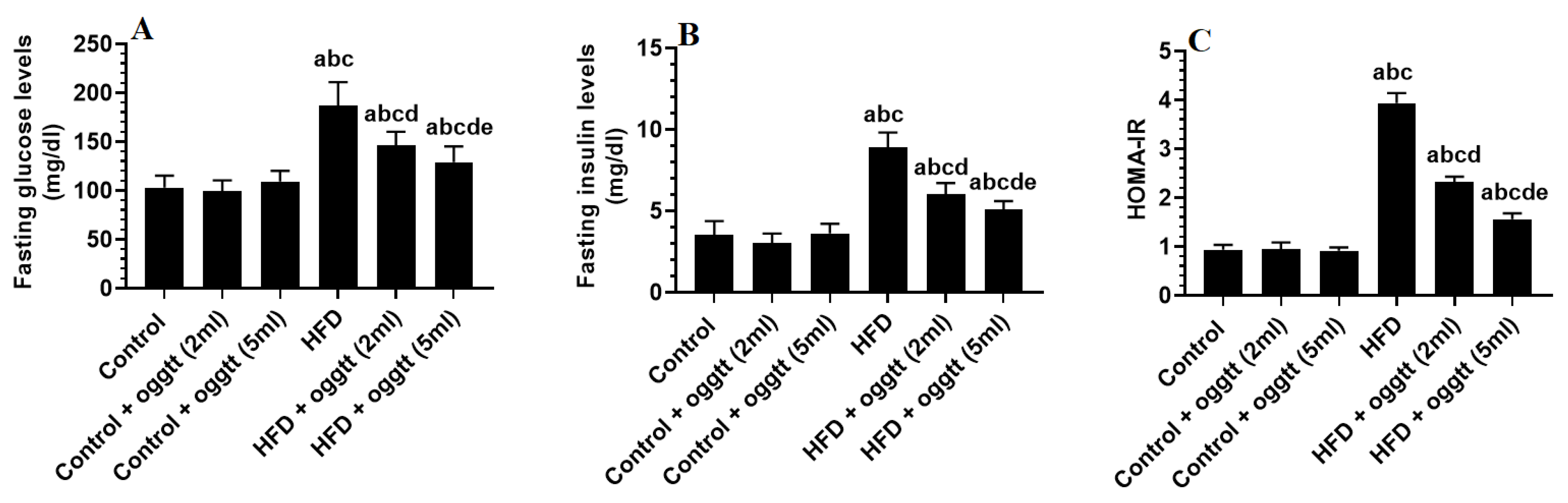
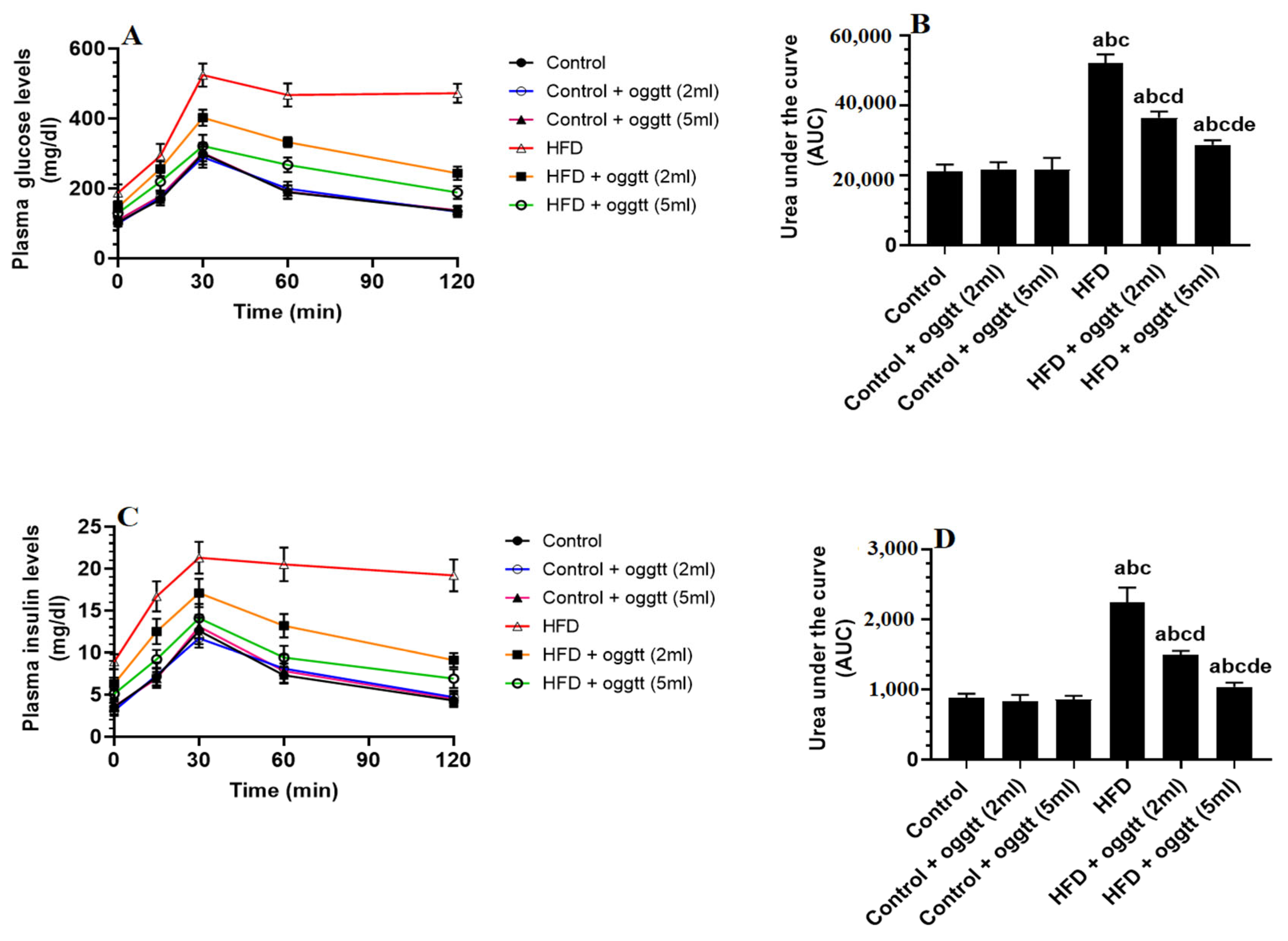
3.1. Oggtt Lowers Glucose and Insulin Levels and Improves HOMA-IR in HFD-Fed Rats

Plasma glucose, insulin, and HOMA-IR levels were significantly increased in HFD-fed rats as compared to control rats. In addition, plasma glucose and insulin levels measured over the 120 min during the OGTT, and their corresponding urea under the curve (UAC) was significantly increased in HFD-fed rats as compared to control (Figure 1A–C and Figure 2A–D). No significant variations in the levels of all these markers were seen between the control, control + oggtt (2 mL), and control + oggtt (5 mL)-treated rats (Figure 1A–C and Figure 2A–D). Blood glucose and insulin levels were significantly and progressively reduced in HFD + oggtt, which received the 2 or 5 mL of oggtt as compared to the HFD-fed rats (Figure 1A–C and Figure 2A–D). The inhibitory effects of oggtt milk on glucose or insulin levels during fasting or the OGTT were more significant in HFD + oggtt (5 mL) as compared to HFD + oggtt (2 mL).

3.2. Oggtt Reduces Body Weights and Has an Anti-Hyperlipidemic Effect in HFd-Fed Rats

Weekly food intake, serum, and hepatic levels of TGs, CHOL, and FFAs as well as hepatic levels of LDL-c and stool levels of TGs and CHOL were significantly higher in HFD-fed rats as compared to the control rats (Table 1). The levels of all these markers were reversed in the groups of rats that received oggtt milk at doses of 2 mL or 5 mL, with a more significant effect being seen with the highest dose (Table 1). No significant change in the food intake, hepatic levels of FFAs, and stool levels of TGs and CHOL were seen between the control and control rats, which received either 2 or 5 mL of oggtt milk (Table 1). Body weights and serum levels of LDL-c as well as in the serum and hepatic levels of TGs and CHOL were significantly decreased in a significant dose-dependent manner in HFD + oggtt-treated rats as compared to control rats (Table 1).

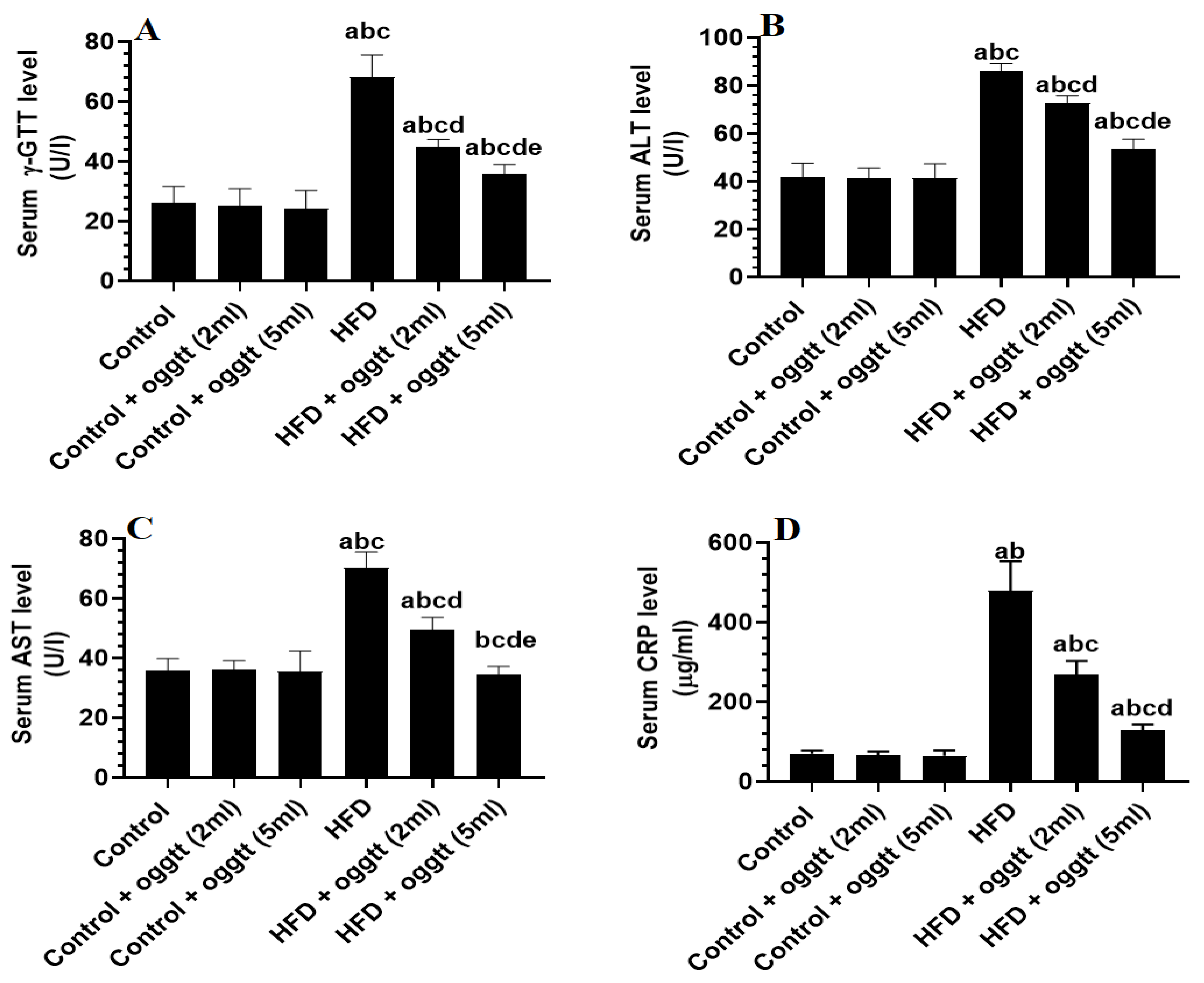
3.3. Oggtt Improves Liver Function in HFD-Fed Rats

Although there was no significant statistical difference between the control and control + oggtt (2 mL or 5 mL)-treated rats for the serum levels of ALT, AST, GTT, and CRP, this was not the case in HFD-fed rats, which showed higher levels of the aforementioned biomarkers (Figure 3A–D). When compared to HFD-fed rats, all of these biochemical markers were significantly and dose-dependently lower in HFD + oggtt (2 mL)- and HFD + oggtt (5 mL)-treated rats (Figure 3A–D).

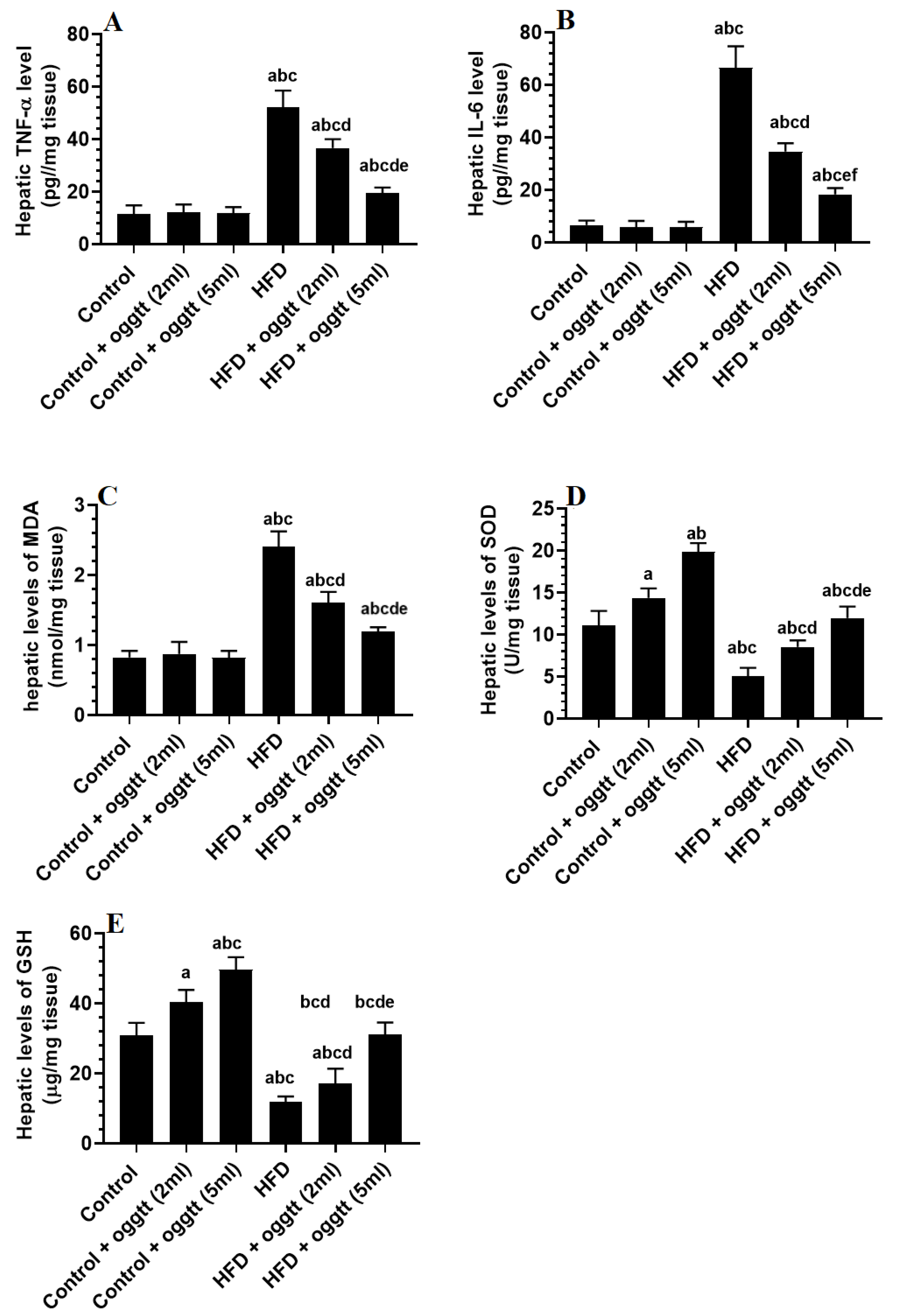
3.4. Oggtt Attenautes Hepatic Oxidaitve Stress and Inflmmation in HFD-Fed Rats

TNF-α, IL-6, and MDA levels in the liver were not significantly different between the control, control + oggtt (2 mL), and control + oggtt (5 mL) rat groups but were significantly lower in the HFD-fed rats (Figure 4A–E). Furthermore, the hepatic levels of SOD and GSH were significantly reduced in HFD-fed rats as compared to all control-treated rats (Figure 4A–E). Moreover, TNF-α, IL-6, and MDA levels in livers of rats given HFD + oggtt (2 mL) or HFD + oggtt (5 mL) were significantly lower than in HFD-fed rats, while GSH and SOD levels were significantly higher. However, the effect was more significant and profound in the HFD-treated rats, which were given the highest dose (Figure 4A–E). Notably, in the same dose-dependent manner, the levels of GSH and SOD were also significantly increased in the livers of the control rats, which were administered with both doses of oggtt, as compared to control rats (Figure 4A–E).

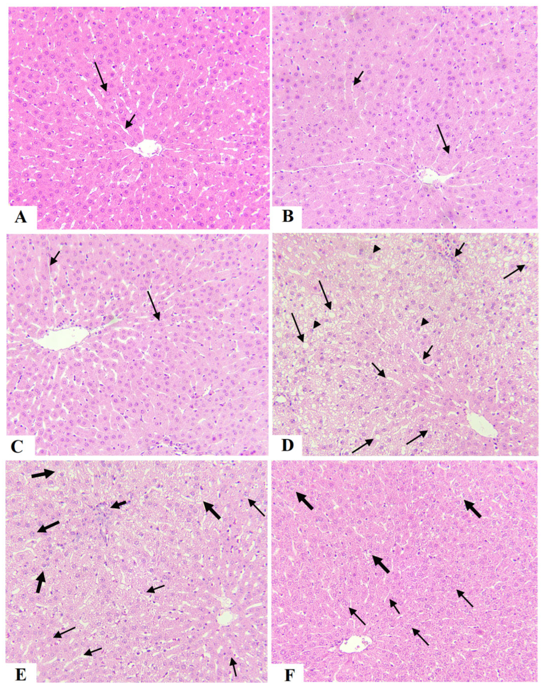
3.5. Oggtt Prevents Hepatic Steatosis in HFD-Fed Rats

Normal liver structure and shapes, including intact hepatocytes and sinusoids, were seen in the control, control + oggtt (2 mL), and control + oggtt (5 mL) (Figure 5A–C). However, increased cytoplasmic fat vacuoles accompanied by dilated sinusoids increased immune cell infiltration, and a higher number of pyknotic cells were seen in the livers of HFD-fed rats (Figure 5D). These effects were significantly reduced in rat livers treated with HFD + oggtt (2 mL) (Figure 5E). HFD + oggtt (5 mL)-treated rats had almost normal liver structures with very few fat vacuoles (Figure 5F).

4. Discussion

Previous retrospective and experimental studies have indicated that fermented milk formed from animal or plant sources is a part of our healthy dietary pattern that could prevent chronic disorders, including T2DM and metabolic syndrome [16,17,23]. Most studies have focused on the fermented yogurt of camels and cows [16]. Water kefir was found to be a less expensive and time-consuming hypoglycaemic and hypolipidemic treatment and can potentially be a useful food for people with diabetes to control body weight, glucose, and lipid levels [30].
However, there is still a lack of experimental evidence to support the clinical use of fermented goat milk as a potential therapy to treat these conditions, especially T2DM, IR, and their associated complications, such as NAFLD.
The present study has examined the effect of prepared oggtt (fermented goat milk) on lipid and glucose homeostasis and IR as well as on hepatic structural changes, inflammatory status, and oxidative/antioxidant balance in rats fed HFD as a model of T2DM and NAFLD. Our data are one of the first reports supporting the recommendations for using oggtt to alleviate NAFLD. Herein, daily treatment with fermented goat milk reduced rats’ weight gain and reserved dyslipidaemia and improved glucose haemostasis and insulin sensitivity by reducing fasting glucose and insulin levels. In addition, treatment with oggtt attenuated steatosis (ballooning) improved hepatocyte structure, reduced inflammation and lipid peroxidation markers, and stimulated endogenous antioxidant levels in the livers of HFD-fed rats. Overall, these findings suggested that oggtt daily treatment could help with obesity and NAFLD by lowering glucose and insulin levels and acting as an anti-inflammatory.
Chronic feeding of HFD to the rats in this study for 12 weeks promoted T2DM features that are characterized by increased body weight, IR, fasting, hyperglycaemia, hyperinsulinemia, and dyslipidemia, which support many previous studies [31,32,33]. The biochemical picture of dyslipidaemia in these HFD-fed rats was reflected by increased TGs, CHOL, and FFA as well as the development of liver ballooning, which is consistent with earlier studies [34,35,36]. This could be explained by the increased influx of FFAs from the insulin-unresponsive adipose tissue and stimulated lipogenesis in response to IR [4,31]. Interestingly, treatment with oggtt significantly attenuated this metabolic picture in HFD-treated rats in a dose-dependent manner and significantly reduced body weights and hepatic steatosis, thus indicating potent anti-obesity and anti-NAFLD potentials. Similar to our data, treatment with fermented camel milk also reduced body weights, attenuated hepatic steatosis, and improved peripheral insulin sensitivity in rats [18]. Similar anti-diabetic, anti-obesity, and anti-NAFLD properties were also reported for fermented soy and cow milk (kefir) and water kefir [20,21,22,30].
However, one important finding in this study is the ability of oggtt to reduce body weights in both the control and HFD-fed rats without altering food intake. These data could be explained by the ability of oggtt to mimic calorie restriction or mobilization of fat in adipose tissue, two major strategies used to reduce body weight [37]. However, the effect of fermented milk on body weight is varied and seems to be sex-dependent [16,38]. For example, some meta-analyses have shown a significant decrease (40.99 g) per year with each incremental serving of fermented cow milk (yogurt) [39]. However, some contradictory studies have found an increased risk of obesity or no effect after prolonged consumption of cow yogurt [38,40,41]. Yet, our data provide evidence for further clinical use of oggtt to reduce body weight, which could be favoured over other fermented animal milks.
On the other hand, while treatment of increasing doses with oggtt reduced fasting glucose and insulin levels and HOMA-IR in HFD-fed rats, they failed to induce this effect in control rats, indicating a glucose-lowering effect only in metabolically disturbed animals. This could be due to improving peripheral and hepatic insulin sensitivity, which could be explained by the antioxidant protective effect of oggtt as discussed below. Indeed, IR has been related to increased production of ROS and the scavenging ability of antioxidants, which impair insulin signalling in the muscles and livers and promote prolonged fasting hyperglycaemia and hyperinsulinemia [42,43]. However, many antioxidant drugs are currently available to improve peripheral insulin action and treat the hyperglycaemia associated with T2DM. In addition, it is well-known that fermentation and the digestion of fermented milk in the gut as well as their action on gut microbes produce several bioactive compounds that may act as glucose-lowering, anti-inflammatory, and antioxidant agents [16]. Indeed, fermented camel and cow milk reduced fasting blood glucose and insulin levels in several experimental and clinical studies [16,18,20,44,45,46]. Based on these data, we could assume that regular intake of oggtt could be an effective strategy to reduce the risk and progression of T2DM as well as an excellent strategy to reduce the complications associated with hyperglycaemia. Several meta-analyses involving 7–9 cohort studies have shown that yogurt supplementation reduces the risk of T2DM by 14–18% [21,47,48].
Nonetheless, dyslipidaemia, increased hepatic DNL, and hepatic steatosis are the major hallmarks of NAFLD and are commonly seen in HFD-experimental animals [49]. Several mechanisms are responsible for developing dyslipidaemia and hepatic steatosis post chronic administration of HFD. Initially, they include increased dietary lipid intake and increased delivery of FFAs from the impaired adipose tissue [7]. Additionally, hyperglycaemia, hyperinsulinemia, and oxidative stress abnormally stimulate DNL by upregulating and activating lipid-related transcription factors such as the sterol regulatory element-binding proteins (SREBP1 and SREBP2), which enhance the synthesis of TGs and CHOL, respectively, by regulating diverse genes [49,50,51]. Hepatic fatty acid transport and oxidation are impaired in NFALD animals and humans due to impaired levels and activities of the peroxisome proliferator-activated receptor-alpha (PPARα), which normally stimulates FAs oxidation [49,51].
Another important finding in this study is the observation that oggtt milk was able to attenuate dyslipidaemia and hepatic steatosis in a dose-dependent manner in the treated HFD-fed rats, indicating a potent hypolipidemic effect that could be related to improving fasting hyperglycaemia and insulin sensitivity as well as the hepatic antioxidant levels. However, the treatment with oggtt not only reduced serum and hepatic TGs, CHOL, and FFAs in HFD-fed rats but also in control rats, where normal glucose and insulin levels were observed. In addition, normal faecal lipid levels were observed in control and HFD-fed rats under oggtt treatments. These data indicated that the effect of oggtt on markers of hepatic steatosis and lipid synthesis is independent and possibly mediated by regulating DNL-related genes (i.e., SREPB-1 and PPARα and their downstream target genes).
Similar to our results, fermented camel and soy milk and kefir also attenuated hyperlipidaemia and hepatic steatosis in HFD-fed rats [18,20,52]. Within this view, the hypolipidemic effect of the fermented camel milk was attributed to its high content of the lipid-lowering bacteria Bifidobacteria, which normally lowers plasma lipids in HFD-fed rats; suppression of gastric emptying downregulation of SREBP1; and activation of PPARα [18,52,53,54]. The main hypolipidemic effect of soy fermented milk was attributed to a decrease in CHOL intestinal absorption caused by pancreatic lipase inhibition as well as an increase in glucose levels and peripheral insulin sensitivity [20]. On the other hand, improving glucose tolerance as well as increasing the availability of the PPARα-stimulating microbes, such as Lactobacillus, Lactococcus, and Candida, were the major mechanisms underlying the hypolipidemic effect of kefir [21,52]. Therefore, further organized studies targeting intestinal microorganism diversity and the expression of lipogenic genes are required to illustrate the possible mechanisms underlying the hypolipidemic effect of oggtt.
Oxidative stress and inflammation are the final damaging effects of hyperglycaemia and IR. Indeed, the increased levels of FFAs in the liver increase the production of ROS by impairing the mitochondria and promoting endoplasmic reticulum (ER) stress [4,6]. In addition, hyperglycaemia generates massive amounts of ROS by activating several pathways, such as advanced glycation end products (AGEs), polyol, hexose monophosphate, and protein kinase-c (PKC) [6]. On the other hand, inflammation in the livers of obese animals is triggered by the influx of cytokines from the inflamed adipose tissue or ROS stimulation. Furthermore, accumulated lipids stimulate the generation of ROS and inflammatory cytokines [55]. These ROS and inflammatory cytokines can damage the membranes, promote DLN, and induce cell necrosis and apoptosis, thus progressing the disease to NASH [6].
Furthermore, accumulated lipid droplets in the hepatocytes can generate a large amount of ROS from the mitochondria and stimulate TNF-α and IL-6 production from the hepatic infiltrating and resident immune cells (i.e., macrophages, dendritic cells, and lymphocytes), thus exacerbating the hepatic inflammation, oxidative stress, and apoptosis [55].
In this study, we discovered that both doses of oggtt suppressed lipid peroxidation and TNF-α and IL-6 levels while increasing SOD and GSH levels in the livers of both control and HFD-fed rats. It also reduced liver marker enzymes in HFD-fed rats. Collectively, these findings suggest a hepatoprotective effect mediated by reducing the generation of ROS, suppressing inflammation, and boosting levels of the endogenous antioxidant defence system. This seems to be an independent antioxidant and anti-inflammatory effect, which could also be mediated by improving glycaemic control in these rats. Fermentation is a process that increases the levels of metabolites (peptides) that are known for their antioxidant, anti-inflammatory, and anti-apoptotic effects [56]. Yogurt from traditional cultures produces specific peptides with potent immunomodulatory and antioxidant properties [16]. In addition, daily consumption of fermented camel milk reduced levels of ALT and AST in patients with metabolic syndrome [19]. It also reduced MDA levels and stimulated GSH, SOD, and CAT in the livers of HFD-fed rats [18]. Further, fermented soy milk prevented renal and hepatic damage induced by HFD by alleviating oxidative stress [20].
In conclusion, the findings of this study remained very interesting and showed the first evidence that supports the protective impact of oggtt against HFD-induced NAFLD disease, such as the hypoglycaemic, hypolipidemic, antioxidant, and anti-inflammatory effects of this fermented goat milk product. However, the study is still an observational study that has some limitations. Importantly, the effect of oggtt with the intact goat milk could be compared in future studies to determine the variations in the proposed effects. In addition, further studies are required, including microbiota analysis as well as molecular experiments to reveal the mechanisms behind the observed effects.

Author Contributions

Conceptualization, G.M.A., K.M.A. and A.E.A.Y.; methodology, K.M.A., A.E.A.Y., A.S. and M.A.Y.; software, S.A.; validation, N.A.A.; formal analysis, K.M.A.; investigation, G.M.A.; resources, G.M.A.; data curation, A.E.A.Y.; writing—original draft preparation, K.M.A. and M.A.Y.; writing—review and editing, A.E.A.Y., S.A. and N.A.A.; visualization, A.E.A.Y.; supervision, G.M.A.; project administration, M.A.Y.; funding acquisition, G.M.A. All authors have read and agreed to the published version of the manuscript.

Funding

This research was funded by Researchers Supporting Project, King Saud University, Riyadh, Saudi Arabia, grant number RSP-2021/84.

Institutional Review Board Statement

All procedures were conducted after obtaining ethical approval from the Research Ethics Committee (REC) at King Saudi University, Riyadh, Saudi Arabia (Ethical Reference No: KSU-SE-22-22).

Informed Consent Statement

Not applicable.

Data Availability Statement

The datasets used and analysed during the current study are available from the corresponding author upon reasonable request.

Acknowledgments

The authors extend thanks to the Researchers Supporting Project number (RSP-2021/84), King Saud University, Riyadh, Saudi Arabia.

Conflicts of Interest

The authors declare no conflict of interest.

References

  1. Obaroakpo, J.U.; Nan, W.; Hao, L.; Liu, L.; Zhang, S.; Lu, J.; Pang, X.; Lv, J. The hyperglycemic regulatory effect of sprouted quinoa yoghurt in high-fat-diet and streptozotocin-induced type 2 diabetic mice via glucose and lipid homeostasis. Food Funct. 2020, 11, 8354–8368. [Google Scholar] [CrossRef] [PubMed]
  2. Blüher, M. Obesity: Global epidemiology and pathogenesis. Nat. Rev. Endocrinol. 2019, 15, 288–298. [Google Scholar] [CrossRef] [PubMed]
  3. Althumiri, N.A.; Basyouni, M.H.; AlMousa, N.; AlJuwaysim, M.F.; Almubark, R.A.; BinDhim, N.F.; Alkhamaali, Z.; Alqahtani, S.A. Obesity in Saudi Arabia in 2020: Prevalence, Distribution, and Its Current Association with Various Health Conditions. Healthcare 2021, 9, 311. [Google Scholar] [CrossRef] [PubMed]
  4. Luci, C.; Bourinet, M.; Leclère, P.S.; Anty, R.; Gual, P. Chronic Inflammation in Non-Alcoholic Steatohepatitis: Molecular Mechanisms and Therapeutic Strategies. Front. Endocrinol. 2020, 11, 597648. [Google Scholar] [CrossRef]
  5. Masarone, M.; Rosato, V.; Dallio, M.; Gravina, A.G.; Aglitti, A.; Loguercio, C.; Federico, A.; Persico, M. Role of Oxidative Stress in Pathophysiology of Nonalcoholic Fatty Liver Disease. Oxidative Med. Cell. Longev. 2018, 2018, 9547613. [Google Scholar] [CrossRef]
  6. Yu, J.; Marsh, S.; Hu, J.; Feng, W.; Wu, C. The Pathogenesis of Nonalcoholic Fatty Liver Disease: Interplay between Diet, Gut Microbiota, and Genetic Background. Gastroenterol. Res. Pract. 2016, 2016, 2862173. [Google Scholar] [CrossRef] [Green Version]
  7. Buzzetti, E.; Pinzani, M.; Tsochatzis, E.A. The multiple-hit pathogenesis of non-alcoholic fatty liver disease (NAFLD). Metab. Clin. Exp. 2016, 65, 1038–1048. [Google Scholar]
  8. Al-Abdulkarim, B.O.; Osman, M.S.; El-Nadeef, M.A. Determination of chemical composition, and storage on dried fermented goat milk product (Oggtt). J. Saudi Soc. Agric. Sci. 2013, 12, 161–166. [Google Scholar] [CrossRef] [Green Version]
  9. Dragomir, K.; Andreja, K. The Effect of Fatty Acids in Goat Milk on Health, Milk Production—An Up-to-Date Overview of Animal Nutrition, Management and Health 2012. Available online: https://www.intechopen.com/chapters/39464 (accessed on 26 September 2012).
  10. Hodgkinson, A.J.; Wallace, O.; Boggs, I.; Broadhurst, M.; Prosser, C.G. Gastric digestion of cow and goat milk: Impact of infant and young child in vitro digestion conditions. Food Chem. 2018, 245, 275–281. [Google Scholar] [CrossRef]
  11. Delgadillo-Puga, C.; Cuchillo-Hilario, M.; León-Ortiz, L.; Ramírez-Rodríguez, A.; Cabiddu, A.; Navarro-Ocaña, A.; Morales-Romero, A.M.; Medina-Campos, O.N.; Pedraza-Chaverri, J. Goats’ Feeding Supplementation with Acacia farnesiana Pods and Their Relationship with Milk Composition: Fatty Acids, Polyphenols, and Antioxidant Activity. Animals 2019, 9, 515. [Google Scholar] [CrossRef] [Green Version]
  12. Delgadillo-Puga, C.; Noriega, L.G.; Morales-Romero, A.M.; Nieto-Camacho, A.; Granados-Portillo, O.; Rodríguez-López, L.A.; Alemán, G.; Furuzawa-Carballeda, J.; Tovar, A.R.; Cisneros-Zevallos, L.; et al. Goat’s Milk Intake Prevents Obesity, Hepatic Steatosis and Insulin Resistance in Mice Fed A High-Fat Diet by Reducing Inflammatory Markers and Increasing Energy Expenditure and Mitochondrial Content in Skeletal Muscle. Int. J. Mol. Sci. 2020, 21, 5530. [Google Scholar] [CrossRef]
  13. Ham, J.S.; Shin, J.H.; Jang, A.; Jeong, S.G.; Parkl, K.W.; Kim, H.U.; Kang, S.Y.; Hwang, H.J.; Lee, W.K. Effect of Goat Milk Yogurt Supplemented with Citrus Concentrate on Blood Glucose Hangug chugsan sigpum haghoeji and Serum Lipids in Diabetic Rats. Korean J. Food Sci. Anim. Resour. 2008, 28, 445–450. [Google Scholar] [CrossRef]
  14. Miglani, S.; Patyar, R.R.; Patyar, S.; Reshi, M.R. Effect of goat milk on hepatotoxicity induced by antitubercular drugs in rats. J. Food Drug Anal. 2016, 24, 716–721. [Google Scholar] [CrossRef] [Green Version]
  15. Zhang, J.; Wang, Z.; Huo, D.; Shao, Y. Consumption of goats’ milk protects mice from carbon tetrachloride-induced acute hepatic injury and improves the associated gut microbiota imbalance. Front. Immunol. 2018, 9, 1034. [Google Scholar] [CrossRef]
  16. Fernandez, M.A.; Marette, A. Novel perspectives on fermented milks and cardiometabolic health with a focus on type 2 diabetes. Nutr. Rev. 2018, 76, 16–28. [Google Scholar] [CrossRef] [Green Version]
  17. Alsayadi, M.M.; Almowallad, S.A.; Yahya, M.A.; Alsanea, E.; Alfardi, T.; Alhaidari, S. Nutritional, Health-Promoting Properties and Antioxidant Activity of Yemeni Fermented Milk (Laban) and A Laban-Pulicaria Jaubertii Mixture. Turk. J. Agric. Food Sci. Technol. 2020, 8, 2049–2058. [Google Scholar] [CrossRef]
  18. Korish, A.A.; Arafah, M.M. Camel milk ameliorates steatohepatitis, insulin resistance and lipid peroxidation in experimental non-alcoholic fatty liver disease. BMC Complement. Altern. Med. 2013, 13, 264. [Google Scholar] [CrossRef] [Green Version]
  19. Fallah, Z.; Feizi, A.; Hashemipour, M.; Kelishadi, R. Positive Effect of Fermented Camel Milk on Liver Enzymes of Adolescents with Metabolic Syndrome: A Double Blind, Randomized, Cross-over Trial. Mater. Socio-Med. 2018, 30, 20–25. [Google Scholar] [CrossRef] [Green Version]
  20. Tiss, M.; Souiy, Z.; Abdeljelil, N.; Njima, M.; Achour, L.; Hamden, K. Fermented soy milk prepared using kefir grains prevents and ameliorates obesity, type 2 diabetes, hyperlipidemia and Liver-Kidney toxicities in HFFD-rats. J. Funct. Foods 2020, 67, 103869. [Google Scholar] [CrossRef]
  21. Chen, H.-L.; Tung, Y.-T.; Tsai, C.-L.; Lai, C.-W.; Lai, Z.-L.; Tsai, H.-C.; Lin, Y.-L.; Wang, C.-H.; Chen, C.-M. Kefir improves fatty liver syndrome by inhibiting the lipogenesis pathway in leptin-deficient ob/ob knockout mice. Int. J. Obes. 2014, 38, 1172–1179. [Google Scholar] [CrossRef]
  22. Punaro, G.R.; Maciel, F.R.; Rodrigues, A.M.; Rogero, M.M.; Bogsan, C.S.B.; Oliveira, M.N.; Ihara, S.S.; Araujo, S.R.R.; Sanches, T.R.C.; Andrade, L.C.; et al. Kefir administration reduced progression of renal injury in STZ-diabetic rats by lowering oxidative stress. Nitric Oxide. Biol. Chem. 2014, 37, 53–60. [Google Scholar]
  23. Alsayadi, M.M.; Al jawfi, Y.; Belarbi, M.; Sabri, F.Z. Antioxidant potency of water kefir. JMBFS 2013, 2, 2444–2447. [Google Scholar]
  24. Bashiti, T.A.; Zabut, B.M. Effect of probiotic fermented milk (Kefir) on some blood biochemical parameters among newly diagnosed type 2 diabetic adult males in Gaza governorate. Curr. Res. Nutr. Food Sci. 2019, 7, 568–575. [Google Scholar] [CrossRef]
  25. de Moura EDias, M.; Dos Reis, S.A.; da Conceição, L.L.; Sediyama, C.; Pereira, S.S.; de Oliveira, L.L.; Gouveia Peluzio, M.D.C.; Martinez, J.A.; Milagro, F.I. Diet-induced obesity in animal models: Points to consider and influence on metabolic markers. Diabetol. Metab. Syndr. 2021, 13, 32. [Google Scholar] [CrossRef] [PubMed]
  26. Yahya, M.A.; Alhaj, O.A.; Al-Khalifah, A.S. Antihypertensive effect of fermented skim camel (Camelus dromedarius) milk on spontaneously hypertensive rats. Nutr. Hosp. 2017, 34, 416–421. [Google Scholar] [CrossRef]
  27. Nagy, C.; Einwallner, E. Study of In Vivo Glucose Metabolism in High-fat Diet-fed Mice Using Oral Glucose Tolerance Test (OGTT) and Insulin Tolerance Test (ITT). J. Vis. Exp. 2018, 131, 56672. [Google Scholar] [CrossRef]
  28. Yoon, H.; Jeon, D.J.; Park, C.E.; You, H.S.; Moon, A.E.M. Relationship between homeostasis model assessment of insulin resistance and beta cell function and serum 25-hydroxyvitamin D in non-diabetic Korean adults. J. Clin. Biochem. Nutr. 2016, 59, 139–144. [Google Scholar] [CrossRef] [Green Version]
  29. Folch, J.; Lees, M.; Sloane Stanley, G.H. A simple method for the isolation and purification of total lipides from animal tissues. J. Biol. Chem. 1957, 226, 497–509. [Google Scholar] [CrossRef]
  30. Alsayadi, M.; Al Jawfi, Y.; Belarbi, M.; Soualem-Mami, Z.; Merzouk, H.; Sari, D.C.; Sabri, F.; Ghalim, M. Evaluation of Anti-Hyperglycemic and Anti-Hyperlipidemic Activities of Water Kefir as Probiotic on Streptozotocin-Induced Diabetic Wistar Rats. J. Diabetes Mellit. 2014, 4, 85–95. [Google Scholar] [CrossRef] [Green Version]
  31. Xu, Z.; Fan, J.; Ding, X.; Qiao, L.; Wang, G. Characterization of high-fat, diet-induced, non-alcoholic steatohepatitis with fibrosis in rats. Dig. Dis. Sci. 2010, 55, 931–940. [Google Scholar] [CrossRef] [Green Version]
  32. Ma, H.; You, G.-P.; Cui, F.; Chen, L.-F.; Yang, X.-J.; Chen, L.-G.; Lu, H.-D.; Zhang, W.-Q. Effects of a low-fat diet on the hepatic expression of adiponectin and its receptors in rats with NAFLD. Ann. Hepatol. 2015, 14, 108–117. [Google Scholar] [CrossRef]
  33. Stott, N.; Marino, J. High Fat Rodent Models of Type 2 Diabetes: From Rodent to Human. Nutrients 2020, 21, 3650. [Google Scholar] [CrossRef]
  34. Song, L.; Qu, D.; Zhang, Q.; Jiang, J.; Zhou, H.; Jiang, R.; Li, Y.; Zhang, Y.; Yan, H. Phytosterol esters attenuate hepatic steatosis in rats with non-alcoholic fatty liver disease rats fed a high-fat diet. Sci. Rep. 2017, 7, srep41604. [Google Scholar] [CrossRef]
  35. Jensen, V.S.; Hvid, H.; Damgaard, J.; Nygaard, H.; Ingvorsen, C.; Wulff, E.M.; Lykkesfeldt, J.; Fledelius, C. Dietary fat stimulates development of NAFLD more potently than dietary fructose in Sprague–Dawley rats. Diabetol. Metab. Syndr. 2018, 10, 4. [Google Scholar] [CrossRef]
  36. Carmiel-Haggai, M.; Cederbaum, A.I.; Nieto, N. A high-fat diet leads to the progression of non-alcoholic fatty liver disease in obese rats. FASEB J. 2005, 19, 136–138. [Google Scholar] [CrossRef]
  37. Jaime, T.J.; Balich, L.L.; Acevedo, G.B.; de la Maza Cave, M.P.; Birn, S.H.; Parada, S.H.; Silva, J.R.; Barnett, D.B. Effect of calorie restriction on energy expenditure in overweight and obese adult women. Nutr. Hosp. 2015, 31, 2428–2436. [Google Scholar] [CrossRef]
  38. Panahi, S.; Doyon, C.Y.; Després, J.P.; Pérusse, L.; Vohl, M.C.; Drapeau, V.; Tremblay, A. Yogurt consumption, body composition, and metabolic health in the Quebec Family Study. Eur. J. Nutr. 2018, 57, 1591–1603. [Google Scholar] [CrossRef]
  39. Schwingshackl, L.; Hoffmann, G.; Schwedhelm, C.; Kalle-Uhlmann, T.; Missbach, B.; Knüppel, S.; Boeing, H. Consumption of dairy products in relation to changes in anthropometric variables in adult populations: Asystematic review and meta-analysis of cohort studies. PLoS ONE 2016, 11, e0157461. [Google Scholar] [CrossRef]
  40. Mozaffarian, D.; Hao, T.; Rimm, E.B.; Willett, W.C.; Hu, F.B. Changes in diet and lifestyle and long-term weight gain in women and men. N. Engl. J. Med. 2011, 364, 2392–2404. [Google Scholar] [CrossRef] [Green Version]
  41. Rautiainen, S.; Wang, L.; Lee, I.M.; Manson, J.E.; Buring, J.E.; Sesso, H.D. Dairy consumption in association with weight change and risk of becoming overweight or obese in middle-aged and older women: A prospective cohort study. Am. J. Clin. Nutr. 2016, 103, 979–988. [Google Scholar] [CrossRef] [Green Version]
  42. Vincent, H.K.; Bourguignon, C.M.; Weltman, A.L.; Vincent, K.R.; Barrett, E.; Innes, K.E.; Taylor, A.G. Effects of antioxidant supplementation on insulin sensitivity, endothelial adhesion molecules, and oxidative stress in normal-weight and overweight young adults. Metabolism 2009, 58, 254–262. [Google Scholar] [CrossRef] [PubMed] [Green Version]
  43. Hoehn, K.L.; Salmon, A.B.; Hohnen-Behrens, C.; Turner, N.; Hoy, A.J.; Maghzal, G.J.; Stocker, R.; Van Remmen, H.; Kraegen, E.W.; Cooney, G.J.; et al. Insulin resistance is a cellular antioxidant defense mechanism. Proc. Natl. Acad. Sci. USA 2009, 106, 17787–17792. [Google Scholar] [CrossRef] [PubMed] [Green Version]
  44. Sadrzadeh-Yeganeh, H.; Elmadfa, I.; Djazayery, A.; Jalali, M.; Heshmat, R.; Chamary, M. The effects of probiotic and conventional yoghurt on lipid profile in women. Br. J. Nutr. 2010, 103, 1778–1783. [Google Scholar] [CrossRef]
  45. Ivey, K.; Hodgson, J.; Kerr, D.; Thompson, P.; Stojceski, B.; Prince, R. The effect of yoghurt and its probiotics on blood pressure and serum lipid profile; a randomised controlled trial. Nutr. Metab. Cardiovasc. Dis. 2015, 25, 46–51. [Google Scholar] [CrossRef] [PubMed] [Green Version]
  46. Ivey, K.; Hodgson, J.M.; Kerr, D.; Lewis, J.R.; Thompson, P.L.; Prince, R.L. The effects of probiotic bacteria on glycaemic control in overweight men and women: A randomised controlled trial. Eur. J. Clin. Nutr. 2014, 68, 447–452. [Google Scholar] [CrossRef] [Green Version]
  47. Aune, D.; Norat, T.; Romundstad, P.R.; Vatten, L.J. Dairy products and the risk of type 2 diabetes: A systematic review and dose-response meta-analysis of cohort studies. Am. J. Clin. Nutr. 2013, 98, 1066–1083. [Google Scholar] [CrossRef] [Green Version]
  48. Gijsbers, L.; Ding, E.L.; Malik, V.S.; De Goede, J.; Geleijnse, J.M.; Soedamah-Muthu, S.S. consumption of dairy foods and diabetes incidence: A dose-response meta-analysis of observational studies. Am. J. Clin. Nutr. 2016, 103, 1111–1124. [Google Scholar] [CrossRef] [Green Version]
  49. Bence, K.K.; Birnbaum, M.J. Metabolic drivers of non-alcoholic fatty liver disease. Mol. Metab. 2021, 50, 101143. [Google Scholar] [CrossRef]
  50. Sekiya, M.; Hiraishi, A.; Touyama, M.; Sakamoto, K. Oxidative stress induced lipid accumulation via SREBP1c activation in HepG2 cells. Biochem. Biophys. Res. Commun. 2008, 375, 602–607. [Google Scholar] [CrossRef] [Green Version]
  51. Liss, K.H.; Finck, B.N. PPARs and nonalcoholic fatty liver disease. Biochimie 2017, 136, 65–74. [Google Scholar] [CrossRef] [Green Version]
  52. Kim, D.-H.; Kim, H.; Jeong, D.; Kang, I.-B.; Chon, J.-W.; Kim, H.-S.; Song, K.-Y.; Seo, K.-H. Kefir alleviates obesity and hepatic steatosis in high-fat diet-fed mice by modulation of gut microbiota and mycobiota: Targeted and untargeted community analysis with correlation of biomarkers. J. Nutr. Biochem. 2017, 44, 35–43. [Google Scholar] [CrossRef]
  53. Mohamed, B.; Idam, N. Effect of camel milk on plasma lipid profile of hypercholesteremic rats. OJVRTM 2011, 15, 314–317. [Google Scholar]
  54. Ziamajidi, N.; Khaghani, S.; Hassanzadeh, G.; Vardasbi, S.; Ahmadian, S.; Nowrouzi, A.; Ghaffari, S.M.; Abdirad, A. Amelioration by chicory seed extract of diabetes- and oleic acid-induced non-alcoholic fatty liver disease (NAFLD)/non-alcoholic steatohepatitis (NASH) via modulation of PPARα and SREBP-1. Food Chem. Toxicol. 2013, 58, 198–209. [Google Scholar] [CrossRef]
  55. Parthasarathy, G.; Revelo, X.; Malh, H. Pathogenesis of Nonalcoholic Steatohepatitis: An Overview. Hepatol. Commun. 2020, 4, 478–492. [Google Scholar] [CrossRef]
  56. Pessione, E. Lactic acid bacteria contribution to gut microbiota complexity: Lights and shadows. Front. Cell Inf. Microbiol. 2012, 2, 86. [Google Scholar] [CrossRef]
Figure 1. Oggtt milk reduces fasting glucose (A) and insulin levels (B) as well as values of homeostasis model assessment of insulin resistance index (HOMA-IR) (C) in high-fat diet (HFD)-fed-rats in a dose-dependent manner. Data were expressed as means ± SD for n = 8 rats/group. a, vs. control; b, vs. control + oggtt (2 mL); c, vs. control + oggtt (5 mL); d, vs. HFD; and e, vs. HFD + oggt (2 mL).
Fermentation 08 00735 g001
Figure 2. Oggtt milk attenuates the impairment in oral glucose tolerance test in the high-fat diet (HFD)-fed rats in a dose-dependent manner. (A,B) Plasma glucose levels and the corresponding area under the curve (UAC) as measured at 0.0., 15, 30, 60, and 120 min in all groups of rats post glucose administration. (C,D) Plasma insulin levels measured at the same time intervals measured post glucose administration in the same groups of rats. Data were expressed as means ± SD for n = 8 rats/group. a, vs. control; b, vs. control + oggtt (2 mL); c, vs. control + oggtt (5 mL); d, vs. HFD; and e, vs. HFD + oggt (2 mL).
Fermentation 08 00735 g002
Figure 3. Oggtt milk prevents the increases in levels of gamma-glutamyl transpeptidase (GGT) (A), alanine aminotransferase (B), aspartate aminotransferase (AST) (C), and C-reactive protein (D) in the serum of high-fat diet (HFD)-fed-rats in a dose-dependent manner. Data were expressed as means ± SD for n = 10 rats/group. a, vs. control; b, vs. control + oggtt (2 mL); c, vs. control + oggtt (5 mL); d, vs. HFD; and e, vs. HFD + oggt (2 mL).
Fermentation 08 00735 g003
Figure 4. Oggtt suppresses the increase in the levels of tumour necrosis factor-α (TNF-α) (A), interleukin-6 (IL-6) (B), and malondialdehyde (C) in the livers of high-fed diet (HFD)-fed rats but stimulates the hepatic levels of superoxide dismutase (SOD) (D) and glutathione (GSH) (E) in both the control and HFD in a dose-dependent manner. The data are expressed as means ± SD for n = 8 rats/group. a, vs. control; b, vs. control + oggtt (2 mL); c, vs. control + oggtt (5 mL); d, vs. HFD; and e, vs. HFD + oggt (2 mL); f, vs. HFD + oggt (5 mL).
Fermentation 08 00735 g004
Figure 5. Photomicrographs of the livers of all groups of rats. H&E (200×). (AC) Control, control + oggtt (2 mL), and control + Oggtt (5 mL) showing normal hepatocytes (long arrow), central vein (CV), and sinusoids (short arrow). (D) HFD-fed rats show increased cytoplasmic fat vacuolization in the hepatocytes (long thin arrow), dilated sinusoids (short thin arrow), immune cell infiltration (short thick arrow), and pyknotic necrotic cells (arrowheads). (E) HFD + oggtt (2 mL) rats show an improvement in the structure of the liver of cells with an obvious increase in the number of normal cells (long arrow), a reduction in the number and size of the fat vacuolated cells (long thick arrow), and reduced size of sinusoids (short arrow). Few immune cells are still seen (short thick arrow). (F) HFD + oggtt (5 mL) rats show the maximum improvement in the liver structure, with most of the hepatocytes being normal (long thin arrow), and sinusoids (short arrow). However, fewer cells still showed fat accumulation but with smaller sizes (long thick arrow).
Fermentation 08 00735 g005
Table 1. Oggtt suppresses the gain in body weight and attenuates hyperlipidaemia and hepatic lipid accumulation in rats fed the control and the high-fat diet (HFD) in a dose-dependent manner.
ControlControl + Oggtt (2 mL)Control + Oggtt (5 mL)HFDHFD + Oggtt (2 mL)HFD + Oggtt (5 mL)
Final body weights (g)422 ± 42.9389 ± 31.9 a351 ± 22.3 ab568 ± 41.3 abc487 ± 36.7 abcd422 ± 31.9 abcde
Weekly food intake (g)234 ± 24.5219 ± 22.7229 ± 26.5322 ± 29 abc339 ± 37.1 abc328 ± 32.2 abc
Liver weight13.8 ± 1.512.9 ± 1.313.7 ± 1.118.5 ± 1.6 abc15.4 ± 1.5 abcd12.8 ± 1.1 cde
SerumTGs (mg/dL)53.2 ± 4.944.6 ± 5.1 a36.9 ± 4.1 ab104 ± 9.8 abc88.5 ± 7.9 abcd63.5 ± 5.8 abcde
CHOL (mg/dL)74.5 ± 5.866.3 ± 5.5 a59.6 ± 4.1 ab146 ± 11.4 abc103 ± 10.5 abcd81.5 ± 7.4 abcde
LDL-c (mg/dL)45.6 ± 4.937.4 ± 4.1 a31.1 ± 3.4 ab83.4 ± 7.1 abc71.2 ± 7.1 abcd59.8 ± 6.1 abcde
FFAs (µmol/L)512 ± 45.3498 ± 42.2523 ± 51.21032 ± 115 abc723 ± 62.4 abcd551 ± 44.8 abcde
LiverTGs (mg/g)4.7 ± 0.383.7 ± 0.41 a3.01 ± 0.29 ab7.8 ± 0.91 abc6.3 ± 0.54 abcd0.52 ± 85 abcde
CHOL (mg/g)2.7 ± 0.292.2 ± 0.21 a1.7 ± 0.27 ab5.1 ± 0.72 abc4.1 ± 0.62 abcd3.2 ± 0.26 abcde
FFA (µmol/g)73.4 ± 8.178.5 ± 6.975.3 ± 6.1149 ± 11.6 abc101 ± 13.2 abcd84.5 ± 7.8 abcde
FaecesTGs (mg/g)4.4 ± 0.354.7 ± 0.54.5 ± 0.7112.3 ± 1.43 abc13.2 ± 1.93 abc11.9 ± 1.6 abc
CHOL (mg/g)5.8 ± 0.725.5 ± 0.786.1 ± 0.8215.4 ± 1.7 abc13.8 ± 1.9 abc14.7 ± 1.8 abc
Data were expressed as means ± SD for n = 8 rats/group. a, vs. control; b, vs. control + oggtt (2 mL); c, vs. control + oggtt (5 mL); d, vs. HFD; and e, vs. HFD + oggt (2 mL).
Publisher’s Note: MDPI stays neutral with regard to jurisdictional claims in published maps and institutional affiliations.

