Open Electronics for Medical Devices: State-of-Art and Unique Advantages

1
Noble Sensors LLC, New York, NY 10010, USA
2
Michigan Technological University, Houghton, MI 49931, USA
*
Author to whom correspondence should be addressed.
Electronics 2019, 8(11), 1256; https://doi.org/10.3390/electronics8111256
Submission received: 30 September 2019
/
Revised: 25 October 2019
/
Accepted: 30 October 2019
/
Published: 1 November 2019
(This article belongs to the Section Bioelectronics)
Abstract
:A wide range of medical devices have significant electronic components. Compared to open-source medical software, open (and open-source) electronic hardware has been less published in peer-reviewed literature. In this review, we explore the developments, significance, and advantages of using open platform electronic hardware for medical devices. Open hardware electronics platforms offer not just shorter development times, reduced costs, and customization; they also offer a key potential advantage which current commercial medical devices lack—seamless data sharing for machine learning and artificial intelligence. We explore how various electronic platforms such as microcontrollers, single board computers, field programmable gate arrays, development boards, and integrated circuits have been used by researchers to design medical devices. Researchers interested in designing low cost, customizable, and innovative medical devices can find references to various easily available electronic components as well as design methodologies to integrate those components for a successful design.
1. Introduction
Healthcare technology is a classic case study for the effectiveness of open and open-source electronics. A high degree of precision, robustness, and reliability are necessary for any healthcare device. An analysis by Winter et al. [1] has projected the enormous cost savings (in billions of US$) of an open-source MRI scanner, which is just a small class of all medical devices which can benefit from open electronic hardware. There is a wide range of open-source software [2,3,4,5,6,7,8,9,10,11,12,13,14] for medical imaging—mainly for image processing (filtering) and visualization. There is some highly encouraging research [15,16,17,18,19,20,21] on artificial intelligence (mainly neural networks and deep learning) for computer-aided diagnostics. Although these research articles on computer aided diagnostics do not necessarily describe open-source software, they are all nevertheless reproducible. Unlike open-source medical software, the field of open-source medical hardware is less developed and less published in peer-reviewed literature. As a recent review by Niezen and co-workers [22] points out, many of the open-source medical device projects are still in nascent stage and unreported in peer-reviewed literature. This is in contrast with the field of open-source medical imaging software; for example, the Medical Imaging Interaction Toolkit (MITK) has been available as an open-source software for almost 16 years now [11]. To take full advantage of the vast developments in the open-source medical software [4,5,6,7,8,9,10,11,12,13,14] and machine learning algorithms [15,16,17,18,19,20,21], it is necessary to develop open-source hardware for medical devices. In fact, with the growing popularity of 3-D printing and open computing platforms such as Arduino® and Raspberry Pi, it is possible to come up with open/open-source electro-mechanical designs for medical devices. In this review, we focus mainly on the open electronic components for medical devices. Readers interested in the mechanical aspect of the open-source medical devices can explore some recent studies [23,24,25,26,27,28,29,30,31] in open-source robotic hands.
During the review, we have found that open electronics not only reduce development cost but in some instances, lead to better hardware and a higher performance solution. The advantages of using open electronics platform are the modularity, ease of repair, data sharing, as well as leveraging machine learning. For example, two healthcare devices made by different manufacturers but using the same open-electronics platform can easily share data or upload their data to a common cloud merely by a firmware update. Such seamless data sharing can take full advantage of the progress in machine learning. Open hardware devices can benefit from the numerous open-source software [2,3,4,5,6,12,13,14] which are very well published in literature. Specifically, recent studies have demonstrated the suitability of Raspberry Pi® [32,33,34] for artificial intelligence and deep learning.
Among the wide range of medical devices, we will further study ultrasound, X-Ray CT, ECG, fluid infusion devices, wearable and posture detection medical devices in particular. This review will serve as a guideline for researchers selecting electronic components for rapid prototyping. While most of the open electronic components are available on popular hobbyist websites such as Sparkfun®, Adafruit® etc. Researchers can also leverage the readily available and professionally designed “demo boards” or “evaluation boards” or “development boards” that can be found on major electronics retailers such as Mouser®, Element14® and Digikey®. As it will be evident in this review, using the appropriate “evaluation board,” in conjunction with a computing/microcontroller/FPGA platform can lead to great results at a very low cost. Such demo/evaluation boards are designed by large semiconductor manufacturers and very professionally and good high-frequency design practices are followed. In addition, such boards have professional soldering for difficult to solder surface mount components such as ball grid arrays. Most of the times, manufacturers also provide printed circuit board designs for these demo boards freely. In our review, we have found numerous examples of researchers utilizing these demo boards for open electronics hardware platforms. Typically these boards are helpful for (1) low noise designs, (2) difficult to solder integrated circuits, (3) saving prototyping time as there is no need to design, order, and assemble printed circuit boards.
To summarize, we have studied a wide range of medical devices which have been prototyped using open electronics platforms. Progress made by both academic research groups as well as the hobbyist community has been reported. In Section 2, we have reviewed the developments in open/open-source medical ultrasound and found that both FPGAs as well as high-frequency microcontrollers have proven quite effective in generating ultrasonic images. Additionally, a number of development boards have been used effectively by open-source developers. Compared to open/open-source ultrasound, developments in field of X-Ray CT scanners have been very few. Historically, even though a hobbyist X-Ray scanner has been described as early as 1974 [35], developments have been fewer yet encouraging. As described in Section 4, numerous variants of ECG/EKG, EEG, and other neurological monitoring devices have been very widely prototyped using open-source electronic hardware. Open electronics can be potentially used for seizure prediction, as reported in some recent research, for example [36]. Developments in open and open-source electronics have now matured to cover measurements which require multi-channel high frequency data-acquisition as well as ultra-low noise amplifications. Such an example is electric impedance tomography covered in Section 5. Infusions pumps are perhaps one of the most widely open-source medical devices with some very popular projects such as [37]. These open electronics infusion pump projects and their potential economic impact is described in Section 6. Various open/open-source wearables, motion sensing, and posture detection/correction devices have been described in Section 7. Quite a wide range of hardware (accelerometers, gyroscopes, piezo-resistive sensors, ultrasonic sensors, cameras) has been used in such open electronic devices. Section 8 lists some unique advantages of using open electronic hardware, other than the obvious cost advantage. In short, open electronics devices have great potential to create large data sets to effectively leverage advances in machine learning and artificial intelligence. In addition, open electronics hardware enables medical innovation, modular design, and easy repairs. This review will be very relevant to engineers, researchers, and other developers of open electronics medical platforms as well as physicians and other healthcare practitioners looking for novel approaches to medical device design.
2. Open Electronics in Medical Ultrasound Imaging
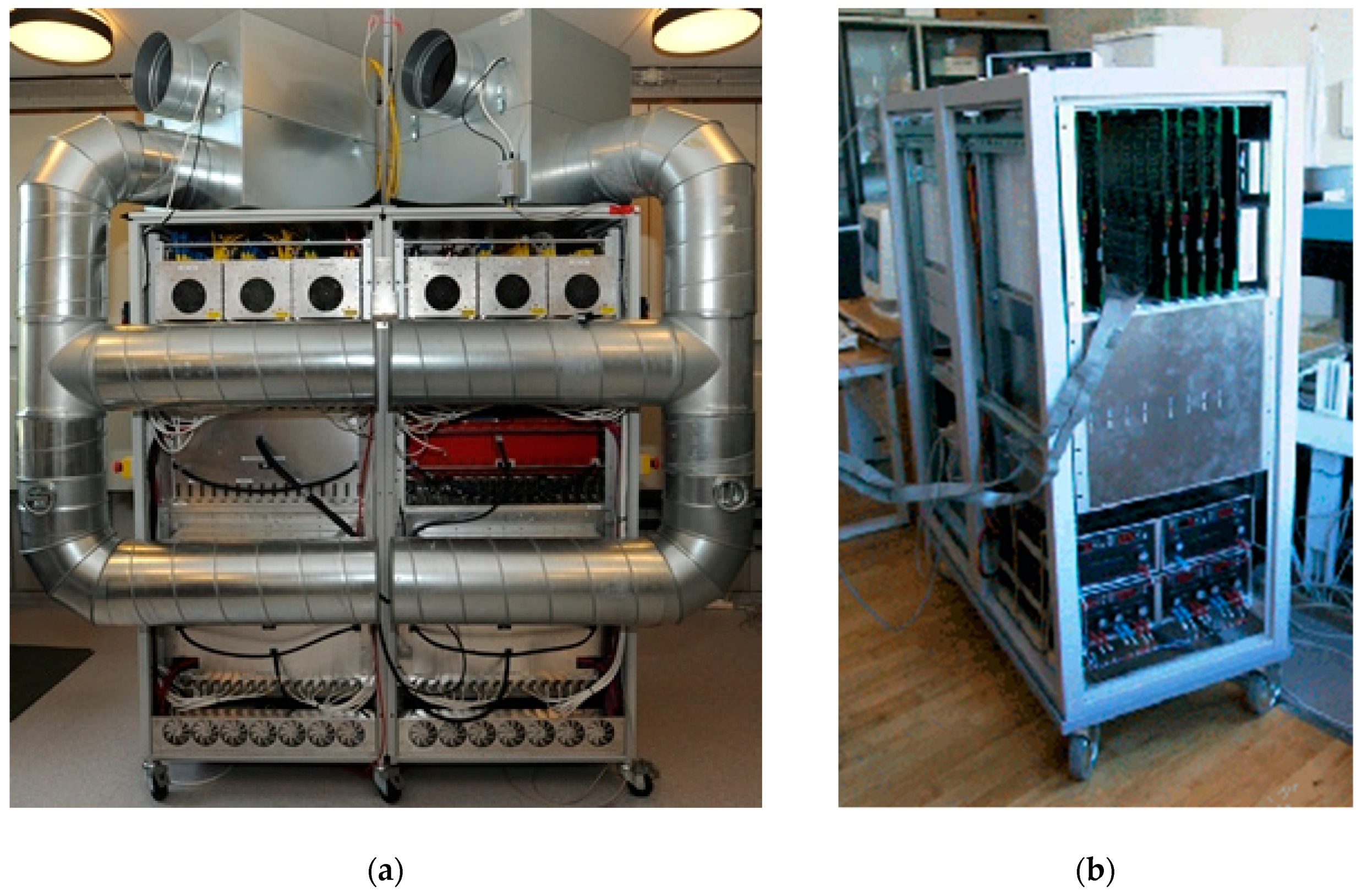
A typical ultrasonic diagnostic system works by exciting an ultrasonic transducer in a 1–100MHz range (2–10 MHz typical) with a peak-peak voltage of 50–200 volts. The system also requires low noise amplifiers of very low signal to noise ratio. The sampling frequency often falls in range of 50–100 MHz for such systems. The sampling frequency is beyond what any hobbyist microcontroller can handle and until a few years back, an open ultrasound would have been inconceivable because of the specialized engineering design involved in the process. A few selected universities have developed their own ultrasound platforms. A review of some of the leading research ultrasound platforms is provided in [38]. Some example are SARUS and RASMUS developed by Danish Technical University [39], UARP developed by Leeds University [40], and ULA-OP 256 [41] by University of Florence. However these ultrasound platforms have been developed at a very high cost and it is not possible for small research groups to reproduce such platforms. Two of these platforms are shown in Figure 1.
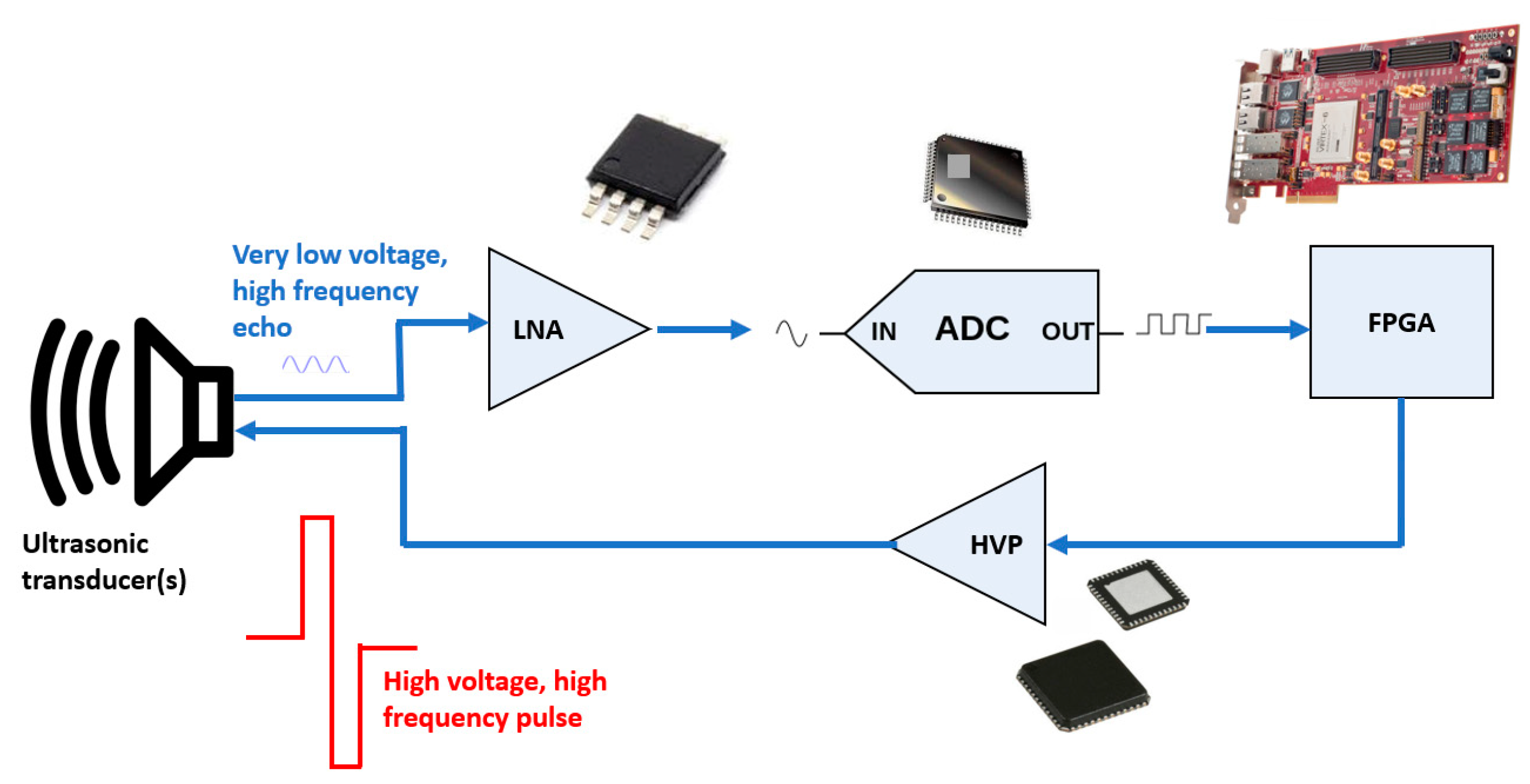
These ultrasound platforms mainly consist of custom-designed printed circuit boards. A typical simplified architecture is shown in Figure 2. As shown in Figure 2, printed circuit boards consist of specialized field programmable gate arrays (FPGAs) and other high-frequency components. Most of these components (especially high voltage pulser (HVP), low noise amplifier (LNA), ADC, and FPGA) have difficult to solder packaging such as QFP, QFN, or BGA. If developed using FPGA development board or module (Figure 2), the ultrasound platforms are easier to assemble. However, if the FPGA-integrated circuit is used individually, the design is still more complicated as FPGAs often require a lot of peripheral components such as resistors, capacitors, well-regulated power supplies, and clock sources.
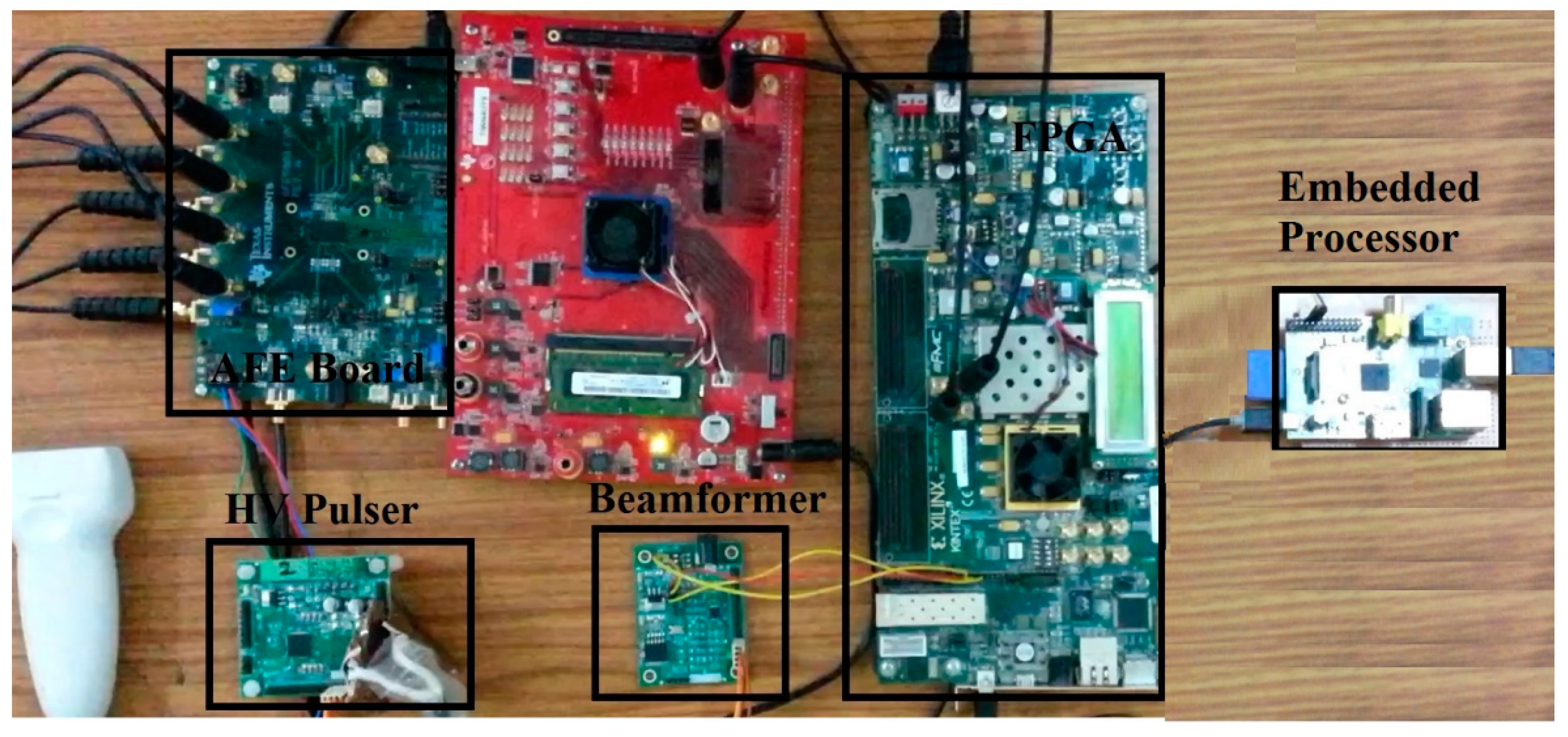
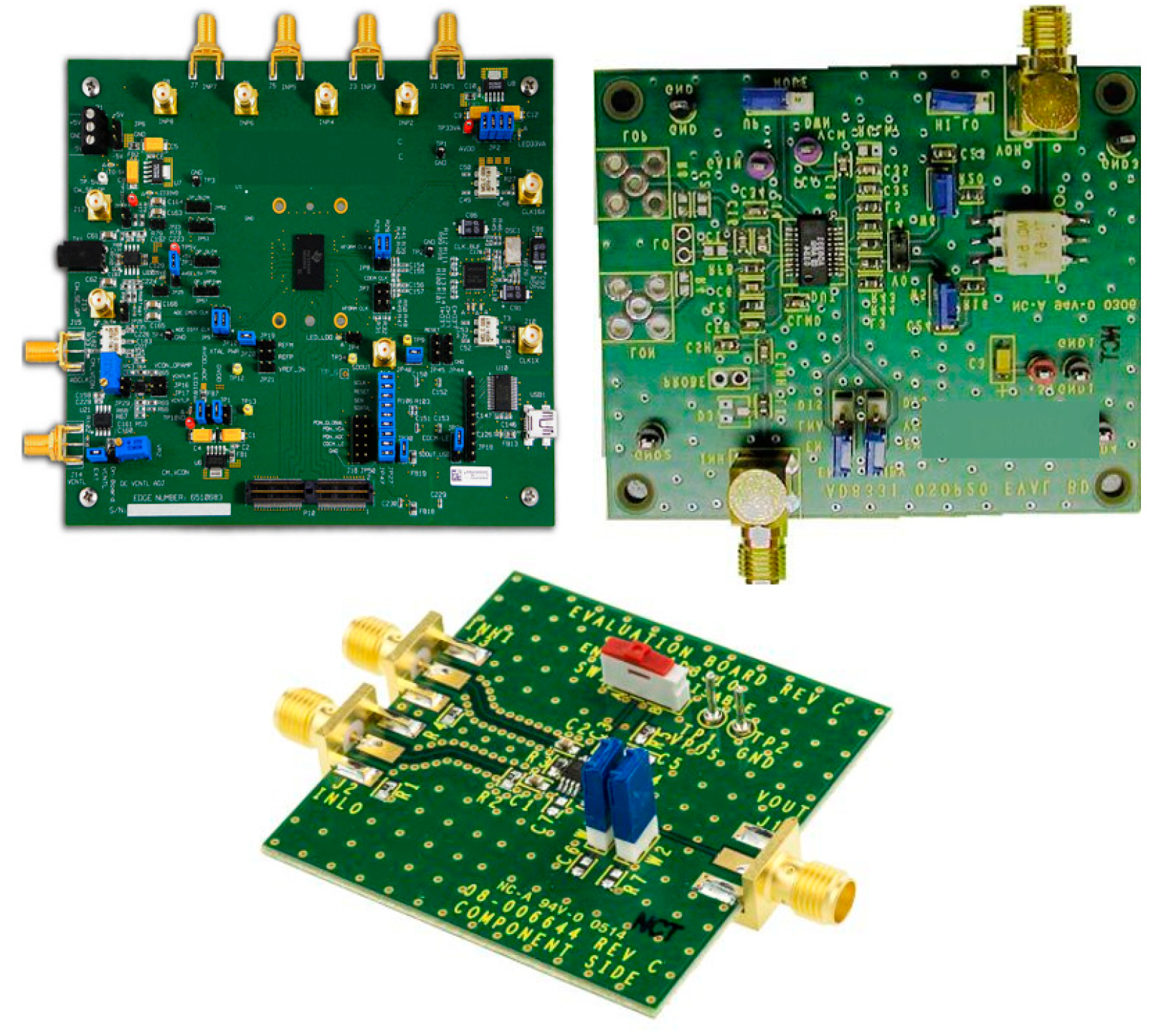
Considering the time and financial investment involved in such a design project, a low-cost open ultrasound seems unviable. However a recent study by K. Divya Krishna and co-workers [42] has developed a multiple element ultrasound using off-the-shelf development kits. The platform is shown in Figure 3. The platform developed by K. Divya Krishna and co-workers uses Xilinx® Kintex® development board in conjunction with a Raspberry Pi. Although not described explicitly in the publication, the research has utilized Texas Instruments evaluation module [43] for AFE5808 analog front end as well as now obsolete Texas Instruments TX-SDK-V1/NOPB-Evaluation Module. AFE is a critical part of any ultrasound system as ultrasonic signals have very high attenuation and so a very low noise amplifier is required to resolve the reflected echoes. An advanced AFE chip such as AFE5808 used in the study has ball-grid array pattern and hence it is very difficult to prototype in a low budget laboratory. However, there are multiple evaluation boards for AFE (Figure 4) which have an appropriate coaxial connection for easy usage. Such low noise AFE evaluation boards (example-AFE 5808, AD8331, and AD8310) are available from semiconductor manufacturers such as Analog Devices and Texas Instruments. Hence evaluation modules provide a very effective way of assembling a functional system at a very low budget. AD8310 is especially useful for ultrasonic applications as it is a logarithmic amplifier and compensates for the increasing (with propagation distance) attenuation of ultrasonic waves.
Other than various research articles published by K. Divya Krishna and co-workers [42,44,45], another study by Techavipoo and co-workers [46] has explicitly used off-the-shelf evaluation boards to design an ultrasonic system from scratch as well as to minimize the design of printed circuit boards.
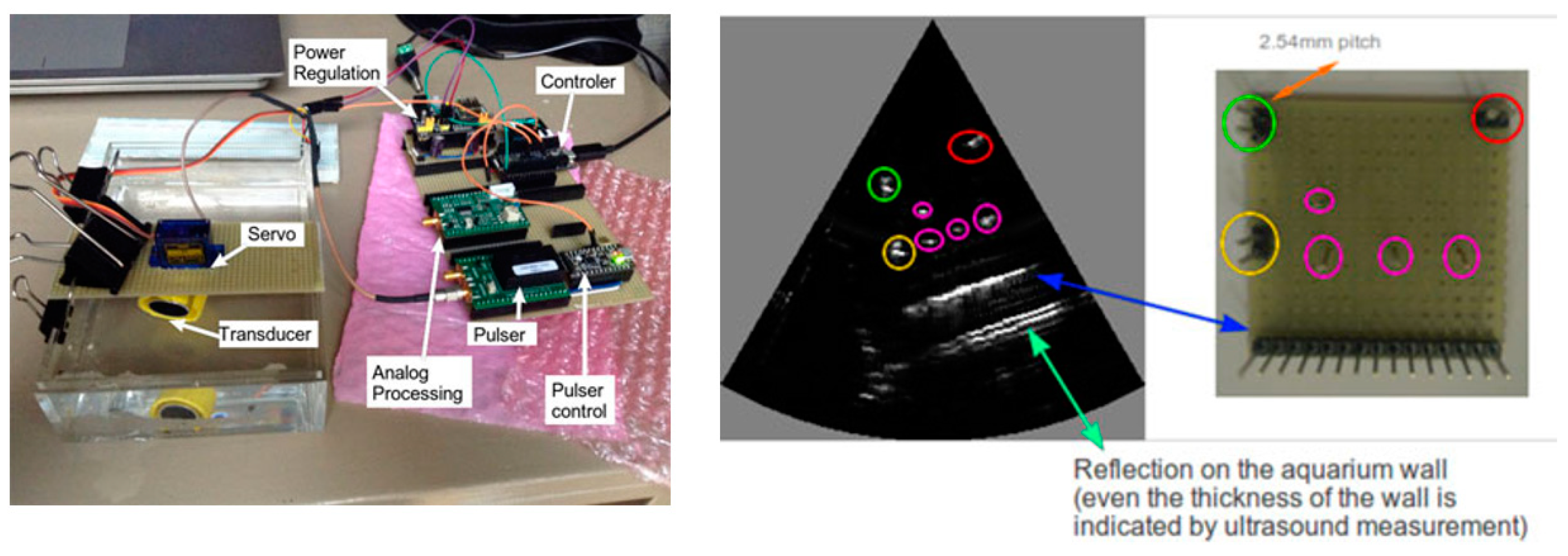
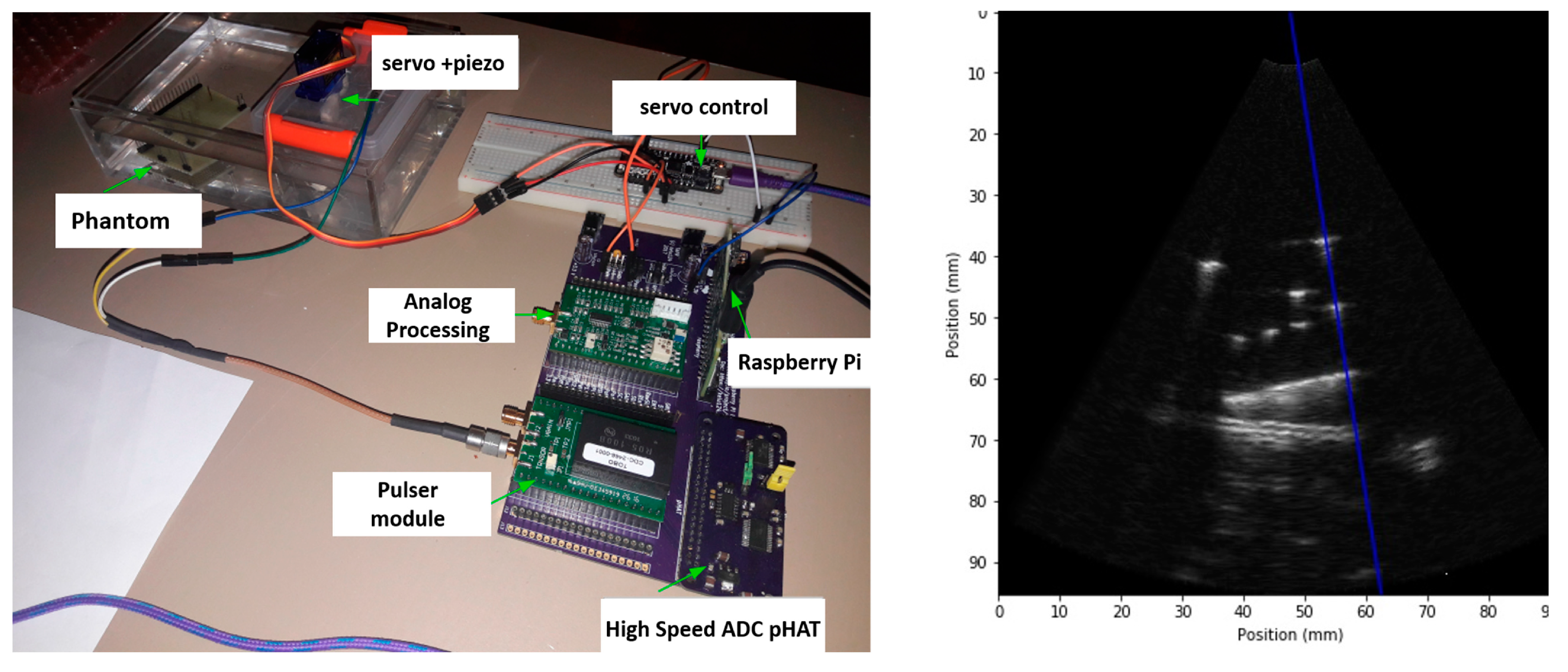
Sobhani et al. [47] have developed an ultrasonic system based on raspberry pi and PIC32 microcontroller. The setup is shown in Figure 5. The research is based on an annular array ultrasonic transducer, which is very promising for both medical as well as non-destructive testing applications. Such annular array transducer research has until now been conducted by using commercial ultrasonic platforms, which cost at-least $50,000. For further research on annular array ultrasonics, the reader can refer to [48,49,50,51,52]. Annular arrays with small number of elements (4–12) and a variable focus offer unique opportunity for development of low-cost yet high-resolution ultrasound. However, despite using specialized and micromachined annular array transducers and inertial tracking system, the ultrasonic images presented in Sobhani et. al. [47] are not very conclusive. In this direction, a significant improvement over [47] is Jonveaux [53]. Jonveaux has described a highly modular ultrasound imaging platform based on microcontrollers and not using FPGAs altogether. Hence it might be very promising to combine the work of Jonveaux [53] with annular array transducers for high-resolution imaging. Further, the work of Jonveaux [53] provides a very comprehensive reference for researchers interested in low-cost microcontroller-based systems and avoiding the complexities associated with using FPGAs.
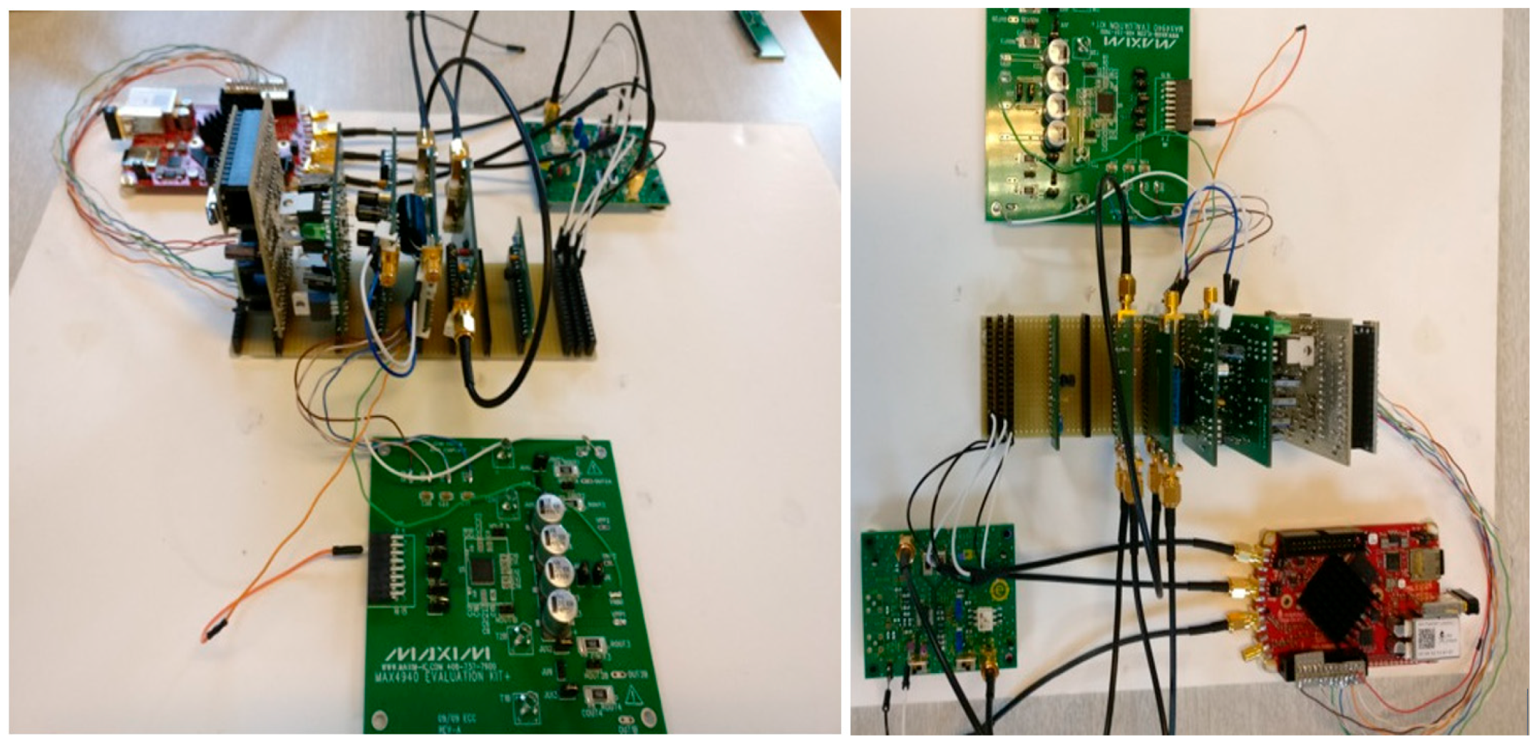
Other than academic research groups, there have been some important efforts by the hobbyist community to design open ultrasound. The open-source project echOpen [54] is developing “echostethoscopy” (ultrasonic imaging tool). The project uses MAX-4940 pulser module, as shown in Figure 6, to produce high frequency (several MHz) and high voltage (−97v) ultrasonic pulses. The project also utilizes Arduino® controlled motors and 3-D printing for producing an ultrasonic b-scan. The project has successfully acquired b-scan images.
Another project “Echomod” [55,56] based on Lattice-FPGA and Raspberry Pi (Figure 7) is perhaps the most advanced among the hobbyist community, with very high resolution images and detailed layouts and software available on its GitHub page. Any hobbyists interested in developing a low cost ultrasound platform will find the project and layouts useful for beginning their design. The project has been verified to work with various used (or refurbished) mechanical ultrasonic probes available off-shelf from websites such as eBay. The project is useful for both medical imaging as well as non-destructive testing (NDT).
While both researchers and hobbyists have made significant progress in developing open and open-source ultrasound devices, so far the prototypes are not compact and miniaturized. One of the challenges is that typical ultrasound frequencies (2–20 MHz) require specialized high-frequency design and researchers have often tackled this challenge by incorporating “development boards.” Such development boards are very common because of the large size of worldwide commercial ultrasound market. However, incorporating multiple development boards often takes a toll on the compactness of the prototype. A greater usage of integrated circuits having multiple channel pulser, receivers, as well as ADC on the same chip, will result in compact open-architecture ultrasound.
To conclude, for a high-frequency low noise healthcare project such as ultrasound, a designer may find it very useful to leverage a range of off-the-shelf evaluation boards for FPGA, microprocessor, System on Chip, analog to digital converter, ultrasonic pulser, analog front end etc. Although FPGA has been used with great success for ultrasonic platforms, it is possible to use high frequency microcontrollers for simpler designs [47,53].
3. Computed Tomography Scanner Based on Open Electronics
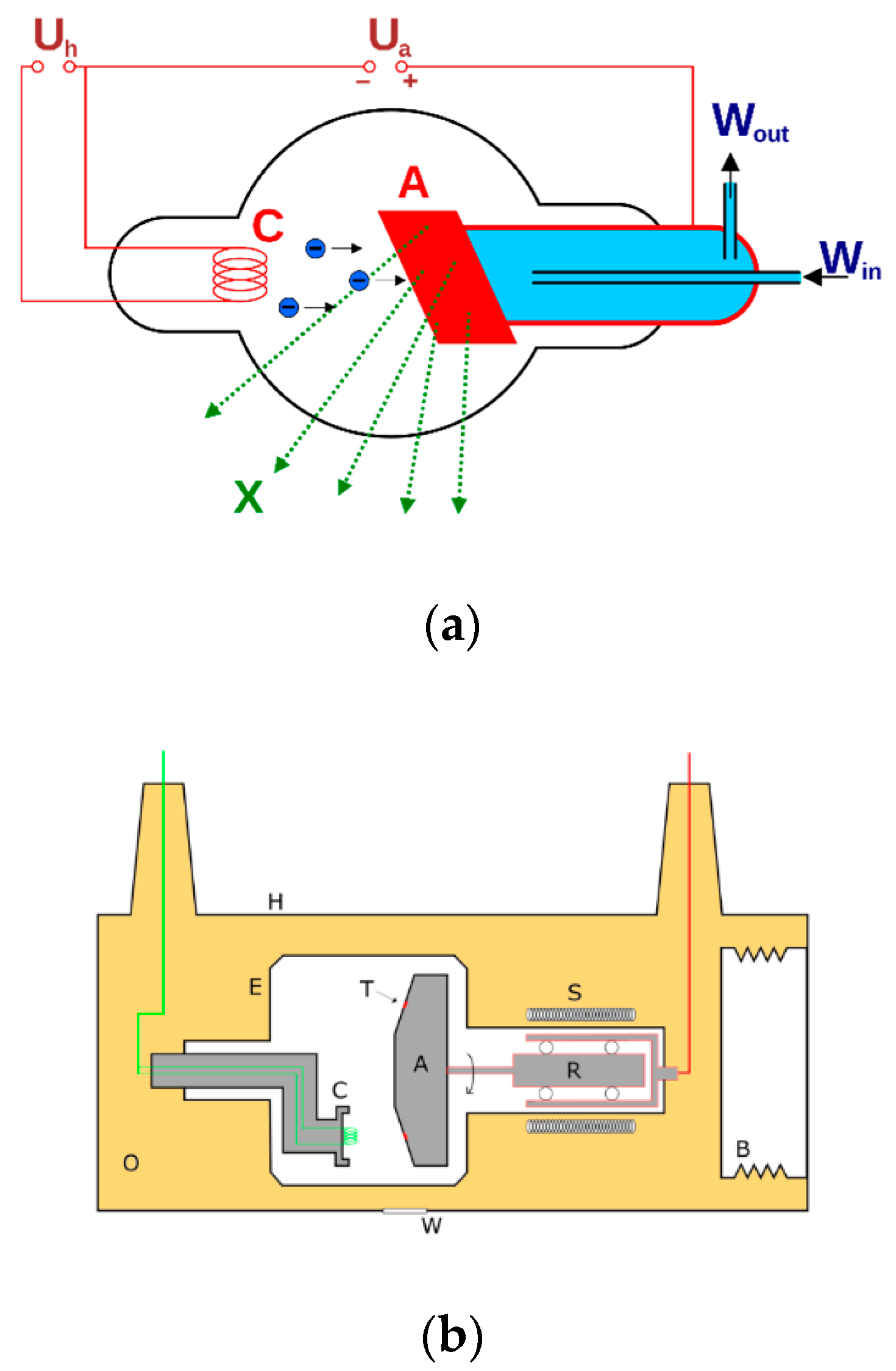
X-ray Computed Tomography Scanner (CT scanner) is an important piece of medical equipment widely used in the healthcare industry. CT Scanning is used for brain [57], neck [58], lungs [59], liver [60], heart [61], and numerous other diagnosis. A typical CT-scan consists of series of planar X-ray scans combined together. One of the earliest X-ray apparatus for hobbyists was described by C.L.Tong in The Amateur Scientist [35] in 1974. A large number of webpages describe X-ray scanners [62,63], which can be built by hobbyists. The main components of an X-Ray scanner are the X-Ray tube (for example-Coolidge tube, rotated anode tube, etc.) and a high voltage power supply, as shown in Figure 8. In the United States, United Nuclear [64] is selling a number of x-ray products-the x-ray tube and the power supply. As per the company website, the components do not require any licensing. Further, a large number of x-ray tubes are easily available on popular websites such as www.ebay.com. Additionally, a number of radioactive sources with low radiation (and hence not requiring licensing) are available from $80–$400 from a number of vendors (for example [65,66]).
Some typical X-ray tubes are shown in Figure 9. The supply voltage and short circuit current specified for a typical tube [64] are 25 KV and 1.5 mA respectively. Further, the minimum input voltage and current as per [64] are mentioned as 12 volts and 1.5 A DC respectively. Hence, we can anticipate that maximum power requirement for a hobbyist/open-source X-Ray to be around 18–20 voltages. Since the power requirement is quite low, in addition to using bench-top power supplies (present state-of-art), it is feasible to use various small form factor DC-DC power conversion modules. Another approach is to design a “ZVS flyback transformer” as described in great detail in [63]. Designing a power supply can further reduce costs as well and result in a compact device.
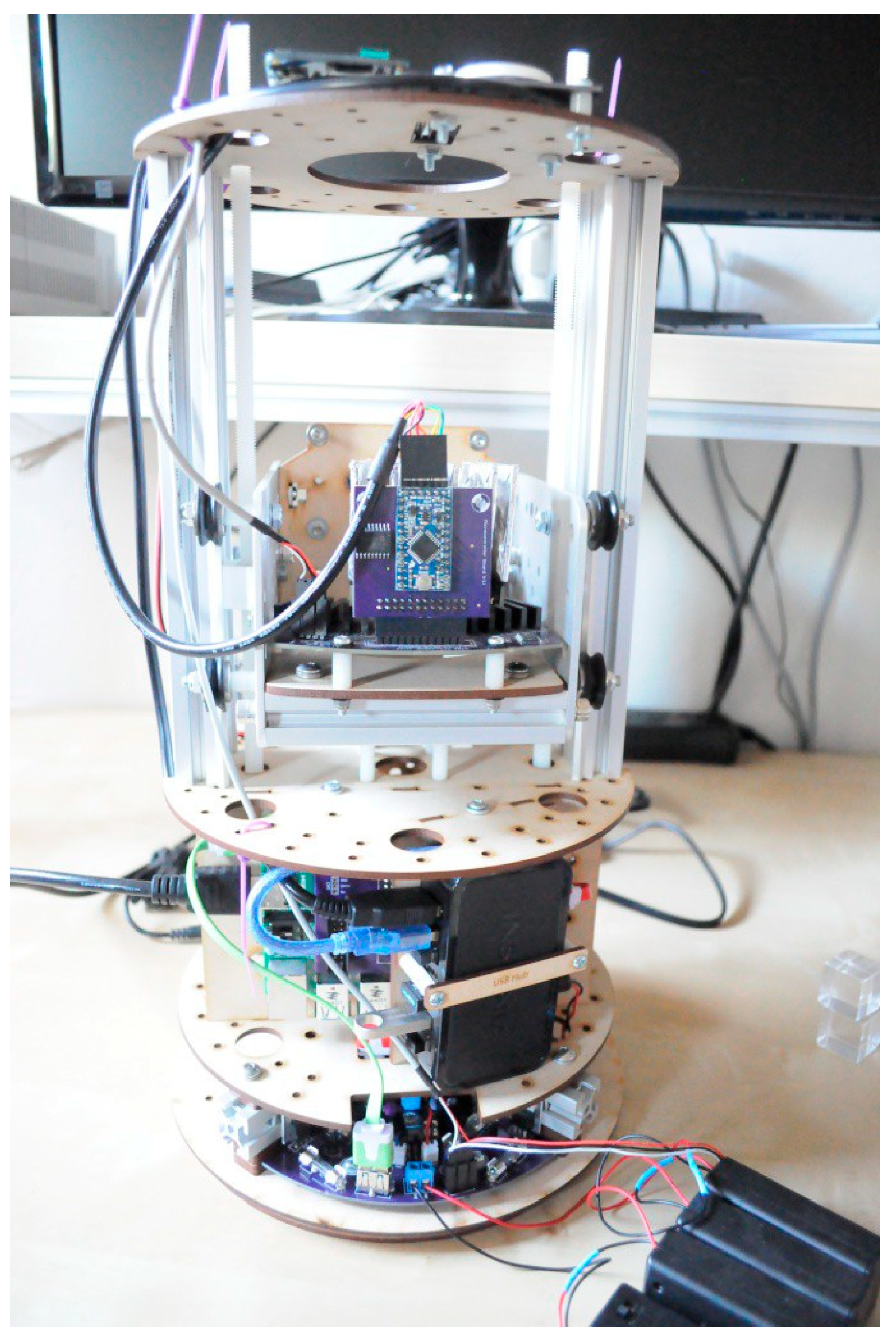
Since the major components of CT scanner, the X-ray tube, and high voltage power supply are already available online, some open-source CT scanner designs [68,69] are freely available. Additionally an optical scanner, shown in Figure 11 is described [70] which can form the basis of x-ray CT scanner, if properly coupled to an X-Ray tube and high voltage power source. One of the scanners from Jansen (shown in Figure 10 and Figure 11) has active open-source repositories. Ben Krasnow has described the details of an Arduino® controlled do it yourself (DIY) CT scanner [71] in a video posted online. Krasnow has also described an innovative use of various image processing software to process CT-scans.
The open-source CT scanner developed by Jansen [68] has a strong potential to be developed for medical tomography and diagnosis. This scanner is currently in the development stage with a small build and scan volume suitable for scanning small objects (size of an apple). The CT scanners used by the medical industry have a large scan volume as they employ a sizeable x-ray source, detector, and related optical setup, which rotates around the specimen object (adult human). On the contrary, the open-source CT scanner ver. 2 [68] consists of a small stationary radioactive source, parallel detectors, and optics but instead has a rotating gantry to rotate the specimen object for imaging, consequently limiting the scan volume. The smaller size of the source (limited by power) and detectors (limited by lower resolution) severely limits the applicability of this open-source CT scanner.
A CT scanner is a sophisticated device integrating complex electronics, mechanical assembly, and software. For the open-source CT scanner to mature in functionality and applicability, the electronics and the related software development needs significant attention and development. Some of the challenges involve developing high-resolution imaging by integrating small x-ray tubes, which will improve the source power, and by increasing the number of parallel detectors comprising of low-cost radiation sensors, the detector array’s resolution can be improved. Furthermore, with additional high-resolution detectors and complex optics, a more sophisticated 3D-slicer software is required for further improvement. The mechanical tomographic platform deployed in the open-source CT scanner is simple, compact, customizable, and modular. Additionally, for integrating x-ray sources, better shielding is required as the current mechanical setup consists of a wooden assembly and framework. Despite all the shortcomings, the developments in open-source CT scanning are promising as a multitude of scans performed on organic samples have been reported [68].
4. Electrocardiograph (ECG/EKG), Electroencephalogram (EEG) and Neurological Monitoring
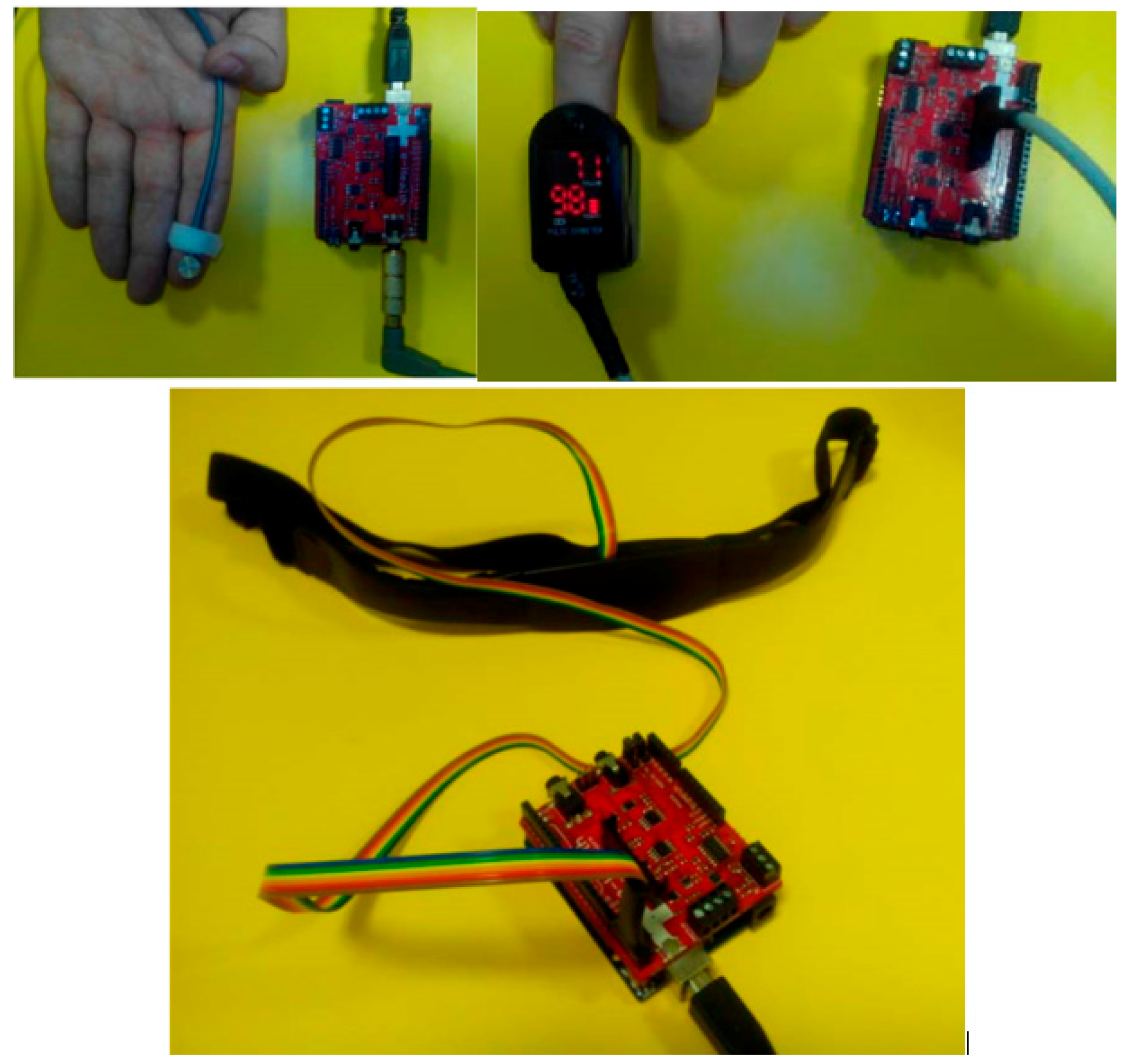
ECG is perhaps one of the most widely prototyped [72,73,74,75,76,77,78,79,80,81] and well-documented medical devices using open electronics. ECG is a widely used for medical diagnosis and monitoring. Most of the open platform ECG devices have the advantage of being readily connected to the Internet using IoT functionalities which are a regular part of open electronics boards such as Arduino® (Figure 12) and Raspberry Pi. ECG signals require a very high signal to noise ratio and very low noise amplification with high common-mode rejection ratio amplifier. Such an amplification system is very difficult to design using discrete electronic components. However, most of the ECG projects have utilized specialized “analog front ends” such as AD8232 and ADAS1000 ECG modules. While the most popular platform for ECG appears to be Arduino® [73,74,75,76,79], some projects have also focused on other platforms such as Raspberry Pi and ESP32 [78,80,81].
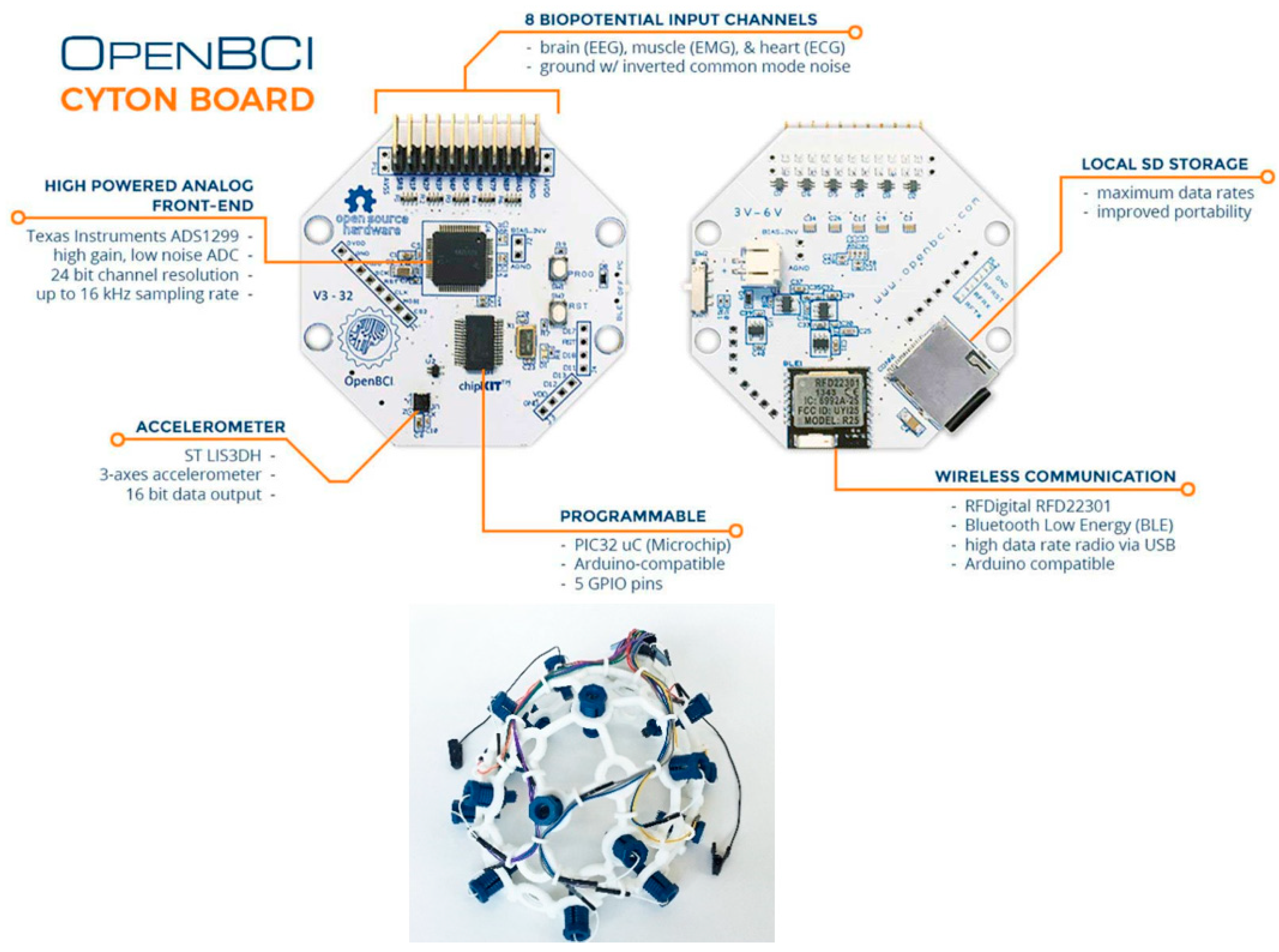
Open-BCI [82] offers EEG, ECG, and EMG for brain, muscle, and heart monitoring on a single platform. The hardware platform is open-source and Arduino®-compatible (Figure 13). The platform has been very successful with the scientific community and has been utilized in many peer-reviewed research articles. Similar to OpenBCI, there are several other platforms offering multiple biosensor integration and the reader is directed to Niezen et al. [22] for further reading.
Other than being a low-cost medical device, thanks to readily available IoT functionality, open-source ECG/EKG offers an unprecedented advantage in diagnosis of epilepsy, strokes etc. Various research papers [83,84,85,86,87] have highlighted the role of ECG in detecting abnormal neurological activity, such as epileptic seizures. Hence, using a low-cost ECG platform, it is possible to not only diagnose abnormal neurological activity but perhaps to forewarn about an impending neurological condition. Open-source electronics are highly relevant to such platforms being widely used by patients all over the world at a low cost. Open electronics platforms also provide the unique opportunity to integrate multiple sensors such as accelerometers, temperature sensors, humidity sensors, galvanic skin response, brain waves, and others to monitor and perhaps predict abnormal neurological activity. An article by Sriram Ramgopal and co-workers [84] provides a highly relevant list of sensor platforms which can be used to manage seizures. Additionally, some open electronics platforms [88,89,90,91] are available for seizure detection. However, more research data are needed before drawing any conclusion about practical utility of such platforms.
Apart from reducing the cost of neurological monitoring, open electronics offers the opportunity to collect data using common data interchange formats such as JSON. Such data can be collected and shared by multiple researchers around the world using machine learning. Open electronics also offers practically unlimited modularity-multiple sensors that can be selectively used, resulting in more data for predicting abnormal neurological activity. Hence open electronics offers a unique opportunity to treat and manage potentially life-threatening medical conditions. Recent works by Vergara and co-workers [36] can be referred for such an IoT-based seizure prediction.
The various open/open-source BCI and neurological devices are significantly mature in their development, and they are employed by medical professionals and researchers globally for EEG, ECG, and EMG measurement and analysis. However, BCI technology does not classify neural activity accurately. It detects the smallest of changes in the energy radiated by the brain when it thinks in a certain way; BCI recognizes specific energy/frequency patterns in the brain. Additionally, the most limiting factor of BCI is interfacing. BCIs placed outside the skull have limited ability to read the brain signals because of high attenuation by the skull. However, with advancements in low noise amplifiers, we can expect more robust open-source BCIs.
For more advanced research, a headset with 32–128 electrodes is required, which is much beyond the capacity of any OpenBCI boards and headsets. The Cyton board offered by OpenBCI, which is a 16-channel board suitable for most applications, but for more complex arrangements of electrodes, a daisy chain or a parallel arrangement setup may be required, thus escalating the cost of hardware. Hence a moderately higher cost of OpenBCI devices is another disadvantage compared to some of the commercial BCI devices with limited functionality such as NeuroSky MindWave headsets.
5. Electric Impedance Tomography
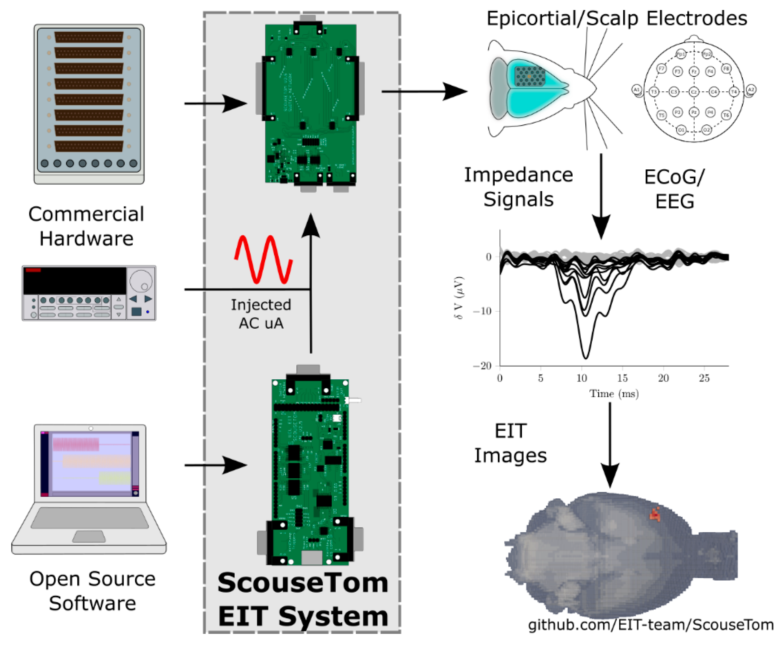
Electrical impedance tomography (EIT) is noninvasive medical imaging to monitor physiological functions on the basis of the conductivity or permittivity of body-tissue. The technique involves taking electrical measurements at the surface of the body by employing conducting electrodes (as shown in Figure 14) that are attached to the skin and applying small (mA) alternating currents. The resulting electrical potentials are measured to infer the electrical conductivity, impedance, and permittivity of a part of the body to form a tomographic image of that part.
In recent years, numerous open-source EIT devices based on Arduino® platforms have been reported for a host of diagnosis applications such as stroke [93], pulmonary diseases [94], epilepsy [95], neurology [92], and generic systems [96]. The EIT system developed by Avery et al. [92], known as ScouseTom for brain imaging application (Figure 14) is one of the most versatile and reproducible devices that can be modified for other diagnostic applications. The versatile applicability of the ScouseTom system can be estimated from work reported by Goren et al. [93], which used the ScouseTom system for stroke patients.
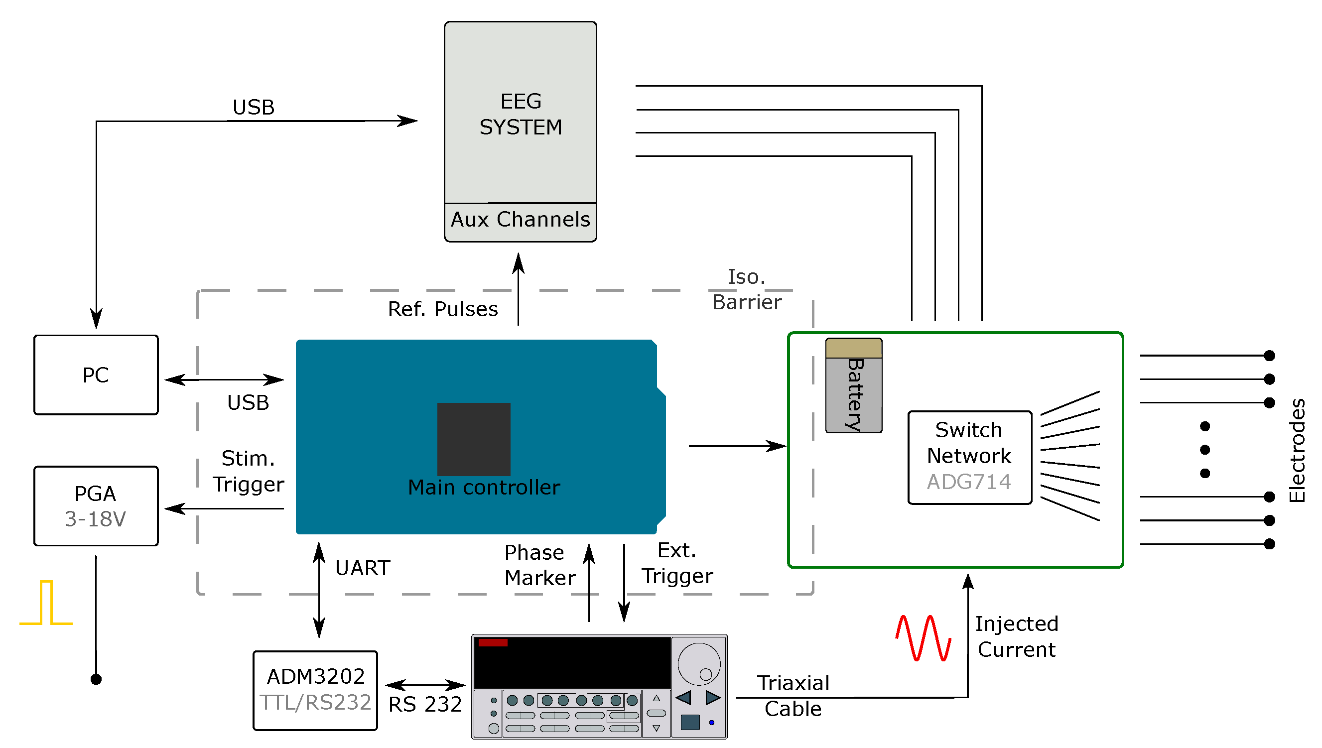
A conventional EIT system constitutes a current source, switching circuitry to address multiple electrodes, a voltage measurement unit, and a controller to automate the measurement process. In a typical EIT system, an alternating current of a defined amplitude and frequency in a range of 10 Hz to 1 MHz is injected through a pair of electrodes, with electric potential measured at all electrodes (generally up to 256). The measurement circuitry employs low noise amplifiers capable of measuring ~100 nV, typically characterized by SNR > 75 dB. These voltage measurements can be obtained either serially or in parallel on all electrodes. The open-source ScouseTom EIT systems (details shown in Figure 15) are compatible with these standards and finding numerous applications in neurological imaging, stroke diagnosis, epilepsy, and other related fields. For more details about the ScouseTom system, the reader can refer to [92]. Dimas et al. [96] have reported an Arduino® based generic EIT system that employs up to 64 electrodes and can be employed for a host of applications such as pulmonary, throat, cervix, and breast diagnosis. Another Arduino® based EIT system developed by Chitturi et al. [94] has been extensively used for pulmonary disease and modeling. This is a simple design to reproduce and getting started as it integrates off-shelf components and open-source software, EIDORS, for image reconstruction.
The open-source EIT devices such as ScouseTom are technologically mature for both research and commercial deployment for a variety of applications, as the front-end electronics and control system involved in this have already been developed and matured more than a decade ago. However, for fast neural activity measurements and imaging, significant research and development work is required. One of the major problems with most EIT systems is the inability to process continuous impedance signals with other additional signals such as EEG, both of which are necessary for EIT of fast-neural activity. FPGA-based data acquisition and processing need to be implemented as Arduino® based systems cannot handle the massive influx of data that drastically increases the complexity and processing as the number of electrodes and scanning frequency increases. Further, performing the signal processing on Arduino® based hardware severely limits the ability to adjust parameters such as carrier frequency, scanning and measurement speed, and filter bandwidth to meet the diverse application requirements. With the recent popularity of various FPGA-based low noise and high frequency open-source projects for ultrasound (Section 2), we can expect open-source EIT to follow the same trend of using off-the-shelf FPGA modules.
6. Infusion Pumps
Infusion pumps or syringe pumps are one of the most widely used medical devices. Infusion pumps are used to deliver nutrients and medications to patients. Some reports have suggested that the global market for infusion pumps are in tens of billions of US$ [97,98]. A 2010 white paper [99] by the United States Food and Drug Administration (FDA) cites a market research estimation of over 2 million external infusion pumps in the United States in the year 2006. Hence, infusion pump devices are a very high impact and still a growing class of medical devices.
Advanced small volume infusion pumps (an example shown in Figure 16) require precise control of fluid dispensation mechanism and cost anywhere between a few hundred dollars to few thousand US dollars. Such smaller pumps are crucial for controlled drug delivery, such as insulin. In 2016, the Time magazine voted a new “smart” insulin pump [37] as one of the top 20 inventions of the year. Millions of people worldwide suffer from diabetes. However, despite such a high global demand, the cost of a typical insulin pump is between $7000–$10,000.
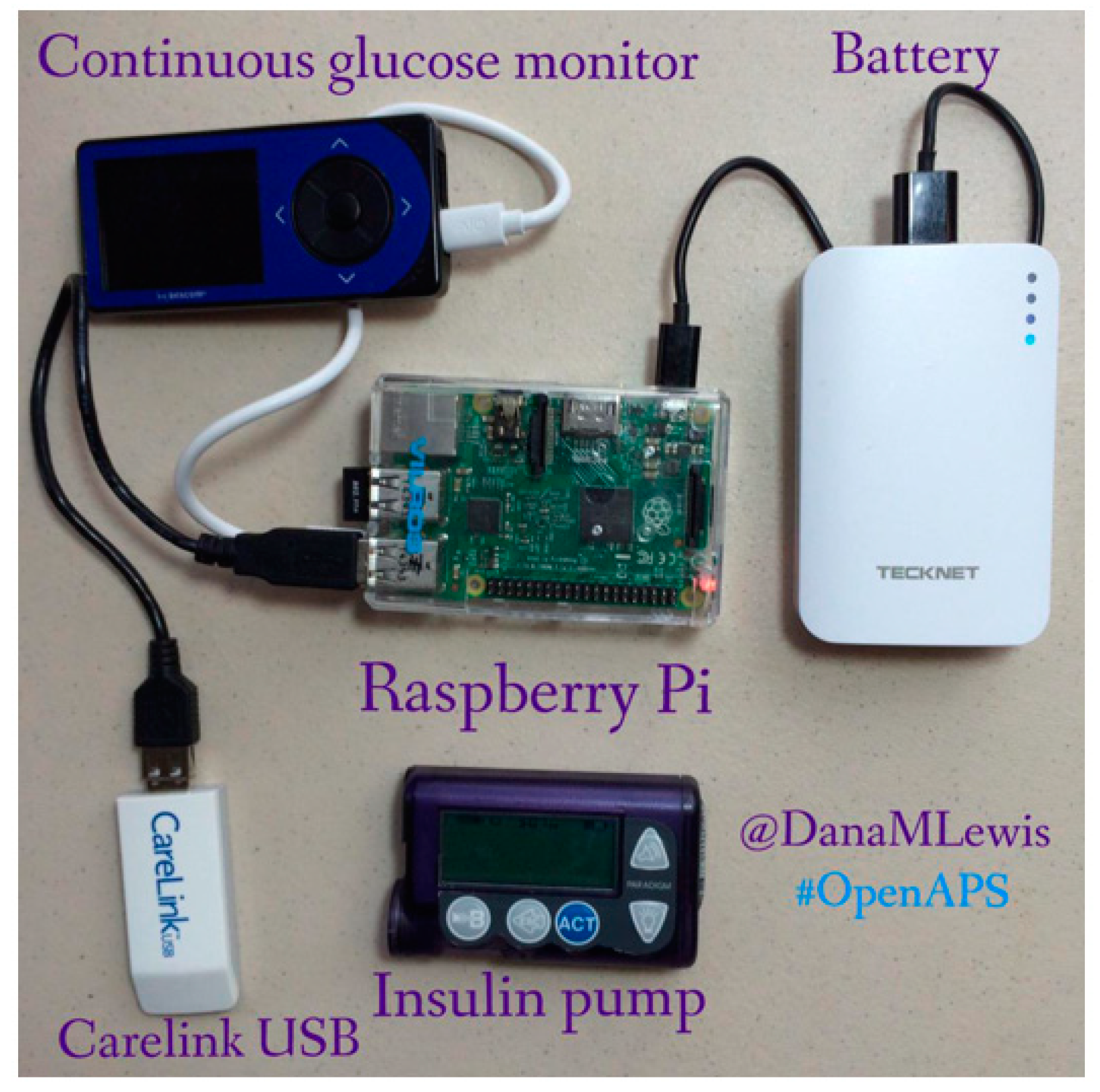
Although small volume drug delivery pumps such as insulin pumps require a high degree of precision and reliability, open electronic platforms have shown tremendous promise in reducing the cost and at the same time a promise of maintaining precision and reliability. Niezen and co-workers [22], in their review of open hardware medical devices have provided an excellent compilation of such medical pumps. The authors have also reviewed glucose monitors such as Diabeto, Nightscout, and X-Drip [102,103,104] which are critical to designing open hardware-based insulin pumps. A recent open-source project [100,101] has used components from commercially available syringe pumps (shown in Figure 16) in combination with open hardware and software.
While the development of open hardware insulin pumps mentioned in the previous projects is encouraging, still, these projects require components from commercial insulin pumps. Such components cost a few hundred to several thousand US dollars. Commercial insulin pumps are microfluidics devices and although it is outside the scope of this study to calculate the precision required for actuating them, it is likely that the actuation precision is comparable to that of 3-D printers. Since most of the 3-D printers now routinely use open electronic hardware such as Arduino®, it is highly likely that in the future, there will be fully open low volume medical pumps. Encouraging developments in this direction are numerous recent studies on open electronics microfluidics [105,106,107,108,109]. White and co-workers [110] have developed an Arduino®-based kit (an example shown in Figure 17) for microfluidics control.
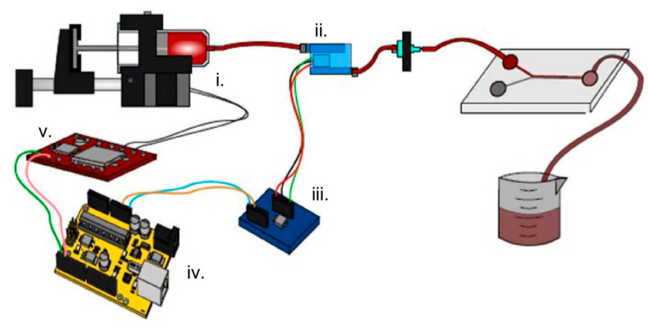
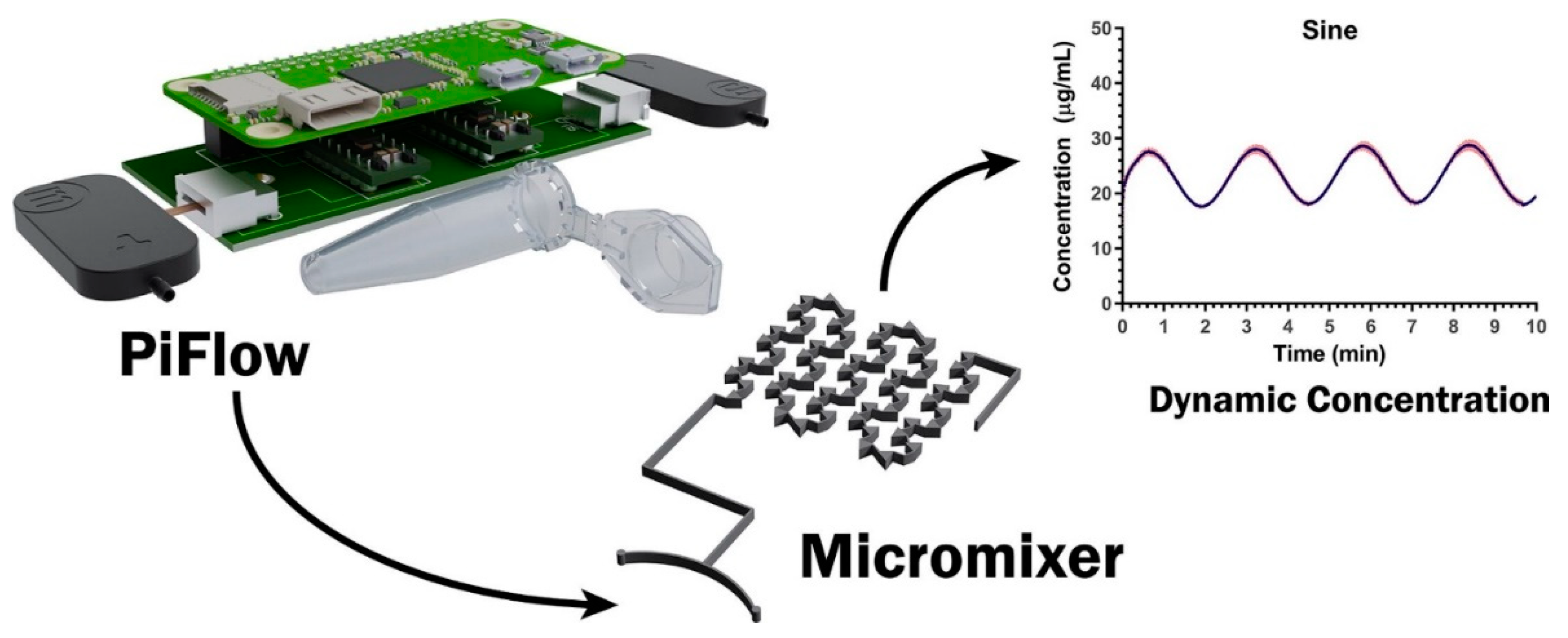
While small volume infusion pumps such as insulin present a challenge, many larger volume peristaltic pumps based on Arduino®, Raspberry pi etc., can be found from popular vendors such as Sparkfun [111], Adafruit [112], etc. Although the hobbyist community has mostly used these pumps for non-medical applications, it is possible that they can be used in medical application after proper scientific and medical studies. Kassis et al. [113] have developed a biocompatible Raspberry pi-based piezoelectric pump driver (Figure 18). Such a pump driver can be very crucial to truly open-source small infusion pumps.
While infusion pumps have been very widely prototyped using open electronic platforms, a key challenge is that for small volume pumps (such as insulin), these open-source projects still rely on components from commercial devices. Therefore, the challenge lies in fabricating biocompatible small-volume liquid dispensing systems. While the hobbyist community has made significant contributions, this is an area of research which would involve significant effort from well-established scientific research groups in the field of microfluidics and nanofluidics. With rapid developments in the field of microfluidics and their associate open electronics-based drivers/controllers such as those described in [113], we can expect more open-source small volume infusion pumps. For larger volume infusion pumps, the open-source/open electronics projects seem to have already reached a significant level of maturity. However, there is still a significant room for improvement in large volume pumps by utilizing full potential of open electronics. For example-integration with various other biomedical sensors, bubble detection, IoT connectivity, incorporating AI and deep learning for optimal dosage. A greater interaction between physicians/healthcare workers and the open-source prototyping community is required for fully utilizing the potential offered by making large volume infusion pumps open source.
7. Miscellaneous Medical Devices: Wearables, Posture Detection, and Sensor Networks
Other than the highly specialized medical devices such as ultrasound, X-ray CT, ECG, and infusion pumps discussed in previous sections, there is a wide range of miscellaneous medical devices that have utilized open electronics platforms. Primarily, such devices constitute wearables (fitness monitoring) and simple sensor networks (accelerometer, temperature, pulse rate, blood pressure, etc.,). Haghi et al. [114] have reviewed a wide range of commercial wearable and motion tracking devices. Accelerometers, gyroscopes, and temperature sensors are the primary components of majority of wearable motion tracking devices. For more advanced wearable devices described in [114], additional sensors such as heart rate sensors are incorporated. Given the easy availability of all these sensors for platforms such as Arduino® and Raspberry Pi, it is possible for researchers to easily put together a wireless wearable platform at a very low cost, for example: Mukherjee and co-workers [115] have utilized Arduino® and accelerometers for a gesture control device; Rakay and co-workers [116] have implemented temperature, air flow and body position sensing on Arduino® (Figure 19); Kemis and co-workers [117] have implemented Arduino® based heart rate monitor; Mallick and Patro [118] have designed an optical heart rate sensor from scratch and utilized it with Arduino®. Many other wearable sensors [119,120,121,122,123,124] based on open electronics are published in scientific literature.
In the field of rehabilitation, there have been a number of studies [125,126,127,128,129,130,131,132,133,134] utilizing open electronics for posture detection, monitoring, and correction. Such posture detection mainly utilizes wearable “smart fabrics” [125,127,128,129,130,132], accelerometers [126], or ultrasonic sensors [133]. Rawal and Nagtilak [135] have used cameras for drowsiness detection. It is possible to use external cameras for posture detection; however, since this review is mainly concerned with electronic hardware, we will skip such methods. Nevertheless, it is important to mention that using open electronics platforms with advanced computing capacities (for example-Raspberry Pi 4) can enable researchers to take advantage of both sensors-based (accelerometer, smart fabrics, ultrasonic, etc.) and camera-based posture detection for robust rehabilitation applications. Using OpenCV and Python which are highly compatible with the Raspberry Pi platform can enable fast development and prototyping.
With growing number of novel sensors (smart fabrics, accelerometers etc.), rapid developments in field of 3-D cameras as well as increasing computing capacities of single board computers, we can expect further open-source projects in wearables for posture monitoring. While present open-source projects have not extensively utilized multi-modal sensing (for example both cameras and wearable sensors), we can expect such multi-modal sensing in the near future (because of higher performance open-source hardware). Further, recently, some semiconductor manufacturers have introduced specialized microcontrollers for multi-modal wearable sensing for example, MAX32600 wellness measurement microcontroller by Maxim IntegratedTM. We expect the open-source community to incorporate such multi-modal microcontrollers in their design effectively.
8. Unique Advantages Offered by Open Electronics Based Medical Devices
8.1. Artificial Intelligence and Machine Learning Compatibility
Other than reduced cost and practically unlimited customization, open hardware medical devices are especially suitable for incorporating artificial intelligence and machine learning. Open electronic platforms have a standard method to update their firmware (USB ports/COM ports (microcontrollers and lower end FPGAs) or file transfer (single board computers or high-end FPGAs with hard processors). Hence, users can easily update firmware to share their raw medical data. The raw medical data can be used by machine learning and artificial intelligence algorithms for better predicting the medical conditions (such as epileptic seizures, impending strokes etc.,). Such a feature to share medical data globally could be optional, based on a firmware update by the users of open hardware medical devices. Typically, raw medical data provided by just a single user may not be sufficient for machine learning algorithms to predict the onset of a medical condition. However, based on a larger data-set, the predictive capabilities are bound to increase. Such potential for sharing data is difficult, if not non-existent for proprietary medical devices.
8.2. Data Synchronization from Multiple Sensors
In addition, using open hardware medical devices, users with multiple medical conditions can easily integrate multiple devices based on the same platform. For example, in a hypothetical scenario, if an EEG and heart rate sensor are both based on a particular microcontroller platform, a patient will have no difficulty in “syncing” the data from both sensors. However, using separate EEG and heart rate devices from different manufacturers, the patient will miss out on this vital feature of “syncing” data. Such syncing of medical data is further crucial for medical devices to benefit from the progress in the field of computer-aided diagnostics. Such advantages have already been demonstrated by various studies utilizing multiple medical sensors [120,122,124]. Such data synchronization cannot be easily accomplished by proprietary medical devices.
8.3. Ease of Repair and Maintenance
A third crucial advantage offered by open electronic medical devices is the ease of repair and maintenance. This is very crucial to reduce the cost of medical care all over the world. When a particular medical device needs maintenance or repair, the whole device has to be shipped to the original manufacturer. The manufacturer, depending on the specific workflow organization has to often disassemble the device, test various components, replace the faulty component, and send it back to the user. The user of the medical device (a physician, hospital or patient) has to wait for either the device to be sent back or have a replacement device. Open hardware medical devices are inherently modular as they consist of a specific electronic board (shield, hat etc.) together with standard computing and control board (Arduino®, Raspberry Pi, FPGA board etc.,). Additionally, there can be one or more mechanical modules (example-robotic hand with motors or infusion pump). In a hypothetical scenario, a user utilizing a medical device consisting of sensor module (SM) for example- blood sugar sensor, mechanical module (MM) for example, insulin pump, power module (PM), and computing module (CM) can test various modules separately to detect faulty module. The faulty module can be either shipped to the module manufacturer or be replaced by off the shelf module. In both scenarios, the repair process is much faster and less expensive than repairing a proprietary and “hard-wired” medical device. A good example of the ease of repair is the popular Arduino® 101 board-boards based on either the Intel® CurieTM or Atmel® ATmega32u4 microcontroller have the same exact functionality when used with a particular “shield.” Hence it is possible to replace a CM based on one microcontroller with another CM based on another microcontroller without the loss of functionality.
8.4. Rapid Innovation
As the review by Neizen et al. [22] explains that open-source medical devices can overcome the slow pace and high cost of innovation. Various other open-source projects in non-medical fields (3D printing, operating system, and computer software, online encyclopedias etc.) have demonstrated the advantage of open innovation by a collaborative community of developers as opposed to internal research and development. Since open hardware medical devices are primarily modular, it gives physicians and surgeons greater opportunities for innovation at a very quick pace and a small cost. The physician can possibly consult with the specifically skilled engineer to modify the required module. This is opposed to the scenario in which the physician/surgeon has to approach the medical device company to consider a complete overhaul of the product.
In light of the above discussion, open electronics-based medical devices are very promising for not just reducing the device cost but also for fostering unprecedented innovation and improving the effectiveness of medical devices. The inherent modularity in open electronics platforms, their worldwide off-the-shelf availability as well seamless firmware update will enable higher quality, well connected and AI capable medical devices.
9. Conclusions
Open electronics platforms have demonstrated their feasibility in a number of fields such as ultrasound, medical infusion pumps, wearable sensors, posture correction, ECG/EKG, EEG and pulse rate measurement. Even in a technologically advanced medical device such as X-Ray CT scan, there are some nascent but very promising open-source projects. While a lot of open (and open source) medical devices are published on online collaborative platforms such as GitHub, a significant number have resulted in several peer-reviewed research articles. The peer reviewed research articles point out that such medical devices can potentially satisfy the requirement for precision, reliability and robustness. In addition, all the open-source projects demonstrate the tremendous cost reduction and modularity of open platform medical devices. There is a wide range of tools available for open electronics developers: microcontroller platforms (such as Arduino®, PIC, ESP etc.), FPGA platforms (Lattice, Altera/Intel, Xilinx etc.), single board computers (Raspberry pi), and development boards (various semiconductor companies). We expect that in future, more and more medical devices will take advantage of open hardware platforms and open-source electronics design to result in lower cost, higher performance, and innovative designs.
Author Contributions
Conceptualization, G.P. and A.V.; methodology, G.P. and A.V.; formal analysis, G.P. and A.V.; investigation, G.P. and A.V.; writing—original draft preparation, G.P. and A.V.; writing—review and editing, A.V.
Funding
This research received no external funding.
Conflicts of Interest
The authors declare no conflict of interest. Opinions expressed by G.P. in this study are entirely individual and have no correlation to his partnership or any other role in Noble Sensors LLC.
References
- Winter, L.; Pellicer-Guridi, R.; Broche, L.; Winkler, S.A.; Reimann, H.M.; Han, H.; Arndt, F.; Hodge, R.; Günyar, S.; Moritz, M.; et al. Open Source Medical Devices for Innovation, Education and Global Health: Case Study of Open Source Magnetic Resonance Imaging. In Co-Creation: Reshaping Business and Society in the Era of Bottom-up Economics; Redlich, T., Moritz, M., Wulfsberg, J.P., Eds.; Springer International Publishing: Cham, Switzerland, 2019; pp. 147–163. ISBN 978-3-319-97788-1. [Google Scholar]
- Gonzalez, D.R.; Carpenter, T.; van Hemert, J.I.; Wardlaw, J. An open source toolkit for medical imaging de-identification. Eur. Radiol. 2010, 20, 1896–1904. [Google Scholar] [CrossRef] [PubMed]
- Björn, K. Evaluation of Open Source Medical Imaging Software: A Case Study on Health Technology Student Learning Experience. Procedia Comput. Sci. 2017, 121, 724–731. [Google Scholar] [CrossRef]
- Norton, I.; Essayed, W.I.; Zhang, F.; Pujol, S.; Yarmarkovich, A.; Golby, A.J.; Kindlmann, G.; Wassermann, D.; Estepar, R.S.J.; Rathi, Y.; et al. SlicerDMRI: Open Source Diffusion MRI Software for Brain Cancer Research. Cancer Res. 2017, 77, e101–e103. [Google Scholar] [CrossRef] [PubMed] [Green Version]
- Fedorov, A.; Khallaghi, S.; Sánchez, C.A.; Lasso, A.; Fels, S.; Tuncali, K.; Sugar, E.N.; Kapur, T.; Zhang, C.; Wells, W.; et al. Open-source image registration for MRI–TRUS fusion-guided prostate interventions. Int. J. Comput. Assist. Radiol. Surg. 2015, 10, 925–934. [Google Scholar] [CrossRef] [PubMed]
- Smith, D.S.; Li, X.; Arlinghaus, L.R.; Yankeelov, T.E.; Welch, E.B. DCEMRI.jl: A fast, validated, open source toolkit for dynamic contrast enhanced MRI analysis. PeerJ 2015, 3, e909. [Google Scholar] [CrossRef]
- Pieper, S.; Lorensen, B.; Schroeder, W.; Kikinis, R. The NA-MIC Kit: ITK, VTK, pipelines, grids and 3D slicer as an open platform for the medical image computing community. In Proceedings of the 3rd IEEE International Symposium on Biomedical Imaging: Nano to Macro, Arlington, VA, USA, 6–9 April 2006; pp. 698–701. [Google Scholar]
- Loening, A.M.; Gambhir, S.S. AMIDE: A Free Software Tool for Multimodality Medical Image Analysis. Mol. Imaging 2003, 2, 15353500200303132. [Google Scholar] [CrossRef]
- Rosset, A.; Spadola, L.; Ratib, O. OsiriX: An Open-Source Software for Navigating in Multidimensional DICOM Images. J. Digit. Imaging 2004, 17, 205–216. [Google Scholar] [CrossRef] [Green Version]
- Hansen, M.S.; Sørensen, T.S. Gadgetron: An open source framework for medical image reconstruction. Magn. Reson. Med. 2013, 69, 1768–1776. [Google Scholar] [CrossRef]
- Nolden, M.; Zelzer, S.; Seitel, A.; Wald, D.; Müller, M.; Franz, A.M.; Maleike, D.; Fangerau, M.; Baumhauer, M.; Maier-Hein, L.; et al. The Medical Imaging Interaction Toolkit: Challenges and advances. Int. J. Comput. Assist. Radiol. Surg. 2013, 8, 607–620. [Google Scholar] [CrossRef]
- De Leener, B.; Lévy, S.; Dupont, S.M.; Fonov, V.S.; Stikov, N.; Louis Collins, D.; Callot, V.; Cohen-Adad, J. SCT: Spinal Cord Toolbox, an open-source software for processing spinal cord MRI data. Neuroimage 2017, 145, 24–43. [Google Scholar] [CrossRef]
- Zöllner, F.G.; Weisser, G.; Reich, M.; Kaiser, S.; Schoenberg, S.O.; Sourbron, S.P.; Schad, L.R. UMMPerfusion: An Open Source Software Tool Towards Quantitative MRI Perfusion Analysis in Clinical Routine. J. Digit. Imaging 2013, 26, 344–352. [Google Scholar] [CrossRef]
- Yoo, T.S.; Ackerman, M.J. Open Source Software for Medical Image Processing and Visualization. Commun. ACM 2005, 48, 55–59. [Google Scholar] [CrossRef]
- Lee, C.-C.; Chung, P.-C.; Tsai, H.-M. Identifying multiple abdominal organs from CT image series using a multimodule contextual neural network and spatial fuzzy rules. IEEE Trans. Inf. Technol. Biomed. 2003, 7, 208–217. [Google Scholar] [PubMed]
- Margeta, J.; Criminisi, A.; Cabrera Lozoya, R.; Lee, D.C.; Ayache, N. Fine-tuned convolutional neural nets for cardiac MRI acquisition plane recognition. Comput. Methods Biomech. Biomed. Eng. Imaging Vis. 2017, 5, 339–349. [Google Scholar] [CrossRef]
- Amsaveni, V.; Singh, N.A. Detection of brain tumor using neural network. In Proceedings of the 2013 Fourth International Conference on Computing, Communications and Networking Technologies (ICCCNT), Tiruchengode, India, 4–6 July 2013; pp. 1–5. [Google Scholar]
- Cicero, M.; Bilbily, A.; Colak, E.; Dowdell, T.; Gray, B.; Perampaladas, K.; Barfett, J. Training and Validating a Deep Convolutional Neural Network for Computer-Aided Detection and Classification of Abnormalities on Frontal Chest Radiographs. Investig. Radiol. 2017, 52, 281–287. [Google Scholar] [CrossRef] [PubMed]
- Kato, H.; Kanematsu, M.; Zhang, X.; Saio, M.; Kondo, H.; Goshima, S.; Fujita, H. Computer-Aided Diagnosis of Hepatic Fibrosis: Preliminary Evaluation of MRI Texture Analysis Using the Finite Difference Method and an Artificial Neural Network. Am. J. Roentgenol. 2007, 189, 117–122. [Google Scholar] [CrossRef] [Green Version]
- Mala, K.; Sadasivam, V. Automatic Segmentation and Classification of Diffused Liver Diseases using Wavelet Based Texture Analysis and Neural Network. In Proceedings of the 2005 Annual IEEE India Conference-Indicon, Chennai, India, 13–15 December 2005; pp. 216–219. [Google Scholar]
- Ohlsson, M. WeAidU—A decision support system for myocardial perfusion images using artificial neural networks. Artif. Intell. Med. 2004, 30, 49–60. [Google Scholar] [CrossRef]
- Niezen, G.; Eslambolchilar, P.; Thimbleby, H. Open-source hardware for medical devices. BMJ Innov. 2016, 2, 78–83. [Google Scholar] [CrossRef] [Green Version]
- Koprnický, J.; Najman, P.; Šafka, J. 3D printed bionic prosthetic hands. In Proceedings of the 2017 IEEE International Workshop of Electronics, Control, Measurement, Signals and their Application to Mechatronics (ECMSM), San Sebastian, Spain, 24–26 May 2017; pp. 1–6. [Google Scholar]
- Krausz, N.E.; Rorrer, R.A.L.; Weir, R.F. f. Design and Fabrication of a Six Degree-of-Freedom Open Source Hand. IEEE Trans. Neural Syst. Rehabil. Eng. 2016, 24, 562–572. [Google Scholar] [CrossRef]
- Dally, C.; Johnson, D.; Canon, M.; Ritter, S.; Mehta, K. Characteristics of a 3D-printed prosthetic hand for use in developing countries. In Proceedings of the 2015 IEEE Global Humanitarian Technology Conference (GHTC), Seattle, WA, USA, 9–12 October 2015; pp. 66–70. [Google Scholar]
- Gretsch, K.F.; Lather, H.D.; Peddada, K.V.; Deeken, C.R.; Wall, L.B.; Goldfarb, C.A. Development of novel 3D-printed robotic prosthetic for transradial amputees. Prosthet. Orthot. Int. 2015, 40, 400–403. [Google Scholar] [CrossRef]
- Kontoudis, G.P.; Liarokapis, M.V.; Zisimatos, A.G.; Mavrogiannis, C.I.; Kyriakopoulos, K.J. Open-source, anthropomorphic, underactuated robot hands with a selectively lockable differential mechanism: Towards affordable prostheses. In Proceedings of the 2015 IEEE/RSJ International Conference on Intelligent Robots and Systems (IROS), Hamburg, Germany, 28 September–3 October 2015; pp. 5857–5862. [Google Scholar]
- Beir, A.; De Caspar, E.; Yernaux, F.; Gama, P.M.D.S.; da Vanderborght, B.; Cleermans, A. Developing new frontiers in the Rubber Hand Illusion: Design of an open source robotic hand to better understand prosthetics. In Proceedings of the 23rd IEEE International Symposium on Robot and Human Interactive Communication, Edinburgh, UK, 25–29 August 2014; pp. 905–910. [Google Scholar]
- Burn, M.B.; Ta, A.; Gogola, G.R. Three-Dimensional Printing of Prosthetic Hands for Children. J. Hand Surg. Am. 2016, 41, e103–e109. [Google Scholar] [CrossRef] [PubMed]
- Zuniga, J.M.; Peck, J.; Srivastava, R.; Katsavelis, D.; Carson, A. An Open Source 3D-Printed Transitional Hand Prosthesis for Children. JPO J. Prosthet. Orthot. 2016, 28, 103–108. [Google Scholar] [CrossRef]
- Slade, P.; Akhtar, A.; Nguyen, M.; Bretl, T. Tact: Design and performance of an open-source, affordable, myoelectric prosthetic hand. In Proceedings of the 2015 IEEE International Conference on Robotics and Automation (ICRA), Seattle, WA, USA, 26–30 May 2015; pp. 6451–6456. [Google Scholar]
- Gupta, I.; Patil, V.; Kadam, C.; Dumbre, S. Face detection and recognition using Raspberry Pi. In Proceedings of the 2016 IEEE International WIE Conference on Electrical and Computer Engineering (WIECON-ECE), Pune, India, 19–21 December 2016; pp. 83–86. [Google Scholar]
- Amato, G.; Carrara, F.; Falchi, F.; Gennaro, C.; Vairo, C. Car parking occupancy detection using smart camera networks and Deep Learning. In Proceedings of the 2016 IEEE Symposium on Computers and Communication (ISCC), Messina, Italy, 27–30 June 2016; pp. 1212–1217. [Google Scholar]
- Sajjad, M.; Nasir, M.; Muhammad, K.; Khan, S.; Jan, Z.; Sangaiah, A.K.; Elhoseny, M.; Baik, S.W. Raspberry Pi assisted face recognition framework for enhanced law-enforcement services in smart cities. Future Gener. Comput. Syst. 2017. [Google Scholar] [CrossRef]
- Stong, C.L. THE AMATEUR SCIENTIST. Sci. Am. 1974, 231, 126–134. [Google Scholar] [CrossRef]
- Vergara, P.M.; de la Cal, E.; Villar, J.R.; González, V.M.; Sedano, J. An IoT Platform for Epilepsy Monitoring and Supervising. J. Sens. 2017, 2017, 1–18. [Google Scholar] [CrossRef] [Green Version]
- The 25 Best Inventions of 2016. Available online: https://time.com/4572079/best-inventions-2016/ (accessed on 5 September 2019).
- Boni, E.; Yu, A.C.H.; Freear, S.; Jensen, J.A.; Tortoli, P. Ultrasound Open Platforms for Next-Generation Imaging Technique Development. IEEE Trans. Ultrason. Ferroelectr. Freq. Control 2018, 65, 1078–1092. [Google Scholar] [CrossRef]
- Center for Fast Ultrasound Imaging-CFU. Available online: http://www.cfu.dtu.dk/ (accessed on 9 September 2019).
- I3S Ultrasound Group. Available online: https://institutes.engineering.leeds.ac.uk/ultrasound/facilities_instrumentation.html (accessed on 19 September 2019).
- ULA-OP 256 System-Ricerca-DINFO: Dipartimento di Ingegneria dell’Informazione-UniFI. Available online: https://www.dinfo.unifi.it/vp-261-ula-op-256-system.html (accessed on 26 September 2019).
- Bharath, R.; Kumar, P.; Dusa, C.; Akkala, V.; Puli, S.; Ponduri, H.; Krishna, D.K.; Rajalakshmi, P.; Merchant, N.S.; Mateen, A.M.; et al. FPGA-Based Portable Ultrasound Scanning System with Automatic Kidney Detection. J. Imaging 2015, 1, 193–219. [Google Scholar] [CrossRef] [Green Version]
- AFE5808AEVM AFE5808A Evaluation Module | TI.com. Available online: http://www.ti.com/tool/AFE5808AEVM (accessed on 12 September 2019).
- Divya Krishna, K.; Akkala, V.; Bharath, R.; Rajalakshmi, P.; Mohammed, A.M.; Merchant, S.N.; Desai, U.B. Computer Aided Abnormality Detection for Kidney on FPGA Based IoT Enabled Portable Ultrasound Imaging System. IRBM 2016, 37, 189–197. [Google Scholar] [CrossRef]
- Krishna, K.D.; Akkala, V.; Bharath, R.; Rajalakshmi, P.; Mohammed, A.M. FPGA based preliminary CAD for kidney on IoT enabled portable ultrasound imaging system. In Proceedings of the 2014 IEEE 16th International Conference on e-Health Networking, Applications and Services (Healthcom), Bogotá, Colombia, 14–16 October 2014; pp. 257–261. [Google Scholar]
- Techavipoo, U.; Keinprasit, R.; Pinunsottikul, P.; Jewajinda, Y.; Punyasai, C.; Thajchayapong, P.; Siritan, T.; Worasawate, D. An ultrasound imaging system prototype for raw data acquisition. In Proceedings of the 5th 2012 Biomedical Engineering International Conference, Chongqing, China, 16–18 October 2012; pp. 1–4. [Google Scholar]
- Sobhani, M.R.; Ozum, H.E.; Yaralioglu, G.G.; Ergun, A.S.; Bozkurt, A. Portable low cost ultrasound imaging system. In Proceedings of the 2016 IEEE International Ultrasonics Symposium (IUS), Tours, France, 18–21 September 2016; pp. 1–4. [Google Scholar]
- Mamou, J.; Ketterling, J.A.; Silverman, R.H. High-frequency Pulse-compression Ultrasound Imaging with an Annular Array. In Acoustical Imaging; Akiyama, I., Ed.; Springer: Dordrecht, The Netherlands, 2009; pp. 81–86. [Google Scholar]
- Ketterling, J.A.; Aristizabal, O.; Turnbull, D.H.; Lizzi, F.L. Design and fabrication of a 40-MHz annular array transducer. IEEE Trans. Ultrason. Ferroelectr. Freq. Control 2005, 52, 672–681. [Google Scholar] [CrossRef]
- Mamou, J.; Aristizabal, O.; Silverman, R.H.; Ketterling, J.A. 40-MHz ultrasound imaging with chirps and annular arrays. In Proceedings of the 2008 30th Annual International Conference of the IEEE Engineering in Medicine and Biology Society, Vancouver, BC, Canada, 20–24 August 2008; pp. 2518–2521. [Google Scholar]
- Mamou, J.; Aristizábal, O.; Silverman, R.H.; Ketterling, J.A.; Turnbull, D.H. High-Frequency Chirp Ultrasound Imaging with an Annular Array for Ophthalmologic and Small-Animal Imaging. Ultrasound Med. Biol. 2009, 35, 1198–1208. [Google Scholar] [CrossRef] [Green Version]
- Brown, J.A.; Demore, C.E.M.; Lockwood, G.R. Design and fabrication of annular arrays for high-frequency ultrasound. IEEE Trans. Ultrason. Ferroelectr. Freq. Control 2004, 51, 1010–1017. [Google Scholar] [CrossRef] [PubMed]
- Jonveaux, L. Arduino-like development kit for single-element ultrasound imaging. J. Open Hardw. 2017, 1. [Google Scholar] [CrossRef]
- echOpen. Available online: http://www.echopen.org/ (accessed on 19 September 2019).
- Ghosh, K. Open Source Ultrasound Processing Modules and Building Blocks. Available online: kelu124/echomods 2019 (accessed on 19 September 2009).
- Experiments. Available online: http://un0rick.cc/FPGA-Rpi (accessed on 25 September 2019).
- Stippler, M.; Smith, C.; McLean, A.R.; Carlson, A.; Morley, S.; Murray-Krezan, C.; Kraynik, J.; Kennedy, G. Utility of routine follow-up head CT scanning after mild traumatic brain injury: A systematic review of the literature. Emerg. Med. J. 2012, 29, 528–532. [Google Scholar] [CrossRef] [PubMed]
- Khan, H.M.; Fraser, A.D.; Daws, S.; Thoppay, J.; Mupparapu, M. Fractured styloid process masquerading as neck pain: Cone-beam computed tomography investigation and review of the literature. Imaging Sci. Dent. 2018, 48, 67–72. [Google Scholar] [CrossRef] [PubMed] [Green Version]
- Swensen, S.J.; Aughenbaugh, G.L.; Douglas, W.W.; Myers, J.L. High-resolution CT of the lungs: Findings in various pulmonary diseases. Am. J. Roentgenol. 1992, 158, 971–979. [Google Scholar] [CrossRef]
- Moghbel, M.; Mashohor, S.; Mahmud, R.; Saripan, M.I. Bin Review of liver segmentation and computer assisted detection/diagnosis methods in computed tomography. Artif. Intell. Rev. 2018, 50, 497–537. [Google Scholar] [CrossRef]
- McKavanagh, P.; Walls, G.; McCune, C.; Malloy, J.; Harbinson, M.T.; Ball, P.A.; Donnelly, P.M. The Essentials of Cardiac Computerized Tomography. Cardiol. Ther. 2015, 4, 117–129. [Google Scholar] [CrossRef] [Green Version]
- How to Build an X-ray Machine. Available online: https://www.sciencebuddies.org/science-fair-projects/project-ideas/Phys_p083/physics/how-to-build-an-x-ray-machine (accessed on 13 September 2019).
- No How to X-Ray: 27 Steps. Available online: https://www.instructables.com/id/How-to-X-Ray/ (accessed on 13 September 2019).
- United Nuclear, Scientific Equipment & Supplies. Available online: www.unitednuclear.com (accessed on 4 September 2019).
- Radioactive Sources; Isotopes and Uranium Ore. Available online: https://www.imagesco.com/geiger/radioactive-sources.html (accessed on 2 September 2019).
- Radioactivity—3B Scientific. Available online: https://www.a3bs.com/radioactivity,pg_825.html (accessed on 19 September 2019).
- X-ray Tube. Available online: https://en.wikipedia.org/w/index.php?title=X-ray_tube&oldid=916432528 (accessed on 29 September 2019).
- Open-Source CT Scanner. Available online: https://makezine.com/2014/04/15/open-source-ct-scanner/ (accessed on 14 September 2019).
- Benchoff, B. [Ben Krasnow] Builds A CT Scanner. Available online: https://hackaday.com/2013/01/09/ben-krasnow-builds-a-ct-scanner/ (accessed on 14 September 2019).
- Desktop CT and 3D Scanner with Arduino. Available online: https://www.instructables.com/id/Desktop-CT-and-3D-Scanner-With-Arduino/ (accessed on 19 September 2019).
- DIY X-ray CT Scanner Controlled by an Arduino. Available online: https://www.youtube.com/watch?v=hF3V-GHiJ78 (accessed on 8 September 2019).
- Build an IoT ECG (Electrocardiogram) System with an AD8232 + ESP32 to Record your Heart’s Electrical Activity. Available online: https://ubidots.com/blog/how-to-build-ecg-system-by-using-ad8232-esp32-and-ubidot/ (accessed on 19 September 2019).
- AD8232 and Arduino ECG Simulator | Arduino. Available online: https://maker.pro/arduino/projects/how-to-build-ecg-heart-measuring-monitor-system (accessed on 5 September 2019).
- ECG Monitoring with AD8232 ECG Sensor & Arduino with ECG Graph. Available online: https://www.how2electronics.com/ecg-monitoring-with-ad8232-ecg-sensor-arduino/ (accessed on 19 September 2019).
- Portable Electrocardiograph (ECG). Available online: https://create.arduino.cc/projecthub/warcraft12321/portable-electrocardiograph-ecg-5a77fd (accessed on 13 September 2019).
- Follow, Sachin0987portfolio Arduino Based ECG & Heartbeat Monitoring Healthcare System. Available online: https://www.instructables.com/id/ECG-Monitoring-System-by-Using-Arduino-or-AD8232/ (accessed on 19 September 2019).
- AD8232 Heart Rate Monitor Hookup Guide-learn.sparkfun.com. Available online: https://learn.sparkfun.com/tutorials/ad8232-heart-rate-monitor-hookup-guide/all (accessed on 19 September 2019).
- ECG Monitoring System Using AD8232 and ADS1015—Raspberry Pi Forums. Available online: https://www.raspberrypi.org/forums/viewtopic.php?t=213640 (accessed on 19 September 2019).
- 8 ecg Projects—Arduino Project Hub. Available online: https://create.arduino.cc/projecthub/projects/tags/ecg (accessed on 19 September 2019).
- Build Your Own Patient Monitor with a Raspberry Pi. Available online: https://www.hackster.io/protocentral/build-your-own-patient-monitor-with-a-raspberry-pi-dab936 (accessed on 19 September 2019).
- Abtahi, F.; Snäll, J.; Aslamy, B.; Abtahi, S.; Seoane, F.; Lindecrantz, K. Biosignal PI, an Affordable Open-Source ECG and Respiration Measurement System. Sensors 2015, 15, 93–109. [Google Scholar] [CrossRef]
- OpenBCI—Open Source Biosensing Tools (EEG, EMG, EKG, and More). Available online: https://openbci.com/ (accessed on 15 September 2019).
- McKinley, J.J. The importance of ECG in first seizure assessment. BMJ Br. Med. J. 2014, 348, g4132. [Google Scholar] [CrossRef]
- Ramgopal, S.; Thome-Souza, S.; Jackson, M.; Kadish, N.E.; Sánchez Fernández, I.; Klehm, J.; Bosl, W.; Reinsberger, C.; Schachter, S.; Loddenkemper, T. Seizure detection, seizure prediction, and closed-loop warning systems in epilepsy. Epilepsy Behav. 2014, 37, 291–307. [Google Scholar] [CrossRef] [Green Version]
- Jansen, K.; Lagae, L. Cardiac changes in epilepsy. Seizure 2010, 19, 455–460. [Google Scholar] [CrossRef] [PubMed] [Green Version]
- Wong, S.H.; Adams, P.; Jackson, M. The electrocardiograph (ECG) in a first seizure clinic. Seizure 2008, 17, 707–710. [Google Scholar] [CrossRef] [PubMed] [Green Version]
- Zijlmans, M.; Flanagan, D.; Gotman, J. Heart Rate Changes and ECG Abnormalities during Epileptic Seizures: Prevalence and Definition of an Objective Clinical Sign. Epilepsia 2002, 43, 847–854. [Google Scholar] [CrossRef]
- Deep Learning for Seizure Prediction Wearable. Available online: https://www.hackster.io/cnns4eegs/deep-learning-for-seizure-prediction-wearable-5ad2d3 (accessed on 19 September 2019).
- Find specific products for children or parents with disabilities., District Of Columbia. Available online: http://washington.dc.demo.nocbeta.com/aging/assistive/list.aspx?cid=265 (accessed on 10 October 2019).
- Sklar, M. Detecting Seizures with a Wristband; Detecting Seizures with a Wristband; Adafruit Industries; Makers, hackers, artists, designers and engineers! Available online: https://blog.adafruit.com/2017/08/27/detecting-seizures-with-a-wristband/ (accessed on 31 October 2019).
- A DIY Seizure Alarm Based on Arduino Micro. Available online: https://blog.arduino.cc/2015/08/11/a-diy-seizure-alarm-based-on-arduino-micro/ (accessed on 7 September 2019).
- Avery, J.; Dowrick, T.; Faulkner, M.; Goren, N.; Holder, D. A Versatile and Reproducible Multi-Frequency Electrical Impedance Tomography System. Sensors 2017, 17, 280. [Google Scholar] [CrossRef] [PubMed]
- Goren, N.; Avery, J.; Dowrick, T.; Mackle, E.; Witkowska-Wrobel, A.; Werring, D.; Holder, D. Multi-frequency electrical impedance tomography and neuroimaging data in stroke patients. Sci. Data 2018, 5, 180112. [Google Scholar] [CrossRef] [PubMed]
- Chitturi, V.; Farrukh, N.; Thiruchelvam, V.; Fei, T.K. A low cost electrical impedance tomography (eit) for pulmonary disease modelling and diagnosis. In Proceedings of the Second International Conference on Technological Advances in Electrical, Electronics and Computer Engineering (TAEECE2014), Kuala Lumpur, Malaysia, 18–20 March 2014; pp. 83–89. [Google Scholar]
- Dowrick, T.; Santos, G.S.; Dos Vongerichten, A.; Holder, D. Parallel, multi frequency EIT measurement, suitable for recording impedance changes during epilepsy. J. Electr. Bioimpedance 2019, 6, 37–43. [Google Scholar] [CrossRef]
- Dimas, C.; Tsampas, P.; Ouzounoglou, N.; Sotiriadis, P.P. Development of a modular 64-electrodes Electrical Impedance Tomography system. In Proceedings of the 2017 6th International Conference on Modern Circuits and Systems Technologies (MOCAST), Thessaloniki, Greece, 4–6 May 2017; pp. 1–4. [Google Scholar]
- Infusion Pump Market Is Determined to Exceed US$ 49 Billion by 2025. Available online: https://www.marketwatch.com/press-release/infusion-pump-market-is-determined-to-exceed-us-49-billion-by-2025-2019-05-01 (accessed on 19 September 2019).
- Infusion Pumps and Devices: Technologies and Global Markets: HLC071C | BCC Research. Available online: https://www.bccresearch.com/market-research/healthcare/infusion-pumps-devices-markets-report.html (accessed on 19 September 2019).
- Health, C. for D. and R. White Paper: Infusion Pump Improvement Initiative. Available online: http://www.fda.gov/medical-devices/infusion-pumps/white-paper-infusion-pump-improvement-initiative (accessed on 19 September 2019).
- Loop: Getting Started. Available online: https://forum.fudiabetes.org/t/loop-getting-started/2216 (accessed on 10 September 2019).
- Artificial Raspberry Pi Pancreas. Available online: https://www.raspberrypi.org/blog/artificial-raspberry-pi-pancreas/ (accessed on 25 September 2019).
- Diabetes Management Software. Available online: http://diabe.to/ (accessed on 19 September 2019).
- Kev, | A Dummy’s Guide to Building an #xDrip–#WeAreNotWaiting. Circles of Blue. Available online: https://circles-of-blue.winchcombe.org/index.php/2015/01/22/a-dummys-guide-to-building-a-dexdrip-wearenotwaiting/ (accessed on 31 October 2019).
- Welcome to Nightscout. Available online: http://www.nightscout.info/ (accessed on 8 September 2019).
- Tebrean, B.; Crisan, S.; Muresan, C.; Crisan, T.E. Low Cost Command and Control System for AutomatedInfusion Devices. In Proceedings of the International Conference on Advancements of Medicine and Health Care through Technology, Cluj-Napoca, Romania, 12–15 October 2016; pp. 81–84. [Google Scholar]
- Lake, J.R.; Heyde, K.C.; Ruder, W.C. Low-cost feedback-controlled syringe pressure pumps for microfluidics applications. PLoS ONE 2017, 12, e0175089. [Google Scholar] [CrossRef]
- Carvalho, M.C.; Murray, R.H. Osmar, the open-source microsyringe autosampler. HardwareX 2018, 3, 10–38. [Google Scholar] [CrossRef]
- Brower, K.; Puccinelli, R.R.; Markin, C.J.; Shimko, T.C.; Longwell, S.A.; Cruz, B.; Gomez-Sjoberg, R.; Fordyce, P.M. An open-source, programmable pneumatic setup for operation and automated control of single- and multi-layer microfluidic devices. HardwareX 2018, 3, 117–134. [Google Scholar] [CrossRef]
- Watson, C.; Senyo, S. All-in-one automated microfluidics control system. HardwareX 2019, 5, e00063. [Google Scholar] [CrossRef]
- White, J.A.; Streets, A.M. Controller for microfluidic large-scale integration. HardwareX 2018, 3, 135–145. [Google Scholar] [CrossRef] [PubMed]
- Bartendro Dispenser—Peristaltic Pump and Controller—WIG-12915—SparkFun Electronics. Available online: https://www.sparkfun.com/products/12915 (accessed on 9 September 2019).
- Industries, A. Peristaltic Liquid Pump with Silicone Tubing—12V DC Power. Available online: https://www.adafruit.com/product/1150 (accessed on 31 October 2019).
- Kassis, T.; Perez, P.M.; Yang, C.J.W.; Soenksen, L.R.; Trumper, D.L.; Griffith, L.G. PiFlow: A biocompatible low-cost programmable dynamic flow pumping system utilizing a Raspberry Pi Zero and commercial piezoelectric pumps. HardwareX 2018, 4, e00034. [Google Scholar] [CrossRef]
- Haghi, M.; Thurow, K.; Stoll, R. Wearable Devices in Medical Internet of Things: Scientific Research and Commercially Available Devices. Healthc. Inform. Res. 2017, 23, 4–15. [Google Scholar] [CrossRef] [PubMed]
- Saikat, M.; Dhar, M.; Ghosh, A. Accelerometer Based Wireless Gesture controlled Robot for Medical assistance using ArduinoLilypad. Int. J. Eng. Technol. Sci. Res. 2018, 5, 155–161. [Google Scholar]
- Rákay, R.; Višňovský, M.; Galajdová, A.; Šimšík, D. Testing Properties of E-health System Based on Arduino. J. Autom. Control 2015, 3, 122–126. [Google Scholar]
- Kemis, H.; Bruce, N.; Ping, W.; Antonio, T.; Gook, L.B.; Lee, H.J. Healthcare monitoring application in ubiquitous sensor network: Design and implementation based on pulse sensor with arduino. In Proceedings of the 2012 6th International Conference on New Trends in Information Science, Service Science and Data Mining (ISSDM2012), Taipei, Taiwan, 23–25 October 2012; pp. 34–38. [Google Scholar]
- Mallick, B.; Patro, A.K. Heart rate monitoring system using finger tip through arduino and processing software. Int. J. Sci. Eng. Technol. Res. 2016, 5, 84–89. [Google Scholar]
- Pawar, P.A. Heart rate monitoring system using IR base sensor & Arduino Uno. In Proceedings of the 2014 Conference on IT in Business, Industry and Government (CSIBIG), Indore, India, 8–9 March 2014; pp. 1–3. [Google Scholar]
- Kioumars, A.H.; Tang, L. Wireless network for health monitoring: Heart rate and temperature sensor. In Proceedings of the 2011 Fifth International Conference on Sensing Technology, Palmerston North, New Zealand, 28 November–1 December 2011; pp. 362–369. [Google Scholar]
- Mansor, H.; Meskam, S.S.; Zamery, N.S.; Rusli, N.Q.A.M.; Akmeliawati, R. Portable heart rate measurement for remote health monitoring system. In Proceedings of the 2015 10th Asian Control Conference (ASCC), Kota Kinabalu, Malaysia, 31 May–3 June2015; pp. 1–5. [Google Scholar]
- Thomas, S.S.; Saraswat, A.; Shashwat, A.; Bharti, V. Sensing heart beat and body temperature digitally using Arduino. In Proceedings of the 2016 International Conference on Signal Processing, Communication, Power and Embedded System (SCOPES), Paralakhemundi, India, 3–4 October 2016; pp. 1721–1724. [Google Scholar]
- Chooruang, K.; Mangkalakeeree, P. Wireless Heart Rate Monitoring System Using MQTT. Procedia Comput. Sci. 2016, 86, 160–163. [Google Scholar] [CrossRef] [Green Version]
- Parihar, V.R.; Tonge, A.Y.; Ganorkar, P.D. Heartbeat and Temperature Monitoring System for Remote Patients using Arduino. Int. J. Adv. Eng. Res. Sci. 2017, 4, 55–58. [Google Scholar] [CrossRef]
- Wang, Q.; Toeters, M.; Chen, W.; Timmermans, A.; Markopoulos, P. Zishi: A Smart Garment for Posture Monitoring. In Proceedings of the Proceedings of the 2016 CHI Conference Extended Abstracts on Human Factors in Computing Systems, San Jose, CA, USA, 7–12 May 2016; ACM: New York, NY, USA, 2016; pp. 3792–3795. [Google Scholar]
- Ma, S.; Cho, W.; Quan, C.; Lee, S. A sitting posture recognition system based on 3 axis accelerometer. In Proceedings of the 2016 IEEE Conference on Computational Intelligence in Bioinformatics and Computational Biology (CIBCB), Chiang Mai, Thailand, 5–7 October 2016; pp. 1–3. [Google Scholar]
- Ma, C.; Li, W.; Gravina, R.; Fortino, G. Posture Detection Based on Smart Cushion for Wheelchair Users. Sensors 2017, 17, 719. [Google Scholar] [CrossRef]
- Coyle, S.; Morris, D.; Lau, K.; Diamond, D.; Moyna, N. Textile-Based Wearable Sensors for Assisting Sports Performance. In Proceedings of the 2009 Sixth International Workshop on Wearable and Implantable Body Sensor Networks, Berkeley, CA, USA, 3–5 June 2009; pp. 307–311. [Google Scholar]
- Xu, W.; Li, Z.; Huang, M.; Amini, N.; Sarrafzadeh, M. eCushion: An eTextile Device for Sitting Posture Monitoring. In Proceedings of the 2011 International Conference on Body Sensor Networks, Dallas, TX, USA, 23–25 May 2011; pp. 194–199. [Google Scholar]
- Xu, W.; Huang, M.; Amini, N.; He, L.; Sarrafzadeh, M. eCushion: A Textile Pressure Sensor Array Design and Calibration for Sitting Posture Analysis. IEEE Sens. J. 2013, 13, 3926–3934. [Google Scholar] [CrossRef]
- Barba, R.; de Madrid, Á.P.; Boticario, J.G. Development of an Inexpensive Sensor Network for Recognition of Sitting Posture. Int. J. Distrib. Sens. Networks 2015, 11, 969237. [Google Scholar] [CrossRef]
- Kim, M.; Kim, H.; Park, J.; Jee, K.-K.; Lim, J.A.; Park, M.-C. Real-time sitting posture correction system based on highly durable and washable electronic textile pressure sensors. Sens. Actuators A Phys. 2018, 269, 394–400. [Google Scholar] [CrossRef]
- Alattas, R. Postuino: Bad Posture Detector using Arduino. Int. J. Innov. Sci. Res. 2014, 3, 208–212. [Google Scholar]
- Fu, T.; Macleod, A. IntelliChair: An Approach for Activity Detection and Prediction via Posture Analysis. In Proceedings of the 2014 International Conference on Intelligent Environments, Roma, Italy, 28–28 June 2014; pp. 211–213. [Google Scholar]
- Rawal, R.; Nagtilak, S. Drowsiness Detection Using RASPBERRY-PI Model Based On Image Processing. Int. Res. J. Eng. Technol. 2016, 3, 328–331. [Google Scholar]
Figure 1.
Ultrasound platforms: (a) RASMUS and (b) SARUS [39] by DTU. Figures used with permission.
Figure 1.
Ultrasound platforms: (a) RASMUS and (b) SARUS [39] by DTU. Figures used with permission.

Figure 2.
Simplified architecture of a typical research ultrasound platform. Typical packaging is shown for each critical component.
Figure 2.
Simplified architecture of a typical research ultrasound platform. Typical packaging is shown for each critical component.

Figure 3.
Ultrasound imaging platform using off the shelf components [42] (Creative Commons CC BY 4.0 license).
Figure 3.
Ultrasound imaging platform using off the shelf components [42] (Creative Commons CC BY 4.0 license).

Figure 4.
Some examples of ultrasound analog front-end evaluation boards.

Figure 5.
Microcontroller-based ultrasound platform with sample images [53]. (Creative Commons CC BY 4.0 license).
Figure 5.
Microcontroller-based ultrasound platform with sample images [53]. (Creative Commons CC BY 4.0 license).

Figure 6.
Open-source project echOpen [54] (Creative Commons CC BY 4.0 license).
Figure 6.
Open-source project echOpen [54] (Creative Commons CC BY 4.0 license).

Figure 7.
Open-source motor driven ultrasound “Echomod” [55,56] (Creative Commons CC BY 4.0 license).

Figure 8.
Basic components of an X-ray tube: (a) Coolidge side window tube-A: anode, C: cathode, X: X-rays, Ua and Uh: voltages, Win and Wout: water inlet and outlet (cooling device) (b) Rotating anode tube: A: anode, C: cathode, T: anode target, W: X-ray window. Images from [67] ((a): public domain image from www.wikipedia.org and (b) reused under Creative Commons license CC BY-SA 3.0).

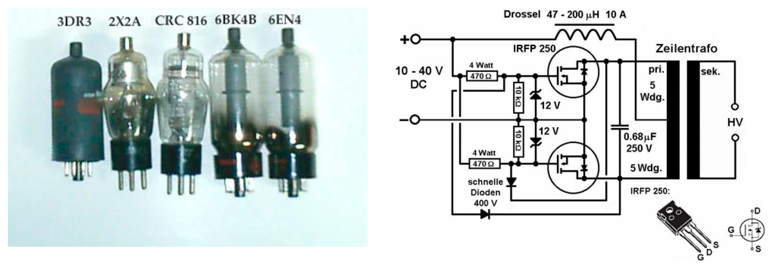
Figure 9.
X-Ray tube and circuit for a typical power supply [63] (Creative Commons CC BY-NC-SA 4.0 license).
Figure 9.
X-Ray tube and circuit for a typical power supply [63] (Creative Commons CC BY-NC-SA 4.0 license).

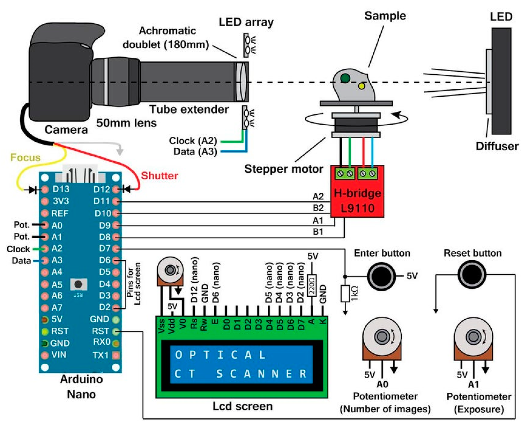
Figure 10.
Arduino® based optical scanner as described in [70] (Creative Commons CC BY 4.0 license).
Figure 10.
Arduino® based optical scanner as described in [70] (Creative Commons CC BY 4.0 license).


Figure 12.
Schematic and final breadboard ECG circuit using an Arduino® [75] (Creative Commons CC BY 3.0 license).
Figure 12.
Schematic and final breadboard ECG circuit using an Arduino® [75] (Creative Commons CC BY 3.0 license).

Figure 13.
OpenBCI platform and headset [82] (Creative Commons CC BY 3.0 license).
Figure 13.
OpenBCI platform and headset [82] (Creative Commons CC BY 3.0 license).

Figure 14.
Overview of the ScouseTom system [92] (Creative Commons CC BY 4.0 license).
Figure 14.
Overview of the ScouseTom system [92] (Creative Commons CC BY 4.0 license).

Figure 15.
Schematic of Arduino®-based electrical impedance tomography (EIT) system [92] (Creative Commons CC BY 4.0 license).
Figure 15.
Schematic of Arduino®-based electrical impedance tomography (EIT) system [92] (Creative Commons CC BY 4.0 license).

Figure 16.
A raspberry pi controlled insulin infusion pump [100,101] (Creative Commons CC BY 4.0 license from www.raspberrypi.org).
Figure 16.
A raspberry pi controlled insulin infusion pump [100,101] (Creative Commons CC BY 4.0 license from www.raspberrypi.org).

Figure 17.
A microfluidics syringe pump described by Lake and co-workers [106] (Creative Commons CC BY 3.0 license).
Figure 17.
A microfluidics syringe pump described by Lake and co-workers [106] (Creative Commons CC BY 3.0 license).

Figure 18.
Pi-flow from Kassis et al. [113] (Creative Commons CC BY 4.0 license).
Figure 18.
Pi-flow from Kassis et al. [113] (Creative Commons CC BY 4.0 license).

Figure 19.
Temperature, pulse + oxygen and body position sensors as described in [116] (Creative Commons CC BY 4.0 license).
Figure 19.
Temperature, pulse + oxygen and body position sensors as described in [116] (Creative Commons CC BY 4.0 license).

© 2019 by the authors. Licensee MDPI, Basel, Switzerland. This article is an open access article distributed under the terms and conditions of the Creative Commons Attribution (CC BY) license (http://creativecommons.org/licenses/by/4.0/).
Share and Cite
MDPI and ACS Style
Pandey, G.; Vora, A. Open Electronics for Medical Devices: State-of-Art and Unique Advantages. Electronics 2019, 8, 1256. https://doi.org/10.3390/electronics8111256
AMA Style
Pandey G, Vora A. Open Electronics for Medical Devices: State-of-Art and Unique Advantages. Electronics. 2019; 8(11):1256. https://doi.org/10.3390/electronics8111256
Chicago/Turabian StylePandey, Gaurav, and Ankit Vora. 2019. "Open Electronics for Medical Devices: State-of-Art and Unique Advantages" Electronics 8, no. 11: 1256. https://doi.org/10.3390/electronics8111256
Note that from the first issue of 2016, this journal uses article numbers instead of page numbers. See further details here.