Review

Dysregulation of miRNAs in DLBCL: Causative Factor for Pathogenesis, Diagnosis and Prognosis

by
Mohammed Alsaadi
1,2,
Muhammad Yasir Khan
1,3,
Mahmood Hassan Dalhat
4,
Salem Bahashwan
2,5,6,
Muhammad Uzair Khan
7,
Abdulgader Albar
1,8,
Hussein Almehdar
1 and
Ishtiaq Qadri
1,*
1
Department of Biological Science, Faculty of Science, King AbdulAziz University, Jeddah 21589, Saudi Arabia
2
Hematology Research Unit, King Fahad Medical Research Center, King AbdulAziz University, Jeddah 21589, Saudi Arabia
3
Vaccine and Immunotherapy Unit, King Fahad Medical Research Center, King AbdulAziz University, Jeddah 21589, Saudi Arabia
4
Department of Biochemistry, Faculty of Science, King AbdulAziz University, Jeddah 21589, Saudi Arabia
5
Department of Hematology, Faculty of Medicine, King AbdulAziz University, Jeddah 21589, Saudi Arabia
6
King AbdulAziz University Hospital, King AbdulAziz University, Jeddah 21589, Saudi Arabia
7
Department of Health Sciences, City University of Science and Information Technology, Peshawar 25000, Pakistan
8
Department of Microbiology, Faculty of Medicine, Jeddah University, Jeddah 23218, Saudi Arabia
*
Author to whom correspondence should be addressed.
Diagnostics 2021, 11(10), 1739; https://doi.org/10.3390/diagnostics11101739
Submission received: 25 July 2021 / Revised: 10 September 2021 / Accepted: 17 September 2021 / Published: 22 September 2021
(This article belongs to the Section Pathology and Molecular Diagnostics)

Abstract

:
MicroRNA is a small non-coding RNA (sncRNA) involved in gene silencing and regulating post-transcriptional gene expression. miRNAs play an essential role in the pathogenesis of numerous diseases, including diabetes, cardiovascular diseases, viral diseases and cancer. Diffuse large B-cell lymphoma (DLBCL) is an aggressive non-Hodgkin’s lymphoma (NHL), arising from different stages of B-cell differentiation whose pathogenesis involves miRNAs. Various viral and non-viral vectors are used as a delivery vehicle for introducing specific miRNA inside the cell. Adenoviruses are linear, double-stranded DNA viruses with 35 kb genome size and are extensively used in gene therapy. Meanwhile, Adeno-associated viruses accommodate up to 4.8 kb foreign genetic material and are favorable for transferring miRNA due to small size of miRNA. The genetic material is integrated into the DNA of the host cell by retroviruses so that only dividing cells are infected and stable expression of miRNA is achieved. Over the years, remarkable progress was made to understand DLBCL biology using advanced genomics and epigenomics technologies enabling oncologists to uncover multiple genetic mutations in DLBCL patients. These genetic mutations are involved in epigenetic modification, ability to escape immunosurveillance, impaired BCL6 and NF-κβ signaling pathways and blocking terminal differentiation. These pathways have since been identified and used as therapeutic targets for the treatment of DLBCL. Recently miRNAs were also identified to act either as oncogenes or tumor suppressors in DLBCL pathology by altering the expression levels of some of the known DLBCL related oncogenes. i.e., miR-155, miR-17-92 and miR-21 act as oncogenes by altering the expression levels of MYC, SHIP and FOXO1, respectively, conversely; miR-34a, mir-144 and miR-181a act as tumor suppressors by altering the expression levels of SIRT1, BCL6 and CARD11, respectively. Hundreds of miRNAs have already been identified as biomarkers in the prognosis and diagnosis of DLBCL because of their significant roles in DLBCL pathogenesis. In conclusion, miRNAs in addition to their role as biomarkers of prognosis and diagnosis could also serve as potential therapeutic targets for treating DLBCL.

1. Introduction

MicroRNA is about 22 nucleotides small non-coding RNA (sncRNA) molecule that functions as a regulator of post-transcriptional gene expression and RNA silencing [1]. In 1998, Andrew Fire and Craig C. Mello discovered the interference RNA(RNAi) in animals [2], and were awarded Nobel prize in physiology or medicine in 2006 [3]. Two main types of sncRNAs, i.e., siRNA and miRNA account for the interference mechanism of RNA [4]. siRNA are small RNA molecules with approximately 25 nucleotides possessing both sense and anti-sense polarity [5]. Primarily it was discovered in plants that were undergoing virus induced gene silencing or co-suppression as compared to control plants. Later on, it was also detected in Drosophila tissue culture by induction of long exogenous dsRNA and, also, from extracts of Drosophila embryo [6]. miRNAs are non-coding RNAs playing a significant role in gene expression and are transcribed from DNA sequences to precursors miRNA and lastly mature miRNAs [7]. miRNAs typically have incomplete base pairing to inhibit many RNAs while siRNA are derived from long double-strand RNA (dsRNA) precursors and base-pair perfectly to cleave mRNA specifically at a single specific site [8].

2. miRNA Biogenesis

The two-step miRNA synthesis is typically a nuclear and cytoplasmic breakdown process involving ribonuclease III, Drosha and Dicer [9]. miRNA synthesis in humans is initiated with RNA polymerase II transcription which binds to promoter region making a hair-pin loop of pre-miRNA. However, RNA polymerase III transcribes some miRNA that initiates primary transcripts [10]. The pri-miRNA contains a hair-pin stem, terminal loop and flanking single stranded sequence from hundred nucleotides to several kilo bases (kb) [11] and at 5 end (5′ UTR), modified nucleotide covers pri-miRNA that makes poly-adenylated (having several adenosine; poly A tail) [11,12]. Nuclear protein DiGeorge critical region 8 (DGCR 8) also known as Pasha in invertebrates and RNA-III enzyme recognizes the hair pin shaped miRNA [13]. Drosha and DGCR8 work with another complex network of proteins known as microprocessors [14]. The pri-mRNA is cleaved by microprocessor to form a shorter hairpin approximately 70 nucleotides in length [15]. The resulting product possesses two nucleotide overhangs at 3′ end (3′UTR) and 5 (5′UTR), this is referred as precursor-miRNA (pre-miRNA) to which downstream are necessary for effective processing [16]. Exportin5 protein (XPO5) is a karyopherin member that recognizes a two-nucleotide overhang at 3′ end of pre-miRNA hairpin deserted by Drosha and this XPO5 protein transports Pre-miRNA from nucleus to cytoplasm. [17]. The transfer of pre-miRNA is an energy dependent pathway using GTP attached to Ran protein [18].
Inside cytoplasm pre-miRNA is cleaved by Dicer (RNAase III enzyme) by interacting with the 5′ and 3′ end, thus removing the loop and resulting in 22 nucleotide miRNA duplex [19]. Either strand of miRNA potentially serve as functional miRNA but just one miRNA strand is merged in RNA-induced silencing complex (RISC) [20].

3. Delivery Platforms for miRNA

One of the important features to performing experiments with miRNA is to select a safe and efficient delivery vehicle. Nowadays, both viral and non-viral vector platforms are used for delivery of miRNA.

3.1. Viral Vectors

Four main types of viral vectors are used in delivering miRNA into cell i.e., Lentiviral vector, adenovirus vector, adeno-associated viral vectors (AAV) and retroviral vectors [21]. Adenoviruses are linear, double-stranded DNA viruses with 35 kb genome size and are extensively used in gene therapy [22]. These vectors are used to express different types of RNAi but the major drawback is Adenovirus associated RNA inhibits RNAi by competing with XPO5 and DICER, thus resulting is suppression of RISC activity [23,24]. Meanwhile, AAVs are more frequently used for miRNA gene therapy as compared to adenoviral vectors. AAVs are single stranded DNA viruses that accommodate up to 4.8 kb foreign genetic material and are favorable for transferring miRNA due to small size of miRNA gene [25]. AAVs are promising gene delivery tools due to their lack of ability to facilitate long-term episomal expression, and minimal oncogenic potential and inflammatory response [26]. Currently many clinical trials are underway utilizing AAV platform for gene therapy. AAVs are commonly used in preclinical studies for cancers, muscular Dystrophies, neurodegenerative disorders and viral infections [26,27].
The tremendous ability to infect post mitotic cells and efficient transduction in neurons make it a promising vector. Pre-existing adaptive and innate immune responses in hosts are main challenges and barriers in AAV gene transfer. To date, 258 studies have been registered in Clinicaltrials.gov using AAV but none have reported encoding miRNA or siRNA and to be administered in humans. Viral delivery systems a remarkably efficient system, however, these may elicit immunogenic responses which halt their effectiveness [26].
Retroviruses are RNA viruses capable of carrying an 8 kb foreign genome and integrate into the host cell genome [28]. The genetic material is integrated into the DNA of the host cell so that only dividing cells are infected and stable expression of miRNA is achieved [29]. Lentiviruses are a subgroup of retroviruses with common features of integration in the host genome but can infect both dividing and non-dividing cells [30]. Due to this ability, they infect terminal differentiated cells to treat neurologic diseases [31].

3.2. Non-Viral Vectors

This delivery approach is less immunogenic, less toxic and has a minimum limitation in terms of size of transferred gene, but it has poor transfection efficiency. The most common is lipid-based approach termed as liposomes, lipid molecules with encapsulated nucleic acids. Liposomes have a shorter life span in vivo due to non-specific binding of proteins in serum [32]. The utilization of polymers i.e., poly lactide-co-glycolide (PLGA), polyethyleneimine (PEI) and cell penetrating peptides (CPP) are reported to be effective in term of less toxicity to membrane and cells [33]. Contrary to usage of polymers, a number of studies have reported usage of nanoparticles [30], the most frequently used nanoparticles are Fe3O4 based nanoparticles and gold nanoparticles (AuNPs) [34].
The oncogenic and tumor suppressing abilities of several miRNA make it promising a candidate for RNA based therapeutics. The only siRNA based FDA-approved drug Patisiran was licensed in 2018 for treating a rare polyneuropathy caused by transthyretin-mediated amyloidosis [35].
No miRNA-based drugs have come to clinic, but many are under clinical trials. MRX34 is liposome encapsulated double stranded RNA drug that mimics (miR-34) that is human tumor suppressor. MRX34 was first miRNA-based drug to enter human clinical trial and showed tumor suppression in 29% of patients [36]. However, FDA stop phase 1b due to severe immune related toxicity in five patients. Further studies proved that lipid encapsulation is not the cause of severe immune toxicity [37,38]. Cobomarsen a locked nucleic acid-based oligonucleotide inhibitor of miRNA-155 was studied to check safety and efficacy in clinical trial phase 1. Result showed reduced lesion burdens and acceptable safety profile in patients with hematological cancer. These results prompt the initiation of clinical trial 2 of cobomarsen versus vorinostat, an FDA-approved drug for CTCL [39]. Thus, non-viral vector platform for delivering miRNAs is accepted extensively due to low immunogenicity and safety profile [39].

4. Diffuse Large B-Cell Lymphoma

Diffuse Large B-cell Lymphoma (DLBCL) is an aggressive cancer of mature B lymphocytes and the most common subtype of non-Hodgkin’s lymphoma, it comprises about 40% of all non-Hodgkin’s lymphoma cases. It has a highly variable clinical course; although most of affected patients initially show a good response to chemotherapy, approximately half of them will achieve a durable remission [40].
DLBCL is genetically heterogeneous disease, which can be divided into ABC-DLBCL and GCB-DLBCL subtypes at different levels of gene expressions, while subgroups are based on the B-cell markers and NF-κB action pathways [41,42].
Heterogeneity in DLBCL is attributed to chromosomal translocation and single nucleotide variation. i.e., mutation in exon 15 of EZH2 results in gain of function of EZH2 implicating suppression of major DLBCL regulator genes [43,44]. The orientation of DLBCLs in specific topographic locality includes CNS and primary cutaneous DLBCL having specific gene expression GEP (genomic profile) different from the nodal DLBCL [44].

5. Etiology of DLBCL

Many chromosomal transitions result in the production of mutated proteins, resulting in the gain or loss of function protein. Mutated proteins downregulate specific pathways which prevent cells from normal cell death/ apoptosis and favor cell growth [45,46]. Some major factors such as immunosuppression and some class of infectious viruses such as human herpesvirus-8, human herpesvirus-6, Epstein-bar virus (EBV) and hepatitis-C virus are associated with human B-cell lymphomas [47,48]. Cancer cell types adopt a pathological mechanism to lower the chances of recognition by NK cells resulting in their prolonged life. A significant role of PDL-1 was found in oncogenic cells which promote immune evasion [49,50]. Genome profiling studies showed high alteration in histone and chromatin enzymes contributing to lymphomagenesis. DLBCL pathogenesis resulted from the alteration of proto-oncogene expression and suppressor genes caused by the accumulation of genetic lesions [51]. Earlier studies only focused on single nucleotide mutation which was later addressed by Bjoern Chap et al. 2018 finding significant driver genes and five new remarkable subsets which could be further used for combinational therapies [52].

6. Epigenetic Modifications in DLBCL

Findings in genome of DLBCL patients have revealed high mutational frequency affecting epigenetic machinery; the most reported epigenetic modifications are histone acetyltransferases (HAT) and histone methyltransferases [53]. In HAT, mutations cause loss of function (LoF) in CREBBP and EP300, detected in about 35% of DLBCL patients with significantly high mutations seen in GCB-subtype [54,55]. The CREBBP/EP300 protein adds acetyl groups to lysine residues on histones and non-histone nuclear proteins, acting as transcriptional coactivators for several transcription factors that bind to DNA [56].
Consistently, CREBBP/EP300 activates transcription through different epigenetic mechanisms, comprising of targeted chromatin acetylation and transcriptional activators such as p53 and BCL6 acetylation-mediated inactivation [56,57,58]. Mutation in the CREBBP could cause C-terminal truncation in the HAT domain or change the amino acid residue(s), which weakens its capacity of acetylating BCL6 and p53 which are its substrates, in turn resulting in the activation of oncoprotein and reduced tumor suppressive role of p53 for the regulation of the DNA damage response reaction which occurs in the germinal center during immunoglobulin genes remodeling [59]. The consequence of BCL6 action suppressing p53 activity could lead to an amplified DNA damage tolerance by deregulation of the apoptotic and cell cycle response arrest [59,60]. The involvement of HATs in chromatin remodeling and transcriptional regulation suggests the consequences of impaired function of CREBBP/EP300 due to alterations. Further studies on the role of CREBBP/EP300 deregulation would help in the mechanistic understanding of all the targets that significantly affect DLBCL pathogenesis [61].
Histone methyltransferases such as MLL2 (mixed lineage leukemia 2) are among the identified candidates for DLBCL pathogenesis whose mutations account for over 30% of DLBCL [52,62]. The MLL2 gene is highly conserved and ubiquitously expresses the MLL2 protein. MLL2 protein regulates gene transcription by adding a methyl group to the 4th position of lysine residue in histone 3 domain (H3K4) [63]. The H3K4 trimethylation is a preserved mark of chromatin that is transcriptionally active and narrowly related to the promoters of active genes and counteracts the transcriptional repression executed by the methylation of H3K9 and H3K27 [63]. Mutation of MLL2 in DLBCL generates a truncated protein that lacks the catalytic SET domain, which is essential for the methyltransferase activity, known to be associated with tumor suppressor activity. It is important to note that MLL3, the paralogue of MLL2 was also found to contain an indel (insertion or deletion) mutation which accounts for approximately 15% of DLBCLs [64,65].
EZH2 encodes for a trimethylated H3K27 histone methyltransferase which causes repressed gene expression [66]. A gain of function (GoF) mutation in EZH2 detected in about 22% of GCB-DLBCL patients is correlated with poor prognosis [67]. The EZH2 GoF implicates in amplified H3K27me3 stage through transformed substrate selectivity due to preserved tyrosine residue (Tyr641) within the domain of EZH2 SET [62]. EZH2 is a promising therapeutic target for DLBCL treatment, as several studies involving EZH2 mutated DLBCL cells have shown that small molecule inhibitors targeting EZH2 could induce apoptosis and cell cycle arrest [68,69]. Taken together, mutational changes in chromatin remodeling genes could have a significant effect on transcriptional regulation and favors the lymphomagenesis through epigenetically remodeling lymphoma cells [70].

7. Immune Escape Pathways in DLBCL

Genetic mutations involved in immune recognition and antigen-presenting functions account for significant mutational frequencies seen in both molecular subtypes of DLBCL. The disruption of beta-2 microglobulin gene is reported in 30% of DLBCL patients which is caused due to the Biallelic deletion and inactivating mutations [71]. The invariant region of the major histocompatibility complex (MHC) class 1 is encoded by B2M gene, found on the surface of nucleated cells. Approximately 75% of DLBCL patients lack expressed B2M gene [71]. B2M is essential for human leukocyte antigen (HLA) development and identification of cytotoxic lymphocytes (CTL) [71]. Involvement of other epigenetic mechanisms was suggested as fractions of DLBCL patients were observed to have reduced expression of B2M but possess elevated HLA-I expression [72]. Truncating mutations and focal deletion were detected in cluster of differentiation 58 (CD58) locus in 20% of DLBCL subjects. [73,74]. CD58 is an immunoglobulin ligand of CD2 expressed on surfaces of T-cells and natural killer cells (NK) that participate in the activation of these cells [74]. The absence of B2M, HLA-I and CD58 in 60% of DLBCL patients signifies the role of lymphomagenesis in protecting cell-mediated lysis of CTL and NK cell [70]. Other mutations disrupting the immune response regulators comprise of alteration in different MHC class II trans-activator genes (CIITA), programmed cell death ligand (PDL2) and PDL1, all of which are preferentially detected in primary mediastinal large B cell lymphoma (PMBCL) [74]. The downregulation of HLA-II reorganizations of CIITA may decrease tumor cells immunogenicity. In contrast, the amplification of PDL1 is shown to be associated with impaired anti-tumor immune responses in different cancer types [75]. These gene mutations are essential in immune response regulated by tumor microenvironment (TME) activities which aid tumor cells to escape immune surveillance mechanisms [76].

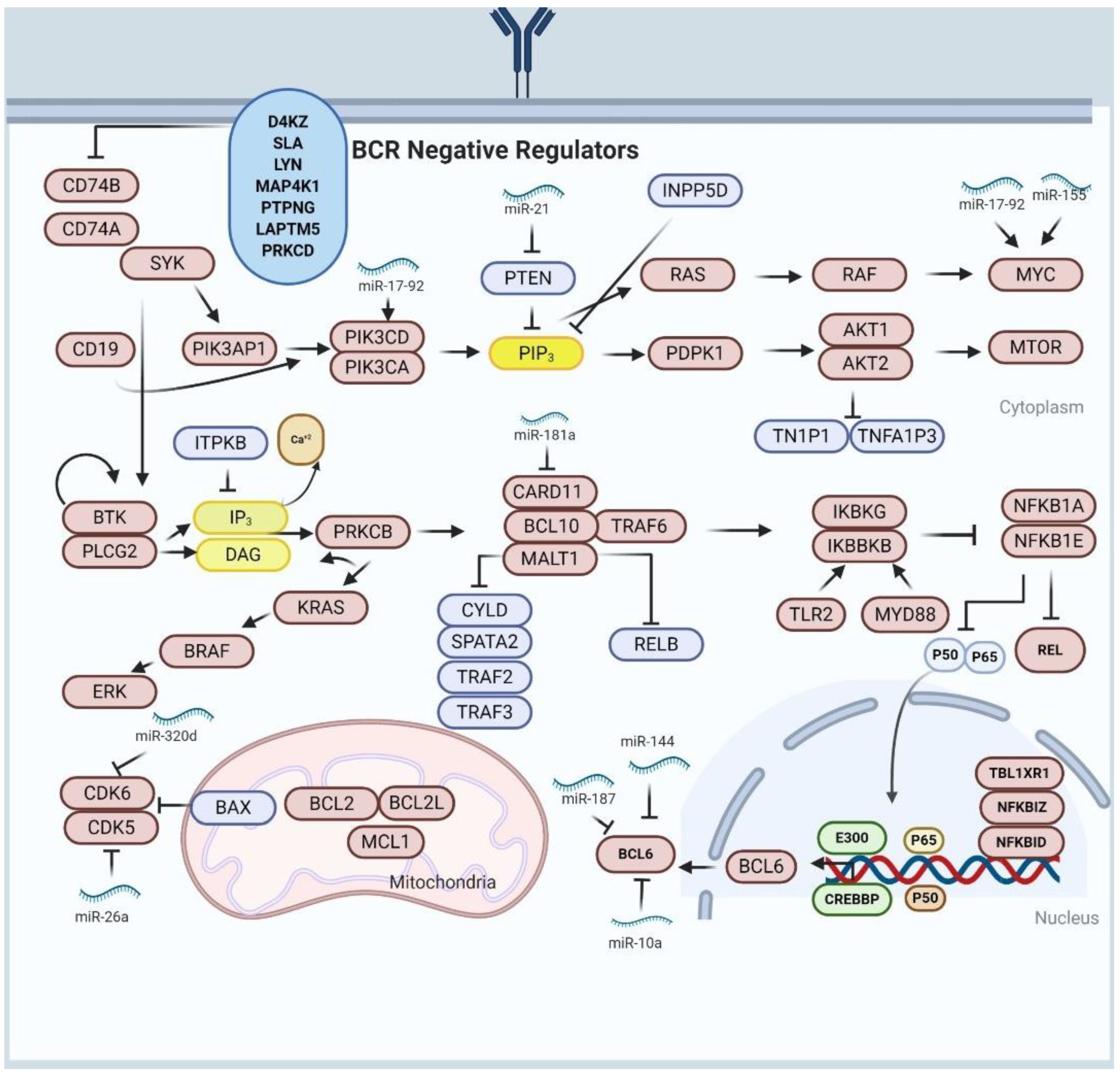
8. Alterations of NF-κβ Activity and BCR Signaling in DLBCL

Gene expression profiling has revealed the role of NF-κB in DLBCL poor prognosis and disease progression, as it was observed that the DLBCL expression profiles are enriched with NF-κB targeted genes resulting in cell survival and cell proliferation of DLBCL cells [77]. Many researchers have shown through molecular profiling that constitutive activation of of NF-κB is associated with interleukin-1 receptor (1L-1 R), Toll-like receptor (TLR) and B-cell receptor (BCR) signaling pathways [78]. In BCR signaling pathway, the existence and development of B-cell lymphomas are associated with mutations in CD79A and CD79B. The CD79A and CD79B mutations account for >20% of overall mutations in ABC-DLBCL [79]. In addition, the loss of function in BCR negative regulators (i.e., DGKZ, SLA MAP4K1, LYN, PTPN, LAPTM5 and PRKCD) result in prolonged BCR signaling [80]. Of note, knockdown of proximal and distal sub-units associated with BCR, of specifically toxic to ABC-DLBCL, contributing to potential to the development of BCR-targeted therapies for ABC-DLBCL subtype [80]. The use of ibrutinib, a BTK inhibitor, was shown to be effective in treating patients with ABC-DLBCL having CD79A/B alterations. Similarly, a small molecule inhibitor of mucosa-associated lymphoid tissue translocation protein 1 (MALT1) showed a selective action against cell lines of ABC-DLBCL in xeno-transplanted tumor and in vitro studies [81].
The alterations in oncogenic myeloid differentiation primary response protein 88 (MYD88) were found in about 30% of patients with ABC-DLBCL [82]. The mutation in MYD88 involves the substitution of leucine with proline at position 265 (L265P), leading to GoF, thereby triggering NF-κβ and JAK/STAT3 transcriptional responses by interacting with lkB kinase (comprising lkBkB and lkBkG) [82].
About 60% of alterations in both negative and positive regulators of NF-κβ were reported in cases of ABC-DLBCL while a lesser percentage were reported in cases of GCB-DLBCL [78]. These mutational changes inactivate certain genes, encoding for tumor-necrosis factor alpha-induced protein 3/A20 (TNFAlP3/A20), which are predominant in ABC-DLBCL [83]. TNFAlP3/A20 is associated with inactivation of NF-κβ, BCR and TLR responses. Moreover, TNFAlP3/A20 inactivates BCR response through its interaction with MALT1 [81]. Therefore, LoF of TNFAlP3/A20 could influence lymphomagenesis by inducing prolonged NF-κβ signaling. Approximately 9% of ABC-DLBCLs patients were shown to possess CARD11 mutations [84]. CARD11 is an essential component of CBM (CARD11/BCL10/MALT1) complex, which alongside TRAF6 is essential for the transduction of signaling from BCR to NF-κβ [84]. While an expected outcome of these genetic mutations initiates responses of NF-κβ signaling pathway similar signals could activate several additional downstream signaling cascades such as PI3K, and ERK/MAP kinase. In summary, BCR and NF-κβ pathways are disrupted by genetic mutations in both ABC-DLBCL and GCB-DLBCL.

9. Impaired BCL6 Activity in DLBCL

BCL6 is a transcription factor controlled by CREBBP/EP300, which binds to conserved sequence in the promoter region of targeted genes, thus controlling gene expression through interaction with specific co-repressor complexes [85]. BCL6 is associated with biological activities of DLBCL cells by suppressing the expression of wide range of genetic factor included in numerous signaling pathways including CD40 in BCR signaling, T-cell mediated B-cells inactivation, the ability to detect as well as respond to DNA damage through repressing of the TP53 protein in TP53/ATR mediated DNA damage response, the induction of apoptosis by suppressing BCL2 in BCL2/BCL2L1 mediated apoptosis and plasma cells differentiation through PRDM1/BLIMP1 suppression [85]. BCL6 associated activities sustain the proliferation of GCB cells while undergoing tolerance for DNA damage and breaks, connected with class switch recombination and somatic hyper-mutation (SHM) without triggering the DNA damage responses. Furthermore, BCL6 causes premature B-cell inactivation and promotes its exit from the germinal centers (GC) before the clone survival selection thereby producing antibodies attracted to antigens [57]. BCL6 chromosomal mutation is observed in 35% of both ABC and GCB DLBCL, with higher mutational frequencies in the ABC-DLBCL subtype [53]. Recombination events in DLBCL cells affect the downstream of integral BCL6 coding domain to heterological sequences, formed by more than 20 different chromosome partners [86]. Moreover, downregulation in BCL-6 expression is correlated with the differentiation of post-GC B-cells, achieved by chromosomal translocation of BCL6 [87]. Less frequently, BCL6 associated mutations prevent IRF4-mediated BCL6 repression, related to CD40/CD40L interaction in light zone GC [85]. BCL6 activity is also impaired by another set of genetic mutations affecting the post-transcriptional regulatory mechanism of CREBBP/EP300, which impairs the acetylation-mediated BCL6 inactivation and FBXO11 mutations that occurs in 5% of DLBCL cases [88]. The FBXO11 mutation is associated with BCL6 proteosomal degradation via SRP1/CUL1/SCF complex. MEF2B is a transcription factor expressed in GC, and also acts as BCL6 transcription regulator. The GoF mutation of MEF2B is seen in approximately 10–18% DLBCL patients; mostly predominant in GCB-subtype [89]. In lymphomagenesis initiation, BCL6 play a critical role confirmed using in vivo mouse model and the results demonstrated that deregulation of BCL6 could promote the increase of human-like DLBCL [69,90]. Connecting all evidence of the critical role of BCL6 in lymphomagenesis, the BCL6 can be used as a promising target for therapeutic intervention [90]. Of note, studies have shown that BCL6 inhibitors have a potential anti-lymphoma capacity and have a strong synergistic effect in DLBCL combinational treatments [90].

10. Blocking the Terminal Differentiation Pathway in DLBCL

The alteration in the terminal differentiation pathway is one of the predominant events that occur in DLBCL. Downregulation of BCL6 is essential to reduce the expression of the terminal differentiation gene targets, including plasma cell master regulator PRDM1/BLIMP1 [91]. The differentiation regulatory axis is altered in majority of ABC-DLBCL cases because of mutually exclusive mutations leading to LoF in BLIMP1. The LoF in BLIMP1 could directly cause deletions and disruptive mutations usually observed in 25% of DLBCL patients or indirectly by BCL6 deregulation through suppression seen in 25% of DLBCL patients [92]. Alternatively, the LoF of PRDM1 and its interaction to BCL6 chromosomal translocation may represent another oncogenic mechanism affecting similar pathway to influence lymphomagenesis by hindering lethal variations. Consistent with the BLIMP1/PRDMl/BCL6 mechanisms, deletion in BLIMP1 causes proliferative lymphoma disease similar to that of human ABC-DLBCL [92].

11. Role of miRNA in DLBCL Pathogenesis

Many aberrantly expressed miRNAs are identified as critical pathogenic factors in DLBCL. It is important to note that many miRNAs are involved in regulating the development of other hematological cancers. These miRNAs have an essential role in the development of normal B-cells in healthy humans. In DLBCL, miR-17-92 is over-expressed, suggesting its role as potential oncogene as shown in vivo using DLBCL induced mice [93].
Remarkably, it was observed that mice overexpressing miR-17-92 developed lymphoproliferation and increased MYC expression. However, lymphocytes from miR-17-92 knockdown mice exhibited increased expression of pro-apoptotic genes, such as PTEN and BIM [94]. Functional genomic studies have shown the transactivation of MYC through miR-17-92 cluster, both of which result in DLBCL progression by silencing tumor suppressor genes [95].
The regulatory axis of miR-17-92 comprises of MYC and its role in the MYC/miR-17-92/E2F circuit resulting in upregulation of miR-17-92 cluster that targets the cell proliferation regulator; E2F1 (Figure 1). Consistently, the supporting proliferation regulator gene E2F3 assists E2F1 in promoting the expression of miR-17-92 cluster. Recently, PTPROT and PP2A were reported to target miR-17-92 clusters and, also, activate the BCR-signaling in DLBCL cells [96]. Furthermore, miR-217 was also reported to stabilize the BCL6 expression as a regulator of germinal center formation. Conversely, another study reported that miR-144 act as tumor suppressor by suppressing the BCL6 function in DLBCL xenografted mice [97]. Both miR-10a and miR-187 act as tumor suppressors by regulating BCL6 expression through induction of cell apoptosis as shown in cell culture and tumor microarray studies suggesting these miRNAs could be exploited as a novel therapeutic target for DLBCL treatment [98,99].
Another miRNA; miRNA-222-3p, was shown to have an oncogenic role in DLBCL by promoting cell proliferation, invasion and inhibiting apoptosis by silencing phosphatase2 regulatory subunit B alpha (PPP2R2A) in DLBCL. Research evidence has shown that miRNA-222-3p promotes downregulation of PPP2R2A and tumor growth in xenografted mice [100].
In cancer, particularly in B-cell lymphoproliferative disease, miR-155 is reported to act as oncogene (oncomiR) miRNA. However, in normal conditions miR-155 plays significant role in the activation and maturation of B-cell lymphocyte. Several studies have attributed miR-155 overexpression to several lymphomas, including DLBCL. miR-155 mediated targeting of SH2 domain which contains C/EBPb and inositol-5-phosphatase-1 (SHIP-1) may increase the accumulation of malignant pre-B cells in mice models due to interaction with miR-155 [101]. In xeno-transplant DLBCL cell model, TNF-α cell growth targeting SHIP1 is regulated by miR-155 overexpression. The miR-155 mediated SHIP2 regulation is demonstrated by the suppressive effect of miR-155 on growth-inhibitory factors such as TGF-β1 and BMP2/4 in DLBCL cell through SMAD5 suppression by targeting PIK3R1 and regulating PI3K-AKT pathways [96]. Moreover, miR-155 also target TGFβR2 suggesting a novel oncogenic pathway contributing to the development of tumors. miR-155 is shown to target HGAL, a GC marker and lymphocyte mobility inhibitors and NF-κβ promote the expression of miR-155 [102]
Another study has reported miR-181a as a negative regulator of NF-κβ signaling [103]. Overexpression of miR-181a significantly decreased the expression and activity of crucial NF-κβ signaling components in DLBCL, leading to reduced tumor cell proliferation and survival by targeting CARD11, NFKB1A, NFKB1, RELA and REL [103]. The expression of miR-181a was shown to be negatively correlated with CARD11; a master regulator of DLBCL [103].
Overexpression of miR-34a is associated with poor prognosis and positive feedback loop in DLBCL. The tumor suppressor, p53; induces the expression of miR-34a which silences a NAD deacetylase called SIRT1 whose effect enhances and stabilizes p53 mediated activators. In DLBCL, high-grade transformation is promoted by low expression of miR-34a through dysregulation of FOXP1 expression which is inhibited by MYC [104]. Downregulation of miR-34a and miR-21 up-regulation was reported to be linked with relapse-free survival. miR-21 acts as an oncogene by directly targeting the tumor suppressors PTEN, PDCD4 and FOXO1, which leads to the activation of PI3K/AKT/mTOR oncogenic pathway. Study involving miR-21 inhibition illustrated the significant decrease in the DLBCL cell lines and sensitivity to R-CHOP treatments and reduction in invasion and proliferation of tumor cell [104].
Recently, studies reported the role of miRNAs as tumor suppressors by inhibiting PD-L1 and long non-coding RNAs (lncRNAs) such as MALAT1 and NEAT1. A study using patients’ tissues and cell culture has shown that downregulation of miR-214 is correlated with PD-L1 upregulation in DLBCL compared with normal control showing that both genes have antagonistic effect [105]. Therefore, overexpression of miR-214 is associated with reduction in cell invasion, cell viability and increased apoptosis [105]. Similarly, PD-L1 and MALAT1 were reported to have antagonistic effect with miR-195, showing the role of miR-195 as tumor suppressor by decreasing expression level of both PD-L1 and MALAT1. This interaction showed that overexpressed miR-195 correlates with cell proliferation, cell migration, immune escape and increased apoptosis in DLBCL [106]. Another miRNA; miR-34b-5p, was shown to silence NEAT1 and GLI1 in the tissues and cell lines of DLBCL by attaching to 3′ UTR mRNA transcripts NEAT1 and GLI1 thereby preventing their expression [107].
In addition, miR-26a was reported to regulate CDK5 activity and p53 expression demonstrating its importance in cell survival, cell proliferation and cell cycle progression. The role of miR-26a in regulating both p53 and CDK5 makes it a suitable therapeutic target for DLBCL treatment [108]. Similarly, upregulation of CDK6 due to downregulation of miRNA-320d was reported in DLBCL [109]. Overexpression of CDK6 is related to poor prognosis in DLBCL patients. miR-320d act as tumor suppressor by silencing the CDK6 mRNA transcripts at 3′ UTR resulting in inhibition of cell proliferation and cell cycle arrest. miRNA-320d is a promising therapeutic target due to the tumor suppressive function [109].
Overexpression of miR-23a was reported to suppress metastasis suppressor 1 (MTSS1) in DLBCL. miR-26a is negatively correlated with MTSS1 expression. MTSS1 functions as a suppressor of cell proliferation, tumor metastasis and, also, play a key role in actin cytoskeleton recombination [110]. It was suggested that miR-23a promotes the proliferation, invasion and metastasis in DLBCL by silencing MTSS1 [110]. Similarly, miR-645 targets the DACH1 by binding to 3′-UTR of DACH1 mRNA transcript leading to reduced DACH1 expression in DLBCL cells. Reduced expression of DACH1 is associated with cell proliferation and survival. DACH1 is expressed in different cancer types, and it is correlated with poor prognosis and tumor progression of cancer patients. Specifically, in DLBCL, decreased DACH1 is inversely correlated with increased miR-645 level. Overexpressed miR-645 is associated with cell proliferation and apoptosis [111].
miR-101 acts as tumor suppressor by targeting the 3′ UTR of KDM1A, thereby reducing tumor growth and progression. In addition, the KDM1A mediated suppression by miR-101 suggests its impact in regulating apoptosis and cell proliferation through MAPK/Erk signaling pathway [112].
Aberrant expression of miR-4638-5p was reported to be associated with high ERG expression. Compared with ERG-negative; the ERG-positive DLBCL is more likely to harbor mutations in genes that are essential in cell cycle control, BCR mediated signaling and β-catenin degradation [113]. Functional and clinicopathological studies associated with ERG-related miRNAs (i.e., miR-4638-5p) and pathways could provide new information into the pathogenesis of DLBCL and reveal novel targets for the management of patients with DLBCL [113]. Similarly, miR-520c-3p acts as tumor suppressor by targeting eIF4GII in xenografted mice [114]. Overexpressed eIF4GII in DLBCL is associated with abnormal protein synthesis, resulting in increased cell proliferation. Thus, the results of in vivo and in vitro studies confirm miR-520c-3p level is correlated with the expression of eIF4GII in DLBCL [114].

12. The Role of miRNAs in Diagnosis and Prognosis in DLBCL

MiRNAs are shown to regulate cancer progression in DLBCL by acting either as oncogenes or tumor suppressors. MiRNAs influence the two major DLBCL molecular subtypes, GCB-DLBCL and ABC-DLBCL. GCB-DLBCL and ABC-DLBCL subtypes are distinguished based on miRNA profiling [115,116,117,118]. Several studies reported the role of miRNAs in DLBCL diagnosis, thereby distinguishing it from other types of lymphomas such as follicular lymphoma (FL) and Burkitt lymphoma (BL) [119,120].
Relapse in central nervous system (CNS) associated with DLBCL, occur in 5% patients with low survival rate [121]. DLBCL patients with CNS relapse tend to have high expression of miR-30d and miR-20a. Therefore, miR-30d and miR-20a expression profiles could be used to stratify DLBCL patients [121]. Similarly, miR-155 is also being linked to prognosis, metastasis and treatment failure in DLBCL patients. A study using mouse model showed overexpression of miR-155 is associated with a high-grade lymphoma development [122].
Conversely, a complete recovery was observed in mice when miR-155 stimulus was removed in an inducible expression system [123]. Moreover, the low expression of both miR-34a and miR-27b are associated with poor prognosis in DLBCL patients [123]. Similarly, shorter relapse-free survival in DLBCL is linked with low miR-21 expression in serum and tissue samples. As a result, miR-21 level is proposed to function as an independent prognostic biomarker for DLBCL [124,125]. In DLBCL patients, the presence of miR-21 is suggested to contribute to increased cellular viability and escape apoptotic in tumor cells by targeting BCL2 and PTEN in apoptotic and cell proliferation pathways. MiR-21 inhibition is reported to increase DLBCL cell lines sensitivity to the R-CHOP regimen (hydroxydaunorubicin, rituximab, oncovin, prednisone, cyclophosphamide regimen), which causes decreased tumor cell proliferation and invasion [126]. Transcriptome-wide association analysis revealed the association between aberrant miRNA expression profile with prognostic result in DLBCL patients treated with R-CHOP regimen [127]. The aberrantly expressed miRNAs included miR-330, miR-199b, miR-27a, miR-519, miR-222, miR-425 and miR-142 associated with overall survival [124], whereas miR-130a and miR-125b were linked with R-CHOP resistance [128]. Overexpression of miR-497 and miR-199a were reported to be associated with increased sensitivity to doxorubicin, rituximab, and vincristine drugs present in the R-CHOP regimen. Similarly, increased sensitivity to doxorubicin and rituximab was attributed to overexpression of miR-409-3p, miR-370-3p and miR-381-3p [129].
In the serum samples of DLBCL patients, the levels of miR-155, miR-210 and miR-21were significantly expressed as compared to healthy controls [130]. Another study using plasma also recorded high expression of miR-124 and miR-532-5p and low expression of miR-424, miR-345, miR-145, miR-122, miR-425, miR-128, miR-197 and miR-141 [131].
In serum samples of DLBCL patients, high expression of miR-155, miR-16, miR-29c, miR-15 and low expression of miR-34a was reported [132]. In DLBCL, a high expression level of serum miR-22 was associated with poor prognostic results [132]. Recently using NGS technology, 51 differentially expressed miRNAs were identified in DLBCL patient samples [133]. Three miRNAs were confirmed by quantitative reverse transcriptase-polymerase chain reaction (qRT-PCR), showing that miR-431-5p and miR-323-3p were downregulated and miR-34a-5p was upregulated (Figure 2) [133].

13. Treatment

Based on the newest classification by World Health Organization (WHO), more than 100 different types of lymphoma, most of which are B-cell lymphomas and different diagnosis, clinical characteristics and treatment choices has been reported [134]. Due to heterogeneity of DLBCL, the prophecy and choice of treatment strategies are difficult [135]. Globally, immunochemotherapy with doxorubicin, cyclophosphamide, prednisone, (R-CHOP) rituximab and vincristine is considered as the current first line treatment [136]. The introduction of rituximab, a chimeric monoclonal antibody immunoglobulin surface CD20, the R-CHOP treatment has become the current standard of treatment of choice for DLBCL irrespective of the disease subtype [126]. However, after primary response, up to 40% of patients experience relapses or early treatment failure with this treatment approach [137].
A study by Lawrie et al. using microarray expression reported different miRNAs that are linked with prognosis of DLBCL patients treated by R CHOP, including miR-519, miR-222, miR-27a, miR-425, miR-199b, miR-330, miR-302 and miR-142 [94].
A similar study reported CHOP treatment of miRNAs with five different levels provided significant results with miRNAs: miR-33a, miR-455-3p, miR 520d-3p, miR-1236 and miR-224 [126]. However, in DLBCL cases, chemoresistance association resulted from miR-130a and miR-125b was found [122]. MiR-34a may be used as a new therapeutic target of DLBCL [133]. Cytotoxic effect of CHOP regimen significantly increased when the cells of CRL2631 were transfected by miR-21 with anti-sense oligonucleotides. By using quantitative reverse transcriptase polymerase chain reaction (qRT-PCR), miR-21 significantly increased DLBCL cell line sensitivity to CHOP treatment and cause tumor cell proliferation reduction [138]. miR-21 inhibition leads to reduced tumor cell proliferation and invasion in DLBCL [125]. The over expression of miR-187 induces cell apoptosis in vitro highlighting its role as therapeutic target of DLBCL [98].
Although there is currently no FDA-approved miRNA-based drug as medical intervention for DLBCL, efforts were made to target some of the important miRNAs known to mediate DLBCL pathogenesis, as shown in clinical trials. For MRX34; a miR-34a mimic and cobomarsen (MRG-106), anti miR-155 drugs were shown to possess anti-tumor activities in clinical trials involving cancer patients including DLBCL patients [36,139].
MRX34, a mimetic of a miR-34a was used in clinical trials for the treatment of cancer patients. Although it showed promising outcomes as an anti-tumor miRNA-based drug, but it was stopped by the FDA due to its immunotoxic effect on cancer patients [36].
Cobomarsen targets and negatively regulates miR-155 expression in DLBCL, which in turn prevents resistance to growth inhibitory factors; TGFβ1 and TGFβ2 and inhibit MYC stability thereby having overall effect on DLBCL cell proliferation. Clinical trial (NCT02580552) studies have reported the safety and efficacy of cobomarsen in DLBCL patients [39].

14. Conclusions

Deep insight into different miRNAs and its expression could be a powerful tool in diagnosing, differentiating between different subtypes of non-Hodgkins lymphoma, and treating DLBCL. Currently, most published data regarding miRNAs and treating DLBCL are carried out in vitro studies. More studies should be conducted on transgenic organisms carrying human genes for better implication and understanding therapeutic roles of miRNAs in DLBCL. Conventional drugs with combination of different miRNAs could be a potential drug of choice for treating DLBCL in future after passing successful clinical trials.

Author Contributions

M.A. and M.Y.K. designed the review format. M.A., M.Y.K. and M.H.D. wrote the first draft of manuscript. S.B., M.U.K. and A.A. edited and reviewed the flow of information in review. H.A. and I.Q. supervised and corrected grammatical errors in manuscript. All authors have read and agreed to the published version of the manuscript.

Funding

This review article received no external funding.

Institutional Review Board Statement

Not applicable.

Informed Consent Statement

No consent of statement to declare.

Data Availability Statement

Not applicable.

Conflicts of Interest

All authors declare no conflict of interest.

References

  1. Vishnoi, A.; Rani, S. MiRNA Biogenesis and Regulation of Diseases: An Overview. In MicroRNA Profiling; Springer: Berlin/Heidelberg, Germany, 2017; pp. 1–10. [Google Scholar]
  2. Fire, A.; Xu, S.; Montgomery, M.K.; Kostas, S.A.; Driver, S.E.; Mello, C.C. Potent and specific genetic interference by double-stranded RNA in Caenorhabditis elegans. Nature 1998, 391, 806–811. [Google Scholar] [CrossRef]
  3. Joga, M.R.; Zotti, M.J.; Smagghe, G.; Christiaens, O. RNAi efficiency, systemic properties, and novel delivery methods for pest insect control: What we know so far. Front. Physiol. 2016, 7, 553. [Google Scholar] [CrossRef] [Green Version]
  4. Michlewski, G.; Cáceres, J.F. Post-transcriptional control of miRNA biogenesis. Rna 2019, 25, 1–6. [Google Scholar] [CrossRef] [Green Version]
  5. O’Brien, J.; Hayder, H.; Zayed, Y.; Peng, C. Overview of microRNA biogenesis, mechanisms of actions, and circulation. Front. Endocrinol. 2018, 9, 402. [Google Scholar] [CrossRef] [Green Version]
  6. Hammond, S.M.; Bernstein, E.; Beach, D.; Hannon, G.J. An RNA-directed nuclease mediates post-transcriptional gene silencing in Drosophila cells. Nature 2000, 404, 293–296. [Google Scholar] [CrossRef] [PubMed]
  7. Treiber, T.; Treiber, N.; Meister, G. Regulation of microRNA biogenesis and its crosstalk with other cellular pathways. Nat. Rev. Mol. Cell Biol. 2019, 20, 5–20. [Google Scholar] [CrossRef] [PubMed]
  8. Li, L.; Song, Y.; Shi, X.; Liu, J.; Xiong, S.; Chen, W.; Fu, Q.; Huang, Z.; Gu, N.; Zhang, R. The landscape of miRNA editing in animals and its impact on miRNA biogenesis and targeting. Genome Res. 2018, 28, 132–143. [Google Scholar] [CrossRef] [PubMed] [Green Version]
  9. Daugaard, I.; Hansen, T.B. Biogenesis and function of ago-associated RNAs. Trends Genet. 2017, 33, 208–219. [Google Scholar] [CrossRef] [PubMed]
  10. Ha, M.; Kim, V.N. Regulation of microRNA biogenesis. Nat. Rev. Mol. Cell Biol. 2014, 15, 509–524. [Google Scholar] [CrossRef]
  11. Greene, J.; Baird, A.M.; Brady, L.; Lim, M.; Gray, S.G.; McDermott, R.; Finn, S.P. Circular RNAs: Biogenesis, function and role in human diseases. Front. Mol. Biosci. 2017, 4, 38. [Google Scholar] [CrossRef]
  12. Vitsios, D.M.; Davis, M.P.; van Dongen, S.; Enright, A.J. Large-scale analysis of microRNA expression, epi-transcriptomic features and biogenesis. Nucleic Acids Res. 2017, 45, 1079–1090. [Google Scholar] [CrossRef] [Green Version]
  13. Asadi, M.; Shanehbandi, D.; Zafari, V.; Khaze, V.; Somi, M.H.; Hashemzadeh, S. Transcript level of MicroRNA processing elements in gastric cancer. J. Gastrointest. Cancer 2019, 50, 855–859. [Google Scholar] [CrossRef] [PubMed]
  14. Lee, Y.; Ahn, C.; Han, J.; Choi, H.; Kim, J.; Yim, J.; Lee, J.; Provost, P.; Rådmark, O.; Kim, S.; et al. The nuclear RNase III Drosha initiates microRNA processing. Nature 2003, 425, 415–419. [Google Scholar] [CrossRef]
  15. Xu, H.; Wang, X.; Wang, Z.; Li, J.; Xu, Z.; Miao, M.; Chen, G.; Lei, X.; Wu, J.; Shi, H.; et al. MicroRNA expression profile analysis in sperm reveals hsa-mir-191 as an auspicious omen of in vitro fertilization. BMC Genom. 2020, 21, 165. [Google Scholar] [CrossRef] [PubMed] [Green Version]
  16. Auyeung, V.C.; Ulitsky, I.; McGeary, S.E.; Bartel, D.P. Beyond secondary structure: Primary-sequence determinants license pri-miRNA hairpins for processing. Cell 2013, 152, 844–858. [Google Scholar] [CrossRef] [PubMed] [Green Version]
  17. Silva, D.C.; Carneiro, F.D.; Almeida, K.C.; Fernandes-Santos, C. Role of miRNAs on the pathophysiology of cardiovascular diseases. Arq. Bras. Cardiol. 2018, 111, 738–746. [Google Scholar] [CrossRef] [PubMed]
  18. Selvarajan, S.; Vijayaraghavan, J.; Bobby, Z.; Ramalingam, J.; Porur, C. Micro RNAs—A review. J. Evol. Med. Dent. Sci. 2019, 8, 2918–2923. [Google Scholar] [CrossRef]
  19. Lund, E.; Dahlberg, J. (Eds.) Substrate Selectivity of Exportin 5 and Dicer in the Biogenesis of microRNAs. Cold Spring Harbor Symposia on Quantitative Biology; Cold Spring Harbor Laboratory Press: Long Island, NY, USA, 2006. [Google Scholar]
  20. Shomron, N.; Levy, C. MicroRNA-biogenesis and Pre-mRNA splicing crosstalk. J. Biomed. Biotechnol. 2009, 2009, 594678. [Google Scholar] [CrossRef] [PubMed]
  21. Nayerossadat, N.; Maedeh, T.; Ali, P.A. Viral and non-viral delivery systems for gene delivery. Adv. Biomed. Res. 2012, 1, 27. [Google Scholar] [CrossRef]
  22. Kamimura, K.; Suda, T.; Zhang, G.; Liu, D. Advances in gene delivery systems. Pharm. Med. 2011, 25, 293–306. [Google Scholar] [CrossRef]
  23. Andersson, M.G.; Haasnoot, P.J.; Xu, N.; Berenjian, S.; Berkhout, B.; Akusjaärvi, G. Suppression of RNA interference by adenovirus virus-associated RNA. J. Virol. 2005, 79, 9556–9565. [Google Scholar] [CrossRef] [Green Version]
  24. Lukac, D.M.; Garibyan, L.; Kirshner, J.R.; Palmeri, D.; Ganem, D. DNA binding by Kaposi’s sarcoma-associated herpesvirus lytic switch protein is necessary for transcriptional activation of two viral delayed early promoters. J. Virol. 2001, 75, 6786–6799. [Google Scholar] [CrossRef] [Green Version]
  25. Schultz, B.R.; Chamberlain, J.S. Recombinant adeno-associated virus transduction and integration. Mol. Ther. 2008, 16, 1189–1199. [Google Scholar] [CrossRef]
  26. Dhungel, B.P.; Bailey, C.G.; Rasko, J.E. Journey to the center of the cell: Tracing the path of AAV transduction. Trends Mol. Med. 2021, 27, 172–184. [Google Scholar] [CrossRef]
  27. Yang, Y.S.; Xie, J.; Chaugule, S.; Wang, D.; Kim, J.M.; Kim, J.; Tai, P.W.; Seo, S.K.; Gravallese, E.; Gao, G.; et al. Bone-targeting AAV-mediated gene silencing in osteoclasts for osteoporosis therapy. Mol. Ther. Methods Clin. Dev. 2020, 17, 922–935. [Google Scholar] [CrossRef]
  28. Liu, Y.P.; Berkhout, B. miRNA cassettes in viral vectors: Problems and solutions. Biochim. Biophys. Acta Gene Regul. Mech. 2011, 1809, 732–745. [Google Scholar] [CrossRef] [PubMed]
  29. Ye, D.; Wang, G.; Liu, Y.; Huang, W.; Wu, M.; Zhu, S.; Jia, W.; Deng, A.M.; Liu, H.; Kang, J. MiR-138 promotes induced pluripotent stem cell generation through the regulation of the p53 signaling. Stem Cells 2012, 30, 1645–1654. [Google Scholar] [CrossRef] [PubMed]
  30. Escors, D.; Breckpot, K. Lentiviral vectors in gene therapy: Their current status and future potential. Arch. Immunol. Ther. Exp. 2010, 58, 107–119. [Google Scholar] [CrossRef] [Green Version]
  31. Lee, S.K.; Teng, Y.; Wong, H.K.; Ng, T.K.; Huang, L.; Lei, P.; Choy, K.W.; Liu, Y.; Zhang, M.; Lam, D.S.; et al. MicroRNA-145 regulates human corneal epithelial differentiation. PLoS ONE 2011, 6, e21249. [Google Scholar] [CrossRef] [PubMed] [Green Version]
  32. Craig, V.; Tzankov, A.; Flori, M.; Schmid, C.; Bader, A.; Müller, A. Systemic microRNA-34a delivery induces apoptosis and abrogates growth of diffuse large B-cell lymphoma in vivo. Leukemia 2012, 26, 2421–2424. [Google Scholar] [CrossRef] [PubMed] [Green Version]
  33. Ibrahim, A.F.; Weirauch, U.; Thomas, M.; Grünweller, A.; Hartmann, R.K.; Aigner, A. MicroRNA replacement therapy for miR-145 and miR-33a is efficacious in a model of colon carcinoma. Cancer Res. 2011, 71, 5214–5224. [Google Scholar] [CrossRef] [Green Version]
  34. Lee, S.W.L.; Paoletti, C.; Campisi, M.; Osaki, T.; Adriani, G.; Kamm, R.D.; Mattu, C.; Chiono, V. MicroRNA delivery through nanoparticles. J. Control. Release 2019, 313, 80–95. [Google Scholar] [CrossRef] [PubMed]
  35. Kristen, A.V.; Ajroud-Driss, S.; Conceição, I.; Gorevic, P.; Kyriakides, T.; Obici, L. Patisiran, an RNAi therapeutic for the treatment of hereditary transthyretin-mediated amyloidosis. Neurodegener. Dis. Manag. 2019, 9, 5–23. [Google Scholar] [CrossRef] [Green Version]
  36. Hong, D.S.; Kang, Y.K.; Borad, M.; Sachdev, J.; Ejadi, S.; Lim, H.Y.; Brenner, A.J.; Park, K.; Lee, J.L.; Kim, T.Y.; et al. Phase 1 study of MRX34, a liposomal miR-34a mimic, in patients with advanced solid tumours. Br. J. Cancer 2020, 122, 1630–1637. [Google Scholar] [CrossRef]
  37. Levin, A.A. Treating disease at the RNA level with oligonucleotides. N. Engl. J. Med. 2019, 380, 57–70. [Google Scholar] [CrossRef]
  38. Voutila, J.; Reebye, V.; Roberts, T.C.; Protopapa, P.; Andrikakou, P.; Blakey, D.C.; Habib, R.; Huber, H.; Saetrom, P.; Rossi, J.J.; et al. Development and mechanism of small activating RNA targeting CEBPA, a novel therapeutic in clinical trials for liver cancer. Mol. Ther. 2017, 25, 2705–2714. [Google Scholar] [CrossRef] [Green Version]
  39. Witten, L.; Slack, F.J. miR-155 as a novel clinical target for hematological malignancies. Carcinogenesis 2020, 41, 2–7. [Google Scholar] [CrossRef] [PubMed]
  40. Aster, J. Disease of white blood cells, lymph nodes, spleen, thymus. In Robins and Cotran Pathologic Basis of Disease, 7th ed.; Kumar, V., Abbas, A.K., Fausto, N., Eds.; Elsevier Saunders: Philadelphia, PA, USA, 2005; pp. 702–705. [Google Scholar]
  41. Baris, D.; Zahm, S.H. Epidemiology of lymphomas. Curr. Opin. Oncol. 2000, 12, 383–394. [Google Scholar] [CrossRef]
  42. Miao, Y.; Medeiros, L.J.; Li, J.; Young, K.H. Diffuse large B-cell lymphoma with molecular variations more than ABC and GCB classification. Gene Expr. 2018, 1400, 8. [Google Scholar] [CrossRef]
  43. Miao, Y.; Medeiros, L.J.; Li, Y.; Li, J.; Young, K.H. Genetic alterations and their clinical implications in DLBCL. Nat. Rev. Clin. Oncol. 2019, 16, 634–652. [Google Scholar] [CrossRef]
  44. Kokich, V.G. Surgical and orthodontic management of impacted maxillary canines. Am. J. Orthod. Dentofac. Orthop. 2004, 126, 278–283. [Google Scholar] [CrossRef]
  45. Pozzato, G.; Mazzaro, C.; Crovatto, M.; Modolo, M.L.; Ceselli, S.; Mazzi, G.; Sulfaro, S.; Franzin, F.; Tulissi, P.; Moretti, M. Low-grade malignant lymphoma, hepatitis C virus infection, and mixed cryoglobulinemia. Blood 1994, 84, 3047–3053. [Google Scholar] [CrossRef] [PubMed] [Green Version]
  46. Mason, D.Y.; Harris, N.L. Human Lymphoma: Clinical Implications of the REAL Classification; Springer: Berlin/Heidelberg, Germany, 2013. [Google Scholar]
  47. Paya, C.V.; Fung, J.J.; Nalesnik, M.A.; Kieff, E.; Green, M.; Gores, G.; Habermann, T.M.; Wiesner, R.H.; Swinnen, L.J.; Woodle, E.S.; et al. Epstein-Barr virus-induced posttransplant lymphoproliferative disorders. Transplantation 1999, 68, 1517–1525. [Google Scholar] [CrossRef] [PubMed]
  48. Oksenhendler, E.; Boulanger, E.; Galicier, L.; Du, M.-Q.; Dupin, N.; Diss, T.C.; Hamoudi, R.; Daniel, M.T.; Agbalika, F.; Boshoff, C.; et al. High incidence of Kaposi sarcoma–associated herpesvirus–related non-Hodgkin lymphoma in patients with HIV infection and multicentric Castleman disease. Blood 2002, 99, 2331–2336. [Google Scholar] [CrossRef]
  49. Campo, E.; Swerdlow, S.H.; Harris, N.L.; Pileri, S.; Stein, H.; Jaffe, E.S. The 2008 WHO classification of lymphoid neoplasms and beyond: Evolving concepts and practical applications. Blood J. Am. Soc. Hematol. 2011, 117, 5019–5032. [Google Scholar] [CrossRef] [Green Version]
  50. Chapuy, B.; Stewart, C.; Dunford, A.J.; Kim, J.; Kamburov, A.; Redd, R.A.; Lawrence, M.S.; Roemer, M.G.; Li, A.J.; Ziepert, M.; et al. Molecular subtypes of diffuse large B cell lymphoma are associated with distinct pathogenic mechanisms and outcomes. Nat. Med. 2018, 24, 679–690. [Google Scholar] [CrossRef]
  51. Parsonnet, J.; Hansen, S.; Rodriguez, L.; Gelb, A.B.; Warnke, R.A.; Jellum, E.; Orentreich, N.; Vogelman, J.H.; Friedman, G.D. Helicobacter pylori infection and gastric lymphoma. N. Engl. J. Med. 1994, 330, 1267–1271. [Google Scholar] [CrossRef]
  52. Ok, C.Y.; Papathomas, T.G.; Medeiros, L.J.; Young, K.H. EBV-positive diffuse large B-cell lymphoma of the elderly. Blood J. Am. Soc. Hematol. 2013, 122, 328–340. [Google Scholar] [CrossRef] [Green Version]
  53. Shaffer, A.L.; Young, R.M.; Staudt, L.M. Pathogenesis of Human B Cell lymphomas. Annu. Rev. Immunol. 2012, 30, 565–610. [Google Scholar] [CrossRef] [PubMed]
  54. Morin, R.D.; Mendez-Lago, M.; Mungall, A.J.; Goya, R.; Mungall, K.L.; Corbett, R.D.; Johnson, N.A.; Severson, T.M.; Chiu, R.; Field, M.; et al. Frequent mutation of histone-modifying genes in non-Hodgkin lymphoma. Nature 2011, 476, 298–303. [Google Scholar] [CrossRef]
  55. Cerchietti, L.C.; Hatzi, K.; Caldas-Lopes, E.; Yang, S.N.; Figueroa, M.E.; Morin, R.D.; Hirst, M.; Mendez, L.; Shaknovich, R.; Cole, P.A.; et al. BCL6 repression of EP300 in human diffuse large B cell lymphoma cells provides a basis for rational combinatorial therapy. J. Clin. Investig. 2010, 120, 4569–4582. [Google Scholar] [CrossRef] [PubMed]
  56. Avantaggiati, M.L.; Ogryzko, V.; Gardner, K.; Giordano, A.; Levine, A.S.; Kelly, K. Recruitment of p300/CBP in p53-dependent signal pathways. Cell 1997, 89, 1175–1184. [Google Scholar] [CrossRef] [Green Version]
  57. Bereshchenko, O.R.; Gu, W.; Dalla-Favera, R. Acetylation inactivates the transcriptional repressor BCL6. Nat. Genet. 2002, 32, 606–613. [Google Scholar] [CrossRef] [PubMed]
  58. Ogryzko, V.V.; Schiltz, R.L.; Russanova, V.; Howard, B.H.; Nakatani, Y. The transcriptional coactivators p300 and CBP are histone acetyltransferases. Cell 1996, 87, 953–959. [Google Scholar] [CrossRef] [Green Version]
  59. Phan, R.T.; Dalla-Favera, R. The BCL6 proto-oncogene suppresses p53 expression in germinal-centre B cells. Nature 2004, 432, 635–639. [Google Scholar] [CrossRef]
  60. Pasqualucci, L.; Compagno, M.; Houldsworth, J.; Monti, S.; Grunn, A.; Nandula, S.V.; Aster, J.C.; Murty, V.V.; Shipp, M.A.; Dalla-Favera, R. Inactivation of the PRDM1/BLIMP1 gene in diffuse large B cell lymphoma. J. Exp. Med. 2006, 203, 311–317. [Google Scholar] [CrossRef] [Green Version]
  61. Lunning, M.A.; Green, M.R. Mutation of Chromatin Modifiers; An Emerging Hallmark of Germinal Center B-Cell Lymphomas. Blood Cancer J. 2015, 5, e361. [Google Scholar] [CrossRef] [PubMed] [Green Version]
  62. Pasqualucci, L.; Dominguez-Sola, D.; Chiarenza, A.; Fabbri, G.; Grunn, A.; Trifonov, V.; Kasper, L.H.; Lerach, S.; Tang, H.; Ma, J.; et al. Inactivating mutations of acetyltransferase genes in B-cell lymphoma. Nature 2011, 471, 189–196. [Google Scholar] [CrossRef] [PubMed] [Green Version]
  63. Shilatifard, A. The COMPASS Family of histone H3K4 methylases: Mechanisms of regulation in development and disease pathogenesis. Annu. Rev. Biochem. 2012, 81, 65–95. [Google Scholar] [CrossRef] [Green Version]
  64. Zhang, J.; Grubor, V.; Love, C.L.; Banerjee, A.; Richards, K.L.; Mieczkowski, P.A.; Dunphy, C.; Choi, W.; Au, W.Y.; Srivastava, G.; et al. Genetic heterogeneity of diffuse large B-cell lymphoma. Proc. Natl. Acad. Sci. USA 2013, 110, 1398–1403. [Google Scholar] [CrossRef] [Green Version]
  65. Pasqualucci, L.; Dalla-Favera, R. Genetics of diffuse large b-cell lymphoma. Blood 2018, 131, 2307–2319. [Google Scholar] [CrossRef]
  66. Sukswai, N.; Lyapichev, K.; Khoury, J.D.; Medeiros, L.J. Diffuse Large B-Cell Lymphoma Variants: An Update; Elsevier: Amsterdam, The Netherlands, 2020; pp. 53–67. [Google Scholar]
  67. Yap, D.B.; Chu, J.; Berg, T.; Schapira, M.; Cheng, S.W.G.; Moradian, A.; Morin, R.D.; Mungall, A.J.; Meissner, B.; Boyle, M.; et al. Somatic mutations at EZH2 Y641 act dominantly through a mechanism of selectively altered PRC2 catalytic activity, to increase H3K27 trimethylation. Blood 2011, 117, 2451–2459. [Google Scholar] [CrossRef] [PubMed] [Green Version]
  68. Knutson, S.K.; Wigle, T.J.; Warholic, N.M.; Sneeringer, C.J.; Allain, C.J.; Klaus, C.R.; Song, J.; Scott, M.P.; Jin, L.; Smith, J.J.; et al. A selective inhibitor of EZH2 blocks H3K27 methylation and kills mutant lymphoma cells. Nat. Chem. Biol. 2012, 8, 890–896. [Google Scholar] [CrossRef] [PubMed]
  69. Sneeringer, C.J.; Scott, M.P.; Kuntz, K.W.; Knutson, S.K.; Pollock, R.M.; Richon, V.M. Coordinated activities of wild-type plus mutant EZH2 drive tumor-associated hypertrimethylation of lysine 27 on histone H3 (H3K27) in human B-cell lymphomas. Proc. Natl. Acad. Sci. USA 2010, 107, 20980–20985. [Google Scholar] [CrossRef] [PubMed] [Green Version]
  70. McCabe, M.T.; Ott, H.M.; Ganji, G.; Korenchuk, S.; Thompson, C.; Aller, G.S.V.; Liu, Y.; Graves, A.P.; Diaz, E.; LaFrance, L.V.; et al. EZH2 inhibition as a therapeutic strategy for lymphoma with EZH2-activating mutations. Nature 2012, 492, 108–112. [Google Scholar] [CrossRef]
  71. Challa-Malladi, M.; Lieu, Y.K.; Califano, O.; Holmes, A.B.; Bhagat, G.; Murty, V.V.; Dominguez-Sola, D.; Pasqualucci, L.; Dalla-Favera, R. Combined genetic inactivation of β2-microglobulin and CD58 reveals frequent escape from immune recognition in diffuse large B cell lymphoma. Cancer Cell 2011, 20, 728–740. [Google Scholar] [CrossRef] [Green Version]
  72. Salles, G.; De Jong, D.; Xie, W.; Rosenwald, A.; Chhanabhai, M.; Gaulard, P.; Klapper, W.; Calaminici, M.; Sander, B.; Thorns, C.; et al. Prognostic significance of immunohistochemical biomarkers in diffuse large B-cell lymphoma: A study from the Lunenburg Lymphoma Biomarker Consortium. Blood 2011, 117, 7070–7078. [Google Scholar] [CrossRef]
  73. Steidl, C.; Shah, S.P.; Woolcock, B.W.; Rui, L.; Kawahara, M.; Farinha, P.; Johnson, N.A.; Zhao, Y.; Telenius, A.; Neriah, S.B.; et al. MHC class II transactivator CIITA is a recurrent gene fusion partner in lymphoid cancers. Nature 2011, 471, 377–383. [Google Scholar] [CrossRef] [Green Version]
  74. Moingeon, P.; Chang, H.C.; Wallner, B.P.; Stebbins, C.; Frey, A.Z.; Reinherz, E.L. CD2-mediated adhesion facilitates T lymphocyte antigen recognition function. Nature 1989, 339, 312–334. [Google Scholar] [CrossRef]
  75. Green, M.R.; Monti, S.; Rodig, S.J.; Juszczynski, P.; Currie, T.; O’Donnell, E.; Chapuy, B.; Takeyama, K.; Neuberg, D.; Golub, T.R.; et al. Integrative analysis reveals selective 9p24.1 amplification, increased PD-1 ligand expression, and further induction via JAK2 in nodular sclerosing Hodgkin lymphoma and primary mediastinal large B-cell lymphoma. Blood 2010, 116, 3268–3277. [Google Scholar] [CrossRef] [Green Version]
  76. Akyol, P.; Albayrak, M.; Maral, S.; Canpolat, F.; Afacan Öztürk, H.B.; Sağlam, B.; Tığlıoğlu, M. Diffuse large B cell lymphoma progression with skin involvement: A case report. J. Tissue Viability 2020, 29, 216–217. [Google Scholar] [CrossRef]
  77. Eric Davis, R.; Brown, K.D.; Siebenlist, U.; Staudt, L.M. Constitutive nuclear factor κB activity is required for survival of activated B cell-like diffuse large B cell lymphoma cells. J. Exp. Med. 2001, 194, 1861–1874. [Google Scholar] [CrossRef]
  78. Compagno, M.; Lim, W.K.; Grunn, A.; Nandula, S.V.; Brahmachary, M.; Shen, Q.; Bertoni, F.; Ponzoni, M.; Scandurra, M.; Califano, A.; et al. Mutations of multiple genes cause deregulation of NF-B in diffuse large B-cell lymphoma. Nature 2009, 459, 717–721. [Google Scholar] [CrossRef] [Green Version]
  79. Yang, Y.; Shaffer, A.L.; Emre, N.C.T.; Ceribelli, M.; Zhang, M.; Wright, G.; Xiao, W.; Powell, J.; Platig, J.; Kohlhammer, H.; et al. Exploiting synthetic lethality for the therapy of ABC diffuse large B cell lymphoma. Cancer Cell 2012, 21, 723–737. [Google Scholar] [CrossRef] [Green Version]
  80. Davis, R.E.; Ngo, V.N.; Lenz, G.; Tolar, P.; Young, R.M.; Romesser, P.B.; Kohlhammer, H.; Lamy, L.; Zhao, H.; Yang, Y.; et al. Chronic active B-cell-receptor signalling in diffuse large B-cell lymphoma. Nature 2010, 463, 88–92. [Google Scholar] [CrossRef] [PubMed] [Green Version]
  81. Fontan, L.; Yang, C.; Kabaleeswaran, V.; Volpon, L.; Osborne, M.J.; Beltran, E.; Garcia, M.; Cerchietti, L.; Shaknovich, R.; Yang, S.N.; et al. MALT1 small molecule inhibitors specifically suppress ABC-DLBCL in vitro and in vivo. Cancer Cell 2012, 22, 812–824. [Google Scholar] [CrossRef] [Green Version]
  82. Ngo, V.N.; Young, R.M.; Schmitz, R.; Jhavar, S.; Xiao, W.; Lim, K.H.; Kohlhammer, H.; Xu, W.; Yang, Y.; Zhao, H.; et al. Oncogenically active MYD88 mutations in human lymphoma. Nature 2011, 470, 115–121. [Google Scholar] [CrossRef] [PubMed] [Green Version]
  83. Kato, M.; Sanada, M.; Kato, I.; Sato, Y.; Takita, J.; Takeuchi, K.; Niwa, A.; Chen, Y.; Nakazaki, K.; Nomoto, J.; et al. Frequent inactivation of A20 in B-cell lymphomas. Nature 2009, 459, 712–716. [Google Scholar] [CrossRef] [PubMed]
  84. Lenz, G.; Davis, R.E.; Ngo, V.N.; Lam, L.; George, T.C.; Wright, G.W.; Dave, S.S.; Zhao, H.; Xu, W.; Rosenwald, A.; et al. Oncogenic CARD11 mutations in human diffuse large B cell lymphoma. Science 2008, 319, 1676–1679. [Google Scholar] [CrossRef] [PubMed]
  85. Basso, K.; Dalla-Favera, R. Roles of BCL6 in normal and transformed germinal center B cells. Immunol. Rev. 2012, 247, 172–183. [Google Scholar] [CrossRef]
  86. Pasqualucci, L.; Migliazza, A.; Basso, K.; Houldsworth, J.; Chaganti, R.S.K.; Dalla-Favera, R. Mutations of the BCL6 proto-oncogene disrupt its negative autoregulation in diffuse large B-cell lymphoma. Blood 2003, 101, 2914–2923. [Google Scholar] [CrossRef]
  87. Migliazza, A.; Martinotti, S.; Chen, W.; Fusco, C.; Ye, B.; Knowles, D.M.; Offit, K.; Chaganti, R.S.; Dalla-Favera, R. Frequent somatic hypermutation of the 5′ non-coding region of the BCL6 gene in B-cell lymphoma. Proc. Natl. Acad. Sci. USA 1995, 92, 12520–12524. [Google Scholar] [CrossRef] [Green Version]
  88. Duan, S.; Cermak, L.; Pagan, J.K.; Rossi, M.; Martinengo, C.; Di Celle, P.F.; Chapuy, B.; Shipp, M.; Chiarle, R.; Pagano, M. FBXO11 targets BCL6 for degradation and is inactivated in diffuse large B-cell lymphomas. Nature 2012, 481, 90–94. [Google Scholar] [CrossRef] [Green Version]
  89. Ying, C.Y.; Dominguez-Sola, D.; Fabi, M.; Lorenz, I.C.; Hussein, S.; Bansal, M.; Califano, A.; Pasqualucci, L.; Basso, K.; Dalla-Favera, R. MEF2B mutations lead to deregulated expression of the oncogene BCL6 in diffuse large B cell lymphoma. Nat. Immunol. 2013, 14, 1084–1092. [Google Scholar] [CrossRef] [PubMed] [Green Version]
  90. Cattoretti, G.; Pasqualucci, L.; Ballon, G.; Tam, W.; Nandula, S.V.; Shen, Q.; Mo, T.; Murty, V.V.; Dalla-Favera, R. Deregulated BCL6 expression recapitulates the pathogenesis of human diffuse large B cell lymphomas in mice. Cancer Cell 2005, 7, 445–455. [Google Scholar] [CrossRef] [PubMed] [Green Version]
  91. Shapiro-Shelef, M.; Lin, K.I.; McHeyzer-Williams, L.J.; Liao, J.; McHeyzer-Williams, M.G.; Calame, K. Blimp-1 is required for the formation of immunoglobulin secreting plasma cells and pre-plasma memory B cells. Immunity 2003, 19, 607–620. [Google Scholar] [CrossRef] [Green Version]
  92. Mandelbaum, J.; Bhagat, G.; Tang, H.; Mo, T.; Brahmachary, M.; Shen, Q.; Chadburn, A.; Rajewsky, K.; Tarakhovsky, A.; Pasqualucci, L.; et al. BLIMP1 is a tumor suppressor gene frequently disrupted in activated B cell-like diffuse large B cell lymphoma. Cancer Cell 2010, 18, 568–579. [Google Scholar] [CrossRef] [PubMed] [Green Version]
  93. Solé, C.; Arnaiz, E.; Lawrie, C.H. MicroRNAs as Biomarkers of B-Cell Lymphoma. Biomark. Insights 2018, 13, 1177271918806840. [Google Scholar] [CrossRef] [Green Version]
  94. Lawrie, C.H.; Chi, J.; Taylor, S.; Tramonti, D.; Ballabio, E.; Palazzo, S.; Saunders, N.J.; Pezzella, F.; Boultwood, J.; Wainscoat, J.S.; et al. Expression of microRNAs in diffuse large B cell lymphoma is associated with immunophenotype, survival and transformation from follicular lymphoma. J. Cell. Mol. Med. 2009, 13, 1248–1260. [Google Scholar] [CrossRef]
  95. Solé, C.; Larrea, E.; Di Pinto, G.; Tellaexte, M.; Lawrie, C.H. miRNAs in B-Cell Lymphoma: Molecular Mechanisms and Biomarker Potential. Cancer Lett. 2017, 405, 79–89. [Google Scholar]
  96. Jablonska, E.; Gorniak, P.; Szydlowski, M.; Sewastianik, T.; Bialopiotrowicz, E.; Polak, A.; Warzocha, K.; Juszczynski, P. MiR-17-92 represses PTPROt and PP2A phosphatases and amplifies tonic BCR signaling in DLBCL cells. Exp. Hematol. 2017, 46, 56–61. [Google Scholar] [CrossRef]
  97. Wang, H.; Wang, A.; Hu, Z.; Xu, X.; Liu, Z.; Wang, Z. A critical role of miR-144 in diffuse large B-cell lymphoma proliferation and invasion. Cancer Immunol. Res. 2016, 4, 337–344. [Google Scholar] [CrossRef]
  98. Huang, F.; Jin, Y.; Wei, Y. Microrna-187 induces diffuse large B-cell lymphoma cell apoptosis via targeting BCL6. Oncol. Lett. 2016, 11, 2845–2850. [Google Scholar] [CrossRef] [Green Version]
  99. Fn, Q.; Meng, X.; Liang, H.; Zhang, H.; Liu, X.; Li, L.; Li, W.; Sun, W.; Zhang, H.; Zen, K.; et al. miR-10a inhibits cell proliferation and promotes cell apoptosis by targeting BCL6 in diffuse large B-cell lymphoma. Protein Cell. 2016, 7, 899–912. [Google Scholar] [CrossRef] [Green Version]
  100. Sun, S.; Wang, H.; Ji, M. Overexpression of miR-222-3p promotes the proliferation and inhibits the apoptosis of diffuse large B-cell lymphoma cells via suppressing PPP2R2A. Technol. Cancer Res. Treat. 2019, 18, 1–12. [Google Scholar] [CrossRef] [Green Version]
  101. Li, X.D.; Li, X.M.; Gu, J.W.; Sun, X.C. MiR-155 regulates lymphoma cell proliferation and apoptosis through targeting SOCS3/JAK-STAT3 signaling pathway. Eur. Rev. Med. Pharmacol. Sci. 2017, 21, 5153–5159. [Google Scholar] [PubMed]
  102. Costinean, S.; Zanesi, N.; Pekarsky, Y.; Tili, E.; Volinia, S.; Heerema, N.; Croce, C.M. Pre-B cell proliferation and lymphoblastic leukemia/high-grade lymphoma in Eμ-miR155 transgenic mice. Proc. Natl. Acad. Sci. USA 2006, 103, 7024–7029. [Google Scholar] [CrossRef] [Green Version]
  103. Kozloski, G.A.; Jiang, X.; Bhatt, S.; Ruiz, J.; Vega, F.; Shaknovich, R. MiR-181a negatively regulates NF-κB signaling and affects activated B-cell-like diffuse large B-cell lymphoma pathogenesis. Blood 2016, 127, 2856–2866. [Google Scholar] [CrossRef] [Green Version]
  104. He, M.; Gao, L.; Zhang, S.; Tao, L.; Wang, J.; Yang, J. Prognostic significance of miR-34a and its target proteins of FOXP1, p53, and BCL2 in gastric MALT lymphoma and DLBCL. Gastric Cancer 2014, 17, 431–441. [Google Scholar] [CrossRef] [PubMed] [Green Version]
  105. Sun, J.R.; Zhang, X.; Zhang, Y. MiR-214 prevents the progression of diffuse large B-cell lymphoma by targeting PD-L1. Cell. Mol. Biol. Lett. 2019, 24, 68. [Google Scholar] [CrossRef] [PubMed]
  106. Wang, Q.M.; Lian, G.Y.; Song, Y.; Huang, Y.F.; Gong, Y. LncRNA MALAT1 promotes tumorigenesis and immune escape of diffuse large B cell lymphoma by sponging miR-195. Life Sci. 2019, 231, 116335. [Google Scholar] [CrossRef]
  107. Qian, C.S.; Li, L.J.; Huang, H.W.; Yang, H.F.; Wu, D.P. MYC-regulated lncRNA NEAT1 promotes B cell proliferation and lymphomagenesis via the miR-34b-5p-GLI1 pathway in diffuse large B-cell lymphoma. Cancer Cell Int. 2020, 20, 87. [Google Scholar] [CrossRef] [Green Version]
  108. Farina, F.M.; Inguscio, A.; Kunderfranco, P.; Cortesi, A.; Elia, L.; Quintavalle, M. MicroRNA-26a/cyclin-dependent kinase 5 axis controls proliferation, apoptosis and in vivo tumor growth of diffuse large B-cell lymphoma cell lines. Cell Death Dis. 2017, 8, e2890. [Google Scholar] [CrossRef]
  109. Su, H.; Chang, J.; Xu, M.; Sun, R.; Wang, J. CDK6 overexpression resulted from microRNA-320d downregulation promotes cell proliferation in diffuse large B-cell lymphoma. Oncol. Rep. 2019, 42, 321–327. [Google Scholar] [CrossRef]
  110. Xu, M.; Xu, T.A.O. Expression and clinical significance of miR-23a and MTSS1 in diffuse large B-cell lymphoma. Oncol. Lett. 2018, 16, 371–377. [Google Scholar] [CrossRef] [PubMed] [Green Version]
  111. Wang, R.; Shen, J.; Su, N.; Wang, Q.; Zhang, M.; Liu, C. MiR-645 regulates the proliferation and apoptosis of diffuse large B-cell lymphoma by targeting DACH1. Human Cell 2020, 33, 1091–1098. [Google Scholar] [CrossRef]
  112. Huang, Y.; Zou, Y.; Lin, L.; Ma, X.; Zheng, R. MiR-101 regulates cell proliferation and apoptosis by targeting KDM1A in diffuse large B cell lymphoma. Cancer Manag. Res. 2019, 11, 2739–2746. [Google Scholar] [CrossRef] [Green Version]
  113. Zhang, S.; Wang, L.; Cheng, L. Aberrant ERG expression associates with downregulation of miR-4638-5p and selected genomic alterations in a subset of diffuse large B-cell lymphoma. Mol. Carcinog. 2019, 58, 1846–1854. [Google Scholar] [CrossRef] [PubMed]
  114. Mazan-Mamczarz, K.; Zhao, X.F.; Dai, B.; Steinhardt, J.J.; Peroutka, R.J.; Berk, K.L.; Landon, A.L.; Sadowska, M.; Zhang, Y.; Lehrmann, E.; et al. Down-Regulation of eIF4GII by miR-520c-3p Represses Diffuse Large B Cell Lymphoma Development. PLoS Genet. 2014, 10, e1004105. [Google Scholar] [CrossRef]
  115. Battistella, M.; Romero, M.; Castro-Vega, L.J.; Gapihan, G.; Bouhidel, F.; Bagot, M.; Feugeas, J.P.; Janin, A. The high expression of the microRNA 17-92 cluster and its paralogs, and the downregulation of the target gene PTEN, is associated with primary cutaneous B-cell lymphoma progression. J. Investig. Dermatol. 2015, 135, 1659–1667. [Google Scholar] [CrossRef] [Green Version]
  116. Zheng, Z.; Li, X.; Zhu, Y.; Gu, W.; Xie, X.; Jisxang, J. Prognostic significance of MiRNA in patients with diffuse large B-cell lymphoma: A meta-analysis. Cell. Physiol. Biochem. 2016, 9, 1891–1904. [Google Scholar] [CrossRef] [Green Version]
  117. Takashima, Y.; Kawaguchi, A.; Iwadate, Y.; Hondoh, H.; Fukai, J.; Kajiwara, K.; Hayano, A.; Yamanaka, R. MiR-101, miR-548b, miR-554, and miR-1202 are reliable prognosis predictors of the miRNAs associated with cancer immunity in primary central nervous system lymphoma. PLoS ONE 2020, 15, e0229577. [Google Scholar] [CrossRef] [PubMed]
  118. Swerdlow, S.H.; Campo, E.; Pileri, S.A.; Lee Harris, N.; Stein, H.; Siebert, R.; Advani, R.; Ghielmini, M.; Salles, G.A.; Zelenetz, A.D.; et al. The 2016 Revision of the World Health Organization Classification of Lymphoid Neoplasms. Blood 2016, 127, 2375–2390. [Google Scholar] [CrossRef] [Green Version]
  119. Iqbal, J.; Shen, Y.; Huang, X.; Liu, Y.; Wake, L.; Liu, C.; Deffenbacher, K.; Lachel, C.M.; Wang, C.; Rohr, J.; et al. Global microRNA expression profiling uncovers molecular markers for classification and prognosis in aggressive B-cell lymphoma. Blood 2015, 125, 1137–1145. [Google Scholar] [CrossRef]
  120. Lenze, D.; Leoncini, L.; Hummel, M.; Volinia, S.; Liu, C.G.; Amato, T.; De Falco, G.; Githanga, J.; Horn, H.; Nyagol, J.; et al. The different epidemiologic subtypes of Burkitt lymphoma share a homogenous micro RNA profile distinct from diffuse large B-cell lymphoma. Leukemia 2011, 25, 1869–1876. [Google Scholar] [CrossRef] [PubMed] [Green Version]
  121. Pillar, N.; Bairey, O.; Goldschmidt, N.; Fellig, Y.; Rosenblat, Y.; Shehtman, I.; Haguel, D.; Raanani, P.; Shomron, N.; Siegal, T. MicroRNAs as predictors for CNS relapse of systemic diffuse large B-cell lymphoma. Oncotarget 2017, 8, 86020–86030. [Google Scholar] [CrossRef] [PubMed] [Green Version]
  122. Sandhu, S.K.; Volinia, S.; Costinean, S.; Galasso, M.; Neinast, R.; Santhanam, R.; Parthun, M.R.; Perrotti, D.; Marcucci, G.; Garzon, R.; et al. MiR-155 targets histone deacetylase 4 (HDAC4) and impairs transcriptional activity of B-cell lymphoma 6 (BCL6) in the Eμ-miR-155 transgenic mouse model. Proc. Natl. Acad. Sci. USA 2012, 109, 20047–20052. [Google Scholar] [CrossRef] [PubMed] [Green Version]
  123. Zhang, J.; Wei, B.; Hu, H.; Liu, F.; Tu, Y.; Zhao, M.; Wu, D. Preliminary study on decreasing the expression of FOXP3 with miR-155 to inhibit diffuse large B-cell lymphoma. Oncol. Lett. 2017, 14, 1711–1718. [Google Scholar] [CrossRef] [PubMed] [Green Version]
  124. Lawrie, C.H.; Saunders, N.J.; Soneji, S.; Palazzo, S.; Dunlop, H.M.; Cooper, C.D.O.; Brown, P.J.; Troussard, X.; Mossafa, H.; Enver, T.; et al. MicroRNA Expression in Lymphocyte Development and Malignancy. Leukemia 2008, 22, 1440–1446. [Google Scholar] [CrossRef]
  125. Gu, L.; Song, G.; Chen, L.; Nie, Z.; He, B.; Pan, Y. Inhibition of miR-21 induces biological and behavioral alterations in diffuse large B-cell lymphoma. Acta Haematol. 2013, 130, 87–94. [Google Scholar] [CrossRef]
  126. Song, G.; Gu, L.; Li, J.; Tang, Z.; Liu, H.; Chen, B.; Sun, X.; He, B.; Pan, Y.; Wang, S.; et al. Serum microRNA expression profiling predict response to R-CHOP treatment in diffuse large B cell lymphoma patients. Ann. Hematol. 2014, 93, 1735–1743. [Google Scholar] [CrossRef]
  127. Yuan, W.X.; Gui, Y.X.; Na, W.N.; Chao, J.; Yang, X. Circulating microRNA-125b and microRNA-130a expression profiles predict chemoresistance to R-CHOP in diffuse large B-cell lymphoma patients. Oncol. Lett. 2016, 11, 423–432. [Google Scholar] [CrossRef] [Green Version]
  128. Troppan, K.; Wenzl, K.; Pichler, M.; Pursche, B.; Schwarzenbacher, D.; Feichtinger, J.; Thallinger, G.G.; Beham-Schmid, C.; Neumeister, P.; Deutsch, A. miR-199a and miR-497 are associated with better overall survival due to increased chemosensitivity in diffuse large b-cell lymphoma patients. Int. J. Mol. Sci. 2015, 16, 18077–18095. [Google Scholar] [CrossRef] [PubMed] [Green Version]
  129. Leivonen, S.-K.; Icay, K.; Jäntti, K.; Siren, I.; Liu, C.; Alkodsi, A.; Cervera, A.; Ludvigsen, M.; Hamilton-Dutoit, S.J.; d’Amore, F.; et al. MicroRNAs regulate key cell survival pathways and mediate chemosensitivity during progression of diffuse large B-cell lymphoma. Blood Cancer J. 2017, 7, 654. [Google Scholar] [CrossRef] [PubMed]
  130. Lawrie, C.H.; Gal, S.; Dunlop, H.M.; Pushkaran, B.; Liggins, A.P.; Pulford, K.; Banham, A.H.; Pezzella, F.; Boultwood, J.; Wainscoat, J.S.; et al. Detection of elevated levels of tumor-associated microRNAs in serum of patients with diffuse large B-cell lymphoma. Br. J. Haematol. 2008, 141, 672–675. [Google Scholar] [CrossRef] [PubMed]
  131. Khare, D.; Goldschmidt, N.; Bardugo, A.; Gur-Wahnon, D.; Ben-Dov, I.Z.; Avni, B. Plasma microRNA profiling: Exploring better biomarkers for lymphoma surveillance. PLoS ONE 2017, 12, e0187722. [Google Scholar] [CrossRef] [PubMed] [Green Version]
  132. Marchesi, F.; Regazzo, G.; Palombi, F.; Terrenato, I.; Sacconi, A.; Spagnuolo, M.; Donzelli, S.; Marino, M.; Ercolani, C.; Di Benedetto, A.; et al. Serum miR-22 as potential non-invasive predictor of poor clinical outcome in newly diagnosed, uniformly treated patients with diffuse large B-cell lymphoma: An explorative pilot study. J. Exp. Clin. Cancer Res. 2018, 37, 95. [Google Scholar] [CrossRef] [Green Version]
  133. Meng, Y.; Quan, L.; Liu, A. Identification of key microRNAs associated with diffuse large B-cell lymphoma by analyzing serum microRNA expressions. Gene 2018, 642, 205–211. [Google Scholar] [CrossRef]
  134. Di Rocco, A.; De Angelis, F.; Ansuinelli, M.; Foà, R.; Martelli, M. Is now the time for molecular driven therapy for diffuse large B-cell lymphoma? Expert Rev. Hematol. 2017, 10, 761–774. [Google Scholar] [CrossRef] [Green Version]
  135. Chaganti, S.; Illidge, T.; Barrington, S.; Mckay, P.; Linton, K.; Cwynarski, K.; McMillan, A.; Davies, A.; Stern, S.; Peggs, K. Guidelines for the management of diffuse large B-cell lymphoma. Br. J. Haematol. 2016, 174, 43–56. [Google Scholar] [CrossRef] [Green Version]
  136. Martelli, M.; Ferreri, A.J.; Agostinelli, C.; Di Rocco, A.; Pfreundschuh, M.; Pileri, S.A. Diffuse large B-cell lymphoma. Crit. Rev. Oncol. Hematol. 2013, 87, 146–171. [Google Scholar] [CrossRef]
  137. Zhong, X.; Zhu, F.; Xu, X.; He, J.; Yu, X.; Chen, X.; Xu, K. The expression and clinical significance of microRNA-34a in diffuse lager B-cell lymphoma. Int. J. Clin. Exp. Med. 2016, 9, 10170–10180. [Google Scholar]
  138. Bai, H.; Wei, J.; Deng, C.; Yang, X.; Wang, C.; Xu, R. MicroRNA-21 regulates the sensitivity of diffuse large B-cell lymphoma cells to the CHOP chemotherapy regimen. Int. J. Hematol. 2013, 97, 223–231. [Google Scholar] [CrossRef] [PubMed]
  139. Witten, L.W.; Cheng, C.J.; Slack, F.J. miR-155 drives oncogenesis by promoting and cooperating with mutations in the c-Kit oncogene. Oncogene 2019, 38, 2151–2161. [Google Scholar] [CrossRef] [PubMed]
Figure 1. The association of miRNAs and altered signaling pathways in DLBCL pathogenesis. The brown colored proteins are the oncogenes, and the blue colored proteins are tumor suppressors. The oncogenic miRNAs promote upregulation of oncogene or inhibit tumor suppressor activity, conversely, the tumor suppressive miRNAs promote upregulation of tumor suppressor or inhibit oncogenes activity. Both oncogenic and tumor suppressive miRNAs are used as biomarkers for prognosis and diagnosis of DLBCL.
Diagnostics 11 01739 g001
Figure 2. Cancerous lymph node and blood diagnosis for DLBCL by histopathological procedures, PCR and next generation sequencing (NGS).
Diagnostics 11 01739 g002
Publisher’s Note: MDPI stays neutral with regard to jurisdictional claims in published maps and institutional affiliations.

