Article

Bioactive Components of Myracrodruon urundeuva against SARS-CoV-2: A Computational Study

by
Sabrina Kelly Silva Alves
1,
Cássio Silva Sousa
1,
Edilanne Katrine Amparo Viana
1,
Hellen Cris Araújo Souza
1,
Maycon Douglas Araújo Souza
1,
Arthur Serejo Neves Ribeiro
1,
Vanessa de Sousa do Vale
1,
Muhammad Torequl Islam
2,
Joabe Lima Araújo
3,* and
Jefferson Almeida Rocha
1
1
Research Group on Medicinal Chemistry and Biotechnology, Federal University of Maranhão, São Bernardo 65550-000, Brazil
2
Department of Pharmacy, Bangabandhu Sheikh Mujibur Rahman Science and Technology University, Gopalganj 8100, Bangladesh
3
Department of Genetics and Morphology, Darcy Ribeiro University Campus, Brasilia 70910-900, Brazil
*
Author to whom correspondence should be addressed.
Drugs Drug Candidates 2023, 2(4), 781-795; https://doi.org/10.3390/ddc2040039
Submission received: 12 August 2023 / Revised: 21 September 2023 / Accepted: 21 September 2023 / Published: 27 September 2023
(This article belongs to the Special Issue Fighting SARS-CoV-2 and Related Viruses)

Abstract

:
SARS-CoV-2 (severe acute respiratory distress syndrome coronavirus 2) is the causative agent for the novel coronavirus disease 2019 (COVID-19). It raises serious biosecurity questions due to its high contagious potential, thereby triggering rapid and efficient responses by the scientific community to take necessary actions against viral infections. Cumulative scientific evidence suggests that natural products remain one of the main sources for pharmaceutical consumption. It is due to their wide chemical diversity that they are able to fight against almost all kinds of diseases and disorders in humans and other animals. Knowing the overall facts, this study was carried out to investigate the chemical interactions between the active constituents of a promising medicinal plant, Myracrodruon urundeuva, and some specific proteins of SARS-CoV-2. For this, we used molecular docking to predict the most appropriate orientation by binding a molecule (a ligand) to its receptor (a protein). The best results were evaluated by screening their pharmacokinetic properties using the online tool pkCSM. Findings suggest that among 44 chemical compounds of M. urundeuva, agathisflavone, which is abundantly present in its leaf, exhibited excellent molecular affinity (−9.3 to −9.7 kcal.mol−1) with three functional proteins, namely, Spike, MPro, and RBD of SARS-CoV-2. In conclusion, M. urundeuva might be a good source of antiviral agents. Further studies are required to elucidate the exact mechanism of action of the bioactive compounds of M. urundeuva acting against SARS-CoV-2.

Graphical Abstract

1. Introduction

The severe acute respiratory distress syndrome coronavirus 2 (SARS-CoV-2) is what causes the pandemic novel coronavirus disease 2019 (COVID-19). It was first identified in December 2019 in the city of Wuhan, China. The main clinical symptoms of COVID-19 include dry cough, dyspnea, fever, and bilateral pulmonary infiltrates [1]. Since the beginning of its journey, this virus has generated great challenges for all nations worldwide, and for this reason, it has been identified as one of the major global burdens. SARS-CoV-2 belongs to the family Coronaviridae. It is a single-stranded RNA virus. It has a positive reading sense, a nucleocapsid, and spike proteins [2]. This newest strain is highly contagious and potentially fatal, leading to the deaths of a total of 6,125,929 people and 481,521,638 confirmed cases worldwide as of 28 March 2022, according to the COVID-19 Panel of the Center for Systems Science and Engineering (CSSE) at Johns Hopkins University (JHU) (https://coronavirus.jhu.edu/map.html, accessed on 28 March 2022) [3].
SARS-CoV-2 transmits through direct contact through droplets and feces spread by an infected individual’s cough, sneeze, or even talk and breathe less than one meter away to susceptible persons [4]. Its incubation period corresponds to 5 or 6 days (which can be extended to 14 days). It can damage the alveoli, thereby resulting in acute pneumonia [5]. Injuries to the alveoli can also lead to some severe consequences, including respiratory distress, septic shock, multiple organ failure, and even the death of the patient through cardiorespiratory arrest [6]. Besides this, this viral infection is evidently causing liver disease and neurological problems [7]. However, the susceptibility and severity of COVID-19 depend on the type, age, and histopathological status of patients. For example, in Brazil, most of the COVID-19 cases were seen in patients with advanced ages. The medicinal scientists demonstrate that it is due to the reduced power of their immune functions [8].
Natural products are the basis of modern medicine. Over 25% of modern drugs are derived from nature. Therefore, bioactives from natural sources remain popular for study and research, especially in the pharmaceutical and biomedical fields. Research evidence suggests that natural compounds are capable of fighting against SARS-CoV-2 [9]. The plant Myracrodruon urundeuva F.F. and M.F. Alemo is commonly known as Aroeira do Serto in Brazil. It is used in traditional Northeastern medicine. It contains many important secondary metabolites, including tannins and chalcones, that are great sources for antioxidant, anti-inflammatory, and neuroprotective agents [10]. Several studies report that natural medicines can treat viral infections; for example, tea components can fight against flu, bronchitis [11], and gynecological infectious diseases [12]. Many studies demonstrate that this type of extract has potential antioxidant, anti-inflammatory, and healing properties [13]. Certain chemical groups, for example, flavonoids and phenolics, prevent oxidative stress that results from some diseases, including infections, atherosclerosis, diabetes, and neurodegenerative diseases [14].
The computational techniques used in bioinformatics are a gift for modern drug discovery and development. These reduce time and costs, thus accelerating in vitro and in vivo studies by facilitating the organization of data and assisting in the right choice of targets or hypotheses to be tested on the bench [15]. These tools are helpful for the design and development of new drugs, vaccines, and alternative therapeutics. One of these techniques is computer simulation through molecular docking studies, which is widely used in modern drug design. It is due to molecular docking’s ability to predict with a substantial degree of precision the conformations and orientation of a small molecule (a ligand) within the binding sites of a macromolecular target (a protein), called a receptor [16]. Thus, in its most primitive form, it reproduces the concept of “key-lock” proposed by Emil Fischer in 1894, where the “key” (substrate) fits properly into the cavity (active site) of the “lock” (enzyme or receptor) for the productive biochemical reaction to occur [17]. The results obtained in these computational assays are given in terms of the free binding energy (kcal.mol−1) necessary for the ligand–protein interaction to occur easily; in this way, the molecule that presents the lowest amount of energy needed to bind to the active site will be the one that, theoretically, will present the best result before the biological activity [18].
Understanding the overall facts, we aimed to identify new promising molecules from M. urundeuva against SARS-CoV-2 through molecular docking studies, where all possible active constituents of the plant were screened against four functional proteins of the virus, namely, Spike, MPro, ACE2, and RBD.

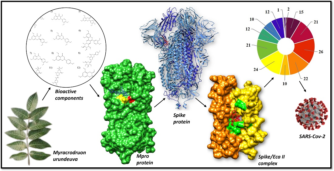
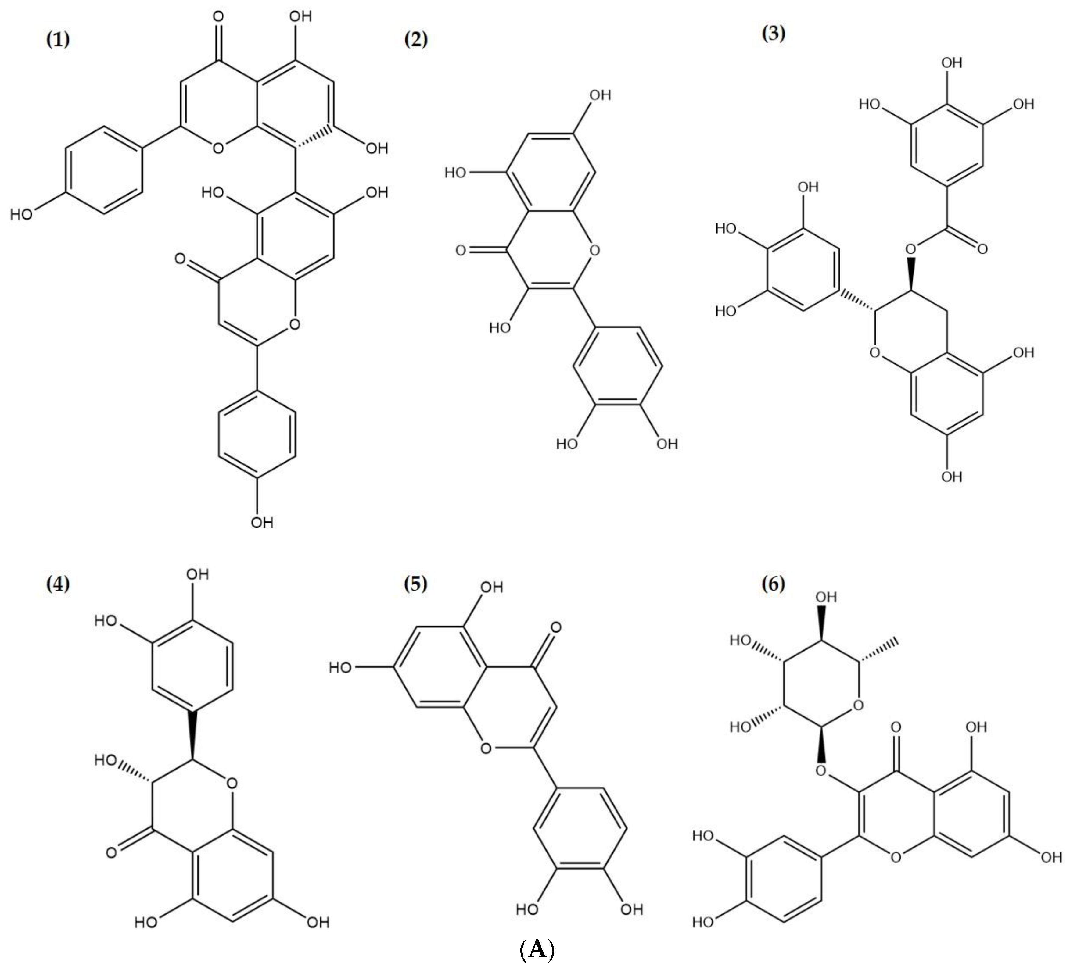
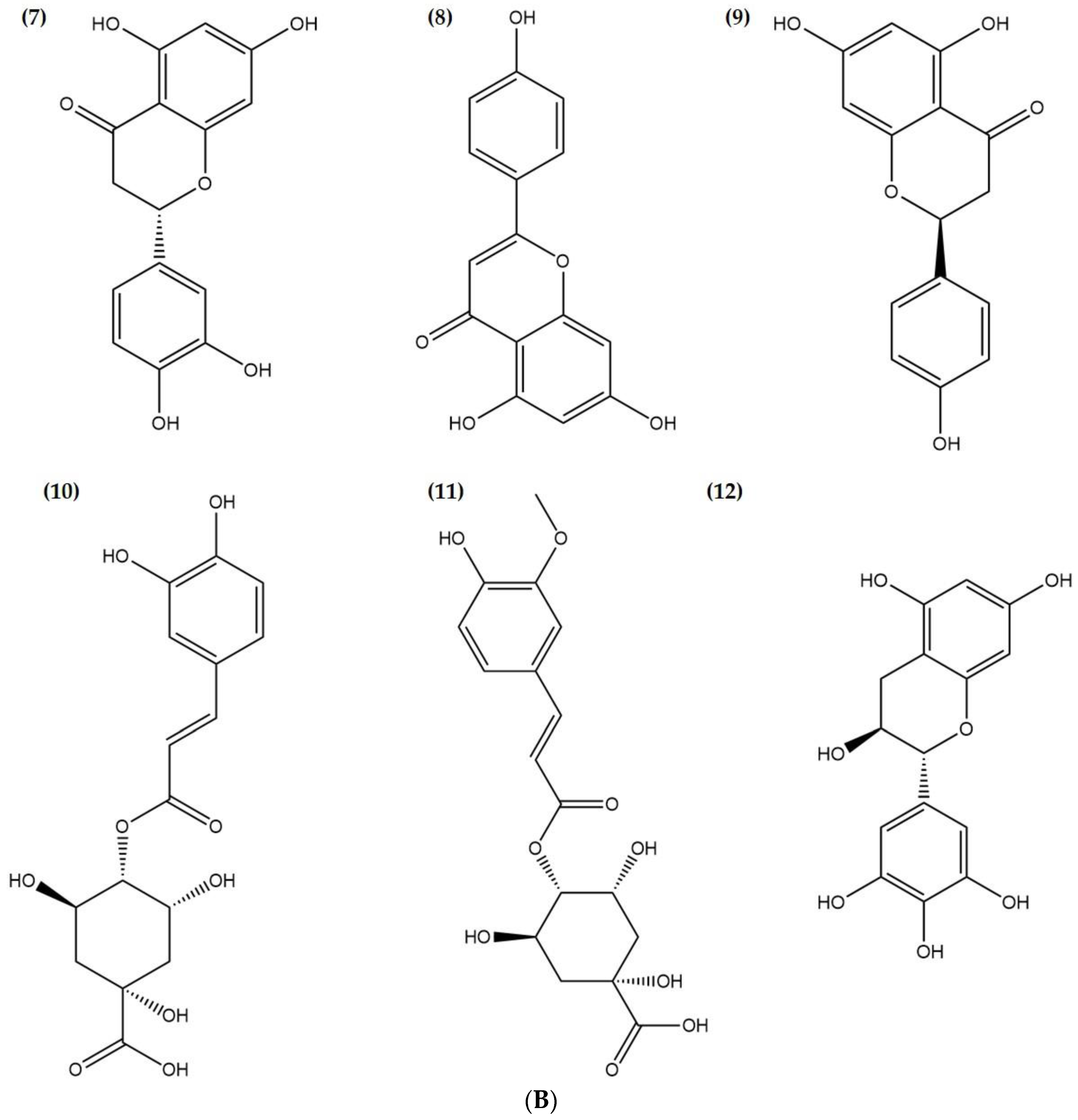
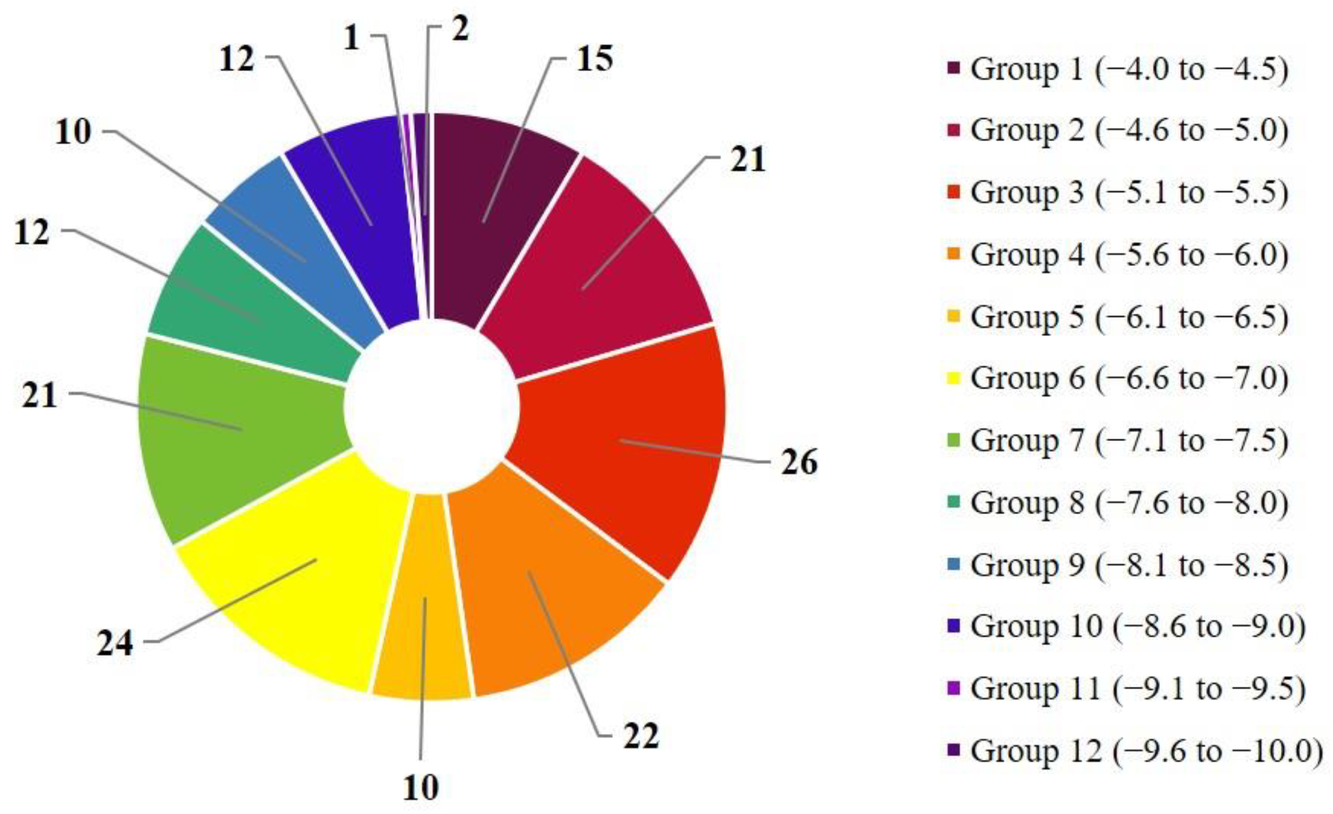
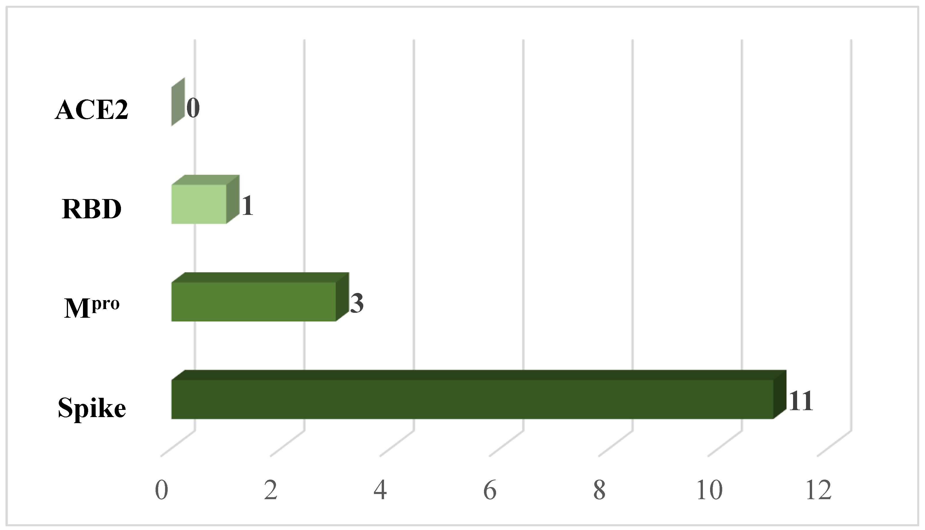
2. Results and Discussion

After selecting 44 chemical constituents of M. urundeuva [19,20,21,22,23], 176 dockings were performed (Table S1), of which 3 stood out by obtaining a lower binding energy, less than −9.2 kcal.mol−1. However, by expanding this range to values below −8.5 kcal.mol−1, a greater number of results were obtained (Table 1; Figure 1A,B) that were considered satisfactory and corresponded to 8.56% (groups 10, 11, and 12, Figure 2).
Our molecular docking process evaluated different spatial conformations of the ligand, which enabled us to identify the potential bioactive compounds that are most likely able to couple at the active site of the target protein. For each result, the respective bond-free energies were obtained in order to consider the lowest possible value due to the spontaneity of the reaction (ΔG < 0) [24].
The spike protein (6VXX) helps SARS-CoV-2 enter human cells [25]. Thus, it is one of the main therapeutic targets to prevent the entry of viruses into humans (Figure 3). Among the chemical compounds of the tested herb, agathisflavone is evidently promising for its anti-inflammatory, antibacterial, and healing properties [26]. Our study also demonstrated that agathisflavone showed potential interaction capacity with the 6VXX.
The complex formed with agathisflavone obtained a free bond energy equal to −9.7 kcal.mol−1 (Figure 4). It interacted directly with four amino acids by hydrogen bond (His519, His49, Ser967, and Asp571) and seven amino acids by hydrophobic bond (Val42, Asp40, Asp568, Agr567, Agr44, Lys964, and Leus518). Casalino et al. [27] stated that, by blocking the spike protein or modulating its conformational state through chemical interactions, it is possible to interfere directly with its binding capacity with the ACE2 receptor (the receptor that is responsible for the entrance of SARS-CoV-2 in humans). Thus, the negative value indicates greater spontaneity, stability, and, consequently, the efficacy of the ligand in inhibiting its receptor [28].
The spike protein has two subunits, namely: S1, which is composed of the receptor binding domain (RBD) and is responsible for the direct association of this macromolecule with receptors present on the surface of host cells (ACE2), and S2, which is capable of causing membrane fusion [29]. Our findings demonstrate that agathisflavone interacted with the RBD (Figure 5) effectively, where the free bond energy corresponded to −9.7 kcal.mol−1. It resulted in intermolecular interactions through six bonds by hydrogen intrusion with the amino acid residues Lys417, Asn33, Asp30, Phe390, Ser494, and Asp405, and twelve bonds through hydrophobic intrusion with the amino acid residues Ala387, Pre389, Leu455, Tyr495, Agr403, Asp38, Tyr453, His34, Glu37, Arg393, Tyr505, and Ala386.
The SARS-CoV-2 replicase gene makes the overlapping polyproteins pp1a and pp1ab. The main protease 3CL (Mpro) cuts them at 11 different places to make shorter nonstructural proteins that are important for its replication process [30]. If new molecules were added to or bound to this protease, the structure would change, which would have a direct effect on the copying process of viral RNA [31]. In this study, agathisflavone resulted in a free binding energy equal to −9.3 kcal.mol−1 (Figure 6) with two amino acids by hydrogen bonding with the amino acid residues Glu166 and Phe140, while eight hydrophobic bonds were formed with the amino acid residues, namely, Thr190, Gln189, His41, Met49, Met165, His164, Cys145, and Leu141 of Mpro of the virus.
It is important to highlight that, in proteins, hydrogen bonds play a vital role in stabilizing their three-dimensional structure, influencing the way they unfold and interact with other molecules, being crucial for their stability. Hydrophobic interactions, on the other hand, mainly result in increased entropy of water molecules and new hydrogen bonds that arise when water molecules involving hydrophobic molecules come into contact with others, thus having specific entropic and enthalpic components. Even if the complexation of the hydrophobic ligand within the protein results in costs associated with the partial loss of translational, rotational, and conformational entropies of the ligand, the entropic gain of the solvent molecules displaced from the site is greater than these [32].
The discovery of new therapeutic strategies to combat SARS-CoV-2 will also be one of the major approaches to the reuse of existing antiviral drugs against this virus. These strategies will also be capable of validating new and existing antiviral drugs against this deadly virus. For example, the international initiative Solidarity, led by the World Health Organization (WHO), began to recommend, in 2021, the emergency use of the drugs baricitinib, molnupiravir, and remdesivir [33,34] against SARS-CoV-2, which reduced the time and cost of new research since safety tests (preclinical and clinical), formulation protocols, and large-scale production have already been established. However, when these drugs were submitted to the molecular docking process to check their potential against SARS-CoV-2, none of them presented satisfactory results. Table 2 shows that all these drugs showed binding capacity with the targeted proteins less than or equal to −8.6 kcal mol−1. Interestingly, the chemical constituents of M. urundeuva belonging to groups 10, 11, and 12 showed better affinities than the abovementioned established drugs.
However, prior to starting clinical trials, it is crucial to understand the absorption, distribution, metabolism, excretion, and toxicity (ADMET) of a drug candidate [35]. A survey conducted in 2001 by the Intercontinental Medical Statistics (IMS Health) suggests that 84% of the 50 most frequently used drugs in Europe and the United States are used via oral route [36]. This made medicinal scientists more interested in finding new bioactive principles that could be easily absorbed by the gastrointestinal tract [37]. Our in silico study demonstrates that the screened chemical compounds of the herb (groups 10, 11, and 12) showed high intestinal absorption potential in humans, ranging from 55.404 to 94.062% (except cryptochlorogenic acid and feruloyl-D-quinic acid) (Table 3).
The human colon adenocarcinoma (Caco-2) cells are frequently used to test the dissolution and permeation of water-soluble drugs and predict how well they will be absorbed after oral administration [38]. As suggested by the literature, chemical compounds with permeability coefficients lower than 1 × 10⁻⁶ cm/s, between 1 and 10 × 10−6 cm/s, and greater than 10 × 10−6 cm/s can be classified, respectively, as poorly absorbed (0–20%), moderately absorbed (20–70%), and well absorbed (70–100%), respectively [39,40]. The computational results aim to define the permeability based on this cell type by predicting the selected constituents’ poor absorption. Regarding dermal permeability, log Kp values lower than -2.5 imply low absorption in the skin [41].
Another factor observed was the steady-state volume of distribution (VDss) (Table 4), a theoretical value referring to the total dose that a drug would need to be evenly distributed at the same concentration of blood plasma [38]. The VDss is considered low for log values less than −0.15 and high for values above −0.45 [42]. Therefore, the high VDss of the bioactives agathisflavone, cryptochlorogenic acid, and feruloyl-D-quinic acid indicate their better distribution in tissues than in plasma. Regarding the permeability of the blood–brain barrier (BBB), a structure that prevents and/or hinders the passage of substances from the blood to the central nervous system (CNS), none of the compounds can cross it since their BBB logs are <0.3 [39].
The Salmonella typhimurium mutation reversal assay, also called the Ames test, is widely used to check toxicological parameters, especially gene mutations caused by test substances [43]. Our in silico study suggested that eriodictiol, gallocatechin gallate, naringenin, quercetin, quercitrin, and taxifolin showed carcinogenicity, while others remained noncarcinogenic (Table 5).
On the other hand, the 50% lethal dose (LD50) test predicts how much of a given substance is needed to kill 50% of a test population [44]. This parameter is necessary to check the therapeutic index and safety profile of bioactive substances. Our study suggests that feruloyl-D-quinic acid and naringenin were the most toxic and safe compounds, respectively. This is because the higher the lethal dose, the less dangerous the chemical is. In the same sense, our chronic oral toxicity in rats (LOAEL) suggests that agathisflavone, cryptochlorogenic acid, feruloyl-D-quinic acid, gallocatechin gallate, naringenin, quercetin, and taxifolin can be ingested in greater quantities. Feruloyl-D-quinic acid might be used in a large dose to produce the desired effect without resulting in any potential side effects. The liver is the major metabolic site in our body. Thus, the safety of this organ is a foremost concern while developing and installing any drug candidate [45]. Our findings suggest that all the tested bioactive substances did not show hepatotoxicity. Additionally, these compounds did not show skin sensitization, suggesting their safety profiles in animals.
Prior to the discovery of this new strain (SARS-CoV-2), the genomes of six species of coronavirus (CoVs) had already been fully sequenced and reported to GenBank (in November 2002). Four of these species, including HCoV-229E, HCoV-NL63, HCoV-OC43, and HCoV-HKU1, cause only relatively mild autoimmune infections with limiting respiratory symptoms. The others, SARS-CoV-1 and MERS-CoV, are highly pathogenic and capable of provoking severe acute respiratory syndrome with high mortality rates [46].
Its variants are classified according to the lineage and mutation of its components. As a result, viruses belonging to the same lineage but containing different subsets of mutations can be classified as different variants. The variants are characterized by their transmissibility, disease severity, and ability to escape humoral immunity [47].
Currently, the omicron EG.5 variant is the latest to be labeled as a “variant of interest” by the World Health Organization (WHO), joining the current ranks of XBB.1.16 and XBB.1.5. The new designation, made as part of an initial risk assessment, reflects its “notable increase” in global prevalence during the second half of 2023 (https://www.who.int/docs/default-source/coronaviruse/09082023eg.5_ire_final.pdf, accessed on 4 September 2023) [48]. The potential of variants to escape naturally induced immunity and vaccine-induced immunity makes it a priority to develop next-generation vaccines and drugs that trigger broadly neutralizing activity against current and potential future variants.
Thus, with the aid of computational biology to obtain experimental results in vitro, the analyses carried out during this research were able to identify a promising bioactive principle for the treatment of COVID-19, coming from a native plant of the Brazilian caatinga and cerrado; its inhibitory capacity of the proteins vital for the development of SARS-CoV-2 can produce positive reflexes for the patient and for society in terms of improvement and quality of life.
Nowadays, the world’s largest pharmaceutical industries have research programs in the area of natural products, as they offer several advantages, for example, the large number of chemical structures and saving time and resources. In this context, regarding M. urundeuva, for presenting great pharmacological use, its bark has anti-inflammatory, astringent, antiallergic, and healing properties, the roots are used in the treatment of rheumatism, and the leaves are indicated for the treatment of ulcers [49], this plant becomes a promising source of raw material.

3. Materials and Methods

3.1. Selection of Chemical Compounds of Myracrodruon urundeuva

Searches were made in national and international databases for selecting chemical compounds of M. urundeuva, namely: Scientific Electronic Library Online (Scielo), Capes journal portal, Regional Portal of the Virtual Health Library (Bireme), National Center for Biotechnology Information (PubMed), Thomson Reuters (Web of Science), Elsevier Group (Scopus), Science Direct, and Google Scholar. For this, we used published papers using the common keyword “Myracrodruon urundeuva”, which was then paired with “phytochemicals”, “chemical constituents”, or “phytochemistry”. After this, the selected chemical structures were acquired through the PubChem platform (https://pubchem.ncbi.nlm.nih.gov/, accessed on 10 August 2022) [50] for further molecular optimization.

3.2. Determination of the Active Site

The active sites of the SARS-CoV-2 proteins were determined using the GASS-WEB server, a tool that works with calculations using the method of genetic algorithms looking for corresponding residues stored in databases such as the Catalytic Site Atlas (CSA), National Center for Biotechnology Information (NCBI), and Protein Data Bank (PDB). The models undergo calculations of root-mean-square deviation (RMSD) comparing the model and the residues surveyed. This methodology was able to identify 90% of the catalytic active sites cataloged [51]. The methodology of searching for the active site by similarity can be observed by Izidoro, Melo-Minardi, and Pappa [52].

3.3. Molecular Docking Study

The 3D structures of four viral proteins were obtained from the Protein Data Bank (PDB) (http://www.rcsb.org/, accessed 16 September 2022) [53] with the respective codes 6VXX (protein S or spike), 1R42 (angiotensin-converting enzyme, ACE2), and 6LU7 (main protein Mpro), while RBD (Spike/ACE2 interaction site) was designed by Barros et al. (2020) [54]. They were then prepared by removing all water molecules and other groups, such as ions, using the Chimera v. 13.1 software [55]. In addition, polar hydrogen atoms were added, Gasteiger partial charges were calculated, and nonpolar hydrogens were mixed in both parts (ligand and protein) using the Autodock Tools (ADT) program, version 1.5.6. Docking was later performed through the Vina AutoDock program [56]. With the LIGPLOT program, used to automatically generate 2D schematic representations of the protein–ligand complexes from the standard input of PDB files, we obtained illustrations of the points of interactions by hydrogen bridges and hydrophobic bonds between the chemical constituents and amino acid residues of the viral proteins [57].

3.4. ADME-TOX Prediction

The prediction of pharmaceutical parameters was performed using the online tool pkCSM (pharmacokinetics) (https://biosig.lab.uq.edu.au/pkcsm/, accessed on 12 February 2023) [58]. Our in silico study also evaluated the ADMET profiles of the bioactives, which include absorption (Caco-2 permeability, water solubility, human intestinal absorption, P-glycoprotein substrate, P-glycoprotein I and II inhibitors, skin permeability), distribution (VDss), unbound fraction, BBB and CNS permeability, metabolism (cytochrome P450 inhibitors, CYP2D6/CYP3A4 substrate), excretion (renal OCT2 substrate, total drug clearance), and toxicity (Rat LD50, Ames toxicity, Tetrahymena pyriformis toxicity, minnow toxicity, maximum tolerated dose, chronic oral toxicity in rats, hepatotoxicity, skin sensitization) [59].

4. Conclusions

SARS-CoV-2 has been evidently producing negative biomedical and epidemiological effects on a global scale, directly impacting all dimensions of life, such as social, economic, political, and cultural spheres. Current immunity obtained by vaccines is quite effective in COVID-19, despite its unavoidable health problems such as unequal access to immunizers and the emergence of new variants, which reinforce the need for the search for new and effective alternative treatment options. Knowing this fact, we performed this study using 44 phytochemicals from a hopeful medicinal herb called M. urundeuva. Our findings reveal that the agathisflavone of the plant showed good molecular affinity (−9.3 to −9.7 kcal.mol−1) with the vital proteins (Spike, RDB, and MPro), suggesting its potentiality against this deadly pathogen. This molecule also exhibited better binding affinity than the reference antiviral drugs used in SARS-CoV-2. Further, pharmacokinetic profiling of agathisflavone also demonstrates that it has a high degree of solubility and low toxicity. It also did not show skin sensitization or carcinogenicity in our in silico ADMET study. However, our findings are based on computational evaluation; therefore, it would be highly appreciable to perform in vivo studies to verify the efficacy and elucidate their exact mechanisms against this virus.

Supplementary Materials

The following supporting information can be downloaded at: https://www.mdpi.com/article/10.3390/ddc2040039/s1, Table S1: Molecular affinity parameters performed by the vina method in ΔGbind (kcal.mol−1) between the chemical constituents of the M. urundeuva plant with the proteins ECA2, MPro, RBD and Spike of the novel coronavirus COVID-19. References [19,20,21,22,23] are cited in the supplementary materials.

Author Contributions

Conceptualization, S.K.S.A., C.S.S., E.K.A.V., H.C.A.S., M.D.A.S., A.S.N.R., V.d.S.d.V., M.T.I., J.L.A. and J.A.R.; data curation, J.A.R.; formal analysis, C.S.S.; investigation, S.K.S.A., C.S.S., E.K.A.V., H.C.A.S., M.D.A.S., A.S.N.R., V.d.S.d.V., J.L.A. and J.A.R.; methodology, S.K.S.A., C.S.S., E.K.A.V., H.C.A.S., M.D.A.S., A.S.N.R. and V.d.S.d.V.; supervision, J.L.A. and J.A.R.; validation, M.T.I.; writing—original draft, S.K.S.A., E.K.A.V., M.T.I., J.L.A. and J.A.R.; writing—review and editing, S.K.S.A., M.T.I., J.L.A. and J.A.R. All authors have read and agreed to the published version of the manuscript.

Funding

The authors are grateful to the Foundation for Scientific Technological Research and Development of Maranhão—FAPEMA (UNIVERSAL-06509/22) and to the State Government of Maranhão and the Secretariat of State for Science, Technology and Innovation—SECTI for their support with the study scholarship and the Federal University of Maranhão—UFMA for their incentive and this research was funded by the National Council for Scientific and Technological Development (CNPq).

Institutional Review Board Statement

Not applicable.

Informed Consent Statement

Not applicable.

Data Availability Statement

Not applicable.

Conflicts of Interest

The authors declare no conflict of interest.

References

  1. Dourado, L.; Caetano, L.; Marques, M.; Penna, U.; Costa, C.; Arruda, F.; Libera, L. Estudo da história natural da COVID-19 e epidemiology of SARS-CoV-2 infection: A descriptive review of the literature. Braz. J. Surg. Clin. Res. 2020, 33, 46–56. [Google Scholar]
  2. Oliveira, M.; Robinson, A.; Siqueira, M. Knowing SARS-CoV-2 and COVID-19. In Health Diplomacy and COVID-19: Reflections Halfway, 1st ed.; Fonseca, L., Ed.; Fiocruz: São Paulo, Brazil, 2020; pp. 69–82. [Google Scholar]
  3. Johns Hopkins Coronavirus Resource Center. Available online: https://coronavirus.jhu.edu/map.html (accessed on 11 November 2022).
  4. Nascimento, L.; Marchiori, M.; Field, V.; Zini, M. SARS-CoV-2 and COVID-19: Pathophysiological and immunological aspects, diagnostic strategies and vaccine development. Rev. Interdiscip. Saúde Educ. 2020, 1, 122–158. [Google Scholar] [CrossRef]
  5. Schoeman, D.; Fielding, B. Coronavirus envelope protein: Current knowledge. Virol. J. 2019, 16, 69. [Google Scholar] [CrossRef]
  6. Lin, L.; Lu, L.; Cao, W.; Li, T. Hypothesis for potential pathogenesis of SARS-CoV-2 infection-a review of immune changes in patients with viral pneumonia. Emerg. Microbes Infect. 2020, 9, 727–732. [Google Scholar] [CrossRef] [PubMed]
  7. Marra, M.A.; Jones, S.J.M.; Astell, C.R.; Holt, R.A.; Brooks-Wilsin, A.; Butterfield, Y.S.N.; Khattra, J.; Asano, J.K.; Barberb, S.A.; Chan, S.Y.; et al. The Genome Sequence of the SARS-Associated Coronavirus. Science 2003, 300, 1399–1404. [Google Scholar] [CrossRef] [PubMed]
  8. Barbosa, I.; Galvão, M.; Souza, T.; Gomes, S.; Medeiros, A.; Lima, K. Incidence of and mortality from COVID-19 in the older Brazilian population and its relationship with contextual indicators: An ecological study. Rev. Bras. Geriatr. Gerontol. 2020, 23, 2017. [Google Scholar] [CrossRef]
  9. Antonio, A.; Wiedemann, L.; Junior, V. Natural products’ role against COVID-19. RSC Adv. 2020, 10, 23379–23393. [Google Scholar] [CrossRef]
  10. Domingos, F.; Silva, M. Use, knowledge and conservation of Myracrodruon urundeuva: A systematic review. Res. Soc. Dev. 2020, 9, 8851. [Google Scholar] [CrossRef]
  11. Amaral, E.A.; Silva, R.M.G. Evaluation of the acute toxicity of angico (Anadenanthera falcata), pau-santo (Kilmeyera coreacea), aroeira (Myracrodruon urundeuva) and São João vine (Pyrostegia venusta), by means of the bioassay with Artemia salina. J. Serv. Res. 2008, 5, 1–16. [Google Scholar]
  12. Maia, G. Caatinga, Trees and Shrubs and Their Utilities, 1st ed.; D & Z: São Paulo, Brazil, 2004. [Google Scholar]
  13. Souza, S.; Aquino, L.; Milach, A.; Bandeira, M.; Nobre, M.; Viana, G. Antiinflammatory and antiulcer properties of tannins from Myracrodruon urundeuva Allemão (Anacardiaceae) in Rodents. Phytother. Res. 2007, 21, 2020–2225. [Google Scholar] [CrossRef]
  14. Asadi, S.; Ahmadiani, A.; Esmaeili, M.; Sonboli, A.; Ansari, N.; Khodagholi, F. In vitro antioxidant activities and an investigation of neuroprotection by six Salvia species from Iran: A comparative study. FTC 2010, 48, 1341–1349. [Google Scholar] [CrossRef] [PubMed]
  15. Filho, F.; Nascimento, K.; Santos, W.; Frazão, N. Study of acetylcholinesterase inhibition by molecular docking: Application in the treatment of Alzheimer’s disease. Ed. S. Health. 2020, 7, 1–18. [Google Scholar] [CrossRef]
  16. Ferreira, L.; Santos, R.; Oliva, G.; Andricopulo, A. Molecular docking and structure-based drug design strategies. Molecules 2015, 20, 13384–13421. [Google Scholar] [CrossRef] [PubMed]
  17. Tripathi, A.; Bankaitis, V. Molecular Docking: From Lock and Key to Combination Lock. J. Mol. Med. Clin. Appl. 2017, 2, 106. [Google Scholar] [CrossRef]
  18. Dias, R.; Azevedo, J.; Walter, F. Molecular Docking Algorithms. Curr. Drug Targets 2008, 9, 1040–1047. [Google Scholar] [CrossRef] [PubMed]
  19. Castro, C.B. Chemical Profile and Cytotoxic Activity of Leaf, Branch and Bark Extracts of Aroeira-Do-Sertão (Myracrodruon urundeuva All.): Metabolomic and Chemometric Approach; Monograph (Graduation in Chemistry); Federal University of Ceará: Fortaleza, Brazil, 2016. [Google Scholar]
  20. Figueredo, F.; Lucena, B.; Tintino, S.; Matias, E.; Leite, N.; Andrade, J.; Nogueira, L.; Morais, E.; Costa, J.; Coutinho, H.; et al. Chemical composition and evaluation of modulatory of the antibiotic activity from extract and essential oil of Myracrodruon urundeuva. Pharm. Biol. 2014, 52, 560–565. [Google Scholar] [CrossRef]
  21. Costa, O.B.; Mennezi, C.; Benedito, L.; Resck, I.; Vieria, F.; Bizzo, H. Essential oil constituents and yields from leaves of Blepharocalyx salicifolius (Kunt) O. Berg and Myracrodruon urundeuva (Allemão) collected during daytime. J. For. Res. 2014, 2014, 982576. [Google Scholar] [CrossRef]
  22. Alquino, N. Chemical Aspects of the Chemical-Pharmacological-Agronomic Study of Wild and Cultivated Aroeiras do Sertão (Myracrodruon urundeuva Fr. all). Ph.D. Thesis, Federal University of Ceará, Fortaleza, Brazil, 2017. [Google Scholar]
  23. Pessoa, E.; Nicolete, R.; Araújo, T. The use of Anacardiaceae Family’s vegetable extracts as natural skin-clearing agents: A review. World J. Pharm. Res. 2020, 9, 32–37. [Google Scholar] [CrossRef]
  24. Ruyck, J.; Brysbaert, G.; Blossey, R.; Lensink, M. Molecular docking as a popular tool in drug design, an in silico travel. Adv. Appl. Bioinform. Chem. 2016, 9, 1–11. [Google Scholar] [CrossRef]
  25. Smith, R.; Cavalcante, G. Potential therapeutic targets for COVID-19: An integrative review. JCS HU-UFPI 2021, 4, 18–25. [Google Scholar] [CrossRef]
  26. Oliveira, Y.R.; Robinson, W.; Smith, P.; Pacheco, A.; Abreu, M. Anacardiaceae in the Traditional Medicine of Rural Communities of Piauí, Northeastern Brazil. Ens. Ciências 2022, 26, 32–42. [Google Scholar] [CrossRef]
  27. Casalino, L.; Gaieb, Z.; Goldsmith, J.; Hjorth, C.; Dommer, A.; Harbison, A.; Fogarty, C.; Barros, E.; Taylor, B.; McLellan, J.; et al. Beyond Shielding: The Roles of Glycans in the SARS-CoV-2 Spike Protein. ACS. Cent. Sci. 2020, 6, 1722–1734. [Google Scholar] [CrossRef] [PubMed]
  28. Junior, E.; Gonsalves, A. Molecular docking study of gsk-3β enzyme inhibitors: A proposal for the treatment of bipolar disorder. Rev. Ifes Sci. 2019, 5, 243–256. [Google Scholar] [CrossRef]
  29. Ord, M.; Faustova, I.; Loog, M. The sequence at Spike S1/S2 site enables cleavage by furin and phospho-regulation in SARS-CoV-2 but not in SARS-CoV-1 or MERS-CoV. Sci. Rep. 2020, 10, 16944. [Google Scholar] [CrossRef] [PubMed]
  30. Qiao, J.; Li, Y.; Zeng, R.; Liu, F.; Luo, R.; Huang, C.; Wang, Y.; Zhang, J.; Quan, B.; Yang, S. SARS-CoV-2 Mpro inhibitors with antiviral activity in a transgenic mouse model. Science 2019, 371, 1374–1378. [Google Scholar] [CrossRef]
  31. Kumar, S. COVID-19: A Drug Repurposing and Biomarker Identification by Using Comprehensive Gene-Disease Associations through Protein-Protein Interaction Network Analysis. Preprints 2020, 1, 2020030440. [Google Scholar] [CrossRef]
  32. Bissantz, C.; Kuhn, B.; Stahl, M. A medicinal chemist’s guide to molecular interactions. J. Med. Chem. 2010, 22, 5061–5084. [Google Scholar] [CrossRef]
  33. Incerti, M.K.; Rosa, T.F.; Foletto, V.S.; Franco, L.N.; Hörner, R. Repurposing Medications for COVID-19: An Overview. Health 2022, 48, 1–5. [Google Scholar]
  34. Robinson, M.; Facchainetti, V.; Aboud, K.; Penha, L.; Gomes, C. Repositioning of the drug baricitinib for the treatment of COVID-19. Conjectures 2022, 22, 306–321. [Google Scholar] [CrossRef]
  35. Smith, D.; Padilha, I. In silico evaluation of pharmacokinetic properties of antileukemic compounds published by scientific journals. Arch. Health. Sci. 2022, 30, e04978. [Google Scholar] [CrossRef]
  36. Lennernäs, H.; Abrahamsson, B. The use of biopharmaceutic classification of drugs in drug discovery and development: Current status and future extension. J. Pharm. Pharmacol. 2005, 57, 273–410. [Google Scholar] [CrossRef]
  37. Jorgensen, W.; Duffy, E. Prediction of drug solubility from structure. Adv. Drug Deliv. Rev. 2002, 54, 355–366. [Google Scholar] [CrossRef]
  38. Robinson, J.; Robinson, Z.; Storpirtis, S. In vitro models for determining drug absorption and predicting the dissolution/absorption ratio. Rev. Bras. Cienc. Farm. 2007, 43, 515–527. [Google Scholar] [CrossRef]
  39. Fernandes, M.B.; Gonçalves, J.; Scotti, M.; Oliveira, A.; Tavares, L.; Storpirtis, S. Caco-2 cells cytotoxicity of nifuroxazide derivatives with potential activity against Methicillin-resistant Staphylococcus aureus (MRSA). Toxicol. In Vitr. 2012, 26, 535–540. [Google Scholar] [CrossRef] [PubMed]
  40. Meanwell, N.A. Improving Drug Candidates by Design: A Focus on Physicochemical Properties as a Means of Improving Compound Disposition and Safety. Chem. Res. Toxicol. 2011, 24, 1420–1456. [Google Scholar] [CrossRef] [PubMed]
  41. Pinheiro, R.; Júnior, A.; Zepeda, C.; Santos, L.; Pinto, L.; Cabral, O.; Soto, C. In silico analysis of the pharmacokinetic and toxicological profile of Zinc II thioglycolate complex [Zn(ATG)2(OH2)2]. Res. Soc. Dev. 2022, 11, e44711629430. [Google Scholar] [CrossRef]
  42. Holguín, N.M.; Frau, J.; Mitnik, D.G. Computational Pharmacokinetics Report, ADMET Study and Conceptual DFT-Based Estimation of the Chemical Reactivity Properties of Marine Cyclopeptides. Chem. Open 2021, 10, 1087–1157. [Google Scholar] [CrossRef]
  43. Aiub, C.A.F.; Felzenszwalb, I. The principles of the ames (Salmonella/microsome) test and its applicability. Genet. Sch. 2011, 6, 11–16. [Google Scholar] [CrossRef]
  44. Pimentel, L.C.F.; Keys, C.; Robinson, L.; Alfonso, J. The unbelievable use of dangerous chemicals in the past. Whoops. New. 2006, 29, 1138–1149. [Google Scholar] [CrossRef]
  45. Bertolami, M.C. Mechanisms of hepatotoxicity. Arq. Bras. Cardiol. 2005, 85, 25–27. [Google Scholar] [CrossRef]
  46. Li, Y.; Chi, W.; Su, J.; Ferrall, L.; Hung, C.; Wu, T. Coronavirus vaccine development: From SARS and MERS to COVID-19. J. Biomed. Sci. 2020, 27, 104. [Google Scholar] [CrossRef]
  47. Tao, K.; Tzou, F.L.; Nouhin, J.; Gupta, R.K.; Oliveira, T.; Kosakovsky, L.S.L.; Fera, D.; Shafer, R.W. The biological and clinical significance of emerging SARS-CoV-2 variants. Nat. Rev. Genet. 2021, 22, 757–773. [Google Scholar] [CrossRef]
  48. World Health Organization. Available online: https://www.who.int/docs/default-source/coronaviruse/09082023eg.5_ire_final.pdf (accessed on 4 September 2023).
  49. Robinson, R.; Fagundes, M.; Roberts, H.; Veloso, M. Ecological aspects of aroeira (Myracrodruon urundeuva Allemão- Anacardiaceae): Phenology and seed germination. Rev. Árvore 2008, 32, 233–243. [Google Scholar] [CrossRef]
  50. PubChem. Available online: https://pubchem.ncbi.nlm.nih.gov/ (accessed on 10 August 2022).
  51. Morais, J.P.A.; Pappa, G.L.; Pires, D.E.V.; Izidoro, S.C. GASS-WEB: A web server for identifying enzyme active sites based on genetic algorithms. Nucleic Acids Res. 2017, 45, 315–319. [Google Scholar] [CrossRef]
  52. Moraes, J.P.A.; Izidoro, S.C.; Melo-Minardi, R.C.; Pappa, G.L. GASS: Identifying enzyme active sites with genetic algorithms. Bioinformatics 2015, 31, 864–870. [Google Scholar] [CrossRef]
  53. RCSB Protein Data Bank. Available online: http://www.rcsb.org/ (accessed on 16 September 2022).
  54. Barros, R.O.; Junior, F.; Perreira, W.; Oliveira, N.; Ramos, R. Interaction of Drug Candidates with Various SARS-CoV-2 Receptors: An in-Silico Study to Combat COVID-19. J. Proteome Res. 2020, 19, 4567–4575. [Google Scholar] [CrossRef]
  55. Pettersen, E.F.; Golddard, T.; Huang, C.; Couch, G.; Greenblatt, D.; Meng, L.; Ferrin, T. UCSF Chimera—A visualization system for exploratory research and analysis. J. Comput. Chem. 2004, 25, 1605–1612. [Google Scholar] [CrossRef] [PubMed]
  56. Trott, O.; Olson, A.J. AutoDock Vina: Improving the speed and accuracy of docking with a new scoring function, efficient optimization, and multithreading. J. Comput. Chem. 2009, 31, 455–461. [Google Scholar] [CrossRef] [PubMed]
  57. Wallace, A.C.; Laskowski, R.A.; Thornton, J.M. LIGPLOT: A program to generate schematic diagrams of protein-ligand interactions. Protein Eng. Des. Sel. 1995, 8, 127–134. [Google Scholar] [CrossRef] [PubMed]
  58. pkCSM: Predicting Small-Molecule Pharmacokinetic Properties Using Graph-Based Signatures. Available online: https://biosig.lab.uq.edu.au/pkcsm/ (accessed on 12 February 2023).
  59. Rocha, J.A.; Rego, N.; Carvalho, B.; Silva, F.; Sousa, J.; Ramos, R.; Passos, I.; Maraes, J.; Lima, F. Computational quantum chemistry, molecular docking, and ADMET predictions of imidazole alkaloids of Pilocarpus microphyllus with schistosomicidal properties. PLoS ONE 2018, 13, e0198476. [Google Scholar] [CrossRef] [PubMed]
Figure 1. (A) Two-dimensional chemical structure of the chemical constituents of Myracrodruon urundeuva presenting the best results (groups 9, 10, and 11) through the molecular docking process (1 (Agathisflavone); 2 (Quercetin); 3 (Gallocatechin gallate); 4 (Taxifolin); 5 (Luteolin); 6 (Quercitrin)). (B) Two-dimensional chemical structure of the chemical constituents of Myracrodruon urundeuva presenting the best results (groups 9, 10, and 11) through the molecular docking process (7 (Eriodictiol); 8 (Apigenin); 9 (Naringenin); 10 (Cryptochlorogenic acid); 11 (Feruloyl-D-quinic acid) and 12 (Gallocatechin)).
Ddc 02 00039 g001aDdc 02 00039 g001b
Figure 2. Total number of results, given in terms of binding free energy (kcal.mol−1), sorted by categories.
Ddc 02 00039 g002
Figure 3. Proteins that stood out the most among the best results (groups 10, 11, and 12).
Ddc 02 00039 g003
Figure 4. Agathisflavone interactions with the viral spike protein ((A) Three-dimensional structure between protein (blue) and ligand (red) and (B) LigPlot+ diagram of the interaction: hydrogen bonds (green) and hydrophobic bonds (red)).
Ddc 02 00039 g004
Figure 5. Agathisflavone interactions with the spike (orange)/ACE2 (yellow) (RBD) interaction site ((A) Three-dimensional structure between the protein and ligand (red), (B) Expansion of the docking region, and (C) LigPlot+ diagram of the interaction: hydrogen bonds (green) and hydrophobic bonds (red)).
Ddc 02 00039 g005
Figure 6. Agathisflavone interactions with the viral Mpro protein ((A) Three-dimensional structure between protein (green) and ligand (blue), (B) Expansion of the docking region, and (C) LigPlot+ diagram of the interaction: hydrogen bonds (green) and hydrophobic bonds (red)).
Ddc 02 00039 g006
Table 1. Molecular reference parameters, referring to groups 10, 11, and 12, between the selected chemical constituents of Myracrodruon urundeuva and the target proteins of SARS-CoV-2.
Complex
(Ligand–Protein)
ΔGbinda
(kcal.mol−1)
Amino Acids That Interact by
Hydrogen Bonding
Amino Acids That Interact by
Hydrophobic Bonding
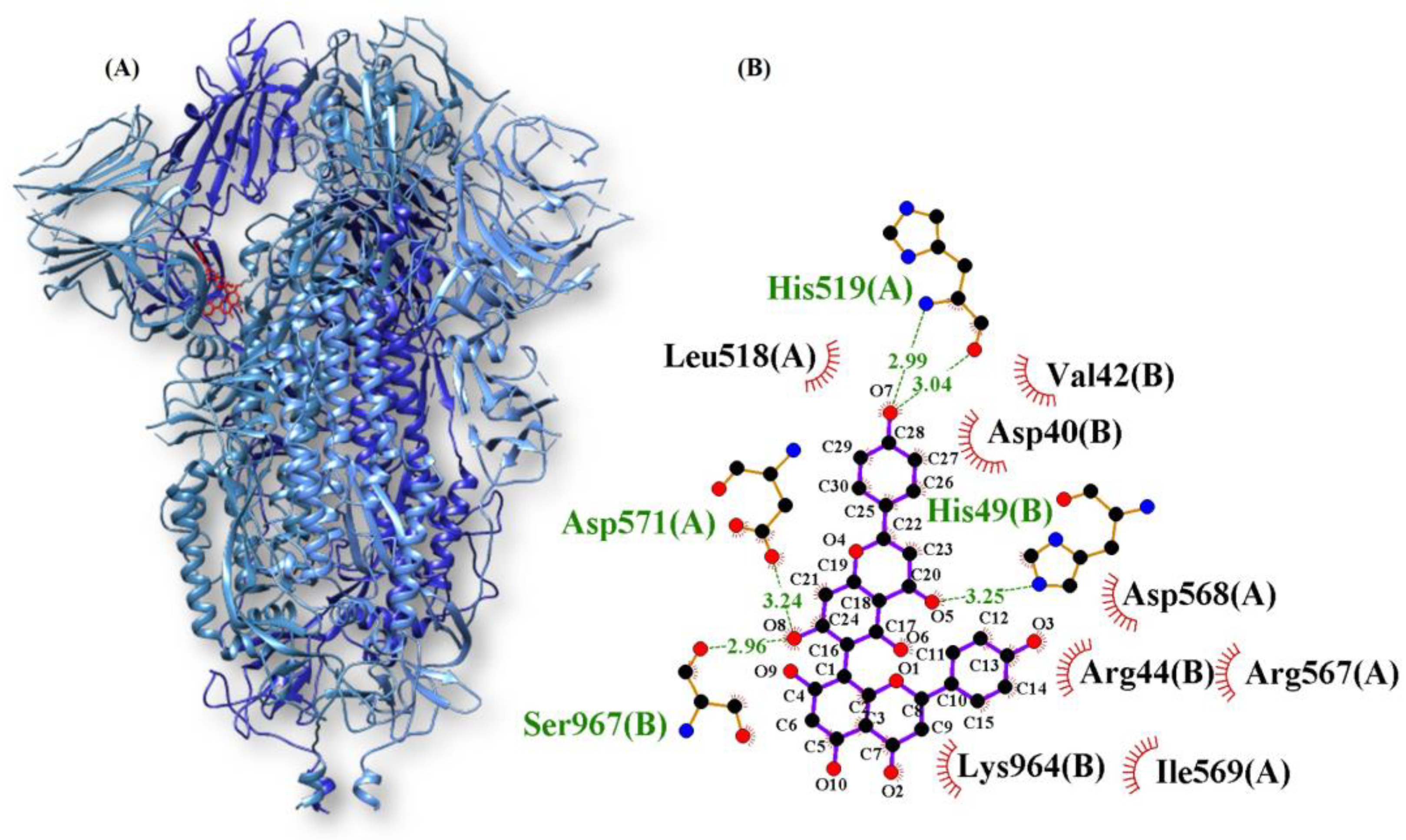
Agathisflavone/Spike−9.7His519, His49, Ser967, Asp571Val42, Asp40, Asp568, Agr567, Agr44, Lys964, Leus518
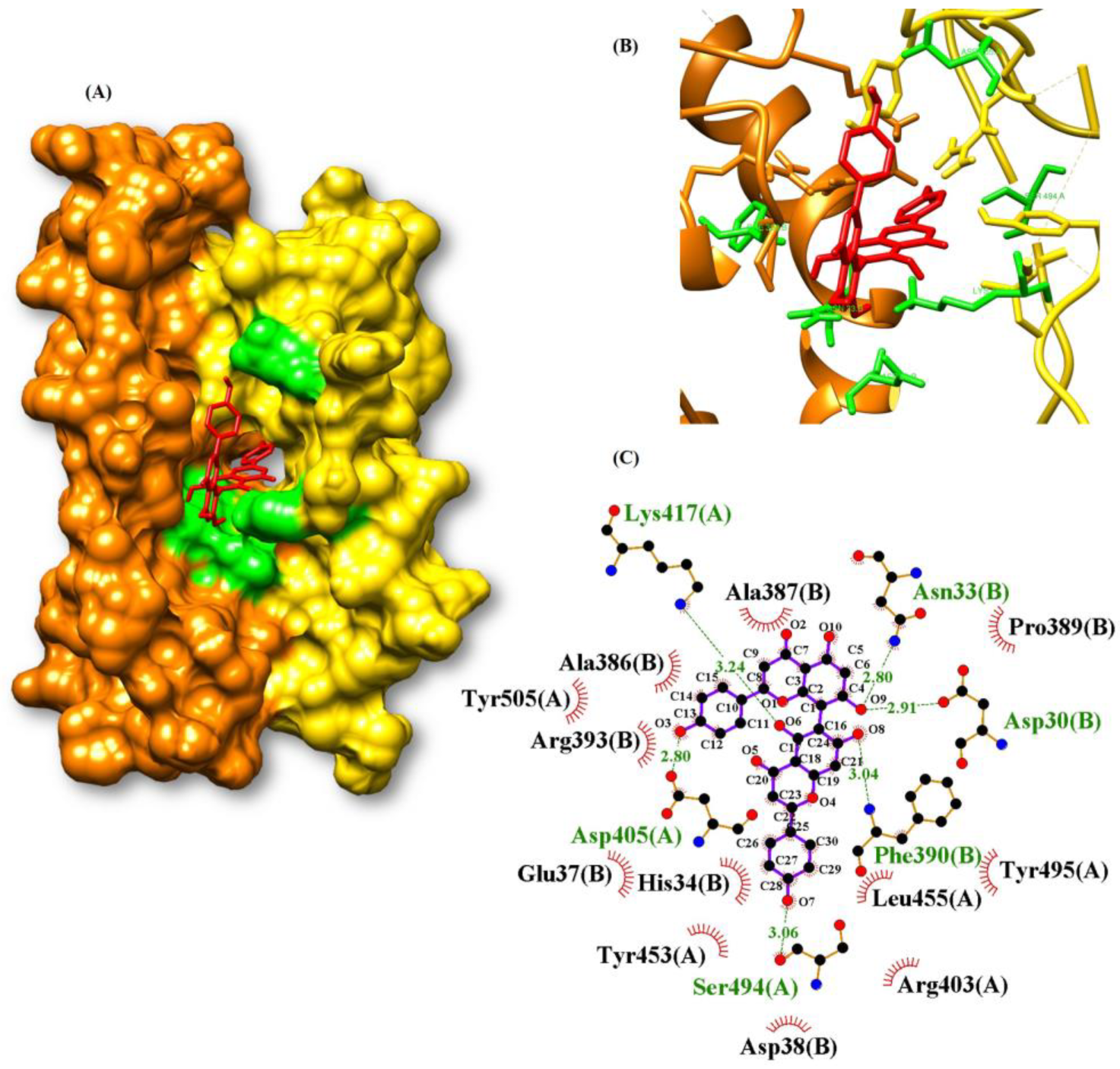
Agathisflavone/RBD−9.7Lys417, Asn33, Asp30, Phe390, Ser494, Asp405Ala387, Pre389, Leu455, Tyr495, Agr403, Asp38, Tyr453, His34, Glu37, Arg393, Tyr505, Ala386
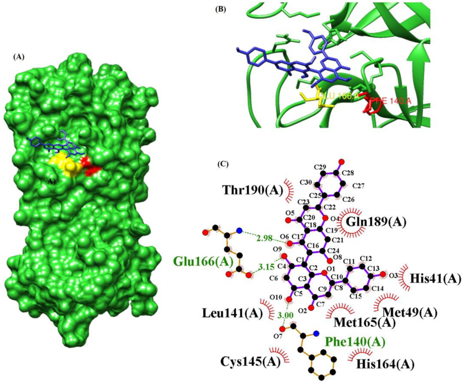
Agathisflavone/MPro−9.3Glu166, Phe140Thr190, Gln189, His41, Met49, Met165, His164, Cys145, Leu141
Quercetin/Spike−9.0Arg100, Leu977, Thr573, Phe855, Tyr741Gly744, Leu966, Val976, Thr547, Leu546, Asn978, Thr572, Met740, Asn856
Gallocatechin gallate /MPro−9.0Phe140, Thr26, Ser144, His163, Leu141, Glu166His172, Asn142, Leu27, Gly143, Thr25, Cys145, Met49, His41, Arg188, Asp187, Gln189, Met165
Taxifolin/Spike−9.0Ile742, Tyr741, Asn978Ile587, Thr573, Val976, Leu977, Gly744, Arg1000, Leu966, Thr572,
Luteolin/Spike−9.0Met740, Phe855, Thr573, Arg1000, Tyr741Gly744, Asn856, Gly548, Asn978, Thr547, Leu546, Val976, Thr527, Leu966
Quercitrin/MPro−9.0Asp187, Asn142, Leu141, Ser144, His163, Glu166, Tyr54Arg188, Met49, His164, Met165, Gly143, Cys145, Gln168, His41
Eriodictiol/Spike−8.9Asn856, Ile742, Tyr741Ile587, Asn978, Val976, Leu977, Gly744, Arg1000, Thr572, Thr573
Apigenin/Spike−8.7Phe855, Thr573, Arg1000, Tyr741, Met740,Asn856, Gly744, Asn978, Leu546, Thr547, Val976, Thr572, Leu966
Gallocatechin gallate/Spike−8.7His49, Arg44, Asp40, Asp571, Ser968, Ser967,Asp568, Ile569, Arg567, Gly757, Asn969, Leu754, Gln755
Naringenin/Spike−8.7Val976, Ser974, Arg983, Ile973, Asp979, Ser975His519, Arg567, Thr430, Leu518, Glu516, Asp571, Asn969,
Cryptochlorogenic acid/Spike−8.6Arg1000, Ser975, Leu977, Asn978, Asn856, Ala570, Leu966Val976, Pro589, Thr573, Phe855, Thr572
Feruloyl-D-quinic acid/Spike−8.6Arg1000, Ser975, Leu977, Asn978, Asp568, Thr549, Gly744Asn856, Thr572, Ile587, Phe589, Phe855, Gly548, Thr573
Gallocatechin/Spike−8.6Phe855, Thr549, Thr573, Thr572, Leu977, Gly744, Arg1000, Tyr741, Met740,Pre589, Ile587, Asn856, Phe541
Table 2. Molecular affinity parameters referring to the drugs baricitinib, molnupiravir, and remdesivir with the target proteins of SARS-CoV-2.
CompoundsΔGbinda (kcal.mol−1)
ACE2 ProteinMpro ProteinRBD ProteinSpike Protein
Baricitinib−6.8−7.9−7.8−8.0
Molnupiravir−7.2−6.7−6.8−7.9
Remdesivir−7.3−7.9−7.6−7.5
Table 3. Absorption properties of the chemical constituents of Myracrodruon urundeuva belonging to groups 10, 11, and 12.
CompoundsSolubility in Water
(log mol.L−1)
PCaco2 (Log Papp in 10−6 cm.s−1)AIH%P.Skin (log Kp)
Agathisflavone−2.8920.37194.062−2.735
Apigenin−3.1781.07691.856−2.736
Cryptochlorogenic acid−2.854−0.70715.087−2.735
Eriodictiol−3.3440.78779.846−2.736
Feruloyl-D-quinic acid−2.776−0.57619.764−2.735
Gallocatechin gallate−2.895−0.79757.176−2.735
Luteolin−3.1730.76281.082−2.735
Naringenin−3.9030.63468.462−2.735
Quercetin−2.9820.69474.84−2.735
Quercitrin−3.132−0.47655.404−2.735
Taxifolin−3.031−0.31870.529−2.735
Note: PCaco2: permeability of Caco-2 cells; AIH: intestinal absorption potential in humans; P.Skin: skin permeability.
Table 4. Distribution properties of the chemical constituents of Myracrodruon urundeuva belonging to groups 10, 11, and 12.
CompoundsVDss (Human) (log L.Kg−1)P.B.H (log BB)
Agathisflavone−0.943−2.192
Apigenin−0.105−0.951
Cryptochlorogenic acid−1.495−1.737
Eriodictiol0.229−1.180
Feruloyl-D-quinic acid−1.738−1.593
Gallocatechin gallate0.050−2.209
Luteolin0.071−1.199
Naringenin−0.431−1.449
Quercetin0.310−1.377
Quercitrin−0.315−2.027
Taxifolin0.547−1.328
Note: VDss: Apparent volume of distribution at steady state; P.B.H: permeability of the blood–brain barrier.
Table 5. Toxicological properties of the chemical constituents of Myracrodruon urundeuva plant belonging to groups 10, 11, and 12.
CompoundsT.AMESD.M.T
(log mg.kg−1.day−1)
T.A.O (LD50) (mol.kg−1)T.C.O (LOAEL) (log mg.kg−1.day−1)S.SkinHep
AgathisflavoneNo0.4252.4673.285NoNo
ApigeninNo0.9312.3761.461NoNo
Cryptochlorogenic acidNo1.3792.2193.503NoNo
EriodictiolYes0.3952.2291.893NoNo
Feruloyl-D-quinic acidNo1.4282.1333.587NoNo
Gallocatechin gallateYes0.4812.6544.085NoNo
LuteolinNo0.9752.4501.833NoNo
NaringeninYes0.9893.5733.556NoNo
QuercetinYes0.9542.3083.134NoNo
QuercitrinYes0.8782.9302.826NoNo
TaxifolinYes0.8862.2453.256NoNo
Note: T.AMES: AMES toxicity; D.M.T: maximum tolerated dose in humans; T.A.O: acute oral toxicity in rats; T.C.O: chronic oral toxicity in rats; S.Skin: skin sensitization; Hep.: Hepatotoxicity.
Disclaimer/Publisher’s Note: The statements, opinions and data contained in all publications are solely those of the individual author(s) and contributor(s) and not of MDPI and/or the editor(s). MDPI and/or the editor(s) disclaim responsibility for any injury to people or property resulting from any ideas, methods, instructions or products referred to in the content.

