
Luminescence Properties of Y3F[Si3O10]:Ln3+ (Ln = Eu, Tb, Er) with Thalenite-Type Host Lattice and Crystal Structure of Tm3F[Si3O10]

Abstract
:1. Introduction
2. Materials and Methods
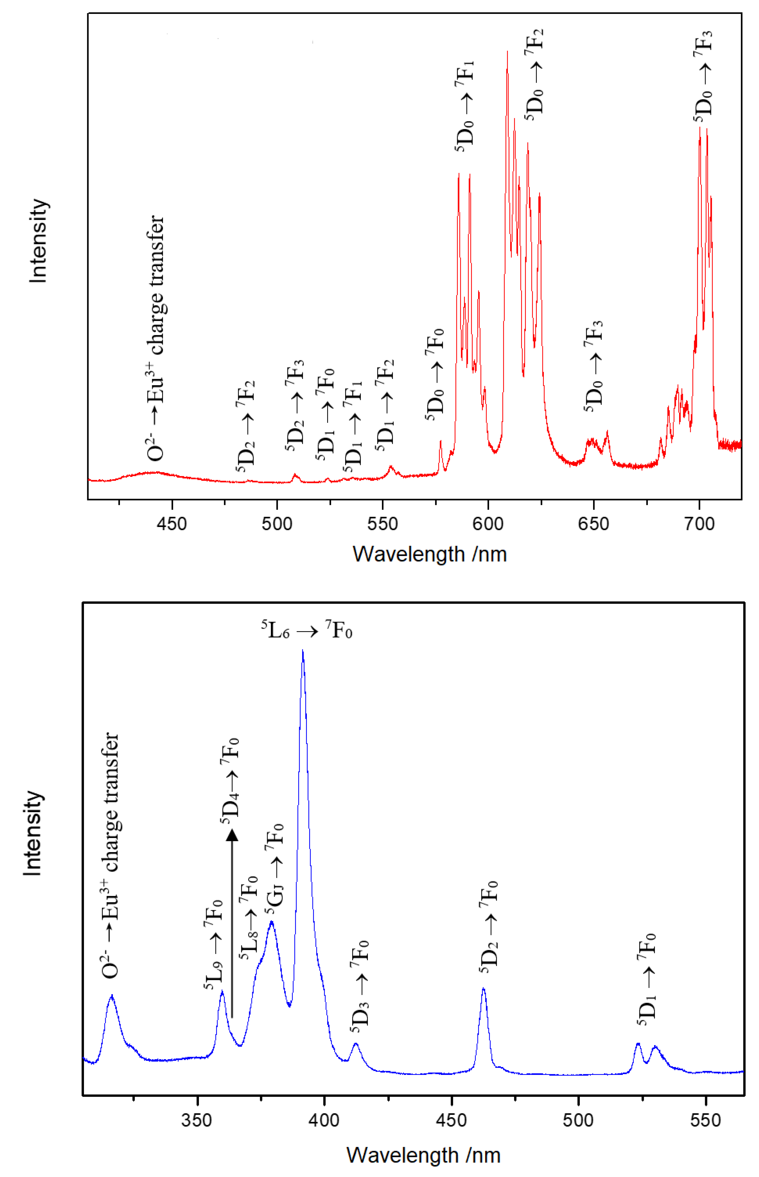
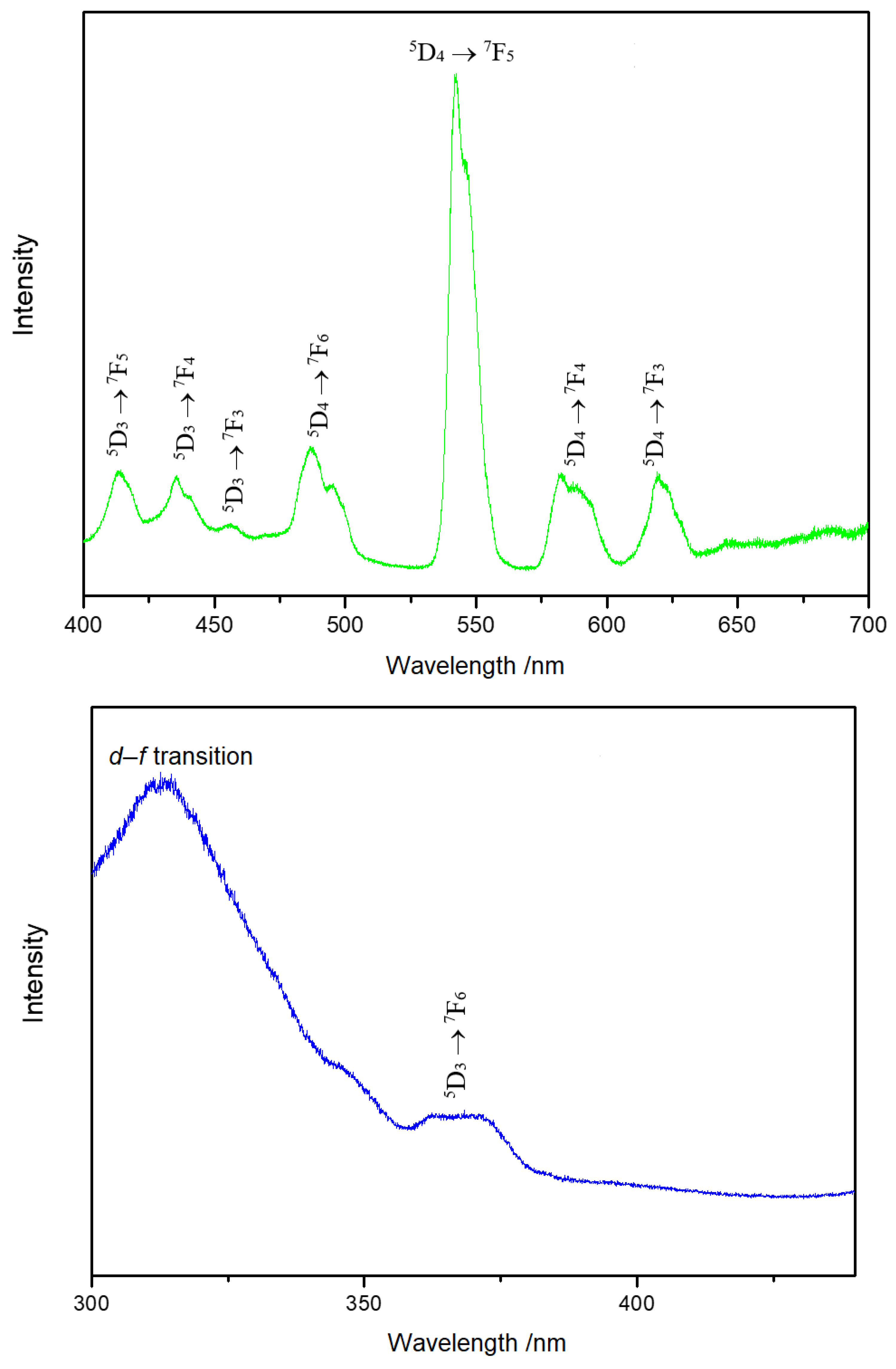
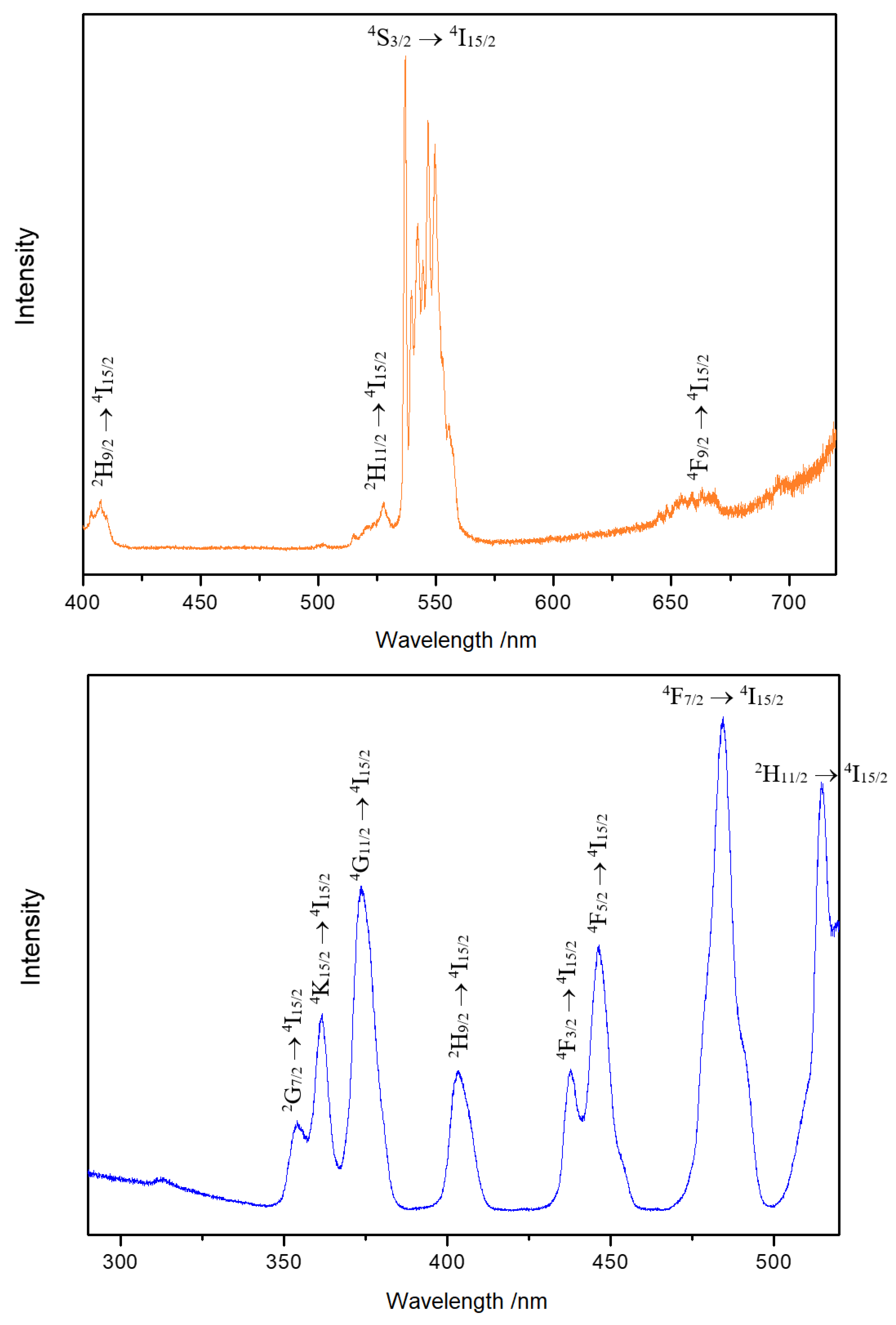
3. Results and Discussion
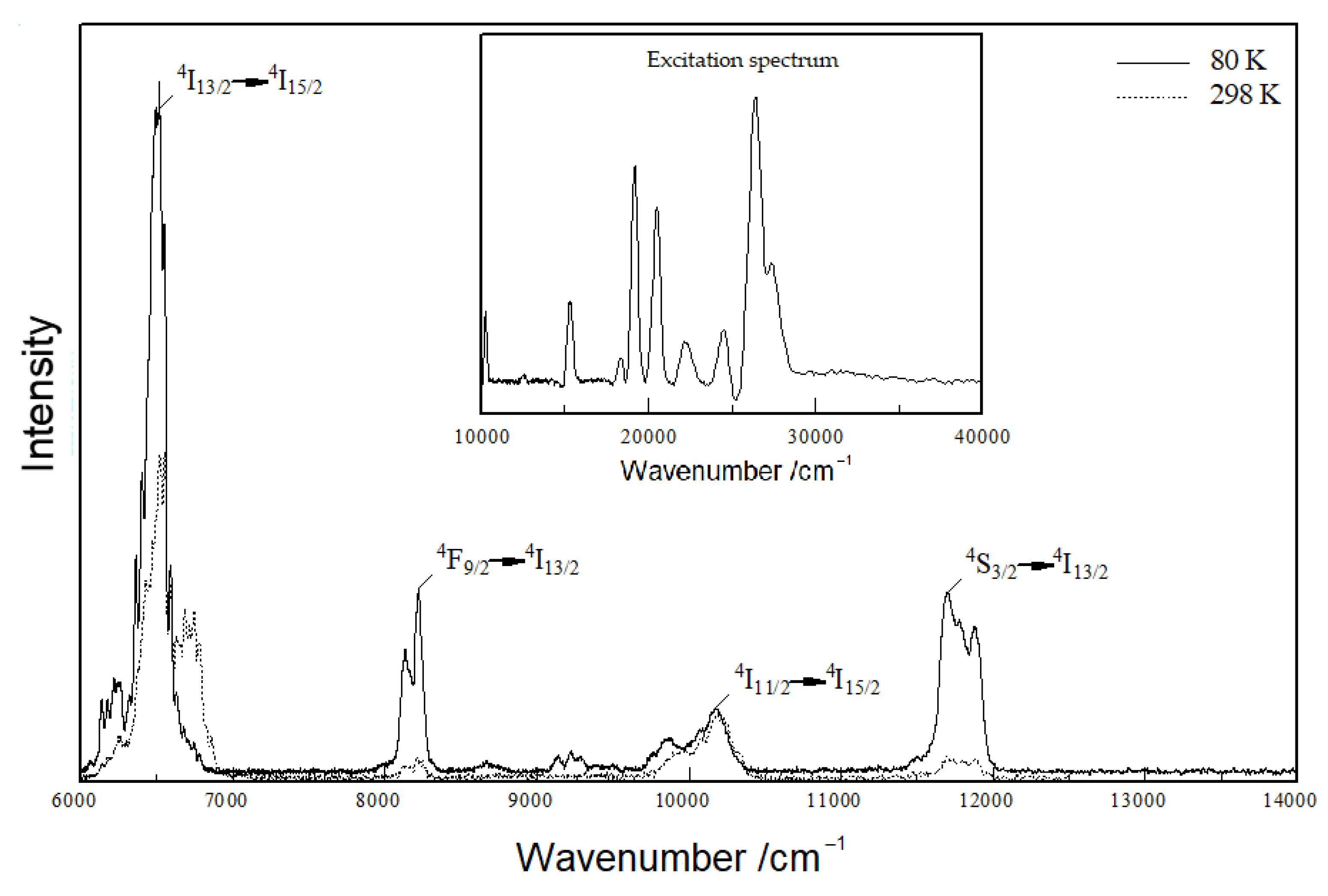
3.1. Structure Description
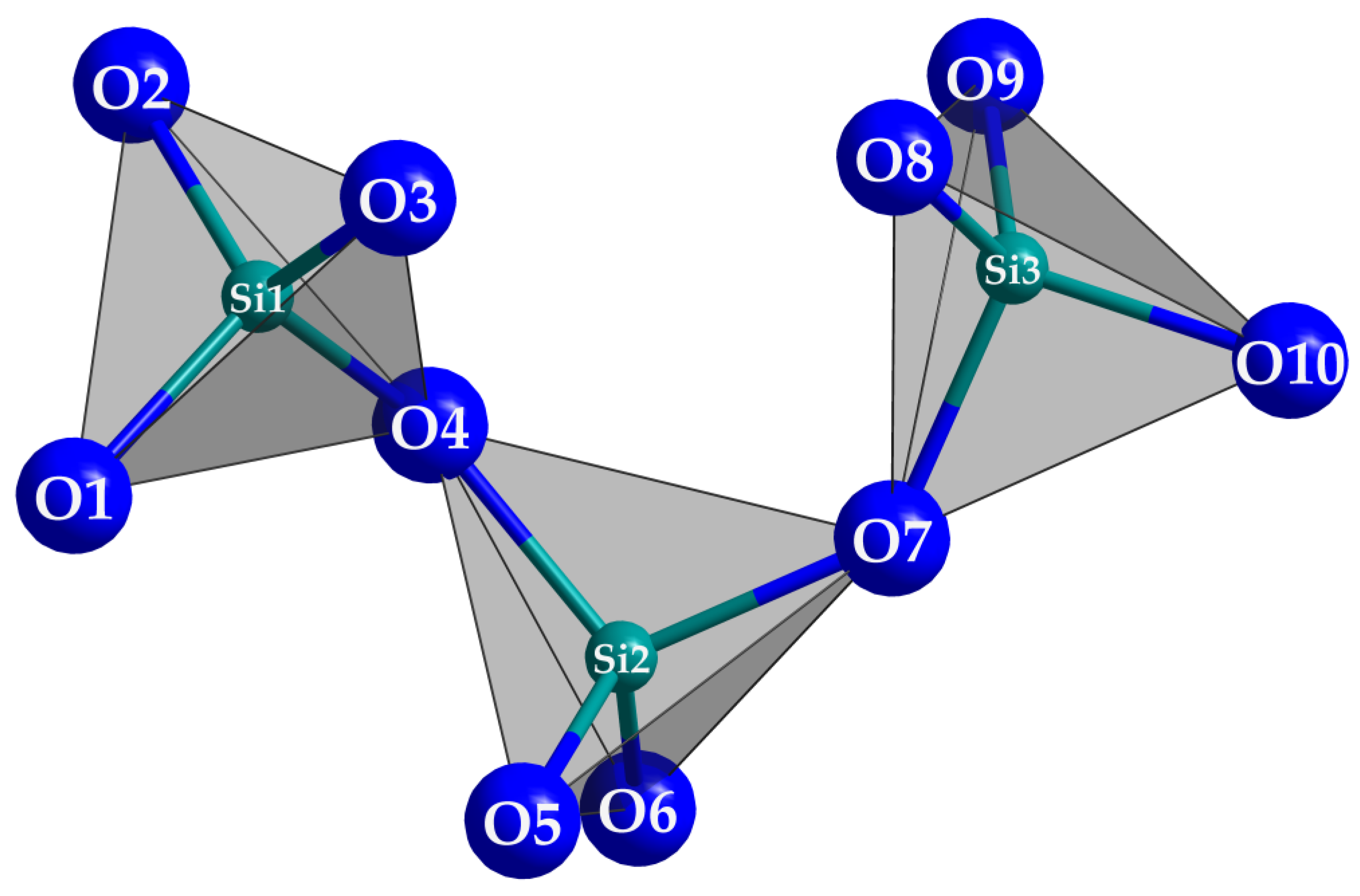
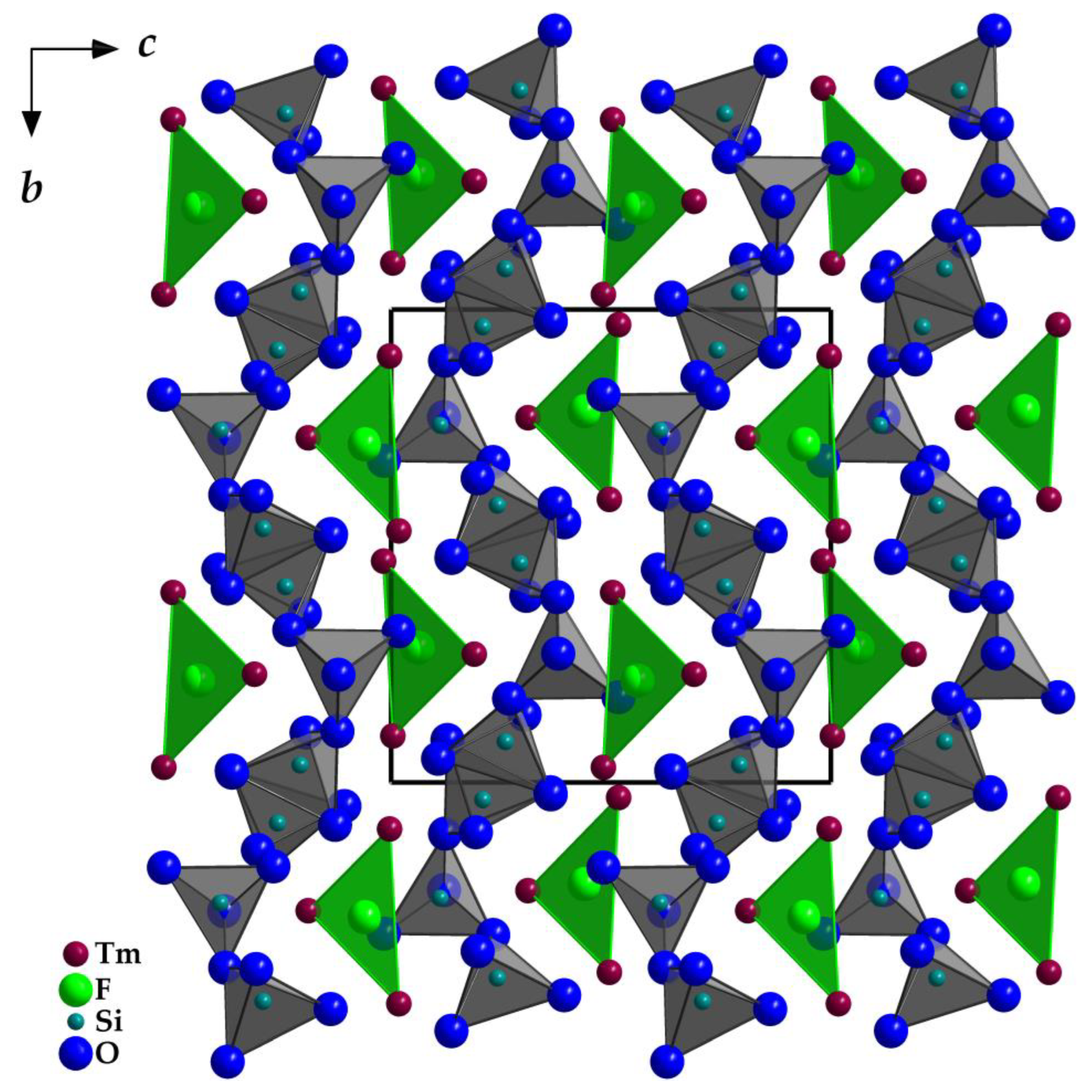
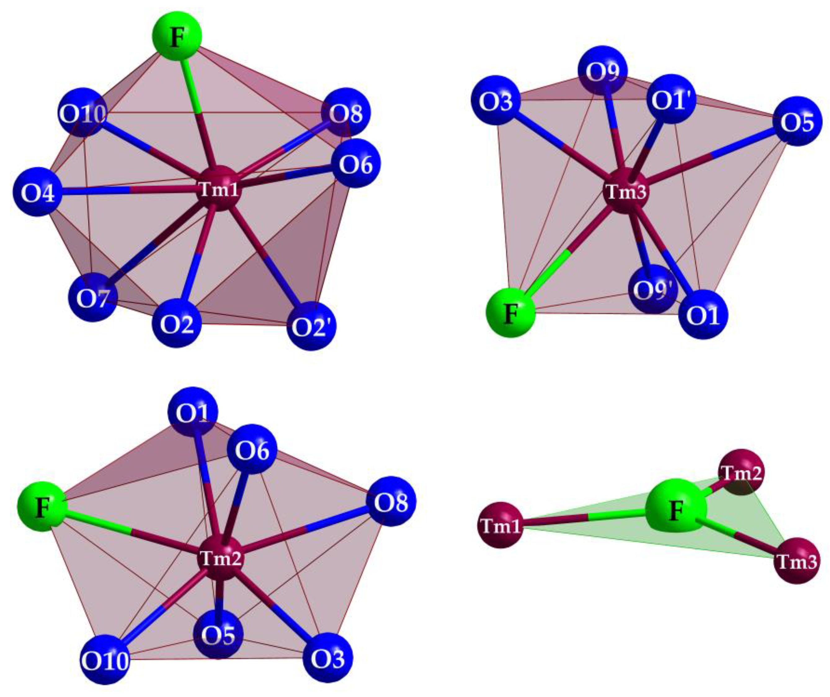
 (Si1–O4–Si2) = 133°), but staggered considering the O7 connection ((Si2–O7–Si3) = 138°). In the unit cell, the opening of the [Si3O10]8− horseshoes is arranged alternatingly to the left and the right (Figure 4) along the [010] direction. The Si–O distances with values of 160–165 pm match very well with the distances of the two known types of thulium oxodisilicates (e.g., B-type Tm2Si2O7: d(Si–O) = 160–165 pm [30] or C-type Tm2Si2O7: d(Si–O) = 161–164 pm [31]). The triangular, almost planar, isolated [FTm3]8+ units (d(F–Tm) = 220–235 pm; Figure 5) are located between the [Si3O10]8− chain fragments (Figure 2). The deflection of the F− anion from the (Tm3+)3 triangle amounts to 20 pm and the Tm–F–Tm angles range from 110 to 134°. In the structure, three crystallographically distinguishable Tm3+ cations are found. (Tm1)3+ is surrounded by one F− and six plus one O2− anions in the shape of a distorted square antiprism. The coordination polyhedra of (Tm2)3+ can be described as a strongly distorted monocapped trigonal antiprism, consisting of one fluorine and six oxygen atoms and (Tm3)3+ shows an environment of one F− and six O2− anions arranged as a distorted monocapped trigonal prism (Figure 5). The Tm–O distances with values of 220–268 plus 292 pm correspond excellently with those in thulium oxodisilicates, oxide ortho-oxosilicate or sulfide oxodisilicate, respectively (e.g., B-type Tm2Si2O7: d(Tm–O) = 220–274 + 287 pm [30], Tm2O[SiO4]: d(Tm–O) = 218–250 + 322 pm [32], Tm4S3[Si2O7]: d(Tm–O) = 225–251 + 317 pm [33]). The distances between thulium and fluorine (d(Tm–F) = 220–235 pm) are also in good agreement with the separations in, for example, BaTm2F8 (d(Tm–F) = 222–231 pm) [34] or TmF[AuF4]2 (d(Tm–F) = 212–238 pm) [35]. For visualization of the crystal-structure features, Figure 3, Figure 4 and Figure 5 were created using the DIAMOND [36] program.
3.2. Spectroscopy
4. Conclusions
Author Contributions
Funding
Institutional Review Board Statement
Informed Consent Statement
Data Availability Statement
Acknowledgments
Conflicts of Interest
References
- Müller-Bunz, H.; Schleid, T. La3F3[Si3O9]: Ein Fluorid-cyclo-Trisilicat des Lanthans. Z. Kristallogr. 1997, S12, 141. [Google Scholar]
- Müller-Bunz, H.; Schleid, T. La3F3[Si3O9]: Das erste Fluoridsilicat aus dem tenären System LaF3/La2O3/SiO2. Z. Anorg. Allg. Chem. 1999, 625, 1377–1383. [Google Scholar] [CrossRef]
- Schleid, T.; Müller-Bunz, H. Einkristalle von Y3F[Si3O10] im Thalenit-Typ. Z. Anorg. Allg. Chem. 1998, 624, 1082–1084. [Google Scholar] [CrossRef]
- Schleid, T.; Müller-Bunz, H. Er3F[Si3O10]: Ein Fluorid-catena-Trisilicat des Erbiums. Z. Kristallogr. 1997, S12, 134. [Google Scholar]
- Müller-Bunz, H.; Schleid, T. Darstellung und Aufbau der Lanthanoidfluorid-catena-Trisilicate M3F[Si3O10] (M = Dy, Ho, Er) im Fluorthalenit-Typ (Y3F[Si3O10]). Z. Anorg. Allg. Chem. 2000, 626, 845–852. [Google Scholar] [CrossRef]
- Kornev, A.N.; Batalieva, N.G.; Maksimov, B.A.; Ilyukhin, V.V.; Belov, N.V. Crystalline structure of thalenite, Y3[Si3O10](OH). Dokl. Akad. Nauk SSSR 1972, 202, 1324–1327. [Google Scholar]
- Yakubovich, O.V.; Voloshin, A.V.; Pakhomovskii, Y.A.; Simonov, M.A. Refined crystal structure of thalenite. Kristallografiya 1988, 33, 605–608. [Google Scholar]
- Schäfer, M.C.; Hartenbach, I.; Schleid, T. Tetrayttrium difluoride disilicate orthosilicate, Y4F2[Si2O7][SiO4]. Acta Crystallogr. 2013, E69, i71. [Google Scholar] [CrossRef] [Green Version]
- Müller-Bunz, H.; Schleid, T. Er4F2[Si2O7][SiO4]: Das erste Selten-Erd-Fluoridsilicat mit zwei verschiedenen Silicat-Anionen. Z. Anorg. Allg. Chem. 2001, 627, 218–223. [Google Scholar] [CrossRef]
- Wickleder, C.; Hartenbach, I.; Lauxmann, P.; Schleid, T. Eu5F[SiO4]3 und Yb5S[SiO4]3. Z. Anorg. Allg. Chem. 2002, 628, 1602–1606. [Google Scholar] [CrossRef]
- Müller-Bunz, H.; Schleid, T. La7OF7[SiO4]3: Das erste Selten-Erd-Oxidfluorid-ortho-Silicat. Z. Kristallogr. 2002, S19, 115. [Google Scholar]
- Zimmerhofer, F.; Netzer, F.; Tribus, M.; Huppertz, H. Crystal strucuture determination and characterization of Sm3SiO5F3. Z. Naturforsch. 2022, 77b, 657–665. [Google Scholar] [CrossRef]
- Cannas, C.; Mainas, M.; Musinu, A.; Piccaluga, G.; Speghini, A.; Bertinelli, M. Nanocrystalline luminescent Eu3+-doped Y2SiO5 prepared by sol-gel technique. Opt. Mater. 2005, 27, 1506–1510. [Google Scholar] [CrossRef]
- Zhang, W.; Xie, P.; Duan, C.; Yan, K.; Yin, M.; Lou, L.; Xia, S.; Krupa, J.-C. Preparation and size effect on concentration quenching of nanocrystalline Y2SiO5:Eu. Chem. Phys. Lett. 1998, 292, 133–136. [Google Scholar] [CrossRef]
- Ananias, D.; Ferdov, S.; Paz, F.A.A.; Sa Ferreira, R.A.; Ferreira, A.; Geraldes, C.F.G.C.; Carlos, L.D.; Lin, Z.; Rocha, J. Photoluminescent Layered Lanthanide Silicate Nanoparticles. Chem. Mater. 2008, 20, 205–212. [Google Scholar] [CrossRef] [Green Version]
- Reichardt, J.; Stiebler, M.; Hirrle, R.; Kemmler-Sack, S. Cathodo- and Photoluminescence in Oxyorthosilicates of X1 and X2 Type: System Y2−xGdxSiO5: Tb3+. Phys. Stat. Sol. 1990, A119, 631–642. [Google Scholar] [CrossRef]
- Lammers, M.J.J.; Blasse, G. Luminescence of Tb3+-and Ce3+-Activated Rare Earth Silicates. J. Electrochem. Soc. 1987, 134, 2068–2072. [Google Scholar] [CrossRef]
- Ding, Y.; Zhao, G.; Xu, X. Crystal growth and spectroscopic properties of erbium doped Lu2SiO5. J. Cryst. Growth 2010, 312, 2103–2106. [Google Scholar] [CrossRef]
- Hayhurst, T.; Shalimoff, G.; Edelstein, N.M.; Boatner, L.A.; Abraham, M.M. Optical spectra and Zeeman effect for Er3+ in LuPO4 and HfSiO4. J. Chem. Phys. 1981, 74, 5449–5452. [Google Scholar] [CrossRef] [Green Version]
- Oskam, K.D.; Kaspers, K.A.; Meijerink, A.; Müller-Bunz, H.; Schleid, T. Luminescence of La3F3[Si3O9]:Ce3+. J. Lumin. 2002, 99, 101–105. [Google Scholar] [CrossRef]
- Schleid, T.; Müller-Bunz, H.; Janka, O. Geo-Inspired Phosphors Based on Rare-Earth Metal(III) Fluorides with Complex Oxoanions: I. Fluoride Oxocarbonates and Oxosilicates. In Minerals as Advanced Materials II; Krivovichev, S.V., Ed.; Springer: Berlin, Heidelberg, Germany, 2011; pp. 353–366. [Google Scholar]
- Schäfer, M.C.; Schleid, T. Synthese und Kristallstruktur des Fluorid-ino-Oxosilicats Cs2YFSi4O10. Z. Anorg. Allg. Chem. 2007, 633, 1018–1023. [Google Scholar] [CrossRef]
- Hoppe, R. The Coordination Number — An “Inorganic Chameleon”. Angew. Chem. Int. Ed. 1970, 9, 25–34. [Google Scholar] [CrossRef]
- Hoppe, R. On the Symbolic Language of the Chemist. Angew. Chem. Int. Ed. 1980, 19, 110–125. [Google Scholar] [CrossRef]
- Shannon, R.D. Revised Effective Ionic Radii and Systematic Studies of Interatomic Distances in Halides and Chalcogenides. Acta Crystallogr. 1975, A32, 751–767. [Google Scholar] [CrossRef]
- Pearson, R.G. Hard and Soft Acids and Bases. J. Am. Chem. Soc. 1963, 85, 3533–3539. [Google Scholar] [CrossRef]
- Herrendorf, W.; Bärnighausen, H. HABITUS: Program for the Optimization of the Crystal Shape for Numerical Absorption Correction in X-SHAPE; Version 1.06; Fa. Stoe: Darmstadt, Germany, 1999. [Google Scholar]
- Sheldrick, G.M. SHELXS-97 and SHELXL-97: Programs for Solution and Refinement of Crystal Structures from X-ray Diffraction Data; University of Göttingen: Göttingen, Germany, 1997. [Google Scholar]
- Wilson, A.J.C. (Ed.) International Tables for Crystallography, 2nd edit.; Kluwer Academic Publishers: Boston, MA, USA; Dordrecht, The Netherlands; London, UK, 1992; Volume C. [Google Scholar]
- Hartenbach, I.; Lissner, F.; Schleid, T. Crystal Structure of B-Type Tm2Si2O7 (≡ Tm4[Si3O10][SiO4]). Z. Naturforsch. 2003, 58b, 925–927. [Google Scholar] [CrossRef]
- Felsche, J. Polymorphism and crystal data of the rare-earth disilicates of type RE2Si2O7. J. Less-Common Met. 1970, 21, 1–14. [Google Scholar] [CrossRef]
- Müller-Bunz, H.; Schleid, T. Über die Oxidsilicate M2O[SiO4] der schweren Lanthanoide (M = Dy–Lu) im A-Typ. Z. Anorg. Allg. Chem. 1999, 625, 613–618. [Google Scholar] [CrossRef]
- Sieke, C.; Hartenbach, I.; Schleid, T. Sulfidisch derivatisierte Oxodisilicate der schweren Lanthanide vom Formeltyp M4S3[Si2O7] (M = Gd–Tm). Z. Naturforsch. 2002, 57b, 1427–1432. [Google Scholar] [CrossRef]
- Izotova, O.E.; Aleksandrov, V.B. Crystalline Structure of BaTm2F8. Dokl. Akad. Nauk SSSR 1970, 192, 1037–1039. [Google Scholar]
- Engelmann, U.; Müller, B.G. Tetrafluoroaurate(III) der Lanthaniden MF[AuF4]2 (M = Tm, Yb, Lu). Z. Anorg. Allg. Chem. 1993, 619, 1661–1668. [Google Scholar] [CrossRef]
- Crystal Impact GbR. DIAMOND: Visual Crystal Structure Information System; Crystal Impact GbR: Bonn, Germany, 1999. [Google Scholar]
- Cao, C.; Xie, Y.; Li, S.-W.; Hong, C. Er3+-Ions-Doped Multiscale Nanoprobes for Fluorescence Imaging in Cellular and Living Mice. Nanomaterials 2021, 11, 2676. [Google Scholar] [CrossRef] [PubMed]


| Crystal system and space group | monoclinic, P21/n (no. 14) | |
| Lattice parameters, | a/pm | 725.04(6) |
| b/pm | 1102.43(9) | |
| c/pm | 1032.57(8) | |
| β/° | 97.185(7) | |
| Formula units, Z | 4 | |
| Calculated density, Dx/g∙cm−3 | 6.246 | |
| Molar volume, Vm/cm3∙mol−1 | 123.28 | |
| Diffractometer and wavelength | κ-CCD (Bruker-Nonius), Mo-Kα: λ = 71.07 pm | |
| ±hmax/±kmax/±lmax | 9/14/13 | |
| Θmax/° | 28.36 | |
| Electron sum, F(000)/e− | 1352 | |
| Absorption coefficient, μ/mm−1 | 32.73 | |
| Absorption correction | HABITUS [27] | |
| Reflections (unique) | 23467 (2046) | |
| Rint/Rσ | 0.082/0.037 | |
| Reflections with |Fo| ≥ 4σ(Fo) | 2007 | |
| Structure determination and refinement | Programs SHELXS-97 and SHELXL-97 [28] | |
| Scattering factors | International Tables, Vol. C [29] | |
| R1/R1 with |Fo| ≥ 4σ(Fo) | 0.027/0.026 | |
| wR2/Goodness of Fit (GooF) | 0.060/1.181 | |
| Extinction coefficient, ε | 0.0031(1) | |
| Residual electron density, ρmax/min/e− · 106 pm−3 | 2.32/−2.65 | |
| CSD number | 380467 | |
| Atom | x/a | y/b | z/c | U11 | U22 | U33 | U23 | U13 | U12 | Ueqa/pm2 |
|---|---|---|---|---|---|---|---|---|---|---|
| Tm1 | 0.30151(4) | 0.40156(3) | 0.49629(3) | 45(5) | 79(2) | 53(2) | 0(1) | 0(1) | −14(1) | 60(1) |
| Tm2 | 0.40500(4) | 0.27124(3) | 0.81112(3) | 50(2) | 63(2) | 42(2) | −2(1) | 6(1) | 4(1) | 52(1) |
| Tm3 | 0.26348(4) | 0.03200(3) | 0.51734(3) | 50(2) | 72(2) | 49(2) | −1(1) | 1(1) | 8(1) | 57(1) |
| F | 0.1957(6) | 0.2150(4) | 0.4397(4) | 62(18) | 62(20) | 150(22) | 17(15) | −21(15) | −7(15) | 94(8) |
| Si1 | 0.0247(3) | 0.0861(2) | 0.7410(2) | 32(8) | 64(20) | 32(8) | −5(6) | 9(6) | 3(6) | 42(3) |
| Si2 | 0.2321(3) | 0.2447(2) | 0.1107(2) | 23(8) | 75(8) | 47(8) | 11(6) | 11(6) | −5(6) | 48(3) |
| Si3 | 0.4943(3) | 0.0367(2) | 0.2079(2) | 24(8) | 57(8) | 47(8) | 3(6) | 6(6) | −4(6) | 42(3) |
| O1 | 0.0134(7) | 0.0252(5) | 0.3630(5) | 45(22) | 73(23) | 85(23) | −6(18) | −27(17) | −10(17) | 71(9) |
| O2 | 0.0423(7) | 0.0501(5) | 0.8934(5) | 44(21) | 93(23) | 46(21) | 16(17) | −13(17) | 44(17) | 62(9) |
| O3 | 0.2099(7) | 0.1431(5) | 0.6967(5) | 51(21) | 84(23) | 70(22) | 7(18) | −7(17) | −29(18) | 70(9) |
| O4 | 0.3448(7) | 0.3225(5) | 0.2319(5) | 94(23) | 103(24) | 60(22) | 9(19) | 26(17) | −16(19) | 84(9) |
| O5 | 0.2695(7) | 0.3198(5) | 0.9829(5) | 123(23) | 80(24) | 49(22) | −2(19) | 46(18) | 11(19) | 81(9) |
| O6 | 0.0155(7) | 0.2277(4) | 0.1200(5) | 81(23) | 83(24) | 41(21) | 0(17) | 32(17) | −8(18) | 66(9) |
| O7 | 0.3194(7) | 0.1082(5) | 0.1224(5) | 45(21) | 79(23) | 97(23) | 8(18) | −10(17) | 5(17) | 75(9) |
| O8 | 0.1860(7) | 0.3946(5) | 0.6927(5) | 83(23) | 72(23) | 70(22) | 12(18) | 19(18) | 17(18) | 74(9) |
| O9 | 0.4666(7) | 0.0208(5) | 0.3621(5) | 43(21) | 127(24) | 50(22) | 11(18) | 23(17) | 2(18) | 72(9) |
| O10 | 0.0267(7) | 0.4103(5) | 0.3721(5) | 42(20) | 81(24) | 40(21) | 17(18) | −16(16) | −35(17) | 56(9) |
/°) for Tm3F[Si3O10].| d/pm | d/pm | d/pm | |||
|---|---|---|---|---|---|
| Tm1–F | 224.7(4) | Tm2–F | 235.1(4) | Tm3–F | 220.4(4) |
| Tm1–O2 | 221.9(5) | Tm2–O5 | 219.9(5) | Tm3–O9 | 226.0(5) |
| Tm1–O2′ | 222.2(5) | Tm2–O6 | 222.1(5) | Tm3–O1 | 226.1(5) |
| Tm1–O10 | 223.3(4) | Tm2–O3 | 223.0(5) | Tm3–O3 | 229.4(5) |
| Tm1–O8 | 229.0(5) | Tm2–O10 | 224.5(5) | Tm3–O9′ | 231.2(5) |
| Tm1–O6 | 236.1(5) | Tm2–O8 | 232.0(5) | Tm3–O5 | 235.2(5) |
| Tm1–O7 | 268.2(5) | Tm2–O1 | 241.6(5) | Tm3–O1′ | 256.4(5) |
| Tm1–O4 | 292.0(5) | ||||
| Si1–O3 | 160.0(5) | Si2–O6 | 159.6(5) | Si3–O8 | 160.8(5) |
| Si1–O2 | 161.2(5) | Si2–O5 | 160.9(5) | Si3–O10 | 161.7(5) |
| Si1–O1 | 163.1(5) | Si2–O7 | 163.1(5) | Si3–O9 | 163.9(5) |
| Si1–O4 | 164.2(5) | Si2–O4 | 164.7(5) | Si3–O7 | 165.2(5) |
| /° | /° | /° | |||
| O2–Si1–O4 | 99.9(3) | O4–Si2–O5 | 103.7(3) | O7–Si1–O10 | 97.0(3) |
| O1–Si1–O3 | 100.7(3) | O6–Si2–O7 | 105.2(3) | O7–Si1–O8 | 109.8(3) |
| O1–Si1–O4 | 110.9(3) | O4–Si2–O7 | 105.9(3) | O8–Si1–O9 | 111.0(3) |
| O2–Si1–O3 | 114.1(3) | O5–Si2–O6 | 112.2(3) | O8–Si1–O10 | 112.5(3) |
| O3–Si1–O4 | 115.6(3) | O4–Si2–O6 | 114.4(3) | O9–Si1–O10 | 112.9(3) |
| O1–Si1–O2 | 116.3(3) | O5–Si2–O7 | 115.6(3) | O7–Si1–O9 | 113.0(3) |
| Si1–O4–Si2 | 132.7(2) | Tm1–F–Tm2 | 109.9(2) | ||
| Si2–O7–Si3 | 138.1(3) | Tm2–F–Tm3 | 114.1(2) | ||
| Tm1–F–Tm3 | 133.6(2) |
| F | O1 | O2 | O3 | O4 | O5 | O6 | O7 | O8 | O9 | O10 | C.N. | |
|---|---|---|---|---|---|---|---|---|---|---|---|---|
| Tm1 | 1/1 | 0/0 | 2/2 | 0/0 | 0 + 1/0 + 1 | 0/0 | 1/1 | 1/1 | 1/1 | 0/0 | 1/1 | 7 + 1 |
| Tm2 | 1/1 | 1/1 | 0/0 | 1/1 | 0/0 | 1/1 | 1/1 | 0/0 | 1/1 | 0/0 | 1/1 | 7 |
| Tm3 | 1/1 | 2/2 | 0/0 | 1/1 | 0/0 | 1/1 | 0/0 | 0/0 | 0/0 | 2/2 | 0/0 | 7 |
| Si1 | 0/0 | 1/1 | 1/1 | 1/1 | 1/1 | 0/0 | 0/0 | 0/0 | 0/0 | 0/0 | 0/0 | 4 |
| Si2 | 0/0 | 0/0 | 0/0 | 0/0 | 1/1 | 1/1 | 1/1 | 1/1 | 0/0 | 0/0 | 0/0 | 4 |
| Si3 | 0/0 | 0/0 | 0/0 | 0/0 | 0/0 | 0/0 | 0/0 | 1/1 | 1/1 | 1/1 | 1/1 | 4 |
| C.N. | 3 | 4 | 3 | 3 | 2 + 1 | 3 | 3 | 3 | 3 | 3 | 3 |
Disclaimer/Publisher’s Note: The statements, opinions and data contained in all publications are solely those of the individual author(s) and contributor(s) and not of MDPI and/or the editor(s). MDPI and/or the editor(s) disclaim responsibility for any injury to people or property resulting from any ideas, methods, instructions or products referred to in the content. |
© 2023 by the authors. Licensee MDPI, Basel, Switzerland. This article is an open access article distributed under the terms and conditions of the Creative Commons Attribution (CC BY) license (https://creativecommons.org/licenses/by/4.0/).
Share and Cite
Schäfer, M.C.; Petter, M.; Hartenbach, I.; Locke, R.J.C.; Zhang, S.; Wickleder, C.; Schleid, T. Luminescence Properties of Y3F[Si3O10]:Ln3+ (Ln = Eu, Tb, Er) with Thalenite-Type Host Lattice and Crystal Structure of Tm3F[Si3O10]. Crystals 2023, 13, 511. https://doi.org/10.3390/cryst13030511
Schäfer MC, Petter M, Hartenbach I, Locke RJC, Zhang S, Wickleder C, Schleid T. Luminescence Properties of Y3F[Si3O10]:Ln3+ (Ln = Eu, Tb, Er) with Thalenite-Type Host Lattice and Crystal Structure of Tm3F[Si3O10]. Crystals. 2023; 13(3):511. https://doi.org/10.3390/cryst13030511
Chicago/Turabian StyleSchäfer, Marion C., Michael Petter, Ingo Hartenbach, Ralf J. C. Locke, Shuang Zhang, Claudia Wickleder, and Thomas Schleid. 2023. "Luminescence Properties of Y3F[Si3O10]:Ln3+ (Ln = Eu, Tb, Er) with Thalenite-Type Host Lattice and Crystal Structure of Tm3F[Si3O10]" Crystals 13, no. 3: 511. https://doi.org/10.3390/cryst13030511